Oral osilodrostat (Isturisa) normalized cortisol levels in Cushing’s disease patients who were ineligible for or not cured with pituitary surgery, according to the phase III LINC 3 trial.
After 24 weeks of open-label treatment with twice-daily osilodrostat, 53% of patients (72 of 137; 95% CI 43.9-61.1) were able to maintain a complete response — marked by mean 24-hour urinary free cortisol concentration of the upper limit of normal or below — without any uptitration in dosage after the initial 12-week buildup phase, reported Rosario Pivonello, MD, of the Università Federico II di Napoli in Italy, and colleagues.
As they explained in their study online in The Lancet Diabetes & Endocrinology, following the 24-week open-label period these complete responders to treatment were then randomized 1:1 to either remain on osilodrostat or be switched to placebo.
During this 10-week randomization phase, 86% of patients maintained their complete cortisol response if they remained on osilodrostat versus only 29% of those who were switched to placebo (odds ratio 13.7, 95% CI 3.7-53.4, P<0.0001) — meeting the trial’s primary endpoint.
As for adverse events, more than half of patients experienced hypocortisolism, and the most common adverse events included nausea (42%), headache (34%), fatigue (28%), and adrenal insufficiency (28%).
“Alongside careful dose adjustments and monitoring of known risks associated with osilodrostat, our findings indicate a positive benefit-risk consideration of treatment for most patients with Cushing’s disease,” the researchers concluded.
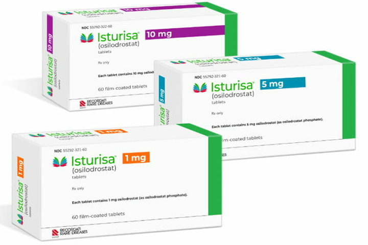
This oral inhibitor of 11β-hydroxylase — the enzyme involved in the last step of cortisol synthesis — was FDA approved in March 2020 based on these findings, and is currently available in 1 mg, 5 mg, and 10 mg film-coated tablets.
The prospective trial, consisting of four periods, included individuals between the ages of 18 and 75 with confirmed persistent or recurrent Cushing’s disease — marked by a mean 24-h urinary free cortisol concentration over 1.5 times the upper limit of normal (50 μg/24 hours), along with morning plasma adrenocorticotropic hormone above the lower limit of normal (9 pg/mL). All individuals had either undergone prior pituitary surgery or irradiation, were not deemed to be candidates for surgery, or had refused to have surgery.
During the first open-label study period, all participants took 2 mg of oral osilodrostat twice daily, spaced 12 hours apart. This dose was then titrated up if the average of three 24-h urinary free cortisol concentration samples exceeded the upper limit of normal. During the second study period, which spanned weeks 12 through 24, all participants remained on their osilodrostat therapeutic dose. By week 24, about 62% of the participants were taking a therapeutic dose of 5 mg or less twice daily; only about 6% of patients needed a dose higher than 10 mg twice daily.
In the third study period, which spanned weeks 26 through 34, “complete responders” who achieved normal cortisol levels were then randomized to continue treatment or be switched to placebo, while those who did not fully respond to treatment continued on osilodrostat. For the fourth study period, from weeks 24 through 48, all participants were switched back to active treatment with osilodrostat.
Overall, 96% of participants were able to achieve a complete response at some point while on osilodrostat treatment, with two-thirds of these responders maintaining this normalized cortisol level for at least 6 months. The median time to first complete response was 41 days.
Metabolic profiles also improved along with this reduction in cortisol levels. These included improvements in body weight, body mass index, fasting plasma glucose, both systolic and diastolic blood pressures, and total cholesterol levels.
“Given the known clinical burden of cardiovascular risk associated with Cushing’s disease, the improvement in clinical features shown here indicates important benefits of osilodrostat,” the researchers said. “By improving multiple cardiovascular risk factors, our findings are likely to be clinically relevant.”
Along with metabolic improvements, patients also had “clinically meaningful improvements” in quality of life, as well as reductions in depressive symptoms measured by the Beck Depression Inventory score, the investigators reported.
One limitation to the trial, they noted, was an inability to control for concomitant medications, since nearly all participants were taking other medications, particularly antihypertensive and antidiabetic therapies.
“Further examination of the effects of osilodrostat on the clinical signs of Cushing’s disease, and the reasons for changes in concomitant medications and the association between such medications and clinical outcomes would be valuable,” Pivonello’s group said.
Despite various approaches to immunoassay and chromatography for monitoring cortisol concentrations, conventional methods require bulky external equipment, which limits their use as mobile health care systems. Here, we describe a human pilot trial of a soft, smart contact lens for real-time detection of the cortisol concentration in tears using a smartphone. A cortisol sensor formed using a graphene field-effect transistor can measure cortisol concentration with a detection limit of 10 pg/ml, which is low enough to detect the cortisol concentration in human tears. In addition, this soft contact lens only requires the integration of this cortisol sensor with transparent antennas and wireless communication circuits to make a smartphone the only device needed to operate the lens remotely without obstructing the wearer’s view. Furthermore, in vivo tests using live rabbits and the human pilot experiment confirmed the good biocompatibility and reliability of this lens as a noninvasive, mobile health care solution.
INTRODUCTION
The steroid hormone, cortisol, which is known as a stress hormone, is secreted by the adrenal gland when people are stressed psychologically or physically (1). This secretion occurs when the adrenal gland is stimulated by adrenocorticotropic hormone, which is secreted by the pituitary gland when it is stimulated by the corticotropin-releasing hormone secreted by the hypothalamus. This serial cortisol secretion system is referred to as a hypothalamus–pituitary gland–adrenal gland axis, which is affected by chronic stress, resulting in abnormal secretion of cortisol (2, 3). The accumulation of cortisol caused by the abnormal secretion of cortisol increases the concentrations of fat and amino acid, which can result in diverse severe diseases (e.g., Cushing’s disease, autoimmune disease, cardiovascular complications, and type 2 diabetes) and neurological disorders (such as depression and anxiety disorders) (2–7). In contrast, abnormally low cortisol levels can lead to Addison’s disease, which results in hypercholesterolemia, weight loss, and chronic fatigue (8). In addition, it was recently reported that plasma cortisol can be correlated to the prognosis of traumatic brain injury (9). Furthermore, the extent of cortisol secretion varies from person to person, and it changes continuously (10, 11). Thus, developing health care systems for real-time monitoring of the cortisol level has been explored extensively over the past decade as the key to the quantitative analysis of stress levels. Although various efforts have led to the development of cortisol sensors that can measure the concentration of cortisol in blood, saliva, sweat, hair, urine, and interstitial fluid (12–17), the accurate measurement of cortisol concentrations has been limited because of the difficulties associated with the transportation and storage of cortisol as well as the instability of the biologically active cortisol in these body fluids at room temperature. In addition, these conventional sensing methods require bulky equipment for the extraction and analysis of these body fluids, which is not suitable for mobile health care systems (12, 18). Therefore, the development of noninvasive and wearable sensors that can monitor cortisol concentration accurately is highly desirable for a smart health care solution. For example, the immunoassay method, which uses an antigen-antibody binding reaction, has been used extensively for electrochemical cortisol immunosensors using saliva and interstitial fluid, except tears (12, 14, 19). However, these immunosensors still require the use of bulky impedance analyzers for the analysis of the Nyquist plot from electrochemical impedance spectroscopy. Although the cyclic voltammetry (CV) technique can be used as an alternative approach for sensing cortisol, additional bulky electrochemical instruments still are necessary for analyzing the CV curves (13, 14, 19). Recently, wearable forms of cortisol sensors that use sweat were developed (15), but they still required bulky measurement equipment (15, 16). Therefore, portable and smart sensors that can monitor the accurate concentration of cortisol in real time are highly desirable for use in mobile health care.
Among the various body fluids, tears, in particular, contain important biomarkers, including cortisol (20, 21). Thus, the integration of biosensors with contact lenses is a potentially attractive candidate for the noninvasive and real-time monitoring of these biomarkers from tears (22–25). However, an approach for fabricating a smart contact lens for sensing the cortisol in tears has not been demonstrated previously. Thus, here, we present an extraordinary approach for the formation of a smart, soft contact lens that enables remote, real-time monitoring of the cortisol level in the wearer’s tears using mobile phones. This smart, soft contact lens is composed of a cortisol sensor, a wireless antenna, capacitors, resistors, and integrated circuit chips that use stretchable interconnects without obstructing the wearer’s view. The components of this device (except the antenna) were protected from mechanical deformations by locating each of the components on discrete, rigid islands and by embedding these islands inside an elastic layer. A graphene field-effect transistor (FET; with the binding of monoclonal antibody) was used as this cortisol immunosensor, which exhibited a sufficiently low detection limit, i.e., 10 pg/ml, for its sensing of cortisol in human tears in which the cortisol concentration ranges from 1 to 40 ng/ml (26). This sensor was integrated with a near-field communication (NFC) chip and antenna inside the soft contact lens for the real-time wireless transmission of the data to the user’s mobile device (e.g., a smart phone or a smart watch). The antenna occupies a relatively large area of this soft lens, so it requires its high stretchability, good transparency, and low resistance for operating a standard NFC chip at 13.56 MHz. In our approach, the hybrid random networks of ultralong silver nanofibers (AgNFs) and fine silver nanowires (AgNWs) enabled high transparency and good stretchability of this antenna and its low sheet resistance for reliable standard NFCs (at 13.56 MHz) inside this smart contact lens. Thus, the fully integrated system of this smart contact lens provided wireless and battery-free operation for the simultaneous detection and transmission of the cortisol concentration from tears to a mobile phone using standard NFC. In addition, a human pilot trial and in vivo tests conducted using live rabbits demonstrated the biocompatibility of this lens, and its safety against inflammation and thermal/electromagnetic field radiation suggests its substantial usability as a noninvasive, mobile health care solution.
RESULTS
Cortisol immunosensor
A graphene FET sensor was fabricated by binding the cortisol monoclonal antibody (C-Mab) to the surface of graphene for the immunosensing of cortisol. Here, graphene acts as a transducer that converts the interaction between cortisol and C-Mab into electrical signals. Figure 1A shows the immobilization process of C-Mab to graphene. Immobilization proceeds through amide bonding of the C-Mab onto the carboxyl group of the graphene surface via the EDC [1-ethyl-3-(3-dimethylaminopropyl) carbodiimide hydrochloride]/NHS (N-hydroxysulfosuccinimide) coupling reaction. A chemical vapor deposition–synthesized graphene layer was transferred onto a desired substrate and exposed to ultraviolet ozone (UVO) to activate the surface of the graphene with the carboxylate group. Figure S1 shows the contact angle between this surface of the graphene and a droplet of deionized (DI) water. Longer exposure time to UVO can decrease the hydrophobicity of graphene with decreasing the contact angle. Table S1 shows the increase in the electrical resistance of graphene that resulted from this UVO treatment. In our experiment, 2 min of exposure time to UVO decreased the contact angle from 70° to 38° without increasing the resistance of the graphene notably. UVO exposure times longer than this threshold time degraded the resistance of the graphene excessively, so the time of exposure of our samples to UVO was limited to 2 min. Figure S2A illustrates the process of immobilizing C-Mab through the EDC/NHS coupling reaction. This two-step coupling reaction of EDC and NHS can mediate the amide bonding between the carboxylate group of the UVO-exposed graphene and the amine group of the protein (12, 17, 27, 28). Here, EDC forms reactive O-acylisourea ester, thereby making the surface unstable. This O-acylisourea ester reacts with the NHS to form amine-reactive NHS ester with the surface still remaining semistable. Then, C-Mab with the amine group reacts with the amine-reactive NHS ester, thereby forming stable amide bonding that can immobilize C-Mab to the NHS on the surface of the graphene. Figure S2B shows the Fourier transform infrared (FTIR) spectroscopy spectra of the DI water after the cortisol sensor had been immersed for 24 hours. The spectra of the DI water in which the sensor was immersed were not significantly different from those of the pristine DI water. However, the C-Mab solution that had a concentration of 1 μg/ml had a significant peak intensity in the range of 3000 to 2800 cm−1, representing the N-H bonding in the C-Mab. These results indicated that C-Mab formed stable bonding on the carboxylated graphene and was negligibly detached by exposure to water.
Severe acute respiratory syndrome coronavirus 2 (SARS-CoV-2), the viral strain that has caused the coronavirus disease 2019 (COVID-19) pandemic, has presented healthcare systems around the world with an unprecedented challenge. In locations with significant rates of viral transmission, social distancing measures and enforced ‘lockdowns’ are the new ‘norm’ as governments try to prevent healthcare services from being overwhelmed. However, with these measures have come important challenges for the delivery of existing services for other diseases and conditions. The clinical care of patients with pituitary disorders typically involves a multidisciplinary team, working in concert to deliver timely, often complex, disease investigation and management, including pituitary surgery. COVID-19 has brought about major disruption to such services, limiting access to care and opportunities for testing (both laboratory and radiological), and dramatically reducing the ability to safely undertake transsphenoidal surgery. In the absence of clinical trials to guide management of patients with pituitary disease during the COVID-19 pandemic, herein the Professional Education Committee of the Pituitary Society proposes guidance for continued safe management and care of this population.
Introduction
In many centers worldwide, the evaluation and treatment of pituitary disorders has already been substantially impacted by severe acute respiratory syndrome coronavirus 2 (SARS-CoV-2), the viral strain that has caused the coronavirus disease 2019 (COVID-19) pandemic. With reduced access to routine clinical services, patients with suspected or confirmed pituitary disease face the prospect of delays in diagnosis and implementation of effective treatment plans. Furthermore, patients undergoing surgery may be at increased risk from COVID-19, whilst the risk of infection to healthcare providers during pituitary surgery is of particular concern.
Herein, we discuss several clinical scenarios where clinical care can be adjusted temporarily without compromising patient outcomes. For this expert guidance, The Pituitary Society Professional Education Committee, which includes neuroendocrinologists and neurosurgeons from four continents, held an online video conference call with subsequent discussions conducted through email communications. The suggestions are not evidence-based due to the novelty and timing of the pandemic; furthermore, re-evaluation every few months in light of emerging data, is recommended. The approach will also likely vary from country to country depending on the risk of viral infection, local rules for “lockdown”, and the capabilities of individual health care systems.
Pituitary surgery challenges during the COVID-19 pandemic
The significant challenges to pituitary surgery presented by COVID-19 can be considered in terms of the phase of the pandemic, the patient, the surgeon, and the healthcare institution (Table 1).
Table 1 Pituitary surgery challenges and recommendations during COVID-19 pandemic
The World Health Organization (WHO) recognizes several phases of a pandemic wave [1]. When the pandemic is in progress (WHO pandemic phase descriptions; Phase 6) [2] there is a high prevalence of active cases. In the immediate post-peak period, the pandemic activity appears to wane, but active cases remain, and additional waves may follow. Previous pandemics have had many such waves, each separated by several months (www.cdc.gov). The corollary is that there will remain a significant possibility of patients and surgeons contracting COVID-19 until a vaccine is developed or herd immunity is achieved by other means.
The patient requiring pituitary surgery may be especially vulnerable to COVID-19 due to age and/or comorbidities. This is particularly true of patients with functioning pituitary adenomas such as those with Cushing’s disease (CD), where cortisol excess results in immunosuppression, hypercoagulability, diabetes mellitus and hypertension, and acromegaly which is also frequently complicated by diabetes mellitus and hypertension. Moreover, the risk for patients undergoing surgery that develop COVID-19 in the perioperative period appears to be very high. In a retrospective analysis of 34 patients who underwent elective—non pituitary—surgeries during the incubation period of COVID-19, 15 (44.1%) patients required admission to the intensive care unit, and 7 (20.5%) died [3]. Although this study included cases of variable technical difficulty, complexity and risk—from excision of breast lump to total hip replacement—we would suggest that patients undergoing pituitary surgery that develop COVID-19 are likely to be at similar or greater risk. These risks must be balanced carefully against the natural history of pituitary disease and, in particular, whether undue delay may result in irreversible morbidity such as visual loss in patients with pituitary apoplexy.
The surgeon remains in direct contact with the patient throughout their operation and is therefore at risk of contracting COVID-19 if the patient has an active infection. Iorio-Morin et al. [4] suggest that surgeons performing transsphenoidal pituitary surgery (TSS) may be at the greatest risk, because such surgery is performed under general anesthesia, requiring intubation and extubation, exposes the colonized nasal mucosa, and usually involves sphenoid drilling, which can result in aerosolization of contaminated tissues.
The healthcare institution will invariably divert resources from elective services to support the care of patients with COVID-19, with a knock-on effect on the capacity to manage patients with pituitary disease (Table 1). Bernstein et al. [5] suggest that surgery is particularly affected in such reorganization, because of both the need for redeployment of anesthesiologists able to manage patient airways, and availability of protective physical resources such as masks, gowns, and gloves (personal protective equipment; PPE). Furthermore, in areas with high number of infections, several operating rooms (OR)s were converted into intensive care units (ICU) to treat patients with COVID-19, thus limiting patients’ access to elective surgery even more.
Recommendations for pituitary surgery
When the viral risk is decreasing in a specific geographic area, we would advocate a stepwise, but flexible normalization of activity, addressing each of the aforementioned factors.
Burke et al. [6] proposed a staged volume limiting approach to scheduling surgical cases depending on the number of community cases and inpatients with COVID-19, and staffing shortages. In extreme cases, where significant assistance is required from outside institutions, only emergent cases can proceed.
Until further data become available, all patients undergoing pituitary surgery should undergo screening for COVID-19, until a vaccine is developed or herd immunity is achieved by other means. At the least, we recommend screening patients for cough, fever, or other recognized symptoms of infection with SARS-CoV-2, and taking swab samples for testing if there is any clinical suspicion. Depending on the level of COVID-19 activity in the community, and available resources, a more exhaustive strategy may be appropriate, including isolation of patients for up to 2 weeks before surgery, paired swabs and/or serological tests for all patients irrespective of symptoms, and routine chest X-ray or chest computed tomography (CT), depending on local guidance. In patients with COVID-19 in whom surgery is indicated, in general we recommend delaying surgery if possible, ideally until patients no longer have symptoms and have a negative swab test result.
The nature of the patient’s pituitary disease is an important consideration, and we propose stratifying cases as emergent, urgent, or elective. We recommend that patients continue to be operated on in an emergent fashion if they present with pituitary apoplexy, acute severe visual loss, or other significant mass effect, or if there is concern regarding malignant pathology. Selected patients with slowly progressive visual loss, functioning tumors with aggressive clinical features, and those with an unclear diagnosis, may also benefit from urgent (but not emergent) surgery, with decisions made on a case-by-case basis. Patients with incidental and asymptomatic tumors, known nonfunctioning adenomas [7] or functioning tumors, which are well controlled with medical therapy, can be scheduled as elective cases.
In most cases, TSS remains the safest, most effective, and most efficient approach to pituitary tumors. In a series of 9 consecutive patients without COVID-19 undergoing pituitary and skull base surgery during the pandemic, Kolias et al. [8] reported that none of the patients or staff contracted COVID-19 following adoption of a standardized risk-mitigation strategy. In the rare instances where a patient with COVID-19 requires emergent surgery that cannot be deferred, alternative transcranial approaches may be considered (avoiding nasal mucosa). To replace high-speed drilling, the use of non-powered tools such as rongeurs and chisels has been recommended. If this is not possible large suction tubes can be used to aspirate as much particulate matter as possible [9]. In such cases, the availability and use of PPE, and in particular filtering facepiece (FFP3) respirators, is mandated. Depending on the level of COVID-19 activity in the community, and the availability and effectiveness of testing, PPE may be appropriate in all cases.
At an institutional level, there must remain flexibility in anticipation of further waves of COVID-19. This necessitates a reduction in capacity, particularly in available ICU beds, that must be recognized when scheduling challenging surgical cases. In the long term, resumption of full elective workloads depends on wider national and international factors, including widespread testing, and widespread immunity through vaccination or other means.
Pituitary diseases diagnosis and management
Acromegaly
Acromegaly, a condition that arises from growth hormone (GH) excess, generally occurs as a result of autonomous GH secretion from a somatotroph pituitary adenoma [10, 11], is associated with substantial morbidity and excess mortality, which can be mitigated by prompt and adequate treatment [12]. Diagnosis is often delayed because of the low prevalence of the disease, the frequently non-specific nature of presenting symptoms, and the typically subtle progression of clinical features [10, 11]. During the COVID-19 pandemic many outpatient clinics have closed or limited work hours. Patients are often reluctant to seek care out of fear of possible exposure to the coronavirus. Therefore, even longer diagnostic delays are anticipated. In addition, patients who present with vision loss and larger tumors encroaching upon the optic apparatus are at risk for experiencing persistent visual compromise unless the optic chiasm and nerves are promptly decompressed.
To improve patient access to care and minimize potentially deleterious delays in diagnosis and treatment, clinicians may conduct virtual visits (VV) using secure, internet-based electronic medical record platforms. A detailed history can be obtained and a limited physical examination is possible, including inspection of the face, skin and extremities.
Diagnosis
Establishing the diagnosis of acromegaly requires testing of serum insulin-like growth factor-I (IGF-I) levels [11] (Box 1). Access to accurate IGF-I assays is critical in light of the substantial analytical and post-analytical problems that have plagued several IGF-I immunoassays. While the oral glucose tolerance test (OGTT) is considered the diagnostic “gold standard”, this test is not essential in many patients, including those with a clear-cut clinical picture and an unequivocally elevated serum IGF-I level. Deferring the lengthy (2-h) OGTT may minimize the risk of potential exposure to infectious agents.
Given the over-representation of macroadenomas in patients with acromegaly, pituitary imaging is indicated, preferably by a pituitary-specific magnetic resonance imaging (MRI) protocol, although CT may be performed to rule out a large tumor if MRI is not feasible. Obtaining imaging at satellite sites detached from major hospitals may also decrease the risk of infection exposure.
Management
Transsphenoidal pituitary surgery remains the treatment of choice for most patients with acromegaly [10, 11], and patients with visual compromise as a result of a pituitary adenoma compressing the optic apparatus should still undergo pituitary surgery promptly. Other patients could be treated medically until the pandemic subsides. Medical treatment options are somatostatin receptor ligands (SRLs), octreotide long-acting release (LAR), lanreotide depot and pasireotide LAR, pegvisomant and cabergoline (used off-label) [13]. Medical therapies can be effective in providing symptomatic relief, control GH excess or action, and potentially reduce tumor size (except pegvisomant, which does not have direct antiproliferative effects). Preoperative medical therapy has been reported to improve surgical outcomes in some, but not all studies. Pasireotide, which potentially can induce QTc prolongation, should be used with caution in patients who are taking, either as prophylaxis or treatment, medications for COVID-19 (azithromycin, hydroxychloroquine), which can also have an effect on QTc interval. Furthermore, as hyperglycemia is very frequent in patients treated with pasireotide and needs close monitoring at start of the treatment, this treatment should be reserved for truly resistant cases, with large tumors and who cannot have surgery yet. Notably, lanreotide depot, cabergoline or pegvisomant can be administered by the patient or a family member and therefore an in-person visit to a clinic is not required. If SRLs that require health care professional administration are required, raising the dose may allow the interval between injections to be extended beyond 4 weeks while maintaining disease control. Virtual visits can be implemented to monitor the patient’s course and response to medical therapy during the pandemic. Careful management of comorbidities associated with acromegaly remains an essential part of patient care [14, 15].
Prolactinomas
Hyperprolactinemia may be physiological in origin or arise because of an underlying pathophysiologic cause, medication use or laboratory artifact. Therefore, an initial evaluation for hyperprolactinemia should include a comprehensive medication history, a thorough evaluation for secondary causes, including primary hypothyroidism, and a careful assessment for clinical features of hyperprolactinemia, including hypogonadism and galactorrhea. Unless a secondary cause of hyperprolactinemia can be established definitively, further investigation is indicated to evaluate the etiology of hyperprolactinemia.
Diagnosis
The diagnosis of a lactotroph adenoma can be inferred in most patients based on the presence of a pituitary adenoma and an elevated prolactin level, which is typically proportionate in magnitude to adenoma size. Pituitary imaging (MRI or CT) is therefore a key step in the investigation of hyperprolactinemia. Evaluation for hypopituitarism is also necessary.
Management
Although observation and routine follow-up with serial prolactin levels and imaging is acceptable for patients who are asymptomatic and who have a microadenoma, most patients diagnosed with a prolactinoma will require treatment. Dopamine-agonists (DA) can normalize prolactin levels and lead to reduction in size of the lactotroph adenoma [16]. In patients who have a microadenoma and who are not seeking fertility, hormone-replacement therapy may also be appropriate if serum prolactin is routinely followed and imaging performed as necessary.
Medical therapy can be managed effectively and efficiently via VVs coupled with laboratory/imaging studies as needed. However, in all patients in whom a DA will be initiated, it is critical that a comprehensive psychiatric history is obtained prior to commencing treatment. Patients may not readily volunteer their psychiatric history and may not appreciate the relevance of such information. For example, until specifically questioned about their psychiatric history, the patient described in the illustrative case (Box 2) did not report a history of severe depression, suicide attempt and prolonged psychiatric hospitalization 8 months prior to presentation with hyperprolactinemia. At the time of the visit, he was not taking any psychiatric medications and was not under the care of a mental health team. Given this patient’s significant psychiatric history, lack of ongoing psychiatric care, and the well-recognized adverse effects of DA therapy, including increased impulsivity, depression and psychosis [17], a DA was not initiated. Counseling on potential DA side-effects is crucial, as they may also present in individuals with no prior psychiatric history [17]. Furthermore, during the COVID-19 pandemic when there is reduced access to routine medical and mental health care, patients who develop symptoms of severe depression may not have ready access to mental health services, or may not seek care. Therefore, it is particularly important to make patients aware of these potential side effects and the critical importance of reporting them.
In the small number of patients for whom medical therapy is not possible and where surveillance is not appropriate (e.g., macroprolactinoma with visual loss) the risks and benefits of surgical intervention will need to be carefully weighed.
Cushing’s disease
Left untreated, CD has significant morbidity and mortality, and delays in diagnosis (from a few months to even years) are common. Clinical presentation is also very variable with some patients having subtle symptoms while others present with more striking/classical features. Severe hypercortisolemia induces immunosuppression, which may place patients with untreated CD at particular risk from COVID-19.
New patients referred for endocrinology evaluation with clinical suspicion of Cushing’s
Diagnosis
Screening for, and confirmation of Cushing’s syndrome (CS) and, furthermore, localization for CD is laborious and requires serial visits and testing procedures [18, 19]. If initial laboratory abnormalities are consistent with hypercortisolemia, a VV should allow for an estimate of the severity of clinical presentation and facilitate planning for further testing and treatment. Careful questioning for potential causes of exogenous CS (including, but not limited to, history of high-dose oral corticosteroids, intraarticular injections or topical preparations) is an important first step. Subsequently, establishing the likelihood and pretest probability of CS is more important than ever now, when testing may be delayed. While presentation varies significantly between patients, some features, although not all highly sensitive, are more specific, e.g. easy bruising, facial plethora, large wide > 1 cm violaceous striae, proximal weakness and hypokalemia. Diagnosis of CS is often challenging even under normal circumstances, however, a diagnosis by VV is more nuanced and difficult. Conversely, if a patient has a high likelihood of CS, we recommend limited laboratory evaluation (urinary free cortisol (UFC), adrenocorticotropic hormone (ACTH), liver panel, basic metabolic panel), preferably at a smaller local laboratory rather than a Pituitary Center, to reduce viral risk exposure. Salivary cortisol samples could represent a hazard for laboratory staff and they are prohibited in some countries [18, 19]. In the US, laboratories have continued to process salivary cortisol samples and salivary cortisol has higher sensitivity compared with UFC and has the convenience of mailing multiple specimens at a time, without travel [18, 19]. Though usually we strongly recommend sequential laboratory testing under normal circumstances, limiting trips to a laboratory is preferred during COVID-19.
If preliminary assessment confirms ACTH-dependent CS [18, 19] and no visual symptoms are reported, imaging may be delayed. However, in the presence of any visual symptoms, and recognizing the challenges of undertaking a formal visual field assessment, proceeding directly with MRI or CT (shorter exam time and easier machine access) imaging, will allow confirmation or exclusion of a large pituitary adenoma compressing the optic chiasm. If the latter is confirmed, the patient will need to be evaluated by a neurosurgeon. In contrast, a small pituitary adenoma may not be visible on CT, but in such cases MRI may be deferred for a few months until COVID-19 restrictions limiting access to care are lifted.
Another VV will help to decide, in conjunction with patient’s preference, the best next step, which in cases of more severe clinical Cushing’s, and in the absence of a large pituitary adenoma, would be medical therapy. The magnitude of 24 h-UFC elevation could also represent a criterion for primary therapy, since higher values have been associated with increased risk of infection.
In parallel, it is also important to address comorbidities including diabetes mellitus, hypertension and hyperlipidemia. In light of the increased risk of venous thromboembolism, in discussion with primary care providers, plans for regular mobilization/exercise as permitted (including at home when orders to stay in are in place) and/or prophylactic low weight molecular heparin should be considered.
Management
First line medical therapy options vary, depending on country availability, regulatory approval and patient comorbidities. Ideally, an oral medication, which is easier to administer is preferred; options include ketoconazole, osilodrostat or metyrapone [20, 21]. Cabergoline therapy, which has lesser efficacy [20, 21] compared with adrenal steroidogenesis inhibitors, can be also attempted in very mild cases. The initial laboratory profile should be reviewed to exclude significant abnormalities of renal and/or liver function prior to commencing treatment. Starting doses of all medications should be the lowest possible to avoid adrenal insufficiency (AI) and up titration should be slow, with VVs weekly if possible. All patients with CS on any type of medical therapy should have prescribed glucocorticoids (GC) both in oral and injectable forms available at home and information regarding AI should be provided during a VV when starting therapy for CS. Down titration of other medications for diabetes and hypertension may also be needed over time. Pasireotide (both subcutaneous and LAR preparations) would be a second line option, reflecting higher risk of significant hyperglycemia that would require treatment [22].
If the clinical features of CS are mild and longstanding, with no acute deterioration, another possibility is to aggressively treat the associated comorbidities for a few months; depending on local circumstances, this may actually be less risky for the patient by avoiding the risk of AI/crisis and the need for an emergency department (ED) visit and/or admission.
For patients with Cushing’s disease with endocrinology chronic care
Patients in remission after surgery with adrenal insufficiency on glucocorticoid replacement
These patients are likely to remain at slightly higher risk of COVID-19 infection due to immunosuppression from previous hypercortisolemia. Furthermore, GC doses should be adjusted to prevent adrenal crisis and visits to an ED. Lower GC daily doses (10–15 mg hydrocortisone/day) are now frequently used for replacement and virtual and/or phone visits are encouraged to evaluate an appropriate regimen and sufficient supplies of medication and injectable GC (at home) should be prescribed. Patients with potential symptoms of under replacement may require an increase in daily dose, while balancing any risk of GC over replacement and possible consequent immunosuppression.
Patients in non-remission treated with medical therapy (dependent on country availability)
Doses may need to be adjusted to reduce the risk of AI/crisis and reduce the need for serial laboratory work. Monthly or bimonthly VVs are appropriate for clinical evaluation and up titration should be slower than usual. Patients with CD on medical therapy need to have at home prescriptions for oral and injectable GC and instruction on AI surveillance. Patients should also be advised, that if they develop a fever, to stop Cushing’s medication for few days; if they develop AI symptoms, GC administration will be required. In some countries, block and replace regimens are also employed to avoid risk of AI. Of note, for mifepristone, a glucocorticoid receptor (GR) antagonist, patients will require much higher doses of GC to reverse the blockade (1 mg of dexamethasone approximately per 400 mg of mifepristone) and for several days, as drug metabolites also have GR antagonist effects.
Furthermore, for all patients who have made dose changes or discontinued medications for Cushing’s, it is essential to follow very closely and consider adjustments in the doses of concomitant medications, especially insulin, other antidiabetic and antihypertensive medications, and potassium supplements.
If patients have history of radiotherapy and are still on medications for CD, a VV every few months should be performed to determine if anti-Cushing’s treatment can be slowly down-titrated (to avoid AI). A morning serum cortisol would be ideal to rule out AI off medications, however, if laboratory testing cannot be undertaken safely, clinical evaluation by serial VVs can be helpful. While head-to-head data will never be available, in COVID-19 hotspots, given the higher risk of infection with laboratory testing or face to face visits, mild hypercortisolemia might be “better” than adrenal crisis, especially in the short term!
Patients with CD have increased rates of depression, anxiety and can have decreased quality of life (QoL) even when in long-term remission, thus in the challenging circumstances of the current pandemic it is it even more important to focus on psychological evaluation during virtual endocrinology visits, with referral to virtual counseling as needed.
Cushing’s syndrome (CS) during pregnancy is very rare with a few cases reported in the literature.
Of great interest, some cases of CS during pregnancy spontaneously resolve after delivery. Most studies suggest that aberrant luteinizing hormone (LH)/human chorionic gonadotropin (hCG) receptor (LHCGR) seems to play a critical role in the pathogenesis of CS during pregnancy.
However, not all women during pregnancy are observed cortisol hypersecretion. Moreover, some cases of adrenal tumors or macronodular hyperplasia with LHCGR expressed, have no response to hCG or LH.
Therefore, alternative pathogenic mechanisms are indicated. It has been recently reported that estrogen binding to estrogen receptor α (ERα) could enhance the adrenocortical adenocarcinoma (ACC) cell proliferation.
Herein, we hypothesize that ERα is probably involved in CS development during pregnancy.
Better understanding of the possible mechanism of ERα on cortisol production and adrenocortical tumorigenesis will contribute to the diagnosis and treatment of CS during pregnancy.