Unmet needs in Cushing’s Syndrome: the Patients

Abstract

Background

Cushing’s syndrome (CS) is a rare condition of chronically elevated cortisol levels resulting in diverse comorbidities, many of which endure beyond successful treatment affecting the quality of life. Few data are available concerning patients’ experiences of diagnosis, care and persistent comorbidities.

Objective

To assess CS patients’ perspectives on the diagnostic and care journey to identify unmet therapeutic needs.

Methods

A 12-item questionnaire was circulated in 2019 by the World Association for Pituitary Organisations. A parallel, 13-item questionnaire assessing physician perceptions on CS patient experiences was performed.

Results

Three hundred twenty CS patients from 30 countries completed the questionnaire; 54% were aged 35–54 and 88% were female; 41% were in disease remission. The most burdensome symptom was obesity/weight gain (75%). For 49% of patients, time to diagnosis was over 2 years. Following treatment, 88.4% of patients reported ongoing symptoms including, fatigue (66.3%), muscle weakness (48.8%) and obesity/weight gain (41.9%). Comparisons with delay in diagnosis were significant for weight gain (P = 0.008) and decreased libido (P = 0.03). Forty physicians completed the parallel questionnaire which showed that generally, physicians poorly estimated the prevalence of comorbidities, particularly initial and persistent cognitive impairment. Only a minority of persistent comorbidities (occurrence in 1.3–66.3%; specialist treatment in 1.3–29.4%) were managed by specialists other than endocrinologists. 63% of patients were satisfied with treatment.

Conclusion

This study confirms the delay in diagnosing CS. The high prevalence of persistent comorbidities following remission and differences in perceptions of health between patients and physicians highlight a probable deficiency in effective multidisciplinary management for CS comorbidities.

Introduction

Cushing’s syndrome (CS) is a morbid endocrine condition due to prolonged exposure to high circulating cortisol levels (123). Hypercortisolism may cause irreversible physical and psychological changes in several tissues, leading to debilitating morbidities which persist over the long term after the resolution of excessive hormone levels, such as cardiovascular complications, metabolic and skeletal disorders, infections and neuropsychiatric disturbances (34). Even patients who have been biochemically ‘cured’ for over 10 years have a residual overall higher risk of mortality, mostly from circulatory disease and diabetes (5). Moreover, people with a history of CS suffer from impaired quality of life (QoL) (6). Several studies suggest that the prevalence of persistent comorbidities is correlated with the duration of exposure to cortisol excess (78). However, as the signs and symptoms of CS overlap with common diseases such as the metabolic syndrome and depression, the time taken to diagnose CS is often long, resulting in a significant number of patients with persistent sequelae and impairments in QoL (69).

Given the burden of the disease, ideal CS treatment would include early diagnosis, curative surgery and multidisciplinary care of comorbidities both pre- and post-cure of CS, including the psychological dimension of the patient’s disease experience (10). Few data are available about patients’ perceptions of the medical journey from first symptoms to diagnosis, treatment and follow-up. The aim of this study was, therefore, to explore CS patients’ experiences of symptoms, diagnosis, care and treatment satisfaction around the world and to compare patients’ perceptions of CS with those of physicians.

Methods

Patient questionnaire design

A 12-item patient questionnaire was developed based on the generally understood clinical characteristics and symptomology of CS, aiming to assess patients’ experiences of symptoms, diagnosis, care and treatment satisfaction (12) (Supplementary File 1, see section on supplementary materials given at the end of this article). The questionnaire was initially offered in English and made available via the SurveyMonkey online platform from March to May 2019. The survey was completed anonymously and required no specific participant identification or any details that could be used to identify individual participants. In addition to basic demographics (i.e. country of residence, sex, age and highest educational level attained), the questionnaire asked ten multiple-choice and two open questions. The survey was shared by the World Association for Pituitary Organisations (WAPO), Adrenal Net, Cushing’s Support & Research Foundation and the Pituitary Foundation, as well as being distributed to local patient associations. As a second step, the questionnaire was translated into eight additional languages (French, Dutch, Spanish, Chinese, Portuguese, Italian and German) and was recirculated by the WAPO, Adrenal Net and China Hypercortisolism Patient Alliance to the different local patient associations for distribution in November 2019. As this was a non-interventional, anonymous patient survey, distributed by the patient associations themselves, and not initiated or funded by a research or educational institution, no ethical review was required. Written consent was obtained from each respondent after full explanation of the purpose and nature of the survey.

Comparative physician survey

In addition, a 13-item physician questionnaire was developed to assess physicians’ perspectives on CS symptoms and comorbidities. This physician questionnaire was conducted by HRA Pharma Rare Diseases at the 2019 European Congress of Endocrinology, in Lyon, France. This anonymous questionnaire was completed by 40 qualified physicians. The responses from the patient survey were compared for context with the physicians’ estimates of the prevalence of CS symptoms and comorbidities. Although the physician questionnaire was conducted independently of the patient questionnaire, and used a different question structure, the comparison with the current patient questionnaire is included to further enrich and contextualise the patient responses.

Data analysis

All responses and answers were collected, coded and analysed using Microsoft Excel. Data preparation involved removing duplicate answers, or where possible analysing and reclassifying qualitative responses reported as ‘other’, based on the accompanying details to new or existing response options.

Statistical methodology

Complementary statistical analyses using SAS software were performed using the chi-square and Fisher tests, depending on the cell counts, to compare (i) the time between first symptoms and diagnosis and the persistence of symptoms and (ii) persistence of symptoms, with the specialities of the physicians currently treating the respondents. Frequency distribution of a particular variable was displayed and compared with the frequency distribution of the comparator variable. A significance level of 0.05 was applied.

Results

Demographic characteristics

Three hundred twenty patients from 30 countries completed the patient questionnaire, with 27% (n  = 87) coming from the United Kingdom and 14% (n  = 44) from the United States of America. More than half (53.7%, n = 172) of the patients were aged between 35 and 54 years, and 88.4% (n  = 283) were female. The majority of patients (53.1%, n = 170) had undergraduate or postgraduate qualifications (Table 1).

Table 1Patient demographics.

Sex N = 319a
 Female 283 (88.4%)
 Male 36 (11.3%)
Age group N = 320
 18–24 years 16
 25–34 years 49
 35–44 years 71
 45–54 years 101
 55–64 years 54
 65–74 years 24
 ≥75 years 5
Regionb N = 320
 Western Europe 222
 North America 60
 China 16
 Australasia 14
 South America 5
 Africa 3
Education N = 320
 High school graduate/secondary education diploma 35%
 Undergraduate degree 25.6%
 Post-graduate degree 27.5%
 Prefer not to say 10.6%
Time from first symptoms to diagnosis N = 320
 0–6 months 18.4%
 6–12 months 15.6%
 1–2 years 14.4%
 2–3 years 18.4%
 3–5 years 11.6%
 5–10 years 8.4%
 10–15 years 7.5%
 15–20 years 0.9%
 20+ years 1.9%
 Unknown 2.8%

aOne patient responded ‘non-binary’. bWestern Europe: United Kingdom (n  = 87), the Netherlands (n  = 38), France (n  = 37), Spain (n  = 12), Denmark (n  = 10), Norway (n  = 9), Germany (n  = 6), Italy (n  = 5), Ireland (n  = 4), Belgium (n  = 4), Poland (n  = 4), Sweden (n  = 2), Malta (n  = 2), Switzerland (n  = 1), Czech Republic (n  = 1); Africa: South Africa (n  = 1), Gabon (n  = 1), Zimbabwe (n  = 1); Australasia: Australia (n  = 8), New Zealand (n  = 6); South America: Colombia (n  = 2), Bolivia (n  = 1), Argentina (n  = 1), Brazil (n  = 1); North America: United States of America (n  = 44), Canada (n  = 13), Costa Rica (n  = 1), Mexico (n  = 1), Dominican Republic (n  = 1).

Time to diagnosis

The time to diagnosis from first reporting of CS symptoms was declared to be within 2 years for 48.4% (n  = 155) (Table 1) and was over 2 years in 48.7% (n  = 156) and over 3 years in 30.3% (n  = 97).

Initial symptoms

A broad range of signs and symptoms were initially noticed by patients, with weight gain, hirsutism or acne, fatigue, sleep disturbances, depressive symptoms, muscle weakness, anxiety and hypertension all being reported in over 50% of patients (Table 2). Obesity/weight gain was most commonly cited (75%, n = 240) as being burdensome. Fatigue, feelings of depression or mood problems, sleep disturbances, muscle weakness and hirsutism were also very commonly (>40%) mentioned as being burdensome. Burdensome symptoms classified as ‘other’ were rare (<1%) and included issues such as hormonal problems and dental problems.

Table 2Patient-reported symptoms (multiple answers were possible).

Symptoms first noticed (%) Most burdensome perceived symptoms before diagnosis (%)
Weight gain 85.0 75.0
Hirsutism/acne 76.3 42.8
Fatigue 66.3 54.1
Sleep disturbances 64.4 41.9
Skin problems 64.7 21.3
Depression/mood problems 58.8 48.1
Muscle weakness 57.8 43.4
Anxiety 54.1 39.1
Hypertension 52.5 22.2
Loss of concentration 45.0 28.4
Memory problems 41.9 30.3
Menstrual disturbances 35.6 12.5
Decreased libido 32.5 12.5
Bone problems 23.1 14.4
Infections 23.8 10.3
Glucose intolerance 17.2 8.4
Blood clot 5.3
Pain(s) 3.1
Vision problems 2.8
Headache 2.5
Cravings 1.6
Other 8.4 1.9

Person who made the initial CS diagnosis

In 53.8% (n  = 172) of cases, an endocrinologist made the initial diagnosis of CS or prescribed the first screening tests, Table 3. General practitioners made 18.1% of diagnoses (n  = 58), in the remaining cases a diversity of other physicians directly or indirectly contributed to make the diagnosis, as indicated in Table 3. A small but noticeable number (5.6%, n = 18) of patients self-diagnosed and then convinced their physician to order the diagnostic tests.

Table 3Patient perception of physician specialty.

Specialty Person who made the initial diagnosis or suspected Cushing’s syndrome (%) (n = 320) Physicians involved in the management of Cushing’s syndrome (%) (n = 320)
Endocrinologist 53.8 97.8
General practitioner/family doctor 18.1 56.3
Self-diagnosed 5.6
Hospital/emergency doctor 3.8
Internist 2.5 0.9
Gynecologist 1.9 14.1
Cardiologist 1.9 13.4
Bone specialist 1.9 14.1
Dermatologist 1.6 11.6
Haematologist 0.9 3.8
Ophthalmologist 0.9 3.1
Nurse 0.9 2.5
Radiologist 0.9 0.6
Family or friend 0.9
Psychiatrist or psycologist 0.9 23.4
Healer 0.6 2.2
Surgeon 0.6
Oncologist 0.3 6.6
Gastroenterologist 0.3 1.3
Neurologist 0.3 4.1
Others 1.6
Physiotherapist 14.4
Dietician 9.7
Neurosurgeon 8.1
Social worker 4.1
Ear, nose and throat specialist 1.6
Sports physician 1.3
Sleep specialist 0.9
Urologist 0.6
Orthopaedic surgeon 0.3

Response to treatment

At the time of answering the questionnaire, 55.8% (n  = 178) of patients were not in remission. 40.8% of patients (n  = 130) were in true biochemical remission (Fig. 1). This latter group was a composite including patients who responded: ‘In remission (no treatment)’ (16.3%, n = 52), ‘Received an operation to remove adrenal glands’ (22.9%, n = 73) and ‘Treated with hydrocortisone’ (1.6%, n = 5). Thirteen percent of the patients (n  = 41) were on cortisol-lowering treatment and 6.6% of the patients (n  = 21) had not had or were awaiting surgery. Following treatment for CS, 11.6% of the patients (n  = 37) reported having no further symptoms related to the condition, with 88.4% (n  = 283) still symptomatic. Of the total population (n  = 320), the most bothersome symptoms were fatigue (66.3%, n = 212), muscle weakness (48.8%, n = 156) and obesity/weight gain (41.9%, n = 134) (Table 4).

Figure 1View Full Size
Figure 1
Patient description of their current clinical situation (n = 319). The category ‘Disease in true remission’ combines scores for ‘In remission (no treatment)’ (16.3%), ‘Received an operation to remove adrenal glands’ (22.9%) and ‘Treated with hydrocortisone’ (1.6%). One person did not complete the question.

Citation: Endocrine Connections 11, 7; 10.1530/EC-22-0027

Table 4Persistent symptoms.

Symptom Persistent bothersome symptomsa (%) (n = 320) Treatment received for symptoms (%) (n = 320)
Fatigue 66.3 15.9
Muscle weakness 48.8 17.2
Weight gain 41.9 8.4
Depression, mood problems 36.9 28.8
Poor concentration 35.9 4.1
Memory problems 33.8 5.6
Sleep problems 33.1 14.1
Anxiety 30.6 14.7
Decreased libido 25.3 4.1
Bone problems 19.1 21.9
Hypertension 18.4 29.4
Hirsutism 17.5 4.1
Skin problems 16.6 6.9
Glucose intolerance 8.8 10
Menstrual problems 9.1 4.7
Infections 7.2 4.7
Blood clot 3.8 2.2
Acne 2.8 1.3
Other 4.4 5.3
No treatment 1.3 8.1
Only hydrocortisone 1.6

aUp to five answers were possible.

Comparison of time to diagnosis and persistence of symptoms

To compare the time to diagnosis and the persistence of symptoms following treatment, an analysis of a number of variables was performed comparing the group with persistent symptoms after treatment (n  = 283) with those who did not (n  = 37) in terms of time to diagnosis. Patients with a longer time to diagnosis reported significantly more frequent weight gain (P = 0.008), and more frequent reduced libido (P = 0.03) after treatment. Although not statistically significant, there was a strong trend towards patients reporting a longer time to diagnosis and a greater frequency of persistent perceived bone issues after treatment (P = 0.053), as well anxiety (P = 0.07) and depression/mood concerns (P = 0.08).

Physicians involved in follow-up

Once diagnosed, almost all patients (97.8%, n = 313) were managed by an endocrinologist, followed by a GP/family doctor (56.3%, n = 180). A psychiatrist/psychologist was involved in 23.4% (n  = 75), followed by a physiotherapist (14.4%, n = 46), rheumatologist (14.4%, n = 46), gynecologist (14.1%, n = 45), cardiologist (13.4%, n = 43), dermatologist (11.6%, n = 37) and a dietician (9.7%, n = 31) (Table 3).

Treatment of persistent symptoms

Table 4 shows the prevalence of persistent symptoms after treatment, common ongoing comorbidities included fatigue, muscle weakness and weight gain. The percentage of patients who were treated for comorbidities is also shown. Noticeable undertreatment occurred for many symptoms, for example, fatigue was a consistent symptom for 66.3% (n  = 212), whereas only 15.9% (n  = 51) were receiving ongoing care for fatigue and persistent muscle weakness was reported in 48.8% (n  = 156) with 17.2% (n  = 55) of patients being treated for this (Table 4).

The high frequency of persistent symptoms suggests that patients were not followed-up by specific specialists, for example of the 212 patients with persistent fatigue, only 60 (28.2%) were seeing a psychiatrist/psychologist (Table 4). Enduring poor concentration and memory problems were relatively frequent (35.9%, 33.8%) but were rarely treated by a specialist (4.1 and 5.6%, respectively).

Three-quarters of patients reported that their work life had been affected (75%, n = 240). Social life (65.3%, n = 209), family life (57.8%, n = 185), interpersonal relationships (51.6%, n = 165), and sexual life (48.8%, n = 155) had also been significantly affected by their illness. Thirty-seven percent of the patients (n  = 118) reported that their economic situation had been negatively affected. ‘Other’ responses for this question included reductions in self-esteem, self-image and self-confidence. Sixty-three percent of patients (193/305) were satisfied with their treatment and 36.7% (n  = 112) were not.

Comparative analysis physician questionnaire

In the complementary physician questionnaire (n  = 40), unlike the patient questionnaire where most respondents were from the United Kingdom, the United States of America, the Netherlands and France, most of the physicians surveyed were from Western Europe, although there were representatives from other parts of the world. In the physician questionnaire, 83% (n  = 33) were endocrinologists, 13% (n  = 5) internal medicine specialists and 5% (n  = 2) other disciplines. Sixty percent (n  = 24) had over 10 years clinical experience, and 93% (n  = 37) were experienced in the treatment of CS, seeing an average of 10 patients per year. Of the specialities involved in the care of CS, 96% of physicians (n  = 38) considered endocrinologists to be involved, 48% (n  = 19) included family doctors/GPs, 20% (n  = 8) cardiologists, 28% (n  = 11) psychiatrists/psychologists and 28% (n  = 11) included dieticians. These results are consistent with the patients’ perceptions, with the exception of dieticians, who only 10% of patients reported seeing (Table 3).

Figure 2A compares the frequency of common symptoms that patients found to be most burdensome during the active phase of the disease, with what physicians thought were the most common symptoms. Although for methodological reasons a statistical comparison was not possible and the comparisons are approximate, these findings suggest that physicians’ perceptions of the prevalence of symptoms were different from those reported by patients. A majority of physicians (Fig. 2A) inadequately estimated (both underestimated and overestimated) the presence of depression, muscle weakness, cognitive impairment, hypertension, bone problems and glucose intolerance. Figure 2B compares the physician’s perception of the frequency of persistent symptoms with the patients’ experience of persistent symptoms. A majority of physicians differently estimated the prevalence of persistent cognitive impairment, muscle weakness, depressive symptoms and weight gain.

Figure 2View Full Size
Figure 2
(A) Physician (n = 40) perception of patient comorbidities (left) and patient reports of the most burdensome symptoms during active CS (right). (B) Physician (n = 40) perception of CS symptoms after cure (right) and patient reports of persistent burdensome symptoms after treatment (left). Only the relevant common results from the physician and patient surveys are shown above. The physician survey included categories ‘insulin resistance’, ‘dyslipidaemia’, ‘cardiovascular complications’ and ‘psychosis’, which are not shown because these same categories were not reported in the patient survey. In the patient survey, responses for the categories: ‘anxiety’ were regrouped with ‘depressive symptoms’ and ‘memory problems’ and ‘poor concentration’ were regrouped into the ‘cognitive impairment’ category for easier comparison with the physician survey.

Citation: Endocrine Connections 11, 7; 10.1530/EC-22-0027

Discussion

This large, international CS patient survey confirms previous findings that despite complaining of multiple symptoms, there is a mean 34-month delay in diagnosis (9). In addition, despite treatment resulting in biochemical remission, patients report persistent comorbidities with associated psychological and social impacts that negatively affect the QoL (1112). In the present survey a majority of patients reported that they are not being managed by the appropriate specialists, suggesting an absence in multidisciplinary care that may be secondary to an underestimation of the sequelae of CS by endocrinologists.

The present survey confirmed that no specific symptom initiated a diagnosis, but rather a range of burdensome symptoms occurring with similar frequency to those reported in previous surveys (12), with the notable difference in that in a USA-German survey, cognitive and psychological symptoms were bothersome for 61% of US and 66% of German patients (13), whereas in the present survey 38% considered depression/mood problems burdensome. Such differences may be a result of different terms being used to describe depression or mood symptoms as well as cultural differences between populations.

The distribution of time to diagnosis, with around 50% diagnosed after 2 years of symptoms and approximately 30% still undiagnosed after 3 years is of a similar magnitude to previous surveys, where 67% of patients waited at least 3 years until diagnosis (14). In the CSFR study in 2014, patients waited a median of 5 years until diagnosis (15). Even though the estimated time to diagnosis may be similar to those in previous studies – 34 months a recent meta-analysis (9) and 2 years in the ERCUSYN database (16) – there is clearly still room for improvement, especially as delayed diagnosis is associated with persistent comorbidities (9171819). Physicians should consider that in patients with diabetes, hypertension and osteoporosis hypercortisolism may be hidden (20). Due to the elevated incidence of mood and cognitive dysfunction at CS diagnosis, questioning the patient whether they feel that ‘something unusual is happening’ such as mood swings and sleeping disorders may be helpful, as a not insignificant proportion of patients self-diagnose CS (15).

Awareness of the clinical presentation patterns of CS should be increased among general practitioners but also in specialists other than endocrinologists. In the current survey, the low proportions of physiotherapists, neurologists, orthopaedic surgeons and psychiatrists identifying CS represent an educational opportunity to improve early diagnosis. It is for instance not widely known that venous thromboembolic events or fragility fractures can be a presenting symptom of CS (2021). It is encouraging that rheumatologists already recommend excluding occult endogenous hypercortisolism as a first cause of muscle weakness (22).

Multidisciplinary care is recommended for the ongoing management of patients after biochemical cure, with a particular emphasis on the QoL, depressive symptoms and anxiety (11). Specialist care is recommended for specific comorbidities, for example physiotherapists are required to help revert musculoskeletal impairment and prevent further deterioration (23), and bone specialists are required to manage the individual patient fracture risk according to the patient’s age and evolution of bone status after surgery (24). In the present survey, almost all patients were treated by endocrinologists and the role of specialists treating particular comorbidities was limited despite the ongoing complaints in patients. This is particularly evident in the high prevalence of muscle weakness, which was rarely managed by physiotherapists. This failure to provide multidisciplinary care may account for why nearly 40% of CS patients were dissatisfied with their treatment.

The exact number of patients with controlled hypercortisolism cannot be evaluated from the questionnaire. The degree of control of hypercortisolism remains debatable in patients treated with cortisol-lowering agents and may not be equivalent to remission following surgery (2526). In the present survey, the vast majority reported persistent and burdensome symptoms despite treatment, which is in line with previous reports of persistent low body satisfaction and high rates of depression and anxiety (27). When compared with longer time to diagnosis, the only comparisons that reached statistical significance were weight gain and decreased libido; whereas, there was a trend towards extended time to diagnosis and worsening of depressive symptoms and anxiety. These findings confirm the need for early diagnosis and treatment as the duration of exposure to hypercortisolism is a predictor of persistent morbidities and long-term impairments in the QoL (15).

Although the parallel physician perception questionnaire was limited by small size and methodological differences in comparison to the patient survey, the results suggest that physicians’ perceptions contrast with patients’ experiences. Physicians tended to underestimate weight gain and cognitive impairment during the active phase of the disease, and underestimate the prevalence of cognitive impairment, depressive symptoms and muscle weakness following treatment. A recent survey on physician vs patient perspectives on postsurgical recovery also highlighted important differences in perceptions, suggestive of poor communication (28). However, these comparisons are limited in that physicians’ estimations may be influenced by the clinical importance of certain symptoms, whereas for patients these may or may not be particularly onerous. Nevertheless, these findings do suggest that some symptoms do not receive enough attention, possibly due to insufficient awareness of these symptoms as real clinical problems.

The strength of this survey is that it includes a large and international population, whereas previous surveys tended to be carried out in individual countries. It informs the quantitative and qualitative understanding of CS patients’ experiences with their treatment journeys and highlights some important lacunae in the management of CS, as well as identifying some differences in physician and patient perceptions about the burden of CS comorbidities.

A limitation in the study design was the inability of the questionnaire to clearly distinguish a subgroup who were biochemically cured and had ongoing symptoms. Indeed, remission was based on patients’ declarations instead of an objective hormone assessment, which is an unavoidable limitation of online surveys. On the other hand, the survey was precisely designed to capture patients’ perceptions about their health status, regardless of having received a diagnosis of “remission” or not from their endocrinologist. Patients who had pituitary surgery were not considered as being “in remission” in order to mitigate the impact of this limitation on the final analysis. The major limitations of this survey also include its cross-sectional design, depending upon an individual assessment at a single time point and relying on patients’ memories. The comparison of the patient and doctor cohorts was limited by having different questionnaire methodologies and the lack of matching of patients and their endocrinologists. The questionnaire results could also not be corroborated against clinical records and no matched control group was assessed. Selection basis was another potential limitation, as patients were recruited through patient associations, which may have skewed the population towards patients with a higher disease burden; moreover, patients with chronic conditions who respond to questionnaires tend to have a low QoL (15).

Conclusion

This international cross-sectional study confirms that symptoms experienced by patients with CS are diverse, burdensome and endure beyond treatment (20). Delays in diagnosis may contribute to persistent symptoms after treatment. Care of patients with persistent comorbidities affecting the QoL (e.g. obesity, cognitive impairment, depression and muscle weakness) could be improved through more frequent multidisciplinary collaboration with healthcare professionals outside of endocrinology.

Supplementary materials

This is linked to the online version of the paper at https://doi.org/10.1530/EC-22-0027.

Declaration of interest

A T participated in research studies, received research grants and honorarium for talks at symposia and boards from HRA Pharma Rare Diseases, Pfizer, Novartis and Recordati Rare Diseases. C A participated in research studies and received honoraria for talks at symposia and participation in advisory boards from HRA Pharma Rare Diseases. E V participated in research studies and received honoraria for talks at symposia and participation in advisory boards from HRA Pharma Rare Diseases and Recordati Rare Diseases. I C is an investigator in studies using relacorilant (Corcept Therapeutics) in patients with hypercortisolism and has received consulting fees from Corcept Therapeutics and HRA Pharma Rare Diseases. R F has received research grants from Strongbridge and Recordati Rare Diseases and honoraria for talks at symposia and for participating in advisory boards from HRA Pharma Rare Diseases, Corcept, Ipsen, Novartis and Recordati Rare Diseases. M A H and S I are employees of HRA Pharma Rare Diseases. R A F is a member of the editorial board of Endocrine Connections. He was not involved in the editorial or review process of this paper, on which he is listed as an authors.

Funding

This work did not receive any specific grant from any funding agency in the public, commercial or not-for-profit sector.

Acknowledgements

The authors would like to thank all the patients involved who responded and the World Association for Pituitary Organisations (WAPO), Adrenal Net, China Hypercortisolism Patient Alliance, the Cushing’s Support & Research Foundation (CSRF) and the Pituitary Foundation for assisting with the distribution of the patient questionnaires. The authors would also like to gratefully acknowledge the contribution of the ApotheCom communications agency for helping to conduct this survey.

References

Ectopic Adrenocorticotropic Hormone-Secreting Pituitary Adenoma in the Clivus Region: A Case Report

Yan Zhang, Danrong Wu, Ruoqiu Wang, Min Luo, Dong Wang, Kaiyue Wang, Yi Ai, Li Zheng, Qiao Zhang, Lixin Shi

Department of Endocrinology and Metabolism, Guiqian International General Hospital, Guiyang, People’s Republic of China

Correspondence: Qiao Zhang; Lixin Shi, Department of Endocrinology and Metabolism, Guiqian International General Hospital, Guiyang, People’s Republic of China, Tel/Fax +86 851-86277666, Email endocrine_zq@126.com; slx1962@medmail.com.cn

Abstract: Ectopic pituitary adenoma (EPA) is a pituitary adenoma unrelated to the intrasellar component and is an extremely rare disease. EPA resembles typical pituitary adenomas in morphology, immunohistochemistry, and hormonal activity, and it may present with specific or non-specific endocrine manifestations. Here, we report a rare case of ectopic adrenocorticotropic hormone (ACTH)-secreting pituitary adenoma in the clival region. Only three patients with ACTH-secreting pituitary adenomas occurring in the clivus have been previously reported, and the present case was diagnosed as a clivus-ectopic ACTH-secreting pituitary macroadenoma. Thus, in addition to the more common organs, such as the lung, thymus, and pancreas, in the diagnosis of ectopic ACTH syndrome, special attention should be paid to the extremely rare ectopic ACTH-secreting pituitary adenoma of the clivus region.

Keywords: ectopic pituitary adenoma, Cushing’s syndrome, clivus, adrenocorticotropic hormone, endocrine

Introduction

The diagnosis of Cushing’s syndrome (CS), particularly its localization diagnosis, has always been a challenge in clinical practice.1,2 Endogenous CS can be divided into adrenocorticotropic hormone (ACTH)-dependent and non-ACTH dependent with the former accounting for 70% of CS cases. Ectopic ACTH syndrome accounts for 5–10% of CS cases, and its lesions are mainly located in the lungs, thymus, pancreas, and the thyroid gland.3 Finding such lesions in non-pituitary intracranial regions is extremely rare, and ectopic ACTH in the clivus region is even rarer. To date, less than 60 cases of ectopic ACTH-secreting pituitary adenomas have been reported,4 and determining their localization is a formidable challenge in CS diagnosis. It is difficult to make an accurate and prompt diagnosis of ectopic ACTH-secreting pituitary adenoma caused by hypercortisolism based on its clinical manifestation, routine laboratory tests, and radiologic examinations.1,4 Ectopic pituitary adenomas (EPAs) are mainly concentrated in the sphenoid sinus, suprasellar region, and cavernous sinus, and rare regions include the clivus, ethmoid sinus, and nasal cavity.5 A literature review showed that only three cases of primary EPA in the clivus region have been reported worldwide.6–8 Recently, we diagnosed a patient with ectopic ACTH-secreting pituitary macroadenoma in the clivus region that was confirmed by surgery and immunohistochemistry.

Case Presentation

A 53-year-old female patient sought medical attention at our hospital for hypertension, headache, and dizziness with a blood pressure as high as 180/100 mmHg. Her medical history showed that she had developed similar symptoms 2 years ago. At that time, she had hypertension (180/100 mmHg), headache, and dizziness, and she was treated with amlodipine (5 mg per day), benazepril hydrochloride (10 mg per day), and metoprolol tartrate (50 mg per day). The patient was not hospitalized for treatment and did not undergo systemic examination. Three months before admission, the patient had a thoracic vertebrae fracture caused by moving heavy objects. One month before admission, she had a bilateral rib fracture due to falling on flat ground. Her physical examination results were as follows: blood pressure, 160/85 mmHg; height, 147 cm; weight, 55.2 kg; and body mass index (BMI), 25.54 kg/m2. In the physical examination, moon facies, buffalo hump, concentric obesity, facial plethora, and large patches of ecchymosis at the blood sampling site were observed. Purple striae were absent below the axilla, abdomen, and limbs. Her hematological examination results were as follows: cortisol (COR) rhythm with 33.52 µg/dL (reference range: 4.26–24.85) at 8:00 AM, 34.3 µg/dL at 4:00 PM, and 33.14 µg/dL at 12:00 AM; 1 mg dexamethasone overnight suppression test indicated 22.21 µg/dL COR at 8:00 AM; 24 h urine COR was 962.16 µg/24 h (reference range: 50–437 µg/24 h); 8:00 AM ACTH at two different times was 74 pg/mL and 90.8 pg/mL (reference range: <46); high-dose dexamethasone suppression test (HDDST) was 21.44 µg/dL COR (serum COR level was not suppressed by more than 50%); serum potassium was 3.38 mmol/L (reference range: 3.5–5.5); insulin-like growth factor-1 (IGF-1) was 106.6 ng/mL (reference range: 84–236); serum luteinizing hormone (LH) was <0.07 IU/L (reference range: 1.9–12.5); serum follicle stimulating hormone (FSH) was 0.37 IU/L (reference range: 2.5–10.2); prolactin (PRL), testosterone, progesterone, and estradiol test results were normal; FT4 was 8.25 pmol/L (reference range: 10.44–24.38); TSH was 1.116 mIU/L (reference range: 0.55–4.78); oral glucose tolerance test (OGTT) indicated that fasting blood glucose was 6.3 mmol/L and 2-h blood glucose was 18.72 mmol/L; and glycated hemoglobin (HbA1c) was 7.1%. A bone mineral density test suggested osteoporosis (dual energy X-rays: L1-L4 T values were −3.4).

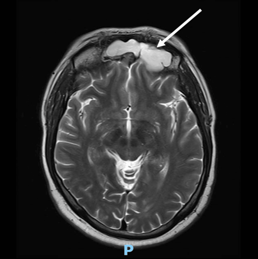
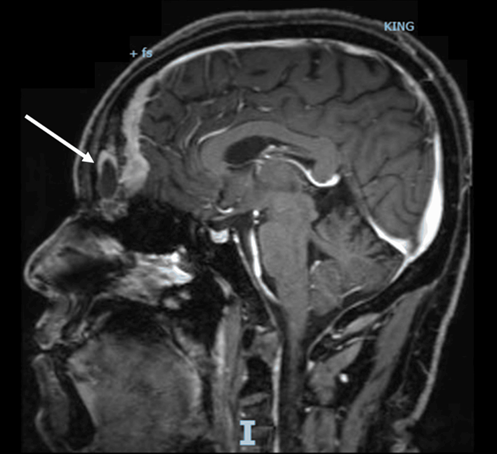
Magnetic resonance (MR) scans were performed using a SIGNA Pioneer 3.0T (GE Healthcare, Waukesha, WI, USA), and computed tomography (CT) scans were performed using a 256 slice CT scanner (Revolution CT; GE Healthcare, Waukesha, WI, USA). The enhanced MR scan of the sellar lesion showed a soft tissue mass with abnormal signals in the occipital bone clivus. T1WI showed an isointense signal, and T2WI showed an isointense/slightly hyperintense signal in a large area of approximately 30 mm × 46 mm. The lesion extended anteriorly to completely fill the entire sphenoidal sinus, and it was in a close proximity to the right internal carotid arteries. Significant invasion, liquefaction, and necrosis were not observed in the bilateral cavernous sinuses. Pituitary gland morphology was normal with a superoinferior diameter of 3.14 mm, and the pituitary gland was located in the center. An occipital bone clival space-occupying lesion was considered with a tendency of low malignancy and a possibility of chordoma (Figure 1A–C). Non-enhanced high-resolution CT scans of the nasal sinuses showed osteolytic destruction, and a soft tissue mass was observed in the occipital bone clivus. The mass had a large area of 20 mm × 30 mm × 46 mm (Figure 1D). Enhanced CT of the adrenals showed bilateral adrenal gland hyperplasia.

Figure 1 (A) MR T1+T2 scan (transverse view). MR T1 scan (left) shows the soft tissue mass of the occipital clivus (white arrow), and MR T2 scan (right) shows that the right internal carotid artery, cavernous sinus, and tumor are within close proximity to each other (white arrow). (B) MR T1 enhanced scan (sagittal view) shows clear demarcation between normal pituitary gland and mass (white arrow). (C) MR T2 scan (sagittal view) shows that the pituitary fossa is normally present (white arrow). (D) CT (sagittal view) shows bony destruction of dorsum sellae, clivus, and sphenoid sinus by mass (white arrow).

Bilateral inferior petrosal sinus sampling (IPSS) combined with a desmopressin stimulation test had the following results: baseline ACTH at left inferior petrosal sinus/periphery (IPS/P), 5.4; post-stimulation IPS/P, 3.42; stimulation corrected (ACTHPRL) IPS/P, 2.8; right baseline IPS/P, 1.64; post-stimulation IPS/P, 9.34; and stimulation corrected IPS/P, 6.92. The left inferior petrosal sinus was the dominant side (Table 1).

Table 1 Bilateral Inferior Petrosal Sinus Sampling Combined with Desmopressin Stimulation Test

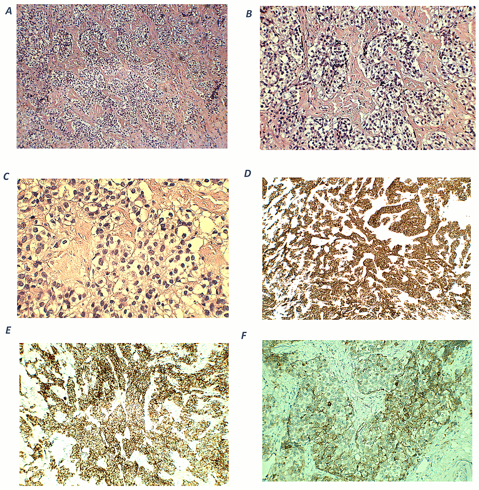
The patient underwent endoscopic transsphenoidal clival lesion resection surgery, and the postoperative pathology test results showed EPA (Figure 2). The immunohistochemistry staining results were as follows: CK (+), SYN (+), CgA (+), ACTH (+), growth hormone (GH) (−), LH (−), TSH (−), PRL (−), FSH (−), and Ki-67 (<1% +). The COR level at 10 days after surgery was 15.87 µg/dL, and the ACTH level was 31.37 pg/mL (Table 2).

Table 2 Changes in COR and ACTH Levels During Course of Treatment
Figure 2 Pathological diagnosis of (clivus) ectopic pituitary adenoma. (A) Pituitary adenoma revealing a trabecular and nested structure revealing vascular invasion (hematoxylin and eosin (HE) stain, 200x) composed of two distinct types of cells. (B) ACTH expression in the EPA (200x, ACTH-antibody, Dako).

After admission, her blood and urine COR levels were significantly elevated, and a qualitative diagnosis of CS was obtained. Etiological examination found that ACTH was also significantly elevated, suggesting that the CS was ACTH dependent. The HDDST results showed that the serum COR level was not suppressed by more than 50% and was accompanied by hypokalemia, suggesting that the ACTH-dependent CS may be ectopic ACTH syndrome. Ectopic ACTH syndrome is relatively rare, and the lesions are caused by non-pituitary tumors. No lesions were identified in the lung, thymus, pancreas, and thyroid of our patient. Regarding the IPSS examination, the IPS/P ratio was greater than 2, which suggested that the ectopic ACTH was located intracranially and not at the periphery. Radiologic testing suggested that the pituitary structure was normal and that a space-occupying lesion in the clivus region was present. Therefore, ectopic ACTH-secreting adenoma in the clivus region was considered, and postoperative pathological biopsy was used to confirm the diagnosis.

Discussion

EPA is an extremely rare disease that occurs outside of the sella turcica, and it is not linked to the intrasellar pituitary. The morphology, immunohistochemistry, and hormone activity of EPAs are similar to typical pituitary adenomas. EPAs can manifest as specific or non-specific endocrine disorders, and they account for 0.48% of all pituitary adenomas.9 The pathogenesis of EPA is still currently unknown. It is generally considered that during the development of the anterior pituitary lobe, the incompletely degraded Rathke cleft cyst remnants of the Rathke pouch lead to the formation of EPAs in the nasopharynx, sphenoid, and clivus.10,11 EPA is rare in China. Zhu et al5 recorded 14,357 pituitary gland patients in the last 20 years; of these patients, only 14 were diagnosed with EPA (0.098% of all cases), but none of the lesions originated from the clivus region. Previous literature reviews4,5 revealed that non-functioning EPAs in the clivus region are the most common (50%); the most common hormone-secreting functional adenomas are PRL adenomas and GH adenomas, which account for 25.0% and 21.4% of EPAs, respectively, whereas ACTH-secreting EPAs are extremely rare and only account for 3.6% of cases.

The postoperative pathological and immunohistochemical results of the tumor tissue in the patient demonstrated that it was an ectopic ACTH-secreting pituitary macroadenoma in the clivus region. Most EPAs are microadenomas (diameter <1 cm), except those in the clivus region, which are macroadenomas.5 Adenoma size generally does not affect the patient’s clinical and biochemical characteristics, and it may be related to tumor location or extension.12 Encasement of the internal carotid artery is a characteristic feature of EPA invasion into surrounding tissues.5 Encasement of the right internal carotid artery by the tumor was also observed in our patient. Therefore, surgery cannot completely remove the tumor and may ultimately affect surgical outcomes, and radiotherapy may even be required in the future. The serum COR and ACTH levels of our patient were evaluated 10 days after surgery. Although the levels were significantly lower than those before the surgery, the COR level was still significantly higher than the cutoff value of 1 µg/dL,13,14 suggesting that the patient may not have complete remission due to the incomplete tumor resection in the area adjacent to the carotid artery during surgery. Another feature that was observed in our patient was bone invasion. Because the clivus is composed of abundant cancellous bone that is connected to surrounding bone structures, EPAs or other tumors may cause bone destruction and affect the sphenoidal sinus and cavernous sinus, which is also consistent with literature reports.15,16

Due to the low incidence of EPAs, most EPA cases are reported as case reports in the literature. We performed an English literature search using the PubMed and Web of Science Core Collection databases with the following predetermined terms: “Cushing’s syndrome”, “pituitary adenomas”, “clivus”, “ectopic pituitary adenoma”, and “adrenocorticotropic”. The literature was included if it met the following criteria: (i) the confirmed diagnosis of CS or ectopic ACTH syndrome was described in the literature; (ii) the diagnosis of EPA was confirmed by postoperative inspection; and (iii) EPA occurred in the clivus. After excluding cases of clival invasion from other sites, we found only three reports of ectopic ACTH-secreting adenoma in the clivus region,6–8 and they were all female patients. Ortiz-Suarez and Erickson6 employed transfrontal craniotomy to demonstrate that the ectopic ACTH-secreting adenoma was an extension of extrasellar lesion to the clivus. In a case report by Pluta et al,7 the patient was found to have cavernous sinus and clival ACTH-positive tumors through transphenoidal surgery. In a case report by Aftab et al,8 the patient only presented a space-occupying lesion with unilateral vision loss; the patient was initially diagnosed with clival chordoma, but the postoperative results supported the diagnosis of EPA. Based on preoperative imaging, the possibility of chordoma was also considered to be high in our patient. We combined the clinical manifestation and laboratory test results of the patient and considered the etiology of CS to conclude that the patient had clival ectopic ACTH-secreting adenoma instead of chordoma.

Hormone tests in our patient suggested secondary pituitary-gonadal axis and decreased pituitary-thyroid axis function. These changes in endocrine function may be due to pituitary suppression by hypercortisolism. After surgery, the corresponding markers recovered, indicating that the suppression was transient. The patient has a history of fracture and a bone mineral density suggestive of osteoporosis, which may also be associated with CS hypercortisolemia.

Treatment modalities for EPA include adenoma resection surgery, radiotherapy, and drugs. The first-line recommended treatment is surgical resection. Craniotomy is considered the surgical procedure of choice for EPA, and endoscopic transsphenoidal surgery (TSS) is considered a feasible method for preserving pituitary function while simultaneously treating EPA. However, due to limitations with the surgical operation space, there are still concerns whether sufficient exploration and effective tumor resection can be achieved.17 Because there are few case reports of such patients, the long-term outcomes of these two surgical procedures require further validation. Due to differences in EPA sites and functions, the efficacy of surgery also differs. Zhu et al5 reported that compared to the radical resection rate of sphenoidal sinus and cavernous sinus EPA (72.3% and 73.3%, respectively), the radical resection rate of clival EPA is only 45.0%, and this difference is statistically significant.

The three clival EPA patients described in the three relevant publications6–8 all showed significant improvements in postoperative signs, symptoms, and hormone levels after complete surgical removal of the lesions or combined with radiation therapy. In our patient, however, radical resection of the tumor could not be achieved due to the close proximity of the tumor mass to the right internal carotid artery, and surgery could not be used to achieve complete remission, which is similar to the case reported by Zhu et al.5 For such patients, radiotherapy can be considered as a second-line treatment for EPA. To control hormone levels, drugs and bilateral adrenalectomy are also treatment options.5,18,19

Conclusion

EPA is a rare disease, and clival EPA is even rarer. From the entire diagnosis and treatment course, this unique and rare EPA case was preliminarily diagnosed through a comprehensive hormone panel and IPSS, and it was confirmed by pathology and immunohistochemistry after surgery. In the diagnosis of ectopic ACTH syndrome, attention should also be paid to extremely rare pituitary ectopic sites, such as the sphenoid sinuses, parasellar region, and the clivus, in addition to common sites, such as the lungs, thymus, pancreas, and thyroid.

Data Sharing Statement

The raw data supporting the conclusions of this article will be made available by the authors without undue reservation.

Informed Consent Statement

Prior written permission was obtained from the patient for treatment as well as for the preparation of this manuscript and for publication. Our institution approved the publication of the case details.

Acknowledgments

We would like to thank the patient and her family.

Author Contributions

All authors made a significant contribution to the work reported, whether that is in the conception, study design, execution, acquisition of data, analysis and interpretation, or in all these areas; took part in drafting, revising or critically reviewing the article; gave final approval of the version to be published; have agreed on the journal to which the article has been submitted; and agree to be accountable for all aspects of the work.

Funding

There is no funding to report.

Disclosure

The authors report no conflicts of interest in this work.

References

1. Senanayake R, Gillett D, MacFarlane J, et al. New types of localization methods for adrenocorticotropic hormone-dependent Cushing’s syndrome. Best Pract Res Clin Endocrinol Metab. 2021;35:101513. doi:10.1016/j.beem.2021.101513

2. Young J, Haissaguerre M, Viera-Pinto O, et al. Management of Endocrine Disease: cushing’s syndrome due to ectopic ACTH secretion: an expert operational opinion. Eur J Endocrinol. 2020;182:R29–r58. doi:10.1530/EJE-19-0877

3. Hayes AR, Grossman AB. The ectopic adrenocorticotropic hormone syndrome: rarely easy, always challenging. Endocrinol Metab Clin North Am. 2018;47:409–425. doi:10.1016/j.ecl.2018.01.005

4. Zhu J, Lu L, Yao Y, et al. Long-term follow-up for ectopic ACTH-secreting pituitary adenoma in a single tertiary medical center and a literature review. Pituitary. 2020;23:149–159. doi:10.1007/s11102-019-01017-y

5. Zhu J, Wang Z, Zhang Y, et al. Ectopic pituitary adenomas: clinical features, diagnostic challenges and management. Pituitary. 2020;23:648–664. doi:10.1007/s11102-020-01071-x

6. Ortiz-Suarez H, Erickson DL. Pituitary adenomas of adolescents. J Neurosurg. 1975;43:437–439. doi:10.3171/jns.1975.43.4.0437

7. Pluta RM, Nieman L, Doppman JL, et al. Extrapituitary parasellar microadenoma in Cushing’s disease. J Clin Endocrinol Metab. 1999;84:2912–2923. doi:10.1210/jcem.84.8.5890

8. Aftab HB, Gunay C, Dermesropian R, et al. “An Unexpected Pit” – ectopic pituitary adenoma. J Endocr Soc. 2021;5:A557–A558. doi:10.1210/jendso/bvab048.1137

9. Li X, Zhao B, Hou B, et al. Case report and literature review: ectopic thyrotropin-secreting pituitary adenoma in the suprasellar region. Front Endocrinol. 2021;12:619161. doi:10.3389/fendo.2021.619161

10. Agely A, Okromelidze L, Vilanilam GK, et al. Ectopic pituitary adenomas: common presentations of a rare entity. Pituitary. 2019;22:339–343. doi:10.1007/s11102-019-00954-y

11. Tajudeen BA, Kuan EC, Adappa ND, et al. Ectopic pituitary adenomas presenting as sphenoid or clival lesions: case series and management recommendations. J Neurol Surg B Skull Base. 2017;78:120–124. doi:10.1055/s-0036-1592081

12. Akirov A, Shimon I, Fleseriu M, et al. Clinical study and systematic review of pituitary microadenomas vs. macroadenomas in cushing’s disease: does size matter? J Clin Med. 2022;11:1558. doi:10.3390/jcm11061558

13. Badiu C. Williams textbook of endocrinology. Acta Endocrinologica. 2019;15:416. doi:10.4183/aeb.2019.416

14. Rollin GA, Ferreira NP, Junges M, et al. Dynamics of serum cortisol levels after transsphenoidal surgery in a cohort of patients with Cushing’s disease. J Clin Endocrinol Metab. 2004;89:1131–1139. doi:10.1210/jc.2003-031170

15. Hu S, Cheng S, Wu Y, et al. A large cavernous sinus giant cell tumor invading clivus and sphenoid sinus masquerading as meningioma: a case report and literature review. Front Surg. 2022;9:861739. doi:10.3389/fsurg.2022.861739

16. Wu X, Ding H, Yang L, et al. Invasive corridor of clivus extension in pituitary adenoma: bony anatomic consideration, surgical outcome and technical nuances. Front Oncol. 2021;11:689943. doi:10.3389/fonc.2021.689943

17. Sun X, Lu L, Feng M, et al. Cushing syndrome caused by ectopic adrenocorticotropic hormone-secreting pituitary adenomas: case report and literature review. World Neurosurg. 2020;142:75–86. doi:10.1016/j.wneu.2020.06.138

18. Szabo Yamashita T, Sada A, Bancos I, et al. Differences in outcomes of bilateral adrenalectomy in patients with ectopic ACTH producing tumor of known and unknown origin. Am J Surg. 2021;221:460–464. doi:10.1016/j.amjsurg.2020.08.047

19. Szabo Yamashita T, Sada A, Bancos I, et al. Bilateral adrenalectomy: differences between cushing disease and Ectopic ACTH-producing tumors. Ann Surg Oncol. 2020;27:3851–3857. doi:10.1245/s10434-020-08451-4

Creative Commons License This work is published and licensed by Dove Medical Press Limited. The full terms of this license are available at https://www.dovepress.com/terms.php and incorporate the Creative Commons Attribution – Non Commercial (unported, v3.0) License. By accessing the work you hereby accept the Terms. Non-commercial uses of the work are permitted without any further permission from Dove Medical Press Limited, provided the work is properly attributed. For permission for commercial use of this work, please see paragraphs 4.2 and 5 of our Terms.


Successful Immunomodulatory Treatment of COVID-19 in a Patient With Severe ACTH-Dependent Cushing’s Syndrome

Introduction: Patients with Cushing’s syndrome (CS) represent a highly sensitive group during corona virus disease 2019 (COVID-19) pandemic. The effect of multiple comorbidities and immune system supression make the clinical picture complicated and treatment challenging.

Case report: A 70-year-old female was admitted to a covid hospital with a severe form of COVID-19 pneumonia that required oxygen supplementation. Prior to her admission to the hospital she was diagnosed with adrenocorticotropic hormone (ACTH)-dependent CS, and the treatment of hypercortisolism had not been started yet. Since the patient’s condition was quickly deteriorating, and with presumend immmune system supression due to CS, we decided on treatement with intraveonus immunoglobulins (IVIg) that enabled quick onset of immunomodulatory effect. All comorbidities were treated with standard of care. The patient’s condition quickly stabilized with no direct side effects of a given treatment.

Conclusion: Treatment of COVID-19 in patients with CS faces many challenges due to the complexity of comorbidity effects, immunosupression and potential interactions of available medications both for treatment of COVID-19 and CS. So far, there are no guidelines for treatment of COVID-19 in patients with active CS. It is our opinion that immunomodulating therapies like IVIg might be an effective and safe treatment modality in this particularly fragile group of patients.

Introduction

Dealing with corona virus disease 2019 (COVID-19) focused medical attention on several sensitive population groups. While the knowledge is still improving, some of the recognized risk factors for severe form of the disease are male sex, older age, obesity, hypertension, diabetes mellitus, and cardio-vascular disease (1). This constellation of morbidities is particularly intriguing from endocrine point of view, since they are all features of patients with Cushing’s syndrome (CS). Another relevant feature of CS is a propensity for infections due to profound immune suppression, with prevalence of 21-51%; even more so, infections are the second cause of death (31%) in CS after disease progression, and are the main cause of death (37%) in patients who died within 90 days of diagnosis (2).

Immune system alterations in CS lead to depression of both innate and adaptive immune responses, favoring not only commonly acquired but also opportunistic bacterial infections, fungal infections, and severe, disseminated viral infections (3). Susceptibility to infections directly positively correlates with cortisol level, and is more frequent in ectopic ACTH secretion (EAS). Hypercortisolism hampers the first-line response to external agents and consequent activation of the adaptive response (3). Consequently, there is a decrease in total number of T-cells and B-cells, as well as a reduction in T-helper cell activation, which might favor opportunistic and intracellular infections. On the other hand, an increase in pro-inflammatory cytokine secretion, including interleukine-6 (IL-6) and tumor necrosis factor-α (TNF-α) leads to persistent, low-grade inflammation. It is important to note that immune system changes are confirmed both during the active phase and while in remission of CS (3).

In view of the aforementioned data, a few topics emerge regarding patients with CS and COVID-19. Initial clinical presentation may be altered – low-grade chronic inflammation and poor immune reaction might limit febrile response in the early phase of infection, aggravating timely diagnosis (4). Increased cytokine levels may put patients with CS at increased risk of severe course and progression to acute respiratory distress syndrome (ARDS). On the other hand, the rise in cytokine levels associated with exposure to external agents is significantly hampered, probably because of persistently elevated pro-inflammatory cytokine secretion (45). Patients with CS have a possibility for prolonged duration of viral infections and risk for superinfections leading to sepsis and increased mortality risk; this is especially relevant for hospitalized patients and mandates empirical prophylaxis with broad-spectrum antibiotics (6). Both COVID-19 and CS individually represent disease states of increased thromboembolic (TE) risk, requiring additional care (6).

Due to very limited data, it is still not possible to address these topics with certainty and make recommendations for optimal management of these patients. Current clinical practice guidance for management of CS during COVID-19 commissioned by the European Society of Endocrinology (ESE) emphasizes prompt and optimal control of hypercortisolism and adequate treatment of all comorbidities (7). Although individual circumstances must always be considered, we need more direct clinical experience, especially regarding the actual treatment of COVID-19 in this sensitive group. So far, there are only five published case studies of patients with CS and COVID-19, with eight patients in total (812). In this study, we present a patient with newly diagnosed ACTH-dependent CS who was diagnosed with COVID-19 before the initiation of specific medical treatment.

Case Report

A 70-year-old female was admitted to our Covid hospital due to bilateral interstitial pneumonia caused by severe acute respiratory syndrome coronavirus 2 (SARS-CoV-2). Six days before she was discharged from endocrinology department of another hospital where she was hospitalized due to newly diagnosed diabetes mellitus. Her personal history was unremarkable, and she was vaccinated with two doses of inactivated COVID-19 vaccine Sinopharm BBIBP. During this hospitalization Cushingoid features were noted (moon face, centripetal obesity, thin extremities with multiple hematomas, bilateral peripheral edema), as well as diabetes mellitus (HbA1c 8.7%), arterial hypertension (BP 180/100 mmHg), hypokalemia (2.0 mmol/L), mild leukocytosis (WBC 12.9x10e9/L) with neutrophilia, and mildly elevated CRP (12.3 mg/L). Hormonal functional testing confirmed ACTH-dependent Cushing’s syndrome: morning ACTH 92.6 pg/mL (reference range 10-60 pg/mL), morning serum cortisol 1239 nmol/L (reference range 131-642 nmol/L), midnight serum cortisol 1241 nmol/L, lack of cortisol suppression in overnight dexamethasone suppression test (978 nmol/L). Pituitary MRI was unremarkable other than empty sella, and CT scan of thorax normal other than left adrenal hyperplasia. Diabetes mellitus was successfully controlled with metformin, hypertension with ACE-inhibitor, Ca-channel blocker and beta-blocker, and hypokalemia with potassium supplementation along with spironolactone. Steroidogenesis inhibitors were not available in this institution, but before referral to a tertiary care hospital she was tested for SARS-CoV-2, and the test came back positive (sample was obtained by nasopharyngeal swab). Since she was asymptomatic, with normal thoracic CT scan and stabile CRP level (9.1 mg/L), she was discharged with detailed recommendations for conduct in case of progression of COVID symptoms.

Next day she started feeling malaise with episodes of fever (up to 38.2°C). Symptomatic therapy was advised in an outpatient clinic (no antiviral therapy was recommended), but 5 days later respiratory symptoms ensued. During examination, the patient was weak, with dyspnea and tachypnea (RR 22/min), afebrile (36.9°C) and with oxygen saturation (SO2) of 85% measured by pulse oximeter. Chest X-ray confirmed bilateral interstitial pneumonia with parenchymal consolidation in the right lower lung lobe, so she was referred to the COVID hospital.

Laboratory analyses upon admission are presented in the Supplementary Table 1. In addition to her previous testing, elevated chromogranin A (CgA) level was verified (538.8 ng/mL, reference range 11-98.1). The patient was treated with supplemental oxygen with maximal flow of 13 l/min. For the reason of previously confirmed severe endogenous hypercortisolism, glucocorticoids were not administered. Due to limited therapeutic options and presumed further clinical deterioration, we decided to treat the patient with intravenous immunoglobulins (IVIg) 30 g iv for 5 days, starting from the 2nd day of hospitalization. We did not observe any side effects of a given treatment. In parallel, the patient received broad-spectrum antibiotics (ceftazidime and levofloxacin), proton pump inhibitor, LMWH in prophylactic dose, oral and parenteral potassium supplementation along with spironolactone. She continued with her previous antihypertensive therapy with good control of blood pressure. While the patient was on oxygen supplementation, glycaemia was controlled with short acting insulin before meals. Following given treatment, we observed clinical, biochemical (Supplementary Table 1.) and radiological improvement (Supplementary Figure 1). Oxygen supplementation was gradually discontinued. With regard to D-dimer levels and risk factors for TE events due to COVID-19 and CS, we performed color Doppler scan of lower extremities veins, and CT pulmonary angiography, but there were no signs of thrombosis. During hospital stay, there were no signs of secondary infection and cotrimoxazole was not added to the current treatment. The patient was discharged with advice to continue her prior medical therapy along with increased dose of spironolactone and initiation of rivaroxaban. She was referred to the tertiary institution for the initiation of steroidogenesis inhibitor and further diagnostics.

Discussion

Endogenous Cushing’s syndrome is a rare disease with an incidence of 0.7-2.4 million person-years in European population-based studies (13). Significant morbidity yields a standard mortality ratio of 3.7 (95%CI 2.3–5.3), with the highest mortality during the first year after initial presentation. COVID-19 pandemic imposes additional challenge to this fragile group of patients. Due to lack of solid experience, it is still difficult to define potential clinical course and outcome of patients with CS and COVID-19. In addition, currently there are no guidelines for management of SARS-CoV-2 infection in patients with active CS.

So far, only two small case series followed patients with Cushing’s disease (CD) in various disease stages (not all were active) during COVID-19 pandemic (912). Small number of SARS-CoV-2 positive cases (3/22 and 2/61) is clearly biased by shortness of analyzed period (one and a half, and three and a half months). Additionally, a small number of patients was actually tested by nasopharyngeal swab for SARS-CoV-2 even in the presence of indicative symptoms, albeit mild. Nevertheless, all these limitations included, it seems that the prevalence of COVID-19 might be greater in patients with CD than in general population (12). This is accordant with studies on patients on exogenous glucocorticoid (GC) treatment. Overall, there is a growing body of evidence that patients on chronic GC therapy are at higher risk for SARS-CoV-2 infection and a severe course of disese, regardless of age and comorbidities (14). In many studies patients on high-dose GC therapy were at particularly high risk for a severe course of disease, so it is reasonable to assume that there is a dose-dependent effect (14).

All patients except one with endogenous CS and COVID-19 presented in literature were hospitalized, with majority of them requiring oxygen supplementation, which classified them as serious cases of disease (812). Parameters of inflammation (namely CRP) were highly variable (from normal to elevated) and did not seem to reflect severity of COVID-19 consistently. Two patients had fatal outcome; one with postoperative hypocortisolism that required stress doses of hydrocortisone, and with terminal kidney failure as significant comorbidity; the other with suspected EAS who developed ARDS in contrast to normal CRP and absence of fever (912). Based on reported cortisol levels in these patients, it seems that the severity of COVID-19 pneumonia depended on severity of hypercortisolism (812). A patient with probable EAS even developed ARDS, which adds to ongoing controversy regarding the risk of ARDS due to SARS-CoV-2 in patients with CS (315). We ourselves have treated a severely obese female patient with active CD on pasireotide, who developed ARDS despite addition of high doses of methylprednisolone (unpublished data). Additional risk imposed by comorbidities cannot be underestimated (1516). This is particularly relevant for obesity, that not only hampers immune system (leading to increased levels of IL-1, IL-6, and TNF-α), but adipocytes represent a reservoir of SARS-CoV-2 thanks to ACE2 receptor, crucial for virus attachment (15).

Majority of depicted patients with active CS were already medically treated for hypercortisolism but with various compliance (sometimes very poor), and two young patients have just started steroidogenesis inhibitors (metyrapone/ketoconazole). Infection with SARS-CoV-2 was treated by national protocols that were mostly based on supportive care. These protocols changed over time, so a few patients received antiviral therapy (favipiravir), and one young patient with suspected EAS was treated with methylprednisolone along with high doses of ketoconazole (10). Treatment was complicated with adrenal insufficiency (AI) in three patients (81112).

We have presented a patient with CS and rapid development of serious case of COVID-19 pneumonia that required hospital admission and oxygen support. She was febrile and had positive laboratory parameters of inflammation. Her CS was active, with very high cortisol levels, no prior medical treatment and with clinical suspicion of EAS (ACTH-dependent disease of short duration, severe hypercortisolism, hypokalemia, very high CgA, no visible pituitary tumor). With this in mind, and with regard to rapid progression of COVID-19 pneumonia, it was our opinion that the patient required treatment with quick onset and presumable immune system modulation.

A logical approach to treatment of CS during COVID-19 pandemic includes meticulous therapy for comorbidities (namely antihypertensives, anti-diabetic drugs, low molecular weight heparin, etc.), and steroidogenesis inhibitors for treatment for hypercortisolemia (7). While some of these drugs demonstrate quick onset of action regarding normalization of cortisol level (and hence improve clinical comorbidities), rapid effects on immune system responses are not likely, which might be of great relevance in case of acute infection. Secondly, adrenolytic therapy increases a risk of AI, which can be even more perilous than CS in case of infection or other stress situations (8121516). A modified “block and replace” approach may be considered, where addition of hydrocortisone could diminish the risk of AI (7). Still, there are a few potential pitfalls with this regimen as well. Some people fail to respond to high doses of adrenal-blocking agents due to genetic differences in the steroidogenic enzymes, since therapeutic responses to metyrapone and ketoconazole in patients with CS are associated with the polymorphism in the CYP17A1 gene (17). Additionally, there are not enough data about possible interactions between adrenolytic drugs (majority of them being metabolized through the CYP450/CYP3A4 pathway) and medications used to treat COVID-19, most of which are only just emerging (18). Special concerns, amplified with similar potential effects of SARS-CoV-2 itself as well as specific therapies are liver dysfunction (metyrapone, ketoconazole), hypokalemia (metyrapone, ketoconazole), QT-interval prolongation (ketoconazole, osilodrostat), gastrointestinal distress (mitotane, osilodrostat, etomidate) (18). Metyrapone may cause accumulation of androgenic precursors secondary to the blockade of cortisol synthesis, that can virtually enhance expression of transmembrane protease serine 2 (TMPRSS2), found to be essential to activate the viral spikes, induce viral spread, and pathogenesis in the infected hosts (19). Another important issue concerns biochemical estimation of disease control (and hence risk for AI), since most commercially available assays can overestimate cortisol level in patients treated with metyrapone due to cross-reactivity with the precursor 11-deoxicortisol (715). Mass spectrometry is a method of choice to overcome this problem, but it is not available in many centers. Some centers advocate titration and/or temporary halting medical therapies in the treatment of patients with CS in the context of COVID-19 infection (20). Treatement was stopped in a few patients with severe COVID-19 symptoms who were then given high dose GC for a few days with no long-term complications, and with full recovery (20).

There are no data about the effect of anti-viral drugs in patients with CS and COVID-19. A special concern refers to adipose tissuse, as adipose tissue is difficult for antiviral drugs to reach. It cannot be excluded that the constant release of viral replicas from the adipose tissue reservoir may interfere with COVID-19 infection treatment, delaying its resolution and favoring a worse prognosis (15). If antiviral drugs are started, it is suggested that immunocompromised patients may require prolonged therapy (18). However, the timing is difficult in practice and candidates for antivirals are limited.

Since the clinical course of COVID-19 only initially depends on viral replication, immunomodulatory therapy emerged as a valuable treatment option to control the host immune response. This became apparent ever since RECOVERY trial proved efficacy of glucocortiods (21). But this therapeutic option is fairly inapplicable in patients with active CS, since glucocorticoid treatment in chronic hypercortisolism seems to enhance immune system alterations (22). In parallel with the development of new agents, it is prudent to study the efficacy of existing therapeutic options with acceptable safety profile (20). Beside glucocorticoids, inflammation blockers, intravenous immunoglobulin and convalescent plasma were used in various settings (23).

Intravenous immunoglobulin (IVIg) is a blood product prepared from the serum pooled from thousands of healthy donors, containing a mixture of polyclonal IgG antibodies, mostly IgG1 and IgG2 subclasses (2425). Initial rationale for its use was immunodefficiency due to hypoglobulinemia. Since then it has been shown that IVIg exerts pleiotropic immunomodulating action involving both innate and adaptive immunity and it has been used in a variety of diseases (26). In previous studies on MERS (Middle East Respiratory Syndrome) and SARS (Severe Acute Respiratory Syndrome) using IVIg showed beneficial clinical effects (25). Although pathogenesis of COVID-19 has not be fully elucidated, there is a consensus that immune-mediated inflammation plays an important role in the progression of this disease, just as it did in prior coronavirus infections (27). In this context, the actual role of IVIg in COVID-19 patients might be not to boost the immune system, but through its immunomodulatory effect to suppress a hyperactive immune response that is seen in some patients (28). So far, a limited number of studies, case series and meta-analyses demonstrate a promising potential of IVIg in patients with COVID-19. The effect was demonstrated in terms of mortality, improvement of clinical symptoms, laboratory examinations, imaging and length of hospital stay, especially in patients with moderate/severe form of the disease, and with emphasis on early administration (within 3 days of admission) (24252731). A recent double blind, placebo-controlled, phase 3, randomized trial tested hyperimmune intravenous immunoglobulin (hIVIg) to SARS-CoV-2 derived from recovered donors with no demonstrated effect compared with standard of care, but therapy was administered in patients symptomatic up to 12 days (32). Additional clinical trials are underway, hopefully with more guidance for proper selection of patients that might benefit from this type of treatment.

Conclusion

To our knowledge, this is the first case of IVIg treatment in a COVID-19 patient with CS. It is our opinion that immune-modulating properties of IVIg might present an attractive treatment option, especially in those CS patients that show rapid clinical progression and positive laboratory parameters of inflammation. While we await for new therapeutic modalities for COVID-19 and while some of the modalities remain not widely available, IVIg is more accessible, safe method, which could be rescuing in carefully selected patients. Of note, we consider our patient’s vaccinal status as an unquestionable positive contributor to the favorable outcome

Data Availability Statement

The raw data supporting the conclusions of this article will be made available by the authors, without undue reservation.

Ethics Statement

Ethical review and approval was not required for the study on human participants in accordance with the local legislation and institutional requirements. The patients/participants provided their written informed consent to participate in this study. Written informed consent was obtained from the individual(s) for the publication of any potentially identifiable images or data included in this article.

Author Contributions

BP, AS, JV, TG, MJ-L, JV, VS, ZG and TA-V analyzed and interpreted the patient data. BP, AP, DI, and DJ were major contributors in writing the manuscript. All authors contributed to the article and approved the submitted version.

Conflict of Interest

The authors declare that the research was conducted in the absence of any commercial or financial relationships that could be construed as a potential conflict of interest.

Publisher’s Note

All claims expressed in this article are solely those of the authors and do not necessarily represent those of their affiliated organizations, or those of the publisher, the editors and the reviewers. Any product that may be evaluated in this article, or claim that may be made by its manufacturer, is not guaranteed or endorsed by the publisher.

Supplementary Material

The Supplementary Material for this article can be found online at: https://www.frontiersin.org/articles/10.3389/fendo.2022.889928/full#supplementary-material

References

1. Hu J, Wang Y. The Clinical Characteristics and Risk Factors of Severe COVID-19. Gerontology (2021) 67(3):255–66. doi: 10.1159/000513400

PubMed Abstract | CrossRef Full Text | Google Scholar

2. Valassi E, Tabarin A, Brue T, Feelders RA, Reincke M, Netea-Maier R, et al. High Mortality Within 90 Days of Diagnosis in Patients With Cushing’s Syndrome: Results From the ERCUSYN Registry. Eur J Endocrinol (2019) 181(5):461–72. doi: 10.1530/EJE-19-0464

PubMed Abstract | CrossRef Full Text | Google Scholar

3. Hasenmajer V, Sbardella E, Sciarra F, Minnetti M, Isidori AM, Venneri MA. The Immune System in Cushing’s Syndrome. Trends Endocrinol Metab (2020) 31(9):655–69. doi: 10.1016/j.tem.2020.04.004

PubMed Abstract | CrossRef Full Text | Google Scholar

4. Pivonello R, Isidori AM, De Martino MC, Newell-Price J, Biller BM, Colao A. Complications of Cushing’s Syndrome: State of the Art. Lancet Diabetes Endocrinol (2016) 4(7):611–29. doi: 10.1016/S2213-8587(16)00086-3

PubMed Abstract | CrossRef Full Text | Google Scholar

5. Martins-Filho PR, Tavares CSS, Santos VS. Factors Associated With Mortality in Patients With COVID-19. A Quantitative Evidence Synthesis of Clinical and Laboratory Data. Eur J Intern Med (2020) 76:97–9. doi: 10.1016/j.ejim.2020.04.043

PubMed Abstract | CrossRef Full Text | Google Scholar

6. Pivonello R, Ferrigno R, Isidori AM, Biller BMK, Grossman AB, Colao A. COVID-19 and Cushing’s Syndrome: Recommendations for a Special Population With Endogenous Glucocorticoid Excess. Lancet Diabetes Endocrinol (2020) 8(8):654–6. doi: 10.1016/S2213-8587(20)30215-1

PubMed Abstract | CrossRef Full Text | Google Scholar

7. Newell-Price J, Nieman LK, Reincke M, Tabarin A. ENDOCRINOLOGY IN THE TIME OF COVID-19: Management of Cushing’s Syndrome. Eur J Endocrinol (2020) 183(1):G1–7. doi: 10.1530/EJE-20-0352

PubMed Abstract | CrossRef Full Text | Google Scholar

8. Beretta F, Dassie F, Parolin M, Boscari F, Barbot M, Busetto L, et al. Practical Considerations for the Management of Cushing’s Disease and COVID-19: A Case Report. Front Endocrinol (Lausanne) (2020) 11:554. doi: 10.3389/fendo.2020.00554

PubMed Abstract | CrossRef Full Text | Google Scholar

9. Belaya Z, Golounina O, Melnichenko G, Tarbaeva N, Pashkova E, Gorokhov M, et al. Clinical Course and Outcome of Patients With ACTH-Dependent Cushing’s Syndrome Infected With Novel Coronavirus Disease-19 (COVID-19): Case Presentations. Endocrine (2021) 72(1):12–9. doi: 10.1007/s12020-021-02674-5

PubMed Abstract | CrossRef Full Text | Google Scholar

10. Rehman T. Image of the Month: Diagnostic and Therapeutic Challenges in the Management of Ectopic ACTH Syndrome: A Perfect Storm of Hypercortisolism, Hyperglycaemia and COVID-19. Clin Med (Lond) (2021) 21(3):231–4. doi: 10.7861/clinmed.2021-0005

PubMed Abstract | CrossRef Full Text | Google Scholar

11. Yuno A, Kenmotsu Y, Takahashi Y, Nomoto H, Kameda H, Cho KY, et al. Successful Management of a Patient With Active Cushing’s Disease Complicated With Coronavirus Disease 2019 (COVID-19) Pneumonia. Endocr J (2021) 68(4):477–84. doi: 10.1507/endocrj.EJ20-0613

PubMed Abstract | CrossRef Full Text | Google Scholar

12. Serban AL, Ferrante E, Carosi G, Indirli R, Arosio M, Mantovani G. COVID-19 in Cushing Disease: Experience of a Single Tertiary Centre in Lombardy. J Endocrinol Invest (2021) 44(6):1335–6. doi: 10.1007/s40618-020-01419-x

PubMed Abstract | CrossRef Full Text | Google Scholar

13. Sharma ST, Nieman LK, Feelders RA. Cushing’s Syndrome: Epidemiology and Developments in Disease Management. Clin Epidemiol (2015) 7:281–93. doi: 10.2147/CLEP.S44336

PubMed Abstract | CrossRef Full Text | Google Scholar

14. Vogel F, Reincke M. Endocrine Risk Factors for COVID-19: Endogenous and Exogenous Glucocorticoid Excess. Rev Endocr Metab Disord (2021) 23(2):233–50. doi: 10.1007/s11154-021-09670-0

PubMed Abstract | CrossRef Full Text | Google Scholar

15. Guarnotta V, Ferrigno R, Martino M, Barbot M, Isidori AM, Scaroni C, et al. Glucocorticoid Excess and COVID-19 Disease. Rev Endocr Metab Disord (2021) 22(4):703–14. doi: 10.1007/s11154-020-09598-x

PubMed Abstract | CrossRef Full Text | Google Scholar

16. Chifu I, Detomas M, Dischinger U, Kimpel O, Megerle F, Hahner S, et al. Management of Patients With Glucocorticoid-Related Diseases and COVID-19. Front Endocrinol (Lausanne) (2021) 12:705214. doi: 10.3389/fendo.2021.705214

PubMed Abstract | CrossRef Full Text | Google Scholar

17. Valassi E, Aulinas A, Glad CA, Johannsson G, Ragnarsson O, Webb SM. A Polymorphism in the CYP17A1 Gene Influences the Therapeutic Response to Steroidogenesis Inhibitors in Cushing’s Syndrome. Clin Endocrinol (Oxf) (2017) 87(5):433–9. doi: 10.1111/cen.13414

PubMed Abstract | CrossRef Full Text | Google Scholar

18. Berlinska A, Swiatkowska-Stodulska R, Sworczak K. Old Problem, New Concerns: Hypercortisolemia in the Time of COVID-19. Front Endocrinol (Lausanne) (2021) 12:711612. doi: 10.3389/fendo.2021.711612

PubMed Abstract | CrossRef Full Text | Google Scholar

19. Barbot M, Ceccato F, Scaroni C. Consideration on TMPRSS2 and the Risk of COVID-19 Infection in Cushing’s Syndrome. Endocrine (2020) 69(2):235–6. doi: 10.1007/s12020-020-02390-6

PubMed Abstract | CrossRef Full Text | Google Scholar

20. Fleseriu M. Pituitary Disorders and COVID-19, Reimagining Care: The Pandemic A Year and Counting. Front Endocrinol (Lausanne) (2021) 12:656025. doi: 10.3389/fendo.2021.656025

PubMed Abstract | CrossRef Full Text | Google Scholar

21. Horby P, Lim WS, Emberson JR, Mafham M, Bell JL, Linsell L, et al. Dexamethasone in Hospitalized Patients With Covid-19. N Engl J Med (2021) 384(8):693–704. doi: 10.1056/NEJMoa2021436

PubMed Abstract | CrossRef Full Text | Google Scholar

22. Bergquist M, Lindholm C, Strinnholm M, Hedenstierna G, Rylander C. Impairment of Neutrophilic Glucocorticoid Receptor Function in Patients Treated With Steroids for Septic Shock. Intensive Care Med Exp (2015) 3(1):59. doi: 10.1186/s40635-015-0059-9

PubMed Abstract | CrossRef Full Text | Google Scholar

23. Cao W, Li T. COVID-19: Towards Understanding of Pathogenesis. Cell Res (2020) 30(5):367–9. doi: 10.1038/s41422-020-0327-4

PubMed Abstract | CrossRef Full Text | Google Scholar

24. Tzilas V, Manali E, Papiris S, Bouros D. Intravenous Immunoglobulin for the Treatment of COVID-19: A Promising Tool. Respiration (2020) 99(12):1087–9. doi: 10.1159/000512727

PubMed Abstract | CrossRef Full Text | Google Scholar

25. Mohtadi N, Ghaysouri A, Shirazi S, Sara A, Shafiee E, Bastani E, et al. Recovery of Severely Ill COVID-19 Patients by Intravenous Immunoglobulin (IVIG) Treatment: A Case Series. Virology (2020) 548:1–5. doi: 10.1016/j.virol.2020.05.006

PubMed Abstract | CrossRef Full Text | Google Scholar

26. Schwab I, Nimmerjahn F. Intravenous Immunoglobulin Therapy: How Does IgG Modulate the Immune System? Nat Rev Immunol (2013) 13(3):176–89. doi: 10.1038/nri3401

PubMed Abstract | CrossRef Full Text | Google Scholar

27. Cao W, Liu X, Hong K, Ma Z, Zhang Y, Lin L, et al. High-Dose Intravenous Immunoglobulin in Severe Coronavirus Disease 2019: A Multicenter Retrospective Study in China. Front Immunol (2021) 12:627844. doi: 10.3389/fimmu.2021.627844

PubMed Abstract | CrossRef Full Text | Google Scholar

28. Bongomin F, Asio LG, Ssebambulidde K, Baluku JB. Adjunctive Intravenous Immunoglobulins (IVIg) for Moderate-Severe COVID-19: Emerging Therapeutic Roles. Curr Med Res Opin (2021) 37(6):903–5. doi: 10.1080/03007995.2021.1903849

PubMed Abstract | CrossRef Full Text | Google Scholar

29. Gharebaghi N, Nejadrahim R, Mousavi SJ, Sadat-Ebrahimi SR, Hajizadeh R. The Use of Intravenous Immunoglobulin Gamma for the Treatment of Severe Coronavirus Disease 2019: A Randomized Placebo-Controlled Double-Blind Clinical Trial. BMC Infect Dis (2020) 20(1):786. doi: 10.1186/s12879-020-05507-4

PubMed Abstract | CrossRef Full Text | Google Scholar

30. Ali S, Uddin SM, Shalim E, Sayeed MA, Anjum F, Saleem F, et al. Hyperimmune Anti-COVID-19 IVIG (C-IVIG) Treatment in Severe and Critical COVID-19 Patients: A Phase I/II Randomized Control Trial. EClinicalMedicine (2021) 36:100926. doi: 10.1016/j.eclinm.2021.100926

PubMed Abstract | CrossRef Full Text | Google Scholar

31. Xiang HR, Cheng X, Li Y, Luo WW, Zhang QZ, Peng WX. Efficacy of IVIG (Intravenous Immunoglobulin) for Corona Virus Disease 2019 (COVID-19): A Meta-Analysis. Int Immunopharmacol (2021) 96:107732. doi: 10.1016/j.intimp.2021.107732

PubMed Abstract | CrossRef Full Text | Google Scholar

32. Polizzotto MN, Nordwall J, Babiker AG, Phillips A, Vock DM, Eriobu N, et al. Hyperimmune Immunoglobulin for Hospitalised Patients With COVID-19 (ITAC): A Double-Blind, Placebo-Controlled, Phase 3, Randomised Trial. Lancet (2022) 399(10324):530–40. doi: 10.1016/S0140-6736(22)00101-5

PubMed Abstract | CrossRef Full Text | Google Scholar

Keywords: Cushing’s syndrome, COVID-19, IVIg, hypercortisolism, immunomodulation, immunosuppression

Citation: Popovic B, Radovanovic Spurnic A, Velickovic J, Plavsic A, Jecmenica-Lukic M, Glisic T, Ilic D, Jeremic D, Vratonjic J, Samardzic V, Gluvic Z and Adzic-Vukicevic T (2022) Successful Immunomodulatory Treatment of COVID-19 in a Patient With Severe ACTH-Dependent Cushing’s Syndrome: A Case Report and Review of Literature. Front. Endocrinol. 13:889928. doi: 10.3389/fendo.2022.889928

Received: 04 March 2022; Accepted: 17 May 2022;
Published: 22 June 2022.

Edited by:

Giuseppe Reimondo, University of Turin, Italy

Reviewed by:

Nora Maria Elvira Albiger, Veneto Institute of Oncology (IRCCS), Italy
Miguel Debono, Royal Hallamshire Hospital, United Kingdom

Copyright © 2022 Popovic, Radovanovic Spurnic, Velickovic, Plavsic, Jecmenica-Lukic, Glisic, Ilic, Jeremic, Vratonjic, Samardzic, Gluvic and Adzic-Vukicevic. This is an open-access article distributed under the terms of the Creative Commons Attribution License (CC BY). The use, distribution or reproduction in other forums is permitted, provided the original author(s) and the copyright owner(s) are credited and that the original publication in this journal is cited, in accordance with accepted academic practice. No use, distribution or reproduction is permitted which does not comply with these terms.

*Correspondence: Bojana Popovic, popbojana@gmail.com

Disclaimer: All claims expressed in this article are solely those of the authors and do not necessarily represent those of their affiliated organizations, or those of the publisher, the editors and the reviewers. Any product that may be evaluated in this article or claim that may be made by its manufacturer is not guaranteed or endorsed by the publisher.

From https://www.frontiersin.org/articles/10.3389/fendo.2022.889928/full

Eyelid Edema Due to Cushing’s Syndrome

Abstract

Cushing’s syndrome (CS) shows diverse signs such as centripetal obesity, moon face, and buffalo hump, which can complicate the diagnosis. Facial features including eyelid edema, as an underrecognized sign, can be diagnostic clues for an excess of corticoids in a CS patient.

A 49-year-old woman presented with amenorrhea and weight gain that had continued for 2 years. Her medical history was dyslipidemia, hypertension, and osteoporosis. Physical examination revealed eyelid edemas (Figure 1A), moon face, buffalo hump, abdominal purple striae, and centripetal obesity (body mass index (BMI), 30.8 kg/m2). Basal plasma adrenocorticotropin was undetectable and serum cortisol level was high (16.9 μg/dl) without circadian rhythms. Free cortisol level in a 24-h urine collection was elevated (158.7 μg/day). Overnight administration of dexamethasone (1 mg) did not reduce serum cortisol level (17.4 μg/dl). Magnetic resonance imaging suggested bilateral adenomas. We made a diagnosis of adrenal Cushing’s syndrome (CS). Since 131l-adosterol scintigraphy showed specific uptake in the left adrenal gland, left adrenalectomy was laparoscopically performed. Histopathology of the tumor was compatible with adrenocortical adenoma. Three months after surgery, her BMI decreased to 25.0 kg/m2 and eyelid edemas were ameliorated (Figure 1B).

Details are in the caption following the image

(A) Bilateral eyelid edemas due to Cushing’s syndrome are shown. (B) These findings were improved three months after surgery for left adrenal adenomas

Eyelid edema, in addition to centripetal obesity, moon face, and buffalo hump, is also a significant sign of CS; however, it has scarcely been reported in countries other than Japan.12 Increased capillary permeability, insufficient venous return due to muscle atrophy, and sodium retention due to mineralocorticoid actions conceivably cause edema in CS.

AUTHORS’ CONTRIBUTIONS

KY wrote the first draft and managed all the submission processes. KO and KH contributed to the clinical management of the patient. FO organized the writing the manuscript.

ACKNOWLEDGMENT

None.

CONFLICT OF INTEREST

The authors declare no conflicts of interest.

ETHICAL APPROVAL

Written informed consent was obtained from the patient to publish this case report.

  • 1Lacroix A, Feelders RA, Stratakis CA, Nieman LK. Cushing’s syndrome. Lancet. 2015; 386: 913- 927.
  • 2Komiya I, Takasu N, Ohara N, et al. Forty-one cases of Cushing’s syndrome: a comparison between Cushing’s syndrome (adrenal adenoma) and Cushing’s disease (adrenal hyperplasia). Nihon Naibunpi Gakkai Zasshi. 1992; 68: 607- 622.

https://doi.org/10.1002/ccr3.5940

From https://onlinelibrary.wiley.com/doi/10.1002/ccr3.5940

Osilodrostat Improves Physical Manifestations of Hypercortisolism for Most Adults

Osilodrostat is associated with improvements in physical manifestations of hypercortisolism and reductions in mean body weight and BMI in adults with Cushing’s syndrome, according to a speaker.

As Healio previously reported, in findings from the LINC 4 phase 3 trial, osilodrostat (Isturisa, Recordati) normalized mean urinary free cortisol level at 12 weeks in more than 75% of adults with Cushing’s disease. In new findings presented at the AACE Annual Scientific and Clinical Conference, most adults with Cushing’s syndrome participating in the LINC 3 phase 3 trial had improvements in physical manifestations of hypercortisolism 72 weeks after initiating osilodrostat, with more than 50% having no dorsal fat pad, supraclavicular fat pad, facial rubor, proximal muscle atrophy, striae, ecchymoses and hirsutism for women at 72 weeks.

Adrenal transparent _Adobe
Source: Adobe Stock

“Many patients with Cushing’s syndrome suffer from clinical manifestations related to hypercortisolism,” Albert M. Pedroncelli, MD, PhD, head of clinical development and medical affairs for Recordati AG in Basel, Switzerland, told Healio. “The treatment with osilodrostat induced a rapid normalization of cortisol secretion, and improvements in physical manifestations associated with hypercortisolism were observed soon after initiation of osilodrostat and were sustained throughout the study.”

Albert M. Pedroncelli

Pedroncelli and colleagues analyzed changes in the physical manifestations of hypercortisolism in 137 adults with Cushing’s syndrome (median age, 40 years; 77.4% women) assigned osilodrostat. Dose titration took place from baseline to 12 weeks, and therapeutic doses were administered from 12 to 48 weeks, with some participants randomly assigned to withdrawal between 26 and 34 weeks. An extension phase of the trial took place from 48 to 72 weeks. Investigators subjectively rated physical manifestations of hypercortisolism in participants as none, mild, moderate or severe. Participants were evaluated at baseline and 12, 24, 34, 48 and 72 weeks.

At baseline, the majority of the study cohort had mild, moderate or severe physical manifestations of hypercortisolism in most individual categories, including dorsal fat pad, central obesity, supraclavicular fat pad, facial rubor, hirsutism in women and striae. Central obesity was the most frequent physical manifestation rated as severe.

The percentage of participants with improvements in physical manifestations of hypercortisolism increased from week 12 on for all individual manifestations evaluated in the study, and improvements were maintained through week 72. At 72 weeks, the percentage of participants who had no individual physical manifestations was higher than 50% for each category except central obesity, where 30.6% of participants had no physical manifestations.

In addition to improvement in physical manifestations, the study cohort had decreases in body weight, BMI and waist circumference at weeks 48 and 72 compared with baseline.

“The main goal of treating patients with Cushing’s syndrome is to normalize cortisol secretion,” Pedroncelli said. “The rapid reduction and normalization of cortisol levels is accompanied by improvement in the associated clinical manifestations. This represents an important objective for patients.”

From https://www.healio.com/news/endocrinology/20220512/osilodrostat-improves-physical-manifestations-of-hypercortisolism-for-most-adults

%d bloggers like this: