Evaluation of Ketoconazole As a Treatment for Cushing’s Disease in a Retrospective Cohort

Objective: The first-line treatment for Cushing’s disease is transsphenoidal surgery, after which the rates of remission are 60 to 80%, with long-term recurrence of 20 to 30%, even in those with real initial remission. Drug therapies are indicated for patients without initial remission or with surgical contraindications or recurrence, and ketoconazole is one of the main available therapies. The objective of this study was to evaluate the safety profile of and the treatment response to ketoconazole in Cushing’s disease patients followed up at the endocrinology outpatient clinic of a Brazilian university hospital.

Patients and methods: This was a retrospective cohort of Cushing’s disease patients with active hypercortisolism who used ketoconazole at any stage of follow-up. Patients who were followed up for less than 7 days, who did not adhere to treatment, or who were lost to follow-up were excluded.

Results: Of the 172 Cushing’s disease patients who were followed up between 2004 and 2020, 38 received ketoconazole. However, complete data was only available for 33 of these patients. Of these, 26 (78%) underwent transsphenoidal surgery prior to using ketoconazole, five of whom (15%) had also undergone radiotherapy; seven used ketoconazole as a primary treatment. Ketoconazole use ranged from 14 days to 14.5 years. A total of 22 patients had a complete response (66%), three patients had a partial response (9%), and eight patients had no response to treatment (24%), including those who underwent radiotherapy while using ketoconazole. Patients whose hypercortisolism was controlled or partially controlled with ketoconazole had lower baseline 24-h urinary free cortisol levels than the uncontrolled group [times above the upper limit of normal: 0.62 (SD, 0.41) vs. 5.3 (SD, 8.21); p < 0.005, respectively] in addition to more frequent previous transsphenoidal surgery (p < 0.04). The prevalence of uncontrolled patients remained stable over time (approximately 30%) despite ketoconazole dose adjustments or association with other drugs, which had no significant effect. One patient received adjuvant cabergoline from the beginning of the follow-up, and it was prescribed to nine others due to clinical non-response to ketoconazole alone. Ten patients (30%) reported mild adverse effects, such as nausea, vomiting, dizziness, and loss of appetite. Only four patients had serious adverse effects that warranted discontinuation. There were 20 confirmed episodes of hypokalemia among 10/33 patients (30%).

Conclusion: Ketoconazole effectively controlled hypercortisolism in 66% of Cushing’s disease patients, being a relatively safe drug for those without remission after transsphenoidal surgery or whose symptoms must be controlled until a new definitive therapy is carried out. Hypokalemia is a frequent metabolic effect not yet described in other series, which should be monitored during treatment.

Introduction

Cushing’s disease (CD) results from a pituitary tumor that secretes adrenocorticotropic hormone (ACTH), which leads to chronic hypercortisolism. It is a potentially fatal disease with high morbidity and a mortality rate of up to 3.7 times than that of the general population (14) associated to several clinical–metabolic disorders caused by excess cortisol and/or loss of circadian rhythm (5). In general, its management is a challenge even in reference centers (67).

Transsphenoidal surgery (TSS), the treatment of choice for CD, results in short-term remission in 60 to 80% of patients (8). However, recurrence rates of 20 to 30% are found in long-term follow-up, even in those with clear initial remission (9). Drug therapies can help control excess cortisol in patients without initial remission, in cases of recurrence, and in those with contraindications or high initial surgical risk (10).

Nevertheless, specific drugs that act on the pituitary adenoma, which could directly treat excess ACTH, have a limited effect, and only pasireotide is approved for this purpose in Brazil (1112). In this scenario, adrenal steroidogenesis blockers are important. One such off-label medication is the antifungal drug ketoconazole, a synthetic imidazole derivative that inhibits the enzymes CYP11A1, CYP17, CYP11B2, and CYP11B1. Because of its hepatotoxicity and the availability of other drugs, it has been withdrawn from the market in several countries (13). In Europe, it is still approved for use in CD, although in the United States, it is recommended for off-label use almost in CD (1416). Due to the potential benefits for hypercortisolism, ketoconazole has been replaced by levoketoconazole, which the European Union has recently approved for CD with a lower expected hepatotoxicity (17).

Thus, when adrenal inhibitors are used as an alternative treatment for CD, information about the outcomes of drugs such as ketoconazole are important. Clinical studies on these effects in CD are scarce, mostly retrospective, multicenter, or from developed countries (1418). A recent meta-analysis on the therapeutic modalities for CD included only four studies (246 patients) that evaluated urinary cortisol response as a treatment outcome and eight studies (366 patients) describing the prevalence of some side effects: change in transaminase activity, digestive symptoms, skin rash, and adrenal insufficiency. Hypokalemia was not mentioned in this meta-analysis (19).

The objective of this study was to evaluate the safety profile of and treatment response to ketoconazole in CD patients followed during a long term in the endocrinology outpatient clinic of a Brazilian university hospital.

Patients and methods

Patients

We retrospectively evaluated 38 patients (27 women) diagnosed with CD. These patients, whose treatment included ketoconazole at any time between 2004 and 2020, are part of a prospective cohort series from the Hospital de Clínicas de Porto Alegre neuroendocrinology outpatient clinic.

The diagnostic criteria for hypercortisolism were based on high 24-h urinary free cortisol levels (24-h UFC) in at least two samples, non-suppression of serum cortisol after low-dose dexamethasone testing (>1.8 µg/dl), and/or loss of cortisol rhythm (midnight serum cortisol >7.5 µg/dl or midnight salivary cortisol >0.208 nmol/L). CD was diagnosed by normal or elevated ACTH levels, evidence of pituitary adenoma >0.6 cm on magnetic resonance image (MRI), and ACTH central/periphery gradient on inferior petrosal sinus catheterization when MRI was normal or showed an adenoma <0.6 cm.

CD was considered to be in remission after the improvement of hypercortisolism symptoms or clinical signs of adrenal insufficiency, associated with serum cortisol within reference values, normalization of 24-h UFC and/or serum cortisol <1.8 μg/dl at 8 am after 1 mg dexamethasone overnight, and/or normalization of midnight serum or salivary cortisol. In patients with active disease, to evaluate the ketoconazole treatment response, 24-h UFC was used as a laboratory parameter, as recommended in similar publications (14162021), but in some cases, we considered elevated late night salivary cortisol and/or 1 mg dexamethasone overnight cortisol (even with normal 24-h UFC), given the greater assessment sensitivity seen through these two methods in the detection of early recurrence when compared with 24-h UFC (22).

Inclusion criteria

We included patients with CD and active hypercortisolism who used ketoconazole either as primary treatment, after TSS without hypercortisolism remission, or after a recurrence.

Exclusion criteria

We excluded patients with CD and active hypercortisolism who used ketoconazole but had <7 days of follow-up, irregular outpatient follow-up, treatment non-adherence, and incomplete medical records or those who were lost to follow-up.

Evaluated parameters

Prior to ketoconazole treatment, all patients underwent an assessment of pituitary function and hypercortisolism, including serum cortisol, ACTH, 24-hour UFC, cortisol suppression after 1 mg dexamethasone overnight, midnight serum cortisol, and/or midnight salivary cortisol. The evaluated parameters were sex, age at diagnosis, weight, height, prevalence and severity of hypertension and DM, pituitary tumor characteristics, prior treatment (surgery, radiotherapy, or other medications), symptoms at disease onset, biochemical tests (renal function, hepatic function, and lipid profile), number of medications used to treat associated comorbidities, data on medication tolerance, and reasons for discontinuation, when necessary.

The clinical parameters observed during treatment were control of blood pressure and hyperglycemia, anthropometric measurements (weight, height, and body mass index), jaundice, and any other symptoms or adverse effects reported by patients.

The biochemical evaluation included fasting glucose, glycated hemoglobin, lipid profile (total cholesterol, high-density lipoprotein, low-density lipoprotein, and triglycerides), markers of liver damage (transaminases, bilirubin, gamma-glutamyl transferase, and alkaline phosphatase), electrolytes (sodium and potassium), and renal function (creatinine and urea). Hypecortisolism was accessed preferentially by 24-h UFC, however, late-night salivary cortisol and cortisol after 1 mg overnight dexamethasone could also be used.

Study design

This retrospective cohort study included patients with CD who were followed up at the Hospital de Clínicas de Porto Alegre Endocrinology Division, with their medical records from the first outpatient visit and throughout clinical follow-up collected. This study was approved by the Hospital de Clínicas de Porto Alegre Research Ethics Committee (number 74555617.0.0000.5327).

Outcomes

Hypercortisolism was considered controlled when the 24-h UFC and/or late-night salivary cortisol (LNSC) and/or overnight 1 mg dexamethasone suppression test (DST) levels were normalized in at least two consecutive assessments. Hypercortisolism was considered partially controlled when there was a 50% over-reduction in 24-h UFC and/or LNSC and/or DST levels but still above normal. A reduction lower than 50% in these parameters was considered as non-response.

We also assessed the ketoconazole doses that resulted in 24-h UFC normalization, maximum dose, medication tolerance, adverse effects, and changes in liver, kidney, and biochemical function. Due to the characteristics of this study, these outcomes were periodically evaluated in all patient consultations, which occurred usually every 2 to 4 months.

Data collection

This retrospective cohort evaluated outpatient medical records and any tests indicated by the attending physician as a pragmatic study. Ketoconazole use followed the department’s care protocol, which is based on national and international guidelines (4), and all patients received a similar care routine: the recommended initial prescription was generally taken in two to six doses at 100 to 300 mg/day. It was then increased by 200 mg every 2 to 4 months until hypercortisolism was controlled or side effects developed, especially those related to liver function. The maximum prescription was 1,200 mg/day. Clinical follow-up of these patients was performed 30 days after starting the medication and every 2–4 months thereafter (23).

Clinical, anthropometric, laboratory, and other exam data were collected through a review of the hospital’s electronic medical records for the entire follow-up period. Data from the first and last consultation were considered in the final analysis of all parameters.

Statistical analysis

Baseline population characteristics were described as mean and standard deviation (SD) or median with interquartile ranges (25–75) for continuous variables. The chi-square test was used to compare qualitative variables, and Student’s t-test or ANOVA was used to compare the quantitative variables. The Mann–Whitney U-test was used for unpaired data. P-values <0.05 were considered significant. Statistical analysis was performed in SPSS 18.0 (SPSS Inc., Chicago, IL, USA) and R package geepack 1.3-1.

Results

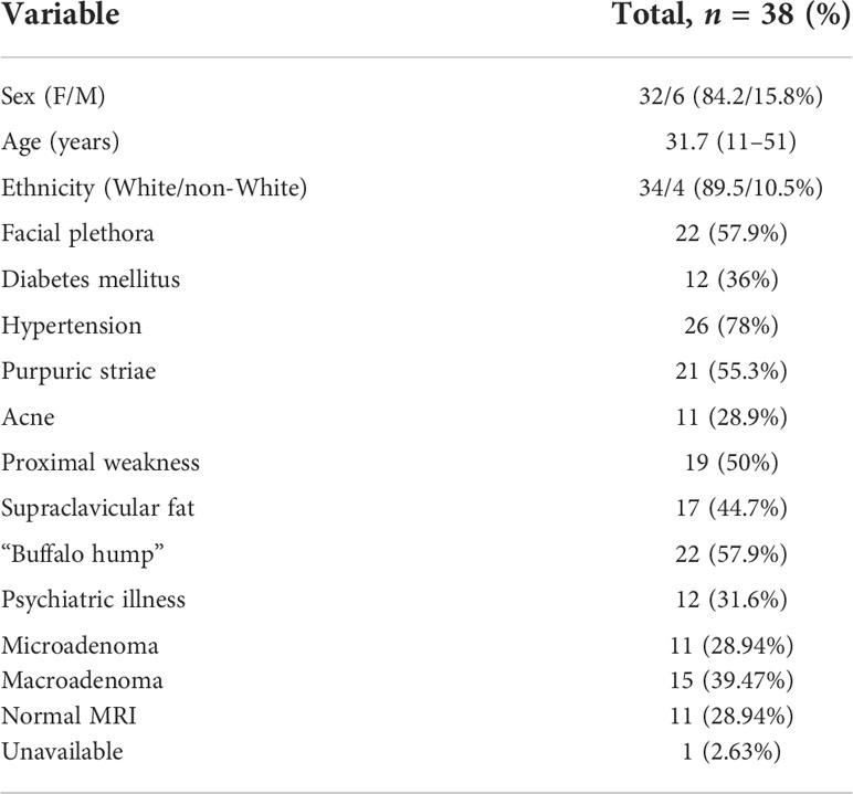
Treatment with ketoconazole was indicated for 41 of the 172 CD patients. In 3/41 patients, ketoconazole was unallowed due to concomitant liver disease, and 38 received ketoconazole during CD treatment between 2004 and 2020. Of these, five were excluded due to insufficient data to determine the response to ketoconazole (short treatment time, irregular follow-up, incomplete medical records, or lost to follow-up). The baseline characteristics of every sample are shown in Table 1. Thus, 33/41 patients were included in the final analysis. The patients were predominantly women (84.2%) and white (89.5%); 11 had microadenoma, 15 had macroadenoma, and 11 had no adenoma visualized. In 12/33 patients, pituitary imaging was not performed immediately before starting ketoconazole. Hypertension was observed in 26 patients (78%) and DM in 12 patients (36%). The mean age at CD diagnosis was 31.7 years.

Table 1
www.frontiersin.orgTABLE 1 Baseline clinical data of Cushing’s disease patients treated with ketoconazole.

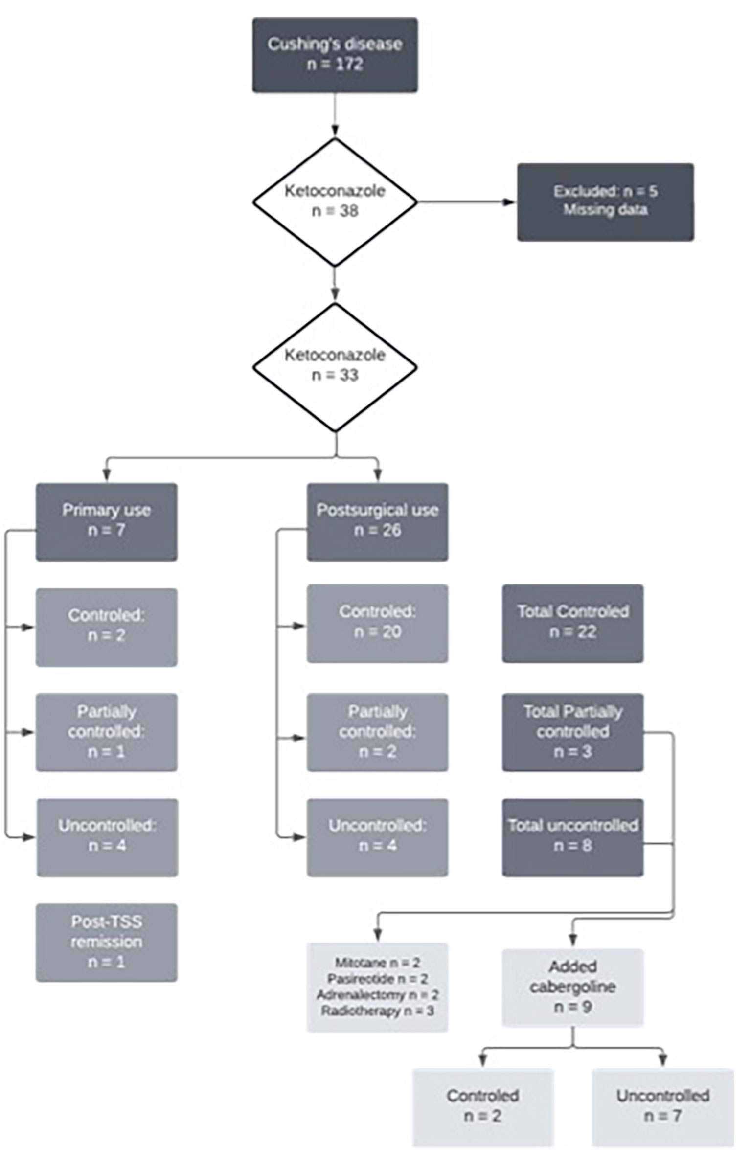
Of the 33 patients with complete data, 26 (78%) underwent TSS prior to starting ketoconazole, five of whom (15%) had also undergone radiotherapy. Thus, seven patients used ketoconazole as primary treatment since performing a surgical procedure was impossible at that time. Of these, four had no response to ketoconazole, one had a partial response, and two had a complete response. At follow-up, four of these patients underwent their first TSS, and three continued the ketoconazole therapy, achieving full UFC control. Among those who used ketoconazole after TSS (n = 26), 20 had a complete response, two had a partial response, and four had no response. Figure 1 shows the study flow chart and patient distribution throughout the treatment.

Figure 1
www.frontiersin.orgFIGURE 1 Flowchart of ketoconazole treatment in Cushing’s disease patients.

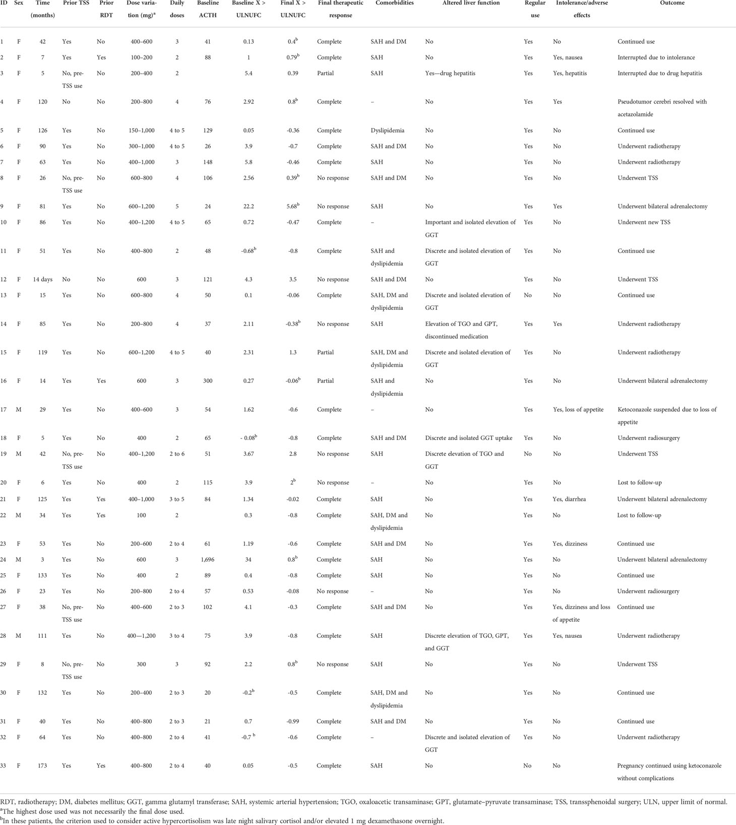
Individual patient data are described in Table 2. The duration of ketoconazole use ranged from 14 days (in one patient who used it pre-TSS) to 14.5 years. The total follow-up time of the 22 patients with controlled CD ranged from 3 months to 14.5 years, with a mean of 5.33 years and a median of 4.8 years.

Table 2
www.frontiersin.orgTABLE 2 Individual data.

Therapeutic response

Relative therapeutic response data are described in Table 3. Patients whose hypercortisolism was controlled or partially controlled with ketoconazole had lower baseline 24-h UFC than the uncontrolled group [times above the upper limit of normal: 0.62 (SD, 0.41) vs. 5.3 (SD, 8.21); p < 0.005, respectively], in addition to more frequent prior TSS (p < 0.04). In some patients (4/33), 24-h UFC was in the normal range at the beginning of ketoconazole therapy, but they were prescribed with the medication due to the clinical recurrence of CD associated to cortisol non-suppression after 1 mg dexamethasone overnight and/or abnormal midnight salivary or serum cortisol.
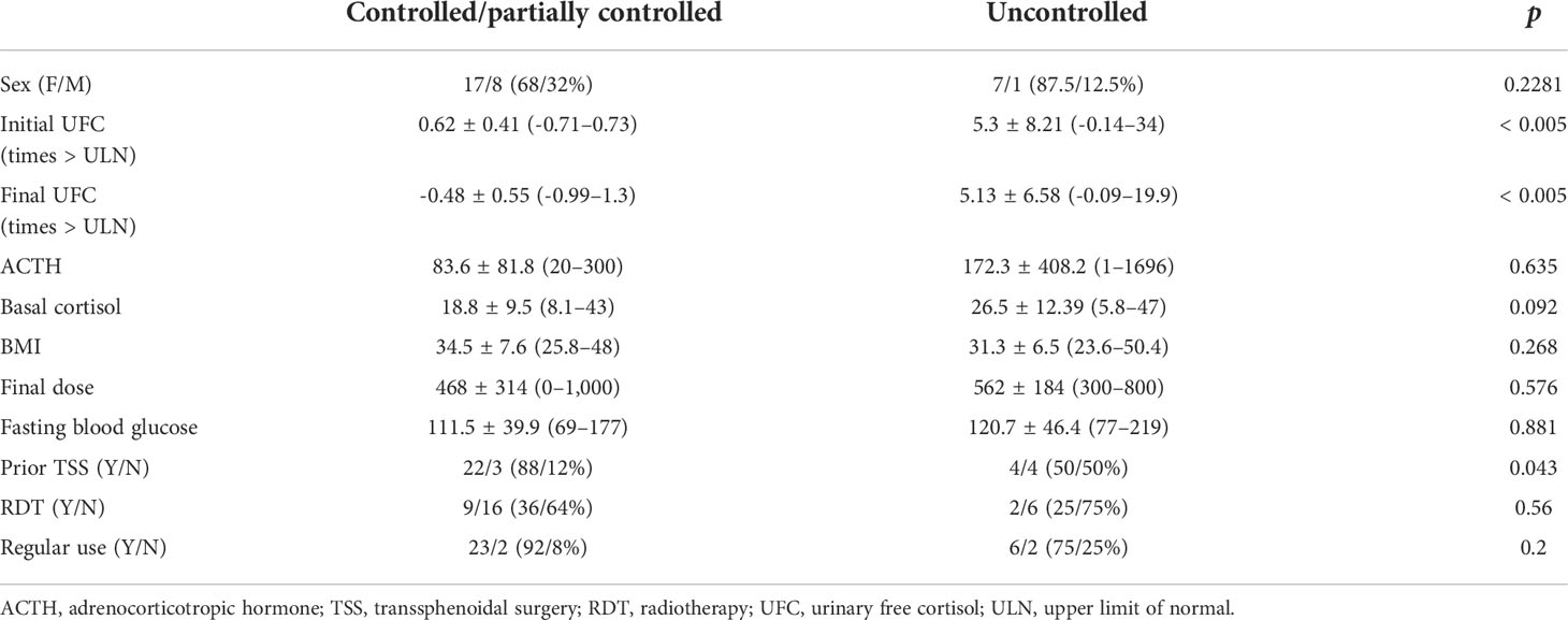
Table 3
www.frontiersin.orgTABLE 3 Baseline characteristics of Cushing’s disease patients according to therapeutic response to ketoconazole.

Figure 2 shows that the prevalence of uncontrolled patients remained stable over time (approximately 30%) despite dose adjustments or association with other drugs, which led to no differences. When analyzing only the results of the last follow-up visit (eliminating fluctuations during follow-up), 22 patients had a complete response (66%), three patients had a partial response (9%), and eight patients had no response to ketoconazole treatment (24%), which includes patients who underwent radiotherapy during ketoconazole treatment.
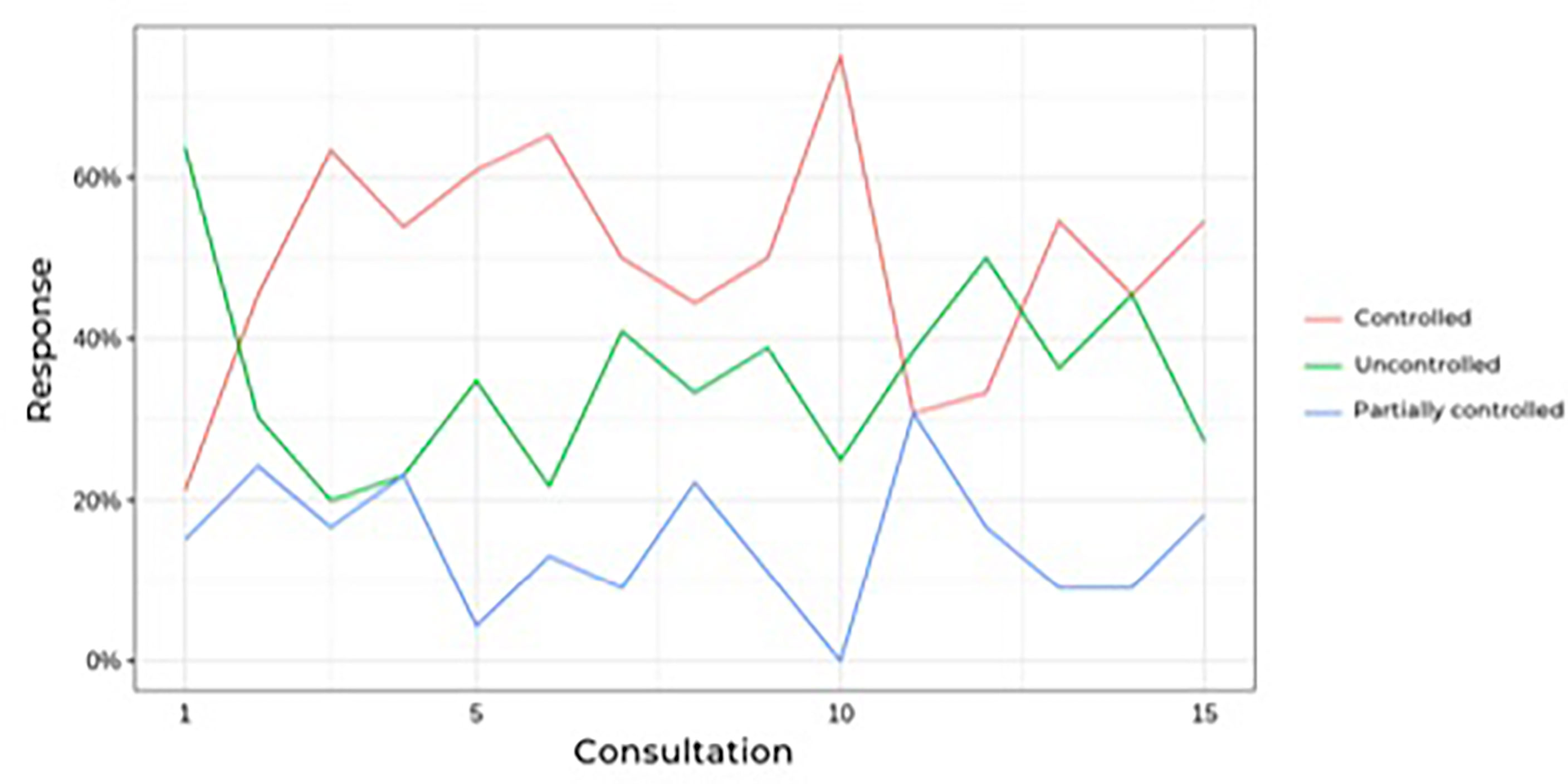
Figure 2
www.frontiersin.orgFIGURE 2 Prevalence of controlled hypercortisolism during follow-up of Cushing’s disease patients treatesd with ketoconazole.

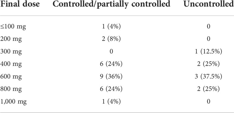
During follow-up, no significant differences were found in blood pressure control or in dehydroepiandrosterone sulfate, cortisol, ACTH, or glucose levels. Worsening of hypertension control was observed in association with hypokalemia in some cases, as described in side effects. The ketoconazole doses ranged from 100 to 1,200 mg per day, and there were no significant dose or response differences between the groups (Table 4). Figure 3 shows the patients, their dosages, and 24-h UFC control at the first and last consultation, showing a trend toward hypercortisolism reduction in approximately 70% of the cohort (25 of 33). Only four patients used doses lower than 300 mg at the end of follow-up. One of them used before TSS and suspended its use after surgery. One patient, who has already undergone radiotherapy, discontinued ketoconazole due to intolerance, despite adequate control of hypercortisolism. Another one, who had also undergone radiotherapy, was lost to follow-up when it was controlled using 100 mg daily, and one remained controlled using 200 mg, without previous radiotherapy.
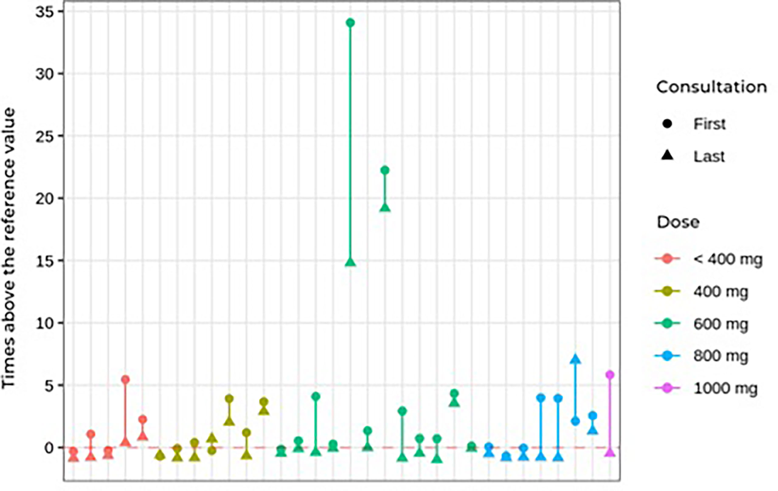
Table 4
www.frontiersin.orgTABLE 4 Final dose of ketoconazole used in patients with Cushing’s disease.

Figure 3
www.frontiersin.orgFIGURE 3 First and last consultation 24çhour UFC results vs. ketoconazole dosage in Cushing’s disease patients.

Side effects

Regarding adverse effects (Table 5), there was no significant difference between the controlled/partially controlled group and the uncontrolled group regarding liver enzyme changes or drug intolerance. Mild adverse effects, including nausea, vomiting, dizziness, and loss of appetite, occurred in 10 patients (30%). Only four patients had serious adverse effects that warranted discontinuing the medication. In two cases, ketoconazole was discontinued due to a significantly acute increase in liver enzymes (drug-induced hepatitis) during the use of 400 and 800 mg of ketoconazole. Non-significant elevation of transaminases (up to three times the normal value) was observed in three cases. A slight increase in gamma-glutamyltransferase occurred in six patients. In these nine patients with elevated liver markers, the daily dose ranged from 400 to 1,200 mg. None of those with mild increases in liver markers needed to discontinue ketoconazole.

Table 5
www.frontiersin.orgTABLE 5 Adverse effects of ketoconazole in Cushing’s disease patients treated with ketoconazole.

One female patient developed pseudotumor cerebri syndrome, which was treated with acetazolamide. She did not need to discontinue ketoconazole, having used it for more than 10 years without new side effects and achieving complete control of hypercortisolism (24). Another patient became pregnant during follow-up while using the medication, but no maternal or fetal complications occurred (25).

Hypokalemia was also observed during follow-up. Twenty episodes of reduced potassium levels occurred in 10 patients over the course of treatment. Of these episodes, six occurred in controlled patients, three in partially controlled patients, and 11 in uncontrolled patients (Table 6). The hypokalemia was managed with spironolactone (25 to 100 mg) and oral potassium supplementation.
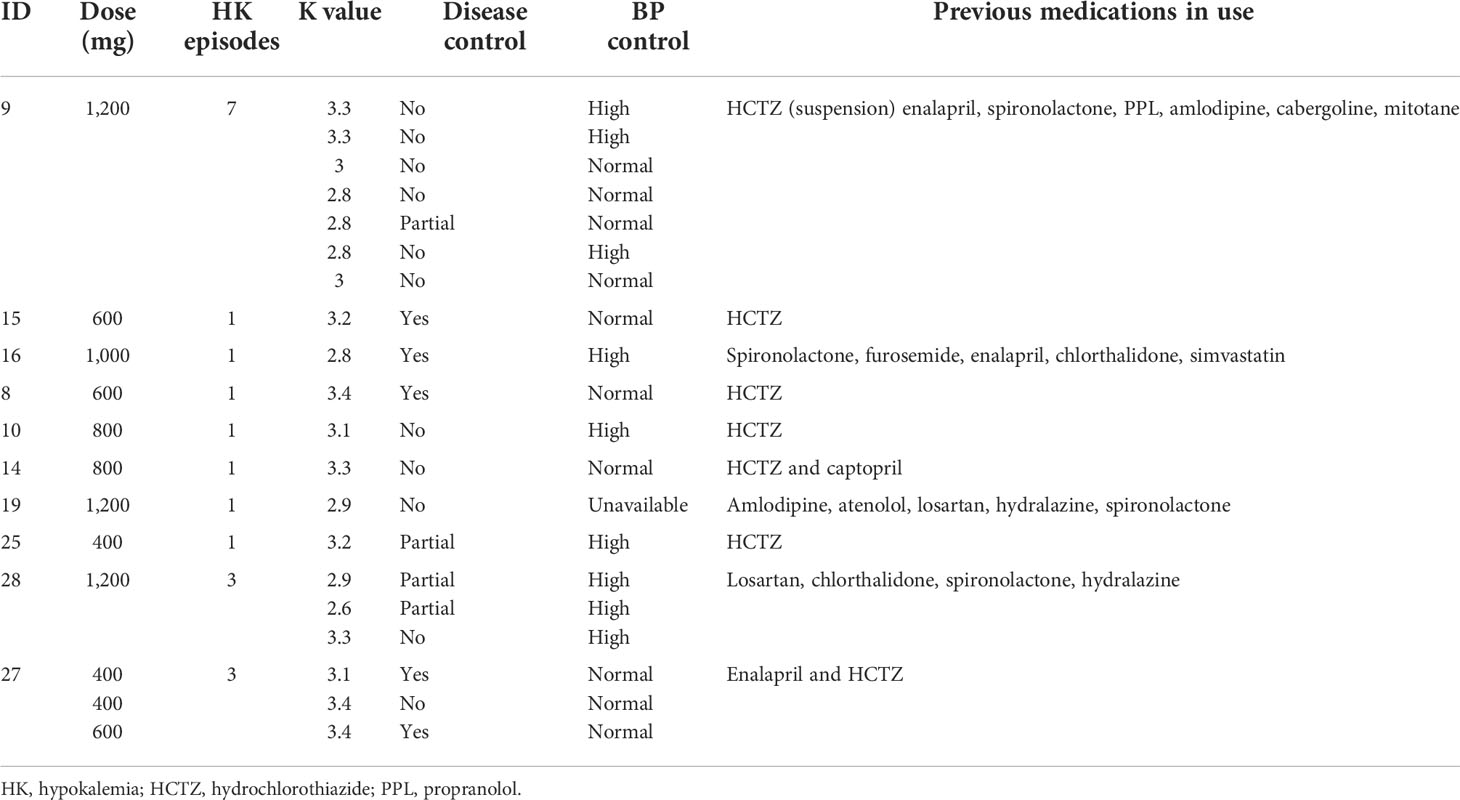
Table 6
www.frontiersin.orgTABLE 6 Characteristics of Cushing’s disease patients who developed hypokalemia during ketoconazole treatment.

Ketoconazole and associations

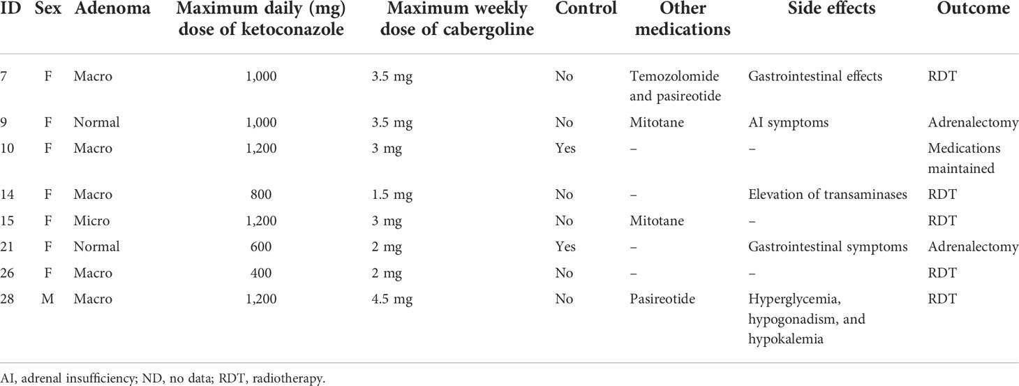
Of the patients who used an association of cabergoline and ketoconazole, one did so since the beginning of follow-up, while another nine were prescribed cabergoline during follow-up due to non-response to ketoconazole alone. Of these 10 patients, two did not start the medication due to problems in obtaining the drug. Thus, in two of the nine patients on the maximum tolerated dose of ketoconazole or who could not tolerate a higher dose due to hepatic enzymatic changes, 1.5–4.5 mg of cabergoline per week was associated. In patients not controlled with ketoconazole plus cabergoline, mitotane (two patients) or pasireotide (two patients) was added. Only two of nine patients responded to the combination of cabergoline and ketoconazole. Data on these associations are shown in Table 7.

Table 7
www.frontiersin.orgTABLE 7 Effects of associating cabergoline with ketoconazole in Cushing’s disease patients.

Considering that one of the indications for the treatment of hypercortisolism may be complementary to radiotherapy, we analyzed the eight patients who underwent radiotherapy after transsphenoidal surgery. In these patients, doses of ketoconazole from 200 to 1,200 mg were used, and in six patients there was a normalization of the UFC in 1 to 60 months of treatment. Thus, the association of ketoconazole with radiotherapy was effective in normalizing the 24-h UFC in 75% of cases.

Clinical follow-up

New therapeutic approaches were attempted in some patients during follow-up: radiotherapy (eight patients), new TSS (five patients), and bilateral adrenalectomy (four patients). At the end of this analysis, 11 patients remained on ketoconazole, all with controlled hypercortisolism. Among the 11 patients who were not fully controlled by the last visit, five were using ketoconazole as pre-TSS therapy and underwent TSS as soon as possible, while three others underwent radiotherapy and two underwent bilateral adrenalectomy. One patient was lost to follow-up.

Discussion

According to the current consensus about CD, drug treatment should be reserved for patients without remission after TSS, those who cannot undergo surgical treatment, or those awaiting the effects of radiotherapy (416). Drugs available in this context may act as adrenal steroidogenesis blockers (ketoconazole, osilodrostat, metyrapone, mitotane, levoketoconazole, and etomidate), in pituitary adenoma (somatostatinergic receptor ligands—pasireotide), dopamine receptor agonists (cabergoline), or glucocorticoid receptor blockers (mifepristone) (1626). Among these alternatives, the drug of choice still cannot be determined. Thus, the best option must be established individually, considering aspects such as remission potential, safety profile, availability, cost, etc. (162728).

For over 30 years, ketoconazole has been prescribed off-label for CD patients with varied rates of remission of hypercortisolism, and it can be used in monotherapy or associated with other drugs (2930). The Brazilian public health system does not provide drugs for the treatment of CD, and among medications with a better profile for controlling hypercortisolism, such as osilodrostat, levoketoconazole, and pasireotide, only pasireotide has been approved by the national regulatory authority (ANVISA). Due to such pragmatic considerations, ketoconazole is among the most commonly used drugs in our health system, whether recently associated or not with cabergoline (7).

In this cohort, the most prevalent response type was complete (66%). Since 75% of the CD patients who used ketoconazole had a complete or partial response, there was a clear trend towards improvement in hypercortisolism. When only those who used ketoconazole post-TSS were evaluated, the rate of control increased to 76%. We found that patients with a higher initial 24-h UFC tended to have less control of excess cortisol, a difference that was not observed when analyzing ketoconazole dose or follow-up time.

In our series and at the prescribed doses, the combination of cabergoline and ketoconazole was not effective in the management of hypercortisolism since only two of nine patients (22%) had their 24-hour UFC normalized. However, it should be observed that this association was used in patients who had more severe CD and, consequently, were less likely to have a favorable response. The effects of cabergoline in CD patients remain controversial, although some studies have shown promising responses (3132).

Previous reviews found that the efficacy of ketoconazole for hypercortisolism control was quite heterogeneous, ranging from 14 to 100% in 99 patients (3334). Our cohort’s response rate was lower than that of Sonino et al. (89%) (20) but higher than that of a multicenter cohort by Castinetti et al. (approximately 50%) (14). Regarding other smaller series (3537) our results reinforce some findings that demonstrate a percentage of control greater than 50% of the cases.

Our analyses showed a trend toward a response that continued, with some oscillations, over time. The rate of uncontrolled patients remained stable over time (approximately 30%), regardless of association with other drugs (cabergoline, mitotane, or pasireotide) or dose adjustments. Speculatively, it would appear that patients who respond to ketoconazole treatment would show some type of response as soon as therapy begins.

Our cohort has the longest follow-up time of any study on ketoconazole use in CD, nearly 15 years. Our results demonstrate that patients who benefit from ketoconazole (i.e., control of hypercortisolism and associated comorbidities) can safely use it for a long term since those who did not experience liver enzyme changes at the beginning of treatment also had no long-term changes.

Another relevant information for clinical practice is the result of treatment with ketoconazole associated with radiotherapy, which demonstrated normalizing the 24-h UFC in 75% of cases, a finding that reinforces the use of this therapeutic combination, especially in cases that are more resistant to different treatment modalities.

As described in the literature, adverse effects, such as nausea, vomiting, dizziness, headache, loss of appetite, and elevated transaminases, are relatively frequent (38). In our cohort, 10 patients (30%) had mild adverse effects, and four (12%) had more serious adverse effects requiring discontinuation. In other studies, up to 20% of patients required discontinuation due to side effects (14). We documented 20 episodes of hypokalemia during ketoconazole treatment, some with worsening blood pressure control. In most cases, hypokalemia has occurred in association with the use of diuretic drugs, which may have potentiated potassium spoliation, reinforcing the need of stringent surveillance in hypertensive Cushing’s disease patients using this combination. It can also result from the enzymatic blockade that could lead to the elevation of adrenal mineralocorticoid precursors (pex. deoxycorticosterone), with consequent sodium retention and worsening hypertension. Although it has not been analyzed in other series with ketoconazole, this side effect has been observed in patients who received other adrenal-blocking drugs, such as osilodrostat and metyrapone (16). This alteration seems to be transient in some patients; in our series, it was managed by suspending drugs that could worsen hypokalemia and introducing spironolactone and/or potassium supplementation. Hypokalemia may also result from continuing intense adrenal stimulation by ACTH and changes in the activity of the 11-beta-hydroxysteroid dehydrogenase enzyme, which increase the mineralocorticoid activity of cortisol, as observed in patients with severe hypercortisolism in uncontrolled CD (39). Hypogonadism occurred in one male patient. In two adolescent patients (one female and one male), hypercortisolism was effectively controlled without altering the progression of puberty. As described in other cohorts, this effect was expected due to the high doses, which block adrenal and testicular androgen production (20).

Thus, our findings confirm previous reports in the literature and add important information about the side effects and safety of long-term ketoconazole use in CD treatment. Our data reinforce the current recommendations about ketoconazole for recurrent cases or those refractory to surgery, including proper follow-up by an experienced team specializing in evaluating clinical and biochemical responses and potential adverse effects (71840). Despite the severity of many of our CD patients, no ketoconazole-related death occurred during follow-up, including long-term observation. On the other hand, no patient progressed to definitive remission of hypercortisolism, even after many years of treatment with ketoconazole.

Conclusions

In our cohort of patients, ketoconazole proved to be an effective and safe alternative for CD treatment, although it can produce side effects that require proper identification and management, allowing effective long-term treatment. We found side effects that have been rarely described in the literature, including hypokalemia and worsening hypertension, which require specific care and management. Thus, ketoconazole is an effective alternative for CD patients who cannot undergo surgery, who do not achieve remission after pituitary surgery, or who have recurrent hypercortisolism.

Data availability statement

The raw data supporting the conclusions of this article will be made available by the authors without undue reservation.

Ethics statement

The studies involving human participants were reviewed and approved by the Hospital de Clínicas de Porto Alegre Research Ethics Committee. Written informed consent for participation was not required for this study in accordance with the national legislation and the institutional requirements.

Author contributions

CV and MAC created the research format. CV, RBM, and MCBC realized the search on medical records. CV performed the statistical analysis. MAC, ACVM, and TCR participated in the final data review and discussion. ACVM participated in the final data review and discussion as volunteer collaborator. All authors contributed to the article and approved the submitted version.

Funding

This work was supported by the “Coordenação de Aperfeiçoamento de Pessoal de Nı́vel Superior” (CAPES), Ministry of Health – Brazil, through a PhD scholarship; and the Research Incentive Fund (FIPE) of Hospital de Clı́nicas de Porto Alegre.

Acknowledgments

The authors would like to thank the HCPA Research and Graduate Studies Group (GPPG) for the statistical technical support provided by Rogério Borges. We also thank the Research Incentive Fund of Hospital de Clínicas de Porto Alegre and Coordenação de Aperfeiçoamento de Pessoal de Nível Superior (CAPES), by funds applied. We also thank the Graduate Program in Endocrinology and Metabolism (PPGEndo UFRGS) for all the support in the preparation of this research.

Conflict of interest

The authors declare that the research was conducted in the absence of any commercial or financial relationships that could be construed as a potential conflict of interest.

Publisher’s note

All claims expressed in this article are solely those of the authors and do not necessarily represent those of their affiliated organizations, or those of the publisher, the editors and the reviewers. Any product that may be evaluated in this article, or claim that may be made by its manufacturer, is not guaranteed or endorsed by the publisher.

References

1. Fleseriu M, Castinetti F. Updates on the role of adrenal steroidogenesis inhibitors in cushing’s syndrome: a focus on novel therapies. Pituitary (2016) 19(6):643–53. doi: 10.1007/s11102-016-0742-1

PubMed Abstract | CrossRef Full Text | Google Scholar

2. Pivonello R, De Leo M, Cozzolino A, Colao A. The treatment of cushing’s disease. Endocr Rev (2015) 36(4):385–486. doi: 10.1210/er.2013-1048

PubMed Abstract | CrossRef Full Text | Google Scholar

3. Nieman LK, Biller BMK, Findling JW, Newell-Price J, Savage MO, Stewart PM, et al. The diagnosis of cushing’s syndrome: An endocrine society clinical practice guideline. J Clin Endocrinol Metab (2008) 93(5):1526–40. doi: 10.1210/jc.2008-0125

PubMed Abstract | CrossRef Full Text | Google Scholar

4. Nieman LK, Biller BMK, Findling JW, Murad MH, Newell-Price J, Savage MO, et al. Treatment of cushing’s syndrome: An endocrine society clinical practice guideline. J Clin Endocrinol Metab (2015) 100(8):2807–31. doi: 10.1210/jc.2015-1818

PubMed Abstract | CrossRef Full Text | Google Scholar

5. Pivonello R, De Martino M, De Leo M, Lombardi G, Colao A. Cushing’s syndrome. Endocrinol Metab Clin North (2008) 37(1):135–49. doi: 10.1016/j.ecl.2007.10.010

CrossRef Full Text | Google Scholar

6. Alexandraki KI, Grossman AB. Therapeutic strategies for the treatment of severe cushing’s syndrome. Drugs (2016) 76(4):447–8. doi: 10.1007/s40265-016-0539-6

PubMed Abstract | CrossRef Full Text | Google Scholar

7. Machado MC, Fragoso MCBV, Moreira AC, Boguszewski CL, Neto LV, Naves LA, et al. A review of cushing’s disease treatment by the department of neuroendocrinology of the Brazilian society of endocrinology and metabolism. Arch Endocrinol Metab (2018) 62(1):87–105. doi: 10.20945/2359-3997000000014

PubMed Abstract | CrossRef Full Text | Google Scholar

8. Rollin G, Ferreira NP, Czepielewski MA. Prospective evaluation of transsphenoidal pituitary surgery in 108 patients with Cushing’s disease. Arq Bras Endocrinol Metabol. (2007) 51(8):1355–61. doi: 10.1590/s0004-27302007000800022

PubMed Abstract | CrossRef Full Text | Google Scholar

9. Patil CG, Prevedello DM, Lad SP, Lee Vance M, Thorner MO, Katznelson L, et al. Late recurrences of cushing’s disease after initial successful transsphenoidal surgery. J Clin Endocrinol Metab (2008) 93(2):358–62. doi: 10.1210/jc.2007-2013

PubMed Abstract | CrossRef Full Text | Google Scholar

10. Rubinstein G, Osswald A, Zopp S, Ritzel K, Theodoropoulou M, Beuschlein F, et al. Therapeutic options after surgical failure in cushing’s disease: A critical review. Best Pract Res Clin Endocrinol Metab (2019) 33(2):101270. doi: 10.1016/j.beem.2019.04.004

PubMed Abstract | CrossRef Full Text | Google Scholar

11. Zhao N, Yang X, Li C, Yin X. Efficacy and safety of pasireotide for Cushing’s disease: A protocol for systematic review and meta-analysis. Medicine (Baltimore). (2020) 99(51):e23824. doi: 10.1097/MD.0000000000023824

PubMed Abstract | CrossRef Full Text | Google Scholar

12. Pivonello R, Fleseriu M, Newell-Price J, Bertagna X, Findling J, Shimatsu A, et al. Efficacy and safety of osilodrostat in patients with cushing’s disease (LINC 3): a multicentre phase III study with a double-blind, randomised withdrawal phase. Lancet Diabetes Endocrinol (2020) 8(9):748–61. doi: 10.1016/S2213-8587(20)30240-0

PubMed Abstract | CrossRef Full Text | Google Scholar

13. Yan JY, Nie XL, Tao QM, Zhan SY, De Zhang Y. Ketoconazole associated hepatotoxicity: A systematic review and meta-analysis. Biomed Environ Sci (2013) 26(7):605–10. doi: 10.3967/0895-3988.2013.07.013

PubMed Abstract | CrossRef Full Text | Google Scholar

14. Castinetti F, Guignat L, Giraud P, Muller M, Kamenicky P, Drui D, et al. Ketoconazole in cushing’s disease: Is it worth a try. J Clin Endocrinol Metab (2014) 99(5):1623–30. doi: 10.1210/jc.2013-3628

PubMed Abstract | CrossRef Full Text | Google Scholar

15. Castinetti F, Nieman LK, Reincke M, Newell-Price J. Approach to the patient treated with steroidogenesis inhibitors. J Clin Endocrinol Metab (2021) 106(7):2114–23. doi: 10.1210/clinem/dgab122

PubMed Abstract | CrossRef Full Text | Google Scholar

16. Fleseriu M, Auchus R, Bancos I, Bem-Shlomo A, Bertherat J, Biermasz NR, et al. Consensus on diagnosis and management of cushing’s disease: a guideline update. Lancet Diabetes Endocrinol (2021) 9(12):847–75. doi: 10.1016/s2213-8587(21)00235-7

PubMed Abstract | CrossRef Full Text | Google Scholar

17. Fleseriu M, Pivonello R, Elenkova A, Salvatori R, Auchus RJ, Feelders RA, et al. Efficacy and safety of levoketoconazole in the treatment of endogenous cushing’s syndrome (SONICS): a phase 3, multicentre, open-label, single-arm trial. Lancet Diabetes Endocrinol (2019) 7(11):855–65. doi: 10.1016/S2213-8587(19)30313-4

PubMed Abstract | CrossRef Full Text | Google Scholar

18. Tritos NA. Adrenally directed medical therapies for cushing syndrome. J Clin Endocrinol Metab (2021) 106(1):16–25. doi: 10.1210/clinem/dgaa778

PubMed Abstract | CrossRef Full Text | Google Scholar

19. Simões Corrêa Galendi J, Correa Neto ANS, Demetres M, Boguszewski CL, dos S V. N. nogueira, “Effectiveness of medical treatment of cushing’s disease: A systematic review and meta-analysis,”. Front Endocrinol (Lausanne) (2021) 12:732240(September). doi: 10.3389/fendo.2021.732240

PubMed Abstract | CrossRef Full Text | Google Scholar

20. Sonino N, Boscaro M, Paoletta A, Mantero F, Zillotto D. Ketoconazole treatment in cushing’s syndrome: experience in 34 patients. Clin Endocrinol (Oxf) (1991) 35(4):347–52. doi: 10.1111/j.1365-2265.1991.tb03547.x

PubMed Abstract | CrossRef Full Text | Google Scholar

21. Costenaro F, Rodrigues TC, Rollin GAF, Czepielewski MA. Avaliação do eixo hipotálamohipófise adrenal no diagnóstico e na remissão da doença de cushing. Arquivos Brasileiros Endocrinologia e Metabologia (2012). doi: 10.1590/S0004-27302012000300002

CrossRef Full Text | Google Scholar

22. Amlashi FG, Swearinger B, Faje AT, Nachtigall LB, Miller KK, Klibanski A, et al. Accuracy of late-night salivary cortisol in evaluating postoperative remission and recurrence in cushing’s disease. J Clin Endocrinol Metab (2015) 100(10):3770–7. doi: 10.1210/jc.2015-2107

PubMed Abstract | CrossRef Full Text | Google Scholar

23. Silveiro SP, Satler F. Rotinas em endocrinologia. (Porto Alegre: Artmed) (2015).

Google Scholar

24. Costenaro F, Rodrigues TC, Ferreira NP, da Costa TG, Schuch T, Boschi V, et al. Pseudotumor cerebri during cushing’s disease treatment with ketoconazole. Arq. Bras Endocrinol Metabol (2011). doi: 10.1590/s0004-27302011000400008

CrossRef Full Text | Google Scholar

25. Costenaro F, Rodrigues TC, De Lima PB, Ruszczyk J, Rollin G, Czepielewski MA. A successful case of cushing’s disease pregnancy treated with ketoconazole. Gynecol Endocrinol (2015) 31(3):176–8. doi: 10.3109/09513590.2014.995615

PubMed Abstract | CrossRef Full Text | Google Scholar

26. Gadelha MR, Neto LV. Efficacy of medical treatment in cushing’s disease: A systematic review. Clin Endocrinol (Oxf) (2014) 80(1):1–12. doi: 10.1111/cen.12345

PubMed Abstract | CrossRef Full Text | Google Scholar

27. Fleseriu M, Petersenn S. New avenues in the medical treatment of cushing’s disease: Corticotroph tumor targeted therapy. J Neurooncol (2013) 114(1):1–11. doi: 10.1007/s11060-013-1151-1

PubMed Abstract | CrossRef Full Text | Google Scholar

28. Fleseriu M, Petersenn S. Medical management of cushing’s disease: What is the future? Pituitary (2012) 15(3):330–41. doi: 10.1007/s11102-012-0397-5

PubMed Abstract | CrossRef Full Text | Google Scholar

29. Feelders RA, De Bruin C, Pereira AM, Romijn JÁ, Netea-Maier RT, Hermus AR, et al. Pasireotide alone or with cabergoline and ketoconazole in cushing’s disease. N Engl J Med (2010) 362(19):1846–8. doi: 10.1056/NEJMc1000094

PubMed Abstract | CrossRef Full Text | Google Scholar

30. Barbot M, Albiger N, Ceccato F, Zilio M, Frigo AC, Denaro Lc, et al. Combination therapy for cushing’s disease: Effectiveness of two schedules of treatment. should we start with cabergoline or ketoconazole? Pituitary (2014) 17(2):109–17. doi: 10.1007/s11102-013-0475-3

PubMed Abstract | CrossRef Full Text | Google Scholar

31. Vilar L, Naves LA, Azevedo MF, Arruda MJ, Arahata CM, Silva LM, et al. Effectiveness of cabergoline in monotherapy and combined with ketoconazole in the management of cushing’s disease. Pituitary (2010) 13(2):123–9. doi: 10.1007/s11102-009-0209-8

PubMed Abstract | CrossRef Full Text | Google Scholar

32. Pivonello R, De Martino MC, Cappabianca P, De Leo M, Faggiano A, Lombardi G, et al. The medical treatment of cushing’s disease: Effectiveness of chronic treatment with the dopamine agonist cabergoline in patients unsuccessfully treated by surgery. J Clin Endocrinol Metab (2009) 94(1):223–30. doi: 10.1210/jc.2008-1533

PubMed Abstract | CrossRef Full Text | Google Scholar

33. Castinetti F, Morange I, Jaquet P, Conte-Devolx B, Brue T. Ketoconazole revisited: A preoperative or postoperative treatment in cushing’s disease. Eur J Endocrinol (2008). doi: 10.1530/EJE-07-0514

PubMed Abstract | CrossRef Full Text | Google Scholar

34. Loli P, Berselli ME, Tagliaferri M. Use of ketoconazole in the treatment of cushing’s syndrome. J Clin Endocrinol Metab (1986) 63(6):1365–71. doi: 10.1210/jcem-63-6-1365

PubMed Abstract | CrossRef Full Text | Google Scholar

35. Kakade HR, Kasaliwal R, Khadilkar KS, Jadhav S, Bukan A, Khare Sc, et al. Clinical, biochemical and imaging characteristics of cushing’s macroadenomas and their long-term treatment outcome. Clin Endocrinol (Oxf) (2014) 81(3):336–42. doi: 10.1111/cen.12442

PubMed Abstract | CrossRef Full Text | Google Scholar

36. Luisetto G, Zangari M, Camozzi V, Boscaro M, Sonino N, Fallo F. Recovery of bone mineral density after surgical cure, but not by ketoconazole treatment, in cushing’s syndrome. Osteoporos Int (2001) 12(11):956–60. doi: 10.1007/s001980170025

PubMed Abstract | CrossRef Full Text | Google Scholar

37. Huguet I, Aguirre M, Vicente A, Alramadan M, Quiroga I, Silva J, et al. Assessment of the outcomes of the treatment of cushing’s disease in the hospitals of castilla-la mancha. Endocrinol y Nutr (2015) 62(5):217–23. doi: 10.1016/j.endonu.2015.02.007

CrossRef Full Text | Google Scholar

38. Tritos NA, Biller BMK. Advances in the medical treatment of cushing disease. Endocrinol Metab Clin North Am (2020) 49(3):401–12. doi: 10.1016/j.ecl.2020.05.003

PubMed Abstract | CrossRef Full Text | Google Scholar

39. Torpy D, Mullen N, Ilias I, Nieman L. Association of hypertension and hypokalemia with cushing’s syndrome caused by ectopic ACTH secretion. Ann N Y Acad Sci (2002) 970:134–44. doi: 10.1111/j.1749-6632.2002.tb04419.x

PubMed Abstract | CrossRef Full Text | Google Scholar

40. Varlamov EV, Han AJ, Fleseriu M. “Updates in adrenal steroidogenesis inhibitors for cushing’s syndrome – a practical guide,”. Best Pract Res Clin Endocrinol Metab (2021) 35(1):101490. doi: 10.1016/j.beem.2021.101490

PubMed Abstract | CrossRef Full Text | Google Scholar

Keywords: Cushing’s disease, Cushing’s syndrome, hypercortisolism, treatment, ketoconazole

Citation: Viecceli C, Mattos ACV, Costa MCB, Melo RBd, Rodrigues TdC and Czepielewski MA (2022) Evaluation of ketoconazole as a treatment for Cushing’s disease in a retrospective cohort. Front. Endocrinol. 13:1017331. doi: 10.3389/fendo.2022.1017331

Received: 11 August 2022; Accepted: 06 September 2022;
Published: 07 October 2022.

Edited by:

Luiz Augusto Casulari, University of Brasilia, Brazil

Reviewed by:

Juliana Drummond, Federal University of Minas Gerais, Brazil
Monalisa Azevedo, University of Brasilia, Brazil

Copyright © 2022 Viecceli, Mattos, Costa, Melo, Rodrigues and Czepielewski. This is an open-access article distributed under the terms of the Creative Commons Attribution License (CC BY). The use, distribution or reproduction in other forums is permitted, provided the original author(s) and the copyright owner(s) are credited and that the original publication in this journal is cited, in accordance with accepted academic practice. No use, distribution or reproduction is permitted which does not comply with these terms.

*Correspondence: Mauro Antonio Czepielewski, maurocze@terra.com.br

Disclaimer: All claims expressed in this article are solely those of the authors and do not necessarily represent those of their affiliated organizations, or those of the publisher, the editors and the reviewers. Any product that may be evaluated in this article or claim that may be made by its manufacturer is not guaranteed or endorsed by the publisher.

From https://www.frontiersin.org/articles/10.3389/fendo.2022.1017331/full

Unmet needs in Cushing’s Syndrome: the Patients

Abstract

Background

Cushing’s syndrome (CS) is a rare condition of chronically elevated cortisol levels resulting in diverse comorbidities, many of which endure beyond successful treatment affecting the quality of life. Few data are available concerning patients’ experiences of diagnosis, care and persistent comorbidities.

Objective

To assess CS patients’ perspectives on the diagnostic and care journey to identify unmet therapeutic needs.

Methods

A 12-item questionnaire was circulated in 2019 by the World Association for Pituitary Organisations. A parallel, 13-item questionnaire assessing physician perceptions on CS patient experiences was performed.

Results

Three hundred twenty CS patients from 30 countries completed the questionnaire; 54% were aged 35–54 and 88% were female; 41% were in disease remission. The most burdensome symptom was obesity/weight gain (75%). For 49% of patients, time to diagnosis was over 2 years. Following treatment, 88.4% of patients reported ongoing symptoms including, fatigue (66.3%), muscle weakness (48.8%) and obesity/weight gain (41.9%). Comparisons with delay in diagnosis were significant for weight gain (P = 0.008) and decreased libido (P = 0.03). Forty physicians completed the parallel questionnaire which showed that generally, physicians poorly estimated the prevalence of comorbidities, particularly initial and persistent cognitive impairment. Only a minority of persistent comorbidities (occurrence in 1.3–66.3%; specialist treatment in 1.3–29.4%) were managed by specialists other than endocrinologists. 63% of patients were satisfied with treatment.

Conclusion

This study confirms the delay in diagnosing CS. The high prevalence of persistent comorbidities following remission and differences in perceptions of health between patients and physicians highlight a probable deficiency in effective multidisciplinary management for CS comorbidities.

Introduction

Cushing’s syndrome (CS) is a morbid endocrine condition due to prolonged exposure to high circulating cortisol levels (123). Hypercortisolism may cause irreversible physical and psychological changes in several tissues, leading to debilitating morbidities which persist over the long term after the resolution of excessive hormone levels, such as cardiovascular complications, metabolic and skeletal disorders, infections and neuropsychiatric disturbances (34). Even patients who have been biochemically ‘cured’ for over 10 years have a residual overall higher risk of mortality, mostly from circulatory disease and diabetes (5). Moreover, people with a history of CS suffer from impaired quality of life (QoL) (6). Several studies suggest that the prevalence of persistent comorbidities is correlated with the duration of exposure to cortisol excess (78). However, as the signs and symptoms of CS overlap with common diseases such as the metabolic syndrome and depression, the time taken to diagnose CS is often long, resulting in a significant number of patients with persistent sequelae and impairments in QoL (69).

Given the burden of the disease, ideal CS treatment would include early diagnosis, curative surgery and multidisciplinary care of comorbidities both pre- and post-cure of CS, including the psychological dimension of the patient’s disease experience (10). Few data are available about patients’ perceptions of the medical journey from first symptoms to diagnosis, treatment and follow-up. The aim of this study was, therefore, to explore CS patients’ experiences of symptoms, diagnosis, care and treatment satisfaction around the world and to compare patients’ perceptions of CS with those of physicians.

Methods

Patient questionnaire design

A 12-item patient questionnaire was developed based on the generally understood clinical characteristics and symptomology of CS, aiming to assess patients’ experiences of symptoms, diagnosis, care and treatment satisfaction (12) (Supplementary File 1, see section on supplementary materials given at the end of this article). The questionnaire was initially offered in English and made available via the SurveyMonkey online platform from March to May 2019. The survey was completed anonymously and required no specific participant identification or any details that could be used to identify individual participants. In addition to basic demographics (i.e. country of residence, sex, age and highest educational level attained), the questionnaire asked ten multiple-choice and two open questions. The survey was shared by the World Association for Pituitary Organisations (WAPO), Adrenal Net, Cushing’s Support & Research Foundation and the Pituitary Foundation, as well as being distributed to local patient associations. As a second step, the questionnaire was translated into eight additional languages (French, Dutch, Spanish, Chinese, Portuguese, Italian and German) and was recirculated by the WAPO, Adrenal Net and China Hypercortisolism Patient Alliance to the different local patient associations for distribution in November 2019. As this was a non-interventional, anonymous patient survey, distributed by the patient associations themselves, and not initiated or funded by a research or educational institution, no ethical review was required. Written consent was obtained from each respondent after full explanation of the purpose and nature of the survey.

Comparative physician survey

In addition, a 13-item physician questionnaire was developed to assess physicians’ perspectives on CS symptoms and comorbidities. This physician questionnaire was conducted by HRA Pharma Rare Diseases at the 2019 European Congress of Endocrinology, in Lyon, France. This anonymous questionnaire was completed by 40 qualified physicians. The responses from the patient survey were compared for context with the physicians’ estimates of the prevalence of CS symptoms and comorbidities. Although the physician questionnaire was conducted independently of the patient questionnaire, and used a different question structure, the comparison with the current patient questionnaire is included to further enrich and contextualise the patient responses.

Data analysis

All responses and answers were collected, coded and analysed using Microsoft Excel. Data preparation involved removing duplicate answers, or where possible analysing and reclassifying qualitative responses reported as ‘other’, based on the accompanying details to new or existing response options.

Statistical methodology

Complementary statistical analyses using SAS software were performed using the chi-square and Fisher tests, depending on the cell counts, to compare (i) the time between first symptoms and diagnosis and the persistence of symptoms and (ii) persistence of symptoms, with the specialities of the physicians currently treating the respondents. Frequency distribution of a particular variable was displayed and compared with the frequency distribution of the comparator variable. A significance level of 0.05 was applied.

Results

Demographic characteristics

Three hundred twenty patients from 30 countries completed the patient questionnaire, with 27% (n  = 87) coming from the United Kingdom and 14% (n  = 44) from the United States of America. More than half (53.7%, n = 172) of the patients were aged between 35 and 54 years, and 88.4% (n  = 283) were female. The majority of patients (53.1%, n = 170) had undergraduate or postgraduate qualifications (Table 1).

Table 1Patient demographics.

Sex N = 319a
 Female 283 (88.4%)
 Male 36 (11.3%)
Age group N = 320
 18–24 years 16
 25–34 years 49
 35–44 years 71
 45–54 years 101
 55–64 years 54
 65–74 years 24
 ≥75 years 5
Regionb N = 320
 Western Europe 222
 North America 60
 China 16
 Australasia 14
 South America 5
 Africa 3
Education N = 320
 High school graduate/secondary education diploma 35%
 Undergraduate degree 25.6%
 Post-graduate degree 27.5%
 Prefer not to say 10.6%
Time from first symptoms to diagnosis N = 320
 0–6 months 18.4%
 6–12 months 15.6%
 1–2 years 14.4%
 2–3 years 18.4%
 3–5 years 11.6%
 5–10 years 8.4%
 10–15 years 7.5%
 15–20 years 0.9%
 20+ years 1.9%
 Unknown 2.8%

aOne patient responded ‘non-binary’. bWestern Europe: United Kingdom (n  = 87), the Netherlands (n  = 38), France (n  = 37), Spain (n  = 12), Denmark (n  = 10), Norway (n  = 9), Germany (n  = 6), Italy (n  = 5), Ireland (n  = 4), Belgium (n  = 4), Poland (n  = 4), Sweden (n  = 2), Malta (n  = 2), Switzerland (n  = 1), Czech Republic (n  = 1); Africa: South Africa (n  = 1), Gabon (n  = 1), Zimbabwe (n  = 1); Australasia: Australia (n  = 8), New Zealand (n  = 6); South America: Colombia (n  = 2), Bolivia (n  = 1), Argentina (n  = 1), Brazil (n  = 1); North America: United States of America (n  = 44), Canada (n  = 13), Costa Rica (n  = 1), Mexico (n  = 1), Dominican Republic (n  = 1).

Time to diagnosis

The time to diagnosis from first reporting of CS symptoms was declared to be within 2 years for 48.4% (n  = 155) (Table 1) and was over 2 years in 48.7% (n  = 156) and over 3 years in 30.3% (n  = 97).

Initial symptoms

A broad range of signs and symptoms were initially noticed by patients, with weight gain, hirsutism or acne, fatigue, sleep disturbances, depressive symptoms, muscle weakness, anxiety and hypertension all being reported in over 50% of patients (Table 2). Obesity/weight gain was most commonly cited (75%, n = 240) as being burdensome. Fatigue, feelings of depression or mood problems, sleep disturbances, muscle weakness and hirsutism were also very commonly (>40%) mentioned as being burdensome. Burdensome symptoms classified as ‘other’ were rare (<1%) and included issues such as hormonal problems and dental problems.

Table 2Patient-reported symptoms (multiple answers were possible).

Symptoms first noticed (%) Most burdensome perceived symptoms before diagnosis (%)
Weight gain 85.0 75.0
Hirsutism/acne 76.3 42.8
Fatigue 66.3 54.1
Sleep disturbances 64.4 41.9
Skin problems 64.7 21.3
Depression/mood problems 58.8 48.1
Muscle weakness 57.8 43.4
Anxiety 54.1 39.1
Hypertension 52.5 22.2
Loss of concentration 45.0 28.4
Memory problems 41.9 30.3
Menstrual disturbances 35.6 12.5
Decreased libido 32.5 12.5
Bone problems 23.1 14.4
Infections 23.8 10.3
Glucose intolerance 17.2 8.4
Blood clot 5.3
Pain(s) 3.1
Vision problems 2.8
Headache 2.5
Cravings 1.6
Other 8.4 1.9

Person who made the initial CS diagnosis

In 53.8% (n  = 172) of cases, an endocrinologist made the initial diagnosis of CS or prescribed the first screening tests, Table 3. General practitioners made 18.1% of diagnoses (n  = 58), in the remaining cases a diversity of other physicians directly or indirectly contributed to make the diagnosis, as indicated in Table 3. A small but noticeable number (5.6%, n = 18) of patients self-diagnosed and then convinced their physician to order the diagnostic tests.

Table 3Patient perception of physician specialty.

Specialty Person who made the initial diagnosis or suspected Cushing’s syndrome (%) (n = 320) Physicians involved in the management of Cushing’s syndrome (%) (n = 320)
Endocrinologist 53.8 97.8
General practitioner/family doctor 18.1 56.3
Self-diagnosed 5.6
Hospital/emergency doctor 3.8
Internist 2.5 0.9
Gynecologist 1.9 14.1
Cardiologist 1.9 13.4
Bone specialist 1.9 14.1
Dermatologist 1.6 11.6
Haematologist 0.9 3.8
Ophthalmologist 0.9 3.1
Nurse 0.9 2.5
Radiologist 0.9 0.6
Family or friend 0.9
Psychiatrist or psycologist 0.9 23.4
Healer 0.6 2.2
Surgeon 0.6
Oncologist 0.3 6.6
Gastroenterologist 0.3 1.3
Neurologist 0.3 4.1
Others 1.6
Physiotherapist 14.4
Dietician 9.7
Neurosurgeon 8.1
Social worker 4.1
Ear, nose and throat specialist 1.6
Sports physician 1.3
Sleep specialist 0.9
Urologist 0.6
Orthopaedic surgeon 0.3

Response to treatment

At the time of answering the questionnaire, 55.8% (n  = 178) of patients were not in remission. 40.8% of patients (n  = 130) were in true biochemical remission (Fig. 1). This latter group was a composite including patients who responded: ‘In remission (no treatment)’ (16.3%, n = 52), ‘Received an operation to remove adrenal glands’ (22.9%, n = 73) and ‘Treated with hydrocortisone’ (1.6%, n = 5). Thirteen percent of the patients (n  = 41) were on cortisol-lowering treatment and 6.6% of the patients (n  = 21) had not had or were awaiting surgery. Following treatment for CS, 11.6% of the patients (n  = 37) reported having no further symptoms related to the condition, with 88.4% (n  = 283) still symptomatic. Of the total population (n  = 320), the most bothersome symptoms were fatigue (66.3%, n = 212), muscle weakness (48.8%, n = 156) and obesity/weight gain (41.9%, n = 134) (Table 4).

Figure 1View Full Size
Figure 1
Patient description of their current clinical situation (n = 319). The category ‘Disease in true remission’ combines scores for ‘In remission (no treatment)’ (16.3%), ‘Received an operation to remove adrenal glands’ (22.9%) and ‘Treated with hydrocortisone’ (1.6%). One person did not complete the question.

Citation: Endocrine Connections 11, 7; 10.1530/EC-22-0027

Table 4Persistent symptoms.

Symptom Persistent bothersome symptomsa (%) (n = 320) Treatment received for symptoms (%) (n = 320)
Fatigue 66.3 15.9
Muscle weakness 48.8 17.2
Weight gain 41.9 8.4
Depression, mood problems 36.9 28.8
Poor concentration 35.9 4.1
Memory problems 33.8 5.6
Sleep problems 33.1 14.1
Anxiety 30.6 14.7
Decreased libido 25.3 4.1
Bone problems 19.1 21.9
Hypertension 18.4 29.4
Hirsutism 17.5 4.1
Skin problems 16.6 6.9
Glucose intolerance 8.8 10
Menstrual problems 9.1 4.7
Infections 7.2 4.7
Blood clot 3.8 2.2
Acne 2.8 1.3
Other 4.4 5.3
No treatment 1.3 8.1
Only hydrocortisone 1.6

aUp to five answers were possible.

Comparison of time to diagnosis and persistence of symptoms

To compare the time to diagnosis and the persistence of symptoms following treatment, an analysis of a number of variables was performed comparing the group with persistent symptoms after treatment (n  = 283) with those who did not (n  = 37) in terms of time to diagnosis. Patients with a longer time to diagnosis reported significantly more frequent weight gain (P = 0.008), and more frequent reduced libido (P = 0.03) after treatment. Although not statistically significant, there was a strong trend towards patients reporting a longer time to diagnosis and a greater frequency of persistent perceived bone issues after treatment (P = 0.053), as well anxiety (P = 0.07) and depression/mood concerns (P = 0.08).

Physicians involved in follow-up

Once diagnosed, almost all patients (97.8%, n = 313) were managed by an endocrinologist, followed by a GP/family doctor (56.3%, n = 180). A psychiatrist/psychologist was involved in 23.4% (n  = 75), followed by a physiotherapist (14.4%, n = 46), rheumatologist (14.4%, n = 46), gynecologist (14.1%, n = 45), cardiologist (13.4%, n = 43), dermatologist (11.6%, n = 37) and a dietician (9.7%, n = 31) (Table 3).

Treatment of persistent symptoms

Table 4 shows the prevalence of persistent symptoms after treatment, common ongoing comorbidities included fatigue, muscle weakness and weight gain. The percentage of patients who were treated for comorbidities is also shown. Noticeable undertreatment occurred for many symptoms, for example, fatigue was a consistent symptom for 66.3% (n  = 212), whereas only 15.9% (n  = 51) were receiving ongoing care for fatigue and persistent muscle weakness was reported in 48.8% (n  = 156) with 17.2% (n  = 55) of patients being treated for this (Table 4).

The high frequency of persistent symptoms suggests that patients were not followed-up by specific specialists, for example of the 212 patients with persistent fatigue, only 60 (28.2%) were seeing a psychiatrist/psychologist (Table 4). Enduring poor concentration and memory problems were relatively frequent (35.9%, 33.8%) but were rarely treated by a specialist (4.1 and 5.6%, respectively).

Three-quarters of patients reported that their work life had been affected (75%, n = 240). Social life (65.3%, n = 209), family life (57.8%, n = 185), interpersonal relationships (51.6%, n = 165), and sexual life (48.8%, n = 155) had also been significantly affected by their illness. Thirty-seven percent of the patients (n  = 118) reported that their economic situation had been negatively affected. ‘Other’ responses for this question included reductions in self-esteem, self-image and self-confidence. Sixty-three percent of patients (193/305) were satisfied with their treatment and 36.7% (n  = 112) were not.

Comparative analysis physician questionnaire

In the complementary physician questionnaire (n  = 40), unlike the patient questionnaire where most respondents were from the United Kingdom, the United States of America, the Netherlands and France, most of the physicians surveyed were from Western Europe, although there were representatives from other parts of the world. In the physician questionnaire, 83% (n  = 33) were endocrinologists, 13% (n  = 5) internal medicine specialists and 5% (n  = 2) other disciplines. Sixty percent (n  = 24) had over 10 years clinical experience, and 93% (n  = 37) were experienced in the treatment of CS, seeing an average of 10 patients per year. Of the specialities involved in the care of CS, 96% of physicians (n  = 38) considered endocrinologists to be involved, 48% (n  = 19) included family doctors/GPs, 20% (n  = 8) cardiologists, 28% (n  = 11) psychiatrists/psychologists and 28% (n  = 11) included dieticians. These results are consistent with the patients’ perceptions, with the exception of dieticians, who only 10% of patients reported seeing (Table 3).

Figure 2A compares the frequency of common symptoms that patients found to be most burdensome during the active phase of the disease, with what physicians thought were the most common symptoms. Although for methodological reasons a statistical comparison was not possible and the comparisons are approximate, these findings suggest that physicians’ perceptions of the prevalence of symptoms were different from those reported by patients. A majority of physicians (Fig. 2A) inadequately estimated (both underestimated and overestimated) the presence of depression, muscle weakness, cognitive impairment, hypertension, bone problems and glucose intolerance. Figure 2B compares the physician’s perception of the frequency of persistent symptoms with the patients’ experience of persistent symptoms. A majority of physicians differently estimated the prevalence of persistent cognitive impairment, muscle weakness, depressive symptoms and weight gain.

Figure 2View Full Size
Figure 2
(A) Physician (n = 40) perception of patient comorbidities (left) and patient reports of the most burdensome symptoms during active CS (right). (B) Physician (n = 40) perception of CS symptoms after cure (right) and patient reports of persistent burdensome symptoms after treatment (left). Only the relevant common results from the physician and patient surveys are shown above. The physician survey included categories ‘insulin resistance’, ‘dyslipidaemia’, ‘cardiovascular complications’ and ‘psychosis’, which are not shown because these same categories were not reported in the patient survey. In the patient survey, responses for the categories: ‘anxiety’ were regrouped with ‘depressive symptoms’ and ‘memory problems’ and ‘poor concentration’ were regrouped into the ‘cognitive impairment’ category for easier comparison with the physician survey.

Citation: Endocrine Connections 11, 7; 10.1530/EC-22-0027

Discussion

This large, international CS patient survey confirms previous findings that despite complaining of multiple symptoms, there is a mean 34-month delay in diagnosis (9). In addition, despite treatment resulting in biochemical remission, patients report persistent comorbidities with associated psychological and social impacts that negatively affect the QoL (1112). In the present survey a majority of patients reported that they are not being managed by the appropriate specialists, suggesting an absence in multidisciplinary care that may be secondary to an underestimation of the sequelae of CS by endocrinologists.

The present survey confirmed that no specific symptom initiated a diagnosis, but rather a range of burdensome symptoms occurring with similar frequency to those reported in previous surveys (12), with the notable difference in that in a USA-German survey, cognitive and psychological symptoms were bothersome for 61% of US and 66% of German patients (13), whereas in the present survey 38% considered depression/mood problems burdensome. Such differences may be a result of different terms being used to describe depression or mood symptoms as well as cultural differences between populations.

The distribution of time to diagnosis, with around 50% diagnosed after 2 years of symptoms and approximately 30% still undiagnosed after 3 years is of a similar magnitude to previous surveys, where 67% of patients waited at least 3 years until diagnosis (14). In the CSFR study in 2014, patients waited a median of 5 years until diagnosis (15). Even though the estimated time to diagnosis may be similar to those in previous studies – 34 months a recent meta-analysis (9) and 2 years in the ERCUSYN database (16) – there is clearly still room for improvement, especially as delayed diagnosis is associated with persistent comorbidities (9171819). Physicians should consider that in patients with diabetes, hypertension and osteoporosis hypercortisolism may be hidden (20). Due to the elevated incidence of mood and cognitive dysfunction at CS diagnosis, questioning the patient whether they feel that ‘something unusual is happening’ such as mood swings and sleeping disorders may be helpful, as a not insignificant proportion of patients self-diagnose CS (15).

Awareness of the clinical presentation patterns of CS should be increased among general practitioners but also in specialists other than endocrinologists. In the current survey, the low proportions of physiotherapists, neurologists, orthopaedic surgeons and psychiatrists identifying CS represent an educational opportunity to improve early diagnosis. It is for instance not widely known that venous thromboembolic events or fragility fractures can be a presenting symptom of CS (2021). It is encouraging that rheumatologists already recommend excluding occult endogenous hypercortisolism as a first cause of muscle weakness (22).

Multidisciplinary care is recommended for the ongoing management of patients after biochemical cure, with a particular emphasis on the QoL, depressive symptoms and anxiety (11). Specialist care is recommended for specific comorbidities, for example physiotherapists are required to help revert musculoskeletal impairment and prevent further deterioration (23), and bone specialists are required to manage the individual patient fracture risk according to the patient’s age and evolution of bone status after surgery (24). In the present survey, almost all patients were treated by endocrinologists and the role of specialists treating particular comorbidities was limited despite the ongoing complaints in patients. This is particularly evident in the high prevalence of muscle weakness, which was rarely managed by physiotherapists. This failure to provide multidisciplinary care may account for why nearly 40% of CS patients were dissatisfied with their treatment.

The exact number of patients with controlled hypercortisolism cannot be evaluated from the questionnaire. The degree of control of hypercortisolism remains debatable in patients treated with cortisol-lowering agents and may not be equivalent to remission following surgery (2526). In the present survey, the vast majority reported persistent and burdensome symptoms despite treatment, which is in line with previous reports of persistent low body satisfaction and high rates of depression and anxiety (27). When compared with longer time to diagnosis, the only comparisons that reached statistical significance were weight gain and decreased libido; whereas, there was a trend towards extended time to diagnosis and worsening of depressive symptoms and anxiety. These findings confirm the need for early diagnosis and treatment as the duration of exposure to hypercortisolism is a predictor of persistent morbidities and long-term impairments in the QoL (15).

Although the parallel physician perception questionnaire was limited by small size and methodological differences in comparison to the patient survey, the results suggest that physicians’ perceptions contrast with patients’ experiences. Physicians tended to underestimate weight gain and cognitive impairment during the active phase of the disease, and underestimate the prevalence of cognitive impairment, depressive symptoms and muscle weakness following treatment. A recent survey on physician vs patient perspectives on postsurgical recovery also highlighted important differences in perceptions, suggestive of poor communication (28). However, these comparisons are limited in that physicians’ estimations may be influenced by the clinical importance of certain symptoms, whereas for patients these may or may not be particularly onerous. Nevertheless, these findings do suggest that some symptoms do not receive enough attention, possibly due to insufficient awareness of these symptoms as real clinical problems.

The strength of this survey is that it includes a large and international population, whereas previous surveys tended to be carried out in individual countries. It informs the quantitative and qualitative understanding of CS patients’ experiences with their treatment journeys and highlights some important lacunae in the management of CS, as well as identifying some differences in physician and patient perceptions about the burden of CS comorbidities.

A limitation in the study design was the inability of the questionnaire to clearly distinguish a subgroup who were biochemically cured and had ongoing symptoms. Indeed, remission was based on patients’ declarations instead of an objective hormone assessment, which is an unavoidable limitation of online surveys. On the other hand, the survey was precisely designed to capture patients’ perceptions about their health status, regardless of having received a diagnosis of “remission” or not from their endocrinologist. Patients who had pituitary surgery were not considered as being “in remission” in order to mitigate the impact of this limitation on the final analysis. The major limitations of this survey also include its cross-sectional design, depending upon an individual assessment at a single time point and relying on patients’ memories. The comparison of the patient and doctor cohorts was limited by having different questionnaire methodologies and the lack of matching of patients and their endocrinologists. The questionnaire results could also not be corroborated against clinical records and no matched control group was assessed. Selection basis was another potential limitation, as patients were recruited through patient associations, which may have skewed the population towards patients with a higher disease burden; moreover, patients with chronic conditions who respond to questionnaires tend to have a low QoL (15).

Conclusion

This international cross-sectional study confirms that symptoms experienced by patients with CS are diverse, burdensome and endure beyond treatment (20). Delays in diagnosis may contribute to persistent symptoms after treatment. Care of patients with persistent comorbidities affecting the QoL (e.g. obesity, cognitive impairment, depression and muscle weakness) could be improved through more frequent multidisciplinary collaboration with healthcare professionals outside of endocrinology.

Supplementary materials

This is linked to the online version of the paper at https://doi.org/10.1530/EC-22-0027.

Declaration of interest

A T participated in research studies, received research grants and honorarium for talks at symposia and boards from HRA Pharma Rare Diseases, Pfizer, Novartis and Recordati Rare Diseases. C A participated in research studies and received honoraria for talks at symposia and participation in advisory boards from HRA Pharma Rare Diseases. E V participated in research studies and received honoraria for talks at symposia and participation in advisory boards from HRA Pharma Rare Diseases and Recordati Rare Diseases. I C is an investigator in studies using relacorilant (Corcept Therapeutics) in patients with hypercortisolism and has received consulting fees from Corcept Therapeutics and HRA Pharma Rare Diseases. R F has received research grants from Strongbridge and Recordati Rare Diseases and honoraria for talks at symposia and for participating in advisory boards from HRA Pharma Rare Diseases, Corcept, Ipsen, Novartis and Recordati Rare Diseases. M A H and S I are employees of HRA Pharma Rare Diseases. R A F is a member of the editorial board of Endocrine Connections. He was not involved in the editorial or review process of this paper, on which he is listed as an authors.

Funding

This work did not receive any specific grant from any funding agency in the public, commercial or not-for-profit sector.

Acknowledgements

The authors would like to thank all the patients involved who responded and the World Association for Pituitary Organisations (WAPO), Adrenal Net, China Hypercortisolism Patient Alliance, the Cushing’s Support & Research Foundation (CSRF) and the Pituitary Foundation for assisting with the distribution of the patient questionnaires. The authors would also like to gratefully acknowledge the contribution of the ApotheCom communications agency for helping to conduct this survey.

References

Persistent vs Recurrent Cushing’s Disease Diagnosed Four Weeks Postpartum

Abstract

Background. Cushing’s disease (CD) recurrence in pregnancy is thought to be associated with estradiol fluctuations during gestation. CD recurrence in the immediate postpartum period in a patient with a documented dormant disease during pregnancy has never been reported. Case Report. A 30-year-old woman with CD had improvement of her symptoms after transsphenoidal resection (TSA) of her pituitary lesion. She conceived unexpectedly 3 months postsurgery and had no symptoms or biochemical evidence of recurrence during pregnancy. After delivering a healthy boy, she developed CD 4 weeks postpartum and underwent a repeat TSA. Despite repeat TSA, she continued to have elevated cortisol levels that were not well controlled with medical management. She eventually had a bilateral adrenalectomy. Discussion. CD recurrence may be higher in the peripartum period, but the link between pregnancy and CD recurrence and/or persistence is not well studied. Potential mechanisms of CD recurrence in the postpartum period are discussed below. Conclusion. We describe the first report of recurrent CD that was quiescent during pregnancy and diagnosed in the immediate postpartum period. Understanding the risk and mechanisms of CD recurrence in pregnancy allows us to counsel these otherwise healthy, reproductive-age women in the context of additional family planning.

1. Introduction

Despite a relatively high prevalence of Cushing’s syndrome (CS) in women of reproductive age, it is rare for pregnancy to occur in patients with active disease [1]. Hypercortisolism leads to infertility through impairment of the hypothalamic gonadal axis. Additionally, while Cushing’s disease (CD) is the leading etiology of CS in nonpregnant adults, it is less common in pregnancy, accounting for only 30–40% of the CS cases in pregnant women [2]. It has been suggested that in CD there is hypersecretion of both cortisol and androgens, impairing fertility to a greater extent, while in CS of an adrenal origin, hypersecretion is almost exclusively of cortisol with minimal androgen production [3]. Regardless of the cause, active CS in pregnancy is associated with a higher maternal and fetal morbidity, hence, prompt diagnosis and treatment are essential.

Pregnancy is considered a physiological state of hypercortisolism, and the peripartum period is a common time for women to develop CD [34]. A recent study reported that 27% of reproductive-age women with CD had onset associated with pregnancy [4]. The high rate of pregnancy-associated CD suggests that the stress of pregnancy and peripartum pituitary corticotroph hyperstimulation may promote or accelerate pituitary tumorigenesis [46]. During pregnancy, the circulating levels of corticotropin-releasing hormone (CRH) in the plasma increase exponentially as a result of CRH production by the placenta, decidua, and fetal membranes rather than by the hypothalamus. Unbound circulating placental CRH stimulates pituitary ACTH secretion and causes maternal plasma ACTH levels to rise [4]. A review of the literature reveals many studies of CD onset during the peripartum period, but CD recurrence in the peripartum period has only been reported a handful of times [710]. Of these, most cases recurred during pregnancy. CD recurrence in the immediate postpartum period has only been reported once [7]. Below, we report for the first time a case of CD recurrence that occurred 4 weeks postpartum, with a documented dormant disease throughout pregnancy.

2. Case Presentation

A 30-year-old woman initially presented with prediabetes, weight gain, dorsal hump, abdominal striae, depression, lower extremity weakness, and oligomenorrhea with a recent miscarriage 10 months ago. Diagnostic tests were consistent with CD. Results included the following: three elevated midnight salivary cortisols: 0.33, 1.38, and 1.10 μg/dL (<0.010–0.090); 1 mg dexamethasone suppression test (DST) with cortisol 14 μg/dL (<1.8); elevated 24 hr urine cortisol (UFC) measuring 825 μg/24 hr (6–42); ACTH 35 pg/mL (7.2–63.3). MRI of the pituitary gland revealed a left 4 mm focal lesion (Figure 1(a)). After transsphenoidal resection (TSA), day 1, 2, and 3 morning cortisol values were 18, 5, and 2 μg/dL, respectively. Pathology did not show a definitive pituitary neoplasm. She was rapidly titrated off hydrocortisone (HC) by six weeks postresection. Her symptoms steadily improved, including improved energy levels, improved mood, and resolution of striae. She resumed normal menses and conceived unexpectedly around 3 months post-TSA. Hormonal evaluation completed a few weeks prior to her pregnancy indicated no recurrence: morning ACTH level, 27.8 pg/mL; UFC, 5 μg/24 hr; midnight salivary cortisol, 0.085 and 0.014 μg/dL. Her postop MRI at that time did not show a definitive adenoma (Figure 1(b)). During pregnancy, she had a normal oral glucose tolerance test at 20 weeks and no other sequela of CD. Every 8 weeks, she had 24-hour urine cortisol measurements. Of these, the highest was 93 μg/24 hr at 17 weeks and none were in the range of CD (Table 1). Towards the end of her 2nd trimester, she started to complain of severe fatigue. Given her low 24 hr urine cortisol level of 15 μg/24 hr at 36 weeks gestation, she was started on HC. She underwent a cesarean section at 40 weeks gestation for oligohydramnios and she subsequently delivered a healthy baby boy weighing 7.6 pounds with APGAR scores at 1 and 5 minutes being 9 and 9. HC was discontinued immediately after delivery. Around four weeks postpartum she developed symptoms suggestive for CD. Diagnostic tests showed an elevated midnight salivary cortisol of 0.206 and 0.723 μg/dL, and 24-hour urine cortisol of 400 μg/24 hr. MRI pituitary illustrated a 3 mm adenoma in the left posterior region of the gland, which was thought to represent a recurrent tumor (Figure 1(c)). A discrete lesion was found and resected during repeat TSA. Pathology confirmed corticotroph adenoma with MIB-1 < 3%. On postoperative days 1, 2, and 3, the cortisol levels were 26, 10, and 2.8 μg/dL, respectively. She was tapered off HC within one month. Her symptoms improved only slightly and she continued to report weight gain, muscle weakness, and fatigue. Three months after repeat TSA, biochemical data showed 1 out of 2 midnight salivary cortisols elevated at 0.124 μg/dL and elevated urine cortisol of 76 μg/24 hr. MRI pituitary demonstrated a 3 × 5 mm left enhancement, concerning for residual or enlarged persistent tumor. Subsequent lab work continued to show a biochemical excess of cortisol, and the patient was started on metyrapone but reported no significant improvement of her symptoms and only mild improvement of excess cortisol. After a multidisciplinary discussion, the patient made the decision to pursue bilateral adrenalectomy, as she refused further medical management and opted against radiation given the risk of hypogonadism.

(a)
(a)
(b)
(b)
(c)
(c)
(a)
(a)(b)
(b)(c)
(c)
Figure 1 
(a) Initial: MRI pituitary with and without contrast showing a coronal T1 postcontrast image immediately prior to our patient’s pituitary surgery. The red arrow points to a 3 × 3 × 5 mm hypoenhancing focus representing a pituitary microadenoma. (b) Postsurgical: MRI pituitary with and without contrast showing a coronal T1 postcontrast image obtained three months after transsphenoidal pituitary surgery. The red arrow shows that a hypoenhancing focus is no longer seen and has been resected. (c) Postpartum: MRI pituitary with and without contrast showing a coronal T1 postcontrast image obtained four weeks postpartum. The red arrow points to a 3 mm relatively hypoenhancing lesion representing a recurrent pituitary adenoma.
Table 1 
24-hour urine-free cortisol measurements collected approximately every 8 weeks throughout our patient’s pregnancy.

3. Discussion

The symptoms and signs of Cushing’s syndrome overlap with those seen in normal pregnancy, making diagnosis of Cushing’s disease during pregnancy challenging [1]. Potential mechanisms of gestational hypercortisolemia include increased systemic cortisol resistance during pregnancy, decreased sensitivity of plasma ACTH to negative feedback causing an altered pituitary ACTH setpoint, and noncircadian secretion of placental CRH during pregnancy causing stimulation of the maternal HPA axis [5]. Consequently, both urinary excretion of cortisol and late-night salivary cortisol undergo a gradual increase during normal pregnancy, beginning at the 11th week of gestation [2]. Cushing’s disease is suggested by 24-hour urinary-free cortisol levels greater than 3-fold of the upper limit of normal [2]. It has also been suggested that nocturnal salivary cortisol be used to diagnose Cushing’s disease by using the following specific trimester thresholds: first trimester, 0.25 μg/dL; second trimester, 0.26 μg/dL; third trimester 0.33, μg/dL [11]. By these criteria, our patient had no signs or biochemical evidence of CD during pregnancy but developed CD 4 weeks postpartum.

A recent study by Tang et al. proposed that there may be a higher risk of developing CD in the peripartum period, but did not test for CD during pregnancy, and therefore was not able to definitively say exactly when CD onset occurred in relation to pregnancy [4]. Previous literature suggests that there may be a higher risk of ACTH-secreting pituitary adenomas following pregnancy as there is a significant surge of ACTH and cortisol hormones at the time of labor. This increased stimulation of the pituitary corticotrophs in the immediate postpartum period may promote tumorigenesis [6]. It has also been suggested that the hormonal milieu during pregnancy may cause accelerated growth of otherwise dormant or small slow-growing pituitary corticotroph adenomas [45]. However, the underlying mechanisms of CD development in the postpartum period have yet to be clarified. We highlight the need for more research to investigate not only the development, but also the risk of CD recurrence in the postpartum period. Such research would be helpful for family planning.

4. Conclusion

Hypothalamic-pituitary-adrenal axis activation during pregnancy and the immediate postpartum period may result in higher rates of CD recurrence in the postpartum period, as seen in our patient. In general, more testing for CS in all reproductive-age females with symptoms suggesting CS, especially during and after childbirth, is necessary. Such testing can also help us determine when CD occurred in relation to pregnancy, so that we can further understand the link between pregnancy and CD occurrence, recurrence, and/or persistence. Learning about the potential mechanisms of CD development and recurrence in pregnancy will help us to counsel these reproductive-age women who desire pregnancy.

Abbreviations

CD: Cushing’s disease
TSA: Transsphenoidal resection
DST: Dexamethasone suppression test
ACTH: Adrenocorticotropic hormone
MRI: Magnetic-resonance imaging
HC: Hydrocortisone
CTH: Corticotroph-releasing hormone
HPA: Hypothalamic-pituitary-adrenal.

Data Availability

The data used to support the findings of this study are included within the article.

Additional Points

Note. Peripartum refers to the period immediately before, during, or after pregnancy and postpartum refers to any period after pregnancy up until 1 year postdelivery.

Disclosure

This case report is a follow up to an abstract that was presented in ENDO 2020 Abstracts. https://doi.org/10.1210/jendso/bvaa046.2128.

Conflicts of Interest

The authors declare that they have no conflicts of interest.

Acknowledgments

The authors thank Dr. Puneet Pawha for his help in reviewing MRI images and his suggestions.

References

  1. J. R. Lindsay and L. K. Nieman, “The hypothalamic-pituitary-adrenal axis in pregnancy: challenges in disease detection and treatment,” Endocrine Reviews, vol. 26, no. 6, pp. 775–799, 2005.View at: Publisher Site | Google Scholar
  2. W. Huang, M. E. Molitch, and M. E. Molitch, “Pituitary tumors in pregnancy,” Endocrinology and Metabolism Clinics of North America, vol. 48, no. 3, pp. 569–581, 2019.View at: Publisher Site | Google Scholar
  3. M. C. Machado, M. C. B. V. Fragoso, M. D. Bronstein, and M. Delano, “Pregnancy in patients with cushing’s syndrome,” Endocrinology and Metabolism Clinics of North America, vol. 47, no. 2, pp. 441–449, 2018.View at: Publisher Site | Google Scholar
  4. K. Tang, L. Lu, M. Feng et al., “The incidence of pregnancy-associated Cushing’s disease and its relation to pregnancy: a retrospective study,” Frontiers in Endocrinology, vol. 11, p. 305, 2020.View at: Publisher Site | Google Scholar
  5. S. K. Palejwala, A. R. Conger, A. A. Eisenberg et al., “Pregnancy-associated Cushing’s disease? an exploratory retrospective study,” Pituitary, vol. 21, no. 6, pp. 584–592, 2018.View at: Publisher Site | Google Scholar
  6. G. Mastorakos and I. Ilias, “Maternal and fetal hypothalamic-pituitary-adrenal axes during pregnancy and postpartum,” Annals of the New York Academy of Sciences, vol. 997, no. 1, pp. 136–149, 2003.View at: Publisher Site | Google Scholar
  7. G. F. Yaylali, F. Akin, E. Yerlikaya, S. Topsakal, and D. Herek, “Cushing’s disease recurrence after pregnancy,” Endocrine Abstracts, vol. 32, 2013.View at: Publisher Site | Google Scholar
  8. C. V. L. Fellipe, R. Muniz, L. Stefanello, N. M. Massucati, and L. Warszawski, “Cushing’s disease recurrence during peripartum period: a case report,” Endocrine Abstracts, vol. 70, 2020.View at: Publisher Site | Google Scholar
  9. P. Recinos, M. Abbassy, V. Kshettry et al., “Surgical management of recurrent Cushing’s disease in pregnancy: a case report,” Surgical Neurology International, vol. 6, no. 26, pp. S640–S645, 2015.View at: Publisher Site | Google Scholar
  10. A. Nakhleh, L. Saiegh, M. Reut, M. S. Ahmad, I. W. Pearl, and C. Shechner, “Cabergoline treatment for recurrent Cushing’s disease during pregnancy,” Hormones, vol. 15, no. 3, pp. 453–458, 2016.View at: Publisher Site | Google Scholar
  11. L. M. L. Lopes, R. P. V. Francisco, M. A. K. Galletta, and M. D. Bronstein, “Determination of nighttime salivary cortisol during pregnancy: comparison with values in non-pregnancy and cushing’s disease,” Pituitary, vol. 19, no. 1, pp. 30–38, 2015.View at: Publisher Site | Google Scholar

Copyright © 2022 Leena Shah et al. This is an open access article distributed under the Creative Commons Attribution License, which permits unrestricted use, distribution, and reproduction in any medium, provided the original work is properly cited.

From https://www.hindawi.com/journals/crie/2022/9236711/

Pregnancy Doesn’t Boost Cushing Disease Recurrences

Researchers published the study covered in this summary on Research Square as a preprint that has not yet been peer reviewed.

Key Takeaways

  • Among women who underwent pituitary surgery to treat Cushing disease subsequent pregnancy had no apparent effect on Cushing disease recurrence, in a single-center review of 113 women treated over a 30-year period.

Why This Matters

  • No single factor predicts the recurrence of Cushing disease during long-term follow-up of patients who have undergone pituitary surgery.
  • This is the first study to assess the effect of pregnancy on Cushing disease recurrence in a group of reproductive-age women who initially showed post-surgical remission.

Study Design

  • Retrospective study of 355 patients with confirmed Cushing disease who were admitted to a single tertiary hospital in Brazil between 1990 and 2020. All patients had transsphenoidal surgery, with a minimum follow-up of 6 months and median follow-up of 83 months. Remission occurred in 246 of these patients.
  • The current analysis focused on 113 of the patients who achieved remission, were women, were 45 years old or younger at time of surgery (median 32 years old), and had information available on their obstetric history.
  • Ninety-one of these women (81%) did not become pregnant after their surgery, and 22 (19%) became pregnant after surgery.

Key Results

  • Among the 113 women in the main analysis 43 (38%) had a Cushing disease recurrence, a median of 48 months after their pituitary surgery.
  • Following surgery, 11 women in each of the two subgroups (recurrence, no recurrence) became pregnant.
  • Although the subgroup with recurrence had a higher incidence of pregnancy (11/43; 26%) compared with those with no recurrence (11/70; 16%) Kaplan-Meier analysis showed that survival free of Cushing disease recurrence was similar and not significantly different in the women with a postsurgical pregnancy and those who did not become pregnant (P=.531).
  • The review also showed that, of the women who became pregnant, several obstetrical measures were similar between patients who had a recurrence and those who remained in remission, including number of pregnancies per patient, maternal weight gain, type of delivery (normal or cesarean), delivery time (term or premature), neonatal weight, and neonatal size. The review also showed roughly similar rates of maternal and fetal complications in these two subgroups of women who became pregnant.

Limitations

  • The study was retrospective and included a relatively small number of patients.
  • The authors collected information on obstetric history for some patients by telephone or email contacts.

Disclosures

  • The study received no commercial funding.
  • None of the authors had disclosures.

This is a summary of a preprint research study ” Pregnancy After Pituitary Surgery Does Not Influence the Recurrence of Cushing s Disease,  written by researchers at the Sao Paulo (Brazil) University Faculty of Medicine on Research Square provided to you by Medscape. This study has not yet been peer reviewed. The full text of the study can be found on researchsquare.com.

Clinical Trial: Multicenter Study of Seliciclib (R-roscovitine) for Cushing Disease

Sponsor:
Information provided by (Responsible Party):
Shlomo Melmed, MD, Cedars-Sinai Medical Center
Brief Summary:

This phase 2 multicenter, open-label clinical trial will evaluate safety and efficacy of 4 weeks of oral seliciclib in patients with newly diagnosed, persistent, or recurrent Cushing disease.

Funding Source – FDA Office of Orphan Products Development (OOPD)

Condition or disease  Intervention/treatment  Phase 
Cushing Disease Drug: Seliciclib Phase 2
Detailed Description:
This phase 2 multicenter, open-label clinical trial will evaluate safety and efficacy of two of three potential doses/schedules of oral seliciclib in patients with newly diagnosed, persistent, or recurrent Cushing disease. Up to 29 subjects will be treated with up to 800 mg/day oral seliciclib for 4 days each week for 4 weeks and enrolled in sequential cohorts based on efficacy outcomes. The study will also evaluate effects of seliciclib on quality of life and clinical signs and symptoms of Cushing disease.
Ages Eligible for Study: 18 Years and older   (Adult, Older Adult)
Sexes Eligible for Study: All
Accepts Healthy Volunteers: No
Criteria

Inclusion criteria:

  • Male and female patients at least 18 years old
  • Patients with confirmed pituitary origin of excess adrenocorticotropic hormone (ACTH) production:
    • Persistent hypercortisolemia established by two consecutive 24 h UFC levels at least 1.5x the upper limit of normal
    • Normal or elevated ACTH levels
    • Pituitary macroadenoma (>1 cm) on MRI or inferior petrosal sinus sampling (IPSS) central to peripheral ACTH gradient >2 at baseline and >3 after corticotropin-releasing hormone (CRH) stimulation
    • Recurrent or persistent Cushing disease defined as pathologically confirmed resected pituitary ACTH-secreting tumor or IPSS central to peripheral ACTH gradient >2 at baseline and >3 after CRH stimulation, and 24 hour UFC above the upper limit of normal reference range beyond post-surgical week 6
    • Patients on medical treatment for Cushing disease. The following washout periods must be completed before screening assessments are performed:
      • Inhibitors of steroidogenesis (metyrapone, ketoconazole): 2 weeks
      • Somatostatin receptor ligand pasireotide: short-acting, 2 weeks; long-acting, 4 weeks
      • Progesterone receptor antagonist (mifepristone): 2 weeks
      • Dopamine agonists (cabergoline): 4 weeks
      • CYP3A4 strong inducers or inhibitors: varies between drugs; minimum 5-6 times the half-life of drug

Exclusion criteria:

  • Patients with compromised visual fields, and not stable for at least 6 months
  • Patients with abutment or compression of the optic chiasm on MRI and normal visual fields
  • Patients with Cushing’s syndrome due to non-pituitary ACTH secretion
  • Patients with hypercortisolism secondary to adrenal tumors or nodular (primary) bilateral adrenal hyperplasia
  • Patients who have a known inherited syndrome as the cause for hormone over secretion (i.e., Carney Complex, McCune-Albright syndrome, Multiple endocrine neoplasia (MEN) 1
  • Patients with a diagnosis of glucocorticoid-remedial aldosteronism (GRA)
  • Patients with cyclic Cushing’s syndrome defined by any measurement of UFC over the previous 1 months within normal range
  • Patients with pseudo-Cushing’s syndrome, i.e., non-autonomous hypercortisolism due to overactivation of the hypothalamic-pituitary-adrenal (HPA) axis in uncontrolled depression, anxiety, obsessive compulsive disorder, morbid obesity, alcoholism, and uncontrolled diabetes mellitus
  • Patients who have undergone major surgery within 1 month prior to screening
  • Patients with serum K+< 3.5 while on replacement treatment
  • Diabetic patients whose blood glucose is poorly controlled as evidenced by HbA1C >8%
  • Patients who have clinically significant impairment in cardiovascular function or are at risk thereof, as evidenced by congestive heart failure (NYHA Class III or IV), unstable angina, sustained ventricular tachycardia, clinically significant bradycardia, high grade atrioventricular (AV) block, history of acute MI less than one year prior to study entry
  • Patients with liver disease or history of liver disease such as cirrhosis, chronic active hepatitis B and C, or chronic persistent hepatitis, or patients with alanine aminotransferase (ALT) or aspartate aminotransferase (AST) more than 1.5 x ULN, serum total bilirubin more than ULN, serum albumin less than 0.67 x lower limit of normal (LLN) at screening
  • Serum creatinine > 2 x ULN
  • Patients not biochemically euthyroid
  • Patients who have any current or prior medical condition that can interfere with the conduct of the study or the evaluation of its results, such as
    • History of immunocompromise, including a positive HIV test result (ELISA and Western blot). An HIV test will not be required, however, previous medical history will be reviewed
    • Presence of active or suspected acute or chronic uncontrolled infection
    • History of, or current alcohol misuse/abuse in the 12 month period prior to screening
  • Female patients who are pregnant or lactating, or are of childbearing potential and not practicing a medically acceptable method of birth control. If a woman is participating in the trial then one form of contraception is sufficient (pill or diaphragm) and the partner should use a condom. If oral contraception is used in addition to condoms, the patient must have been practicing this method for at least two months prior to screening and must agree to continue the oral contraceptive throughout the course of the study and for 3 months after the study has ended. Male patients who are sexually active are required to use condoms during the study and for three month afterwards as a precautionary measure (available data do not suggest any increased reproductive risk with the study drugs)
  • Patients who have participated in any clinical investigation with an investigational drug within 1 month prior to screening or patients who have previously been treated with seliciclib
  • Patients with any ongoing or likely to require additional concomitant medical treatment to seliciclib for the tumor
  • Patients with concomitant treatment of strong CYP3A4 inducers or inhibitors.
  • Patients who were receiving mitotane and/or long-acting somatostatin receptor ligands octreotide long-acting release (LAR) or lanreotide
  • Patients who have received pituitary irradiation within the last 5 years prior to the baseline visit
  • Patients who have been treated with radionuclide at any time prior to study entry
  • Patients with known hypersensitivity to seliciclib
  • Patients with a history of non-compliance to medical regimens or who are considered potentially unreliable or will be unable to complete the entire study
  • Patients with presence of Hepatitis B surface antigen (HbsAg)
  • Patients with presence of Hepatitis C antibody test (anti-HCV)