The Genomic Landscape of Corticotroph Tumors: From Silent Adenomas to ACTH-Secreting Carcinomas

Abstract

Corticotroph cells give rise to aggressive and rare pituitary neoplasms comprising ACTH-producing adenomas resulting in Cushing disease (CD), clinically silent ACTH adenomas (SCA), Crooke cell adenomas (CCA) and ACTH-producing carcinomas (CA). The molecular pathogenesis of these tumors is still poorly understood. To better understand the genomic landscape of all the lesions of the corticotroph lineage, we sequenced the whole exome of three SCA, one CCA, four ACTH-secreting PA causing CD, one corticotrophinoma occurring in a CD patient who developed Nelson syndrome after adrenalectomy and one patient with an ACTH-producing CA. The ACTH-producing CA was the lesion with the highest number of single nucleotide variants (SNV) in genes such as USP8, TP53, AURKA, EGFR, HSD3B1 and CDKN1A. The USP8 variant was found only in the ACTH-CA and in the corticotrophinoma occurring in a patient with Nelson syndrome. In CCA, SNV in TP53, EGFR, HSD3B1 and CDKN1A SNV were present. HSD3B1 and CDKN1A SNVs were present in all three SCA, whereas in two of these tumors SNV in TP53, AURKA and EGFR were found. None of the analyzed tumors showed SNV in USP48, BRAF, BRG1 or CABLES1. The amplification of 17q12 was found in all tumors, except for the ACTH-producing carcinoma. The four clinically functioning ACTH adenomas and the ACTH-CA shared the amplification of 10q11.22 and showed more copy-number variation (CNV) gains and single-nucleotide variations than the nonfunctioning tumors.

1. Introduction

The pathological spectrum of the corticotroph includes ACTH (adrenocorticotropic hormone)-secreting pituitary adenomas (PA), causing Cushing disease (CD), silent corticotroph adenomas (SCA), Crooke cell adenomas (CCA) and the rare ACTH-secreting carcinoma (ACTH-CA). Pituitary carcinomas account for 0.1 to 0.2% of all pituitary tumors and are defined by the presence of craniospinal or distant metastasis [1,2,3]. Most pituitary carcinomas are of corticotroph or lactotrope differentiation [3]. Although a few cases present initially as CA, the majority develop over the course of several months or years from apparently benign lesions [3,4]. CCA are characterized by the presence of hyaline material in more than 50% of the cells of the lesion, and most of them arise from silent corticotroph adenomas (SCA) or CD-provoking ACTH-secreting adenomas [5]. SCA are pituitary tumors with positive immunostaining for ACTH but are not associated with clinical or biochemical evidence of cortisol excess; they are frequently invasive lesions and represent up to 19% of clinically non-functioning pituitary adenomas (NFPA) [6]. ACTH-secreting PA represents up to 6% of all pituitary tumors and causes eloquent Cushing disease (CD), which is characterized by symptoms and signs of cortisol hypersecretion, including a two- to fivefold increase in mortality [7,8]. The 2017 World Health Organization (WHO) classification of PA considers not only the hormones these tumors synthesize but also the transcription factors that determine their cell lineage [9]. TBX19 is the transcription factor responsible for the terminal differentiation of corticotrophs [9]. All tumor lesions of corticotroph differentiation are positive for both ACTH and TBX19.
ACTH-secreting PA causing CD are among the best genetically characterized pituitary tumors, with USP8 somatic variants occurring in up to 25–35% of sporadic cases [9]. Yet, information regarding the molecular pathogenesis of the lesions conforming to the whole pathological spectrum of the corticotroph is scarce. The aim of the present study is to characterize the genomic landscape of pituitary tumors of corticotroph lineage. For this purpose, we performed whole exome sequencing to uncover the mutational burden (single-nucleotide variants, SNV) and copy-number variations (CNVs) of these lesions.

2. Results

2.1. Clinical and Demographic Characteristics of the Patients

A total of 10 tumor samples from 10 patients were evaluated: 4 ACTH-secreting adenomas causing clinically evident CD, three non-functioning adenomas that proved to be SCA upon immunohistochemistry (IHC), one ACTH-secreting CA with a prepontine metastasis, one rapidly growing ACTH-secreting adenoma after bilateral adrenalectomy (Nelson syndrome) in a patient with CD and one non-functioning, ACTH-producing CCA (Table 1). All except one patient were female; the mean age was 38.8 ± 16.5 years (range 17–61) (Table 1). They all harbored macroadenomas with a mean maximum diameter of 31.9 ± 13 mm (range 18–51). Cavernous sinus invasion was evident on MRI in all but one of the patients (Table 1). Homonymous hemianopia was present in seven patients, whereas right optic nerve atrophy and amaurosis were evident in patient with the ACTH-CA, and in patient with CD and pituitary apoplexy (Table 1). Detailed clinical data are included in Supplementary Table S1. Death was documented in only the patient with pituitary apoplexy, and one patient was lost during follow-up, as of October 2018.
Table 1. Clinical features of the tumors analyzed and SNV present in each tumor.
Table

2.2. General Genomic Characteristics of Neoplasms of Corticotrophic Lineage

Overall, approximately 18,000 variants were found, including missense, nonsense and splice-site variants as well as frameshift insertions and deletions. Of these alterations, the majority corresponded to single-nucleotide variants, followed by insertions and deletions. The three most common base changes were transitions C > T, T > C and C > G; most of the genetic changes were base transitions rather than transversions (Figure 1). There were several genes across the whole genome affected in more than one way, meaning that the same gene presented missense and nonsense variants, insertions, deletions and splice-site variants (Figure 2). Many of these variants are of unknown pathogenicity and require further investigation. Gains in genetic material were found in 44 cytogenetic regions, whereas 72 cytogenetic regions showed loss of genetic material in all corticotroph tumors.
Ijms 23 04861 g001 550
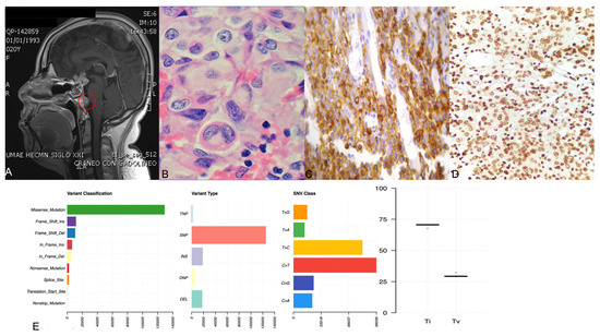
Figure 1. Panel (A) shows the gadolinium-enhanced magnetic resonance imaging of the patient with ACTH-CA, highlighting in red the metastatic lesion in the prepontine area. Panel (B) shows the hematoxylin and eosin staining displaying the hyaline structures in the perinuclear areas denoting a Crooke cell adenoma. Panel (C,D) depict a representative corticotroph tumor with positive ACTH and TBX19 immunohistochemistry, respectively. Panel (E) shows four graphics: variant classification, variant type, SNV class and transition (ti) or transversion (tv) describing the general results of exome sequencing of the corticotroph tumors.
Ijms 23 04861 g002 550
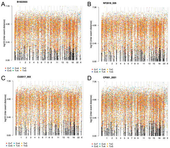
Figure 2. Representative rainfall plots showing the SNV alterations throughout the whole genome of corticotroph tumors (A) CCA, (B) SCA, (C) CD and (D) ACTH-CA, displaying all base changes, including transversions and transitions. No kataegis events were found. Alterations across the genome were seen in all corticotroph tumors.

2.3. ACTH-Secreting Carcinoma (Tumor 1)

SNV missense variants were found in the genes encoding TP53 (c.215G > C [rs1042522], p.Pro72Arg); AURKA (c.91T > A [rs2273535], p.Phe31Ile); EGFR (epidermal growth factor receptor, c.1562G > A [rs2227983], p.Arg521Lys); HSD3B1 (3-ß-hydroxisteroid dehydrogenase, c.1100C > A [rs1047303], p.Thr367Asn); CDKN1A (cyclin-dependent kinase inhibitor 1A or p21, c.93C > A [rs1801270], p.Ser31Arg); and USP8 (c.2159C > G [rs672601311], p.Pro720Arg). Interestingly, the previously reported USP48, BRAF, BRG1 and CABLES1 variants in pituitary CA cases were not found in this patient’s tumor (Figure 3). All SNV detected in WES experiments were validated by Sanger sequencing. The variants described were selected due to their potential pathogenic participation in other tumors and the allelic-risk association with tumorigenesis. Hereafter, all the mentioned variants in other corticotroph tumors are referred to by these aforementioned variants. Even though these same genes presented other variants, currently the significance of those variants is unknown.
Ijms 23 04861 g003 550
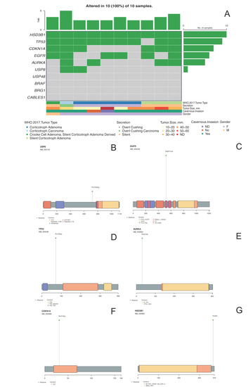
Figure 3. Panel (A) shows the oncoplot from the missense variants of the selected genes and their clinical–pathological features. Panels (BG) depict USP8, EGFR, TP53, AURKA, CDKN1A and HSD3B1 proteins, respectively, with the changes found in DNA impacting aminoacidic changes.
In general, the pituitary CA presented more CNV alterations than the benign tumors, with 27 and 32 cytogenetic regions showing gains and losses of genetic material, respectively. The cytogenetic regions showing gains were 10q11.22, 15q11.2, 16p12.3, 1p13.2 and 20p, where genes SYT15, POTEB, ARL6IP1, HIPK1 and CJD6 are coded, respectively. By contrast, 8p21.2 was the cytogenetic region showing loss of genetic material. The previously reported amplification of 1p13.2 was also detected in this tumor (Figure 4) [10].
Ijms 23 04861 g004 550
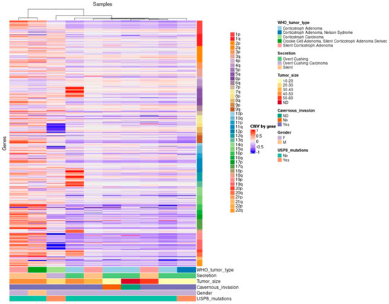
Figure 4. Hierarchical clustering of corticotroph tumors according to their gains and losses across the whole genome (somatic chromosomes only). High contrast was used to enhance potential CNV alterations; nevertheless, there were only 44 unique cytogenetic regions that showed gains in genetic material with statistical significance, whereas only 72 unique cytogenetic regions showed loss of genetic material with statistical significance.

2.4. Crooke Cell Adenoma (Tumor 2)

The CCA showed SNV in the genes encoding TP53, EGFR, HSD3B1 and CDKN1A. However, neither the genes encoding AURKA and USP8 nor those encoding USP48, BRAF, BRG1 and CABLES were affected in this tumor. In CCA, only two and fifteen gains and losses were observed in copy-number variation, respectively. CNVs only showed gains in cytogenetic regions 17q12 and 10q11.22, harboring genes CCL3L1 and NPY4R, respectively, whereas losses were found in cytogenetic regions 18q21.1, 15q12 and 2q11.2, harboring genes KATNAL2, TUBGCP5 and ANKRD36.

2.5. Silent Corticotroph Adenomas (Tumors 3–5)

The three SCA shared SNVs in the genes encoding HSD3B1 and CDKN1A. SCA 4 and 5 showed SNV in the genes encoding EGFR, whereas SNV in the genes encoding AURKA and TP53 were present in SCA 3 and 5. None of the SCA were found to have SNV in the genes encoding USP8, USP48, BRAF, BRG1 or CABLES1.
The SCA presented only two and eighteen gains and losses (CNV), respectively. In regard to CNV, the these clinically silent tumors presented gains of genetic material in cytogenetic regions 17q22 and 10q11.22, which harbor genes encoding CCL3L1 and NPY4R. Eighteen losses were found distributed in cytogenetic regions 18q21.1, 15q12 and 2q11.2, encompassing the genes encoding KATNAL2, TUBGCP5 and ANKRD36. This CNV pattern closely resembles the one found in the CCA, which is somewhat expected if we consider that both neoplasms are clinically non-functioning

2.6. ACTH-Secreting Adenomas (Cushing Disease) (Tumors 6–9)

SNV of the genes encoding TP53 and HSD3B1 were present in tumor samples from all four CD patients, whereas none of these patients harbored adenomas with SNV in the genes encoding USP8 or CDKN1A. An SNV in the gene encoding AURKA was identified in only one of these tumors (tumor 8). EGFR SNV were found in tumors 7 and 9. None of the CD-causing ACTH-secreting adenomas showed the previously reported SNV in the genes encoding USP48, BRAF, BRG1 and CABLES1.
CNV analysis in this group of eloquent-area corticotroph tumors revealed 25 gains and 55 losses of genetic material. The gains occurred in cytogenetic regions 17q12, 2p12, 9p24 and 10q11.22, where genes CCL3L1, CTNNA2, FOXD4 and NPY4R are coded, respectively. The losses were localized in cytogenetic regions 21p12, 15q11.2, and 8p23, harboring genes USP16, KLF13 and DEF130A, respectively. We also detected the previously reported 20p13 amplification [10].

2.7. ACTH-Secreting Adenoma Causing Nelson Syndrome (Tumor 10)

This patient’s tumor showed SNV in the genes encoding USP8, TP53, HSD3B1 and CDKN1A but no alterations were found in the genes encoding EGFR and AURKA. This tumor and the ACTH-CA were the only two neoplasms that harbored a USP8 variant. No SNV were identified in the genes encoding USP48, BRAF, BRG1 and CABLES1. Interestingly, CNV analysis revealed the same gains and losses of genetic material found in tumors from other patients with CD.

2.8. Tumor Phylogenic Analysis

We performed a phylogenetic inference analysis to unravel a hypothetical sequential step transformation from an SCA to a functioning ACTH-secreting adenoma and finally to an ACTH-CA. The theoretical evolutive development of the ACTH CA, departing from the SCA, shows two main clades, with the smallest one comprising two of the three SCA and two of the five ACTH-adenomas causing CD. Since these four tumors have the same SNV profile, we can assume that they harbor the genes that must be altered to make possible the transition from a silent to a clinically eloquent adenoma; the gene encoding ATF7IP (c.1589A > G [rs3213764], p.K529R) characterizes this clade. The second and largest clade includes the CCA, the ACTH-CA, one of the three SCA and three of the five most aggressive ACTH adenomas causing CD, including the adenoma of the patient with Nelson syndrome. This clade represents the molecular alterations required to evolve from a CD-causing ACTH-adenoma to a more aggressive tumor, or even to a CA and is characterized by the gene encoding MSH3 (c.235A > G [rs1650697], p.I79V) (Figure 5).
Ijms 23 04861 g005 550
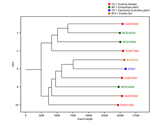
Figure 5. Phylogenetic analysis of the corticotroph tumors. The theoretical evolutive development of the ACTH-CA, departing from the SCA shows two main clades. The first clade, characterized by ATF7IP gene, comprises 2 of the 3 SCA and 2 of the 5 ACTH-adenomas causing CD. The second clade is characterized by the gene encoding MSH3 and includes the CCA, the ACTH-CA, one of the 3 SCA and 3 of the 5 most aggressive ACTH adenomas causing CD, including the adenoma of the patient with Nelson syndrome. Red dots represent the Cushing Disease provoking adenomas, green dots represent the silent corticotroph tumors, brown dot represent the Crooke cell adenoma and the blue dot represent the corticotroph carcinoma.

2.9. Correlation between Gene Variants and Clinicopathological Features

The USP8 variant positively correlated with increased tumor mass (p = 0.019). The CDKN1A variant was significantly associated with silent tumors (p = 0.036). The rest of the genetic variants did not correlate with any of the clinicopathological features tested. The presence of the EGFR variant was not distinctly associated with any of the clinical parameters and was equally present in functional as well as non-functional tumors (p = 0.392). AURKA SNV did not correlate with any of the features, including recurrence (p = 0.524). Detailed statistical results are presented in Supplementary Table S2.

3. Discussion

Corticotrophs are highly specialized cells of the anterior pituitary that synthesize and secrete hormones that are essential for the maintenance of homeostasis. In this study, we sequenced the exome of 10 corticotroph tumors, including three SCA, four ACTH adenomas causing CD, an ACTH adenoma in a patient with Nelson syndrome, a CCA and an ACTH-CA in total, representing the broad pathological spectrum of this cell. Our results portray the genomic landscape of all the neoplasms that are known to affect the corticotroph.
The neoplasm with the highest number of genomic abnormalities, including SNV and CNV, was the ACTH-CA, followed by the CCA and the CD tissues. Of all the genes harboring SNVs, six were found to be present in at least two of our tumor samples: HSD3B1, TP53, CDKN1A, EGFR, AURKA and USP8.
The HSD3B1 gene encodes a rate-limiting enzyme required for all pathways of dihydrotestosterone synthesis and is abundantly expressed in adrenal tumors. Gain of function of this HSD3B1 variant, which has a global allelic prevalence of 0.69678 [11], results in resistance to proteasomal degradation with the consequent accumulation of the enzyme and has been associated with a poor prognosis in patients with prostate cancer [12]. Nine of the ten corticotroph tumors in our cohort harbored an SNV of the tumor suppressor gene TP53. The TP53 variant described in our cohort has been reported to be present in 80% of non-functioning pituitary adenomas and is apparently associated with a younger age at presentation and with cavernous sinus invasion [13]. Furthermore, this TP53 variant results in a reduced expression of CDKN1A and an increased expression of vascular endothelial growth factor (VEGF) as well as an increased cellular proliferation rate [13]. CDKN1A (also known as p21) is a cyclin-dependent kinase inhibitor regulating cell cycle progression. The SNV described in our study was reported to alter DNA binding ability and expression and has a global allelic frequency of 0.086945 [14]. This cyclin-dependent kinase inhibitor SNV was found to be associated with breast carcinoma [15] and lung cancer [16]. The presence of this SNV has not been previously explored in pituitary adenomas, although CDKN1A is downregulated in clinically non-functioning pituitary adenomas of gonadotrophic lineage but not in hormone-secreting tumors [17]. EGFR encodes a transmembrane tyrosine kinase receptor, activation of which leads to mitogenic signaling [18]. This gene is upregulated in several cancers and represents a target for molecular therapies [19]. The EGFR SNV described in our corticotroph tumor series was found to be associated with the response to neoadjuvant chemotherapy in patients with breast and lung cancer [18]. EGFR is normally expressed in corticotrophs, where it participates in the regulation of POMC (proopiomelanocortin) gene transcription and cellular proliferation [20]. The EGFR rs2227983 has a 0.264334 global allelic frequency [21]. AURKA is a cell-cycle regulatory serine/threonine kinase that promotes cell cycle progression by the establishment of the mitotic spindle and centrosome separation [22]. Alterations of these gene are related to centrosomal amplification, dysfunction of cytokinesis and aneuploidy [22]; it has a global allelic frequency of 0.18078 [23]. This same SNV has been associated with overall cancer risk, particularly breast, gastric, colorectal, liver and endometrial carcinomas, but it has never been formally studied in pituitary tumors [22]. Activating somatic variants of the gene encoding USP8 were recently found in 25–40% of ACTH-secreting adenomas causing CD [24,25]. Patients harboring these variants are usually younger, more frequently females and were found to have higher long-term recurrence rates in some but not all studies [26,27]. USP8 mediates the deubiquitination of EGFR by inhibiting its interaction with protein 14-3-3, which in turn prevents its proteosomal degradation. Signaling through the recycled deubiquitinated EGFR is increased, leading to increased POMC transcription and cellular proliferation. Most activating USP8 variants are located within its 14-3-3 binding motif [24,25]. Recently, USP8 and TP53 SNV were described in corticotroph tumors as drivers of aggressive lesions [28]. To our knowledge, USP8 variants have not been evaluated in patients with pituitary carcinomas, and none of the previously mentioned studies have included patients with Nelson syndrome. In our cohort, neither the CCA nor the SCA showed variants in USP8, in concordance with previously published studies [25,29], or in the genes USP48, BRAF, BRG1 and CABLES1 [9], and none of them were present in our cohort.
Genetic structural variations in the human genome can be present in many forms, from SNV to large chromosomal aberrance [30]. CNV are structurally variant regions, including unbalanced deletions, duplications and amplifications of DNA segments ranging from a dozen to several hundred base pairs, in which copy-number differences have been observed between two or more genomes [31,32]. CNV are involved in the development and progression of many tumors and occur frequently in PA [30,33]. Hormone-secreting pituitary tumors show more CNV than non-functioning tumors [34]. Accordingly, our non-functioning SCA and CCA had considerably fewer chromosomal gains and losses than the CD-causing adenomas and the ACTH-CA. Expectedly, the ACTH-CA had significantly more cytogenetic abnormalities than any other tumor in our series. Interestingly, the ACTH-adenomas causing CD, the SCA and the CCA shared the gain of genetic material in 17q12, highlighting their benign nature. The 17q12 amplification has been described in gastric neoplasms [35]. The only cytogenetic abnormality shared by all types of corticotroph tumors was the gain of genetic material in 10q11.22. Amplification of 10q11.22 was previously described in Li–Fraumeni cancer predisposition syndrome [36]. The ACTH-CA, the CCA and one SCA clustered together showing a related CNV pattern; this CNV profile could be reflective of the aggressive nature of these neoplasms, since both CCA and SCA can follow a clinically aggressive course [5,6].
Our results show that all lesions conforming to the pathological spectrum of the corticotroph share some of the SNV and CNV profiles. These genomic changes are consistent with the potential existence of a continuum, whereby silent tumors can transform into a clinically eloquent tumor and finally to carcinoma, or at least a more aggressive tumor. It can also be interpreted as the common SNV shared by aggressive tumors. It is known that silent corticotroph adenomas may switch into a hormone-secreting tumor [37] and are considered a marker for aggressiveness and a risk factor for malignancy since most of the carcinomas are derived from functioning hormone-secreting adenomas. Our phylogenetic inference analysis showed that the genes ATF7IP and MSH3 could participate in a tumor transition ending in aggressive entities or even carcinomas. ATF7IP is a multifunctional nuclear protein mediating heterochromatin formation and gene regulation in several contexts [38], while MSH3 is a mismatch-repair gene [39]. Events related to heterochromatin remodeling and maintenance have been related to aggressive pituitary adenomas and carcinomas [40]. Additionally, alterations in mismatch-repair genes are related to pituitary tumor aggressiveness and resistance to pharmacologic treatment [41,42]. The variants described in ATF7IP and MSH3 are related to prostate and colorectal cancer, respectively [43,44]. There is evidence suggesting that the ATF7IP variant could be deleterious because it leads to a negative regulation of transcription [45]. Thus, these events could be biologically relevant to corticotroph tumorigenesis, although more research is needed.

4. Conclusions

We have shown genomic evidence that within the tumoral spectrum of the corticotroph, functioning ACTH-secreting lesions harbor more SNV and CNV than non-functioning ACTH adenomas. The ACTH-secreting CA shows more genomic abnormalities than the other lesions, underscoring its more aggressive biological behavior. Phylogenetic inference analysis of our data reveals that silent corticotroph lesions may transform into functioning tumors, or at least potentially, into more aggressive lesions. Alterations in genes ATF7IP and MSH3, related to heterochromatin formation and mismatch repair, could be important in corticotroph tumorigenesis. The main drawback of our study is the limited sample size. We are currently increasing the number of samples to corroborate our findings and to be able to perform a more comprehensive complementary phylogenetic analysis of our data. Finally, further research is needed to uncover the roles of these variants in corticotroph tumorigenesis.

5. Materials and Methods

5.1. Patients and Tumor Tissue Samples

Ten pituitary tissues were collected: one ACTH-CA, one CCA, three SCA, and five ACTH-secreting PA causing CD, including the tumor of a patient who developed Nelson syndrome after bilateral adrenalectomy. All tumors included in the study were sporadic and were collected from patients diagnosed, treated and followed at the Endocrinology Service and the Neurosurgical department of Hospital de Especialidades, Centro Médico Nacional Siglo XXI of the Instituto Mexicano del Seguro Social, Hospital General de Mexico “Dr. Eduardo Liceaga” and Instituto Nacional de Neurologia y Neurocirugia “Manuel Velazquez”. All participating patients were recruited with signed informed consent and ethical approval from the Comisión Nacional de Ética e Investigación Científica of the Instituto Mexicano del Seguro Social, in accordance with the Helsinki declaration.
CD was diagnosed according to our standard protocol. Briefly, the presence of hypercortisolism was documented based on two screening tests, namely a 24 h urinary free-cortisol level above 130 µg and the lack of suppression of morning (7:00–8:00) cortisol after administration of 1 mg dexamethasone the night before (23:00) to less than 1.8 µg/dL, followed by a normal or elevated plasma ACTH to ascertain ACTH-dependence. Finally, an overnight, high-dose (8 mg) dexamethasone test, considered indicative of a pituitary source, and a cortisol suppression > 69%, provided that a pituitary adenoma was clearly present on magnetic resonance imaging (MRI) of the sellar region. In none of the 10 patients included in the study was inferior petrosal venous sampling necessary to confirm the pituitary origin of the ACTH excess. Invasiveness was defined by the presence of tumor within the cavernous sinuses (CS).
DNA was extracted from paraffin-embedded tumor tissues using the QIAamp DNA FFPE tissue kit. From frozen tumors, DNA was obtained using the Proteinase K-ammonium acetate protocol.

5.2. Construction and Sequencing of Whole Exome Libraries

Exome libraries were prepared according to the Agilent SureSelect XT HS Human All exon v7 instructions. Briefly, 200 ng of DNA was enzymatically fragmented with Agilent SureSelect Enzymatic Fragmentation Kit. Fragmented DNA was end-repaired and dA-tail was added at DNA ends; then, molecular barcode adaptors were added, followed by AMPure XP bead purification. The adaptor-ligated library was amplified by PCR and purified by AMPure XP beads. DNA libraries were hybridized with targeting exon probes and purified with streptavidin-coated magnetic beads. The retrieved libraries were amplified by PCR and purified by AMPure XP beads and pooled for sequencing in NextSeq 500 using Illumina flow cell High Output 300 cycles chemistry. All quality controls of the libraries were carried out using Screen tape assays and quantified by Qubit fluorometer. Quality parameters included a DNA integrity number above 8 and a 100X sequencing depth aimed with at least 85% of coverage.

5.3. Bioinformatics Analysis

The fastq files were subjected to quality control using FastQC v0.11.9, the adapters were removed using Cutadapt v3.4, the alignment was carried out with Burrows–Wheeler Alignment Tool v0.7.17 with the -M option to ensure compatibility with Picard and GRCh38 as a reference genome. The marking of duplicates as well as the sorting was carried out with Picard v2.26.4 with the AddOrReplaceReadGroups programs with the option SORT_ORDER = coordinate and MarkDuplicates, respectively. Variant calling was carried out using Genomic Analysis Toolkit (GATK) v4.2.2.0 following the Best Practices guide (available at https://gatk.broadinstitute.org/) [46] and with the parameters used by Genomic Data Commons (GDC), available at https://docs.gdc.cancer.gov/ [47]. The GATK tools used were CollectSequencingArtifactMetrics, GetPileupSummaries, CalculateContamination and Mutect2. Mutect2 was run with the latest filtering recommendations, including a Panel of Normal and a Germline Reference from the GATK database. Filtering was performed with the CalculateContamination, LearnReadOrientationModel and FilterMutectCalls tools with the default parameters. For the calculation of CNV GISTIC v2.0.23 was used with the parameters used by GDC. Catalog of Somatic Mutation in Cancer (COSMIC) was used to uncover pathogenic variants. For the analysis of variants and CNV, the maftool v2.10.0 and ComplexHeatmap 2.10.0 packages were used. All analyses were carried out on the GNU/Linux operating system under Ubuntu v20.01.3 or using the R v4.0.2 language in Rstudio v2021.09.0+351. A second bioinformatics pipeline was also used, SureCall software (Agilent) with the default parameters used for SNV variant calling. The variants found by both algorithms were taken as reliable SNV. Data were deposited in Sequence Read Archive hosted by National Center for Biotechnology Information under accession number PRJNA806516.
Phylogenetic tree inference (PTI) was run by means of the default parameters using matrices for each sample. These matrices contain an identifier for each variant, mutant read counts, counts of reference reads and the gene associated with the variant. The only PTI parameter was Allele Frequency of Mutation and was used to improve the speed of the algorithm. Briefly, PTI uses an iterative process on the variants shared between the samples. First, it builds the base of the tree using the variants shared by all the samples; second, it eliminates these variants and establishes a split node; and third, it eliminates the variants of the sample that produced the division (split). PTI iteratively performs these three steps for all division possibilities. Each tree is given a score based on an aggregated variant count, and the tree with the highest score is chosen as the optimal tree.

5.4. Sanger Sequencing forConfirmation of Exome Findings

Exome variant findings in exome sequencing were validated by Sanger sequencing using BigDye Terminator v3.1 Cycle Sequencing kit (ThermoFischer) in a 3500 Genetic Analyzer. Primers used for USP8 [48], TP53 [49], EGFR [50], AURKA [51], CDKN1A [52,53] and HSD3B1 sequencing have been previously reported.

5.5. Hormone and Transcription Factor Immunohistochemistry

Paraffin-embedded, formalin-fixed tissue blocks were stained with hematoxylin–eosin and reviewed by a pathologist. Tumors were represented with a 2-fold redundancy. Sections (3 μm) were cut and placed onto coated slides. Immunostaining was performed by means of the HiDef detection HRP polymer system (Cell Marque, CA, USA), using specific antibodies against each pituitary hormone (TSH, GH, PRL, FSH, LH and ACTH) and the lineage-specific transcription factors TBX19, POU1F1 and NR5A1, as previously described [54]. Two independent observers performed assessment of hormones and transcription factors expression at different times.

5.6. Statistical Analysis

Two-tailed Fisher exact tests and Student’s t tests were used to evaluate the relationship between the identified gene variants and clinicopathological features. A p value of <0.05 was considered statistically significant. Statistical software consisted of SPSS v28.0.1

Supplementary Materials

The following supporting information can be downloaded at: https://www.mdpi.com/article/10.3390/ijms23094861/s1.

Author Contributions

D.M.-R., K.T.-P. and M.M. conceived, designed and coordinated the project, performed experiments, analyzed, discussed data and prepared the manuscript. S.A.-E., G.S.-R., E.P.-M., S.V.-P., R.S., L.B.-A., C.G.-T., J.G.-C. and J.T.A.-S. performed DNA purification, library preparation, sequencing experiments, bioinformatics analysis and wrote the manuscript. A.-L.E.-d.-l.-M., I.R.-S., E.G.-A., L.A.P.-O., G.G., S.M.-J., L.C.-M., B.L.-F. and A.B.-L. provided biological samples and detailed patient information. All authors have read and agreed to the published version of the manuscript.

Funding

This work was partially supported by grants 289499 from Fondos Sectoriales Consejo Nacional de Ciencia y Tecnologia, Mexico, and R-2015-785-015 from Instituto Mexicano del Seguro Social (MM).

Institutional Review Board Statement

Protocol approved by the Comisión Nacional de Ética e Investigación Científica of the Instituto Mexicano del Seguro Social, in accordance with the Helsinki declaration (R-2019-785-052).

Informed Consent Statement

Informed consent was obtained from all subjects involved in the study.

Data Availability Statement

Data were deposited in Sequence Read Archive hosted by National Center for Biotechnology Information under accession number PRJNA806516.

Acknowledgments

Sergio Andonegui-Elguera is a doctoral student from Programa de Doctorado en Ciencias Biomédicas, Universidad Nacional Autónoma de México (UNAM) and received fellowship 921084 from CONACYT. KTP is a recipient of Consejo Nacional de Ciencia y Tecnología (CONACyT) fellowship “Estáncias posdoctorales por Mexico 2021” program. DMR is a recipient of the National Council for Science and Technology Fellowship “Catedra CONACyT” program.

Conflicts of Interest

The authors declare no conflict of interest.

References

  1. Heaney, A. Clinical review: Pituitary carcinoma: Difficult diagnosis and treatment. J. Clin. Endocrinol. Metab. 2011, 96, 3649–3660. [Google Scholar] [CrossRef] [PubMed]
  2. Raverot, G.; Burman, P.; McCormack, A.; Heaney, A.; Petersenn, S.; Popovic, V.; Trouillas, J.; Dekkers, O.M.; The European Society of Endocrinology. European Society of Endocrinology Clinical Practice Guidelines for the management of aggressive pituitary tumours and carcinomas. Eur. J. Endocrinol. 2018, 178, G1–G24. [Google Scholar] [CrossRef]
  3. Todeschini, A.B.; Beer-Furlan, A.; Montaser, A.S.; Jamshidi, A.O.; Ghalib, L.G.; Chavez, J.A.; Lehman, N.L.; Prevedello, D.M. Pituitary carcinomas: Review of the current literature and report of atypical case. Br. J. Neurosurg. 2020, 34, 528–533. [Google Scholar] [CrossRef]
  4. Dudziak, K.; Honegger, J.; Bornemann, A.; Horger, M.; Müssig, K. Pituitary carcinoma with malignant growth from first presentation and fulminant clinical course-case report and review of the literature. J. Clin. Endocrinol. Metab. 2011, 96, 2665–2669. [Google Scholar] [CrossRef] [PubMed]
  5. Di Ieva, A.; Davidson, J.; Syro, L.; Rotondo, F.; Montoya, J.; Horvath, E.; Cusimano, M.D.; Kovacs, K. Crooke’s cell tumors of the pituitary. Neurosurgery 2015, 76, 616–622. [Google Scholar] [CrossRef] [PubMed]
  6. Fountas, A.; Lavrentaki, A.; Subramanian, A.; Toulis, K.; Nirantharakumar k Karavitaki, N. Recurrence in silent corticotroph adenomas after primary treatment: A systematic review and meta-analysis. J. Clin. Endocrinol. Metab. 2019, 104, 1039–1048. [Google Scholar] [CrossRef] [PubMed]
  7. Molitch, M. Diagnosis and Treatment of Pituitary Adenomas: A Review. JAMA 2017, 317, 516–524. [Google Scholar] [CrossRef]
  8. Melmed, S. Pituitary-Tumor Endocrinopathies. N. Engl. J. Med. 2020, 382, 937–950. [Google Scholar] [CrossRef] [PubMed]
  9. Lopes, M.B.S. The 2017 World Health Organization classification of tumors of the pituitary gland: A sumary. Acta Neuropathol. 2017, 134, 521–535. [Google Scholar] [CrossRef] [PubMed]
  10. Song, Z.-J.; Reitman, Z.; Ma, Z.-Y.; Chen, J.-H.; Zhang, Q.-L.; Shou, X.-F.; Huang, C.X.; Wang, Y.F.; Li, S.Q.; Mao, Y.; et al. The genome-wide mutational landscape of pituitary adenomas. Cell Res. 2016, 26, 1255–1259. [Google Scholar] [CrossRef]
  11. NCBI. Available online: https://www.ncbi.nlm.nih.gov/snp/rs1047303#frequency_tab (accessed on 23 February 2022).
  12. Shiota, M.; Narita, S.; Akamatsu, S.; Fujimoto, N.; Sumiyoshi, T.; Fujiwara, M.; Uchiumi, T.; Habuchi, T.; Ogawa, O.; Eto, M. Association of Missense Polymorphism in HSD3B1 With Outcomes Among Men With Prostate Cancer Treated With Androgen-Deprivation Therapy or Abiraterone. JAMA Netw. Open 2019, 2, e190115. [Google Scholar] [CrossRef]
  13. Yagnik, G.; Jahangiri, A.; Chen, R.; Wagner, J.; Aghi, M. Role of a p53 polymorphism in the development of nonfunctional pituitary adenomas. Mol. Cell Endocrinol. 2017, 446, 81–90. [Google Scholar] [CrossRef]
  14. Heidari, Z.; Harati-Sadegh, M.; Arian, A.; Maruei-Milan, R.; Salimi, S. The effect of TP53 and P21 gene polymorphisms on papillary thyroid carcinoma susceptibility and clinical/pathological features. IUBMB Life 2020, 72, 922–930. [Google Scholar] [CrossRef]
  15. Akhter, N.; Dar, S.; Haque, S.; Wahid, M.; Jawed, A.; Akhtar, M.S.; A Alharbi, R.; A A Sindi, A.; Alruwetei, A.; Choudhry, H.M.Z.; et al. Crosstalk of Cyclin-dependent kinase inhibitor 1A (CDKN1A) gene polymorphism with p53 and CCND1 polymorphism in breast cancer. Eur. Rev. Med. Pharmacol. Sci. 2021, 25, 4258–4273. [Google Scholar]
  16. Wang, C.; Nie, H.; Li, Y.; Liu, G.; Wang, X.; Xing, S.; Zhang, L.; Chen, X.; Chen, Y.; Li, Y. The study of the relation of DNA repair pathway genes SNPs and the sensitivity to radiotherapy and chemotherapy of NSCLC. Sci. Rep. 2016, 6, 26526. [Google Scholar] [CrossRef] [PubMed]
  17. Taniguchi-Ponciano, K.; Portocarrero-Ortiz, L.A.; Guinto, G.; Moreno-Jimenez, S.; Gomez-Apo, E.; Chavez-Macias, L.; Peña-Martínez, E.; Silva-Román, G.; Vela-Patiño, S.; Ordoñez-García, J.; et al. The kinome, cyclins and cyclin-dependent kinases of pituitary adenomas, a look into the gene expression rofile among tumors different lineages. BMC Med. Genom. 2022, 15, 52. [Google Scholar] [CrossRef]
  18. Sobral-Leite, M.; Lips, E.; Vieira-Monteiro, H.; Giacomin, L.; Freitas-Alves, D.; Cornelissen, S.; Mulder, L.; Wesseling, J.; Schmidt, M.K.; Vianna-Jorge, R. Evaluation of the EGFR polymorphism R497K in two cohorts of neoadjuvantly treated breast cancer patients. PLoS ONE 2017, 12, e0189750. [Google Scholar] [CrossRef]
  19. Zhang, H.; Berezov, A.; Wang, Q.; Zhang, G.; Drebin, J.; Murali, R.; Greene, M. ErbB receptors: From oncogenes to targeted cancer therapies. J. Clin. Investig. 2007, 117, 2051–2058. [Google Scholar] [CrossRef] [PubMed]
  20. Liu, X.; Feng, M.; Dai, C.; Bao, X.; Deng, K.; Yao, Y.; Wang, R. Expression of EGFR in Pituitary Corticotroph Adenomas and Its Relationship With Tumor Behavior. Front. Endocrinol. 2019, 10, 785. [Google Scholar] [CrossRef]
  21. NCBI. Available online: https://www.ncbi.nlm.nih.gov/snp/rs2227983#frequency_tab (accessed on 23 February 2022).
  22. Wang, S.; Qi, J.; Zhu, M.; Wang, M.; Nie, J. AURKA rs2273535 T>A Polymorphism Associated With Cancer Risk: A Systematic Review With Meta-Analysis. Front. Oncol. 2020, 10, 1040. [Google Scholar] [CrossRef] [PubMed]
  23. NCBI. Available online: https://www.ncbi.nlm.nih.gov/snp/rs2273535#frequency_tab (accessed on 23 February 2022).
  24. Reincke, M.; Sbiera, S.; Hayakawa, A.; Theodoropoulou, M.; Osswald, A.; Beuschlein, F.; Meitinger, T.; Mizuno-Yamasaki, E.; Kawaguchi, K.; Saeki, Y.; et al. Mutations in the deubiquitinase gene USP8 cause Cushing’s disease. Nat. Genet. 2015, 47, 31–38. [Google Scholar]
  25. Perez-Rivas, L.; Theodoropoulou, M.; Ferraù, F.; Nusser, C.; Kawaguchi, K.; Faucz, F.; Nusser, C.; Kawaguchi, K.; Stratakis, C.A.; Faucz, F.R.; et al. The Gene of the Ubiquitin-Specific Protease 8 Is Frequently Mutated in Adenomas Causing Cushing’s Disease. J. Clin. Endocrinol. Metab 2015, 100, E997–E1004. [Google Scholar] [CrossRef] [PubMed]
  26. Albani, A.; Pérez-Rivas, L.G.; Dimopoulou, C.; Zopp, S.; Colón-Bolea, P.; Roeber, S.; Honegger, J.; Flitsch, J.; Rachinger, W.; Buchfelder, M.; et al. The USP8 mutational status may predict long-term remission in patients with Cushing’s disease. Clin. Endocrinol. 2018, 89, 454–458. [Google Scholar] [CrossRef] [PubMed]
  27. Wanichi, I.Q.; de Paula Mariani, B.M.; Frassetto, F.P.; Siqueira, S.A.C.; de Castro Musolino, N.R.; Cunha-Neto, M.B.C.; Ochman, G.; Cescato, V.A.S.; Machado, M.C.; Trarbach, E.B.; et al. Cushing’s disease due to somatic USP8 mutations: A systematic review and meta-analysis. Pituitary 2019, 22, 435–442. [Google Scholar] [CrossRef]
  28. Uzilov, A.; Taik, P.; Cheesman, K.; Javanmard, P.; Ying, K.; Roehnelt, A.; Wang, H.; Fink, M.Y.; Lau, C.Y.; Moe, A.S.; et al. USP8 and TP53 Drivers are Associated with CNV in a Corticotroph Adenoma Cohort Enriched for Aggressive Tumors. J. Clin. Endocrinol. Metab. 2021, 106, 826–842. [Google Scholar] [CrossRef]
  29. Hayashi, K.; Inoshita, N.; Kawaguchi, K.; Ibrahim, A.; Suzuki, H.; Fukuhara, N.; Okada, M.; Nishioka, H.; Takeuchi, Y.; Komada, M.; et al. The USP8 mutational status may predict drug susceptibility in corticotroph adenomas of Cushing’s disease. Eur. J. Endocrinol. 2016, 174, 213–226. [Google Scholar] [CrossRef] [PubMed]
  30. Shao, X.; Lv, N.; Liao, J.; Long, J.; Xue, R.; Ai, N.; Xu, D.; Fan, X. Copy number variation is highly correlated with differential gene expression: A pan-cancer study. BMC Med Genet. 2019, 20, 175. [Google Scholar] [CrossRef] [PubMed]
  31. Shlien, A.; Malkin, D. Copy number variations and cancer. Genome Med. 2009, 1, 62. [Google Scholar] [CrossRef]
  32. Pös, O.; Radvanszky, J.; Styk, J.; Pös, Z.; Buglyó, G.; Kajsik, M.; Budis, J.; Nagy, B.; Szemes, T. Copy Number Variation: Methods and Clinical Applications. Appl. Sci. 2021, 11, 819. [Google Scholar] [CrossRef]
  33. Cui, Y.; Li, C.; Jiang, Z.; Zhang, S.; Li, Q.; Liu, X.; Zhou, Y.; Li, R.; Wei, L.; Li, L.; et al. Single-cell transcriptome and genome analyses of pituitary neuroendocrine tumors. Neuro-Oncol. 2021, 23, 1859–1871. [Google Scholar] [CrossRef]
  34. Neou, M.; Villa, C.; Armignacco, R.; Jouinot, A.; Raffin-Sanson, M.-L.; Septier, A.; Letourneur, F.; Diry, S.; Diedisheim, M.; Izac, B.; et al. Pangenomic Classification of Pituitary Neuroendocrine Tumors. Cancer Cell 2020, 37, 123–134.e5. [Google Scholar] [CrossRef] [PubMed]
  35. Varis, A.; Wolf, M.; Monni, O.; Vakkari, M.-L.; Kokkola, A.; Moskaluk, C.; Frierson, H.; Powell, S.M.; Knuutila, S.; Kallioniemi, A.; et al. Targets of gene amplification and overexpression at 17q in gastric cancer. Cancer Res. 2002, 62, 2625–2629. [Google Scholar] [PubMed]
  36. Shlien, A.; Tabori, U.; Marshall, C.; Pienkowska, M.; Feuk, L.; Novokmet, A.; Nanda, S.; Druker, H.; Scherer, S.W.; Malkin, D. Excessive genomic DNA copy number variation in the Li-Fraumeni cancer predisposition syndrome. Proc. Natl. Acad. Sci. USA 2008, 105, 11264–11269. [Google Scholar] [CrossRef] [PubMed]
  37. McCormack, A.; Dekkers, O.; Petersenn, S.; Popovic, V.; Trouillas, J.; Raverot, G.; Burman, P. Treatment of aggressive pituitary tumours and carcinomas: Results of a European Society of Endocrinology (ESE) survey 2016. Eur. J. Endocrinol. 2018, 178, 265–276. [Google Scholar] [CrossRef]
  38. Trouillas, J.; Jaffrain-Rea, M.; Vasiljevic, A.; Raverot, G.; Roncaroli, F.; Villa, C. How to Classify the Pituitary Neuroendocrine Tumors (PitNET)s in 2020. Cancers 2020, 12, 514. [Google Scholar] [CrossRef]
  39. Hu, H.; Khodadadi-Jamayran, A.; Dolgalev, I.; Cho, H.; Badri, S.; Chiriboga, L.A.; Zeck, B.; Gregorio, M.L.D.R.; Dowling, C.M.; Labbe, K.; et al. Targeting the Atf7ip-Setdb1 Complex Augments Antitumor Immunity by Boosting Tumor Immunogenicity. Cancer Immunol. Res. 2021, 9, 1298–1315. [Google Scholar] [CrossRef]
  40. Park, J.; Huang, S.; Tougeron, D.; Sinicrope, F. MSH3 mismatch repair protein regulates sensitivity to cytotoxic drugs and a histone deacetylase inhibitor in human colon carcinoma cells. PLoS ONE 2013, 8, e65369. [Google Scholar] [CrossRef]
  41. Raverot, G.; Ilie, M.; Lasolle, H.; Amodru, V.; Trouillas, J.; Castinetti, F.; Brue, T. Aggressive pituitary tumours and pituitary carcinomas. Nat. Rev. Endocrinol. 2021, 17, 671–684. [Google Scholar] [CrossRef]
  42. Syro, L.; Rotondo, F.; Camargo, M.; Ortiz, L.; Serna, C.; Kovacs, K. Temozolomide and Pituitary Tumors: Current Understanding, Unresolved Issues, and Future Directions. Front. Endocrinol. 2018, 9, 318. [Google Scholar] [CrossRef]
  43. Mamidi, T.K.K.; Wu, J.; Hicks, C. Integrating germline and somatic variation information using genomic data for the discovery of biomarkers in prostate cancer. BMC Cancer 2019, 19, 229. [Google Scholar] [CrossRef]
  44. Caja, F.; Vodickova, L.; Kral, J.; Vymetalkova, V.; Naccarati, A.; Vodicka, P. Mismatch repair gene variant in sporadic solid cancers. Int. J. Mol. Sci. 2020, 21, 5561. [Google Scholar] [CrossRef]
  45. Song, G.G.; Kim, J.H.; Lee, H. Genome-wide pathway analysis in major depresive disorder. J. Mol. Neurosci. 2013, 51, 428–436. [Google Scholar] [CrossRef] [PubMed]
  46. GATK. Available online: https://gatk.broadinstitute.org/ (accessed on 6 October 2021).
  47. GDC. Available online: https://docs.gdc.cancer.gov/ (accessed on 6 October 2021).
  48. Chang, M.; Yang, C.; Bao, X.; Wang, R. Genetic and Epigenetic Causes of Pituitary Adenomas. Front. Endocrinol. 2021, 11, 596554. [Google Scholar] [CrossRef] [PubMed]
  49. Ballmann, C.; Thiel, A.; Korah, H.E.; Reis, A.C.; Saeger, W.; Stepanow, S.; Köhrer, K.; Reifenberger, G.; Knobbe-Thomsen, C.B.; Knappe, U.J.; et al. USP8 Mutations in Pituitary Cushing Adenomas-Targeted Analysis by Next-Generation Sequencing. J. Endocr. Soc. 2018, 2, 266–278. [Google Scholar] [CrossRef] [PubMed]
  50. Naidoo, P.; Naidoo, R.; Ramkaran, P.; Chuturgoon, A. Effect of maternal HIV infection, BMI and NOx air pollution exposure on birth outcomes in South African pregnant women genotyped for the p53 Pro72Arg (rs1042522). Int. J. Immunogenet. 2020, 47, 414–429. [Google Scholar] [CrossRef] [PubMed]
  51. Leite, M.; Giacomin, L.; Piranda, D.; Festa-Vasconcellos, J.; Indio-do-Brasil, V.; Koifman, S.; de Moura-Neto, R.S.; de Carvalho, M.A.; Vianna-Jorge, R. Epidermal growth factor receptor gene polymorphisms are associated with prognostic features of breast cancer. BMC Cancer 2014, 14, 190. [Google Scholar] [CrossRef]
  52. Baumann, A.; Buchberger, A.; Piontek, G.; Schüttler, D.; Rudelius, M.; Reiter, R.; Gebel, L.; Piendl, G.; Brockhoff, G.; Pickhard, A. The Aurora-Kinase A Phe31-Ile polymorphism as possible predictor of response to treatment in head and neck squamous cell carcinoma. Oncotarget 2018, 9, 12769–12780. [Google Scholar] [CrossRef]
  53. Vargas-Torres, S.L.; Portari, E.A.; Silva, A.L.; Klumb, E.M.; da Rocha Guillobel, H.C.; de Camargo, M.J.; Santos-Rebouças, C.B.; Russomano, F.B.; Macedo, J.M.B. Roles of CDKN1A gene polymorphisms (rs1801270 and rs1059234) in the development of cervical neoplasia. Tumour Biol. 2016, 37, 10469–10478. [Google Scholar] [CrossRef]
  54. Taniguchi-Ponciano, K.; Andonegui-Elguera, S.; Peña-Martínez, E.; Silva-Román, G.; Vela-Patiño, S.; Gomez-Apo, E.; Chavez-Macias, L.; Vargas-Ortega, G.; Espinosa-de-Los-Monteros, L.; Gonzalez-Virla, B.; et al. Transcriptome and methylome analysis reveals three cellular origins of pituitary tumors. Sci. Rep. 2020, 10, 19373. [Google Scholar] [CrossRef]
Publisher’s Note: MDPI stays neutral with regard to jurisdictional claims in published maps and institutional affiliations.

Glucocorticoid Withdrawal Syndrome following treatment of endogenous Cushing Syndrome

Abstract

Purpose:

Literature regarding endogenous Cushing syndrome (CS) largely focuses on the challenges of diagnosis, subtyping, and treatment. The enigmatic phenomenon of glucocorticoid withdrawal syndrome (GWS), due to rapid reduction in cortisol exposure following treatment of CS, is less commonly discussed but also difficult to manage. We highlight the clinical approach to navigating patients from GWS and adrenal insufficiency to full hypothalamic-pituitary-adrenal (HPA) axis recovery.

Methods:

We review the literature on the pathogenesis of GWS and its clinical presentation. We provide strategies for glucocorticoid dosing and tapering, HPA axis testing, as well as pharmacotherapy and ancillary treatments for GWS symptom management.

Results:

GWS can be difficult to differentiate from adrenal insufficiency and CS recurrence, which complicates glucocorticoid dosing and tapering regimens. Monitoring for HPA axis recovery requires both clinical and biochemical assessments. The most important intervention is reassurance to patients that GWS symptoms portend a favorable prognosis of sustained remission from CS, and GWS typically resolves as the HPA axis recovers. GWS also occurs during medical management of CS, and gradual dose titration based primarily on symptoms is essential to maintain adherence and to eventually achieve disease control. Myopathy and neurocognitive dysfunction can be chronic complications of CS that do not completely recover.

Conclusions:

Due to limited data, no guidelines have been developed for management of GWS. Nevertheless, this article provides overarching themes derived from published literature plus expert opinion and experience. Future studies are needed to better understand the pathophysiology of GWS to guide more targeted and optimal treatments.

Introduction

Endogenous neoplastic hypercortisolism – Cushing syndrome (CS) – is one of the most challenging diagnostic and management problems in clinical endocrinology. CS may be due to either a pituitary tumor (Cushing disease, CD), or a non-pituitary (ectopic) tumor secreting ACTH. ACTH-independent hypercortisolism due to unilateral or bilateral adrenal nodular disease has been increasingly recognized as an important cause of CS. Regardless of the cause of CS, the clinical manifestations are protean and include a myriad of clinical, biochemical, neurocognitive, and neuropsychiatric abnormalities. The catabolic state of hypercortisolism causes signs and symptoms including skin fragility, bruising, delayed healing, violaceous striae, muscle weakness, and low bone mass with fragility fractures. Other clinical features include weight gain, fatigue, depression, difficulty concentrating, insomnia, facial plethora, and fat redistribution to the head and neck with resultant supraclavicular and dorsocervical fullness[1]. Metabolic consequences of hypercortisolism including hypertension, diabetes, and dyslipidemia are common. In addition, women often experience hirsutism and menstrual irregularity, while men may have hypogonadism.

Management options of CS include surgery, medications, and radiation. The preferred first line treatment, regardless of source, is surgery, which offers the potential for remission[2,3,4]. The primary literature, reviews, and clinical practice guidelines for CS have traditionally focused on the diagnosis, subtyping, and surgical approach to CS. This bias derives first from the profound diagnostic challenge posed in the evaluation of cortisol production and dynamics, given that circulating cortisol follows a circadian rhythm, exhibits extensive protein binding and metabolism, and rises acutely with stress. CD and ectopic ACTH syndrome may be difficult to distinguish clinically and biochemically, and inferior petrosal sinus sampling is required in many patients to resolve this differential diagnosis. Ectopic ACTH-producing tumors can also be small, and these tumors can escape localization despite the best current methods. Although diagnosis and initial surgical remission can be achieved in the majority of patient with CS at experienced centers, up to 50% of patients with CD will require additional therapies after unsuccessful primary surgeries or recurrence up to many years later[5]. For patients who do not achieve surgical cure or who are not surgical candidates, several medical treatment options are now available. Pharmacotherapies directed at the pituitary include pasireotide[67] (FDA approved) and cabergoline[8]. Adrenal steroidogenesis inhibitors such as osilodrostat[9] (FDA approved), metyrapone[10], levoketoconazole[11] (FDA approved) and ketoconazole[12], as well as the glucocorticoid antagonist, mifepristone[13] (FDA approved), are now widely used to treat CS. Pituitary radiotherapy is an additional treatment option for CD but can take months to years to lower cortisol production. Bilateral adrenalectomy (BLA) provides immediate, reliable correction of hypercortisolism but mandates life-long corticosteroid replacement therapy, and, in patients with CD, may be complicated by corticotroph tumor progression syndrome in 25–40% of patients[14].

After successful surgery for CS, the rapid onset of adrenal insufficiency (AI) is anticipated and usually portends a favorable prognosis [15,16,17,18]; however, despite the use of post-operative corticosteroid replacement, the rapid reduction in cortisol exposure often results in an enigmatic phenomenon referred to as the glucocorticoid withdrawal syndrome (GWS). This article addresses the clinical presentation and the pathogenesis of GWS, as well as its distinction from AI. When available, appropriate references are provided. Statements and guidance provided without references are derived from expert opinion and experience.

Clinical Presentation and Pathogenesis of GWS

GWS occurs following withdrawal of supraphysiologic exposure to either exogenous or endogenous glucocorticoids of at least several months duration[19]. After surgical cure of endogenous CS, GWS is usually characterized by biochemical evidence of hypothalamic-pituitary-adrenal (HPA) axis suppression with many signs and symptoms consistent with cortisol deficiency despite the use of supraphysiologic glucocorticoid replacement therapy. The degree of physical or psychologic glucocorticoid dependence experienced by patients may not correlate with the degree of HPA axis suppression[2021].

GWS symptom onset is typically 3–10 days postoperatively, often after the patient has been discharged from the hospital. The first symptoms of GWS vary but usually consist of myalgias, muscle weakness, fatigue, and hypersomnolence. Anorexia, nausea, and abdominal discomfort are common, but vomiting should raise concern for hyponatremia, cerebrospinal fluid leak, hydrocephalus, or other perioperative complications. Mood changes develop more gradually and range from mood swings to depression, and the fatigue with myalgias can exacerbate mood changes. An atypical depressive disorder has been described in many patients after CD surgery[22]. Weight loss should ensue in most patients but gradually and proportionate to the reduction in glucocorticoid exposure. It is important to complete a thorough symptom review and physical exam at postoperative visits, as the differentiation between GWS and bona fide AI – and even between GWS and recurrence of CS – can be challenging (Fig. 1). All three conditions are associated with symptoms of myalgias, weakness, and fatigue; however, rapid weight loss, hypoglycemia, and hypotension are suggestive of AI and the need for an increase in the glucocorticoid dose. In parallel, hypersomnia is more suggestive of GWS, while insomnia is more associated with recurrence of CS. Given the anticipation of GWS onset shortly after discharge and the potential for hyponatremia during this time, a widely employed strategy is a generous glucocorticoid dose for the first 2–3 weeks, at least until the first postoperative outpatient visit (Table 1).

Fig. 1

figure 1

Overlapping clinical features of Cushing syndrome (CS), glucocorticoid withdrawal syndrome (GWS), and adrenal insufficiency (AI)

Table 1 Glucocorticoid Therapy Options After Surgery for CS

The mechanisms responsible for the precipitation of the GWS after surgery for CS and the variability in its manifestations are not completely understood, yet alterations in the regulation of cortisol and cortisol-responsive genes appear to contribute. Down-regulation of corticotropin-releasing hormone (CRH) and proopiomelanocortin (POMC) expression, combined with up-regulation of cytokines and prostaglandins are likely to be important components of GWS. Low CRH has been associated with atypical depression[23], and CRH levels in cerebrospinal fluid of patients with CD are significantly lower compared to healthy subjects[24]. CRH suppression gradually resolves after surgical cure over 12 months during glucocorticoid replacement[25], illustrative of the slow recovery process. The expression of POMC, the ACTH precursor molecule, is also suppressed with chronic glucocorticoid exposure[26], and the normalization of POMC-associated peptides mirrors HPA axis recovery[19]. In the acute phase of glucocorticoid withdrawal, interleukins IL-6 and IL-1β, as well as tumor-necrosis factor alpha (TNFα) have been observed to rise[27], suggesting that glucocorticoid-mediated suppression of cytokines and prostaglandins is then released in GWS, and these cytokines induce the associated flu-like symptoms. Glucocorticoid replacement with dexamethasone 0.5 mg/d reduced but did not normalize IL-6 after 4–5 days[27], consistent with resistance to suppression during GWS.

Acute Care: Perioperative Planning, Coaching, and Management

For patients with CD, transsphenoidal surgery performed by an experienced surgeon achieves remission in about 80% of pituitary microadenomas and 60% of macroadenomas[28,29,30,31]. Post-operative AI and GWS are some of the most challenging phases of management for endocrinologists and one of the most disheartening for CS patients. Many patients report feeling unprepared for the postsurgical recovery process[32]. For these reasons, it is important to prepare the patient prior to surgery for the difficult months ahead, and the same considerations apply to the commencement of medical therapies, as will be discussed later. On the one hand, more potent glucocorticoids and higher doses reliably mitigate symptoms, but on the other hand, substitution of exogenous for endogenous CS delays recovery of the HPA axis and perpetuates CS-related co-morbidities. Limited data that compare management strategies preclude evidence-based decisions, yet some themes can be derived from expert opinion and extensive experience from CS centers.

In centers dedicated to the management of CS, surgeons and endocrinologists work closely together through all phases of the process. Although the goal of primary surgery for CD is adenoma resection, the tumor might not be found and/or removed completely after initial exploration. To prepare for this possibility, the surgeon should determine in advance with the patient and endocrinologist what to do next in this situation – dissect further, perform a hypophysectomy or hemi-hypophysectomy, or stop the operation. The plan for perioperative testing and glucocorticoid treatment varies widely among centers. The conundrum faced in the immediate perioperative period is that withholding glucocorticoids allows for rapid testing and demonstration of remission; however, complete resection of the causative tumor causes AI from prolonged suppression of the HPA axis and concerns for acute decompensation. Abundant evidence has shown that post-pituitary adenomectomy patients are not at risk for an adrenal crisis when monitored closely in an intensive care unit or equivalent setting[33]. Many studies have confirmed that post-operative AI almost always suggests a remission of CD[15,16,17,1834]. A standard protocol includes securing serum electrolytes and cortisol, plasma ACTH, capillary blood glucose, blood pressure, and urine specific gravity every 6 h for 24–48 h while withholding all glucocorticoids. Consecutive serum cortisol values less than 2–5 µg/dL (we use < 3 µg/dL) are sufficient to document successful tumor resection and to begin glucocorticoid therapy[35]. Post-operative signs and symptoms of AI including vomiting, hyponatremia, hypoglycemia, and hypotension should also mandate immediate glucocorticoid support. Although not clinically useful in the immediate post-operative period, some investigators have shown that low ACTH and DHEAS levels may be better predictors of long-term remission than serum cortisol[36]. A similar strategy for the management of possible post-operative AI/GWS following unilateral adrenalectomy for nodular adrenal disease has recently been reported. A post-operative day 1 basal cortisol and its response to cosyntropin stimulation can reliably segregate those patients with HPA axis suppression requiring cortisol replacement from those with an intact HPA axis who do not need to be discharged with glucocorticoid therapy[37].

Once remission is achieved, exogenous glucocorticoid replacement should be initiated and maintained during the months required for HPA axis recovery. Several glucocorticoids and dosing options are available (Table 1), and the initial dose is generally 3- to 4-fold higher than the physiologic range and graded based on age, comorbidities, and severity of disease. Fludrocortisone acetate should also be initiated following BLA for patients who receive glucocorticoids other than hydrocortisone, the only glucocorticoid with mineralocorticoid activity. By comparison, post-BLA patients receiving supraphysiologic hydrocortisone doses usually do not need mineralocorticoid support until their dose is tapered to near physiologic replacement. In the acute postoperative period, several medical comorbidities accompanying CS may reverse rapidly and require medication adjustments[35]. In particular, insulin and oral hypoglycemic drugs, potassium-sparing diuretics such as spironolactone, and other cardiovascular drugs are typically tapered or discontinued as glucose counter-regulation and electrolyte balance change rapidly upon cortisol reduction. Due to the high risk of postoperative venous thromboembolism[38,39,40], prophylaxis is frequently recommended and continued for several weeks after discharge. Posterior pituitary manipulation can disturb water balance and result in serum sodium alterations, including transient or permanent central diabetes insipidus, and in rare cases the triphasic response of diabetes insipidus, followed by syndrome of inappropriate secretion of antidiuretic hormone (SIADH), and finally permanent diabetes insipidus[4142]. In the first week or two after discharge, the most common cause for readmission is hyponatremia[4344], although the mechanisms responsible for this transient SIADH state are not known. For this reason, patients should be instructed to drink only when thirsty and not as an alternative to solid foods or for social reasons for 7–10 days after the surgery. Both diabetes insipidus and SIADH may not manifest for weeks after surgery; consequently, serum sodium should be monitored after hospital discharge as well [42].

Subacute Care: The GWS and HPA Axis Recovery

When managing GWS symptoms, it is important to repeatedly emphasize to the patient that not only are GWS symptoms to be expected, but in fact these manifestations portend a favorable prognosis of sustained remission from CS. The most important treatment intervention is frequent reassurance to the patient that GWS typically resolves as the HPA axis recovers. Family members must be included in the conversation to help provide as much support as possible, as patients report that support from family and friends is the most helpful coping mechanism during the recovery process[32]. When appropriate, it may be necessary to provide the patient with temporary disability documentation, since GWS symptoms may be so severe to preclude gainful employment. The patient must know that the myalgias reflect the body’s attempts to repair the muscle damage, similar to the soreness experienced the day after resistance weight training, and these aches will eventually subside. Due to the challenges of differentiating between GWS and AI, a higher glucocorticoid dose can be briefly trialed to assess if this increased glucocorticoid exposure improves symptoms, but late-day dosing should be avoided to support recovery of the circadian rhythm. In parallel, the patient should be encouraged to adequately rest, particularly going to sleep early but limiting daytime sleep to short naps.

Several other classes of medications can be trialed to target specific patient symptoms (Table 2). Antidepressants such as fluoxetine, sertraline, and trazodone might help to improve mood, sleep and appetite. A non-steroidal anti-inflammatory medication to address the musculoskeletal discomfort might be used early in the GWS, with the cyclooxygenase type 2 (COX-2) inhibitor celecoxib (100–200 mg once or twice daily) preferred when several weeks of daily treatment is needed, generally not more than 3 months. With anorexia and reduced food intake, adequate protein intake is necessary to allow muscle recovery. Egg whites, nuts, and lean meats are nutritionally dense and generally easy to tolerate despite poor appetite.

Table 2 Pharmacotherapy and Ancillary Treatment Options for GWS Symptoms

Following surgical remission, the duration of glucocorticoid taper can vary from 6 to 12 months or more, depending on age, severity of disease, and duration of disease [4546]. Monitoring for HPA axis recovery involves both clinical and biochemical assessments. Since the HPA axis is likely to remain suppressed with prolonged supraphysiologic glucocorticoid replacement, the first goal is to shift from all-day dosing to a circadian schedule as soon as possible, such as hydrocortisone 20 mg on rising and 10 mg in the early afternoon by 2–6 weeks after surgery. The advantages of hydrocortisone include rapid absorption for symptom mitigation, the ability to measure serum cortisol as a measure of drug exposure when helpful, and the relatively short half-life [47], which ensures a glucocorticoid-free period in the early morning when it is most critical to avoid prolonged HPA axis suppression and to enhance recovery. The second goal, which should not be attempted until GWS symptoms – particularly the anorexia and myalgias – are considerably improved, is to limit replacement to a single morning dose.

Biochemical assessment should begin once patients are taking a physiologic dose of glucocorticoid replacement (total daily dose of hydrocortisone 15 to 20 mg per day) and clinically feel well enough to begin the final stage to discontinuation of glucocorticoid replacement (Fig. 2). Biochemical evaluation begins with basal testing, and dynamic assessment of adrenal function might be necessary to confirm completion of recovery. For basal testing, patients should not take their afternoon hydrocortisone dose (if prescribed) the day before testing and then have a blood draw by 0830 prior to the morning hydrocortisone dose on the day of testing. While a serum cortisol alone is adequate to taper hydrocortisone, a simultaneous plasma ACTH assists in gauging the state of HPA axis recovery. Often the ACTH and cortisol rise gradually in parallel, but sometimes the ACTH rises above the normal range despite a low cortisol, which indicates recovery of the hypothalamus (CRH neuron) and pituitary corticotrophs in advance of adrenal function. Serum DHEAS can remain suppressed for months to years after cortisol normalization, and a low DHEAS does not indicate continued HPA axis suppression. A rapid rise in DHEAS, in contrast, is concerning for disease recurrence, but a slow drift to a measurable amount in parallel with the cortisol rise is consistent with HPA axis recovery. Periodic assessment of electrolytes is prudent to screen for hyponatremia and hypo- or hyperkalemia as medications are changed, particularly diuretics. Hypercalcemia that is parathyroid-hormone independent might be observed during the recovery phase, probably related to the rise in cytokines that accompany resolution of hypercortisolemia[4849].

Fig. 2

figure 2

Glucocorticoid withdrawal algorithm. TDD, total daily dose

Basal testing is performed at 4- to 6-week intervals during glucocorticoid replacement. A rule of thumb is that the AM cortisol in µg/dL plus the morning dose of hydrocortisone in milligrams should sum to 15–20. Thus, once endogenous cortisol production is measurable, the hydrocortisone dose should be not more than 20 mg on arising. Once the AM cortisol rises to near 5 and then 10 µg/dL, the AM hydrocortisone dose is dropped to 15 and then 10 mg, respectively. Once the AM cortisol is 12–14 µg/dL, recovery is essentially complete, and the morning hydrocortisone dose is dropped to 5 mg for 4–6 weeks and then stopped or held for dynamic testing (Fig. 2). A clinical pearl related to HPA axis recovery is that patients who state that they are finally feeling better and getting over the GWS usually have started to make some endogenous cortisol, yet not enough to stop glucocorticoid tapering. Nevertheless, a smidgeon of endogenous cortisol production with the waning of GWS symptoms is a harbinger that HPA axis recovery is imminent. If basal testing is equivocal, dynamic testing might be necessary. The gold standard testing for central AI is the insulin tolerance test, which is rarely used, and metyrapone testing might be employed once the basal cortisol is > 10 µg/dL. Although designed to test for primary adrenal insufficiency, the cosyntropin stimulation test is often employed in this setting due to greater availability, simplicity, and safety than insulin or metyrapone testing. The duration of full HPA axis recovery can be highly variable depending on the individual and postoperative glucocorticoid dosing[50].

GWS During Medical Management of CS

Patients who are not surgical candidates or do not have successful remission of CS following surgery may be offered medical treatment or BLA. After BLA, the GWS will ensue without eventual recovery of the HPA axis, so glucocorticoids are tapered until a chronic physiologic replacement dose is reached as described previously. With medical management, patients might also experience GWS, particularly at the onset of treatment. Therefore, patients must be counseled that the typical symptoms of fatigue, myalgias, and anorexia are not only possible but indeed expected, rather than “side effects” of the medication, with two caveats. First, as described for glucocorticoid replacement following surgical remission, the endocrinologist must distinguish GWS from AI due to over-treatment of CS. The same parameters of vomiting, hypotension, and hypoglycemia favor inadequate cortisol exposure and the need for dose reduction or treatment pause and/or supplementation with a potent glucocorticoid such as dexamethasone to reverse an acute event. Second, known adverse effects of the specific drug in use should be considered and excluded. The quandary of distinguishing GWS from over-treatment raises an important principle of medical management: under-dose initially and gauge primarily the severity of GWS symptoms in the first several days. The initial goal of medical therapy is not to rapidly achieve normal cortisol milieu, but rather to “dial in” just enough inhibition of cortisol production or receptor antagonism to precipitate mild to moderate GWS symptoms. Once GWS symptoms appear and/or a typical dose of the medication is achieved, further assessments, including glucose, serum cortisol and/or UFC (except when treated with mifepristone), clinical appearance, and body weight are conducted while the dose is maintained constant until GWS symptoms begin to dissipate. If the patient is not experiencing adequate clinical and/or biochemical benefit from the medication in the absence of GWS symptoms, the dose is gradually raised incrementally. This iterative process might require periodic dose reduction or perhaps even temporarily discontinuing the medication if the patient’s daily living activities are affected at any point in the process.

For several medications, a block-and-replacement strategy is an option[3], particularly for very compliant patients for whom a priority is placed on avoidance of over-treatment. This strategy resembles thionamide-plus-levothyroxine therapy for the treatment of Graves disease. The patient is given both a generous dose of medication to completely block endogenous glucocorticoid production, plus simultaneous exogenous glucocorticoid therapy, titrated to replacement dose or greater. This approach allows for greater control over glucocorticoid exposure and low risk of AI, as long as the patient always takes both medications each day. Long-acting pasireotide, for example, would not be an appropriate drug for the block-and-replace strategy. Based on the drug mechanism of action, this block-and-replace strategy is feasible with ketoconazole or levoketoconazole, the 11β-hydroxylase inhibitors osilodrostat and metyrapone, and the adrenolytic agent mitotane (the latter three are off-label uses). Alternatively, the patient might be given a double replacement dose of glucocorticoid to take only if symptoms concerning for over-treatment occur, and the medical therapy for hypercortisolemia is then interrupted until the patient communicates with the endocrinologist.

Treatment monitoring with medical management includes biochemical and symptom assessment. For all medications other than mifepristone, normalization of 24-hour UFC is the minimal goal [2]. Basal morning cortisol and late-night salivary cortisol may be more challenging to interpret in the setting of diurnal rhythm loss characteristic of CS. Because mifepristone blocks glucocorticoid receptors, ACTH and cortisol increase with treatment for most forms of CS; dose titration therefore relies on assessment of clinical features, glycemia, body weight, and other metabolic parameters [2]. For occult tumors, periodic imaging to screen for a surgical target and/or tumor regrowth is prudent, and a pause in treatment for repeat surgery might be indicated.

The End Game: Comprehensive Recovery for the Patient with CS

Besides navigating the GWS and shepherding recovery of the HPA axis, recovery from co-morbidities of CS must be addressed to the extent possible. Hypertension, hyperglycemia, hypokalemia, and dyslipidemia often improve substantially but do not always resolve. Insomnia, skin thinning and bruising, and risk of thrombosis also generally resolve, and associated treatments might be discontinued. Although there is usually an improvement in bone density and decreased fracture risk following correction of CS, anabolic and/or anti-resorptive therapies may be warranted in some patients. The deformities of vertebral compression fractures may be permanent, and some authors have recommended the use of vertebroplasty for symptom relief[51]. Violaceous striae and chronic skin tears might heal with hyperpigmentation, leaving “the scars of Cushing’s,” which can persist for a lifetime. These milestones or minor victories can be used as evidence of healing and encouragement for the patient during the dark days of the GWS, and these changes herald further improvements. Fat redistribution and significant weight loss take some weeks to manifest and usually follow next.

The myopathy from CS is an example of a co-morbidity that rarely improves without targeted treatment, and the German Cushing’s Registry has provided evidence for chronic muscle dysfunction following cure of CS[52]. Recent data indicate that a low IGF-1 after curative surgery is associated with long-term myopathy [53]. This persistent myopathy is a common source of chronic fatigue following HPA axis recovery, which is unresponsive to glucocorticoids. For these reasons, an important ancillary modality is physical therapy, and an ideal time to initiate this treatment is at the first signs of HPA axis recovery when the GWS symptoms have subsided. A complete evaluation from an experienced physical therapist should focus on core and proximal muscle strength, balance, and other factors that limit function. Exercises targeting these factors (stand on one foot, sit-to-stand, straight-arm raises with 1- to 5-pound weights) rather than traditional gym exercises (arm curls, bench press, treadmill) are necessary to restore functional status and avoid frustration and injury when the patient is not yet prepared for the latter stages of recovery. Professional supervision of this initial phase is a critical component of the recovery process, and failure to attend to musculoskeletal rehabilitation – as would be routine following survival of a critical illness – risks long-term morbidities from a curable disease.

Patients with CS often complain of cognitive defects, which usually improve but may not completely recover following treatment[5455]. Glucocorticoids are toxic to the hippocampus, and both rats treated with high-dose corticosterone and patients with CD experience reductions in hippocampal volume, which does not completely return to normal even with correction of hypercortisolemia[5657]. Because the hippocampus is an important brain region for memory, the main complaint is impaired formation of new memories and recall of recent events. When significant cognitive dysfunction persists, a formal neuropsychologic testing session is prudent, both to screen for additional sources of memory loss (degenerative brain diseases) and to identify aspects that might be amenable to functional management approaches. Cognitive therapy can be effective for mental health and overall disease coping strategies as well.

Finally, for patients undergoing transsphenoidal surgery for CD, complications associated with pituitary surgeries in general should also be considered. Anterior pituitary hormone axes should be assessed biochemically and symptomatically for hypothyroidism and hypogonadism, as hypopituitarism is an independent predictor of decreased quality of life after surgical cure [58]. Hypopituitarism can not only complicate the assessment of GWS with overlapping symptoms such as fatigue, but treatment of hypopituitarism can also be important for GWS recovery. Prior to initiating physical therapy, testosterone replacement in male patients with hypogonadism should be optimized. Hypothyroidism can contribute to hyponatremia and can also slow the metabolism of glucocorticoids. Therefore, optimizing the treatment of hypothyroidism and hypogonadism prior to completing glucocorticoid taper is prudent. Growth hormone deficiency may also be evaluated in symptomatic patients in the setting of other anterior pituitary hormone deficiencies, although formal evaluation is best delayed for at least 6–12 months when HPA axis recovery has occurred or at least the glucocorticoid dose is reduced to a physiologic range [2].

Summary and Final Thoughts

After a diagnosis of CS has been well established, a multidisciplinary team of endocrinologists and surgeons must design the best treatment strategy for the patient. Expectations and possible adverse side effects of surgery or pharmacotherapy should be reviewed with the patient. The GWS is a very difficult concept for patients to understand. It seems inconceivable to them that they could possibly feel worse (and that this is a good omen) six weeks after resolution of their hypercortisolism than they do pre-operatively; however, there are no studies that address whether comprehensive pre-operative patient education regarding GWS has any impact on the patient’s post-operative perception and outcome after successful surgery. An addiction metaphor is sometimes helpful: the patient’s body and brain has become addicted to steroids (cortisol) and after steroids are abruptly reduced, their body and brain are dysphoric — much like removal of any other addictive substance (e.g., opioids, alcohol, nicotine). The patient and their care team need to know that this treatment odyssey will be a marathon, not a sprint. It may take as long as 12–18 months for patients to have full HPA axis recovery, regression of GWS, and, most importantly, resolution of the devastating effects of chronic excessive glucocorticoid exposure.

Conclusions

GWS following surgery or during medical treatment of CS can be challenging to manage. There are currently no standard guidelines for management of GWS, but various available medical and ancillary therapies are discussed here. Studies are needed to better understand the pathophysiology of GWS to guide more targeted treatments. There may be yet unrecognized steroids produced by the adrenal glands, the withdrawal of which contributes to GWS symptoms[59]. Future observational and interventional studies would be beneficial for identifying optimal management options.

References

  1. Carroll TB, Findling JW (2010) The diagnosis of Cushing’s syndrome. Rev Endocr Metab Disord 11:147–153. https://doi.org/10.1007/s11154-010-9143-3

    Article PubMed Google Scholar

  2. Fleseriu M, Auchus R, Bancos I et al (2021) Consensus on diagnosis and management of Cushing’s disease: a guideline update. Lancet Diabetes Endocrinol 9:847–875. https://doi.org/10.1016/S2213-8587(21)00235-7

    Article PubMed Google Scholar

  3. Nieman LK, Biller BMK, Findling JW et al (2015) Treatment of Cushing’s Syndrome: An Endocrine Society Clinical Practice Guideline. J Clin Endocrinol Metab 100:2807–2831. https://doi.org/10.1210/jc.2015-1818

    CAS Article PubMed PubMed Central Google Scholar

  4. Biller BMK, Grossman AB, Stewart PM et al (2008) Treatment of adrenocorticotropin-dependent Cushing’s syndrome: a consensus statement. J Clin Endocrinol Metab 93:2454–2462. https://doi.org/10.1210/jc.2007-2734

    CAS Article PubMed PubMed Central Google Scholar

  5. Geer EB, Shafiq I, Gordon MB et al (2017) BIOCHEMICAL CONTROL DURING LONG-TERM FOLLOW-UP OF 230 ADULT PATIENTS WITH CUSHING DISEASE: A MULTICENTER RETROSPECTIVE STUDY. Endocr Pract 23:962–970. https://doi.org/10.4158/EP171787.OR

    Article PubMed Google Scholar

  6. Colao A, Petersenn S, Newell-Price J et al (2012) A 12-Month Phase 3 Study of Pasireotide in Cushing’s Disease. N Engl J Med 366:914–924. https://doi.org/10.1056/NEJMoa1105743

    CAS Article PubMed Google Scholar

  7. Lacroix A, Gu F, Gallardo W et al (2018) Efficacy and safety of once-monthly pasireotide in Cushing’s disease: a 12 month clinical trial. Lancet Diabetes Endocrinol 6:17–26. https://doi.org/10.1016/S2213-8587(17)30326-1

    CAS Article PubMed Google Scholar

  8. Pivonello R, De Martino MC, Cappabianca P et al (2009) The medical treatment of Cushing’s disease: effectiveness of chronic treatment with the dopamine agonist cabergoline in patients unsuccessfully treated by surgery. J Clin Endocrinol Metab 94:223–230. https://doi.org/10.1210/jc.2008-1533

    CAS Article PubMed Google Scholar

  9. Pivonello R, Fleseriu M, Newell-Price J et al (2020) Efficacy and safety of osilodrostat in patients with Cushing’s disease (LINC 3): a multicentre phase III study with a double-blind, randomised withdrawal phase. Lancet Diabetes Endocrinol 8:748–761. https://doi.org/10.1016/S2213-8587(20)30240-0

    CAS Article PubMed Google Scholar

  10. Ceccato F, Zilio M, Barbot M et al (2018) Metyrapone treatment in Cushing’s syndrome: a real-life study. Endocrine 62:701–711. https://doi.org/10.1007/s12020-018-1675-4

    CAS Article PubMed Google Scholar

  11. Fleseriu M, Pivonello R, Elenkova A et al (2019) Efficacy and safety of levoketoconazole in the treatment of endogenous Cushing’s syndrome (SONICS): a phase 3, multicentre, open-label, single-arm trial. Lancet Diabetes Endocrinol 7:855–865. https://doi.org/10.1016/S2213-8587(19)30313-4

    CAS Article PubMed Google Scholar

  12. Castinetti F, Guignat L, Giraud P et al (2014) Ketoconazole in Cushing’s disease: is it worth a try? J Clin Endocrinol Metab 99:1623–1630. https://doi.org/10.1210/jc.2013-3628

    CAS Article PubMed Google Scholar

  13. Fleseriu M, Biller BMK, Findling JW et al (2012) Mifepristone, a glucocorticoid receptor antagonist, produces clinical and metabolic benefits in patients with Cushing’s syndrome. J Clin Endocrinol Metab 97:2039–2049. https://doi.org/10.1210/jc.2011-3350

    CAS Article PubMed Google Scholar

  14. Reincke M, Albani A, Assie G et al (2021) Corticotroph tumor progression after bilateral adrenalectomy (Nelson’s syndrome): systematic review and expert consensus recommendations. Eur J Endocrinol 184:P1–P16. https://doi.org/10.1530/EJE-20-1088

    CAS Article PubMed PubMed Central Google Scholar

  15. Lindsay JR, Oldfield EH, Stratakis CA, Nieman LK (2011) The Postoperative Basal Cortisol and CRH Tests for Prediction of Long-Term Remission from Cushing’s Disease after Transsphenoidal Surgery. J Clin Endocrinol Metab 96:2057–2064. https://doi.org/10.1210/jc.2011-0456

    CAS Article PubMed PubMed Central Google Scholar

  16. Hameed N, Yedinak CG, Brzana J et al (2013) Remission rate after transsphenoidal surgery in patients with pathologically confirmed Cushing’s disease, the role of cortisol, ACTH assessment and immediate reoperation: a large single center experience. Pituitary 16:452–458. https://doi.org/10.1007/s11102-012-0455-z

    CAS Article PubMed Google Scholar

  17. Ramm-Pettersen J, Halvorsen H, Evang JA et al (2015) Low immediate postoperative serum-cortisol nadir predicts the short-term, but not long-term, remission after pituitary surgery for Cushing’s disease. BMC Endocr Disord 15:62. https://doi.org/10.1186/s12902-015-0055-9

    CAS Article PubMed PubMed Central Google Scholar

  18. Ironside N, Chatain G, Asuzu D et al (2018) Earlier post-operative hypocortisolemia may predict durable remission from Cushing’s disease. Eur J Endocrinol 178:255–263. https://doi.org/10.1530/EJE-17-0873

    CAS Article PubMed PubMed Central Google Scholar

  19. Hochberg Z, Pacak K, Chrousos GP (2003) Endocrine Withdrawal Syndromes. Endocr Rev 24:523–538. https://doi.org/10.1210/er.2001-0014

    Article PubMed Google Scholar

  20. Dixon RB, Christy NP (1980) On the various forms of corticosteroid withdrawal syndrome. Am J Med 68:224–230. https://doi.org/10.1016/0002-9343(80)90358-7

    CAS Article PubMed Google Scholar

  21. AMATRUDA TT ND JR (1965) Certain Endocrine and Metabolic Facets of the Steroid Withdrawal Syndrome. J Clin Endocrinol Metab 25:1207–1217. https://doi.org/10.1210/jcem-25-9-1207

    Article PubMed Google Scholar

  22. Dorn LD, Burgess ES, Friedman TC et al (1997) The Longitudinal Course of Psychopathology in Cushing’s Syndrome after Correction of Hypercortisolism. J Clin Endocrinol Metab 82:912–919. https://doi.org/10.1210/jcem.82.3.3834

    CAS Article PubMed Google Scholar

  23. Chrousos GP, Gold PW (1992) The Concepts of Stress and Stress System Disorders: Overview of Physical and Behavioral Homeostasis. JAMA 267:1244–1252. https://doi.org/10.1001/jama.1992.03480090092034

    CAS Article PubMed Google Scholar

  24. Kling MA, Roy A, Doran AR et al (1991) Cerebrospinal fluid immunoreactive corticotropin-releasing hormone and adrenocorticotropin secretion in Cushing’s disease and major depression: potential clinical implications. J Clin Endocrinol Metab 72:260–271. https://doi.org/10.1210/jcem-72-2-260

    CAS Article PubMed Google Scholar

  25. Gomez MT, Magiakou MA, Mastorakos G, Chrousos GP (1993) The pituitary corticotroph is not the rate limiting step in the postoperative recovery of the hypothalamic-pituitary-adrenal axis in patients with Cushing syndrome. J Clin Endocrinol Metab 77:173–177. https://doi.org/10.1210/jcem.77.1.8392083

    CAS Article PubMed Google Scholar

  26. Young EA, Kwak SP, Kottak J (1995) Negative feedback regulation following administration of chronic exogenous corticosterone. J Neuroendocrinol 7:37–45. https://doi.org/10.1111/j.1365-2826.1995.tb00665.x

    CAS Article PubMed Google Scholar

  27. Papanicolaou DA, Tsigos C, Oldfield EH, Chrousos GP (1996) Acute glucocorticoid deficiency is associated with plasma elevations of interleukin-6: does the latter participate in the symptomatology of the steroid withdrawal syndrome and adrenal insufficiency? J Clin Endocrinol Metab 81:2303–2306. https://doi.org/10.1210/jcem.81.6.8964868

    CAS Article PubMed Google Scholar

  28. Ciric I, Zhao J-C, Du H et al (2012) Transsphenoidal surgery for Cushing disease: experience with 136 patients. Neurosurgery 70:70–80 discussion 80–81. https://doi.org/10.1227/NEU.0b013e31822dda2c

    Article PubMed Google Scholar

  29. Alexandraki KI, Kaltsas GA, Isidori AM et al (2013) Long-term remission and recurrence rates in Cushing’s disease: predictive factors in a single-centre study. Eur J Endocrinol 168:639–648. https://doi.org/10.1530/EJE-12-0921

    CAS Article PubMed Google Scholar

  30. Capatina C, Hinojosa-Amaya JM, Poiana C, Fleseriu M (2020) Management of patients with persistent or recurrent Cushing’s disease after initial pituitary surgery. Expert Rev Endocrinol Metab 15:321–339. https://doi.org/10.1080/17446651.2020.1802243

    CAS Article PubMed Google Scholar

  31. Stroud A, Dhaliwal P, Alvarado R et al (2020) Outcomes of pituitary surgery for Cushing’s disease: a systematic review and meta-analysis. Pituitary 23:595–609. https://doi.org/10.1007/s11102-020-01066-8

    Article PubMed Google Scholar

  32. Acree R, Miller CM, Abel BS et al (2021) Patient and Provider Perspectives on Postsurgical Recovery of Cushing Syndrome. J Endocr Soc 5:bvab109. https://doi.org/10.1210/jendso/bvab109

    Article PubMed PubMed Central Google Scholar

  33. AbdelMannan D, Selman WR, Arafah BM (2010) Peri-operative management of Cushing’s disease. Rev Endocr Metab Disord 11:127–134. https://doi.org/10.1007/s11154-010-9140-6

    Article PubMed Google Scholar

  34. Costenaro F, Rodrigues TC, Rollin GAF et al (2014) Evaluation of Cushing’s disease remission after transsphenoidal surgery based on early serum cortisol dynamics. Clin Endocrinol (Oxf) 80:411–418. https://doi.org/10.1111/cen.12300

    CAS Article Google Scholar

  35. Varlamov EV, Vila G, Fleseriu M (2022) Perioperative Management of a Patient With Cushing Disease. J Endocr Soc 6:bvac010. https://doi.org/10.1210/jendso/bvac010

    Article PubMed PubMed Central Google Scholar

  36. El Asmar N, Rajpal A, Selman WR, Arafah BM (2018) The Value of Perioperative Levels of ACTH, DHEA, and DHEA-S and Tumor Size in Predicting Recurrence of Cushing Disease. J Clin Endocrinol Metab 103:477–485. https://doi.org/10.1210/jc.2017-01797

    Article PubMed Google Scholar

  37. DeLozier OM, Dream SY, Findling JW et al (2022) Selective Glucocorticoid Replacement Following Unilateral Adrenalectomy for Hypercortisolism and Primary Aldosteronism. J Clin Endocrinol Metab 107:e538–e547. https://doi.org/10.1210/clinem/dgab698

    Article PubMed Google Scholar

  38. Stuijver DJF, van Zaane B, Feelders RA et al (2011) Incidence of venous thromboembolism in patients with Cushing’s syndrome: a multicenter cohort study. J Clin Endocrinol Metab 96:3525–3532. https://doi.org/10.1210/jc.2011-1661

    CAS Article PubMed Google Scholar

  39. van der Pas R, Leebeek FWG, Hofland LJ et al (2013) Hypercoagulability in Cushing’s syndrome: prevalence, pathogenesis and treatment. Clin Endocrinol (Oxf) 78:481–488. https://doi.org/10.1111/cen.12094

    CAS Article Google Scholar

  40. van der Pas R, de Bruin C, Leebeek FWG et al (2012) The hypercoagulable state in Cushing’s disease is associated with increased levels of procoagulant factors and impaired fibrinolysis, but is not reversible after short-term biochemical remission induced by medical therapy. J Clin Endocrinol Metab 97:1303–1310. https://doi.org/10.1210/jc.2011-2753

    CAS Article PubMed Google Scholar

  41. Kristof RA, Rother M, Neuloh G, Klingmüller D (2009) Incidence, clinical manifestations, and course of water and electrolyte metabolism disturbances following transsphenoidal pituitary adenoma surgery: a prospective observational study: Clinical article. J Neurosurg 111:555–562. https://doi.org/10.3171/2008.9.JNS08191

    Article PubMed Google Scholar

  42. Yuen KCJ, Ajmal A, Correa R, Little AS (2019) Sodium Perturbations After Pituitary Surgery. Neurosurg Clin 30:515–524. https://doi.org/10.1016/j.nec.2019.05.011

    Article Google Scholar

  43. Ghiam MK, Chyou DE, Dable CL et al (2021) 30-Day Readmissions and Coordination of Care Following Endoscopic Transsphenoidal Pituitary Surgery: Experience with 409 Patients. J Neurol Surg Part B Skull Base. https://doi.org/10.1055/s-0041-1729980

    Article Google Scholar

  44. Bohl MA, Ahmad S, Jahnke H et al (2016) Delayed Hyponatremia Is the Most Common Cause of 30-Day Unplanned Readmission After Transsphenoidal Surgery for Pituitary Tumors. Neurosurgery 78:84–90. https://doi.org/10.1227/NEU.0000000000001003

    Article PubMed Google Scholar

  45. Doherty GM, Nieman LK, Cutler GB et al (1990) Time to recovery of the hypothalamic-pituitary-adrenal axis after curative resection of adrenal tumors in patients with Cushing’s syndrome. Surgery 108:1085–1090

    CAS PubMed Google Scholar

  46. Sippel RS, Elaraj DM, Kebebew E et al (2008) Waiting for change: Symptom resolution after adrenalectomy for Cushing’s syndrome. Surgery 144:1054–1061. https://doi.org/10.1016/j.surg.2008.08.024

    Article PubMed Google Scholar

  47. Derendorf H, Möllmann H, Barth J et al (1991) Pharmacokinetics and Oral Bioavailability of Hydrocortisone. J Clin Pharmacol 31:473–476. https://doi.org/10.1002/j.1552-4604.1991.tb01906.x

    CAS Article PubMed Google Scholar

  48. Suzuki K, Nonaka K, Ichihara K et al (1986) Hypercalcemia in Glucocorticoid Withdrawal. Endocrinol Jpn 33:203–209. https://doi.org/10.1507/endocrj1954.33.203

    CAS Article PubMed Google Scholar

  49. Oyama Y, Iwafuchi Y, Narita I (2021) A case of hypercalcemia because of adrenal insufficiency induced by glucocorticoid withdrawal in a patient undergoing hemodialysis. CEN Case Rep. https://doi.org/10.1007/s13730-021-00619-5

    Article PubMed PubMed Central Google Scholar

  50. Berr CM, Di Dalmazi G, Osswald A et al (2015) Time to Recovery of Adrenal Function After Curative Surgery for Cushing’s Syndrome Depends on Etiology. J Clin Endocrinol Metab 100:1300–1308. https://doi.org/10.1210/jc.2014-3632

    CAS Article PubMed Google Scholar

  51. Gad HEM, Ismail AM (2020) The role of vertebroplasty in steroid-induced vertebral osteoporotic fractures. Egypt Spine J 35:41–52. https://doi.org/10.21608/esj.2020.34844.1140

    Article Google Scholar

  52. Vogel F, Braun LT, Rubinstein G et al (2020) Persisting Muscle Dysfunction in Cushing’s Syndrome Despite Biochemical Remission. J Clin Endocrinol Metab 105:e4490–e4498. https://doi.org/10.1210/clinem/dgaa625

    Article PubMed Central Google Scholar

  53. Vogel F, Braun L, Rubinstein G et al (2021) Patients with low IGF-I after curative surgery for Cushing’s syndrome have an adverse long-term outcome of hypercortisolism-induced myopathy. Eur J Endocrinol 184:813–821. https://doi.org/10.1530/EJE-20-1285

    CAS Article PubMed Google Scholar

  54. Andela CD, van Haalen FM, Ragnarsson O et al (2015) MECHANISMS IN ENDOCRINOLOGY: Cushing’s syndrome causes irreversible effects on the human brain: a systematic review of structural and functional magnetic resonance imaging studies. Eur J Endocrinol 173:R1–R14. https://doi.org/10.1530/EJE-14-1101

    CAS Article PubMed Google Scholar

  55. Bride MM, Crespo I, Webb SM, Valassi E (2021) Quality of life in Cushing’s syndrome. Best Pract Res Clin Endocrinol Metab 35:101505. https://doi.org/10.1016/j.beem.2021.101505

    CAS Article PubMed Google Scholar

  56. Starkman MN, Gebarski SS, Berent S, Schteingart DE (1992) Hippocampal formation volume, memory dysfunction, and cortisol levels in patients with Cushing’s syndrome. Biol Psychiatry 32:756–765. https://doi.org/10.1016/0006-3223(92)90079-F

    CAS Article PubMed Google Scholar

  57. McEwen BS, Gould EA, Sakai RR (1992) The Vulnerability of the Hippocampus to Protective and Destructive Effects of Glucocorticoids in Relation to Stress. Br J Psychiatry 160:18–23. https://doi.org/10.1192/S0007125000296645

    Article Google Scholar

  58. van Aken MO, Pereira AM, Biermasz NR et al (2005) Quality of Life in Patients after Long-Term Biochemical Cure of Cushing’s Disease. J Clin Endocrinol Metab 90:3279–3286. https://doi.org/10.1210/jc.2004-1375

    CAS Article PubMed Google Scholar

  59. Zorumski CF, Paul SM, Izumi Y et al (2013) Neurosteroids, stress and depression: potential therapeutic opportunities. Neurosci Biobehav Rev 37:109–122. https://doi.org/10.1016/j.neubiorev.2012.10.005

    CAS Article PubMed Google Scholar

Download references

Acknowledgements

We thank Recordati Rare Diseases for their support with literature review and figure preparation to the authors’ designs.

Funding

XH is supported by grant T32DK07245 from the National Institutes of Diabetes and Digestive and Kidney Diseases.

Author information

Affiliations

  1. Department of Internal Medicine, Division of Metabolism, Endocrinology and Diabetes, University of Michigan Medical School, Ann Arbor, MI, USA

    Xin He & Richard J. Auchus

  2. Department of Medicine, Division of Endocrinology and Molecular Medicine, Medical College of Wisconsin, Milwaukee, WI, USA

    James W. Findling

  3. Endocrinology Center and Clinics, Medical College of Wisconsin, Milwaukee, WI, USA

    James W. Findling

  4. Department of Pharmacology, University of Michigan Medical School, Ann Arbor, MI, USA

    Richard J. Auchus

  5. Lieutenant Colonel Charles S. Kettles Veterans Affairs Ann Arbor Healthcare System, Ann Arbor, MI, USA

    Richard J. Auchus

Contributions

All authors contributed to the manuscript conception, design, and content. All authors read, edited, and approved the final manuscript.

Corresponding author

Correspondence to Richard J. Auchus.

Ethics declarations

Financial Interests

Dr. Auchus has received research support from Novartis Pharmaceuticals, Corcept Therapeutics, Spruce Biosciences, and Neurocrine Biosciences and has served as a consultant for Corcept Therapeutics, Janssen Pharmaceuticals, Novartis Pharmaceuticals, Quest Diagnostics, Adrenas Therapeutics, Crinetics Pharmaceuticals, PhaseBio Pharmaceuticals, OMass Therapeutics, Recordati Rare Diseases, Strongbridge Biopharma, and H Lundbeck A/S. Dr. Findling has received research support from Novartis Pharmaceuticals and has served as a consultant for Corcept Therapeutics and Recordati Rare Diseases.

Human Subjects and Animals

No human subjects or animals were used to collect data for this manuscript.

Additional information

Publisher’s Note

Springer Nature remains neutral with regard to jurisdictional claims in published maps and institutional affiliations.

Electronic Supplementary Material

Below is the link to the electronic supplementary material.

About this article

Cite this article

He, X., Findling, J.W. & Auchus, R.J. Glucocorticoid Withdrawal Syndrome following treatment of endogenous Cushing Syndrome. Pituitary (2022). https://doi.org/10.1007/s11102-022-01218-y

Download citation

From https://link.springer.com/article/10.1007/s11102-022-01218-y

Levoketoconazole improves cortisol control in endogenous Cushing’s syndrome

Compared with placebo, levoketoconazole improved cortisol control and serum cholesterol levels for adults with endogenous Cushing’s syndrome, according to results from the LOGICS study presented here.

Safety and efficacy of levoketoconazole (Recorlev, Xeris Biopharma) for treatment of Cushing’s syndrome were established in the pivotal phase 3, open-label SONICS study. The phase 3, double-blind LOGICS study sought to demonstrate the drug specificity of levoketoconazole in normalizing mean urinary free cortisol (mUFC) level.

“Treatment with levoketoconazole benefited patients with Cushing’s syndrome of different etiologies and a wide range in UFC elevations at baseline by frequent normalization of UFC,” Ilan Shimon, MD, professor at the Sackler Faculty of Medicine at Tel Aviv University and associate dean of the Faculty of Medicine at Rabin Medical Center and director of the Institute of Endocrinology in Israel, told Healio. “This is a valuable Cushing’s study as it includes a placebo-controlled randomized withdrawal phase.”

LOGICS participants were drawn from a cohort of 79 adults with Cushing’s syndrome with a baseline mUFC at least 1.5 times the upper limit of normal who participated in a single-arm, open-label titration and maintenance phase of approximately 14 to 19 weeks. Researchers randomly assigned 39 of those participants plus five from SONICS who had normalized mUFC levels on stable doses of levoketoconazole for at least 4 weeks to continue to receive the medication (n = 22) or to receive placebo with withdrawal of the medication (n = 22) for 8 weeks. At the end of the withdrawal period, all participants received levoketoconazole for 8 more weeks. Primary endpoint was proportion of participants who lost mUFC normalization during the randomized withdrawal period, and secondary endpoints included proportion with normalized mUFC and changes in total and LDL cholesterol at the end of the restoration period.

During the withdrawal period, 95.5% of participants receiving placebo vs. 40.9% of those receiving levoketoconazole experienced loss of mUFC response, for a treatment difference of –54.5% (95% CI, –75.7 to –27.4; P = .0002). At the end of the withdrawal period, 4.5% of participants receiving placebo vs. 50% of those receiving levoketoconazole maintained normalized mUFC, for a treatment difference of 45.5% (95% CI, 19.2-67.9; P = .0015).

Among participants who had received placebo and lost mUFC response, 60% regained normalized mUFC at the end of the restoration period.

During the withdrawal period, participants in the placebo group had increases of 0.9 mmol/L in total cholesterol and 0.6 mmol/L in LDL cholesterol vs. decreases of 0.04 mmol/L (P = .0004) and 0.006 mmol/L (P = .0056), respectively, for the levoketoconazole group. The increases seen in the placebo group were reversed when participants restarted the medication.

The most common adverse events with levoketoconazole were nausea (29%) and hypokalemia (26%). Prespecified adverse events of special interest were liver-related (10.7%), QT interval prolongation (10.7%) and adrenal insufficiency (9.5%).

“This study has led to the FDA decision to approve levoketoconazole for the treatment of Cushing’s syndrome after surgical failure or if surgery is not possible,” Shimon said.

From https://www.healio.com/news/endocrinology/20220512/logics-levoketoconazole-improves-cortisol-control-in-endogenous-cushings-syndrome

Osilodrostat Resolves Most Physical Signs of Cushing’s Disease

— More than half of patients saw physical manifestations fully resolve by week 72

by Kristen Monaco, Staff Writer, MedPage Today May 16, 2022

SAN DIEGO — Osilodrostat (Isturisa) improved many physical features associated with Cushing’s disease, according to additional findings from the phase III LINC-3 study.

Among 137 adults with Cushing’s disease, a 39.5% improvement in central obesity scores was observed from baseline to week 72 with osilodrostat, reported Alberto Pedroncelli, MD, PhD, of Recordati AG in Basel, Switzerland.

Not only was central obesity the most common physical manifestation associated with hypercortisolism among these Cushing’s disease patients, but it was also more frequently rated as severe at baseline, Pedroncelli explained during the American Association of Clinical Endocrinology (AACE) annual meeting.

Osilodrostat treatment also led to a 34.9% improvement in proximal muscle atrophy at week 72, along with a 34.4% improvement in hirsutism scores.

By week 72, nearly all physical manifestations of hypercortisolism saw significant improvement — marked by more than 50% of patients scoring these physical traits as nonexistent:

  • Dorsal fat pat: 50.6%
  • Central obesity: 30.6%
  • Supraclavicular fat pad: 51.8%
  • Facial rubor: 64.7%
  • Hirsutism in women: 53.1%
  • Proximal muscle atrophy: 61.2%
  • Striae: 63.5%
  • Ecchymoses: 87.1%

Most of these physical manifestation improvements were notable soon after treatment initiation with osilodrostat, Pedroncelli pointed out.

When stratified according to testosterone levels, hirsutism scores remained either stable or improved in the majority of patients who had normal or above normal testosterone levels. More women with normal testosterone levels over time experienced improvements in hirsutism versus those with levels above the upper limit of normal, who mostly remained stable.

Osilodrostat is an oral agent that was first FDA approved in March 2020 for adults with Cushing’s disease who either cannot undergo pituitary gland surgery or have undergone the surgery but still have the disease. Available in 1 mg, 5 mg, and 10 mg film-coated tablets, the drug acts as a potent oral 11-beta-hydroxylase inhibitor — the enzyme involved in the last step of cortisol synthesis.

Osilodrostat is taken orally twice daily, once in the morning and once in the evening.

Approval was based upon findings from the LINC-3 and LINC-4 trials, which found osilodrostat was able to normalize cortisol levels in 53% of patients, based on mean 24-hour urinary free cortisol (UFC) concentrations. During an initial 10-week randomization phase, 86% of patients maintained their complete cortisol response if they remained on osilodrostat versus only 29% of those who were switched to placebo.

As expected, 77.4% of the 137 adults included in the trial were women. The median participant age was 40 and about 47 months had passed since their initial diagnosis. A total of 87.6% underwent previous pituitary surgery and 16.1% underwent previous pituitary irradiation. At baseline, median and mean 24-hour UFC levels were 3.5 nmol and 7.3 nmol, respectively, based on two or three urine samples.

Participants had an average body weight of 176.4 lb, body mass index (BMI) of 30, and 41 in waist circumference at baseline. Throughout the trial, all measures dropped, reaching the nadir at week 72: body weight of 165 lb, BMI of 27, and 37.8 in waist circumference.

The most common side effects reported with the agent include adrenal insufficiency, fatigue, nausea, headache, and edema.

  • author['full_name']

    Kristen Monaco is a staff writer, focusing on endocrinology, psychiatry, and nephrology news. Based out of the New York City office, she’s worked at the company since 2015.

Disclosures

The study was supported by Recordati AG.

Pedroncelli reported employment with Recordati.

Safety of Long-Term Growth Hormone Treatment Assessed

A worldwide, observational study of adults and adolescents with growth hormone deficiency (GHD) found long-term GH replacement was safe. These findings were published in the Journal of Clinical Endocrinology & Metabolism.

Data for this long-term follow-up study were sourced from the KIMS Pfizer International Metabolic Database cohort. Patients (N=15,809) with confirmed GHD were prescribed GH by their primary care physician. Adverse events were evaluated at up to 18 years (mean, 5.3 years).

The median age of study participants was 44.8 (range, 5.6-91.2) years, 50.5% were girls or women, 94.4% were White, 57.6% were true-naive to treatment at baseline, 59.7% had pituitary or hypothalamic tumor, 21.6% had idiopathic or congenital GHD, and 67.8% had at least 2 pituitary deficiencies.

Patients were administered a mean GH dosage of 0.30±0.30 mg/d.

At year 15, patients (n=593) had a 1.7-kg/m2 increase in body mass index (BMI), a 4.3-kg increase in weight, a 0.4-cm decrease in height, a 6.2-cm increase in waist circumference, a 0.03 increase in waist to hip ratio, a 6.3-mm Hg increase in systolic blood pressure, a 1.0-mm Hg increase in diastolic blood pressure, and a 0.5-bpm decrease in heart rate.

Approximately one-half of the patients (51.2%) experienced at least 1 adverse event, but few patients (18.8%) reported treatment-related adverse events.

The most common all-cause adverse events included arthralgia (4.6%), peripheral edema (3.9%), headache (3.6%), influenza (2.8%), depression (2.8%), and recurrence of pituitary tumor (2.7%). The most common treatment-related adverse events were peripheral edema (3.1%) and arthralgia (2.6%).

The rate of all-cause (P =.0141) and related (P =.0313) adverse events was significantly related with age at enrollment, with older patients (aged ³45 years) having higher rates than younger patients.

The rate of all-cause and related adverse events was higher among patients with pituitary or hypothalamic tumor, adult-onset GHD, and insulin-like growth factor 1 standard deviation score greater than 0; those who had prior pituitary radiation treatment; and those who took a GH dosage of no more than 0.30 mg/d (all P £..014).

A total of 1934 patients discontinued treatment, and 869 patients reduced their dose due to adverse events. Study discontinuation was highest among patients with idiopathic or congenital GHD (45.0%).

At least 1 serious adverse event occurred among 4.3% of patients. The most common serious events included recurrence of pituitary tumor (n=154; 1.0%) and death (n=21; 0.1%). The highest mortality rate was observed among patients who enrolled at 45 years of age and older (4.7%).

In total, 418 patients who had no history of cancer at baseline were diagnosed with cancer after starting GH treatment, which equated to a standardized incidence ratio of 0.92 (95% CI, 0.83-1.01).

This study was limited as data were collected during routine clinical practice and no predefined windows or reporting were set.

This study found that GH replacement therapy was safe at up to an 18-year follow-up among adolescents and adults.

Disclosure: Multiple authors declared affiliations with industry. Please refer to the original article for a full list of disclosures.

Reference

Johannsson G, Touraine P, Feldt-Rasmussen U, et al. Long-term safety of growth hormone in adults with growth hormone deficiency: overview of 15,809 GH-treated patients. J Clin Endocrinol Metab. Published online April 3, 2022. doi:10.1210/clinem/dgac199

From https://www.endocrinologyadvisor.com/home/topics/general-endocrinology/safety-of-long-term-growth-hormone-treatment-assessed/