Cushing’s Syndrome caused by ACTH Precursors Secreted from a Pancreatic Yolk Sac Tumor in an Adult

Here, we report the first adult case of pancreatic yolk sac tumor with ectopic adrenocorticotropic hormone (ACTH) syndrome. The patient was a 27-year-old woman presenting with abdominal distension, Cushingoid features, and hyperpigmentation. Endogenous Cushing’s syndrome was biochemically confirmed. The ACTH level was in the normal range, which raised the suspicion of ACTH precursor-dependent disease. Elevated ACTH precursors were detected, supporting the diagnosis of ectopic ACTH syndrome. Functional imaging followed by tissue sampling revealed a pancreatic yolk sac tumor. The final diagnosis was Cushing’s syndrome due to a yolk sac tumor. The patient received a steroidogenesis inhibitor and subsequent bilateral adrenalectomy for control of hypercortisolism. Her yolk sac tumor was treated with chemotherapy and targeted therapy. Cushing’s syndrome secondary to a yolk sac tumor is extremely rare. This case illustrated the utility of ACTH precursor measurement in confirming an ACTH-related pathology and distinguishing an ectopic from a pituitary source for Cushing’s syndrome.

Introduction

Ectopic adrenocorticotrophic hormone (ACTH) syndrome, also termed paraneoplastic Cushing’s syndrome, can be caused by the secretion of ACTH and/or ACTH precursors from ectopic tumors. The tumors concerned secrete ACTH precursors, including unprocessed proopiomelanocortin (POMC) and POMC-derived peptides, owing to the altered post-translational processing of POMC (1). These tumors are associated with intense hypercortisolism and various complications, such as hypertension, hyperglycemia, osteoporosis, infection risks, and thrombotic tendencies (2). Distinguishing ectopic from pituitary-dependent Cushing’s syndrome is often challenging. The two conditions are classically distinguished by their variable responses to dynamic endocrine tests, including the high-dose dexamethasone suppression test, the corticotrophin-releasing-factor (CRF) test, and the desmopressin test (3). Pituitary imaging may sometimes provide a diagnosis if a pituitary macroadenoma is identified at this juncture. The gold standard for diagnosing pituitary Cushing’s is a positive inferior petrosal sinus sampling (IPSS) result. The measurement of ACTH precursors is reported to have diagnostic value in this scenario (4).

The most common source of ectopic ACTH is intrathoracic tumors, including bronchial carcinoid and small cell lung cancers. Other possible sources include gut neuroendocrine tumors and medullary thyroid cancer. Recognizing the potential causes of ectopic ACTH syndrome is essential as this provides guidance in locating the causative tumor and allows tumor-directed therapies. A yolk sac tumor as a cause of ectopic ACTH syndrome has only been reported in a 2-year-old child but not in adults (5). Here, we present a case of a 27-year-old Chinese woman who had Cushing’s syndrome due to ectopic ACTH precursor production from a pancreatic yolk sac tumor.

Case description

A 27-year-old Chinese woman, who had unremarkable past health and family history, presented with right upper quadrant abdominal pain and nausea in early 2020. Abdominal ultrasonography was unrevealing. A few months later, she developed Cushingoid features and oligomenorrhea. At presentation, her blood pressure was 160/95 mmHg, body weight was 65.6 kg, and body mass index was 23.2 kg/m2. She had a moon face, hirsutism, proximal myopathy, bruising, thinning of the skin, and acne. She also had hyperpigmentation on the nails and knuckles of both hands (Figure 1).

Figure 1
www.frontiersin.org
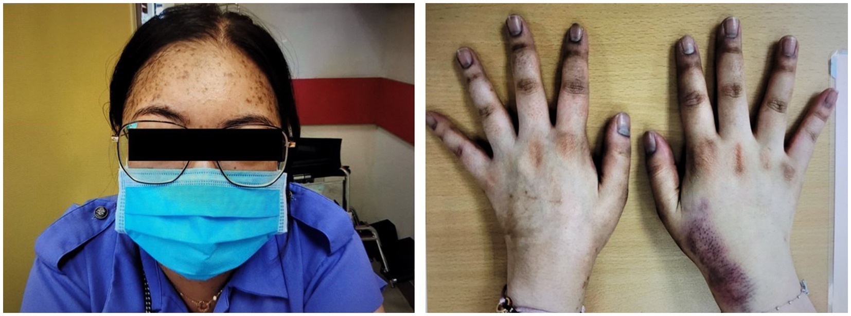
Figure 1. Cushingoid features at presentation include moon face, acne, thin skin, and easy bruising. Hyperpigmentation on the nails and knuckles was also noted.

Diagnostic assessments

Her 9 am and 9 pm cortisol were both >1,700 nmol/L. Her 24-h urine-free cortisol was beyond the upper measurable limit at >1,500 nmol/L. Her serum cortisol was 759 nmol/L after a 1 mg overnight-dexamethasone suppression test, confirming endogenous Cushing’s syndrome. The morning ACTH was 35 pg/mL (upper limit of normal is 46 pg/mL). After excluding a high dose-hook effect, her blood sample was concomitantly sent for ACTH measurement using two different platforms to eliminate possible interference, which might cause a falsely low ACTH reading. ACTH was 19 pg/mL (upper limit of normal is 46 pg/mL) using an IMMULITE 2000 XPI, Siemens Healthineers, Erlangen, Germany, and 17 pg/mL (reference range: 7–63 pg/mL) using a Cobas e-801, Roche Diagnostics, Indianapolis, IN, United States, therefore verifying the ACTH measurement.

In view of this being ACTH-dependent Cushing’s syndrome, a high-dose-dexamethasone suppression test (HDDST) was performed, and her cortisol was not suppressed at 890 nmol/L, with ACTH 42 pg/mL. The serum cortisol day profile showed a mean cortisol level of >1,700 nmol/L (i.e., higher than the upper measurable limit of the assay) and an ACTH of 17 pg/mL. A CRF test using 100 μg of corticorelin showed less than a 50% rise in ACTH and no rise in cortisol levels (Supplementary Table S1). She suffered from multiple complications of hypercortisolism, including thoracic vertebral collapse with back pain, diabetes mellitus (HbA1c 6.7% and fasting glucose 7.6 mmol/L), and hypokalemic hypertension, with a lowest potassium level of 2.3 mmol/L.

The rapid onset of intense hypercortisolism and refractory hypokalemia, as well as the responses in the HDDST and CRF tests raised the suspicion of ectopic ACTH syndrome. Tumor markers were measured. Alpha-fetoprotein (AFP) was markedly raised at 33,357 ng/mL (reference range: <9 ng/mL). Beta-human chorionic gonadotropin (beta-hCG) was not elevated. Carcinoembryonic antigen (CEA) was 4.0 ng/mL (reference range: <3 ng/mL) and CA 19–9 was 57 U/mL (reference range: <37 U/mL). The marked hyperpigmentation in the context of normal ACTH levels pointed to the presence of an underlying tumor producing circulating ACTH precursors. Hence, magnetic resonance imaging (MRI) of the pituitary gland was not performed at this juncture. ACTH precursors were measured using a specialized immunoenzymatic assay (IEMA) employing in-house monoclonal antibodies against the ACTH region and the gamma MSH region. Both monoclonal antibodies have to bind to these regions in POMC and pro-ACTH to create a signal. The patient had a level of 4,855 pmol/L (upper limit of normal is 40 pmol/L) (6). This supported Cushing’s syndrome from an ectopic source secondary to an excess in ACTH precursors.

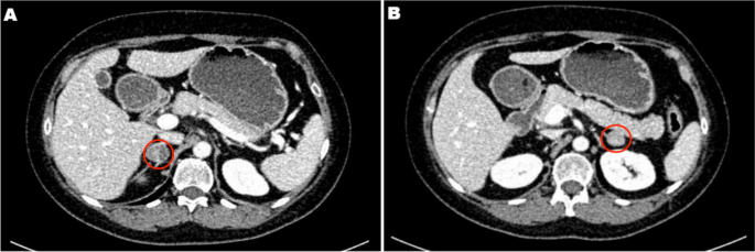
Localization studies were arranged to identify the source of ectopic ACTH precursors. Computed tomography (CT) of the thorax did not show any significant intrathoracic lesion but incidentally revealed a pancreatic mass. Dedicated CT of the abdomen confirmed the presence of a 7.9 × 5.6 cm lobulated mass in the pancreatic body; the adrenal glands were unremarkable. 18-FDG and 68Ga-DOTATATE dual-tracer positron-emission tomography-computed tomography (PET-CT) showed that the pancreatic mass was moderately FDG-avid and non-avid for DOTATATE (Supplementary Figure S1). Multiple FDG-avid nodal metastases were also present, including left supraclavicular fossa lymph nodes.

Fine needle aspiration of the left supraclavicular fossa lymph node yielded tumor cells featuring occasional conspicuous nucleoli, granular coarse chromatin, irregular nuclei, and a high nuclear-to-cytoplasmic ratio. Mitotic figures were infrequent. On immunostaining, the tumor cells were positive for cytokeratin 7 and negative for cytokeratin 20. Focal expression of CDX-2, chromogranin, and synaptophysin was noted. They were negative for TTF-1, GCDPF, Gata 3, Pax-8, CD56, ACTH, inhibin, and S-100 protein. Further immunostaining was performed in view of highly elevated AFP. The tumor cells expressed AFP, Sall4, and MNF-116. They were negative for c-kit, calretinin, Melan A and SF-1. Placental ALP (PLAP) was weak and equivocal. The features were in keeping with a yolk sac tumor.

Therapeutic intervention and outcome

The patient had significant hypokalemic hypertension requiring losartan 100 mg daily, spironolactone 100 mg daily, and a potassium supplement of 129 mmol/day. Co-trimoxazole was given for prophylaxis against Pneumocystis jirovecii pneumonia. Metyrapone was started and up-titrated to 1 gram three times per day. However, in view of persistent hypercortisolism, with urinary free cortisol persistently above the upper measurable limit of the assay, bilateral adrenalectomy was performed. The tumor was mainly in the periadrenal soft tissue, with vascular invasion. The tumor formed cords, nests, and ill-defined lumen (Figure 2). The tumor cells were polygonal and contained pale to eosinophilic cytoplasm and pleomorphic nuclei, some with large nucleoli. Mitosis was present while tumor necrosis was not obvious. The stroma was composed of vascular fibrous tissue, with minimal inflammatory reaction. Immunohistochemical study showed that the tumor was positive for cytokeratin 7, MNF-116, AFP, and glypican-3, and also positive for Sall4 and HNF1β. The tumor cells were negative for cytokeratin 20, PLAP, CD30, negative for neuroendocrine markers including S100 protein, synaptophysin, chromogranin, and also negative for Melan-A, inhibin, and ACTH. Histochemical study for Periodic acid–Schiff–diastase (PAS/D) showed no cytoplasmic zymogen granules like those of acinar cell tumor. The features were compatible with yolk sac tumor. She was put on glucocorticoid and mineralocorticoid replacements post-operatively.

Figure 2
www.frontiersin.org
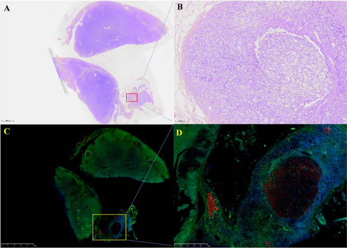
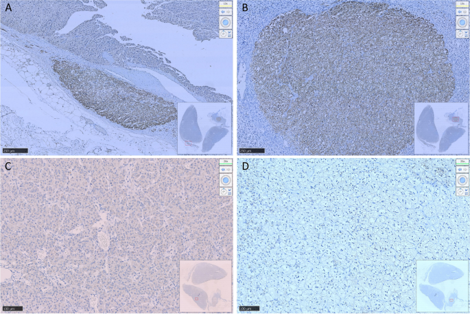
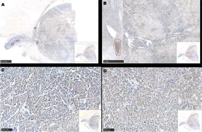
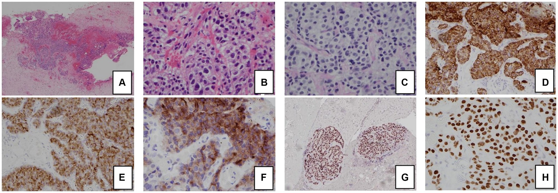
Figure 2. Histology and immunohistochemical staining pattern of tumor specimen. (A) HE stain x 40 showing tumor cells in the soft tissue and peritoneum. (B) HE × 400 showing that the tumor forms cords, nests, and ill-formed lumen in the vascular stroma. The tumor cells are polygonal with pale cytoplasm and pleomorphic nuclei. (C) PAS/D stain showing no cytoplasmic zymogen granules. (D) Tumor is diffusely positive for cytokeratin 7. (E) Tumor is positive for AFP. (F) Tumor is positive for glypican-3. (G) Tumor is diffusely positive for HNF1β. (H) Tumor is diffusely positive for SALL4.

Regarding her oncological management, she received multiple lines of chemotherapy, but the response was poor. Due to limited access to the ACTH precursor assay, serial measurement was unavailable. Treatment response was monitored by repeated imaging and monitoring of AFP. Figure 3 shows a timeline indicating the key events of the disease, showing the trends of the AFP and cortisol levels. Apart from (i) bleomycin, etoposide, and platinum, she was sequentially treated with (ii) etoposide, ifosfamide with cisplatin, and (iii) palliative gemcitabine with oxaliplatin. Next-generation sequencing showed a BRAF V600E mutation, for which (iv) dabrafenib and trametinib were given. Unfortunately, the disease progressed, and the patient succumbed approximately one year after the disease was diagnosed.

Figure 3
www.frontiersin.org
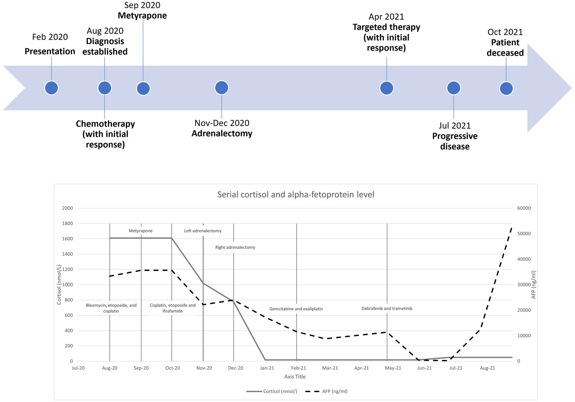
Figure 3. Timeline with serial cortisol and alpha-fetoprotein levels from diagnosis to patient death.

Discussion

This case demonstrates the diagnostic value of ACTH precursor measurement in the diagnosis of ectopic Cushing’s syndrome. ACTH precursors are raised in all ectopic tumors responsible for Cushing’s syndrome and could be useful in distinguishing ectopic from pituitary Cushing’s syndrome (4). Moreover, Cushing’s syndrome due to a yolk sac tumor has been reported only once in a pediatric case, and this is the first adult case reported in the literature (5).

POMC is sequentially cleaved in the anterior pituitary into pro-ACTH and then into ACTH, which is released into the circulation and binds to ACTH receptors in the adrenal cortex, leading to glucocorticoid synthesis (57). Due to incomplete processing, ACTH precursors are found in normal subjects at a concentration of 5–40 pmol/L (6). Pituitary tumors are traditionally well-differentiated and can also relatively efficiently process ACTH precursors. However, this processing is less efficient in ectopic tumors that cause Cushing’s syndrome (8). Some less differentiated pituitary macroadenomas can secrete ACTH precursors into the circulation; however, these tumors are diagnosed by imaging and so do not, in general, cause problems with differential diagnosis (9).

Measurement of ACTH precursors by immunoradiometric assay (IRMA) was first described by Crosby et al. (10). The assay utilized monoclonal antibodies specific for ACTH and the other binding gamma-MSH. The assay only detects peptides expressing both epitopes and therefore measures POMC and pro-ACTH. The assay does not cross-react with other POMC-derived peptides such as beta-lipotropin, ACTH, and N-POMC.

Oliver et al. demonstrated that, compared to the pituitary adenomas in Cushing’s disease, all ectopic tumors responsible for Cushing’s syndrome in their study produce excessive POMC and pro-ACTH (4). The excessive production of ACTH precursors may reflect neoplasm-induced modification and amplification of POMC production. It is suggested that POMC binds to and activates the ACTH receptor because it contains the ACTH amino-acid sequence, or it is cleaved to ACTH in the adrenal glands to cause hypercortisolism (5) (Figure 4). Moreover, cleavage of POMC may produce peptides that exert mitogenic actions on adrenal cells and lead to adrenocortical growth. Outside the adrenal tissue, excessive ACTH precursors in Cushing’s syndrome caused by ectopic tumors can lead to marked hyperpigmentation. Both hypercortisolism and hyperpigmentation were observed in the reported case.

Figure 4
www.frontiersin.org
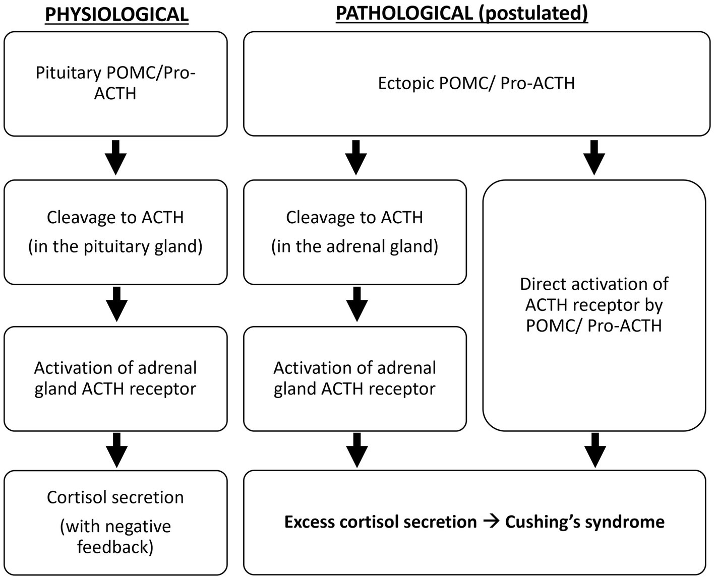
Figure 4. Postulated pathological mechanism of ectopic ACTH precursors.

In patients with ACTH-dependent Cushing’s syndrome, ectopic tumors should be distinguished from pituitary tumors. The HDDST, at a cut-off of 50% cortisol suppression, gives a sensitivity of 81% and a specificity of 67% for pituitary dependent Cushing’s syndrome (11). The CRF test provides 82% sensitivity and 75% specificity for pituitary disease (8). IPSS is the gold standard in distinguishing pituitary from ectopic tumors in Cushing’s syndrome. Utilization of CRF-stimulated IPSS provides 93% sensitivity and 100% specificity for pituitary disease. It also allows correct lateralization in 78% of patients with pituitary tumors. However, it is only available in specialized centers.

In a retrospective cohort, the ACTH precursor level distinguished well between Cushing’s disease and ectopic ACTH syndrome (4). With a cut-off of 100 pmol/L, the test achieved 100% sensitivity and specificity for ectopic ACTH syndrome. More recently, this assay has been used to diagnose patients with occult ectopic ACTH syndrome, with ACTH precursors above 36 pmol/L (8). Unfortunately, the immunoassay for ACTH precursor measurement utilizes in-house monoclonal antibodies, which are not widely available.

Cross-reactivity of POMC in commercially available ACTH assays ranges from 1.6% to 4.7% (12). In cases of ectopic tumors causing Cushing’s syndrome with markedly raised ACTH-precursors and intense hypercortisolism, the cross-reactivity would give significantly high ‘ACTH’ measurements to suggest an ACTH-related pathology. The degree of cross-reactivity, which is variable, should ideally be provided by the assay manufacturer as it affects result interpretation. Lower levels of ACTH precursor production might not be detected, especially by assays with low precursor cross-reactivity. Clinical vigilance is crucial in reaching the correct diagnosis. In patients with marked hypercortisolism and a normal ACTH concentration, like in this case, the measurement of ACTH precursors would allow the accurate diagnosis of Cushing’s syndrome caused by ACTH precursors.

Ectopic tumors causing Cushing’s syndrome are associated with more intense hypercortisolism than Cushing’s disease (11). However, due to variable cross-reactivity, commercial ACTH assays might not accurately detect the excessive ACTH precursors responsible for the clinical syndrome. For this reason, ACTH measurements in these two conditions can significantly overlap and may not differentiate between ectopic and pituitary diseases (4). On the other hand, the more specific POMC assay described in 1996, which does not cross-react with pro-ACTH, has a low sensitivity of 80% for ectopic Cushing’s syndrome and is not now available (13). Hence, the ACTH precursor assay used in this reported case, which detects POMC and pro-ACTH, appears to provide the best diagnostic accuracy from the available literature.

Serial measurement of ACTH precursors may play a role in monitoring the treatment response in an ACTH precursor secreting tumor. In the case of ectopic ACTH secretion, the corticotropic axis is slowed down and ACTH is almost exclusively of paraneoplastic origin. Immunotherapy is known to alter the functioning of the hypothalamic–pituitary corticotropic axis; however, its effect on ectopic secretions is not known. More data is required before the role of ACTH precursor measurement for disease monitoring in these scenarios can be ascertained.

The incidence of endogenous Cushing’s syndrome is reported to be 2 to 4 per million people per year (14). Ectopic sources of Cushing’s syndrome are responsible for 9 to 18% of these cases. Typical sources of these ectopic tumors include bronchial carcinoid tumors, small-cell lung cancer, and gut neuroendocrine tumors. Notably, germ cell tumors, including teratomas, ovarian epithelial tumors, and ovarian endometrial tumors, are also possible ectopic sources of Cushing’s syndrome.

The histological diagnosis of germ cell tumor in a non-genital site is challenging, especially for the poorly differentiated, or with somatic differentiation. Immunostaining, chromosomal, or genetic study are very important in confirming the diagnosis. AFP elevation in our case limited the differential diagnoses to germ cell tumors/yolk sac tumors, hepatocellular carcinoma, and rare pancreatic tumors. The specimen was biopsied from the retroperitoneum, and the morphology was a dominant trabecular pattern or a hepatoid pattern. It showed diffuse positive immunostaining for cytokeratin, AFP, and glypican-3. It was also diffusely and strongly positive for HNF1β and SALL4, supporting the diagnosis of yolk sac tumor. Both HNF1β and SALL4, being related with the expression of genes associated with stem cells or progenitor cells, are used as sensitive and specific markers for germ cell tumors/yolk sac tumors (1516).

Staining related to pancreatic acinar cell carcinoma and neuroendocrine tumor were performed. PAS/D staining showed a lack of zymogen granules. A lack of nuclear β-catenin positivity was shown. Staining for neuroendocrine markers, including chromogranin and synaptophysin, was negative. Bcl-10 and trypsin were not available in the local setting.

Cushing’s syndrome due to a yolk sac tumor was reported only once, in a 2-year-old child (5). The abdominal yolk sac tumor was resistant to cisplatin, with rapid disease progression, and the patient succumbed 1.5 years after initial presentation. Yolk sac tumor in the pancreas is also rare, with only 4 cases reported so far. The first case was reported in a 57-year-old woman with an incidentally detected abdominal mass (17). The tumor stained positive for AFP, PLAP, and CEA. The second case was a 70-year-old asymptomatic woman with histology showing a group of tumor cells with features of a yolk sac tumor, and another group showing features of pancreatic ductal adenocarcinoma with mucin production, suggesting a yolk sac tumor derived from pancreatic ductal adenocarcinoma (18). The tumor showed partial positivity for AFP, Sall4, glypican-3, and cytokeratin 7, as found in our case, while MNF-116 and PLAP staining results were not described. The third was in a 33-year-old man with a solitary pancreatic head mass with obstructive jaundice (19). The patient had undergone Whipple’s procedure followed by cisplatin-based chemotherapy, resulting in at least 5 years of disease remission. The latest reported case was in a 32-year-old man presenting with abdominal pain (20). Notably, initial imaging showed diffuse enlargement of the pancreas and increased FDG uptake without a distinct mass. Reassessment imaging 11 months later showed a 13 cm pancreatic mass. The initial imaging findings suggested initial intraductal growth of the tumor, as reported in some subtypes of pancreatic carcinoma. None of the reported cases of adult pancreatic yolk sac tumors were associated with abnormal hormone secretion. We reported the first adult case of pancreatic yolk sac tumor with ectopic ACTH syndrome. The case represents an overlap of two rarities. It demonstrates that pancreatic yolk sac tumor is a possible cause of ectopic ACTH syndrome.

Conclusion

ACTH precursor measurement helps to distinguish ectopic ACTH syndrome from Cushing’s disease. The test has superior diagnostic performance and is less invasive than IPSS. Nonetheless, the limited availability of the assay may restrict its broader use in patient management. We describe the first adult case of pancreatic yolk sac tumor with ACTH precursor secretion resulting in Cushing’s syndrome. This adds to the list of origins of ectopic ACTH syndrome in adults.

Data availability statement

The original contributions presented in the study are included in the article/Supplementary material, further inquiries can be directed to the corresponding author.

Ethics statement

Written informed consent was obtained from the individual to publish any potentially identifiable images or data in this article.

Author contributions

JC wrote the manuscript. JC, CW, WC, AW, KW, and PT researched the data. WC, AL, EL, YW, KT, KL, and CL critically reviewed and edited the manuscript. DL initiated and conceptualized this case report and is the guarantor of this work. All authors contributed to the article and approved the submitted version.

Funding

The author(s) declare that no financial support was received for the research, authorship, and/or publication of this article.

Conflict of interest

The authors declare that the research was conducted in the absence of any commercial or financial relationships that could be construed as a potential conflict of interest.

Publisher’s note

All claims expressed in this article are solely those of the authors and do not necessarily represent those of their affiliated organizations, or those of the publisher, the editors and the reviewers. Any product that may be evaluated in this article, or claim that may be made by its manufacturer, is not guaranteed or endorsed by the publisher.

Supplementary material

The Supplementary material for this article can be found online at: https://www.frontiersin.org/articles/10.3389/fmed.2023.1246796/full#supplementary-material

References

1. Stewar, PM, Gibson, S, Crosby, SR, Pennt, R, Holder, R, Ferry, D, et al. ACTH precursors characterize the ectopic ACTH syndrome. Clin Endocrinol. (1994) 40:199–204. doi: 10.1111/j.1365-2265.1994.tb02468.x

PubMed Abstract | CrossRef Full Text | Google Scholar

2. Young, J, Haissaguerre, M, Viera-Pinto, O, Chabre, O, Baudin, E, and Tabarin, A. MANAGEMENT OF ENDOCRINE DISEASE: Cushing’s syndrome due to ectopic ACTH secretion: an expert operational opinion. Eur J Endocrinol. (2020) 182:R29–58. doi: 10.1530/EJE-19-0877

PubMed Abstract | CrossRef Full Text | Google Scholar

3. Fleseriu, M, Auchus, R, Bancos, I, Ben-Shlomo, A, Bertherat, J, Biermasz, NR, et al. Consensus on diagnosis and management of Cushing’s disease: a guideline update. Lancet Diabetes Endocrinol. (2021) 9:847–75. doi: 10.1016/S2213-8587(21)00235-7

PubMed Abstract | CrossRef Full Text | Google Scholar

4. Oliver, RL, Davis, JR, and White, A. Characterisation of ACTH related peptides in ectopic Cushing’s syndrome. Pituitary. (2003) 6:119–26. doi: 10.1023/B:PITU.0000011172.26649.df

PubMed Abstract | CrossRef Full Text | Google Scholar

5. Gevers, EF, Meredith, S, Shah, P, Torpiano, J, Peters, C, Sebire, NJ, et al. Cushing syndrome in a child due to pro-opiomelanocortin (POMC) secretion from a yolk sac tumor. Eur J Endocrinol. (2017) 176:K1–7. doi: 10.1530/EJE-16-0776

PubMed Abstract | CrossRef Full Text | Google Scholar

6. Gibson, S, Crosby, SR, Stewart, MF, Jennings, AM, McCall, E, and White, A. Differential release of proopiomelanocortin-derived peptides from the human pituitary: evidence from a panel of two-site immunoradiometric assays. J Clin Endocrinol Metab. (1994) 78:835–41.

PubMed Abstract | Google Scholar

7. Harno, E, Gali Ramamoorthy, T, Coll, AP, and White, A. POMC: the physiological power of hormone processing. Physiol Rev. (2018) 98:2381–430. doi: 10.1152/physrev.00024.2017

CrossRef Full Text | Google Scholar

8. Page-Wilson, G, Freda, PU, Jacobs, TP, Khandji, AG, Bruce, JN, Foo, ST, et al. Clinical utility of plasma POMC and AgRP measurements in the differential diagnosis of ACTH-dependent Cushing’s syndrome. J Clin Endocrinol Metab. (2014) 99:E1838–45. doi: 10.1210/jc.2014-1448

PubMed Abstract | CrossRef Full Text | Google Scholar

9. Gibson, S, Ray, DW, Crosby, SR, Dornan, TL, Jennings, AM, Bevan, JS, et al. Impaired processing of proopiomelanocortin in corticotroph macroadenomas. J Clin Endocrinol Metab. (1996) 81:497–502.

PubMed Abstract | Google Scholar

10. Crosby, SR, Stewart, MF, Ratcliffe, JG, and White, A. Direct measurement of the precursors of adrenocorticotropin in human plasma by two-site immunoradiometric assay. J Clin Endocrinol Metab. (1988) 67:1272–7. doi: 10.1210/jcem-67-6-1272

PubMed Abstract | CrossRef Full Text | Google Scholar

11. Aron, DC, Raff, H, and Findling, JW. Effectiveness versus efficacy: the limited value in clinical practice of high dose dexamethasone suppression testing in the differential diagnosis of adrenocorticotropin-dependent Cushing’s syndrome. J Clin Endocrinol Metab. (1997) 82:1780–5. doi: 10.1210/jc.82.6.1780

PubMed Abstract | CrossRef Full Text | Google Scholar

12. Monaghan, PJ, Kyriacou, A, Sturgeon, C, Davies, A, Trainer, PJ, White, A, et al. Proopiomelanocortin interference in the measurement of adrenocorticotrophic hormone: a United Kingdom National External Quality Assessment Service study. Clin Endocrinol. (2016) 85:569–74. doi: 10.1111/cen.13118

CrossRef Full Text | Google Scholar

13. Raffin-Sanson, ML, Massias, JF, Dumont, C, Raux-Demay, MC, Proeschel, MF, Luton, JP, et al. High plasma proopiomelanocortin in aggressive adrenocorticotropin-secreting tumors. J Clin Endocrinol Metab. (1996) 81:4272–7.

PubMed Abstract | Google Scholar

14. Beuschlein, F, and Hammer, GD. Ectopic pro-opiomelanocortin syndrome. Endocrinol Metab Clin N Am. (2002) 31:191–234. doi: 10.1016/S0889-8529(01)00025-1

CrossRef Full Text | Google Scholar

15. Yu, DD, Guo, SW, Jing, YY, Dong, YL, and Wei, LX. A review on hepatocyte nuclear factor-1beta and tumor. Cell Biosci. (2015) 5:58. doi: 10.1186/s13578-015-0049-3

PubMed Abstract | CrossRef Full Text | Google Scholar

16. Miettinen, M, Wang, Z, McCue, PA, Sarlomo-Rikala, M, Rys, J, Biernat, W, et al. SALL4 expression in germ cell and non-germ cell tumors: a systematic immunohistochemical study of 3215 cases. Am J Surg Pathol. (2014) 38:410–20. doi: 10.1097/PAS.0000000000000116

PubMed Abstract | CrossRef Full Text | Google Scholar

17. Zhang, B, Gao, S, Chen, Y, and Wu, Y. Primary yolk sac tumor arising in the pancreas with hepatic metastasis: a case report. Korean J Radiol. (2010) 11:472–5. doi: 10.3348/kjr.2010.11.4.472

PubMed Abstract | CrossRef Full Text | Google Scholar

18. Yonemaru, J, Takahashi, M, Nara, S, Ichikawa, H, Ishigamori, R, Imai, T, et al. A yolk sac tumor of the pancreas and derived xenograft model effectively responded to VIP chemotherapy. Pancreatology. (2020) 20:551–7. doi: 10.1016/j.pan.2019.12.021

PubMed Abstract | CrossRef Full Text | Google Scholar

19. Galanis, I, Floros, G, Simou, M, Kyriakopoulos, G, and Stylianidis, G. An extremely rare case of a primary pancreatic yolk sac tumor. Cureus. (2022) 14:e26007. doi: 10.7759/cureus.26007

PubMed Abstract | CrossRef Full Text | Google Scholar

20. Sui, H, Zhu, Z, Li, Z, and Luo, Y. Primary pancreatic yolk sac tumor presenting as diffusely enlarged pancreas in initial 18F-FDG PET/CT. Clin Nucl Med. (2020) 45:483–6. doi: 10.1097/RLU.0000000000003038

PubMed Abstract | CrossRef Full Text | Google Scholar

Keywords: Cushing’s syndrome, ectopic ACTH syndrome, yolk sac tumor, pancreatic tumor, ACTH precursor

Citation: Chang JYC, Woo CSL, Chow WS, White A, Wong KC, Tsui P, Lee ACH, Leung EKH, Woo YC, Tan KCB, Lam KSL, Lee CH and Lui DTW (2023) Cushing’s syndrome caused by ACTH precursors secreted from a pancreatic yolk sac tumor in an adult—a case report and literature review. Front. Med. 10:1246796. doi: 10.3389/fmed.2023.1246796

Received: 18 July 2023; Accepted: 20 November 2023;
Published: 05 December 2023.

Edited by:

Alessandro Vanoli, University of Pavia, Italy

Reviewed by:

Petar Brlek, St. Catherine Specialty Hospital, Croatia
Wafa Alaya, Hospital University Tahar Sfar, Tunisia

Copyright © 2023 Chang, Woo, Chow, White, Wong, Tsui, Lee, Leung, Woo, Tan, Lam, Lee and Lui. This is an open-access article distributed under the terms of the Creative Commons Attribution License (CC BY). The use, distribution or reproduction in other forums is permitted, provided the original author(s) and the copyright owner(s) are credited and that the original publication in this journal is cited, in accordance with accepted academic practice. No use, distribution or reproduction is permitted which does not comply with these terms.

*Correspondence: David Tak Wai Lui, dtwlui@hku.hk

Disclaimer: All claims expressed in this article are solely those of the authors and do not necessarily represent those of their affiliated organizations, or those of the publisher, the editors and the reviewers. Any product that may be evaluated in this article or claim that may be made by its manufacturer is not guaranteed or endorsed by the publisher.

From https://www.frontiersin.org/articles/10.3389/fmed.2023.1246796/full

FDA Grants Breakthrough Therapy Designation for Oral Congenital Adrenal Hyperplasia Drug

Key takeaways:

  • Crinecerfont was granted FDA breakthrough therapy designation for the treatment of congenital adrenal hyperplasia.
  • The medication met primary and secondary endpoints in a pair of phase 3 trials.

The FDA granted breakthrough therapy designation for an oral non-glucocorticoid medication for the treatment of congenital adrenal hyperplasia, according to an industry press release.

Crinecerfont (Neurocrine Biosciences) is a selective corticotropin-releasing factor type 1 receptor antagonist under development to lower excess adrenal androgens for people with congenital adrenal hyperplasia due to 21-hyroxylase deficiency.

The medication met its primary and secondary endpoints in two phase 3 CAHtalyst trials, one assessing use of crinecerfont by children and the other by adults. In the pediatric trial, children and adolescents receiving crinecerfont had a decrease in serum androstenedione from baseline to 4 weeks. Participants receiving the medication also had a greater reduction in daily glucocorticoid at 28 weeks than placebo. As Healio previously reported, in the adult trial, crinecerfont was associated with a greater reduction in daily glucocorticoid while maintaining androgen control compared with placebo. The most common adverse events in the pediatric study were headache, fever, vomiting, upper respiratory tract infection and nasopharyngitis. Among adults, the most common adverse events were fatigue, headache and COVID-19 infection. No serious adverse events related to crinecerfont were reported.

Breakthrough therapy is the latest designation granted to crinecerfont by the FDA. The medication was previously granted fast track and rare pediatric disease designations.

“We are very pleased that the FDA granted breakthrough therapy designation for crinecerfont, thus recognizing both the seriousness of congenital adrenal hyperplasia and the significant unmet need currently faced by patients and families living with this condition,” Eiry W. Roberts, MD, Chief Medical Officer for Neurocrine Biosciences, said in a press release. “The outstanding safety and efficacy results from the phase 3 CAHtalyst studies in pediatric and adult patients suggest that crinecerfont has the potential to represent a substantial improvement over current standard of care in congenital adrenal hyperplasia by controlling androgen levels and allowing for reduced steroid doses. We remain on track to submit the new drug application in 2024.”

From https://www.healio.com/news/endocrinology/20231206/fda-grants-breakthrough-therapy-designation-for-oral-congenital-adrenal-hyperplasia-drug?utm_source=selligent&utm_medium=email&utm_campaign=news&fbclid=IwAR2WXDd3ajhKG0s2h0XD9ZQAstUkSotJYl1KLicH3gmxEPF6hvg6sZu2dCU

Evaluation of Psoriasis Patients With Long-Term Topical Corticosteroids for Their Risk of Developing Adrenal Insufficiency, Cushing’s Syndrome and Osteoporosis

In this study, we will investigate the possible side effects of psoriasis patients using long-term topical corticosteroids (TCS) such as adrenal insufficiency, Cushing’s Syndrome (CS) and osteoporosis and determine how these side effects develop.

Forty-nine patients were included in the study. The patients were divided into two groups based on the potency of the topical steroid they took and the patients’ ACTH, cortisol and bone densitometer values were evaluated.

There was no significant difference between the two groups regarding the development of surrenal insufficiency, CS and osteoporosis. One patient in group 1 and 4 patients in group 2 were evaluated as iatrogenic CS. ACTH stimulation tests of these patients in group 2 showed consistent results with adrenal insufficiency, while no adrenal insufficiency was detected in the patient in Group 1. Patients who used more than 50g of superpotent topical steroids per week compared to patients who used 50g of superpotent topical steroids per week. It was identified that patients who used more than 50g of superpotent topical steroids had significantly lower cortisol levels, with a negatively significant correlation between cortisol level and the amount of topical steroid use ( < .01).Osteoporosis was detected in 3 patients in group 1 and 8 patients in Group 2. Because of the low number of patients between two groups, statistical analysis could not be performed to determine the risk factors.

Our study is the first study that we know of that investigated these three side effects. We have shown that the development of CS, adrenal insufficiency and osteoporosis in patients who use topical steroids for a long time depends on the weekly TCS dosage and the risk increases when it exceeds the threshold of 50 grams per week. therefore, our recommendation would be to avoid long-term use of superpotent steroids and to choose from the medium-potent group if it is to be used.

ABOUT THE CONTRIBUTORS

Betul Erdem

Department of Dermatology, Van Training and Research Hospital, Van, Turkey.

Muzeyyen Gonul

Department of Dermatology, Ministry of Health, Ankara Etlik City Hospital, Ankara, Turkey.

Ilknur Ozturk Unsal

Department of Endocrine and Metabolic Disease, Ministry of Health, Ankara Etlik City Hospital, Ankara, Turkey.

Seyda Ozdemir Sahingoz

Bilateral Co-Secretory Lesions Presenting with Coexisting Cushing Syndrome and Primary Aldosteronism


Abstract

Background

There is an increasing number of cases of aldosterone- and cortisol-producing adenomas (A/CPAs) reported in the context of primary aldosteronism (PA). Most of these patients have PA complicated with subclinical Cushing’s syndrome; cases of apparent Cushing’s syndrome (CS) complicated with aldosteronism are less reported. However, Co-secretory tumors were present in the right adrenal gland, a cortisol-secreting adenoma and an aldosterone-producing nodule (APN) were present in the left adrenal gland, and aldosterone-producing micronodules (APMs) were present in both adrenal glands, which has not been reported. Here, we report such a case, offering profound insight into the diversity of clinical and pathological features of this disease.

Case presentation

The case was a 45-year-old female from the adrenal disease diagnosis and treatment centre in West China Hospital of Sichuan University. The patient presented with hypertension, moon-shaped face, central obesity, fat accumulation on the back of the neck, disappearance of cortisol circadian rhythm, ACTH < 5 ng/L, failed elevated cortisol inhibition by dexamethasone, orthostatic aldosterone/renin activity > 30 (ng/dL)/(ng/mL/h), and plasma aldosterone concentration > 10 ng/dL after saline infusion testing. Based on the above, she was diagnosed with non-ACTH-dependent CS complicated with PA. Adrenal vein sampling showed no lateralization for cortisol and aldosterone secretion in the bilateral adrenal glands. The left adrenocortical adenoma was removed by robot-assisted laparoscopic resection. However, hypertension, fatigue and weight gain were not alleviated after surgery; additionally, purple striae appeared in the lower abdomen, groin area and inner thigh, accompanied by systemic joint pain. One month later, the right adrenocortical adenoma was also removed. CYP11B1 were expressed in the bilateral adrenocortical adenomas, and CYP11B2 was also expressed in the right adrenocortical adenomas. APN existed in the left adrenal gland and APMs in the adrenal cortex adjacent to bilateral adrenocortical adenomas. After another surgery, her serum cortisol and plasma aldosterone returned to normal ranges, except for slightly higher ACTH.

Conclusions

This case suggests that it is necessary to assess the presence of PA, even in CS with apparent symptoms. As patients with CS and PA may have more complicated adrenal lesions, more data are required for diagnosis.

Peer Review reports

Background

Because both adrenal Cushing’s syndrome and primary aldosteronism (PA) can manifest as adrenocortical adenomas, it is difficult to distinguish between them on the sole basis of adrenal computed tomography (CT). There may also be multiple adenomas with different functions in the same adrenal gland [1], which also leads to the difficulty in the interpretation of adrenal vein blood collection results. With the increased reports on cases of PA complicated with subclinical Cushing’s syndrome in clinical practice, increasing attention is being given to the screening of PA complicated with subclinical Cushing’s syndrome. However, PA screening may be ignored in the diagnosis and treatment of adrenal Cushing’s syndrome.

Although it has been reported that PA with a diameter > 2 cm may be complicated with aldosterone- and cortisol-producing adenomas (A/CPAs) [2], cases of apparent Cushing’s syndrome complicated with PA are less well known.

Recently, Y. Fushimi et al. [3] reported a case of apparent Cushing’s syndrome complicated with PA. The cortisol-producing enzyme cytochrome P450 (CYP) 11B1 was diffusely expressed in the adenoma, but based on staining, the aldosterone synthase CYP11B2 was significantly expressed in the adjacent adrenal cortex. This finding indicated that aldosterone-producing micronodules (APMs) in the adjacent adrenal cortex may be the pathological basis of PA.

Here, a case of bilateral co-secretory lesions presenting with coexisting Cushing syndrome and primary aldosteronism detected by AVS and confirmed by immunohistochemical analysis after surgical resection is reported. Moreover, APMs were found in the adrenal cortex adjacent to bilateral adrenocortical adenomas; an aldosterone-producing nodule was detected adjacent to the unilateral adenoma.

Case presentation

A 45-year-old female patient was admitted to the adrenal disease diagnosis and treatment centre in West China Hospital of Sichuan University due to “increased blood pressure, weight gain for one year and facial oedema for half a year”. After nifedipine controlled-release tablets 30 mg daily and terazosin 2 mg daily were applied, the blood pressure of this patient was still as high as 179/113 mmHg. She had no family history of endocrine disease or malignant tumour. Her body mass index (BMI) was 25.6 kg/m2 at admission, with a moon-shaped face, fat accumulation on the back of the neck and thin skin. Hormonal, glucose, renal function, lipid, and blood electrolyte tests were completed, and the physiological rhythm of cortisol had disappeared. Aldosterone-renin-angiotensin system (RAAS) results showed a significant decrease in renin activity and a significantly higher aldosterone/renin ratio (ARR) (as provided in Table 1). Dynamic testing for hormones was conducted, and the results were as follows: (i) in terms of the saline infusion test (SIT) in supine position, the before and after aldosterone level was 17.03 ng/dL and 15.45 ng/dL, respectively; (ii) in terms of the captopril challenge test (CCT), the before and after aldosterone level was 18.49 ng/dl and 15.25 ng/mL, respectively, with an inhibition rate of 17.52%; (iii) in terms of the standard low-dose dexamethasone suppression test, the before and after serum cortisol level was 467.9 nmol/L and 786.3 nmol/L, respectively; the before and after 24-h urine free cortisol (24-h UFC) level was 332.3 µg/24 and 480.4 µg/24, respectively. An enhanced CT scan revealed adenoma lesions in both adrenal glands (Fig. 1a and b). Bone mineral density measurement with dual-energy X-ray absorptiometry indicated osteoporosis. Chest CT showed old fractures of the 9th rib on the left side and the 2nd rib on the right side.

Table 1 Peripheral blood laboratory data for this case
Fig. 1


figure 1

Adrenal CT of the patient: A nodule with a size of approximately 1.6 × 1.5 cm was found in the left adrenal gland, and a nodule with a size of approximately 2.2 × 1.8 cm was found in the right adrenal gland. Irregular mild to moderate enhancement was on enhanced CT, and the surrounding fat gap was clear

Based on the above clinical features, the patient was diagnosed with “non-ACTH-dependent Cushing’s syndrome complicated with PA”. To assess lateralization, adrenal vein sampling (AVS) stimulated by ACTH was performed after obtaining informed consent. The results showed no lateralization of cortisol and aldosterone secretion (Table 2).

Table 2 Results of AVS

After communicating with the patient, the left adrenocortical adenoma was first removed by robot-assisted laparoscopic resection; the thickened adrenal cortex near the left adrenocortical adenoma was also resected during the surgery. The pathological report revealed adrenocortical adenoma, the Weiss score was 1, and immunohistochemistry showed weak CYP11B1 expression in the adenoma and positive CYP11B2 expression in an adjacent nodule. Hypertension was not alleviated after surgery. One month later, purple lines appeared on both sides of the lower abdomen, groin area and inner thigh, accompanied by weight gain, apparent systemic joint pain and fatigue in both lower limbs. The patient was readmitted to the hospital, and examination revealed orthostatic ALD at 11.99 ng/dL, PRA at 0.08 ng/mL/h, angiotensin II at 39.38 ng/L (reference range: 55.3–115.3 ng/L) and ARR at 149.88 (ng/dL)/(ng/mL/h). In addition, ACTH was 2.37 ng/L, serum cortisol was 352.30–353.50–283.90 nmol/L at 8 h-16 h-24 h, 24-h UFC was 112.8 µg, and serum cortisol was 342.10 nmol/L in the morning after the 1 mg dexamethasone suppression test. Enhanced CT of the kidneys and adrenal glands showed no solid nodules or masses in the left adrenal gland, though a nodule with a size of approximately 2.2*1.8 cm was detected in the right adrenal gland. Enhanced CT showed irregular mild to moderate enhancement. Therefore, the diagnosis was still “non-ACTH-dependent Cushing’s syndrome complicated with PA”. Subsequently, the right adrenocortical adenoma and the thickened adrenal cortex near the right adrenocortical adenoma were removed by robot-assisted laparoscopic resection. The pathological report indicated adrenocortical adenoma, and immunohistochemistry showed diffuse homogeneous expression of CYP11B1 and CYP11B2. Antibodies against CYP11B1 (MABS502) and CYP11B1 (MABS1251) were purchased from the Millipore Corporation. There were APMs in the adrenal cortex adjacent to the bilateral cortical adenomas. The fluorescence staining image of the left cortical adenoma is shown in Fig. 2. The immunohistochemistry image of the left adrenal gland is given in Fig. 3 and that of the right adrenal gland in Fig. 4. The immunofluorescence method used in this study was indirect immunofluorescence double staining procedure. Paraffin-embedded human adrenal tissues were prepared using heat-induced epitope retrieval after deparaffinization. Tissue sections were blocked with 5% goat serum in PBS, pH 7.4, containing 0.5% SDS, for 1 h. The slides were incubated with individual primary antibodies at 4℃ overnight, followed by incubation with Alexa Fluor 488-, and Alexa Fluor 647-conjugated secondary antibodies specific to the species of the primary antibodies with DAPI for immunofluorescence staining. Antibodies used included anti-CYP11B1 (Millipore, Cat. No. MABS502, 1:100), anti-CYP11B2(Millipore, Cat. No. MABS1251, 1:100), Alexa Fluor 488-conjugated anti-rat IgG secondary antibody (CYP11B1; Green) and Alexa Fluor 647-conjugated anti-mouse IgG secondary antibody (CYP11B2; Red). Nuclei were stained with DAPI.

Fig. 2

figure 2

Routine hematoxylin and eosin (H&E) staining and immunofluorescence of the left adrenocortical adenoma (green represents expression of CYP11B1 and red that of CYP11B2). This adrenocortical adenoma and the surrounding cortex was cut into three parts. A and C show the overall appearance of the resected portion, with a nodule adjacent to the adenoma. B shows a neoplastic lesion formed by clear cells (aldosterone-producing cell) within nodules, lacking a fibrous envelope. C clearly shows the weak and diffuse expression of CYP11B1 in adrenocortical adenoma and CYP11B2 expression in a nodule in the cortex adjacent to the adenoma. D shows local enlargement of the aldosterone-producing nodule and three aldosterone-producing micronodules adjacent to it

Fig. 3

figure 3

Resected adrenocortical adenoma and part of the adrenal cortex on the left side. A shows expression of Aldosterone-producing micronodule CYP11B2 in the cortex adjacent to the adenoma. B shows an aldosterone-producing nodule with a diameter of approximately 2 mm. C shows weak positive expression of CYP11B1 in the adenoma and D negative expression of CYP11B1 in the aldosterone-producing nodule

Fig. 4

figure 4

Resected adrenocortical adenoma and part of the adrenal cortex on the right side. A and B show several Aldosterone-producing micronodules (positive expression of CYP11B2) in the cortex adjacent to the adenoma. C shows diffuse expression of CYP11B1 in the adenoma. D shows diffuse expression of CYP11B2 in the adenoma

The Cushing’s syndrome in this patient disappeared after surgery, and glucocorticoids were discontinued after 15 months according to medical advice. Follow-up was conducted for half a year after drug discontinuance, and the patient had no fatigue or dizziness; she was satisfied with the outcomes. Her systolic and diastolic blood pressure remained at 100–120 mmHg and 70–80 mmHg, respectively. During the most recent re-examination, the following results were obtained: (1) orthostatic ALD of 19.1 ng/dL and orthostatic renin concentration of 12.59 µIU/mL, with an aldosterone/renin ratio (ARR) of 1.52; (2) PTC at 8 AM of 247 nmol/L, ACTH of 93.55 ng/L and 24-h UFC of 26.8 µg; (3) parathyroid hormone of 3.86 pmol/L; (4) 25-OH-VitD of 119.5 nmol/L; (5) serum creatinine of 60 µmol/L; (6) serum sodium of 140.4 nmol/L, serum potassium of 3.87 mmol/L and serum calcium of 2.27 mmol/L.

Discussion and conclusions

Adrenal Cushing’s syndrome is caused by excessive autonomic secretion of cortisol induced by adrenal cortical tumours or adrenal cortical hyperplasia; primary aldosteronism (PA) is caused by excessive autonomic secretion of aldosterone induced by adrenal cortical tumours or adrenal cortical hyperplasia. More adverse symptoms occur if aldosterone and cortisol-producing adenomas are present. Specifically, (1) it is more difficult to control hypertension; (2) the incidence of major adverse cardiovascular and cerebrovascular events would increase [4]; (3) glucose intolerance and other metabolic complications would be aggravated [56]; (4) patients would be prone towards osteoporosis [78]; (5) adrenal vein sampling results may be misinterpreted [9]; and (6) adrenal insufficiency may occur after surgery. Therefore, it is of great clinical significance to avoid missed diagnosis of A/CPAs.

Despite many reports on A/CPAs, the majority of these patients may have subclinical Cushing’s syndrome (SCS), and cases of apparent Cushing’s syndrome complicated with PA are rarely reported. In the present case, the clinical manifestation of Cushing’s syndrome were more apparent, and it would be appropriate to call it cortisol-aldosterone cosecretoma. Naoyoshi Onoda et al. [10] reported a case of Cushing’s syndrome caused by a left adrenocortical adenoma (30 mm in diameter) and PA caused by a right adrenocortical adenoma (20 mm in diameter), and Fushimi et al. [3] reported a case of right A/CPA (25 mm*22 mm in size). Interestingly, in the present report, the patient had bilateral A/CPAs, and the clinical manifestations of Cushing’s syndrome became more apparent after unilateral resection was performed. Similar to the above two cases, APMs were found in the adrenal cortex adjacent to the A/CPAs, but aldosterone-producing nodules were found near the cortisol-producing adenoma on the left side.

The biochemical phenotype of APM-inducing autonomic aldosterone secretion has not been clarified. APMs can also be found in the adrenal tissue of 30% of individuals with normal blood pressure [11] and surrounding areas of APA [1213]. APMs do not express CYP11B1 or CYP17A1, which are necessary for the generation of cortisol [1214]. In our patient, the aldosterone-producing nodule in the left adrenal gland may have developed from APM. More than one-third of APMs carry known mutations in CACNA1D and ATP1A1, promoting the generation of aldosterone [1415]. Unfortunately, we did not perform whole-exome sequencing on the DNA of the peripheral blood and adenoma tissues of this patient. Due to the existence of APMs adjacent to the adenoma, it remains unclear whether there is a risk of the relapse of PA in these cases after resection of adrenal the adenoma. Therefore, it was necessary to conduct medical follow-up for this patient.

Remi Goupil et al. performed AVS on 8 patients with cortisol-producing adenoma (CPA), and the results showed that cortisol on the CPA side was higher than that on the contralateral side (median, 6.7 times [range: 2.4–27.2]); P = 0.012]) [16]. There was no significant difference in bilateral cortisol and aldosterone concentrations after AVS in this patient, which is consistent with bilateral A/CPA. Although immunohistochemical results revealed weak expression of CYP11B1 for the first time, expression of cortisol in bilateral adrenal venous blood samples increased significantly after ACTH stimulation. Hence, cortisol was over-synthesized on both sides, and bilateral A/CPAs was definitively diagnosed.

In summary, this case highlights the need for A/CPA screening. The complicated pathological features of these cases impose challenges to our understanding of this disease. Due to the presence of APMs in the adrenal cortex near bilateral adrenocortical adenomas, more clinical data are required to identify whether the disease might relapse after simple resection of the adenoma in these patients. Therefore, further medical follow-up of these patient is needed.

Availability of data and materials

Not applicable.

Abbreviations

CS:
Cushing’s syndrome
PA:
Primary aldosteronism
ACTH:
Adrenocorticotropic hormone
UFC:
Urinary free cortisol
AVS:
Adrenal vein sampling
A/CPA:
Aldosterone-and cortisol producing adenoma
APN:
Aldosterone-producing nodules
APM:
Aldosterone-producing micronodule
CYP:
Cytochrome P450
CT:
Computed tomography
PAC:
Plasma aldosterone concentration
PRA:
Plasma renin activity
ARR:
Aldosterone /renin ratio

References

  1. Stenman A, Shabo I, Ramström A, Zedenius J, Juhlin CC: Synchronous aldosterone- and cortisol-producing adrenocortical adenomas diagnosed using CYP11B immunohistochemistry. SAGE open medical case reports. 2019, 7:2050313×19883770.

  2. Hiraishi K, Yoshimoto T, Tsuchiya K, Minami I, Doi M, Izumiyama H, Sasano H, Hirata YJ. Clinicopathological features of primary aldosteronism associated with subclinical Cushing’s syndrome. Endocr J. 2011;58(7):543–51.

    Article CAS PubMed Google Scholar

  3. Fushimi Y, Tatsumi F, Sanada J, Shimoda M, Kamei S, Nakanishi S, Kaku K, Mune T, Kaneto H. Concurrence of overt Cushing’s syndrome and primary aldosteronism accompanied by aldosterone-producing cell cluster in adjacent adrenal cortex: case report. BMC Endocr Disord. 2021;21(1):163.

    Article PubMed PubMed Central Google Scholar

  4. Araujo-Castro M, BengoaRojano N, FernándezArgüeso M, Pascual-Corrales E, Jiménez Mendiguchía L. García Cano AMCardiometabolic risk in patients with primary aldosteronism and autonomous cortisol secretion. Case-control study. Med Clin (Barc). 2021;157(10):473–9.

    Article CAS PubMed Google Scholar

  5. Petramala L, Olmati F, Concistrè A, Russo R, Mezzadri M, Soldini M, De Vincentis G, Iannucci G, De Toma G, Letizia C. Cardiovascular and metabolic risk factors in patients with subclinical Cushing. Endocrine. 2020;70(1):150–63.

    Article CAS PubMed Google Scholar

  6. Akehi Y, Yanase T, Motonaga R, Umakoshi H, Tsuiki M, Takeda Y, Yoneda T, Kurihara I, Itoh H, Katabami T, et al. High Prevalence of Diabetes in Patients With Primary Aldosteronism (PA) Associated With Subclinical Hypercortisolism and Prediabetes More Prevalent in Bilateral Than Unilateral PA: A Large Multicenter Cohort Study in Japan. Diabetes Care. 2019;42(5):938–45.

    Article CAS PubMed Google Scholar

  7. Shi S, Lu C, Tian H, Ren Y, Chen T. Primary Aldosteronism and Bone Metabolism: A Systematic Review and Meta-Analysis. Front Endocrinol (Lausanne). 2020;11:574151.

    Article PubMed Google Scholar

  8. Petramala L, Zinnamosca L, Settevendemmie A, Marinelli C, Nardi M, Concistrè A, Corpaci F, Tonnarini G, De Toma G, Letizia C. Bone and mineral metabolism in patients with primary aldosteronism. Int J Endocrinol. 2014;2014:836529.

    Article PubMed PubMed Central Google Scholar

  9. Späth M, Korovkin S, Antke C, Anlauf M, Willenberg HS. Aldosterone- and cortisol-co-secreting adrenal tumors: the lost subtype of primary aldosteronism. Eur J Endocrinol. 2011;164(4):447–55.

    Article PubMed Google Scholar

  10. Onoda N, Ishikawa T, Nishio K, Tahara H, Inaba M, Wakasa K, Sumi T, Yamazaki T, Shigematsu K, Hirakawa K. Cushing’s syndrome by left adrenocortical adenoma synchronously associated with primary aldosteronism by right adrenocortical adenoma: report of a case. Endocr J. 2009;56(3):495–502.

    Article CAS PubMed Google Scholar

  11. Williams T, Gomez-Sanchez C, Rainey W, Giordano T, Lam A, Marker A, Mete O, Yamazaki Y, Zerbini M, Beuschlein F, et al. International Histopathology Consensus for Unilateral Primary Aldosteronism. J Clin Endocrinol Metab. 2021;106(1):42–54.

    Article PubMed Google Scholar

  12. Nishimoto K, Tomlins SA, Kuick R, Cani AK, Giordano TJ, Hovelson DH, Liu CJ, Sanjanwala AR, Edwards MA, Gomez-Sanchez CE, et al. Aldosterone-stimulating somatic gene mutations are common in normal adrenal glands. Proc Natl Acad Sci U S A. 2015;112(33):E4591-4599.

    Article CAS PubMed PubMed Central Google Scholar

  13. Omata K, Anand SK, Hovelson DH, Liu CJ, Yamazaki Y, Nakamura Y, Ito S, Satoh F, Sasano H, Rainey WE, et al. Aldosterone-producing cell clusters frequently harbor somatic mutations and accumulate with age in normal adrenals. J Endocr Soc. 2017;1(7):787–99.

    Article CAS PubMed PubMed Central Google Scholar

  14. Omata K, Tomlins SA, Rainey WE. Aldosterone-producing cell clusters in normal and pathological States. Horm Metab Res. 2017;49(12):951–6.

    Article CAS PubMed PubMed Central Google Scholar

  15. Lalli E, Barhanin J, Zennaro MC, Warth R. Local Control of Aldosterone Production and Primary Aldosteronism. Trends Endocrinol Metab. 2016;27(3):123–31.

    Article CAS PubMed Google Scholar

  16. Goupil R, Wolley M, Ahmed AH, Gordon RD, Stowasser M. Does concomitant autonomous adrenal cortisol overproduction have the potential to confound the interpretation of adrenal venous sampling in primary aldosteronism? Clin Endocrinol (Oxf). 2015;83(4):456–61.

    Article CAS PubMed Google Scholar

Download references

Acknowledgements

Not applicable

Funding

This study was supported by the Discipline Excellence Development 1.3.5 Project of West China Hospital, Sichuan University (No. ZYGD18022).

Author information

Authors and Affiliations

  1. Department of Endocrinology and Metabolism, Adrenal Center, West China Hospital of Sichuan University, Chengdu, 610041, Sichuan, ChinaHongjiao Gao, Yan Ren, Tao Chen & Haoming Tian
  2. Department of Endocrinology and Metabolism, The Third Affiliated Hospital of Zunyi Medical University (The First People’s Hospital of Zunyi), Zunyi, Guizhou, ChinaHongjiao Gao
  3. Institute of Clinical Pathology, West China Hospital of Sichuan University, Chengdu, Sichuan, ChinaLi Li & Fei Chen

Contributions

HG, TC researched data and/or wrote the manuscript. LL, FC contributed to immumohistochemical staining. HT, TC, YR contributed to discussion. All authors have read and approved the manuscript.

Corresponding authors

Correspondence to Tao Chen or Haoming Tian.

Ethics declarations

Ethics approval and consent to participate

Not applicable.

Consent for publication

Written informed consent was obtained from the patient for publication of this Case report and any accompanying images. A copy of the written consent is available for review by the Editor of this journal.

Competing interests

We do not have any potential conflicts of interest relevant to this article.

Additional information

Publisher’s Note

Springer Nature remains neutral with regard to jurisdictional claims in published maps and institutional affiliations.

About this article

Cite this article

Gao, H., Li, L., Chen, F. et al. Bilateral co-secretory lesions presenting with coexisting Cushing syndrome and primary aldosteronism: a case report. BMC Endocr Disord 23, 263 (2023). https://doi.org/10.1186/s12902-023-01454-8

Download citation

Share this article

Anyone you share the following link with will be able to read this content:

Get shareable linkProvided by the Springer Nature SharedIt content-sharing initiative

Keywords

Radiation-induced Undifferentiated Malignant Pituitary Tumor After 5 Years of Treatment for Cushing Disease

Abstract

The occurrence of a second neoplasm possibly constitutes an adverse and uncommon complication after radiotherapy. The incidence of a second pituitary tumor in patients irradiated for adrenocorticotropic hormone secreting pituitary adenoma is rare. We report a case of a 40-year-old female with Cushing disease who underwent surgical management followed by radiotherapy. After 5 years of initial treatment, an increase in tumor size was evident at the same location, with a significant interval growth of the parasellar component of the lesion. Histology revealed an undifferentiated highly malignant sarcoma. In the span of next 2 years, the patient was followed with 2 repeat decompression surgeries and radiotherapy because of significant recurrent compressive symptoms by locally invasive malignant tumor. Despite the best efforts, the patient remained unresponsive to multiple treatment strategies (eg, surgical resections and radiotherapy) and succumbed to death.

Introduction

Radiation therapy is a commonly used modality for primary or adjuvant treatment of pituitary adenoma. It is also used as an adjuvant therapy for Cushing disease with persistent or aggressive tumor growth or recurrent disease after surgery. The immediate sequelae of radiotherapy for pituitary tumors include nausea, fatigue, diminished taste and olfaction, and hair loss [1]. One frequent long-term side effect is hypopituitarism. The incidence rate of new-onset hypopituitarism after conventional radiotherapy is approximately 30% to 100% after a follow-up of 10 years, whereas after stereotactic radiosurgery or fractionated radiotherapy, the incidence is approximately 10% to 40% at 5 years [2].

The occurrence of a second neoplasm after cranial radiotherapy constitutes possibly one of the most adverse complications. Tumors such as meningioma, glioma, and sarcoma are the most frequently reported secondary neoplasms after pituitary irradiation [3]. The cumulative probability of a second brain tumor in patients irradiated for pituitary adenoma and craniopharyngioma is approximately 4% [4].

We report 1 such case with detailed clinical, histopathological, and radiological characteristics because of its rarity and associated high mortality of radiation-induced sarcoma.

Case Presentation

The patient first presented at 40 years of age with complaints of weight gain, new-onset diabetes mellitus, hypertension, and cushingoid features in 2014. She was diagnosed with Cushing disease (24-hour urinary cortisol 1384 mcg/24 hours [3819 nmol/24 hours; reference >2 upper limit of normal], low-dose dexamethasone suppression test serum cortisol 16.6 mcg/dL [457.9 nmol/L], ACTH 85 pg/mL [18.7 pmol/L; reference range, <46 pg/mL, <10 pmol/L]) caused by invasive adrenocorticotropic hormone-secreting giant adenoma. The initial imaging revealed a homogenously enhanced pituitary macroadenoma with a size of 42 × 37 × 35 mm with suprasellar extension and encasing both the internal carotid arteries with mass effect on optic chiasma and sellar erosion. The patient underwent tumor excision by endoscopic transsphenoidal transnasal approach. Partial excision of the tumor was achieved because of cavernous sinus invasion. Histopathology and immunohistochemical stains demonstrated a corticotrophin-secreting (ACTH-staining positive) pituitary adenoma with MIB labeling index of 1% to 2%. Because biochemical remission was not achieved (urinary cortisol 794 mcg/24 hours [2191 nmol/24 hours]; ACTH 66 pg/mL [14.5 pmol/L; reference range, <46 pg/mL, <10 pmol/L]), the patient was started on ketoconazole and was received fractionated radiotherapy with a dose of 5040 cGy in 28 fractions.

Diagnostic Assessment

For the next 5 years, at yearly follow-up, 400 mg ketoconazole was continued in view of insufficient control of ACTH secretion. During follow-up, the size of the tumor was stable at approximately 23 × 16 × 33 mm after radiotherapy with no significant clinical and biochemical changes.

Five years after surgery and radiotherapy, the patient developed cerebrospinal fluid rhinorrhea; imaging revealed a cystic transformation of the suprasellar component and increase in the size of the tumor to 39 × 22 × 26 mm, which included visualization of a parasellar component of size 29 × 19 × 15 mm. The patient continued on ketoconazole. The patient was also advised to undergo hypofractionated radiotherapy but did not return for follow-up.

Treatment

In 2021, 1.5 years after the last visit, the patient developed severe headache, altered sensorium, ptosis, focal seizures, and left-sided hemiparesis. During this episode, the patient had an ACTH of 66 pg/mL (14.53 pmol/L; reference range, <46 pg/mL [<10 pmol/L]) and baseline cortisol of 25 mcg/dL (689 nmol/L; reference range, 4-18 mcg/dL [110-496 nmol/L]). Repeat imaging revealed a significant decrease in the suprasellar cystic component but an increase in the size of the parasellar component to 38 × 21 × 25 mm from 29 × 19 × 15 mm, which was isointense on T1 and T2 with heterogeneous enhancement. Significant brain stem compression and perilesional edema was also visible. The patient underwent urgent frontotemporal craniotomy and decompression of the tumor. On pathological examination, the tumor tissue was composed of small pleomorphic round cells arranged in sheets and cords separated by delicate fibrocollagenous stroma. Cells had a round to oval hyperchromatic nucleus with scanty cytoplasm. Areas of hemorrhage, necrosis, and a few apoptotic bodies were seen. The tumor tissue had very high mitotic activity of >10/10 hpf and MIB labeling index of 70%. Immunohistochemistry demonstrated positivity for vimentin, CD99, and TLE-1. Dot-like positivity was present for HMB 45, synaptophysin. INI-1 loss was present in some cells. Ten percent patchy positivity was present for p53. The tumor cells, however, consistently failed to express smooth muscle actin, CD34, Myf-4, epithelial membrane antigen, desmin, LCA, SADD4, CD138, and S-100 protein. ACTH and staining for other hormones was negative. Based on the immunological and histochemical patterns, a diagnosis of high-grade poorly differentiated malignant tumor with a probability of undifferentiated sarcoma was made.

Because of the invasion of surrounding structures and surgical inaccessibility, repeat fractionated radiotherapy was given with a dose of 4500 cGy over 25 fractions at 1.8 Gy daily to the planned target volume via image-guided fractionated radiotherapy. During the next 1.5 years, patient improved clinically with no significant increase in the size of tumor (Fig. 1). The patient was gradually tapered from ketoconazole and developed hypopituitarism requiring levothyroxine and glucocorticoid replacement. There was a significant improvement in the power of the left side and ptosis.


Figure 1.

Contrast-enhanced T1 magnetic resonance imaging dynamic pituitary scan (A, sagittal; B, axial; C, coronal sections) reveals postoperative changes with residual enhancing tumor in the right lateral sella cavity with extension into the right cavernous sinus and parasellar region encasing the cavernous and inferiorly extends through the foramen ovale below the skull base up to approximately 1.5 cm. Anteriorly, it extends up to the right orbital apex and posteriorly extends along the right dorsal surface of clivus.

Outcome and Follow-up

After 1.5 years of reradiation in 2022, the patient again developed palsies of the abducens, trigeminal, oculomotor, and trochlear cranial nerve on the right side and left-sided hemiparesis. A significant increase in tumor size to 50 × 54 × 45 mm with anterior, parasellar, and infratentorial extension was seen (Fig. 2). Again, repeat decompression surgery was done. Two months after surgery, there was no improvement in clinical features and repeat imaging suggested an increased size of the tumor by 30%, to approximately 86 × 68 × 75 mm. Nine years after initial presentation, the patient had an episode of aspiration pneumonia and died.


Figure 2.

Contrast-enhanced T1 magnetic resonance imaging dynamic pituitary images (A, sagittal; B, axial; C, coronal sections) after 1.5 years of a second session of radiotherapy reveal a significant interval increase in size of heterogeneously enhancing irregular soft tissue in sellar cavity with extension into the right cavernous sinus and parasellar region when compared with previous imaging. Superiorly, it extends in the suprasellar region, causing mass effect on the optic chiasma with encasement of the right prechiasmatic optic nerve and right-sided optic chiasma. Inferiorly, the lesion extends into the sphenoid sinus. Posteriorly, there is interval increase in the lesion involving the clivus and extending into the prepontine and interpeduncular cistern. Anteriorly, mass has reached up to the right orbital apex optic nerve canal, which shows mild interval increase.

Discussion

Radiation-induced tumors were initially described by Cahan et al in 1948. They also described the prerequisites for a tumor to be classified as a radiation-induced sarcoma [5]. The modified Cahan criteria state that (1) the presence of nonmalignancy or malignancy of a different histological type before irradiation, (2) development of sarcoma within or adjacent to the area of the radiation beam, (3) a latent period of at least 3 years between irradiation and diagnosis of secondary tumor, and (4) histological diagnosis of sarcoma, can be classified as radiation-induced sarcoma [5].

Our patient fulfilled the criteria for a radiation-induced sarcoma with a highly malignant tumor on histopathology. Radiation-induced sarcomas after functional pituitary tumors, especially Cushing disease, are rarely reported. One of the case reports revealed a high-grade osteoblastic osteosarcoma 30 years after treatment for Cushing disease with transsphenoidal resection and external beam radiotherapy [6]. In our case, there was a lag period of approximately 5 years before the appearance of a second highly undifferentiated, malignant, histologically distinct tumor. The cellular origin of this relatively undifferentiated tumor cannot be determined with certainty. However, the interlacing sarcomatous and adenomatous components resulting from distinct positive immunohistochemistry may indicate that the sarcomatous component may be derived from the preexisting pituitary adenoma.

A hormonally functional pituitary tumor is not itself expected to be associated with an increased risk of secondary malignancy, except in the case of GH-secreting tumors and those with a hereditary cancer syndrome. Although not proven, immunosuppression from hypercortisolism in Cushing disease has been proposed as a contributor to secondary tumor development [7]. Other mechanisms causing increased risk of secondary malignancy can be double-stranded DNA damage and genomic instability caused by ionizing radiation and germline mutations in tumor suppressor genes such as TP53 and Rb [7].

Radiation-induced intracranial tumors were studied in a multicenter, retrospective cohort of 4292 patients with pituitary adenoma or craniopharyngioma. Radiotherapy exposure was associated with an increased risk of a second brain tumor with a rate ratio of 2.18 (95% CI, 1.31-3.62, P < .0001). The cumulative probability of a second brain tumor was 4% for the irradiated patients and 2.1% for the controls at 20 years [7]. In another study including 426 patients irradiated for pituitary adenoma between 1962 and 1994, the cumulative risk of second brain tumors was 2.0% (CI, 0.9-4.4) at 10 years and 2.4% (95% CI, 1.2-5.0) at 20 years. The relative risk of a second brain tumor compared with the incidence in the normal population is 10.5 (95% CI, 4.3-16.7) [8].

The incidence of radiation-induced sarcomas has been estimated at 0.03% to 0.3% of patients who have undergone radiation therapy. The risk of radiation-induced sarcomas increases with field size and dose. In a systemic review and analysis of 180 cases of radiation-induced intracranial sarcomas, the average dose of radiation delivered was 51.4 ± 18.6 Gy and latent period of sarcoma onset was 12.4 ± 8.6 years. A total of 49 cases were developed after radiation treatment of pituitary adenomas (27.2%). The median overall survival time for all patients with sarcoma was 11 months, with a 5-year survival rate of 14.3% [9].

Our patient received approximately 50 Gy twice through fractionated radiotherapy, resulting in larger field size and significantly higher dose than one would expect with a modern stereotactic treatment. Such a high dose of radiation is indeed a risk factor for secondary malignancy. In our patient, in a period of 2 months, there was already >30% tumor growth after recent repeat decompression surgery.

The risk of secondary malignancy is thought to be much lower with stereotactic radiosurgery than conventional external beam radiation therapy, with an estimated cumulative incidence of 0.045% over 10 years (95% CI, 0.00-0.34) [10]. However, long-term follow-up data for patients receiving stereotactic radiation therapy are shorter and thus definitive conclusions cannot be made at this stage.

Our case highlights a rare but devastating long-term complication of pituitary tumor irradiation after Cushing disease. The limited response to various available treatment options defines the aggressive nature of radiation-induced malignancy.

Learning Points

  • The occurrence of a second neoplasm constitutes possibly one of the most adverse and rare complication after radiotherapy.
  • The incidence of radiation-induced sarcomas has been estimated at 0.03% to 0.3% of patients, but cases after Cushing disease are rarely reported.
  • Patients often present with advanced disease unresponsive to various treatment modalities because of aggressive clinical course.
  • New modalities with stereotactic radiosurgery and proton beam therapy are to be reviewed closely for risk assessment of secondary tumor.

Acknowledgments

The authors acknowledge Dr. Ishani Mohapatra for her support with histopathology and interpretation.

Contributors

All authors made individual contributions to authorship. G.B., S.K.M., and V.A.R. were involved in diagnosis and management of the patient. G.B. was involved in the writing of this manuscript and submission. V.P.S. was responsible for patient surgeries. All authors reviewed and approved the final draft.

Funding

The authors received no financial support for the research, authorship, and/or publication of this article.

Disclosures

The authors have nothing to disclose.

Informed Patient Consent for Publication

Signed informed consent could not be obtained from the patient or a proxy but was approved by the treating institute.

Data Availability Statement

Data sharing is not applicable to this article as no data sets were generated or analyzed during the current study.

© The Author(s) 2023. Published by Oxford University Press on behalf of the Endocrine Society.
This is an Open Access article distributed under the terms of the Creative Commons Attribution License (https://creativecommons.org/licenses/by/4.0/), which permits unrestricted reuse, distribution, and reproduction in any medium, provided the original work is properly cited.