Subclinical Hemorrhage of ACTH-secreting Pituitary Adenomas in Children and Adolescents Changes Their Biochemical Profile

Journal of the Endocrine Society, Volume 6, Issue 7, July 2022, bvac080, https://doi.org/10.1210/jendso/bvac080

Abstract

Context

Subclinical pituitary hemorrhage, necrosis, and/or cystic degeneration (SPH) presents mainly in large tumors and prolactinomas. The characteristics of patients with Cushing disease (CD) and SPH are not known.

Objective

To determine if SPH affects the presentation and biochemical profile of young patients with CD.

Methods

Pediatric and adolescent patients who were diagnosed with CD between 2005 and 2021 and available magnetic resonance imaging images were evaluated for SPH. The clinical and biochemical characteristics of patients with and without SPH were compared.

Results

Evidence of possible SPH was present in 12 out of 170 imaging studies (7.1%). Patients with and without SPH had similar age at diagnosis and sex distribution but differed in disease duration (median duration: 1.0 year [1.0-2.0] in the SPH group vs 2.5 years [1.5-3.0] in the non-SPH group, P = .014). When comparing their biochemical evaluation, patients with SPH had higher levels of morning adrenocorticotropin (ACTH) (60.8 pg/mL [43.5-80.3]) compared to patients without SPH (39.4 pg/mL [28.2-53.2], P = .016) and the degree of cortisol reduction after overnight high dose (8 mg or weight-based equivalent) dexamethasone was lower (–58.0% [–85.4 to –49.7]) compared to patients without SPH (85.8 [–90.5 to –76.8], P = .035). The presence of SPH did not affect the odds of remission after surgery or the risk of recurrence after initial remission.

Conclusion

SPH in ACTH-secreting pituitary adenomas may affect their biochemical response during endocrine evaluations. They may, for example, fail to suppress to dexamethasone which can complicate diagnosis. Thus, SPH should be mentioned on imaging and taken into consideration in the work up of pediatric patients with CD.

Acute hemorrhage or necrosis of pituitary adenomas (PAs), defined as pituitary apoplexy, is a rare life-threatening condition that requires emergent neurosurgical evaluation [1]. However, subclinical hemorrhage, necrosis, intratumor cystic degeneration, and/or infarct of PAs, herein all events included in the term subclinical pituitary hemorrhage (SPH) for brevity, may occur in up to 7% to 22% of all pituitary tumors [2-9]. SPH is not associated with significant clinical symptoms and is often discovered at the time of routine diagnostic evaluation [2-9].

Previous studies suggested that SPH is more common in large tumors, prolactin-secreting or nonfunctioning PAs, while other factors, such as initiation or withdrawal of treatment with dopamine agonists, use of anticoagulants and others, have also been hypothesized to be involved in this process [56]. Overall, adrenocorticotropin (ACTH)-secreting PAs represent small percentage of SPH (0%-3.2% of cases reported) [3568]. Although pituitary apoplexy is associated with pituitary hormone deficiencies, SPH has a lower if any effect on the function of the remaining pituitary gland when it occurs in nonfunctioning adenomas [34].

The diagnosis of Cushing syndrome (CS) involves elaborate and time-dependent tests that are based on cortisol secretion and its regulation by ACTH [10]. Furthermore, the differential diagnosis of ACTH-dependent causes between ACTH-secreting PAs (Cushing disease, CD) and ectopic ACTH secretion is based on several dynamic tests, such as corticotropin-releasing hormone (CRH) stimulation and dexamethasone suppression [11]. The biochemical profile of corticotropinomas with SPH to both baseline and dynamic endocrine tests is not known.

Materials and Methods

Participants

Individuals enrolled under the protocol 97-CH-0076 (ClinicalTrials.gov identifier NCT00001595) at the Eunice Kennedy Shriver National Institute of Child Health and Human Development (NICHD), from 2005 to 2020 with confirmed diagnosis of CD, were screened for eligibility in the study. Pediatric and adolescent patients (diagnosis age < 21 years) with imaging studies available before any surgical intervention were included in the analysis. Patients with previous surgery of the pituitary gland or who were evaluated during recurrence were excluded from the study since postoperative changes make imaging findings difficult to distinguish from true SPH, and biochemical presentation at recurrence often differs in severity from initial diagnosis. CS diagnosis was based on criteria defined by the Endocrine Society guidelines and adjusted for the pediatric and adolescent population as needed (abnormal measurements in at least 2 of the following criteria: elevated 24 hour urinary free cortisol [UFC], elevated midnight serum cortisol [> 4.4 mcg/dL in children or > 7.5 mcg/dL for patients age > 18 years], and/or failure to suppress cortisol to 1 mg [or weight-based equivalent dose] overnight dexamethasone suppression test [postdexamethasone cortisol > 1.8 mcg/dL]). CD diagnosis was based on postoperative histologic confirmation of ACTH-secreting PA in most cases, or remission after pituitary surgery even if histologic report failed to identify a PA in the studied material. Remission was defined as postoperative cortisol nadir of less than 2 mcg/dL and/or clinical/biochemical remission during follow-up.

Informed consent was obtained from parents and assent from patients if developmentally appropriate. Study procedures were approved by the NICHD and/or central National Institutes of Health Institutional Review Board.

Magnetic Resonance Imaging Scans

SPH was defined as minimal or no clinical symptoms reported by the patients (apart from those commonly associated with hypercortisolemia) and magnetic resonance imaging (MRI) findings consistent with hemorrhage, intratumor infarction, and/or intratumor cyst formation (suggesting old infarcts) based on the radiologist and the principal investigator’s (C.A.S.) assessment [1213]. MRI scans were performed based on standard clinical protocols as previously described [14]. Briefly, MRIs at the National Institutes of Health were performed before and after intravenous administration of gadolinium contrast material, with a gradient echo sequence and thin slices (≤ 1.5 mm). MRIs were performed in either a 1.5 Tesla or 3 Tesla MR machine from various manufacturers over time.

Clinical and Biochemical Data

Clinical and biochemical data were extracted from electronic medical records. Tumor size was recorded as the maximum dimension retrieved from the histology report. In cases where histologic report was not available, failed to identify a PA in the studied material, or if the histology report recorded only dimensions on fragments of the tumor, the maximum dimension was retrieved from the MRI images. If the MRI was negative and the histology was negative (but the patient achieved remission after surgery), the tumor size was recorded as missing.

Serum cortisol and plasma ACTH levels were calculated as the average value of the corresponding levels performed at 23:30 h and 00:00 h (reported as midnight values) and 07:30 h and 08:00 h (reported as morning values). Twenty-four–hour UFC was calculated as the average of the first 2 or 3 samples reported in the electronic medical records. Given the possible differences in the assays and reported reference range for UFC, we calculated the increase of UFC based on the upper limit of normal according to the following formula: UFC fold change = UFC/upper limit of the reference range. Serum cortisol was measured with solid-phase, competitive chemiluminescent enzyme immunoassay on a Siemens Immulite 2500 analyzer. Plasma ACTH was measured with a chemiluminescence immunoassay on a Siemens Immulite 200 XPi analyzer. UFC was measured with a chemiluminescent enzyme immunoassay until 2011 and with high-performance liquid chromatography–tandem mass spectrometry since 2011. High-dose dexamethasone suppression test was performed as previously described. Briefly, oral dexamethasone (120 mcg/kg, max 8 mg) was administered at 23:00 and cortisol was measured before (8 AM the day of administration) and after (9 AM the day after dexamethasone administration). The change of cortisol was calculated as: 100* [(postdexamethasone cortisol – predexamethasone cortisol)/predexamethasone cortisol]. Levels of cortisol lower than the lower limit of detection of the assay (< 1 mcg/dL) were substituted with the intermediate value (0.5) for all analyses. CRH stimulation test was performed as previously described. Briefly, an intravenous catheter was placed in the forearm the night before testing; the patient was fasting and lying in bed, and ovine CRH (oCRH) was administered (1 mcg/kg, max 100 mcg). Samples for cortisol and ACTH were taken at –5, 0, 15, 30, and 45 minutes after the administration of oCRH. The response to the last was expressed as the percentage change from baseline by subtracting the pretest cortisol and ACTH values from the posttest values and dividing by the former. The mean cortisol increase was estimated at 30 and 45 minutes from baseline. For ACTH the mean increase was estimated at 15 and 30 minutes after the administration of oCRH. CRH stimulation test was not performed after the discontinuation of oCRH by the company in November 2020.

Statistical Analysis

Categorical data are described as counts (proportions) and were compared between groups using the Fisher exact test. Fisher odds ratio (OR) was used to assess the odds of remission based on the presence of SPH and is presented as OR and 95% CI.

Continuous data were checked for normality and not normally distributed data are presented as median (first quartile to third quartile) and were compared between 2 groups using Wilcoxon rank-sum test. The Cox proportional hazards test was used to assess the risk of recurrence based on the presence of SPH and is presented as hazard ratio (HR) and 95% CI. Statistical analyses were performed in R.

Results

Clinical Data

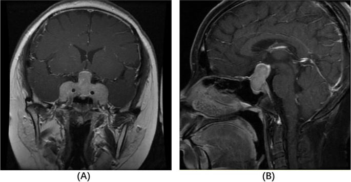
Out of 170 patients with available MRI before first surgery, 12 patients had evidence of possible SPH (7.1%) (Table 1). Various MRI findings were noted (Fig. 1), most commonly hyperintense lesions in T1 and T2 precontrast images (Fig. 1A and 1B), while some patients had intratumor heterogeneity suggestive of cystic formation (Fig. 1C-1F). As expected, the tumor size of patients with SPH as noted in histology reports or MRI images was higher than that in patients without SPH (median size: 8.5 mm [7.0-11.25] in the SPH group vs 5.4 mm [4-8] in the non-SPH group; P < .001).


Table 1.

Characteristics of patients with and without subclinical pituitary hemorrhage, necrosis, and/or cystic degeneration

No SPH (N = 158) SPH (N = 12) P
Age at diagnosis, y 13.0 (10.6 to 15.4) 12.5 (10.6 to 15.6) .95
Sex
 Female 89 (56.3%) 5 (41.7%) .49
 Male 69 (43.7%) 7 (58.3%)
Disease duration, y 2.50 (1.5 to 3.0)
n = 138
1.00 (1.0 to 2.0)
n = 10
.014
Morning cortisol, mcg/dL 16.2 (12.6 to 20.4)
n = 139
18.4 (13.7 to 27.5)
n = 10
.28
Midnight cortisol, mcg/dL 14.0 (10.7 to 19.7)
n = 133
16.3 (11.0 to 23.0)
n = 9
.52
UFC fold change 4.89 (2.51 to 8.50)
n = 131
8.81 (6.86 to 9.42)
n = 9
.18
Morning ACTH, pg/mL 39.4 (28.2 to 53.2)
n = 143
60.8 (43.5 to 80.3)
n = 10
.016
Cortisol change during CRH stimulation test, % 68.7 (33.3 to 111)
n = 128
46.0 (–7.46 to 90.7)
n = 8
.22
ACTH change during CRH stimulation test, % 145 (59.6 to 260)
n = 127
103 (–5.75 to 278)
n = 8
.55
Cortisol change during high-dose dexamethasone suppression test, % –85.8 (–90.5 to –76.8)
n = 120
–58.0 (–85.4 to –49.7)
n = 9
.035
Remission
 Yes 127 (80.4%) 10 (83.3%) .99
 No 25 (15.8%) 2 (16.7%)

Number in each cell reports the number of patients with available results.

Abbreviations: ACTH, adrenocorticotropin; CRH, corticotropin-releasing hormone; SPH, subclinical pituitary hemorrhage, necrosis, and/or cystic degeneration; UFC, urinary free cortisol.


Figure 1.

Imaging findings in patients with subclinical pituitary hemorrhage, necrosis, and/or cystic degeneration. A, T1, and B, T2 magnetic resonance imaging (MRI) scans of the same patient showing area of high intensity inside the tumor suggesting acute/subacute episode. C, T2, and D, T1 MRI scans of the same patient showing heterogeneity in a large tumor with high-intensity areas suggesting blood-filled cavities and/or necrosis. E, T1, and F, T2 MRI scans of the same patient showing fluid inside the tumor.

Imaging findings in patients with subclinical pituitary hemorrhage, necrosis, and/or cystic degeneration. A, T1, and B, T2 magnetic resonance imaging (MRI) scans of the same patient showing area of high intensity inside the tumor suggesting acute/subacute episode. C, T2, and D, T1 MRI scans of the same patient showing heterogeneity in a large tumor with high-intensity areas suggesting blood-filled cavities and/or necrosis. E, T1, and F, T2 MRI scans of the same patient showing fluid inside the tumor.

Patients with and without SPH were similar in age (median age: 12.5 years [10.6-15.6] in the SPH group vs 13.0 years [10.6-15.4] in the non-SPH group; P = .95) and sex distribution (n of female = 5 (41.7%) in the SPH group vs 89 (56.3%) in the non-SPH group; P = .49). Patients with SPH had a shorter duration of disease as noted by changes in their growth chart parameters (median duration: 1.0 [1.0-2.0] year in the SPH group vs 2.5 [1.5-3.0] years in the non-SPH group; P = .014).

Patients in the 2 groups did not differ on their anthropometric characteristics, including height and body mass index z scores(P > .05). They also had similar blood pressure parameters and did not differ in terms of the frequency of hypertension diagnosis. No patient was on anticoagulation treatment nor had received radiation at the time of the MRI. One patient in the SPH group had a history of lower leg deep vein thrombosis and was previously on low-heparin therapy, but he had stopped treatment at least 6 months before the MRI.

Biochemical Evaluation of Hypercortisolemia

Morning and midnight serum cortisol and 24-hour UFC levels were similar in both groups, but patients with SPH had higher levels of morning ACTH (60.8 pg/mL [43.5-80.3]) compared to patients without SPH (39.4 pg/mL [28.2-53.2]; P = .016). Changes in cortisol and ACTH levels during the CRH stimulation test were similar between the 2 groups, but patients with SPH who underwent the overnight high-dose dexamethasone suppression test (n = 8) had lower suppression of cortisol after dexamethasone (–58.0% [–85.4 to –49.7]) compared to patients without SPH (n = 120) (–86.0% [–90.5 to –76.7]; P = .035) (Fig. 2). When the cutoff of suppression of more than 69% was considered, patients with SPH had a lower chance of suppressing more than 69% compared to patients without SPH (OR: 0.18; 95% CI, 0.03-0.95).


Figure 2.

Cortisol levels before and after high-dose dexamethasone suppression test in patients with and without subclinical pituitary hemorrhage, necrosis, and/or cystic degeneration (SPH).

 Cortisol levels before and after high-dose dexamethasone suppression test in patients with and without subclinical pituitary hemorrhage, necrosis, and/or cystic degeneration (SPH).

Remission After Surgical Treatment and Risk for Recurrence

The chance of immediate postoperative remission after surgery was similar in patients with and without SPH. For patients with initial remission and follow-up of at least 3 months, analysis of the risk of recurrence did not show a difference in recurrence rate between the 2 groups (HR: 1.12; 95% CI, 0.13-9.4, in the SPH group compared to the non-SPH group, adjusting for the neurosurgeon).

Discussion

SPH is often an incidental finding in the imaging evaluation of patients with PAs. The frequency of SPH in patients with CD is low (7.1% in our study) but these patients may differ in terms of their history of shorter duration of symptoms and the biochemical evaluation. More specifically, patients with CD and SPH showed higher ACTH levels and lower suppression of cortisol to high-dose dexamethasone. This, however, did not affect their prognosis in terms of immediate postoperative remission and long-term risk of recurrence.

SPH has been mainly studied in cohorts of patients with various types of PAs. From these studies important conclusions have been made suggesting that the risk of SPH is higher in patients with large nonfunctioning PAs or prolactinomas. Other risk factors for pituitary apoplexy are thought to be size of the adenoma, change in size, initiation, and withdrawal of dopamine agonists, type of dopamine agonist, use of anticoagulants, diabetes mellitus, hypertension, head trauma, radiotherapy, and preceding dynamic endocrine testing.

Patients with CD often represent a small portion of these cohorts, and to our knowledge there is no study to investigate how these patients respond to stimulation/suppression tests. For that reason, we evaluated these findings in our cohort of only patients with ACTH-secreting adenomas. As most of our referrals involve pediatric patients, we limited our cohort only to patients diagnosed at younger than 21 years to have a more homogeneous group.

The pathogenesis of pituitary apoplexy has been hypothesized to lie in more friable vessels in PAs, which, while the tumor increases in size, are more susceptible to rupture or cause surrounding feeding vessels to extend and bleed [715]. CS, because of the coexisting hypercortisolemia, leads independently to endothelial dysfunction and coagulation defects, which are often apparent as easy bruising, thrombotic events, and other signs. However, review of the literature and the estimated frequency of SPH in our cohort suggest that patients with CD are not at increased risk for SPH, potentially related to the relatively small adenomas present in these patients [1617].

The main difference of patients with CD and SPH compared to those without lies in their biochemical testing, more characteristically in lower suppression to dexamethasone. The overnight high-dose dexamethasone suppression test was originally designed to differentiate various types of CS [18]. Although originally described as a highly accurate test, in clinical practice, cutoffs of 50% to 80% have shown variable sensitivity and specificity and certain centers opt not to use this test unless all other diagnostic evaluations yield confounding results [19-21]. In previous studies a threshold of suppression of more than 69% showed the highest accuracy (sensitivity: 71%, specificity: 100%), and we have incorporated this in our diagnostic algorithm (acknowledging the limitations of the test) [2223]. In our analysis, patients with SPH had lower suppression of cortisol under the effect of high-dose dexamethasone and a higher chance of not passing the aforementioned threshold. The mechanism for the lower suppression to dexamethasone of these tumors may be due to lower vascular circulation of dexamethasone at the level of the tumor, and/or the lower sensitivity of necrotic cells to the negative feedback by circulating glucocorticoids.

A limitation of this study was that the diagnosis of SPH was based on MRI findings. However, MRI sequences and machines differed between patients and over time. Thus, although large hemorrhagic/necrotic lesions are probably accurately identified, it is possible that smaller lesions are misclassified as negative; the effect of smaller hemorrhagic areas to the biochemical testing may however be smaller as well. Further, the MRIs were not read by a central radiologist, but rather from the radiologist on call at each time point, and this could lead to discrepancies in readings. In addition, our cohort’s data may not be generalized to the pediatric or adult CD population, as often our referrals consist of patients with difficult to treat, small, or otherwise unusual tumors.

In conclusion, SPH may be incidentally identified in up to 7% of patients with CD. Patients with CD and SPH may differ in terms of their response to endocrine tests, and this finding should be incorporated in their evaluation.

Abbreviations


  • ACTH

    adrenocorticotropin

  • CD

    Cushing disease

  • CRH

    corticotropin-releasing hormone

  • CS

    Cushing syndrome

  • HR

    hazard ratio

  • MRI

    magnetic resonance imaging

  • NICHD

    Eunice Kennedy Shriver National Institute of Child Health and Human Development

  • OR

    odds ratio

  • PA

    pituitary adenoma

  • SPH

    subclinical pituitary hemorrhage

  • UFC

    urinary free cortisol

Financial Support

This work was supported by the intramural research program of the Eunice Kennedy Shriver NICHD, NIH, Bethesda, MD 20892, USA.

Disclosures

Dr Stratakis holds patents on the function of the PRKAR1APDE11A, and GPR101 genes and related issues; his laboratory has also received research funding on the GPR101 gene, and on abnormal growth hormone secretion and its treatment by Pfizer, Inc. He is currently employed by ELPEN, SA and has been consulting for Lundbeck Pharmaceuticals and Sync, SA. The other authors have nothing to disclose.

Data Availability

Some or all datasets generated during and/or analyzed during the current study are not publicly available but may be available from the corresponding author on reasonable request.

Published by Oxford University Press on behalf of the Endocrine Society 2022.
This work is written by (a) US Government employee(s) and is in the public domain in the US.
Published by Oxford University Press on behalf of the Endocrine Society 2022.

Long-term Outcomes and Complications from Endoscopic Versus Microscopic Transsphenoidal Surgery for Cushing’s Disease – a 15-year Single-center Study

Abstract

Background

Endoscopic endonasal surgery is the main transsphenoidal approach for pituitary surgery in many centers, however few studies compare the endoscopic and microscopic surgical approach with regard to long-term follow-up. This single-center study aimed to compare the two techniques over 15 years.

Methods

Medical records and magnetic resonance images from 40 patients with primary transsphenoidal surgery for Cushing’s disease at Sahlgrenska University Hospital between 2003 and 2018 were reviewed. Fourteen patients who underwent microscopic surgery and 26 patients who underwent endoscopic surgery were included in this study.

Results

In the microscopic group, 12 of 14 patients achieved endocrine remission, compared to 19 of 26 patients in the endoscopic group (n. s.). Three patients in each group developed a late recurrence. Complications were seen in 5 patients in the microscopic group and in 8 patients in the endoscopic group (n. s.). No serious complications, such as carotid artery damage, cerebrovascular fluid leakage, epistaxis, or meningitis, occurred in any group. The postoperative hospital stay was shorter in the endoscopic than the microscopic group.

Conclusion

Endoscopic endonasal surgery for Cushing’s disease showed no difference in remission, recurrence, and complication rates compared to the microscopic approach. The endoscopic group had a shorter postoperative hospital stay than the microscopic group, which in part may be due to the minimal invasiveness of the endoscopic approach.

References (0)

Cited by (0)

Conflicts of interest

The authors have no conflicts of interest.

Author statements

Conceptualization: D. Farahmand, E. Backlund, O. Ragnarsson and P. Trimpou

Data curation: Dan Farahmand, Erica Backlund, J. Carlqvist, T. Skoglund, T. Hallén, O. Ragnarsson, P. Trimpou.

Formal Analysis: D. Farahmand, E. Backlund

Funding acquisition: D. Farahmand

Investigation: D. Farahmand, E. Backlund, O. Ragnarsson and P. Trimpou

Methodology: D. Farahmand, E. Backlund, O. Ragnarsson and P. Trimpou

Project administration: D. Farahmand, E. Backlund, O. Ragnarsson and P. Trimpou

Supervision: D. Farahmand

Writing – original draft: Penelope Trimpou

Writing – review & editing: E. Backlund, O. Ragnarsson, T. Skoglund, T. Hallén, G. Gudnadottir, J. Carlqvist and D. Farahmand.

View full text

From https://www.sciencedirect.com/science/article/abs/pii/S1878875022009640

MRI-negative Cushing’s Disease

Background

Cushing’s disease (CD) is among the most common etiologies of hypercortisolism. Magnetic resonance imaging (MRI) is often utilized in the diagnosis of CD, however, up to 64% of adrenocorticotropic hormone (ACTH)-producing pituitary microadenomas are undetectable on MRI. We report 15 cases of MRI negative CD who underwent surgical resection utilizing a purely endoscopic endonasal approach.

Methods

Endoscopic endonasal transsphenoidal surgery (EETS) was performed on 134 CD cases by a single surgeon. Fifteen cases met inclusion criteria: no conclusive MRI studies and no previous surgical treatment. Data collected included signs/symptoms, pre- and post-operative hormone levels, and complications resulting from surgical or medical management. Data regarding tumor diameter, location, and tumor residue/recurrence was obtained from both pre- and post-operative MRI. Immunohistochemistry was performed to assess for tumor hormone secretion.

Results

Aside from a statistically significant difference (P = 0.001) in histopathological results between patients with negative and positive MRI, there were no statistically significant difference between these two groups in any other demographic or clinical data point. Inferior petrosal sinus sampling (IPSS) with desmopressin (DDAVP®) administration was performed on the 15 patients with inconclusive MRIs to identify the origin of ACTH hypersecretion via a central/peripheral (C/P) ratio. IPSS in seven, five and three patients showed right, left, and central side lateralization, respectively. With a mean follow-up of 5.5 years, among MRI-negative patients, 14 (93%) and 12 patients (80%) achieved early and long-term remission, respectively. In the MRI-positive cohort, over a mean follow-up of 4.8 years, 113 patients (94.9%) and 102 patients (85.7%) achieved initial and long-term remission, respectively.

Conclusions

Surgical management of MRI-negative/inconclusive Cushing’s disease is challenging scenario requiring a multidisciplinary approach. An experienced neurosurgeon, in collaboration with a dedicated endocrinologist, should identify the most likely location of the adenoma utilizing IPSS findings, followed by careful surgical exploration of the pituitary to identify the adenoma.

Peer Review reports

Introduction

Cushing’s disease (CD) is the most common cause of hypercortisolism [1]. Left untreated, CD can result in multiple complications, most often cardiovascular disease or infection, and has a mortality rate 1.7–4.8-times higher than the general population [2,3,4]. Although MRI is the imaging modality of choice for identifying these tumors, imaging is often inconclusive [5].

Prior studies have shown that adrenocorticotropic hormone (ACTH)-producing pituitary microadenomas are undetectable on MRI in 36–64% of cases [5]. However, the development and widespread utilization of 3-T MRI (3TMRI) has led to much higher tumor detection rates [67]. With a negative predictive value of approximately 19–94% and variable sensitivity and specificity, anywhere from 4 to 54% of MRIs are incorrectly reported, especially in the setting of ACTH-secreting pituitary adenomas [89]. With such variation in radiographic appearance, reliance on imaging for the management of CD patients can cause significant uncertainty for neurosurgeons and endocrinologists alike.

The choice approach in the surgical management of these adenomas is via an endoscopic endonasal transsphenoidal surgery (EETS) [21011], resulting in overall post-operative remission rates of 64–93% globally and 50–71% for cases without a conclusive MRI [12,13,14,15]. Inconclusive MRIs pose a significant challenge in the surgical management of CD, with the decision to pursue surgery for MRI-negative CD remaining highly controversial [8101416]. In this study, we report 15 cases of CD without positive MRIs who underwent adenoma resection via EETS.

Patients, materials and methods

Patients population

Between January 2005 and December 2018, EETS was performed in 134 CD cases by a single surgeon at Loghman hakim and Erfan hospitals. Of those patients, 15 cases met inclusion criteria: inconclusive MRI studies and no prior surgical treatment. The population consisted of 12 women (mean age 32.5 years; range 14–65 years) and 3 men (mean age 35 years; range 22–60 years). Data collected included signs/symptoms, pre- and post-operative hormone levels, and complications resulting from surgical or medical management. Data regarding tumor diameter, location, and tumor residue/recurrence was obtained from both pre- and post-operative MRIs. Immunohistochemistry was performed to assess for tumor hormone secretion.

Ethics approval and consent to participate

All procedures performed in this study involving human participants were in accordance with the ethical standards and approved by the Shahid Beheshti Medical University (SBMU) Ethical Committee and the 1964 Helsinki Declaration and its later amendments or comparable ethical standards. Also, a written informed consent was obtained from all subjects (or their parent or legal guardian in the case of children under 16).

Imaging

All patients underwent pre- and post-operative dynamic pituitary MRI via a superconducting 1.5-T scanner. Prior to gadolinium injection, T1-weighted Spin Echo (SE) and T2-weighted turbo SE images, followed by coronal dynamic acquisition (T1-weighted turbo SE), were obtained in the coronal plane using the following protocol: TR/TE, 400/20 ms; 288 · 192 matrix; two excitations; 18 · 18 cm field of view (FOV); 3 mm in thickness with 0.3-mm intersection gap. Afterwards, with simultaneous gadolinium injection, coronal and sagittal T1-weighted SE images were obtained 2 minutes following injection. All images were independently reviewed by both a radiologist and a neurosurgeon.

MRIs studies were categorized into direct and indirect signs of CD. Direct signs consisted of any inhomogeneity found in the pituitary, such as a lesions with diminished enhancement. Indirect signs included pituitary stalk deviation and bulging or erosion of the sellar contour. MRI studies were considered negative (normal) if no direct or indirect signs were identified.

In some cases, small lesions with diameters under 6 mm may be seen on MRI however are not considered indicative of CD due to the high prevalence of incidentalomas in this region. MRIs in which these lesions were present were classified as inconclusive.

Any uncertainty in interpreting the MRIs by any of the reviewers resulted in exclusion of the image from this study.

Pre-operative endocrine examination

All cases were ACTH-dependent Cushing syndrome showing clinical features including weight gain, proximal myopathy, and wide base purple striae. Furthermore, all cases demonstrated laboratory abnormalities consistent with CD, including increased 24-hour urinary free cortisol (UFC) excretion, loss of the cortisol circadian rhythm, high basal ACTH level, failure of low-dose dexamethasone to suppress cortisol secretion in addition to serum suppression or 24-hour UFC after high-dose dexamethasone. Additionally, pre- and post-operative levels of anterior pituitary hormone including prolactin, growth hormone (GH), insulin-like growth factor I (IGF-I), thyroid stimulating hormone (TSH), free/total Triiodothyronine (T3)/ Thyroxine (T4), follicle-stimulating hormone (FSH), Luteinizing hormone (LH), and free/total testosterone (men) or estradiol (premenopausal women) were measured.

The 15 cases of MRI negative CD were diagnosed and categorized according to their endocrine profile in order to distinguish the ACTH-dependent CD from pseudo-cushing syndrome.

Bilateral inferior petrosal sinus sampling (BIPSS)

All 15 cases of MRI-negative ACTH-dependent Cushing’s syndrome underwent bilateral inferior petrosal sinus sampling (BIPSS). To confirm that the elevated ACTH secretion originated from the pituitary, BIPSS was simultaneously performed with central/peripheral (C/P) ACTH gradient measurement, utilizing the calculations described by Oldfield et al. [17].

No significant complications occurred in performing the procedures. A petrosal to peripheral ACTH ratio ≥ 2.0 in the basal state, a peak ratio ≥ 3.0 after desmopressin (DDAVP®) administration, or a normalized IPS:P ratio > 0.8 were considered diagnostic of CD. Additionally, tumor lateralization was specified when the interpetrosal gradient ratio of ACTH was ≥1.4 [18].

Endoscopic Endonasal Transsphenoidal surgical approach

All patients underwent surgery by a single neurosurgeon and otolaryngologist (ENT) with extensive experience in pituitary tumor excision via EETS. Exposure to the sellar floor was provided by an ENT surgeon while drilling of the sella was performed by the neurosurgeon. Extensive drilling of the sellar floor laterally up to the carotid artery bilaterally provided a wide view of the medial wall of the cavernous sinus as well as exposure of the anterior and posterior intercavernous sinuses was performed in all cases. The dura was then opened to expose the pituitary gland. Following tumor identification, adenomectomy was performed with selective removal of a rim of normal pituitary tissue. In cases where a tumor was not visualized on initial exposure of the pituitary, the pituitary gland was explored laterally via a horizontal paramedian incision on the IPSS suggesting side. If a tumor was not visualized at this stage, a vertical paramedian incision was then performed. In some cases, a cream-like substance was drained from the pituitary incision. Although this was suspicious of a tumor and tissue biopsy was obtained, it was not considered a definite tumor diagnosis and thus surgical exploration (EXP) was done in the same manner on the other side of the pituitary. In the scenario where no distinct adenoma was found, both sides of the pituitary gland underwent EXP with emphasis on lateralizing sides distinguished by IPSS. However, we did not rely solely on IPSS lateralization, as whole gland EXP was performed in all cases. Although ACTH secreting pituitary adenomas are the most common cause of Cushing syndrome, pituitary adenomas can also be ectopic, forming outside of the sella turcica with no direct connection to the pituitary gland [19]. After EXP of each side of the gland, ipsilateral periglandular inspection with visualization of the medial wall of the cavernous sinus and diaphragm was performed to identify a potential ectopic microadenoma in the periglandular region. Although the exact origin of ectopic ACTH-producing pituitary adenomas is unclear, they likely emerge from remnants of Rathke’s pouch during its development course [20]. As a result, these tumors can be discovered in the nasopharynx, sphenoid sinus, cavernous sinus, clivus, or suprasellar area [21]. Detecting an adenoma at this stage may prevent further unnecessary EXP of pituitary gland. If a visible tumor was still not detected, a vertical medial incision was made on the pituitary gland adjacent to the pituitary stalk and neurohypophysis. If a tumor could not be reliably identified by extensive EXP of the entire pituitary gland or BIPSS failed to localize a pituitary adenoma, we did not progress to performing incomplete or complete hypophysectomy. Figures 1 and 2, respectively, demonstrate the surgical management algorithm and pituitary incisions for MRI-negative CD.

Fig. 1

figure 1

Eight-step MRI negative Cushing’s disease surgical management

Fig. 2

figure 2

Schematic illustration of 8 steps in endoscopic endonasal approach to MRI inconclusive Cushing’s disease (Resembling half Georgia flag)

If an ectopic ACTH-secreting adenoma is not easily found, permanent destructive or ablative surgeries such as bilateral adrenalectomy and hypophysectomy may be required [20]. Despite the danger of Nelson syndrome, bilateral adrenalectomy remains a feasible option in the management of refractory CD [2223].

Histological examination

All intraoperative tissue specimens obtained underwent histological examination by a pathologist. Pituitary specimens were fixed in buffered 10% formalin and embedded in paraffin wax. All specimens were first examined by Hematoxylin and Eosin (H&E) staining to detect regions which had loss of acinar organization. Additionally, reticulin and periodic Acid-Schiff (PAS) staining was implemented for a more accurate histopathologic diagnosis. Immunohistochemistry staining was used to identify cytokeratin and anterior pituitary hormones, including ACTH, in the case of a pituitary adenoma not being detected by H&E staining. The presence of ACTH-secreting cells was examined via immunocytochemistry using specific anti-ACTH antibodies.

Post-operative endocrinologic assessment and follow up

Serum cortisol and ACTH levels were monitored for 2–5 days following surgery. Initial follow-up occurred 2 weeks post-operatively with a subsequent visit occurring 3 months postoperatively, during each visit a complete pituitary hormonal evaluation was performed. This evaluation was repeated every 3 months for up to 2 years and every 6 months after that. An initial postoperative pituitary MRI was typically performed within 3 months after surgery. For patients to be considered to be in initial post-operation remission, a basal plasma cortisol level lower than 140 nmol/L (5 μg/dL) or adequate suppression of plasma cortisol (≤56 nmol/L) (≤1.8 μg/dL) following the 1-mg dexamethasone suppression test was necessary during the first month following surgery. Long term remission was defined as a plasma cortisol lower than 84 nmol/L (3 μg/dL) after a 1-mg dexamethasone suppression test at the final visit. Recurrence was defined as a recurring case of hypercortisolism with insufficient suppression of plasma cortisol (> 140 nmol/L) after a 1-mg dexamethasone suppression test. Clinical criteria for remission included significant symptomatic improvement or resolution without additional therapy (radiotherapy, adrenalectomy). Patients achieving remission had to meet both laboratory and clinical criteria to be classified as such. Glucocorticoids were not given postoperatively except when there was laboratory evidence of hypercortisolism and/or clinical manifestations of glucocorticoid insufficiency. Additionally, 4 to 6 weeks post-operatively, thyroid and gonadal axis function was assessed by measuring free T4, TSH, FSH, and LH levels in addition to end-organ hormones (estradiol in women and testosterone in men).

Statistical analysis

SPSS software (version 26, Chicago, IL) was used to analyze the data. For continuous data, we calculated descriptive statistics, mean and standard deviation (SD), and for categorical variables, frequency and percentages were calculated. The chi-square or Fisher’s exact test was used to analyze categorical data, while the student’s t-test or Mann- Whitney U test was used to analyze continuous variables’ means, depending on the distribution’s normality. Statistical significance was defined by a p value of < 0.05.

Results

Demographic and clinical data of 134 patients with CD who underwent EETS are shown in Table 1. Fifteen (11.2%) of the 134 CD patients who underwent EETS were MRI-negative and 119 patients (88.8%) were MRI positive. The female/male ratio in the MRI-negative group was four to one while this ratio in the MRI-positive cohort was 2.6. With regards to sex distribution, Fisher’s exact test found no statistically significant difference between these two groups (P = 0.565). All patients had clinical manifestation of Cushing’s syndrome including obesity, hirsutism, glucose intolerance, and hypertension. As shown in Table 1, pre-operative ACTH level was 134.02 ± 21.78 ng/l and 151.76 ± 44.17 ng/l in MRI-negative and MRI-positive patients, respectively, and no statistically significant difference was observed between these two groups (P = 0.781). As demonstrated in Table 1, UFC was 462.3 ± 43.98 μg/24 h and 478.4 ± 73.02 in MRI-negative and MRI-positive patients, respectively, and no statistically significant difference was observed between these two groups (P = 0.832).

Table 1 Demographic and clinical data

IPSS with DDAVP® administration was performed on the 15 MRI-negative patients to identify the origin of ACTH hypersecretion via the C/P ratio. Seven patients showed right-sided lateralization and five patients showed left-sided lateralization. In remaining three patients, IPSS did not show an ACTH interpetrosal gradient ratio greater than the cutoff point, which was interpreted as an ACTH hypersecretion with central origin. On EXP, adenomas were found in 2 of the 3 patients, with no adenoma being found in the 3rd. The IPSS results were in concordance with our observations during EXPs in 60% of patients. However, in 13% of patients, no adenoma was detected, and in 26% an adenoma was found on the opposite side of the pituitary where pre-operative IPSS results initially reported a tumor or was suggestive of one being present. In 60% of MRI-negative patients, histological examination demonstrated an adrenocorticotropic pituitary adenoma, but in 40% no adenoma was found after pathological examinations. In MRI-positive patients, positive histology was observed in 112 patients (94.1%), while in 7 patients (5.9%) histopathological studies were negative. Fisher’s exact test revealed that the difference between MRI-negative and MRI-positive patients in terms of histopathological result was statistically significant (P = 0.001).

In all four patients who had discordant IPSS results as well as the patients who had negative or inconclusive findings on EXP, tissue samples were obtained from suspicious sites during EXP and were sent for histopathological examination. Histopathology demonstrated adrenocorticotropic adenoma tissues in 3 of them on the opposite side of the IPSS suggested region, while in 1 of them the histological results were inconclusive. This patient (case 10) achieved initial remission, however she experienced recurrence after 25 months, and similarly to her initial presentation, MRI findings were negative and IPSS suggested right sided lateralization. She underwent revision surgery, and a distinct adenoma was detected on the right side, which was confirmed by histological examination, after which she went into remission following selective adenectomy (Table 2).

Table 2 Presents summary of patients’ demographics, IPSS and surgical exploration results

Among the patients with inconclusive MRI, 14 (93%) achieved initial remission, 12 of which (80%) went on to long term remission with a mean follow up of 5.5 years. Two patients (cases 10 and 11) developed recurrence following initial remission; according to the IPSS suggested side, partial hypophysectomy was performed in both cases however neither was able to achieve remission afterwards. One patient (case 13) was unable to achieve initial remission following the initial surgery and thus required continued medical management. With a mean follow-up of 4.8 years among the 119 patients with positive MRI, 113 patients (94.9%) and 102 patients (85.7%) achieved initial and long-term remission, respectively. There were no statistically significant differences between these two groups in terms of either initial (P = 0.767) or long-term remission (P = 0.457). Among the 102 patients who achieved long-term remission, 12 patients (11.7%) experienced disease recurrence. With regards to recurrence rate, there was no statistically significant difference between patients with either positive or negative MRI (P = 0.542).

In two patients (cases 2 and 6) the adenoma was not found during EXP, however tissue samples obtained from the IPSS suggested side demonstrated adrenocorticotropic pituitary adenoma in both patients on histopathological examination.

Diabetes insipidus (DI) was the most frequent complication associated with CD. Transient DI occurred in seven cases with resolution prior to discharge. There was one case of permanent DI diagnosed in follow-up. Additionally, one patient developed symptomatic adrenal insufficiency requiring glucocorticoid replacement. Two patients developed hypothyroidism requiring hormone replacement. Panhypopituitarism was not seen following the initial surgeries however occurred in one case following revision surgery (partial hypophysectomy) which required hormone replacement therapy. Cerebrospinal fluid (CSF) leak resulting in meningitis was seen in one patient, however no other complications occurred during the post-operative period. None of our patients demonstrated clinical or endocrinological signs of gonadal insufficiency in follow-up aside from the aforementioned case of panhypopituitarism following revision partial hypophysectomy. In the MRI-positive cohort, 51 patients showed transients DI (42.8%), with 4 of the patients (3.4%) experiencing DI till last follow-up. Partial anterior pituitary insufficiency and complete anterior pituitary insufficiency was observed in one (0.8%) and two (1.6%) patients, respectively. Syndrome of inappropriate antidiuretic hormone (SIADH) secretion was observed in 3 patients (2.5%).

Discussion

In this study we present the outcomes of pure endoscopic endonasal surgical treatment of fifteen patients with MRI-negative Cushing’s disease. Due to the arduous nature of treatment in this patient population, we used a precise method of EXP as described above, resulting in initial remission in 93% of patients post-operatively. Based on the work of Bansal et al., patients with a definite adenoma on MRI who underwent microscopic transsphenoidal surgery had a statistically significant greater rate of early remission and lower rates of persistent disease than those with negative/equivocal findings [24]. However, in terms of late remission and recurrence, there was no statistically significant difference between these two groups [24]. Negative/equivocal MRI results and the incidence of macroadenoma, particularly in patients with cavernous sinus invasion, were found to predict poor remission rates [24]. According to some investigations, MRI-negative CD patients had a poorer remission rate [2526]. In other studies, however, there was no statistically significant difference in remission between those who had MRI-negative CD and those who had a MRI-positive CD, which is consistent with our result [1427,28,29,30,31,32]. Recurrence occurred in 2 patients, while 12 patients showed no clinical or endocrinological signs of recurrence during the mean follow-up of 5 years, and one patient did not go to remission. Aside from one CSF leak leading to meningitis and one case of permanent DI, there were no major surgery related complications. Pituitary CD is a common and potentially lethal condition that, if left untreated, can lead to sequelae such as morbid obesity, hypertension, and diabetes mellitus. Diagnosis and treatment of CD is more challenging than other functional pituitary adenomas. Currently, trans-sphenoidal pituitary EXP is considered the standard of care for CD [33,34,35]. CD is typically diagnosed by endocrinologist through clinical symptoms, and supported by laboratory tests such as the 8 AM blood or saliva cortisol level, 24 hours urinary free cortisol level, low- and high-dose dexamethasone suppression tests, and the corticotropin-releasing hormone (CRH) stimulating test [36,37,38]. When ACTH-dependent CD is diagnosed, or clinical signs and symptoms are highly suggestive of it, MRI imaging of the pituitary is often the next step to identify the causative agent i.e., a pituitary adenoma. With regards to pituitary lesions, MRI is considered the most sensitive imaging modality, however reported sensitivity varies significantly between studies, with reported rates ranging from 22 to 92% [39,40,41].

The rate of MRI-negative microadenomas is reported to be between 36 to 63% [5]. Hofmann et al. reported no identified tumor in 49.3% of 270 MRIs [29]. Yamada et al. reported a lower frequency (17%) of MRI-negative CD in their series [42]. In our series, only 15 out of 134 (11.19%) CD patients were MRI-negative. In general, negative-MRIs could be explained by several factors such as field strength, technique (the correct pulse sequence and parameters), radiologist interpretation errors, or tumor size. Identifying tumors smaller than 3 mm in diameter is difficult in MRIs with 2.5- to 3-mm-thick image sections [29]. Dynamic MRI and 3-TMRI can result in a higher sensitivity in identifying ACTH-secreting microadenomas [6743]. In addition, spoiled gradient-recalled echo sequence (SPGR) view can help to increase sensitivity [44]. The relatively low number of negative-MRIs in our study can be attributed to the more extensive review of MRI images, utilization of high-field strength MRI (1.5 T), as well as the implementation of SPGR dynamic studies with 1.5- to 2.0-mm-thick sections, in addition to standard methods. Additionally, assessment of images by experienced pituitary neuroradiologists may have reduced the negative-MRI rate in our series. Although small tumor size is a likely factor in MRI-negative CD, prior studies have reported examples of MRI-negative microadenomas 4-6 mm in size, typically large enough to be easily identified on EXP [42].

If MRI is unable to identify the tumor definitively, the next best step is venous sampling to confirm CD. There are various indication for BIPSS, including patients who have clinical and laboratory findings of CD but normal or inconclusive MRI results [45], cases that do not have a clear hormone test response, or cases where there are inconsistencies between laboratory and imaging results [46]. BIPSS is also recommended by some as standard for any case of confirmed ACTH-dependent Cushing’s syndrome [4748]. In our institution, BIPSS is reserved for MRI-negative Cushing’s patients. Newell-Price et al. reviewed 21 studies with 569 total patients, and found that BIPSS with CRH stimulation had a 96% sensitivity and 100% specificity in separating CD from pseudo-Cushing’s states [49]. Most studies report a 90–100% sensitivity and specificity for BIPSS [50,51,52]. In the majority of cases of CD, a pituitary microadenoma can be found eccentric to one side of the pituitary, having venous drainage directly into the ipsilateral inferior petrosal sinus (IPS) [53].

This phenomenon is the basis for utilizing BIPSS as a means of lateralizing ACTH secreting pituitary tumors. There are many instances where EXP fails to detect a pituitary adenoma, despite conformation of pituitary origin of ACTH secretion via BIPSS. Evidence of lateralization prior to surgery can convince the surgeon to perform a guided hemi hypophysectomy. In our series, the accuracy of BIPSS for lateralizing adenomas was 60%, similar to the reported accuracy in the literature of approximately 70% [17]. Inaccurate lateralization from BIPSS has been attributed to asymmetrical venous drainage with shunting of blood toward the dominant side. Thus, BIPSS appears to be a superior diagnostic tool compared to other means of lateralization, and neurosurgeons should be wary of making operative decisions solely from BIPSS data [49].

The standard of care for MRI-negative CD is highly disputed. There is evidence suggesting surgical exploration is more problematic than watchful waiting [8], or that it is not indicated in MRI-negative CD [54]. Many advancements have led to the widespread adoption of transsphenoidal approach during the last three decades, especially the endoscope [31]. Regardless of the width or depth of access, the endoscopic approach allows the surgeon to have a large panoramic view. Many cases in the literature have reported successfully treating functional pituitary tumors via endoscopic surgery [273155,56,57,58]. The results suggest that they are on par with, if not superior to, traditional microscopic approaches. When patients were operated on utilizing a microscopic technique assisted by a pre-operative ACTH gradient, the overall rate of partial adenomectomy (partial hypophysectomy) was 30%, including 19% in patients with positive MRIs and 40% in those with negative MRIs [28]. However, endoscopic visualization of pituitary adenomas has allowed for the need for partial adenomectomy to be reduced to less than 2%, limiting the damage to the normal pituitary gland during operation [28]. A recently published meta-analysis demonstrated that although there was no statistically significant differences between EETS and microscopic endonasal transsphenoidal surgery in the sub-analysis with regards to recurrence rate, remission rate, and persistence rate, the recurrence rate in the microscopic endonasal transsphenoidal surgery group was almost three times higher than in the EETS group [11]. As a result, EETS appears to be a possible suggested therapeutic method, while more studies are needed to establish the therapeutic method of choice [59].

In general, pituitary surgery is not advisable in cases of MRI-negative CD where IPSS is not able to prove a central origin of ACTH secretion [42]. However, when IPSS demonstrates central ACTH secretion, surgical intervention has been proposed as a first line treatment in MRI-negative CD [25324260]. The outcome of surgical intervention in MRI-negative patients is variable in the literature. Some reports indicated lower remission rate in these patients [4261], while others have concluded that EXP results in greater complications in this population [815]. Additionally, several studies have shown no significant difference in outcomes of pituitary surgery between MRI-negative and MRI-positive patients [142532]. Pivonello et al. found the lack of tumor detection on pre-operative MRI operation to be a negative prognostic factor in surgical management [62]. In the present study, surgery was performed for all MRI-negative Cushing’s patients with positive IPSS results. We achieved 93% initial remission and 80% long term remission rates, comparable to mean remission rates in patients with preoperative identification of tumor, as reported in the literature, ranging from 52.6–100% [62].

Failure to identify an adenoma on EXP or in histologic examination is not uncommon in the surgical management of CD. Intraoperative detection of the adenoma has been shown to be a factor of favorable prognosis [63,64,65]. Similarly, failure to identify an adenoma on histopathology has been found to be a negative prognostic indicator. Specifically, remission rates were significantly lower in cases where no histological tumor identification could be provided [146366]. In our study, two cases revealed no adenoma on EXP, however the tissue samples subsequently obtained from the IPSS suggesting side were consistent with pituitary adenoma on histologic examinations. In six cases, a cream-like substance was identified within the pituitary following incision, however histologic examination failed to demonstrate adrenocorticotropic adenoma in any of them. Nonetheless, 5 of the 6 patients went into remission following surgery, potentially due to the small size of tissue samples obtained which in turn made accurate histopathological assessment more difficult [1467].

In cases where EXP does not result in localization of an adenoma, surgical decision making becomes complicated. Generally, total hypophysectomy is not advisable due to high rates of endocrine complications as well as failing to provide significantly increased remission rates over partial hypophysectomy [6268]. In this scenario, multiple studies have recommended partial hypophysectomy based on IPSS lateralization as the next best step in management [6369]. Carr et al. suggested the advantage of 2/3 gland resection on remission rate in MRI-negative CD [60], but as previously discussed, IPSS may incorrectly lateralize adenomas, and thus surgeons should be hesitant when making decisions regarding tumor lateralization based solely on BIPSS data [1749]. Moreover, both adenomectomy and hypophysectomy are not without risks and potential complications. Surgical aggressiveness is correlated with increased likelihood of pituitary loss-of-function, supported by literature showing that the larger the amount of resection, the higher the rate of hypopituitarism. It has been reported that patients undergoing adenomectomy, hemi-hypophysectomy, and-total hypophysectomy had mean rates of hypopituitarism of 6.6, 20.2, and 80.2%, respectively [637071]. As most CD patients are females of reproductive age, preserving child-bearing capacity is an important consideration, one which results in reluctance to perform hemi-hypophysectomy. In our series, we performed selective adenectomy when distinct adenomas were found, and in the cases where no adenoma was detected, meticulous EXP of pituitary gland bilaterally was performed. Subsequently, if EXP was inconclusive, a vertical median incision was made near the pituitary stalk to explore central part of the gland, which is believed to be the nest for adrenocorticotropic cells. Although an important step in localizing the adenoma, this also likely explains the high rate of postoperative DI in our study. No additional hemi-hypophysectomy was performed during the initial surgery in our study. With this technique, we achieved acceptable results with regards to remission rates, and none of our patients experienced panhypopituitarism in postoperative follow-ups. In one patient where CD recurred 2 years post-operatively, inadequate bony exposure and limited visualization of the medial wall of the right cavernous sinus resulted in failure to identify the adenoma during the initial surgery, further supporting the strategy of creating extensive exposure of the operative field in MRI-negative CD. Another possible reason for recurrence in this patient would be growth of a previously undetected microadenoma.

Conclusion

Surgical treatment of MRI-negative Cushing’s disease is a demanding scenario necessitating multidisciplinary management. An experienced neurosurgeon working in collaboration with an endocrinologist should specify the most likely region of the tumor via IPSS. Additionally, surgical exploration of the pituitary is an invaluable tool in identifying adenomas while reducing the need for aggressive hypophysectomy, thus decreasing the likelihood of complications. Although MRI-negative Cushing’s disease presents significant challenges to neurosurgeons, surgical management remains essential in achieving remission.

Availability of data and materials

The authors confirm that the data supporting the findings of this study are available within the article.

References

  1. Mehta GU, Lonser RR, Oldfield EH. The history of pituitary surgery for Cushing disease: historical vignette. J Neurosurg. 2012;116(2):261–8.

    PubMed Article Google Scholar

  2. Hammer GD, Tyrrell JB, Lamborn KR, Applebury CB, Hannegan ET, Bell S, et al. Transsphenoidal microsurgery for Cushing’s disease: initial outcome and long-term results. J Clin Endocrinol Metabol. 2004;89(12):6348–57.

    CAS Article Google Scholar

  3. Nieman LK. Cushing’s syndrome: update on signs, symptoms and biochemical screening. Eur J Endocrinol. 2015;173(4):M33–8.

    CAS PubMed PubMed Central Article Google Scholar

  4. Swearingen B, Biller BM, Barker FG, Katznelson L, Grinspoon S, Klibanski A, et al. Long-term mortality after transsphenoidal surgery for Cushing disease. Ann Intern Med. 1999;130(10):821–4.

    CAS PubMed Article Google Scholar

  5. Lüdecke DK, Flitsch J, Knappe UJ, Saeger W. Cushing’s disease: a surgical view; this publication is dedicated to our professor emeritus Dr. med. Rudolf Kautzky. J Neuro-Oncol. 2001;54(2):151–66.

    Article Google Scholar

  6. Bartynski WS, Lin L. Dynamic and conventional spin-echo MR of pituitary microlesions. Am J Neuroradiol. 1997;18(5):965–72.

    CAS PubMed PubMed Central Google Scholar

  7. Erickson D, Erickson B, Watson R, Patton A, Atkinson J, Meyer F, et al. 3 tesla magnetic resonance imaging with and without corticotropin releasing hormone stimulation for the detection of microadenomas in Cushing’s syndrome. Clin Endocrinol. 2010;72(6):793–9.

    CAS Article Google Scholar

  8. Semple PL, Vance ML, Findling J, Laws ER Jr. Transsphenoidal surgery for Cushing’s disease: outcome in patients with a normal magnetic resonance imaging scan. Neurosurgery. 2000;46(3):553–9.

    CAS PubMed Article Google Scholar

  9. Tabarin A, Laurent F, Catargi B, Olivier-Puel F, Lescene R, Berge J, et al. Comparative evaluation of conventional and dynamic magnetic resonance imaging of the pituitary gland for the diagnosis of Cushing’s disease. Clin Endocrinol. 1998;49(3):293–300.

    CAS Article Google Scholar

  10. Nieman LK, Biller BM, Findling JW, Murad MH, Newell-Price J, Savage MO, et al. Treatment of Cushing’s syndrome: an endocrine society clinical practice guideline. J Clin Endocrinol Metab. 2015;100(8):2807–31.

    CAS PubMed PubMed Central Article Google Scholar

  11. Sabahi M, Shahbazi T, Maroufi SF, Vidal K, Recinos PF, Kshettry VR, et al. MRI-negative Cushing’s disease: a review on therapeutic management. World Neurosurg. 2022.

  12. Ciric I, Zhao J-C, Du H, Findling JW, Molitch ME, Weiss RE, et al. Transsphenoidal surgery for Cushing disease: experience with 136 patients. Neurosurgery. 2012;70(1):70–81.

    PubMed Article Google Scholar

  13. Netea-Maier R, Van Lindert E, Den Heijer M, Van der Eerden A, Pieters G, Sweep C, et al. Transsphenoidal pituitary surgery via the endoscopic technique: results in 35 consecutive patients with Cushing’s disease. Eur J Endocrinol. 2006;154(5):675–84.

    CAS PubMed Article Google Scholar

  14. Salenave S, Gatta B, Pecheur S, Fo S-G, Visot A, Lasjaunias P, et al. Pituitary magnetic resonance imaging findings do not influence surgical outcome in adrenocorticotropin-secreting microadenomas. J Clin Endocrinol Metab. 2004;89(7):3371–6.

    CAS PubMed Article Google Scholar

  15. Semple PL, Laws ER. Complications in a contemporary series of patients who underwent transsphenoidal surgery for Cushing’s disease. J Neurosurg. 1999;91(2):175–9.

    CAS PubMed Article Google Scholar

  16. Lonser RR, Nieman L, Oldfield EH. Cushing’s disease: pathobiology, diagnosis, and management. J Neurosurg. 2017;126(2):404–17.

    PubMed Article Google Scholar

  17. EH OLDFIELD, ME GIRTON, JL DOPPMAN. Absence of intercavernous venous mixing: evidence supporting lateralization of pituitary microadenomas by venous sampling. J Clin Endocrinol Metab. 1985;61(4):644–7.

    Article Google Scholar

  18. Mamelak AN, Dowd CF, Tyrrell JB, McDonald JF, Wilson CB. Venous angiography is needed to interpret inferior petrosal sinus and cavernous sinus sampling data for lateralizing adrenocorticotropin-secreting adenomas. J Clin Endocrinol Metab. 1996;81(2):475–81.

    CAS PubMed Google Scholar

  19. Thompson LD, Seethala RR, Müller S. Ectopic sphenoid sinus pituitary adenoma (ESSPA) with normal anterior pituitary gland: a clinicopathologic and immunophenotypic study of 32 cases with a comprehensive review of the English literature. Head and neck pathology. 2012;6(1):75–100.

    PubMed PubMed Central Article Google Scholar

  20. Seltzer J, Lucas J, Commins D, Lerner O, Lerner A, Carmichael JD, et al. Ectopic ACTH-secreting pituitary adenoma of the sphenoid sinus: case report of endoscopic endonasal resection and systematic review of the literature. Neurosurg Focus. 2015;38(2):E10.

    PubMed Article Google Scholar

  21. Langford L, Batsakis JG. Pituitary gland involvement of the sinonasal tract. Ann Otology, Rhinology & Laryngology. 1995;104(2):167–9.

    CAS Article Google Scholar

  22. Gurazada K, Ihuoma A, Galloway M, Dorward N, Wilhelm T, Khoo B, et al. Nasally located ectopic ACTH-secreting pituitary adenoma (EAPA) causing Nelson’s syndrome: diagnostic challenges. Pituitary. 2014;17(5):423–9.

    CAS PubMed Article Google Scholar

  23. Pluta RM, Nieman L, Doppman JL, Watson JC, Tresser N, Katz DA, et al. Extrapituitary parasellar microadenoma in Cushing’s disease. J Clin Endocrinol Metabol. 1999;84(8):2912–23.

    CAS Google Scholar

  24. Bansal P, Lila A, Goroshi M, Jadhav S, Lomte N, Thakkar K, et al. Duration of post-operative hypocortisolism predicts sustained remission after pituitary surgery for Cushing’s disease. Endocrine connections. 2017;6(8):625–36.

    CAS PubMed PubMed Central Article Google Scholar

  25. Jagannathan J, Sheehan JP, Jane JA. Evaluation and management of Cushing syndrome in cases of negative sellar magnetic resonance imaging. Neurosurg Focus. 2007;23(3):1–7.

    Article Google Scholar

  26. Pouratian N, Prevedello DM, Jagannathan J, Lopes MB, Vance ML, Laws ER Jr. Outcomes and management of patients with Cushing’s disease without pathological confirmation of tumor resection after transsphenoidal surgery. J Clin Endocrinol Metab. 2007;92(9):3383–8.

    CAS PubMed Article Google Scholar

  27. Cebula H, Baussart B, Villa C, Assié G, Boulin A, Foubert L, et al. Efficacy of endoscopic endonasal transsphenoidal surgery for Cushing’s disease in 230 patients with positive and negative MRI. Acta Neurochir. 2017;159(7):1227–36.

    PubMed Article Google Scholar

  28. Cristante J, Lefournier V, Sturm N, Passagia JG, Gauchez AS, Tahon F, et al. Why we should still treat by neurosurgery patients with Cushing disease and a normal or inconclusive pituitary MRI. J Clin Endocrinol Metab. 2019;104(9):4101–13.

    Article Google Scholar

  29. Hofmann BM, Hlavac M, Martinez R, Buchfelder M, Müller OA, Fahlbusch R. Long-term results after microsurgery for Cushing disease: experience with 426 primary operations over 35 years. J Neurosurg. 2008;108(1):9–18.

    PubMed Article Google Scholar

  30. Testa R, Albiger N, Occhi G, Sanguin F, Scanarini M, Berlucchi S, et al. The usefulness of combined biochemical tests in the diagnosis of Cushing’s disease with negative pituitary magnetic resonance imaging. Eur J Endocrinol. 2007;156(2):241–8.

    CAS PubMed Article Google Scholar

  31. Starke RM, Reames DL, Chen C-J, Laws ER, Jane JA Jr. Endoscopic transsphenoidal surgery for Cushing disease: techniques, outcomes, and predictors of remission. Neurosurgery. 2013;72(2):240–7.

    PubMed Article Google Scholar

  32. Sun Y, Sun Q, Fan C, Shen J, Zhao W, Guo Y, et al. Diagnosis and therapy for Cushing’s disease with negative dynamic MRI finding: a single-Centre experience. Clin Endocrinol. 2012;76(6):868–76.

    Article Google Scholar

  33. Esposito F, Dusick JR, Cohan P, Moftakhar P, McArthur D, Wang C, et al. Early morning cortisol levels as a predictor of remission after transsphenoidal surgery for Cushing’s disease. J Clin Endocrinol Metab. 2006;91(1):7–13.

    CAS PubMed Article Google Scholar

  34. Ram Z, Nieman LK, Cutler GB, Chrousos GP, Doppman JL, Oldfield EH. Early repeat surgery for persistent Cushing’s disease. J Neurosurg. 1994;80(1):37–45.

    CAS PubMed Article Google Scholar

  35. Rollin GAFS, Ferreira NP, Junges M, Gross JL, Czepielewski MA. Dynamics of serum cortisol levels after transsphenoidal surgery in a cohort of patients with Cushing’s disease. J Clin Endocrinol Metab. 2004;89(3):1131–9.

    CAS PubMed Article Google Scholar

  36. Minami I, Tateno T, Yoshimoto T, Doi M, Izumiyama H, Akashi T, et al. Subclinical Cushings disease with amelioration of metabolic comorbidities after removal of pituitary tumor. Intern Med. 2006;45(21):1231–5.

    PubMed Article Google Scholar

  37. Nakane T, Kuwayama A, Watanabe M, Takahashi T, Kato T, Ichihara K, et al. Long term results of transsphenoidal adenomectomy in patients with Cushing’s disease. Neurosurgery. 1987;21(2):218–22.

    CAS PubMed Article Google Scholar

  38. Buchfelder M, Nistor R, Fahlbusch R, Huk WJ. The accuracy of CT and MR evaluation of the Sella turcica for detection of adrenocorticotropic hormone-secreting adenomas in Cushing disease. Am J Neuroradiol. 1993;14(5):1183–90.

    CAS PubMed PubMed Central Google Scholar

  39. Nieman LK. Medical therapy of Cushing’s disease. Pituitary. 2002;5(2):77–82.

    CAS PubMed Article Google Scholar

  40. Escourolle H, Abecassis J, Bertagna X, Guilhaume B, Pariente D, Derome P, et al. Comparison of computerized tomography and magnetic resonance imaging for the examination of the pituitary gland in patients with Cushing’s disease. Clin Endocrinol. 1993;39(3):307–13.

    CAS Article Google Scholar

  41. Findling JW, Doppman JL. Biochemical and radiologic diagnosis of Cushing’s syndrome. Endocrinol Metab Clin N Am. 1994;23(3):511–37.

    CAS Article Google Scholar

  42. Yamada S, Fukuhara N, Nishioka H, Takeshita A, Inoshita N, Ito J, et al. Surgical management and outcomes in patients with Cushing disease with negative pituitary magnetic resonance imaging. World Neurosurg. 2012;77(3–4):525–32.

    PubMed Article Google Scholar

  43. Portocarrero-Ortiz L, Bonifacio-Delgadillo D, Sotomayor-González A, Garcia-Marquez A, Lopez-Serna R. A modified protocol using half-dose gadolinium in dynamic 3-tesla magnetic resonance imaging for detection of ACTH-secreting pituitary tumors. Pituitary. 2010;13(3):230–5.

    CAS PubMed Article Google Scholar

  44. Patronas N, Bulakbasi N, Stratakis CA, Lafferty A, Oldfield EH, Doppman J, et al. Spoiled gradient recalled acquisition in the steady state technique is superior to conventional postcontrast spin echo technique for magnetic resonance imaging detection of adrenocorticotropin-secreting pituitary tumors. J Clin Endocrinol Metabol. 2003;88(4):1565–9.

    CAS Article Google Scholar

  45. Findling JW, Raff H. Cushing’s syndrome: important issues in diagnosis and management. J Clin Endocrinol Metab. 2006;91(10):3746–53.

    CAS PubMed Article Google Scholar

  46. Arnaldi G, Angeli A, Atkinson A, Bertagna X, Cavagnini F, Chrousos G, et al. Diagnosis and complications of Cushing’s syndrome: a consensus statement. J Clin Endocrinol Metab. 2003;88(12):5593–602.

    CAS PubMed Article Google Scholar

  47. McCANCE DR, McILRATH E, McNEILL A, DS GORDON, DR HADDEN, KENNEDY L, et al. Bilateral inferior petrosal sinus sampling as a routine procedure in ACTH-dependent Cushing’s syndrome. Clin Endocrinol. 1989;30(2):157–66.

    CAS Article Google Scholar

  48. JW FINDLING, ME KEHOE, JL SHAKER, RAFF H. Routine inferior petrosal sinus sampling in the differential diagnosis of adrenocorticotropin (ACTH)-dependent Cushing’s syndrome: early recognition of the occult ectopic ACTH syndrome. J Clin Endocrinol Metabol. 1991;73(2):408–13.

    Article Google Scholar

  49. Newell-Price J, Trainer P, Besser M, Grossman A. The diagnosis and differential diagnosis of Cushing’s syndrome and pseudo-Cushing’s states. Endocr Rev. 1998;19(5):647–72.

    CAS PubMed Google Scholar

  50. Machado MC, De Sa SV, Domenice S, Fragoso MCBV, Puglia P Jr, Pereira MAA, et al. The role of desmopressin in bilateral and simultaneous inferior petrosal sinus sampling for differential diagnosis of ACTH-dependent Cushing’s syndrome. Clin Endocrinol. 2007;66(1):136–42.

    CAS Google Scholar

  51. Hernandez I, Espinosa-de-los-Monteros AL, Mendoza V, Cheng S, Molina M, Sosa E, et al. Ectopic ACTH-secreting syndrome: a single center experience report with a high prevalence of occult tumor. Arch Med Res. 2006;37(8):976–80.

    CAS PubMed Article Google Scholar

  52. Invitti C, Giraldi FP, Cavagnini F. Endocrinology SGotHPAAotISo: inferior petrosal sinus sampling in patients with Cushing’s syndrome and contradictory responses to dynamic testing. Clin Endocrinol. 1999;51(2):255–7.

    CAS Article Google Scholar

  53. Obuobie K, Davies J, Ogunko A, Scanlon M. Venous thrombo-embolism following inferior petrosal sinus sampling in Cushing’s disease. J Endocrinol Investig. 2000;s23(8):542–4.

    Article Google Scholar

  54. Barrou Z, Abecassis J, Guilhaume B, Thomopoulos P, Bertagna X, Derome P, et al. Magnetic resonance imaging in Cushing disease. Prediction Surg Results. Presse medicale. 1997;26(1):7.

    CAS Google Scholar

  55. Guaraldi F, Zoli M, Asioli S, Corona G, Gori D, Friso F, et al. Results and predictors of outcome of endoscopic endonasal surgery in Cushing’s disease: 20-year experience of an Italian referral pituitary center. J Endocrinol Investig. 2020;43(10):1463–71.

    CAS Article Google Scholar

  56. Shin S, Gardner P, Ng J, Faraji A, Agarwal N, Chivukula S. Endoscopic endonasal approach for ACTH-secreting pituitary adenomas: outcomes and analysis of remission rates and tumor biochemical activity with respect to tumor invasiveness. World Neurosurg. 2015.

  57. Wagenmakers M, Boogaarts H, Roerink S, Timmers H, Stikkelbroeck N, Smit J, et al. Endoscopic transsphenoidal pituitary surgery: a good and safe primary treatment option for Cushing’s disease, even in case of macroadenomas or invasive adenomas. Eur J Endocrinol. 2013;169(3):329–37.

    CAS PubMed Article Google Scholar

  58. Zhang K, Shen M, Qiao N, Chen Z, He W, Ma Z, et al. Surgical outcomes and multidisciplinary management strategy of Cushing’s disease: a single-center experience in China. Neurosurg Focus. 2020;48(6):E7.

    PubMed Article Google Scholar

  59. Sabahi M, Shahbazi T, Maroufi SF, Vidal K, Recinos P, Kshettry V, et al. MRI-negative Cushing’s disease: a systematic review and Meta-analysis. J Neurological Surg Part B: Skull Base. 2022;83(S 01):A169.

    Google Scholar

  60. Carr SB, Kleinschmidt-DeMasters BK, Kerr JM, Kiseljak-Vassiliades K, Wierman ME, Lillehei KO. Negative surgical exploration in patients with Cushing’s disease: benefit of two-thirds gland resection on remission rate and a review of the literature. J Neurosurg. 2017;129(5):1260–7.

    Article Google Scholar

  61. Bochicchio D, Losa M, Buchfelder M. Factors influencing the immediate and late outcome of Cushing’s disease treated by transsphenoidal surgery: a retrospective study by the European Cushing’s disease survey group. J Clin Endocrinology Metab. 1995;80(11):3114–20.

    CAS Google Scholar

  62. Pivonello R, De Leo M, Cozzolino A, Colao A. The treatment of Cushing’s disease. Endocr Rev. 2015;36(4):385–486.

    CAS PubMed PubMed Central Article Google Scholar

  63. Prevedello DM, Pouratian N, Sherman J, Jane JA, Vance ML, Lopes MB, et al. Management of Cushing’s disease: outcome in patients with microadenoma detected on pituitary magnetic resonance imaging. J Neurosurg. 2008;109(4):751–9.

    PubMed Article Google Scholar

  64. Jagannathan J, Smith R, DeVroom HL, Vortmeyer AO, Stratakis CA, Nieman LK, et al. Outcome of using the histological pseudocapsule as a surgical capsule in Cushing disease. J Neurosurg. 2009;111(3):531–9.

    PubMed PubMed Central Article Google Scholar

  65. Burke C, Adams C, Esiri M, Morris C, Bevan J. Transsphenoidal surgery for Cushing’s disease: does what is removed determine the endocrine outcome? Clin Endocrinol. 1990;33(4):525–37.

    CAS Article Google Scholar

  66. Acebes J, Martino J, Masuet C, Montanya E, Soler J. Early post-operative ACTH and cortisol as predictors of remission in Cushing’s disease. Acta Neurochir. 2007;149(5):471–9.

    CAS PubMed Article Google Scholar

  67. Mampalam TJ, Tyrrell JB, Wilson CB. Transsphenoidal microsurgery for Cushing disease: a report of 216 cases. Ann Intern Med. 1988;109(6):487–93.

    CAS PubMed Article Google Scholar

  68. Tyrrell JB, Brooks RM, Fitzgerald PA, Cofoid PB, Forsham PH, Wilson CB. Cushing’s disease: selective trans-sphenoidal resection of pituitary microadenomas. N Engl J Med. 1978;298(14):753–8.

    CAS PubMed Article Google Scholar

  69. Fomekong E, Maiter D, Grandin C, Raftopoulos C. Outcome of transsphenoidal surgery for Cushing’s disease: a high remission rate in ACTH-secreting macroadenomas. Clin Neurol Neurosurg. 2009;111(5):442–9.

    PubMed Article Google Scholar

  70. Rees D, Hanna F, Davies J, Mills R, Vafidis J, Scanlon M. Long-term follow-up results of transsphenoidal surgery for Cushing’s disease in a single Centre using strict criteria for remission. Clin Endocrinol. 2002;56(4):541–51.

    CAS Article Google Scholar

  71. Trainer P, Lawrie H, Verhelst J, Howlett T, Lowe D, Grossman A, et al. Transsphenoidal resection in Cushing’s disease: undetectable serum cortisol as the definition of successfuI treatment. Clin Endocrinol. 1993;38(1):73–8.

    CAS Article Google Scholar

Download references

Acknowledgments

We are grateful to all those who have helped us to accomplish and fulfil this project.

Funding

None.

Author information

Authors and Affiliations

  1. Department of Neurosurgery, Loghman Hakim Hospital, Shahid Beheshti University of Medical Sciences, Tehran, Iran

    Guive Sharifi, Amir Arsalan Amin & Seyed Ali Mousavinejad

  2. Skull Base Research Center, Loghman Hakim Hospital, Shahid Beheshti University of Medical Sciences, Tehran, Iran

    Guive Sharifi, Amir Arsalan Amin, Nader Akbari Dilmaghani & Seyed Ali Mousavinejad

  3. Neurosurgery Research Group (NRG), Student Research Committee, Hamadan University of Medical Sciences, Hamadan, Iran

    Mohammadmahdi Sabahi

  4. Department of Neurosurgery, Rutgers-New Jersey Medical School, Newark, NJ, USA

    Nikolas B. Echeverry

  5. Department of Otolaryngology, Head and Neck Surgery, Loghman Hakim Hospital, Shahid Beheshti University of Medical Sciences, Tehran, Iran

    Nader Akbari Dilmaghani

  6. Obesity Research Center, Research Institute for Endocrine Sciences, Shahid Beheshti University of Medical Sciences, Tehran, Iran

    Majid Valizadeh

  7. Department of Endocrinology, Loghman Hakim Hospital, Shahid Beheshti Medical University, Tehran, Iran

    Zahra Davoudi

  8. Department of Neurological Surgery, Pauline Braathen Neurological Center, Cleveland Clinic Florida, Weston, Florida, USA

    Badih Adada & Hamid Borghei-Razavi

  9. Cleveland Clinic Lerner College of Medicine of Case Western Reserve University, Director of Minimally Invasive Cranial and Pituitary Surgery Program, Research Director of Neuroscience Institute, Cleveland Clinic Florida Region, 2950 Cleveland Clinic Blvd. Weston, Cleveland, FL, 33331, USA

    Hamid Borghei-Razavi

Contributions

Guive Sharifi, Mohammadmahdi Sabahi and Amirarsalan Amin have given substantial contributions to the conception and the design of the manuscript, Mohammadmahdi Sabahi, Nikolas B. Echeverry, Nader Akbari Dilmaghani, Ali Mousavi Nejad, and Zahra Davoudi to the acquisition, analysis, and interpretation of the data. All authors have participated in drafting the manuscript. Mohammadmahdi Sabahi, Majid Valizadeh, and Badih Adada revised it critically. Hamid Borghei-Razavi supervised this project. All authors read and approved the final version of the manuscript. All authors contributed equally to the manuscript and read and approved the final version of the manuscript.

Corresponding author

Correspondence to Hamid Borghei-Razavi.

Ethics declarations

Ethics approval and consent to participate

All procedures performed in this study involving human participants were in accordance with the ethical standards and approved by the Shahid Beheshti Medical University (SBMU) Ethical Committee and the 1964 Helsinki Declaration and its later amendments or comparable ethical standards. Also, informed consent to participate in this study was obtained from participants included in the (or their parent or legal guardian in the case of children under 16).

Consent for publication

Not applicable.

Competing interests

All authors certify that they have no affiliations with or involvement in any organization or entity with any financial interest (such as honoraria; educational grants; participation in speakers’ bureaus; membership, employment, consultancies, stock ownership, or other equity interest; and expert testimony or patent-licensing arrangements), or non-financial interest (such as personal or professional relationships, affiliations, knowledge or beliefs) in the subject matter or materials discussed in this manuscript.

Additional information

Publisher’s Note

Springer Nature remains neutral with regard to jurisdictional claims in published maps and institutional affiliations.

About this article

Cite this article

Sharifi, G., Amin, A.A., Sabahi, M. et al. MRI-negative Cushing’s Disease: Management Strategy and Outcomes in 15 Cases Utilizing a Pure Endoscopic Endonasal Approach. BMC Endocr Disord 22, 154 (2022). https://doi.org/10.1186/s12902-022-01069-5

Download citation

Concurrent Mutations of Germline GPR101 and Somatic USP8 in a Pediatric Giant Pituitary ACTH Adenoma

Abstract

Background

Cushing’s disease (CD) is rare in pediatric patients. It is characterized by elevated plasma adrenocorticotropic hormone (ACTH) from pituitary adenomas, with damage to multiple systems and development. In recent years, genetic studies have shed light on the etiology and several mutations have been identified in patients with CD.

Case presentation

A girl presented at the age of 10 years and 9 months with facial plethora, hirsutism and acne. Her vision and eye movements were impaired. A quick weight gain and slow growth were also observed. Physical examination revealed central obesity, moon face, buffalo hump, supra-clavicular fat pads and bruising. Her plasma ACTH level ranged between 118 and 151 pg/ml, and sella enhanced MRI showed a giant pituitary tumor of 51.8 × 29.3 × 14.0 mm. Transsphenoidal pituitary debulk adenomectomy was performed and immunohistochemical staining confirmed an ACTH-secreting adenoma. Genetic analysis identified a novel germline GPR101 (p.G169R) and a somatic USP8 (p. S719del) mutation. They were hypothesized to impact tumor growth and function, respectively.

Conclusions

We reported a rare case of pediatric giant pituitary ACTH adenoma and pointed out that unusual concurrent mutations might contribute to its early onset and large volume.

Peer Review reports

Background

Cushing’s disease (CD) is caused by the overproduction of adrenocorticotropic hormone (ATCH) by pituitary adenomas (PAs). It is rare in children and accounts for approximately 75% of pediatric Cushing’s syndrome from 7 to 17 years of age [1]. Weight gain and facial changes are more common in children than in adults [2]. Growth retardation is also a characteristic of children with hypercortisolemia [3]. Genetic alterations such as somatic USP8RASD1TP53 mutations, and germline AIPMEN1, and CABLES1 mutations have been identified in CD patients [4]. Here we report a case of pediatric invasive pituitary ACTH macroadenoma associated with a novel germline GPR101 (p. G169R) and a somatic USP8 (p. S719del) mutation.

Case presentation

The girl was born at full term with a length of 48 cm and a weight of 2900 g. Her neuromotor and cognitive development was comparable to those of children of the same age. At the age of 9 years and 4 months she developed plethora, hirsutism, facial acne, rapid weight gain, and increased abdominal circumference. Her skin darkened, and purple striae appeared on thighs and in the armpits. She became dull and less talkative, as indicated by her parents. At 10 years and 3 months, the patient complained of pain around the left orbit with an intensity of 4–5 points on a numerical rating scale (NRS). Five months later bilateral blepharoptosis appeared, with significantly impaired vision of the left eye. Soon both eyes failed to rotate in all directions.

On admission the patient was 10 years and 9 months, with a height of 144 cm (90–97th percentile) and a weight of 48 kg (25–50th percentile). Her weight gain was 20 kg, while the height increased by only 2–3 cm in 18 months. Her blood pressure was 115/76mmHg, and her heart rate was 80 bpm. Apart from the signs mentioned above, physical examination revealed central obesity (BMI 23.1 kg/m2), moon face, buffalo hump, supra-clavicular fat pads and bruising at the left fossa cubitalis. Her pupils were 7 mm in diameter and barely reacted to light. There was a fan-shaped visual field defect in the left eye. Her breasts were Tanner stage III and pubic hair was Tanner stage II, although menarche had not yet occurred. The parents and her younger brother at 6 years of age did not have symptoms related to Cushing syndrome, acromegaly or gigantism. There was no family history of pituitary tumor or other endocrine tumors.

She had increased midnight serum cortisol (24.35 µg/dL, normal range < 1.8 µg/mL) and 24-hour urine free cortisol (24hUFC) (308.0 µg, normal range 12.3–103.5). The plasma ACTH level ranged from 118 to 151 pg/mL (< 46pg/mL). The 24hUFC was not suppressed (79.2 µg) after 48 h low-dose dexamethasone suppression test (LDDST), but suppressed to 32.8 µg (suppression rate 89.4%) after 48 h high-dose dexamethasone. Sella enhanced MRI showed a giant pituitary tumor measured 51.8 × 29.3 × 14.0 mm with heterogeneous density (Fig. 1). The mass compressed the optic chiasma and surrounded the bilateral cavernous sinus (Knosp 4). Therefore, an invasive giant pituitary ACTH adenoma was clinically diagnosed. The morning growth hormone (GH) was 1.0ng/ml (< 2 ng/ml) and insulin-like growth factor 1 416 ng/ml (88–452 ng/ml). The prolactin (PRL), luteinizing hormone (LH), follicle-stimulating hormone (FSH) and thyroid stimulating hormone (TSH) were all in normal ranges, as well as serum sodium, potassium, blood glucose and urine osmolality. Abdominal ultrasonography revealed a fatty liver. Tests concerning type 1 multiple endocrine neoplasia included serum calcium, phosphate, parathyroid hormone, gastrin and glucagon, which were all unremarkable (Table 1).

Fig. 1

figure 1

Contrast-enhanced coronal (A) and sagittal (B) T1-weighted MRI on admission. The sellar mass measured 51.8 × 29.3 × 14.0 cm (TD × VD × APD) with a heterogeneous density in the enhanced scan. The diaphragma sellea was dramatically elevated, with optic chiasm compressed. The sellar floor was sunken and bilateral cavernous sinus was surrounded (Knosp 4)

Table 1 Laboratory data on admission

Transsphenoidal pituitary debulk adenomectomy was performed immediately due to multiple cranial nerve involvement and the negative results of Sandostatin loading test. A decompression resection was done. The plasma ACTH level declined to 77 pg/ml and serum cortisol 30.2 µg/dl three days after the operation. Vision, pupil dilation, eye movements and blepharoptosis also partially improved. Histopathology and immunohistochemical staining confirmed a densely–granulated corticotroph adenoma (Fig. 2, NanoZoomer S360 digital slide scanner and NDP.view 2.9.25 software, Hamamatsu, Japan). Neither necrosis nor mitotic activity was observed. The immunostaining for somatostatin receptor SSTR2A was positive with a cytoplasmic pattern, while GH, PRL, TSH, FSH, LH and PIT were all negative. The Ki 67 index was found to be 10%. One month after the operation the ACTH level increased to 132 pg/mL again, and the parents agreed to refer their child for radiotherapy to control the residual tumor.

Fig. 2

figure 2

Histopathology and immunohistochemistry staining results of the pituitary tumor. By light microscopy, the tumor cells were mostly basophilic and arranged in papillary architecture. Neither necrosis nor mitotic activity was observed (A hematoxylin-eosin, ×200). Immunohistochemistry staining was positive for ACTH (B immunoperoxidase, ×200) and transcription factor T-PIT (C immunoperoxidase, ×200). Cytoplasmic staining of SSTR2A was observed in around 1/3 tumor cells besides the strong staining of endothelial cells (D immunoperoxidase, ×200). The Ki-67 index was 10% (E immunoperoxidase, ×200). Cytokeratin CAM5.2 was diffusely positive in the cytoplasm (F immunoperoxidase, ×200). The positive control for ACTH and T-PIT was the human anterior pituitary gland, and for SSRT2, Ki-67 and CAM5.2 were cerebral cortex, tonsil and colonic mucosa, respectively

The early onset and invasive behavior of this tumor led to the consideration of whether there was a genetic defect. Genetic studies were recommended for the families and they all agreed and signed the written informed consent forms. Whole exome sequencing (WES) was performed on the patient’s blood sample using an Illumina HiSeq sequencer to an average read depth of at least 90 times per individual. Raw sequence files were mapped to the GRCH37 human reference genome and analyzed using the Sentieon software. The results revealed a germline heterozygous GPR101 gene mutation c.505G > C (p.Gly169Arg), which was subsequently confirmed to be of maternal origin by Sanger sequencing. Meanwhile WES of the tumor tissue identified an additional somatic heterozygous c.2155_2157delTCC (p.S719del) mutation of the USP8 gene .

Discussion and conclusions

In this report, we described an extremely giant and invasive pituitary ACTH adenoma in a 10-year-old girl. According to Trouillas et al., invasive and proliferative pituitary tumors have a poor prognosis [5]. CD is rare among children, and the fast-growing and invasive nature of the tumor in this case led to the investigation of genetic causes. The somatic USP8 gene mutation has been recently reported to be associated with the pathogenesis of CD [67]. This gene encodes ubiquitin-specific protease 8 (USP8). S718, S719 and P720 are hotspots in different studies [6,7,8,9,10,11,12,13,14]. They are located at the 14-3-3 binding motif, and the mutations disrupt the binding between USP8 and 14-3-3 protein, which leads to increased deubiquitination and EGFR signaling. High levels of EGFR consequently trigger proopiomelanocortin (POMC) transcription and ACTH secretion [67]. The p.S719del mutation has been previously reported and its pathogenicity has been confirmed [7]. Thus, we speculate the p.S719del mutation plays a role in this patient with CD.

It is noteworthy that in our case, the pituitary corticotrophin adenoma was extremely giant and bilaterally invasive. USP8 mutations have been found in 31% of pediatric CD patients [10]. It is well known that microadenomas are most common in adult and pediatric CD patients. Previously, the Chinese and Japanese cohorts observed smaller sizes of USP8-mutated PAs than wild-type PAs [79]. The Chinese cohort also reported a lower rate of invasive adenomas in USP8-mutated PAs [7]. This may be explained by the finding that UPS8 mutations did not significantly promote cell proliferation more than the wild-type ones [6]. Other cohorts suggested no difference in tumor size or invasiveness between USP8-mutated and wild-type PAs [81012,13,14], which may be partially explained by the differences in sample sizes and ethnic backgrounds. Owing to the lack of evidence of USP8 mutations significantly contributing to tumor growth and invasiveness, additional pathogenesis should be investigated in this case.

The p.Gly169Arg mutation of the GPR101 gene has not been reported in patients with pituitary tumors. In silico predictions were performed using Polyphen-2, Mutation Taster and PROVEAN, and all of the programs reported it to be pathogenic. The GPR101 gene encodes an orphan G protein-coupled receptor (GPCR) and microduplication encompassing the gene has been proven to be the cause of X-linked acrogigantism (XLAG) [15]. XLAG is characterized by the early onset of pituitary GH-secreting macroadenomas. Point mutations of GPR101 have been found in patients with PAs that are mostly GH-secreting [15,16,17]. Although their prevalence is very low, an in vitro study supported the pathogenic role of p.E308D, the most common mutation of GPR101. This led to increased cell proliferation and GH production in rat pituitary GH3 cells [15]. Rare cases of PRL, ACTH or TSH-secreting PAs with GPR101 variants were also documented [1618]. To date, there have been five cases of ACTH-secreting PAs with four different germline GPR101 mutations: two cases of p.E308D, p.I122T, p.T293I and p.G31S, although in silico predictions and in vitro evaluations using AtT-20 cells have respectively determined the latter two mutations to be non-pathogenic [1618]. These patients were mainly children and young adults. Unlike pituitary GH-secreting tumors, the role of GPR101 mutations in the pathophysiology of CD is still questionable. Trivellin et al. demonstrated no statistically significant difference in GPR101 expression between corticotropinomas and normal human pituitaries. No significant correlation between GPR101 and POMC expression levels was found neither [18].

Given the evidences above, we hypothesize that the somatic USP8 mutation is responsible for the overexpression of ACTH in this CD girl while the germline GPR101 mutation contributes to the early onset and fast-growing nature of the tumor. Similarly, a 27-year-old woman with Nelson’s syndrome originally considered to be associated with a germline AIP variant (p.Arg304Gln) was recently reported to have a somatic USP8 mutation. The patient progressed rapidly and underwent multiple transsphenoidal surgeries [19]. Since germline AIP mutations are more commonly seen in GH-secreting PAs [20], the authors proposed that the USP8 mutation might have shifted the tumor towards ACTH-secreting [19]. Further investigations into the pathogenicity of GPR101 p.Gly169Arg and AIP p.Arg304Gln mutations are required to support the hypothesis.

In summary, we report a novel germline GPR101 and somatic USP8 mutation in a girl with an extremely giant pituitary ACTH adenoma. The concurrent mutations may lead to the growth and function of the tumor, respectively. Further investigations should be carried out to verify the role of the concurrent mutations in the pathogenesis of pediatric CD.

Availability of data and materials

The WES data of the blood sample of the patient is available in the NGDC repository (https://ngdc.cncb.ac.cn/gsa-human/) and the accession number is HRA002396. Any additional information is available from the authors upon reasonable request.

Abbreviations

CD:
Cushing’s disease
ACTH:
adrenocorticotropic hormone
PA:
pituitary adenoma
NRS:
numerical rating scale
24hUFC:
24-hour urine free cortisol
LDDST:
low-dose dexamethasone suppression test
USP8:
ubiquitin-specific protease 8
POMC:
proopiomelanocortin
GPCR:
G protein-coupled receptor
XLAG:
X-linked acrogigantism

References

  1. Weber A, Trainer PJ, Grossman AB, Afshar F, Medbak S, Perry LA, et al. Investigation, management and therapeutic outcome in 12 cases of childhood and adolescent Cushing’s syndrome. Clin Endocrinol (Oxf). 1995;43(1):19–28.

    CAS Article Google Scholar

  2. Storr HL, Alexandraki KI, Martin L, Isidori AM, Kaltsas GA, Monson JP, et al. Comparisons in the epidemiology, diagnostic features and cure rate by transsphenoidal surgery between paediatric and adult-onset Cushing’s disease. Eur J Endocrinol. 2011;164(5):667–74.

    CAS Article Google Scholar

  3. Magiakou MA, Mastorakos G, Oldfield EH, Gomez MT, Doppman JL, Cutler GB Jr, et al. Cushing’s syndrome in children and adolescents. Presentation, diagnosis, and therapy. N Engl J Med. 1994;331(10):629–36.

    CAS Article Google Scholar

  4. Hernández-Ramírez LC, Stratakis CA. Genetics of Cushing’s Syndrome. Endocrinol Metab Clin North Am. 2018;47(2):275–97.

    Article Google Scholar

  5. Trouillas J, Roy P, Sturm N, Dantony E, Cortet-Rudelli C, Viennet G, et al. A new prognostic clinicopathological classification of pituitary adenomas: a multicentric case-control study of 410 patients with 8 years post-operative follow-up. Acta Neuropathol. 2013;126(1):123–35.

    Article Google Scholar

  6. Reincke M, Sbiera S, Hayakawa A, Theodoropoulou M, Osswald A, Beuschlein F, et al. Mutations in the deubiquitinase gene USP8 cause Cushing’s disease. Nat Genet. 2015;47(1):31–8.

    CAS Article Google Scholar

  7. Ma Z-Y, Song Z-J, Chen J-H, Wang Y-F, Li S-Q, Zhou L-F, et al. Recurrent gain-of-function USP8 mutations in Cushing’s disease. Cell Res. 2015;25(3):306–17.

    CAS Article Google Scholar

  8. Perez-Rivas LG, Theodoropoulou M, Ferrau F, Nusser C, Kawaguchi K, Stratakis CA, et al. The Gene of the Ubiquitin-Specific Protease 8 Is Frequently Mutated in Adenomas Causing Cushing’s Disease. J Clin Endocrinol Metab. 2015;100(7):E997–1004.

    CAS Article Google Scholar

  9. Hayashi K, Inoshita N, Kawaguchi K, Ibrahim Ardisasmita A, Suzuki H, Fukuhara N, et al. The USP8 mutational status may predict drug susceptibility in corticotroph adenomas of Cushing’s disease. Eur J Endocrinol. 2016;174(2):213–26.

    CAS Article Google Scholar

  10. Faucz FR, Tirosh A, Tatsi C, Berthon A, Hernandez-Ramirez LC, Settas N, et al. Somatic USP8 Gene Mutations Are a Common Cause of Pediatric Cushing Disease. J Clin Endocrinol Metab. 2017;102(8):2836–43.

    Article Google Scholar

  11. Albani A, Perez-Rivas LG, Dimopoulou C, Zopp S, Colon-Bolea P, Roeber S, et al. The USP8 mutational status may predict long-term remission in patients with Cushing’s disease. Clin Endocrinol (Oxf). 2018;89:454–8.

    CAS Article Google Scholar

  12. Ballmann C, Thiel A, Korah HE, Reis AC, Saeger W, Stepanow S, et al. USP8 Mutations in Pituitary Cushing Adenomas-Targeted Analysis by Next-Generation Sequencing. J Endocr Soc. 2018;2(3):266–78.

    CAS Article Google Scholar

  13. Losa M, Mortini P, Pagnano A, Detomas M, Cassarino MF, Pecori Giraldi F. Clinical characteristics and surgical outcome in USP8-mutated human adrenocorticotropic hormone-secreting pituitary adenomas. Endocrine. 2019;63(2):240–6.

    CAS Article Google Scholar

  14. Weigand I, Knobloch L, Flitsch J, Saeger W, Monoranu CM, Hofner K, et al. Impact of USP8 Gene Mutations on Protein Deregulation in Cushing Disease. J Clin Endocrinol Metab. 2019;104(7):2535–46.

    Article Google Scholar

  15. Trivellin G, Daly AF, Faucz FR, Yuan B, Rostomyan L, Larco DO, et al. Gigantism and acromegaly due to Xq26 microduplications and GPR101 mutation. N Engl J Med. 2014;371(25):2363–74.

    CAS Article Google Scholar

  16. Lecoq AL, Bouligand J, Hage M, Cazabat L, Salenave S, Linglart A, et al. Very low frequency of germline GPR101 genetic variation and no biallelic defects with AIP in a large cohort of patients with sporadic pituitary adenomas. Eur J Endocrinol. 2016;174(4):523–30.

    CAS Article Google Scholar

  17. Iacovazzo D, Caswell R, Bunce B, Jose S, Yuan B, Hernández-Ramírez LC, et al. Germline or somatic GPR101 duplication leads to X-linked acrogigantism: a clinico-pathological and genetic study. Acta Neuropathol Commun. 2016;4(1):56.

    Article Google Scholar

  18. Trivellin G, Correa RR, Batsis M, Faucz FR, Chittiboina P, Bjelobaba I, et al. Screening for GPR101 defects in pediatric pituitary corticotropinomas. Endocr Relat Cancer. 2016;23(5):357–65.

    CAS Article Google Scholar

  19. Perez-Rivas LG, Theodoropoulou M, Puar TH, Fazel J, Stieg MR, Ferrau F, et al. Somatic USP8 mutations are frequent events in corticotroph tumor progression causing Nelson’s tumor. Eur J Endocrinol. 2018;178(1):57–63.

    CAS Article Google Scholar

  20. Tatsi C, Stratakis CA. The Genetics of Pituitary Adenomas. J Clin Med. 2019;9(1).

Download references

Acknowledgements

We thanked Dr. Xiaohua Shi and Dr. Yu Xiao from the Department of Pathology, Peking Union Medical College Hospital for their expertise in pituitary pathology and critical help in accomplishment of our manuscript.

Funding

This research was supported by “The National Key Research and Development Program of China” (No. 2016YFC0901501), “CAMS Innovation Fund for Medical Science” (CAMS-2017-I2M–1–011). They mainly covered the fees for genetic analysis and publications.

Author information

Authors and Affiliations

  1. Department of Pediatrics, Peking Union Medical College Hospital, Chinese Academy of Medical Science and Peking Union Medical College, Beijing, 100730, China

    Xu-dong Bao

  2. Department of Endocrinology, Key Laboratory of Endocrinology of National Health Commission, Peking Union Medical College Hospital, Chinese Academy of Medical Science and Peking Union Medical College, Beijing, 100730, China

    Lin Lu, Hui-juan Zhu, Xiao Zhai, Yong Fu, Feng-ying Gong & Zhao-lin Lu

  3. Department of Neurosurgery, Peking Union Medical College Hospital, Chinese Academy of Medical Science and Peking Union Medical College, Beijing, 100730, China

    Yong Yao, Ming Feng & Ren-zhi Wang

Contributions

XB and LL contributed to the study design and manuscript writing. HZ and FG performed genetic analysis. XZ and YF collected the clinical data. YY, MF and RW provided the tumor tissue and histopathology data. ZL revised the manuscript. All authors have read and approved the final manuscript.

Corresponding author

Correspondence to Lin Lu.

Ethics declarations

Ethics approval and consent to participate

This study was approved by the Ethics Committee of Peking Union Medical College Hospital. The parents of the patient provided written informed consent for research participation.

Consent for publication

The parents of the patient provided written informed consent for the publication of indirectly identifiable data in this research.

Competing interests

The authors declare that they have no competing interests.

Additional information

Publisher’s note

Springer Nature remains neutral with regard to jurisdictional claims in published maps and institutional affiliations.

The Genomic Landscape of Corticotroph Tumors: From Silent Adenomas to ACTH-Secreting Carcinomas

Abstract

Corticotroph cells give rise to aggressive and rare pituitary neoplasms comprising ACTH-producing adenomas resulting in Cushing disease (CD), clinically silent ACTH adenomas (SCA), Crooke cell adenomas (CCA) and ACTH-producing carcinomas (CA). The molecular pathogenesis of these tumors is still poorly understood. To better understand the genomic landscape of all the lesions of the corticotroph lineage, we sequenced the whole exome of three SCA, one CCA, four ACTH-secreting PA causing CD, one corticotrophinoma occurring in a CD patient who developed Nelson syndrome after adrenalectomy and one patient with an ACTH-producing CA. The ACTH-producing CA was the lesion with the highest number of single nucleotide variants (SNV) in genes such as USP8, TP53, AURKA, EGFR, HSD3B1 and CDKN1A. The USP8 variant was found only in the ACTH-CA and in the corticotrophinoma occurring in a patient with Nelson syndrome. In CCA, SNV in TP53, EGFR, HSD3B1 and CDKN1A SNV were present. HSD3B1 and CDKN1A SNVs were present in all three SCA, whereas in two of these tumors SNV in TP53, AURKA and EGFR were found. None of the analyzed tumors showed SNV in USP48, BRAF, BRG1 or CABLES1. The amplification of 17q12 was found in all tumors, except for the ACTH-producing carcinoma. The four clinically functioning ACTH adenomas and the ACTH-CA shared the amplification of 10q11.22 and showed more copy-number variation (CNV) gains and single-nucleotide variations than the nonfunctioning tumors.

1. Introduction

The pathological spectrum of the corticotroph includes ACTH (adrenocorticotropic hormone)-secreting pituitary adenomas (PA), causing Cushing disease (CD), silent corticotroph adenomas (SCA), Crooke cell adenomas (CCA) and the rare ACTH-secreting carcinoma (ACTH-CA). Pituitary carcinomas account for 0.1 to 0.2% of all pituitary tumors and are defined by the presence of craniospinal or distant metastasis [1,2,3]. Most pituitary carcinomas are of corticotroph or lactotrope differentiation [3]. Although a few cases present initially as CA, the majority develop over the course of several months or years from apparently benign lesions [3,4]. CCA are characterized by the presence of hyaline material in more than 50% of the cells of the lesion, and most of them arise from silent corticotroph adenomas (SCA) or CD-provoking ACTH-secreting adenomas [5]. SCA are pituitary tumors with positive immunostaining for ACTH but are not associated with clinical or biochemical evidence of cortisol excess; they are frequently invasive lesions and represent up to 19% of clinically non-functioning pituitary adenomas (NFPA) [6]. ACTH-secreting PA represents up to 6% of all pituitary tumors and causes eloquent Cushing disease (CD), which is characterized by symptoms and signs of cortisol hypersecretion, including a two- to fivefold increase in mortality [7,8]. The 2017 World Health Organization (WHO) classification of PA considers not only the hormones these tumors synthesize but also the transcription factors that determine their cell lineage [9]. TBX19 is the transcription factor responsible for the terminal differentiation of corticotrophs [9]. All tumor lesions of corticotroph differentiation are positive for both ACTH and TBX19.
ACTH-secreting PA causing CD are among the best genetically characterized pituitary tumors, with USP8 somatic variants occurring in up to 25–35% of sporadic cases [9]. Yet, information regarding the molecular pathogenesis of the lesions conforming to the whole pathological spectrum of the corticotroph is scarce. The aim of the present study is to characterize the genomic landscape of pituitary tumors of corticotroph lineage. For this purpose, we performed whole exome sequencing to uncover the mutational burden (single-nucleotide variants, SNV) and copy-number variations (CNVs) of these lesions.

2. Results

2.1. Clinical and Demographic Characteristics of the Patients

A total of 10 tumor samples from 10 patients were evaluated: 4 ACTH-secreting adenomas causing clinically evident CD, three non-functioning adenomas that proved to be SCA upon immunohistochemistry (IHC), one ACTH-secreting CA with a prepontine metastasis, one rapidly growing ACTH-secreting adenoma after bilateral adrenalectomy (Nelson syndrome) in a patient with CD and one non-functioning, ACTH-producing CCA (Table 1). All except one patient were female; the mean age was 38.8 ± 16.5 years (range 17–61) (Table 1). They all harbored macroadenomas with a mean maximum diameter of 31.9 ± 13 mm (range 18–51). Cavernous sinus invasion was evident on MRI in all but one of the patients (Table 1). Homonymous hemianopia was present in seven patients, whereas right optic nerve atrophy and amaurosis were evident in patient with the ACTH-CA, and in patient with CD and pituitary apoplexy (Table 1). Detailed clinical data are included in Supplementary Table S1. Death was documented in only the patient with pituitary apoplexy, and one patient was lost during follow-up, as of October 2018.
Table 1. Clinical features of the tumors analyzed and SNV present in each tumor.
Table

2.2. General Genomic Characteristics of Neoplasms of Corticotrophic Lineage

Overall, approximately 18,000 variants were found, including missense, nonsense and splice-site variants as well as frameshift insertions and deletions. Of these alterations, the majority corresponded to single-nucleotide variants, followed by insertions and deletions. The three most common base changes were transitions C > T, T > C and C > G; most of the genetic changes were base transitions rather than transversions (Figure 1). There were several genes across the whole genome affected in more than one way, meaning that the same gene presented missense and nonsense variants, insertions, deletions and splice-site variants (Figure 2). Many of these variants are of unknown pathogenicity and require further investigation. Gains in genetic material were found in 44 cytogenetic regions, whereas 72 cytogenetic regions showed loss of genetic material in all corticotroph tumors.
Ijms 23 04861 g001 550
Figure 1. Panel (A) shows the gadolinium-enhanced magnetic resonance imaging of the patient with ACTH-CA, highlighting in red the metastatic lesion in the prepontine area. Panel (B) shows the hematoxylin and eosin staining displaying the hyaline structures in the perinuclear areas denoting a Crooke cell adenoma. Panel (C,D) depict a representative corticotroph tumor with positive ACTH and TBX19 immunohistochemistry, respectively. Panel (E) shows four graphics: variant classification, variant type, SNV class and transition (ti) or transversion (tv) describing the general results of exome sequencing of the corticotroph tumors.
Ijms 23 04861 g002 550
Figure 2. Representative rainfall plots showing the SNV alterations throughout the whole genome of corticotroph tumors (A) CCA, (B) SCA, (C) CD and (D) ACTH-CA, displaying all base changes, including transversions and transitions. No kataegis events were found. Alterations across the genome were seen in all corticotroph tumors.

2.3. ACTH-Secreting Carcinoma (Tumor 1)

SNV missense variants were found in the genes encoding TP53 (c.215G > C [rs1042522], p.Pro72Arg); AURKA (c.91T > A [rs2273535], p.Phe31Ile); EGFR (epidermal growth factor receptor, c.1562G > A [rs2227983], p.Arg521Lys); HSD3B1 (3-ß-hydroxisteroid dehydrogenase, c.1100C > A [rs1047303], p.Thr367Asn); CDKN1A (cyclin-dependent kinase inhibitor 1A or p21, c.93C > A [rs1801270], p.Ser31Arg); and USP8 (c.2159C > G [rs672601311], p.Pro720Arg). Interestingly, the previously reported USP48, BRAF, BRG1 and CABLES1 variants in pituitary CA cases were not found in this patient’s tumor (Figure 3). All SNV detected in WES experiments were validated by Sanger sequencing. The variants described were selected due to their potential pathogenic participation in other tumors and the allelic-risk association with tumorigenesis. Hereafter, all the mentioned variants in other corticotroph tumors are referred to by these aforementioned variants. Even though these same genes presented other variants, currently the significance of those variants is unknown.
Ijms 23 04861 g003 550
Figure 3. Panel (A) shows the oncoplot from the missense variants of the selected genes and their clinical–pathological features. Panels (BG) depict USP8, EGFR, TP53, AURKA, CDKN1A and HSD3B1 proteins, respectively, with the changes found in DNA impacting aminoacidic changes.
In general, the pituitary CA presented more CNV alterations than the benign tumors, with 27 and 32 cytogenetic regions showing gains and losses of genetic material, respectively. The cytogenetic regions showing gains were 10q11.22, 15q11.2, 16p12.3, 1p13.2 and 20p, where genes SYT15, POTEB, ARL6IP1, HIPK1 and CJD6 are coded, respectively. By contrast, 8p21.2 was the cytogenetic region showing loss of genetic material. The previously reported amplification of 1p13.2 was also detected in this tumor (Figure 4) [10].
Ijms 23 04861 g004 550
Figure 4. Hierarchical clustering of corticotroph tumors according to their gains and losses across the whole genome (somatic chromosomes only). High contrast was used to enhance potential CNV alterations; nevertheless, there were only 44 unique cytogenetic regions that showed gains in genetic material with statistical significance, whereas only 72 unique cytogenetic regions showed loss of genetic material with statistical significance.

2.4. Crooke Cell Adenoma (Tumor 2)

The CCA showed SNV in the genes encoding TP53, EGFR, HSD3B1 and CDKN1A. However, neither the genes encoding AURKA and USP8 nor those encoding USP48, BRAF, BRG1 and CABLES were affected in this tumor. In CCA, only two and fifteen gains and losses were observed in copy-number variation, respectively. CNVs only showed gains in cytogenetic regions 17q12 and 10q11.22, harboring genes CCL3L1 and NPY4R, respectively, whereas losses were found in cytogenetic regions 18q21.1, 15q12 and 2q11.2, harboring genes KATNAL2, TUBGCP5 and ANKRD36.

2.5. Silent Corticotroph Adenomas (Tumors 3–5)

The three SCA shared SNVs in the genes encoding HSD3B1 and CDKN1A. SCA 4 and 5 showed SNV in the genes encoding EGFR, whereas SNV in the genes encoding AURKA and TP53 were present in SCA 3 and 5. None of the SCA were found to have SNV in the genes encoding USP8, USP48, BRAF, BRG1 or CABLES1.
The SCA presented only two and eighteen gains and losses (CNV), respectively. In regard to CNV, the these clinically silent tumors presented gains of genetic material in cytogenetic regions 17q22 and 10q11.22, which harbor genes encoding CCL3L1 and NPY4R. Eighteen losses were found distributed in cytogenetic regions 18q21.1, 15q12 and 2q11.2, encompassing the genes encoding KATNAL2, TUBGCP5 and ANKRD36. This CNV pattern closely resembles the one found in the CCA, which is somewhat expected if we consider that both neoplasms are clinically non-functioning

2.6. ACTH-Secreting Adenomas (Cushing Disease) (Tumors 6–9)

SNV of the genes encoding TP53 and HSD3B1 were present in tumor samples from all four CD patients, whereas none of these patients harbored adenomas with SNV in the genes encoding USP8 or CDKN1A. An SNV in the gene encoding AURKA was identified in only one of these tumors (tumor 8). EGFR SNV were found in tumors 7 and 9. None of the CD-causing ACTH-secreting adenomas showed the previously reported SNV in the genes encoding USP48, BRAF, BRG1 and CABLES1.
CNV analysis in this group of eloquent-area corticotroph tumors revealed 25 gains and 55 losses of genetic material. The gains occurred in cytogenetic regions 17q12, 2p12, 9p24 and 10q11.22, where genes CCL3L1, CTNNA2, FOXD4 and NPY4R are coded, respectively. The losses were localized in cytogenetic regions 21p12, 15q11.2, and 8p23, harboring genes USP16, KLF13 and DEF130A, respectively. We also detected the previously reported 20p13 amplification [10].

2.7. ACTH-Secreting Adenoma Causing Nelson Syndrome (Tumor 10)

This patient’s tumor showed SNV in the genes encoding USP8, TP53, HSD3B1 and CDKN1A but no alterations were found in the genes encoding EGFR and AURKA. This tumor and the ACTH-CA were the only two neoplasms that harbored a USP8 variant. No SNV were identified in the genes encoding USP48, BRAF, BRG1 and CABLES1. Interestingly, CNV analysis revealed the same gains and losses of genetic material found in tumors from other patients with CD.

2.8. Tumor Phylogenic Analysis

We performed a phylogenetic inference analysis to unravel a hypothetical sequential step transformation from an SCA to a functioning ACTH-secreting adenoma and finally to an ACTH-CA. The theoretical evolutive development of the ACTH CA, departing from the SCA, shows two main clades, with the smallest one comprising two of the three SCA and two of the five ACTH-adenomas causing CD. Since these four tumors have the same SNV profile, we can assume that they harbor the genes that must be altered to make possible the transition from a silent to a clinically eloquent adenoma; the gene encoding ATF7IP (c.1589A > G [rs3213764], p.K529R) characterizes this clade. The second and largest clade includes the CCA, the ACTH-CA, one of the three SCA and three of the five most aggressive ACTH adenomas causing CD, including the adenoma of the patient with Nelson syndrome. This clade represents the molecular alterations required to evolve from a CD-causing ACTH-adenoma to a more aggressive tumor, or even to a CA and is characterized by the gene encoding MSH3 (c.235A > G [rs1650697], p.I79V) (Figure 5).
Ijms 23 04861 g005 550
Figure 5. Phylogenetic analysis of the corticotroph tumors. The theoretical evolutive development of the ACTH-CA, departing from the SCA shows two main clades. The first clade, characterized by ATF7IP gene, comprises 2 of the 3 SCA and 2 of the 5 ACTH-adenomas causing CD. The second clade is characterized by the gene encoding MSH3 and includes the CCA, the ACTH-CA, one of the 3 SCA and 3 of the 5 most aggressive ACTH adenomas causing CD, including the adenoma of the patient with Nelson syndrome. Red dots represent the Cushing Disease provoking adenomas, green dots represent the silent corticotroph tumors, brown dot represent the Crooke cell adenoma and the blue dot represent the corticotroph carcinoma.

2.9. Correlation between Gene Variants and Clinicopathological Features

The USP8 variant positively correlated with increased tumor mass (p = 0.019). The CDKN1A variant was significantly associated with silent tumors (p = 0.036). The rest of the genetic variants did not correlate with any of the clinicopathological features tested. The presence of the EGFR variant was not distinctly associated with any of the clinical parameters and was equally present in functional as well as non-functional tumors (p = 0.392). AURKA SNV did not correlate with any of the features, including recurrence (p = 0.524). Detailed statistical results are presented in Supplementary Table S2.

3. Discussion

Corticotrophs are highly specialized cells of the anterior pituitary that synthesize and secrete hormones that are essential for the maintenance of homeostasis. In this study, we sequenced the exome of 10 corticotroph tumors, including three SCA, four ACTH adenomas causing CD, an ACTH adenoma in a patient with Nelson syndrome, a CCA and an ACTH-CA in total, representing the broad pathological spectrum of this cell. Our results portray the genomic landscape of all the neoplasms that are known to affect the corticotroph.
The neoplasm with the highest number of genomic abnormalities, including SNV and CNV, was the ACTH-CA, followed by the CCA and the CD tissues. Of all the genes harboring SNVs, six were found to be present in at least two of our tumor samples: HSD3B1, TP53, CDKN1A, EGFR, AURKA and USP8.
The HSD3B1 gene encodes a rate-limiting enzyme required for all pathways of dihydrotestosterone synthesis and is abundantly expressed in adrenal tumors. Gain of function of this HSD3B1 variant, which has a global allelic prevalence of 0.69678 [11], results in resistance to proteasomal degradation with the consequent accumulation of the enzyme and has been associated with a poor prognosis in patients with prostate cancer [12]. Nine of the ten corticotroph tumors in our cohort harbored an SNV of the tumor suppressor gene TP53. The TP53 variant described in our cohort has been reported to be present in 80% of non-functioning pituitary adenomas and is apparently associated with a younger age at presentation and with cavernous sinus invasion [13]. Furthermore, this TP53 variant results in a reduced expression of CDKN1A and an increased expression of vascular endothelial growth factor (VEGF) as well as an increased cellular proliferation rate [13]. CDKN1A (also known as p21) is a cyclin-dependent kinase inhibitor regulating cell cycle progression. The SNV described in our study was reported to alter DNA binding ability and expression and has a global allelic frequency of 0.086945 [14]. This cyclin-dependent kinase inhibitor SNV was found to be associated with breast carcinoma [15] and lung cancer [16]. The presence of this SNV has not been previously explored in pituitary adenomas, although CDKN1A is downregulated in clinically non-functioning pituitary adenomas of gonadotrophic lineage but not in hormone-secreting tumors [17]. EGFR encodes a transmembrane tyrosine kinase receptor, activation of which leads to mitogenic signaling [18]. This gene is upregulated in several cancers and represents a target for molecular therapies [19]. The EGFR SNV described in our corticotroph tumor series was found to be associated with the response to neoadjuvant chemotherapy in patients with breast and lung cancer [18]. EGFR is normally expressed in corticotrophs, where it participates in the regulation of POMC (proopiomelanocortin) gene transcription and cellular proliferation [20]. The EGFR rs2227983 has a 0.264334 global allelic frequency [21]. AURKA is a cell-cycle regulatory serine/threonine kinase that promotes cell cycle progression by the establishment of the mitotic spindle and centrosome separation [22]. Alterations of these gene are related to centrosomal amplification, dysfunction of cytokinesis and aneuploidy [22]; it has a global allelic frequency of 0.18078 [23]. This same SNV has been associated with overall cancer risk, particularly breast, gastric, colorectal, liver and endometrial carcinomas, but it has never been formally studied in pituitary tumors [22]. Activating somatic variants of the gene encoding USP8 were recently found in 25–40% of ACTH-secreting adenomas causing CD [24,25]. Patients harboring these variants are usually younger, more frequently females and were found to have higher long-term recurrence rates in some but not all studies [26,27]. USP8 mediates the deubiquitination of EGFR by inhibiting its interaction with protein 14-3-3, which in turn prevents its proteosomal degradation. Signaling through the recycled deubiquitinated EGFR is increased, leading to increased POMC transcription and cellular proliferation. Most activating USP8 variants are located within its 14-3-3 binding motif [24,25]. Recently, USP8 and TP53 SNV were described in corticotroph tumors as drivers of aggressive lesions [28]. To our knowledge, USP8 variants have not been evaluated in patients with pituitary carcinomas, and none of the previously mentioned studies have included patients with Nelson syndrome. In our cohort, neither the CCA nor the SCA showed variants in USP8, in concordance with previously published studies [25,29], or in the genes USP48, BRAF, BRG1 and CABLES1 [9], and none of them were present in our cohort.
Genetic structural variations in the human genome can be present in many forms, from SNV to large chromosomal aberrance [30]. CNV are structurally variant regions, including unbalanced deletions, duplications and amplifications of DNA segments ranging from a dozen to several hundred base pairs, in which copy-number differences have been observed between two or more genomes [31,32]. CNV are involved in the development and progression of many tumors and occur frequently in PA [30,33]. Hormone-secreting pituitary tumors show more CNV than non-functioning tumors [34]. Accordingly, our non-functioning SCA and CCA had considerably fewer chromosomal gains and losses than the CD-causing adenomas and the ACTH-CA. Expectedly, the ACTH-CA had significantly more cytogenetic abnormalities than any other tumor in our series. Interestingly, the ACTH-adenomas causing CD, the SCA and the CCA shared the gain of genetic material in 17q12, highlighting their benign nature. The 17q12 amplification has been described in gastric neoplasms [35]. The only cytogenetic abnormality shared by all types of corticotroph tumors was the gain of genetic material in 10q11.22. Amplification of 10q11.22 was previously described in Li–Fraumeni cancer predisposition syndrome [36]. The ACTH-CA, the CCA and one SCA clustered together showing a related CNV pattern; this CNV profile could be reflective of the aggressive nature of these neoplasms, since both CCA and SCA can follow a clinically aggressive course [5,6].
Our results show that all lesions conforming to the pathological spectrum of the corticotroph share some of the SNV and CNV profiles. These genomic changes are consistent with the potential existence of a continuum, whereby silent tumors can transform into a clinically eloquent tumor and finally to carcinoma, or at least a more aggressive tumor. It can also be interpreted as the common SNV shared by aggressive tumors. It is known that silent corticotroph adenomas may switch into a hormone-secreting tumor [37] and are considered a marker for aggressiveness and a risk factor for malignancy since most of the carcinomas are derived from functioning hormone-secreting adenomas. Our phylogenetic inference analysis showed that the genes ATF7IP and MSH3 could participate in a tumor transition ending in aggressive entities or even carcinomas. ATF7IP is a multifunctional nuclear protein mediating heterochromatin formation and gene regulation in several contexts [38], while MSH3 is a mismatch-repair gene [39]. Events related to heterochromatin remodeling and maintenance have been related to aggressive pituitary adenomas and carcinomas [40]. Additionally, alterations in mismatch-repair genes are related to pituitary tumor aggressiveness and resistance to pharmacologic treatment [41,42]. The variants described in ATF7IP and MSH3 are related to prostate and colorectal cancer, respectively [43,44]. There is evidence suggesting that the ATF7IP variant could be deleterious because it leads to a negative regulation of transcription [45]. Thus, these events could be biologically relevant to corticotroph tumorigenesis, although more research is needed.

4. Conclusions

We have shown genomic evidence that within the tumoral spectrum of the corticotroph, functioning ACTH-secreting lesions harbor more SNV and CNV than non-functioning ACTH adenomas. The ACTH-secreting CA shows more genomic abnormalities than the other lesions, underscoring its more aggressive biological behavior. Phylogenetic inference analysis of our data reveals that silent corticotroph lesions may transform into functioning tumors, or at least potentially, into more aggressive lesions. Alterations in genes ATF7IP and MSH3, related to heterochromatin formation and mismatch repair, could be important in corticotroph tumorigenesis. The main drawback of our study is the limited sample size. We are currently increasing the number of samples to corroborate our findings and to be able to perform a more comprehensive complementary phylogenetic analysis of our data. Finally, further research is needed to uncover the roles of these variants in corticotroph tumorigenesis.

5. Materials and Methods

5.1. Patients and Tumor Tissue Samples

Ten pituitary tissues were collected: one ACTH-CA, one CCA, three SCA, and five ACTH-secreting PA causing CD, including the tumor of a patient who developed Nelson syndrome after bilateral adrenalectomy. All tumors included in the study were sporadic and were collected from patients diagnosed, treated and followed at the Endocrinology Service and the Neurosurgical department of Hospital de Especialidades, Centro Médico Nacional Siglo XXI of the Instituto Mexicano del Seguro Social, Hospital General de Mexico “Dr. Eduardo Liceaga” and Instituto Nacional de Neurologia y Neurocirugia “Manuel Velazquez”. All participating patients were recruited with signed informed consent and ethical approval from the Comisión Nacional de Ética e Investigación Científica of the Instituto Mexicano del Seguro Social, in accordance with the Helsinki declaration.
CD was diagnosed according to our standard protocol. Briefly, the presence of hypercortisolism was documented based on two screening tests, namely a 24 h urinary free-cortisol level above 130 µg and the lack of suppression of morning (7:00–8:00) cortisol after administration of 1 mg dexamethasone the night before (23:00) to less than 1.8 µg/dL, followed by a normal or elevated plasma ACTH to ascertain ACTH-dependence. Finally, an overnight, high-dose (8 mg) dexamethasone test, considered indicative of a pituitary source, and a cortisol suppression > 69%, provided that a pituitary adenoma was clearly present on magnetic resonance imaging (MRI) of the sellar region. In none of the 10 patients included in the study was inferior petrosal venous sampling necessary to confirm the pituitary origin of the ACTH excess. Invasiveness was defined by the presence of tumor within the cavernous sinuses (CS).
DNA was extracted from paraffin-embedded tumor tissues using the QIAamp DNA FFPE tissue kit. From frozen tumors, DNA was obtained using the Proteinase K-ammonium acetate protocol.

5.2. Construction and Sequencing of Whole Exome Libraries

Exome libraries were prepared according to the Agilent SureSelect XT HS Human All exon v7 instructions. Briefly, 200 ng of DNA was enzymatically fragmented with Agilent SureSelect Enzymatic Fragmentation Kit. Fragmented DNA was end-repaired and dA-tail was added at DNA ends; then, molecular barcode adaptors were added, followed by AMPure XP bead purification. The adaptor-ligated library was amplified by PCR and purified by AMPure XP beads. DNA libraries were hybridized with targeting exon probes and purified with streptavidin-coated magnetic beads. The retrieved libraries were amplified by PCR and purified by AMPure XP beads and pooled for sequencing in NextSeq 500 using Illumina flow cell High Output 300 cycles chemistry. All quality controls of the libraries were carried out using Screen tape assays and quantified by Qubit fluorometer. Quality parameters included a DNA integrity number above 8 and a 100X sequencing depth aimed with at least 85% of coverage.

5.3. Bioinformatics Analysis

The fastq files were subjected to quality control using FastQC v0.11.9, the adapters were removed using Cutadapt v3.4, the alignment was carried out with Burrows–Wheeler Alignment Tool v0.7.17 with the -M option to ensure compatibility with Picard and GRCh38 as a reference genome. The marking of duplicates as well as the sorting was carried out with Picard v2.26.4 with the AddOrReplaceReadGroups programs with the option SORT_ORDER = coordinate and MarkDuplicates, respectively. Variant calling was carried out using Genomic Analysis Toolkit (GATK) v4.2.2.0 following the Best Practices guide (available at https://gatk.broadinstitute.org/) [46] and with the parameters used by Genomic Data Commons (GDC), available at https://docs.gdc.cancer.gov/ [47]. The GATK tools used were CollectSequencingArtifactMetrics, GetPileupSummaries, CalculateContamination and Mutect2. Mutect2 was run with the latest filtering recommendations, including a Panel of Normal and a Germline Reference from the GATK database. Filtering was performed with the CalculateContamination, LearnReadOrientationModel and FilterMutectCalls tools with the default parameters. For the calculation of CNV GISTIC v2.0.23 was used with the parameters used by GDC. Catalog of Somatic Mutation in Cancer (COSMIC) was used to uncover pathogenic variants. For the analysis of variants and CNV, the maftool v2.10.0 and ComplexHeatmap 2.10.0 packages were used. All analyses were carried out on the GNU/Linux operating system under Ubuntu v20.01.3 or using the R v4.0.2 language in Rstudio v2021.09.0+351. A second bioinformatics pipeline was also used, SureCall software (Agilent) with the default parameters used for SNV variant calling. The variants found by both algorithms were taken as reliable SNV. Data were deposited in Sequence Read Archive hosted by National Center for Biotechnology Information under accession number PRJNA806516.
Phylogenetic tree inference (PTI) was run by means of the default parameters using matrices for each sample. These matrices contain an identifier for each variant, mutant read counts, counts of reference reads and the gene associated with the variant. The only PTI parameter was Allele Frequency of Mutation and was used to improve the speed of the algorithm. Briefly, PTI uses an iterative process on the variants shared between the samples. First, it builds the base of the tree using the variants shared by all the samples; second, it eliminates these variants and establishes a split node; and third, it eliminates the variants of the sample that produced the division (split). PTI iteratively performs these three steps for all division possibilities. Each tree is given a score based on an aggregated variant count, and the tree with the highest score is chosen as the optimal tree.

5.4. Sanger Sequencing forConfirmation of Exome Findings

Exome variant findings in exome sequencing were validated by Sanger sequencing using BigDye Terminator v3.1 Cycle Sequencing kit (ThermoFischer) in a 3500 Genetic Analyzer. Primers used for USP8 [48], TP53 [49], EGFR [50], AURKA [51], CDKN1A [52,53] and HSD3B1 sequencing have been previously reported.

5.5. Hormone and Transcription Factor Immunohistochemistry

Paraffin-embedded, formalin-fixed tissue blocks were stained with hematoxylin–eosin and reviewed by a pathologist. Tumors were represented with a 2-fold redundancy. Sections (3 μm) were cut and placed onto coated slides. Immunostaining was performed by means of the HiDef detection HRP polymer system (Cell Marque, CA, USA), using specific antibodies against each pituitary hormone (TSH, GH, PRL, FSH, LH and ACTH) and the lineage-specific transcription factors TBX19, POU1F1 and NR5A1, as previously described [54]. Two independent observers performed assessment of hormones and transcription factors expression at different times.

5.6. Statistical Analysis

Two-tailed Fisher exact tests and Student’s t tests were used to evaluate the relationship between the identified gene variants and clinicopathological features. A p value of <0.05 was considered statistically significant. Statistical software consisted of SPSS v28.0.1

Supplementary Materials

The following supporting information can be downloaded at: https://www.mdpi.com/article/10.3390/ijms23094861/s1.

Author Contributions

D.M.-R., K.T.-P. and M.M. conceived, designed and coordinated the project, performed experiments, analyzed, discussed data and prepared the manuscript. S.A.-E., G.S.-R., E.P.-M., S.V.-P., R.S., L.B.-A., C.G.-T., J.G.-C. and J.T.A.-S. performed DNA purification, library preparation, sequencing experiments, bioinformatics analysis and wrote the manuscript. A.-L.E.-d.-l.-M., I.R.-S., E.G.-A., L.A.P.-O., G.G., S.M.-J., L.C.-M., B.L.-F. and A.B.-L. provided biological samples and detailed patient information. All authors have read and agreed to the published version of the manuscript.

Funding

This work was partially supported by grants 289499 from Fondos Sectoriales Consejo Nacional de Ciencia y Tecnologia, Mexico, and R-2015-785-015 from Instituto Mexicano del Seguro Social (MM).

Institutional Review Board Statement

Protocol approved by the Comisión Nacional de Ética e Investigación Científica of the Instituto Mexicano del Seguro Social, in accordance with the Helsinki declaration (R-2019-785-052).

Informed Consent Statement

Informed consent was obtained from all subjects involved in the study.

Data Availability Statement

Data were deposited in Sequence Read Archive hosted by National Center for Biotechnology Information under accession number PRJNA806516.

Acknowledgments

Sergio Andonegui-Elguera is a doctoral student from Programa de Doctorado en Ciencias Biomédicas, Universidad Nacional Autónoma de México (UNAM) and received fellowship 921084 from CONACYT. KTP is a recipient of Consejo Nacional de Ciencia y Tecnología (CONACyT) fellowship “Estáncias posdoctorales por Mexico 2021” program. DMR is a recipient of the National Council for Science and Technology Fellowship “Catedra CONACyT” program.

Conflicts of Interest

The authors declare no conflict of interest.

References

  1. Heaney, A. Clinical review: Pituitary carcinoma: Difficult diagnosis and treatment. J. Clin. Endocrinol. Metab. 2011, 96, 3649–3660. [Google Scholar] [CrossRef] [PubMed]
  2. Raverot, G.; Burman, P.; McCormack, A.; Heaney, A.; Petersenn, S.; Popovic, V.; Trouillas, J.; Dekkers, O.M.; The European Society of Endocrinology. European Society of Endocrinology Clinical Practice Guidelines for the management of aggressive pituitary tumours and carcinomas. Eur. J. Endocrinol. 2018, 178, G1–G24. [Google Scholar] [CrossRef]
  3. Todeschini, A.B.; Beer-Furlan, A.; Montaser, A.S.; Jamshidi, A.O.; Ghalib, L.G.; Chavez, J.A.; Lehman, N.L.; Prevedello, D.M. Pituitary carcinomas: Review of the current literature and report of atypical case. Br. J. Neurosurg. 2020, 34, 528–533. [Google Scholar] [CrossRef]
  4. Dudziak, K.; Honegger, J.; Bornemann, A.; Horger, M.; Müssig, K. Pituitary carcinoma with malignant growth from first presentation and fulminant clinical course-case report and review of the literature. J. Clin. Endocrinol. Metab. 2011, 96, 2665–2669. [Google Scholar] [CrossRef] [PubMed]
  5. Di Ieva, A.; Davidson, J.; Syro, L.; Rotondo, F.; Montoya, J.; Horvath, E.; Cusimano, M.D.; Kovacs, K. Crooke’s cell tumors of the pituitary. Neurosurgery 2015, 76, 616–622. [Google Scholar] [CrossRef] [PubMed]
  6. Fountas, A.; Lavrentaki, A.; Subramanian, A.; Toulis, K.; Nirantharakumar k Karavitaki, N. Recurrence in silent corticotroph adenomas after primary treatment: A systematic review and meta-analysis. J. Clin. Endocrinol. Metab. 2019, 104, 1039–1048. [Google Scholar] [CrossRef] [PubMed]
  7. Molitch, M. Diagnosis and Treatment of Pituitary Adenomas: A Review. JAMA 2017, 317, 516–524. [Google Scholar] [CrossRef]
  8. Melmed, S. Pituitary-Tumor Endocrinopathies. N. Engl. J. Med. 2020, 382, 937–950. [Google Scholar] [CrossRef] [PubMed]
  9. Lopes, M.B.S. The 2017 World Health Organization classification of tumors of the pituitary gland: A sumary. Acta Neuropathol. 2017, 134, 521–535. [Google Scholar] [CrossRef] [PubMed]
  10. Song, Z.-J.; Reitman, Z.; Ma, Z.-Y.; Chen, J.-H.; Zhang, Q.-L.; Shou, X.-F.; Huang, C.X.; Wang, Y.F.; Li, S.Q.; Mao, Y.; et al. The genome-wide mutational landscape of pituitary adenomas. Cell Res. 2016, 26, 1255–1259. [Google Scholar] [CrossRef]
  11. NCBI. Available online: https://www.ncbi.nlm.nih.gov/snp/rs1047303#frequency_tab (accessed on 23 February 2022).
  12. Shiota, M.; Narita, S.; Akamatsu, S.; Fujimoto, N.; Sumiyoshi, T.; Fujiwara, M.; Uchiumi, T.; Habuchi, T.; Ogawa, O.; Eto, M. Association of Missense Polymorphism in HSD3B1 With Outcomes Among Men With Prostate Cancer Treated With Androgen-Deprivation Therapy or Abiraterone. JAMA Netw. Open 2019, 2, e190115. [Google Scholar] [CrossRef]
  13. Yagnik, G.; Jahangiri, A.; Chen, R.; Wagner, J.; Aghi, M. Role of a p53 polymorphism in the development of nonfunctional pituitary adenomas. Mol. Cell Endocrinol. 2017, 446, 81–90. [Google Scholar] [CrossRef]
  14. Heidari, Z.; Harati-Sadegh, M.; Arian, A.; Maruei-Milan, R.; Salimi, S. The effect of TP53 and P21 gene polymorphisms on papillary thyroid carcinoma susceptibility and clinical/pathological features. IUBMB Life 2020, 72, 922–930. [Google Scholar] [CrossRef]
  15. Akhter, N.; Dar, S.; Haque, S.; Wahid, M.; Jawed, A.; Akhtar, M.S.; A Alharbi, R.; A A Sindi, A.; Alruwetei, A.; Choudhry, H.M.Z.; et al. Crosstalk of Cyclin-dependent kinase inhibitor 1A (CDKN1A) gene polymorphism with p53 and CCND1 polymorphism in breast cancer. Eur. Rev. Med. Pharmacol. Sci. 2021, 25, 4258–4273. [Google Scholar]
  16. Wang, C.; Nie, H.; Li, Y.; Liu, G.; Wang, X.; Xing, S.; Zhang, L.; Chen, X.; Chen, Y.; Li, Y. The study of the relation of DNA repair pathway genes SNPs and the sensitivity to radiotherapy and chemotherapy of NSCLC. Sci. Rep. 2016, 6, 26526. [Google Scholar] [CrossRef] [PubMed]
  17. Taniguchi-Ponciano, K.; Portocarrero-Ortiz, L.A.; Guinto, G.; Moreno-Jimenez, S.; Gomez-Apo, E.; Chavez-Macias, L.; Peña-Martínez, E.; Silva-Román, G.; Vela-Patiño, S.; Ordoñez-García, J.; et al. The kinome, cyclins and cyclin-dependent kinases of pituitary adenomas, a look into the gene expression rofile among tumors different lineages. BMC Med. Genom. 2022, 15, 52. [Google Scholar] [CrossRef]
  18. Sobral-Leite, M.; Lips, E.; Vieira-Monteiro, H.; Giacomin, L.; Freitas-Alves, D.; Cornelissen, S.; Mulder, L.; Wesseling, J.; Schmidt, M.K.; Vianna-Jorge, R. Evaluation of the EGFR polymorphism R497K in two cohorts of neoadjuvantly treated breast cancer patients. PLoS ONE 2017, 12, e0189750. [Google Scholar] [CrossRef]
  19. Zhang, H.; Berezov, A.; Wang, Q.; Zhang, G.; Drebin, J.; Murali, R.; Greene, M. ErbB receptors: From oncogenes to targeted cancer therapies. J. Clin. Investig. 2007, 117, 2051–2058. [Google Scholar] [CrossRef] [PubMed]
  20. Liu, X.; Feng, M.; Dai, C.; Bao, X.; Deng, K.; Yao, Y.; Wang, R. Expression of EGFR in Pituitary Corticotroph Adenomas and Its Relationship With Tumor Behavior. Front. Endocrinol. 2019, 10, 785. [Google Scholar] [CrossRef]
  21. NCBI. Available online: https://www.ncbi.nlm.nih.gov/snp/rs2227983#frequency_tab (accessed on 23 February 2022).
  22. Wang, S.; Qi, J.; Zhu, M.; Wang, M.; Nie, J. AURKA rs2273535 T>A Polymorphism Associated With Cancer Risk: A Systematic Review With Meta-Analysis. Front. Oncol. 2020, 10, 1040. [Google Scholar] [CrossRef] [PubMed]
  23. NCBI. Available online: https://www.ncbi.nlm.nih.gov/snp/rs2273535#frequency_tab (accessed on 23 February 2022).
  24. Reincke, M.; Sbiera, S.; Hayakawa, A.; Theodoropoulou, M.; Osswald, A.; Beuschlein, F.; Meitinger, T.; Mizuno-Yamasaki, E.; Kawaguchi, K.; Saeki, Y.; et al. Mutations in the deubiquitinase gene USP8 cause Cushing’s disease. Nat. Genet. 2015, 47, 31–38. [Google Scholar]
  25. Perez-Rivas, L.; Theodoropoulou, M.; Ferraù, F.; Nusser, C.; Kawaguchi, K.; Faucz, F.; Nusser, C.; Kawaguchi, K.; Stratakis, C.A.; Faucz, F.R.; et al. The Gene of the Ubiquitin-Specific Protease 8 Is Frequently Mutated in Adenomas Causing Cushing’s Disease. J. Clin. Endocrinol. Metab 2015, 100, E997–E1004. [Google Scholar] [CrossRef] [PubMed]
  26. Albani, A.; Pérez-Rivas, L.G.; Dimopoulou, C.; Zopp, S.; Colón-Bolea, P.; Roeber, S.; Honegger, J.; Flitsch, J.; Rachinger, W.; Buchfelder, M.; et al. The USP8 mutational status may predict long-term remission in patients with Cushing’s disease. Clin. Endocrinol. 2018, 89, 454–458. [Google Scholar] [CrossRef] [PubMed]
  27. Wanichi, I.Q.; de Paula Mariani, B.M.; Frassetto, F.P.; Siqueira, S.A.C.; de Castro Musolino, N.R.; Cunha-Neto, M.B.C.; Ochman, G.; Cescato, V.A.S.; Machado, M.C.; Trarbach, E.B.; et al. Cushing’s disease due to somatic USP8 mutations: A systematic review and meta-analysis. Pituitary 2019, 22, 435–442. [Google Scholar] [CrossRef]
  28. Uzilov, A.; Taik, P.; Cheesman, K.; Javanmard, P.; Ying, K.; Roehnelt, A.; Wang, H.; Fink, M.Y.; Lau, C.Y.; Moe, A.S.; et al. USP8 and TP53 Drivers are Associated with CNV in a Corticotroph Adenoma Cohort Enriched for Aggressive Tumors. J. Clin. Endocrinol. Metab. 2021, 106, 826–842. [Google Scholar] [CrossRef]
  29. Hayashi, K.; Inoshita, N.; Kawaguchi, K.; Ibrahim, A.; Suzuki, H.; Fukuhara, N.; Okada, M.; Nishioka, H.; Takeuchi, Y.; Komada, M.; et al. The USP8 mutational status may predict drug susceptibility in corticotroph adenomas of Cushing’s disease. Eur. J. Endocrinol. 2016, 174, 213–226. [Google Scholar] [CrossRef] [PubMed]
  30. Shao, X.; Lv, N.; Liao, J.; Long, J.; Xue, R.; Ai, N.; Xu, D.; Fan, X. Copy number variation is highly correlated with differential gene expression: A pan-cancer study. BMC Med Genet. 2019, 20, 175. [Google Scholar] [CrossRef] [PubMed]
  31. Shlien, A.; Malkin, D. Copy number variations and cancer. Genome Med. 2009, 1, 62. [Google Scholar] [CrossRef]
  32. Pös, O.; Radvanszky, J.; Styk, J.; Pös, Z.; Buglyó, G.; Kajsik, M.; Budis, J.; Nagy, B.; Szemes, T. Copy Number Variation: Methods and Clinical Applications. Appl. Sci. 2021, 11, 819. [Google Scholar] [CrossRef]
  33. Cui, Y.; Li, C.; Jiang, Z.; Zhang, S.; Li, Q.; Liu, X.; Zhou, Y.; Li, R.; Wei, L.; Li, L.; et al. Single-cell transcriptome and genome analyses of pituitary neuroendocrine tumors. Neuro-Oncol. 2021, 23, 1859–1871. [Google Scholar] [CrossRef]
  34. Neou, M.; Villa, C.; Armignacco, R.; Jouinot, A.; Raffin-Sanson, M.-L.; Septier, A.; Letourneur, F.; Diry, S.; Diedisheim, M.; Izac, B.; et al. Pangenomic Classification of Pituitary Neuroendocrine Tumors. Cancer Cell 2020, 37, 123–134.e5. [Google Scholar] [CrossRef] [PubMed]
  35. Varis, A.; Wolf, M.; Monni, O.; Vakkari, M.-L.; Kokkola, A.; Moskaluk, C.; Frierson, H.; Powell, S.M.; Knuutila, S.; Kallioniemi, A.; et al. Targets of gene amplification and overexpression at 17q in gastric cancer. Cancer Res. 2002, 62, 2625–2629. [Google Scholar] [PubMed]
  36. Shlien, A.; Tabori, U.; Marshall, C.; Pienkowska, M.; Feuk, L.; Novokmet, A.; Nanda, S.; Druker, H.; Scherer, S.W.; Malkin, D. Excessive genomic DNA copy number variation in the Li-Fraumeni cancer predisposition syndrome. Proc. Natl. Acad. Sci. USA 2008, 105, 11264–11269. [Google Scholar] [CrossRef] [PubMed]
  37. McCormack, A.; Dekkers, O.; Petersenn, S.; Popovic, V.; Trouillas, J.; Raverot, G.; Burman, P. Treatment of aggressive pituitary tumours and carcinomas: Results of a European Society of Endocrinology (ESE) survey 2016. Eur. J. Endocrinol. 2018, 178, 265–276. [Google Scholar] [CrossRef]
  38. Trouillas, J.; Jaffrain-Rea, M.; Vasiljevic, A.; Raverot, G.; Roncaroli, F.; Villa, C. How to Classify the Pituitary Neuroendocrine Tumors (PitNET)s in 2020. Cancers 2020, 12, 514. [Google Scholar] [CrossRef]
  39. Hu, H.; Khodadadi-Jamayran, A.; Dolgalev, I.; Cho, H.; Badri, S.; Chiriboga, L.A.; Zeck, B.; Gregorio, M.L.D.R.; Dowling, C.M.; Labbe, K.; et al. Targeting the Atf7ip-Setdb1 Complex Augments Antitumor Immunity by Boosting Tumor Immunogenicity. Cancer Immunol. Res. 2021, 9, 1298–1315. [Google Scholar] [CrossRef]
  40. Park, J.; Huang, S.; Tougeron, D.; Sinicrope, F. MSH3 mismatch repair protein regulates sensitivity to cytotoxic drugs and a histone deacetylase inhibitor in human colon carcinoma cells. PLoS ONE 2013, 8, e65369. [Google Scholar] [CrossRef]
  41. Raverot, G.; Ilie, M.; Lasolle, H.; Amodru, V.; Trouillas, J.; Castinetti, F.; Brue, T. Aggressive pituitary tumours and pituitary carcinomas. Nat. Rev. Endocrinol. 2021, 17, 671–684. [Google Scholar] [CrossRef]
  42. Syro, L.; Rotondo, F.; Camargo, M.; Ortiz, L.; Serna, C.; Kovacs, K. Temozolomide and Pituitary Tumors: Current Understanding, Unresolved Issues, and Future Directions. Front. Endocrinol. 2018, 9, 318. [Google Scholar] [CrossRef]
  43. Mamidi, T.K.K.; Wu, J.; Hicks, C. Integrating germline and somatic variation information using genomic data for the discovery of biomarkers in prostate cancer. BMC Cancer 2019, 19, 229. [Google Scholar] [CrossRef]
  44. Caja, F.; Vodickova, L.; Kral, J.; Vymetalkova, V.; Naccarati, A.; Vodicka, P. Mismatch repair gene variant in sporadic solid cancers. Int. J. Mol. Sci. 2020, 21, 5561. [Google Scholar] [CrossRef]
  45. Song, G.G.; Kim, J.H.; Lee, H. Genome-wide pathway analysis in major depresive disorder. J. Mol. Neurosci. 2013, 51, 428–436. [Google Scholar] [CrossRef] [PubMed]
  46. GATK. Available online: https://gatk.broadinstitute.org/ (accessed on 6 October 2021).
  47. GDC. Available online: https://docs.gdc.cancer.gov/ (accessed on 6 October 2021).
  48. Chang, M.; Yang, C.; Bao, X.; Wang, R. Genetic and Epigenetic Causes of Pituitary Adenomas. Front. Endocrinol. 2021, 11, 596554. [Google Scholar] [CrossRef] [PubMed]
  49. Ballmann, C.; Thiel, A.; Korah, H.E.; Reis, A.C.; Saeger, W.; Stepanow, S.; Köhrer, K.; Reifenberger, G.; Knobbe-Thomsen, C.B.; Knappe, U.J.; et al. USP8 Mutations in Pituitary Cushing Adenomas-Targeted Analysis by Next-Generation Sequencing. J. Endocr. Soc. 2018, 2, 266–278. [Google Scholar] [CrossRef] [PubMed]
  50. Naidoo, P.; Naidoo, R.; Ramkaran, P.; Chuturgoon, A. Effect of maternal HIV infection, BMI and NOx air pollution exposure on birth outcomes in South African pregnant women genotyped for the p53 Pro72Arg (rs1042522). Int. J. Immunogenet. 2020, 47, 414–429. [Google Scholar] [CrossRef] [PubMed]
  51. Leite, M.; Giacomin, L.; Piranda, D.; Festa-Vasconcellos, J.; Indio-do-Brasil, V.; Koifman, S.; de Moura-Neto, R.S.; de Carvalho, M.A.; Vianna-Jorge, R. Epidermal growth factor receptor gene polymorphisms are associated with prognostic features of breast cancer. BMC Cancer 2014, 14, 190. [Google Scholar] [CrossRef]
  52. Baumann, A.; Buchberger, A.; Piontek, G.; Schüttler, D.; Rudelius, M.; Reiter, R.; Gebel, L.; Piendl, G.; Brockhoff, G.; Pickhard, A. The Aurora-Kinase A Phe31-Ile polymorphism as possible predictor of response to treatment in head and neck squamous cell carcinoma. Oncotarget 2018, 9, 12769–12780. [Google Scholar] [CrossRef]
  53. Vargas-Torres, S.L.; Portari, E.A.; Silva, A.L.; Klumb, E.M.; da Rocha Guillobel, H.C.; de Camargo, M.J.; Santos-Rebouças, C.B.; Russomano, F.B.; Macedo, J.M.B. Roles of CDKN1A gene polymorphisms (rs1801270 and rs1059234) in the development of cervical neoplasia. Tumour Biol. 2016, 37, 10469–10478. [Google Scholar] [CrossRef]
  54. Taniguchi-Ponciano, K.; Andonegui-Elguera, S.; Peña-Martínez, E.; Silva-Román, G.; Vela-Patiño, S.; Gomez-Apo, E.; Chavez-Macias, L.; Vargas-Ortega, G.; Espinosa-de-Los-Monteros, L.; Gonzalez-Virla, B.; et al. Transcriptome and methylome analysis reveals three cellular origins of pituitary tumors. Sci. Rep. 2020, 10, 19373. [Google Scholar] [CrossRef]
Publisher’s Note: MDPI stays neutral with regard to jurisdictional claims in published maps and institutional affiliations.