Bilateral Co-Secretory Lesions Presenting with Coexisting Cushing Syndrome and Primary Aldosteronism


Abstract

Background

There is an increasing number of cases of aldosterone- and cortisol-producing adenomas (A/CPAs) reported in the context of primary aldosteronism (PA). Most of these patients have PA complicated with subclinical Cushing’s syndrome; cases of apparent Cushing’s syndrome (CS) complicated with aldosteronism are less reported. However, Co-secretory tumors were present in the right adrenal gland, a cortisol-secreting adenoma and an aldosterone-producing nodule (APN) were present in the left adrenal gland, and aldosterone-producing micronodules (APMs) were present in both adrenal glands, which has not been reported. Here, we report such a case, offering profound insight into the diversity of clinical and pathological features of this disease.

Case presentation

The case was a 45-year-old female from the adrenal disease diagnosis and treatment centre in West China Hospital of Sichuan University. The patient presented with hypertension, moon-shaped face, central obesity, fat accumulation on the back of the neck, disappearance of cortisol circadian rhythm, ACTH < 5 ng/L, failed elevated cortisol inhibition by dexamethasone, orthostatic aldosterone/renin activity > 30 (ng/dL)/(ng/mL/h), and plasma aldosterone concentration > 10 ng/dL after saline infusion testing. Based on the above, she was diagnosed with non-ACTH-dependent CS complicated with PA. Adrenal vein sampling showed no lateralization for cortisol and aldosterone secretion in the bilateral adrenal glands. The left adrenocortical adenoma was removed by robot-assisted laparoscopic resection. However, hypertension, fatigue and weight gain were not alleviated after surgery; additionally, purple striae appeared in the lower abdomen, groin area and inner thigh, accompanied by systemic joint pain. One month later, the right adrenocortical adenoma was also removed. CYP11B1 were expressed in the bilateral adrenocortical adenomas, and CYP11B2 was also expressed in the right adrenocortical adenomas. APN existed in the left adrenal gland and APMs in the adrenal cortex adjacent to bilateral adrenocortical adenomas. After another surgery, her serum cortisol and plasma aldosterone returned to normal ranges, except for slightly higher ACTH.

Conclusions

This case suggests that it is necessary to assess the presence of PA, even in CS with apparent symptoms. As patients with CS and PA may have more complicated adrenal lesions, more data are required for diagnosis.

Peer Review reports

Background

Because both adrenal Cushing’s syndrome and primary aldosteronism (PA) can manifest as adrenocortical adenomas, it is difficult to distinguish between them on the sole basis of adrenal computed tomography (CT). There may also be multiple adenomas with different functions in the same adrenal gland [1], which also leads to the difficulty in the interpretation of adrenal vein blood collection results. With the increased reports on cases of PA complicated with subclinical Cushing’s syndrome in clinical practice, increasing attention is being given to the screening of PA complicated with subclinical Cushing’s syndrome. However, PA screening may be ignored in the diagnosis and treatment of adrenal Cushing’s syndrome.

Although it has been reported that PA with a diameter > 2 cm may be complicated with aldosterone- and cortisol-producing adenomas (A/CPAs) [2], cases of apparent Cushing’s syndrome complicated with PA are less well known.

Recently, Y. Fushimi et al. [3] reported a case of apparent Cushing’s syndrome complicated with PA. The cortisol-producing enzyme cytochrome P450 (CYP) 11B1 was diffusely expressed in the adenoma, but based on staining, the aldosterone synthase CYP11B2 was significantly expressed in the adjacent adrenal cortex. This finding indicated that aldosterone-producing micronodules (APMs) in the adjacent adrenal cortex may be the pathological basis of PA.

Here, a case of bilateral co-secretory lesions presenting with coexisting Cushing syndrome and primary aldosteronism detected by AVS and confirmed by immunohistochemical analysis after surgical resection is reported. Moreover, APMs were found in the adrenal cortex adjacent to bilateral adrenocortical adenomas; an aldosterone-producing nodule was detected adjacent to the unilateral adenoma.

Case presentation

A 45-year-old female patient was admitted to the adrenal disease diagnosis and treatment centre in West China Hospital of Sichuan University due to “increased blood pressure, weight gain for one year and facial oedema for half a year”. After nifedipine controlled-release tablets 30 mg daily and terazosin 2 mg daily were applied, the blood pressure of this patient was still as high as 179/113 mmHg. She had no family history of endocrine disease or malignant tumour. Her body mass index (BMI) was 25.6 kg/m2 at admission, with a moon-shaped face, fat accumulation on the back of the neck and thin skin. Hormonal, glucose, renal function, lipid, and blood electrolyte tests were completed, and the physiological rhythm of cortisol had disappeared. Aldosterone-renin-angiotensin system (RAAS) results showed a significant decrease in renin activity and a significantly higher aldosterone/renin ratio (ARR) (as provided in Table 1). Dynamic testing for hormones was conducted, and the results were as follows: (i) in terms of the saline infusion test (SIT) in supine position, the before and after aldosterone level was 17.03 ng/dL and 15.45 ng/dL, respectively; (ii) in terms of the captopril challenge test (CCT), the before and after aldosterone level was 18.49 ng/dl and 15.25 ng/mL, respectively, with an inhibition rate of 17.52%; (iii) in terms of the standard low-dose dexamethasone suppression test, the before and after serum cortisol level was 467.9 nmol/L and 786.3 nmol/L, respectively; the before and after 24-h urine free cortisol (24-h UFC) level was 332.3 µg/24 and 480.4 µg/24, respectively. An enhanced CT scan revealed adenoma lesions in both adrenal glands (Fig. 1a and b). Bone mineral density measurement with dual-energy X-ray absorptiometry indicated osteoporosis. Chest CT showed old fractures of the 9th rib on the left side and the 2nd rib on the right side.

Table 1 Peripheral blood laboratory data for this case
Fig. 1


figure 1

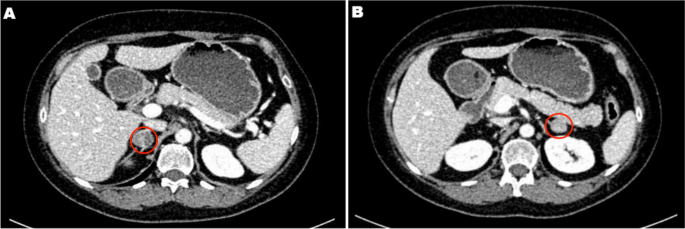
Adrenal CT of the patient: A nodule with a size of approximately 1.6 × 1.5 cm was found in the left adrenal gland, and a nodule with a size of approximately 2.2 × 1.8 cm was found in the right adrenal gland. Irregular mild to moderate enhancement was on enhanced CT, and the surrounding fat gap was clear

Based on the above clinical features, the patient was diagnosed with “non-ACTH-dependent Cushing’s syndrome complicated with PA”. To assess lateralization, adrenal vein sampling (AVS) stimulated by ACTH was performed after obtaining informed consent. The results showed no lateralization of cortisol and aldosterone secretion (Table 2).

Table 2 Results of AVS

After communicating with the patient, the left adrenocortical adenoma was first removed by robot-assisted laparoscopic resection; the thickened adrenal cortex near the left adrenocortical adenoma was also resected during the surgery. The pathological report revealed adrenocortical adenoma, the Weiss score was 1, and immunohistochemistry showed weak CYP11B1 expression in the adenoma and positive CYP11B2 expression in an adjacent nodule. Hypertension was not alleviated after surgery. One month later, purple lines appeared on both sides of the lower abdomen, groin area and inner thigh, accompanied by weight gain, apparent systemic joint pain and fatigue in both lower limbs. The patient was readmitted to the hospital, and examination revealed orthostatic ALD at 11.99 ng/dL, PRA at 0.08 ng/mL/h, angiotensin II at 39.38 ng/L (reference range: 55.3–115.3 ng/L) and ARR at 149.88 (ng/dL)/(ng/mL/h). In addition, ACTH was 2.37 ng/L, serum cortisol was 352.30–353.50–283.90 nmol/L at 8 h-16 h-24 h, 24-h UFC was 112.8 µg, and serum cortisol was 342.10 nmol/L in the morning after the 1 mg dexamethasone suppression test. Enhanced CT of the kidneys and adrenal glands showed no solid nodules or masses in the left adrenal gland, though a nodule with a size of approximately 2.2*1.8 cm was detected in the right adrenal gland. Enhanced CT showed irregular mild to moderate enhancement. Therefore, the diagnosis was still “non-ACTH-dependent Cushing’s syndrome complicated with PA”. Subsequently, the right adrenocortical adenoma and the thickened adrenal cortex near the right adrenocortical adenoma were removed by robot-assisted laparoscopic resection. The pathological report indicated adrenocortical adenoma, and immunohistochemistry showed diffuse homogeneous expression of CYP11B1 and CYP11B2. Antibodies against CYP11B1 (MABS502) and CYP11B1 (MABS1251) were purchased from the Millipore Corporation. There were APMs in the adrenal cortex adjacent to the bilateral cortical adenomas. The fluorescence staining image of the left cortical adenoma is shown in Fig. 2. The immunohistochemistry image of the left adrenal gland is given in Fig. 3 and that of the right adrenal gland in Fig. 4. The immunofluorescence method used in this study was indirect immunofluorescence double staining procedure. Paraffin-embedded human adrenal tissues were prepared using heat-induced epitope retrieval after deparaffinization. Tissue sections were blocked with 5% goat serum in PBS, pH 7.4, containing 0.5% SDS, for 1 h. The slides were incubated with individual primary antibodies at 4℃ overnight, followed by incubation with Alexa Fluor 488-, and Alexa Fluor 647-conjugated secondary antibodies specific to the species of the primary antibodies with DAPI for immunofluorescence staining. Antibodies used included anti-CYP11B1 (Millipore, Cat. No. MABS502, 1:100), anti-CYP11B2(Millipore, Cat. No. MABS1251, 1:100), Alexa Fluor 488-conjugated anti-rat IgG secondary antibody (CYP11B1; Green) and Alexa Fluor 647-conjugated anti-mouse IgG secondary antibody (CYP11B2; Red). Nuclei were stained with DAPI.

Fig. 2

figure 2

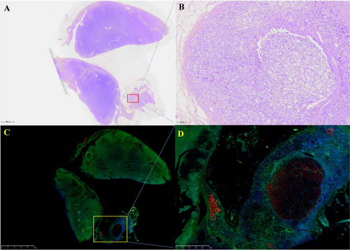
Routine hematoxylin and eosin (H&E) staining and immunofluorescence of the left adrenocortical adenoma (green represents expression of CYP11B1 and red that of CYP11B2). This adrenocortical adenoma and the surrounding cortex was cut into three parts. A and C show the overall appearance of the resected portion, with a nodule adjacent to the adenoma. B shows a neoplastic lesion formed by clear cells (aldosterone-producing cell) within nodules, lacking a fibrous envelope. C clearly shows the weak and diffuse expression of CYP11B1 in adrenocortical adenoma and CYP11B2 expression in a nodule in the cortex adjacent to the adenoma. D shows local enlargement of the aldosterone-producing nodule and three aldosterone-producing micronodules adjacent to it

Fig. 3

figure 3

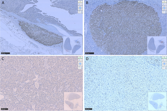
Resected adrenocortical adenoma and part of the adrenal cortex on the left side. A shows expression of Aldosterone-producing micronodule CYP11B2 in the cortex adjacent to the adenoma. B shows an aldosterone-producing nodule with a diameter of approximately 2 mm. C shows weak positive expression of CYP11B1 in the adenoma and D negative expression of CYP11B1 in the aldosterone-producing nodule

Fig. 4

figure 4

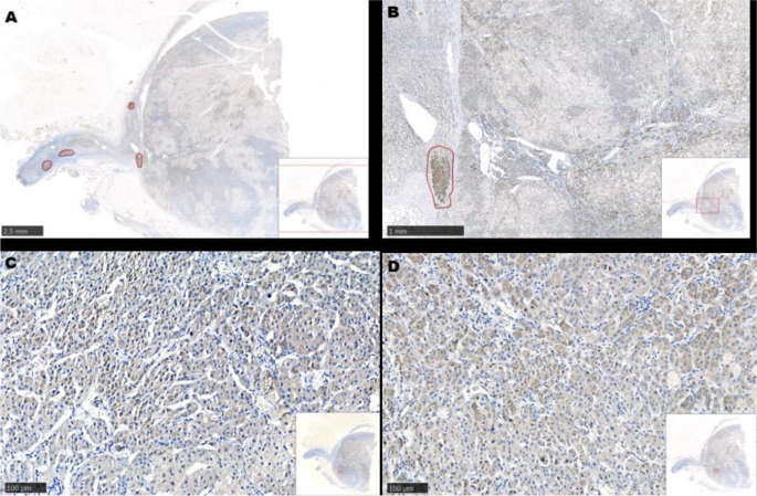
Resected adrenocortical adenoma and part of the adrenal cortex on the right side. A and B show several Aldosterone-producing micronodules (positive expression of CYP11B2) in the cortex adjacent to the adenoma. C shows diffuse expression of CYP11B1 in the adenoma. D shows diffuse expression of CYP11B2 in the adenoma

The Cushing’s syndrome in this patient disappeared after surgery, and glucocorticoids were discontinued after 15 months according to medical advice. Follow-up was conducted for half a year after drug discontinuance, and the patient had no fatigue or dizziness; she was satisfied with the outcomes. Her systolic and diastolic blood pressure remained at 100–120 mmHg and 70–80 mmHg, respectively. During the most recent re-examination, the following results were obtained: (1) orthostatic ALD of 19.1 ng/dL and orthostatic renin concentration of 12.59 µIU/mL, with an aldosterone/renin ratio (ARR) of 1.52; (2) PTC at 8 AM of 247 nmol/L, ACTH of 93.55 ng/L and 24-h UFC of 26.8 µg; (3) parathyroid hormone of 3.86 pmol/L; (4) 25-OH-VitD of 119.5 nmol/L; (5) serum creatinine of 60 µmol/L; (6) serum sodium of 140.4 nmol/L, serum potassium of 3.87 mmol/L and serum calcium of 2.27 mmol/L.

Discussion and conclusions

Adrenal Cushing’s syndrome is caused by excessive autonomic secretion of cortisol induced by adrenal cortical tumours or adrenal cortical hyperplasia; primary aldosteronism (PA) is caused by excessive autonomic secretion of aldosterone induced by adrenal cortical tumours or adrenal cortical hyperplasia. More adverse symptoms occur if aldosterone and cortisol-producing adenomas are present. Specifically, (1) it is more difficult to control hypertension; (2) the incidence of major adverse cardiovascular and cerebrovascular events would increase [4]; (3) glucose intolerance and other metabolic complications would be aggravated [56]; (4) patients would be prone towards osteoporosis [78]; (5) adrenal vein sampling results may be misinterpreted [9]; and (6) adrenal insufficiency may occur after surgery. Therefore, it is of great clinical significance to avoid missed diagnosis of A/CPAs.

Despite many reports on A/CPAs, the majority of these patients may have subclinical Cushing’s syndrome (SCS), and cases of apparent Cushing’s syndrome complicated with PA are rarely reported. In the present case, the clinical manifestation of Cushing’s syndrome were more apparent, and it would be appropriate to call it cortisol-aldosterone cosecretoma. Naoyoshi Onoda et al. [10] reported a case of Cushing’s syndrome caused by a left adrenocortical adenoma (30 mm in diameter) and PA caused by a right adrenocortical adenoma (20 mm in diameter), and Fushimi et al. [3] reported a case of right A/CPA (25 mm*22 mm in size). Interestingly, in the present report, the patient had bilateral A/CPAs, and the clinical manifestations of Cushing’s syndrome became more apparent after unilateral resection was performed. Similar to the above two cases, APMs were found in the adrenal cortex adjacent to the A/CPAs, but aldosterone-producing nodules were found near the cortisol-producing adenoma on the left side.

The biochemical phenotype of APM-inducing autonomic aldosterone secretion has not been clarified. APMs can also be found in the adrenal tissue of 30% of individuals with normal blood pressure [11] and surrounding areas of APA [1213]. APMs do not express CYP11B1 or CYP17A1, which are necessary for the generation of cortisol [1214]. In our patient, the aldosterone-producing nodule in the left adrenal gland may have developed from APM. More than one-third of APMs carry known mutations in CACNA1D and ATP1A1, promoting the generation of aldosterone [1415]. Unfortunately, we did not perform whole-exome sequencing on the DNA of the peripheral blood and adenoma tissues of this patient. Due to the existence of APMs adjacent to the adenoma, it remains unclear whether there is a risk of the relapse of PA in these cases after resection of adrenal the adenoma. Therefore, it was necessary to conduct medical follow-up for this patient.

Remi Goupil et al. performed AVS on 8 patients with cortisol-producing adenoma (CPA), and the results showed that cortisol on the CPA side was higher than that on the contralateral side (median, 6.7 times [range: 2.4–27.2]); P = 0.012]) [16]. There was no significant difference in bilateral cortisol and aldosterone concentrations after AVS in this patient, which is consistent with bilateral A/CPA. Although immunohistochemical results revealed weak expression of CYP11B1 for the first time, expression of cortisol in bilateral adrenal venous blood samples increased significantly after ACTH stimulation. Hence, cortisol was over-synthesized on both sides, and bilateral A/CPAs was definitively diagnosed.

In summary, this case highlights the need for A/CPA screening. The complicated pathological features of these cases impose challenges to our understanding of this disease. Due to the presence of APMs in the adrenal cortex near bilateral adrenocortical adenomas, more clinical data are required to identify whether the disease might relapse after simple resection of the adenoma in these patients. Therefore, further medical follow-up of these patient is needed.

Availability of data and materials

Not applicable.

Abbreviations

CS:
Cushing’s syndrome
PA:
Primary aldosteronism
ACTH:
Adrenocorticotropic hormone
UFC:
Urinary free cortisol
AVS:
Adrenal vein sampling
A/CPA:
Aldosterone-and cortisol producing adenoma
APN:
Aldosterone-producing nodules
APM:
Aldosterone-producing micronodule
CYP:
Cytochrome P450
CT:
Computed tomography
PAC:
Plasma aldosterone concentration
PRA:
Plasma renin activity
ARR:
Aldosterone /renin ratio

References

  1. Stenman A, Shabo I, Ramström A, Zedenius J, Juhlin CC: Synchronous aldosterone- and cortisol-producing adrenocortical adenomas diagnosed using CYP11B immunohistochemistry. SAGE open medical case reports. 2019, 7:2050313×19883770.

  2. Hiraishi K, Yoshimoto T, Tsuchiya K, Minami I, Doi M, Izumiyama H, Sasano H, Hirata YJ. Clinicopathological features of primary aldosteronism associated with subclinical Cushing’s syndrome. Endocr J. 2011;58(7):543–51.

    Article CAS PubMed Google Scholar

  3. Fushimi Y, Tatsumi F, Sanada J, Shimoda M, Kamei S, Nakanishi S, Kaku K, Mune T, Kaneto H. Concurrence of overt Cushing’s syndrome and primary aldosteronism accompanied by aldosterone-producing cell cluster in adjacent adrenal cortex: case report. BMC Endocr Disord. 2021;21(1):163.

    Article PubMed PubMed Central Google Scholar

  4. Araujo-Castro M, BengoaRojano N, FernándezArgüeso M, Pascual-Corrales E, Jiménez Mendiguchía L. García Cano AMCardiometabolic risk in patients with primary aldosteronism and autonomous cortisol secretion. Case-control study. Med Clin (Barc). 2021;157(10):473–9.

    Article CAS PubMed Google Scholar

  5. Petramala L, Olmati F, Concistrè A, Russo R, Mezzadri M, Soldini M, De Vincentis G, Iannucci G, De Toma G, Letizia C. Cardiovascular and metabolic risk factors in patients with subclinical Cushing. Endocrine. 2020;70(1):150–63.

    Article CAS PubMed Google Scholar

  6. Akehi Y, Yanase T, Motonaga R, Umakoshi H, Tsuiki M, Takeda Y, Yoneda T, Kurihara I, Itoh H, Katabami T, et al. High Prevalence of Diabetes in Patients With Primary Aldosteronism (PA) Associated With Subclinical Hypercortisolism and Prediabetes More Prevalent in Bilateral Than Unilateral PA: A Large Multicenter Cohort Study in Japan. Diabetes Care. 2019;42(5):938–45.

    Article CAS PubMed Google Scholar

  7. Shi S, Lu C, Tian H, Ren Y, Chen T. Primary Aldosteronism and Bone Metabolism: A Systematic Review and Meta-Analysis. Front Endocrinol (Lausanne). 2020;11:574151.

    Article PubMed Google Scholar

  8. Petramala L, Zinnamosca L, Settevendemmie A, Marinelli C, Nardi M, Concistrè A, Corpaci F, Tonnarini G, De Toma G, Letizia C. Bone and mineral metabolism in patients with primary aldosteronism. Int J Endocrinol. 2014;2014:836529.

    Article PubMed PubMed Central Google Scholar

  9. Späth M, Korovkin S, Antke C, Anlauf M, Willenberg HS. Aldosterone- and cortisol-co-secreting adrenal tumors: the lost subtype of primary aldosteronism. Eur J Endocrinol. 2011;164(4):447–55.

    Article PubMed Google Scholar

  10. Onoda N, Ishikawa T, Nishio K, Tahara H, Inaba M, Wakasa K, Sumi T, Yamazaki T, Shigematsu K, Hirakawa K. Cushing’s syndrome by left adrenocortical adenoma synchronously associated with primary aldosteronism by right adrenocortical adenoma: report of a case. Endocr J. 2009;56(3):495–502.

    Article CAS PubMed Google Scholar

  11. Williams T, Gomez-Sanchez C, Rainey W, Giordano T, Lam A, Marker A, Mete O, Yamazaki Y, Zerbini M, Beuschlein F, et al. International Histopathology Consensus for Unilateral Primary Aldosteronism. J Clin Endocrinol Metab. 2021;106(1):42–54.

    Article PubMed Google Scholar

  12. Nishimoto K, Tomlins SA, Kuick R, Cani AK, Giordano TJ, Hovelson DH, Liu CJ, Sanjanwala AR, Edwards MA, Gomez-Sanchez CE, et al. Aldosterone-stimulating somatic gene mutations are common in normal adrenal glands. Proc Natl Acad Sci U S A. 2015;112(33):E4591-4599.

    Article CAS PubMed PubMed Central Google Scholar

  13. Omata K, Anand SK, Hovelson DH, Liu CJ, Yamazaki Y, Nakamura Y, Ito S, Satoh F, Sasano H, Rainey WE, et al. Aldosterone-producing cell clusters frequently harbor somatic mutations and accumulate with age in normal adrenals. J Endocr Soc. 2017;1(7):787–99.

    Article CAS PubMed PubMed Central Google Scholar

  14. Omata K, Tomlins SA, Rainey WE. Aldosterone-producing cell clusters in normal and pathological States. Horm Metab Res. 2017;49(12):951–6.

    Article CAS PubMed PubMed Central Google Scholar

  15. Lalli E, Barhanin J, Zennaro MC, Warth R. Local Control of Aldosterone Production and Primary Aldosteronism. Trends Endocrinol Metab. 2016;27(3):123–31.

    Article CAS PubMed Google Scholar

  16. Goupil R, Wolley M, Ahmed AH, Gordon RD, Stowasser M. Does concomitant autonomous adrenal cortisol overproduction have the potential to confound the interpretation of adrenal venous sampling in primary aldosteronism? Clin Endocrinol (Oxf). 2015;83(4):456–61.

    Article CAS PubMed Google Scholar

Download references

Acknowledgements

Not applicable

Funding

This study was supported by the Discipline Excellence Development 1.3.5 Project of West China Hospital, Sichuan University (No. ZYGD18022).

Author information

Authors and Affiliations

  1. Department of Endocrinology and Metabolism, Adrenal Center, West China Hospital of Sichuan University, Chengdu, 610041, Sichuan, ChinaHongjiao Gao, Yan Ren, Tao Chen & Haoming Tian
  2. Department of Endocrinology and Metabolism, The Third Affiliated Hospital of Zunyi Medical University (The First People’s Hospital of Zunyi), Zunyi, Guizhou, ChinaHongjiao Gao
  3. Institute of Clinical Pathology, West China Hospital of Sichuan University, Chengdu, Sichuan, ChinaLi Li & Fei Chen

Contributions

HG, TC researched data and/or wrote the manuscript. LL, FC contributed to immumohistochemical staining. HT, TC, YR contributed to discussion. All authors have read and approved the manuscript.

Corresponding authors

Correspondence to Tao Chen or Haoming Tian.

Ethics declarations

Ethics approval and consent to participate

Not applicable.

Consent for publication

Written informed consent was obtained from the patient for publication of this Case report and any accompanying images. A copy of the written consent is available for review by the Editor of this journal.

Competing interests

We do not have any potential conflicts of interest relevant to this article.

Additional information

Publisher’s Note

Springer Nature remains neutral with regard to jurisdictional claims in published maps and institutional affiliations.

About this article

Cite this article

Gao, H., Li, L., Chen, F. et al. Bilateral co-secretory lesions presenting with coexisting Cushing syndrome and primary aldosteronism: a case report. BMC Endocr Disord 23, 263 (2023). https://doi.org/10.1186/s12902-023-01454-8

Download citation

Share this article

Anyone you share the following link with will be able to read this content:

Get shareable linkProvided by the Springer Nature SharedIt content-sharing initiative

Keywords

Radiation-induced Undifferentiated Malignant Pituitary Tumor After 5 Years of Treatment for Cushing Disease

Abstract

The occurrence of a second neoplasm possibly constitutes an adverse and uncommon complication after radiotherapy. The incidence of a second pituitary tumor in patients irradiated for adrenocorticotropic hormone secreting pituitary adenoma is rare. We report a case of a 40-year-old female with Cushing disease who underwent surgical management followed by radiotherapy. After 5 years of initial treatment, an increase in tumor size was evident at the same location, with a significant interval growth of the parasellar component of the lesion. Histology revealed an undifferentiated highly malignant sarcoma. In the span of next 2 years, the patient was followed with 2 repeat decompression surgeries and radiotherapy because of significant recurrent compressive symptoms by locally invasive malignant tumor. Despite the best efforts, the patient remained unresponsive to multiple treatment strategies (eg, surgical resections and radiotherapy) and succumbed to death.

Introduction

Radiation therapy is a commonly used modality for primary or adjuvant treatment of pituitary adenoma. It is also used as an adjuvant therapy for Cushing disease with persistent or aggressive tumor growth or recurrent disease after surgery. The immediate sequelae of radiotherapy for pituitary tumors include nausea, fatigue, diminished taste and olfaction, and hair loss [1]. One frequent long-term side effect is hypopituitarism. The incidence rate of new-onset hypopituitarism after conventional radiotherapy is approximately 30% to 100% after a follow-up of 10 years, whereas after stereotactic radiosurgery or fractionated radiotherapy, the incidence is approximately 10% to 40% at 5 years [2].

The occurrence of a second neoplasm after cranial radiotherapy constitutes possibly one of the most adverse complications. Tumors such as meningioma, glioma, and sarcoma are the most frequently reported secondary neoplasms after pituitary irradiation [3]. The cumulative probability of a second brain tumor in patients irradiated for pituitary adenoma and craniopharyngioma is approximately 4% [4].

We report 1 such case with detailed clinical, histopathological, and radiological characteristics because of its rarity and associated high mortality of radiation-induced sarcoma.

Case Presentation

The patient first presented at 40 years of age with complaints of weight gain, new-onset diabetes mellitus, hypertension, and cushingoid features in 2014. She was diagnosed with Cushing disease (24-hour urinary cortisol 1384 mcg/24 hours [3819 nmol/24 hours; reference >2 upper limit of normal], low-dose dexamethasone suppression test serum cortisol 16.6 mcg/dL [457.9 nmol/L], ACTH 85 pg/mL [18.7 pmol/L; reference range, <46 pg/mL, <10 pmol/L]) caused by invasive adrenocorticotropic hormone-secreting giant adenoma. The initial imaging revealed a homogenously enhanced pituitary macroadenoma with a size of 42 × 37 × 35 mm with suprasellar extension and encasing both the internal carotid arteries with mass effect on optic chiasma and sellar erosion. The patient underwent tumor excision by endoscopic transsphenoidal transnasal approach. Partial excision of the tumor was achieved because of cavernous sinus invasion. Histopathology and immunohistochemical stains demonstrated a corticotrophin-secreting (ACTH-staining positive) pituitary adenoma with MIB labeling index of 1% to 2%. Because biochemical remission was not achieved (urinary cortisol 794 mcg/24 hours [2191 nmol/24 hours]; ACTH 66 pg/mL [14.5 pmol/L; reference range, <46 pg/mL, <10 pmol/L]), the patient was started on ketoconazole and was received fractionated radiotherapy with a dose of 5040 cGy in 28 fractions.

Diagnostic Assessment

For the next 5 years, at yearly follow-up, 400 mg ketoconazole was continued in view of insufficient control of ACTH secretion. During follow-up, the size of the tumor was stable at approximately 23 × 16 × 33 mm after radiotherapy with no significant clinical and biochemical changes.

Five years after surgery and radiotherapy, the patient developed cerebrospinal fluid rhinorrhea; imaging revealed a cystic transformation of the suprasellar component and increase in the size of the tumor to 39 × 22 × 26 mm, which included visualization of a parasellar component of size 29 × 19 × 15 mm. The patient continued on ketoconazole. The patient was also advised to undergo hypofractionated radiotherapy but did not return for follow-up.

Treatment

In 2021, 1.5 years after the last visit, the patient developed severe headache, altered sensorium, ptosis, focal seizures, and left-sided hemiparesis. During this episode, the patient had an ACTH of 66 pg/mL (14.53 pmol/L; reference range, <46 pg/mL [<10 pmol/L]) and baseline cortisol of 25 mcg/dL (689 nmol/L; reference range, 4-18 mcg/dL [110-496 nmol/L]). Repeat imaging revealed a significant decrease in the suprasellar cystic component but an increase in the size of the parasellar component to 38 × 21 × 25 mm from 29 × 19 × 15 mm, which was isointense on T1 and T2 with heterogeneous enhancement. Significant brain stem compression and perilesional edema was also visible. The patient underwent urgent frontotemporal craniotomy and decompression of the tumor. On pathological examination, the tumor tissue was composed of small pleomorphic round cells arranged in sheets and cords separated by delicate fibrocollagenous stroma. Cells had a round to oval hyperchromatic nucleus with scanty cytoplasm. Areas of hemorrhage, necrosis, and a few apoptotic bodies were seen. The tumor tissue had very high mitotic activity of >10/10 hpf and MIB labeling index of 70%. Immunohistochemistry demonstrated positivity for vimentin, CD99, and TLE-1. Dot-like positivity was present for HMB 45, synaptophysin. INI-1 loss was present in some cells. Ten percent patchy positivity was present for p53. The tumor cells, however, consistently failed to express smooth muscle actin, CD34, Myf-4, epithelial membrane antigen, desmin, LCA, SADD4, CD138, and S-100 protein. ACTH and staining for other hormones was negative. Based on the immunological and histochemical patterns, a diagnosis of high-grade poorly differentiated malignant tumor with a probability of undifferentiated sarcoma was made.

Because of the invasion of surrounding structures and surgical inaccessibility, repeat fractionated radiotherapy was given with a dose of 4500 cGy over 25 fractions at 1.8 Gy daily to the planned target volume via image-guided fractionated radiotherapy. During the next 1.5 years, patient improved clinically with no significant increase in the size of tumor (Fig. 1). The patient was gradually tapered from ketoconazole and developed hypopituitarism requiring levothyroxine and glucocorticoid replacement. There was a significant improvement in the power of the left side and ptosis.


Figure 1.

Contrast-enhanced T1 magnetic resonance imaging dynamic pituitary scan (A, sagittal; B, axial; C, coronal sections) reveals postoperative changes with residual enhancing tumor in the right lateral sella cavity with extension into the right cavernous sinus and parasellar region encasing the cavernous and inferiorly extends through the foramen ovale below the skull base up to approximately 1.5 cm. Anteriorly, it extends up to the right orbital apex and posteriorly extends along the right dorsal surface of clivus.

Outcome and Follow-up

After 1.5 years of reradiation in 2022, the patient again developed palsies of the abducens, trigeminal, oculomotor, and trochlear cranial nerve on the right side and left-sided hemiparesis. A significant increase in tumor size to 50 × 54 × 45 mm with anterior, parasellar, and infratentorial extension was seen (Fig. 2). Again, repeat decompression surgery was done. Two months after surgery, there was no improvement in clinical features and repeat imaging suggested an increased size of the tumor by 30%, to approximately 86 × 68 × 75 mm. Nine years after initial presentation, the patient had an episode of aspiration pneumonia and died.


Figure 2.

Contrast-enhanced T1 magnetic resonance imaging dynamic pituitary images (A, sagittal; B, axial; C, coronal sections) after 1.5 years of a second session of radiotherapy reveal a significant interval increase in size of heterogeneously enhancing irregular soft tissue in sellar cavity with extension into the right cavernous sinus and parasellar region when compared with previous imaging. Superiorly, it extends in the suprasellar region, causing mass effect on the optic chiasma with encasement of the right prechiasmatic optic nerve and right-sided optic chiasma. Inferiorly, the lesion extends into the sphenoid sinus. Posteriorly, there is interval increase in the lesion involving the clivus and extending into the prepontine and interpeduncular cistern. Anteriorly, mass has reached up to the right orbital apex optic nerve canal, which shows mild interval increase.

Discussion

Radiation-induced tumors were initially described by Cahan et al in 1948. They also described the prerequisites for a tumor to be classified as a radiation-induced sarcoma [5]. The modified Cahan criteria state that (1) the presence of nonmalignancy or malignancy of a different histological type before irradiation, (2) development of sarcoma within or adjacent to the area of the radiation beam, (3) a latent period of at least 3 years between irradiation and diagnosis of secondary tumor, and (4) histological diagnosis of sarcoma, can be classified as radiation-induced sarcoma [5].

Our patient fulfilled the criteria for a radiation-induced sarcoma with a highly malignant tumor on histopathology. Radiation-induced sarcomas after functional pituitary tumors, especially Cushing disease, are rarely reported. One of the case reports revealed a high-grade osteoblastic osteosarcoma 30 years after treatment for Cushing disease with transsphenoidal resection and external beam radiotherapy [6]. In our case, there was a lag period of approximately 5 years before the appearance of a second highly undifferentiated, malignant, histologically distinct tumor. The cellular origin of this relatively undifferentiated tumor cannot be determined with certainty. However, the interlacing sarcomatous and adenomatous components resulting from distinct positive immunohistochemistry may indicate that the sarcomatous component may be derived from the preexisting pituitary adenoma.

A hormonally functional pituitary tumor is not itself expected to be associated with an increased risk of secondary malignancy, except in the case of GH-secreting tumors and those with a hereditary cancer syndrome. Although not proven, immunosuppression from hypercortisolism in Cushing disease has been proposed as a contributor to secondary tumor development [7]. Other mechanisms causing increased risk of secondary malignancy can be double-stranded DNA damage and genomic instability caused by ionizing radiation and germline mutations in tumor suppressor genes such as TP53 and Rb [7].

Radiation-induced intracranial tumors were studied in a multicenter, retrospective cohort of 4292 patients with pituitary adenoma or craniopharyngioma. Radiotherapy exposure was associated with an increased risk of a second brain tumor with a rate ratio of 2.18 (95% CI, 1.31-3.62, P < .0001). The cumulative probability of a second brain tumor was 4% for the irradiated patients and 2.1% for the controls at 20 years [7]. In another study including 426 patients irradiated for pituitary adenoma between 1962 and 1994, the cumulative risk of second brain tumors was 2.0% (CI, 0.9-4.4) at 10 years and 2.4% (95% CI, 1.2-5.0) at 20 years. The relative risk of a second brain tumor compared with the incidence in the normal population is 10.5 (95% CI, 4.3-16.7) [8].

The incidence of radiation-induced sarcomas has been estimated at 0.03% to 0.3% of patients who have undergone radiation therapy. The risk of radiation-induced sarcomas increases with field size and dose. In a systemic review and analysis of 180 cases of radiation-induced intracranial sarcomas, the average dose of radiation delivered was 51.4 ± 18.6 Gy and latent period of sarcoma onset was 12.4 ± 8.6 years. A total of 49 cases were developed after radiation treatment of pituitary adenomas (27.2%). The median overall survival time for all patients with sarcoma was 11 months, with a 5-year survival rate of 14.3% [9].

Our patient received approximately 50 Gy twice through fractionated radiotherapy, resulting in larger field size and significantly higher dose than one would expect with a modern stereotactic treatment. Such a high dose of radiation is indeed a risk factor for secondary malignancy. In our patient, in a period of 2 months, there was already >30% tumor growth after recent repeat decompression surgery.

The risk of secondary malignancy is thought to be much lower with stereotactic radiosurgery than conventional external beam radiation therapy, with an estimated cumulative incidence of 0.045% over 10 years (95% CI, 0.00-0.34) [10]. However, long-term follow-up data for patients receiving stereotactic radiation therapy are shorter and thus definitive conclusions cannot be made at this stage.

Our case highlights a rare but devastating long-term complication of pituitary tumor irradiation after Cushing disease. The limited response to various available treatment options defines the aggressive nature of radiation-induced malignancy.

Learning Points

  • The occurrence of a second neoplasm constitutes possibly one of the most adverse and rare complication after radiotherapy.
  • The incidence of radiation-induced sarcomas has been estimated at 0.03% to 0.3% of patients, but cases after Cushing disease are rarely reported.
  • Patients often present with advanced disease unresponsive to various treatment modalities because of aggressive clinical course.
  • New modalities with stereotactic radiosurgery and proton beam therapy are to be reviewed closely for risk assessment of secondary tumor.

Acknowledgments

The authors acknowledge Dr. Ishani Mohapatra for her support with histopathology and interpretation.

Contributors

All authors made individual contributions to authorship. G.B., S.K.M., and V.A.R. were involved in diagnosis and management of the patient. G.B. was involved in the writing of this manuscript and submission. V.P.S. was responsible for patient surgeries. All authors reviewed and approved the final draft.

Funding

The authors received no financial support for the research, authorship, and/or publication of this article.

Disclosures

The authors have nothing to disclose.

Informed Patient Consent for Publication

Signed informed consent could not be obtained from the patient or a proxy but was approved by the treating institute.

Data Availability Statement

Data sharing is not applicable to this article as no data sets were generated or analyzed during the current study.

© The Author(s) 2023. Published by Oxford University Press on behalf of the Endocrine Society.
This is an Open Access article distributed under the terms of the Creative Commons Attribution License (https://creativecommons.org/licenses/by/4.0/), which permits unrestricted reuse, distribution, and reproduction in any medium, provided the original work is properly cited.

Restoration of Intra-patient Variability and Diurnal Range of ACTH with Remission in Cushing’s Disease

The following is a summary of “Diurnal Range and Intra-patient Variability of ACTH Is Restored With Remission in Cushing’s Disease,” published in the November 2023 issue of Endocrinology by Alvarez, et al.


Distinguishing Cushing’s disease (CD) remission from other conditions using single adrenocorticotropic hormone (ACTH) measurements poses challenges. For a study, researchers sought to analyze changes in ACTH levels before and after transsphenoidal surgery (TSS) to identify trends confirming remission and establish ACTH cutoffs for targeted clinical trials.

A retrospective analysis involved 253 CD patients undergoing TSS at a referral center from 2005 to 2019. Remission outcomes were assessed based on postoperative ACTH levels.

Among 253 patients, 223 achieved remission post-TSS. The remission group exhibited higher ACTH variability at morning (AM) (P = .02) and evening (PM) (< .001) time points compared to the nonremission group. Nonremission cases had a significantly narrower diurnal ACTH range (P < .0001). A ≥50% decrease in plasma ACTH from mean preoperative levels, especially in PM values, predicted remission. Absolute plasma ACTH concentration and the ratio of preoperative to postoperative values were associated with nonremission (adj P < .001 and .001, respectively).

ACTH variability suppression was observed in CD, with remission linked to restored variability. A ≥50% decrease in plasma ACTH may predict CD remission post-TSS. The insights can guide clinicians in developing rational outcome measures for interventions targeting CD adenomas.

Source: academic.oup.com/jcem/article-abstract/108/11/2812/7187942?redirectedFrom=fulltext

Adrenocorticotropin-Dependent Ectopic Cushing’s Syndrome: A Case Report

Abstract

Paraneoplastic syndromes are rare and diverse conditions caused by either an abnormal chemical signaling molecule produced by tumor cells or a body’s immune response against the tumor itself. These syndromes can manifest in a variable, multisystemic and often nonspecific manner posing a diagnostic challenge.

We report the case of an 81-year-old woman who exhibited severe hypokalemia, metabolic alkalosis, and worsening hyperglycemia. The investigation was consistent with adrenocorticotropin (ACTH)-dependent Cushing’s syndrome and, eventually, the patient was diagnosed with stage IV primary small-cell lung cancer (SCLC).

SCLC is known to be associated with paraneoplastic syndromes, including Cushing’s syndrome caused by ectopic adrenocorticotropin (ACTH) secretion. Despite being associated with very poor outcomes, managing these syndromes can be challenging and may hold prognostic significance.

Introduction

Adrenocorticotropin (ACTH)-dependent Cushing’s syndrome (CS) is caused by excessive ACTH production by corticotroph (Cushing’s disease (CD)) or nonpituitary (ectopic) tumors, leading to excessive cortisol production. Ectopic ACTH syndrome (EAS) is a rare condition, accounting for 10 to 20% of all cases of ACTH-dependent CS and 5 to 10% of all types of CS [1]. The normal glucocorticoid-induced suppression of ACTH is reduced in ACTH-dependent CS, especially with ectopic ACTH production. Studies show that a wide variety of neoplasms, usually carcinomas rather than sarcomas or lymphomas, have been associated with EAS. Most cases are caused by neuroendocrine tumors of the lung, pancreas, or thymus, in which the hypercortisolism state is not apparent clinically, resulting, all too often, in delayed diagnosis [2,3].

Current diagnostic tests for EAS aim to confirm high cortisol levels, the absence of a cortisol circadian rhythm, as well as the reduced response to negative feedback from glucocorticoid administration, and imaging to identify the site of ACTH production.

Prompt diagnosis and management are crucial in EAS, highlighting the importance of physician awareness and early recognition of this syndrome.

Treatment options depend on the underlying tumor. Surgical removal is often the primary approach, followed by radiation therapy or chemotherapy. Additionally, medications to control cortisol levels may be necessary to manage the various comorbid conditions associated with CS, such as cardiovascular disease, diabetes, electrolyte imbalances, infections and thrombotic risk [4,5].

Case Presentation

We report the case of an 81-year-old woman with a fully active performance status (ECOG 0) and a medical history of diabetes, hypertension, dyslipidemia, and depressive disorder. She was admitted to an internal medicine ward due to an acute hydroelectrolytic disorder, including metabolic alkalosis, severe hypokalemia (2 mmol/L), hypochloremia (85 mmol/L), hypocalcemia (0.95 mmol/L), hypophosphatemia (1.4 mg/dL), hypomagnesemia (0.9 mg/dL), and hyperlactatemia (5.8 mmol/L), after she reportedly self-medicated herself with higher doses of metformin (four to five pills a day) due to high blood glucose levels. The patient presented with asthenia, nausea, vomiting, and diarrhea for three days and reported uncontrolled blood glucose levels for the last eight days.

The physical examination was unremarkable, without any altered mental status or signs of infection. Arterial blood gas samples showed metabolic alkalemia (pH 7.59) and hyperlactatemia, associated with severe hypokalemia, normal bicarbonate (27 mmol/L), and mildly elevated glycemia and ketonemia (232 mg/dL and 1.7 mmol/L, respectively). Lab tests confirmed the serum potassium levels as well as the other aforementioned electrolyte disturbances. Kidney function and hepatic enzymes were normal. Considering the possible relationship between the electrolyte disorder and the gastrointestinal presentation, the patient was given intravenous (IV) fluids and received potassium and magnesium replacement therapy.

Despite receiving 200 milliequivalents (mEq) of IV potassium chloride and 4 grams of magnesium sulfate, in the first 48 hours, the ion deficits persisted. Given the persistent electrolyte derangement, the patient was admitted to the Internal Medicine ward for etiological investigation and monitoring of ionic correction. The initial period was remarkable for refractory hypokalemia and uncontrolled diabetes under respective therapeutic measures, including 80 to 130 mEq of IV potassium chloride and progressive titration of spironolactone to 200 mg a day. Laboratory investigation revealed high parathormone levels (PTHi 167 pg/mL; reference range: 10-65 pg/mL), vitamin D deficiency (3.3 ng/mL; reference range >20 ng/mL) and apparent ACTH-dependent hypercortisolism (serum cortisol 80.20 ug/dL; ACTH 445 pg/mL), as well as high urinary potassium and glucose concentrations (190 mEq/24 h and 21161 mg/24 h). A dexamethasone suppression test was performed twice (standard low and high dose) without any changes in cortisol levels, leading to the suspicion of a CS caused by abnormally high ACTH production. Cranioencephalic computed tomography (CT) and magnetic resonance imaging (MRI) were performed, excluding the presence of pituitary anomalies. A follow-up whole-body CT scan was performed, revealing a suspicious pulmonary mass in the left lower lobe, associated with ipsilateral hilar lymphadenopathy and hepatic and adrenal gland lesions suggestive of secondary involvement. An endobronchial ultrasound bronchoscopy and biopsy were performed, documenting anatomopathological findings of small-cell lung carcinoma with a Ki67 expression of 100% (Figures 13).

Pulmonary-mass-(SCLC)-in-the-left-lower-lobe-with-ipsilateral-hilar-lymphadenopathy-and-pleural-effusion.
Figure 1: Pulmonary mass (SCLC) in the left lower lobe with ipsilateral hilar lymphadenopathy and pleural effusion.

SCLC: small-cell lung cancer.

Secondary-involvement-of-the-liver-with-hypodense-multilobar-hepatic-lesions-(arterial-phase).
Figure 2: Secondary involvement of the liver with hypodense multilobar hepatic lesions (arterial phase).
Bilateral-suprarenal-lesions-suggestive-of-secondary-involvement.
Figure 3: Bilateral suprarenal lesions suggestive of secondary involvement.

The patient was referred to oncology, and chemotherapy was deferred, considering the infectious risk associated with hypercortisolism.

The patient started metyrapone 500 mg every eight hours, resulting in a reduction in cortisol levels and control of hypokalemia. Later on, a fluorodeoxyglucose-positron emission tomography (FDG-PET) scan was performed, confirming disseminated disease with additional bone involvement. Unfortunately, despite endocrinological stabilization, the patient’s condition worsened, and she ended up dying one month after the diagnosis.

Discussion

When this patient was admitted, it was assumed that the metabolic alkalosis and various electrolyte disturbances were related to the gastrointestinal presentation and hyperlactatemia secondary to metformin overdose. However, the unusual persistence and refractory hypokalaemia raised some concerns that an alternative etiology might be involved and incited subsequent testing.

The high cortisol levels were unexpected given the subclinical presentation, which seems to be more frequent in cases of EAS. In fact, because of this, the true incidence of EAS is unknown and probably underdiagnosed since patients often have subclinical presentations and do not exhibit catabolic features.

Since the patient wasn’t on any steroid medication, the association between the high cortisol and ACTH levels, non-responsive to the dexamethasone suppression test, along with the absence of a pituitary lesion, raised suspicion of a probable EAS, which was later confirmed by the body CT scan and endobronchial ultrasound (EBUS).

EAS is a rare disease with a poor prognosis. It reportedly occurs in 3.2 to 6% of neuroendocrine neoplasms, and the tumor often originates in the lung, thyroid, stomach, and pancreas. Locoregional and/or distant metastasis can be seen at the time of diagnosis in 15% of typical carcinoids and about half of atypical carcinoids with visible primaries [6,7].

The presence of a typical CS presentation, with or without electrolyte abnormalities, should raise suspicion and serum levels of both ACTH and cortisol should be assessed to determine if they are elevated and to distinguish between an ACTH-dependent (pituitary or nonpituitary ACTH-secreting tumor) and an independent mechanism (e.g., from an adrenal source). The diagnosis of CS is established when at least two different first-line tests are unequivocally abnormal and cannot be explained by any other conditions that cause physiologic hypercortisolism. Additional evaluation is performed to rule out a pituitary origin (with brain MRI) and to assess for a possible ectopic ACTH-secreting tumor.

In the aforementioned case, the production of ACTH was caused by primary neuroendocrine SCLC. The recommended approach to EAS involves the initial normalization of serum cortisol levels and the treatment of related comorbidities before performing a complete diagnostic evaluation and addressing the underlying cause [5-7]. This approach seems to improve survival and prevent complications such as sepsis following a combined steroid-induced immunosuppression and chemotherapy-induced agranulocytosis [6,7].

Direct therapies vary according to the tumor, but surgery is usually the first line of treatment (transsphenoidal surgery in cases of CD or tumor resection in cases of non-metastatic EAS). However, our patient presented with stage IV SCLC with EAS, in which chemotherapy remains the first-line treatment. SCLC patients with EAS have a poorer prognosis than those without EAS, with a life expectancy of only three to six months. This makes early diagnosis more important [2,7], as controlling the high cortisol levels and then administering systemic chemotherapy may achieve longer survival [8].

Apart from systemic chemotherapy, ketoconazole (widely accepted but highly toxic), metyrapone, mitotane (adrenocortical suppressant drug with significant side effects), and mifepristone (glucocorticoid antagonist, mainly used for the treatment of hyperglycemia in CS) can be used to reduce circulating glucocorticoids. Moreover, thromboprophylaxis and Pneumocystis jirovecii pneumonia prophylaxis should be started.

Because ketoconazole may increase the risk of chemotherapy toxicity by inhibiting cytochrome P450 3A4, metyrapone has been reported to be a better choice [5,7].

Nonetheless, administration of chemotherapy in the setting of a hypercortisolism-induced immunosuppressive state, cancerous background and metabolic disorders featuring electrolyte disturbance and hyperglycemia, aggravate the condition and can be life-threatening. Thus, a palliative approach can sometimes be reasonable.

Conclusions

The diagnosis of CS is a three-step process that includes its suspicion based on the patient’s laboratory and semiologic findings, the documentation of hypercortisolism, and the identification of its cause, which can be either ACTH-dependent or independent.

The ectopic secretion of ACTH (EAS) by nonpituitary tumors is a relatively rare cause of CS and often presents as paraneoplastic syndromes, adding therapeutic and prognostic concerns.

This case, in particular, highlights the importance of seeking alternative explanations for common electrolyte disturbances, particularly when they don’t resolve promptly. Clinicians should be aware of EAS and its frequent subclinical presentation in order to initiate the diagnostic workup as soon as suspicion arises.

References

  1. Hayes AR, Grossman AB: The ectopic adrenocorticotropic hormone syndrome: rarely easy, always challenging. Endocrinol Metab Clin North Am. 2018, 47:409-25. 10.1016/j.ecl.2018.01.005
  2. Ilias I, Torpy DJ, Pacak K, Mullen N, Wesley RA, Nieman LK: Cushing’s syndrome due to ectopic corticotropin secretion: twenty years’ experience at the National Institutes of Health. J Clin Endocrinol Metab. 2005, 90:4955-62. 10.1210/jc.2004-2527
  3. Lacroix A, Feelders RA, Stratakis CA, Nieman LK: Cushing’s syndrome. Lancet. 2015, 29:913-27. 10.1016/S0140-6736(14)61375-1
  4. Nieman LK: Molecular derangements and the diagnosis of ACTH-dependent Cushing’s syndrome. Endocr Rev. 2022, 43:852-77. 10.1210/endrev/bnab046
  5. Nieman LK, Biller BM, Findling JW, Murad MH, Newell-Price J, Savage MO, Tabarin A: Treatment of Cushing’s syndrome: an endocrine society clinical practice guideline. J Clin Endocrinol Metab. 2015, 100:2807-31. 10.1210/jc.2015-1818
  6. Bostan H, Duger H, Akhanli P, et al.: Cushing’s syndrome due to adrenocorticotropic hormone-secreting metastatic neuroendocrine tumor of unknown primary origin: a case report and literature review. Hormones (Athens). 2022, 21:147-54. 10.1007/s42000-021-00316-z
  7. Richa CG, Saad KJ, Halabi GH, Gharios EM, Nasr FL, Merheb MT: Case-series of paraneoplastic Cushing syndrome in small-cell lung cancer. Endocrinol Diabetes Metab Case Rep. 2018, 2018:4. 10.1530/EDM-18-0004
  8. Zhang HY, Zhao J: Ectopic Cushing syndrome in small cell lung cancer: a case report and literature review. Thorac Cancer. 2017, 8:114-7. 10.1111/1759-7714.12403

From https://www.cureus.com/articles/198133-adrenocorticotropin-dependent-ectopic-cushings-syndrome-a-case-report#!/

Interpetrosal Sphingosine-1-Phosphate Ratio Predicting Cushing’s Disease Tumor Laterality and Remission After Surgery

Background: Cushing’s disease (CD) poses significant challenges in its treatment due to the lack of reliable biomarkers for predicting tumor localization or postoperative clinical outcomes. Sphingosine-1-phosphate (S1P) has been shown to increase cortisol biosynthesis and is regulated by adrenocorticotropic hormone (ACTH).

Methods: We employed bilateral inferior petrosal sinus sampling (BIPSS), which is considered the gold standard for diagnosing pituitary sources of CD, to obtain blood samples and explore the clinical predictive value of the S1P concentration ratio in determining tumor laterality and postoperative remission. We evaluated 50 samples from 25 patients who underwent BIPSS to measure S1P levels in the inferior petrosal sinuses bilaterally.

Results: Serum S1P levels in patients with CD were significantly higher on the adenoma side of the inferior petrosal sinus than on the nonadenoma side (397.7 ± 15.4 vs. 261.9 ± 14.88; P < 0.05). The accuracy of diagnosing tumor laterality with the interpetrosal S1P and ACTH ratios and the combination of the two was 64%, 56% and 73%, respectively. The receiver operating characteristic curve analysis revealed that the combination of interpetrosal S1P and ACTH ratios, as a predictor of tumor laterality, exhibited a sensitivity of 81.82% and a specificity of 75%, with an area under the curve value of 84.09%. Moreover, we observed that a high interpetrosal S1P ratio was associated with nonremission after surgery. Correlation analyses demonstrated that the interpetrosal S1P ratio was associated with preoperative follicle-stimulating hormone (FSH), luteinizing hormone (LH), and postoperative ACTH 8 am levels (P < 0.05).

Conclusion: Our study demonstrated a significant association between the interpetrosal S1P ratio and tumor laterality, as well as postoperative remission in CD, suggesting that the interpetrosal S1P ratio could serve as a valuable biomarker in clinical practice.

1 Introduction

Cushing’s disease (CD), also known as adrenocorticotropic hormone (ACTH)-secreting pituitary adenoma, arises from the pituitary corticotroph cells and induces endogenous hypercortisolism by stimulating the adrenal glands to produce excessive amount of cortisol (1). Patients with CD typically exhibit symptoms of hypercortisolism, such as hypertension, diabetes, purplish skin striae, mental disturbances, hyposexuality, hirsutism, menstrual disorders, acne, fatigue, obesity, and osteoporosis (1). The overall mortality of patients with CD is twice that of the general population, and if left untreated, hypercortisolism resulting from CD increases this rate to approximately four times the expected value (24). Transsphenoidal surgery continues to be the primary treatment for CD (5). However, previous studies reported variable remission rates, ranging from 45% to 95% (68). Long-term follow-up data have revealed recurrence in 3–66% of patients who had initially achieved complete remission (910). The rate of surgical remission in CD can be influenced by various factors, including the size and location of the tumor, expertise of the neurosurgeon, and criteria used for assessing remission (11). Preoperative clinical variables, such as age, gender, disease duration, and severity of clinical signs and symptoms, cannot reliably identify patients at a higher risk of nonremission (1213). Therefore, predicting postsurgical remission in CD remains a challenging goal.

Accumulating evidence has shown that sphingosine-1-phosphate (S1P), an intracellular pleiotropic bioactive sphingolipid metabolite synthesized by sphingosine kinase 1 (SPHK1), plays a pivotal role in diverse endocrine disorders (1416). Overexpression of SPHK1 promotes the progression of multiple neuroendocrine tumors (1718). ACTH can rapidly activate sphingolipid metabolism, causing an increase in S1P secretion in the adrenal cortex (19). Furthermore, the activation of S1P signaling in H295R cells, a human adrenocortical tumor cell line, has been suggested to induce increased transcription of hormone-sensitive lipase and steroidogenic acute regulatory protein, ultimately elevating cortisol production (20). Recently, surgical removal of ACTH-secreting adenoma has been reported to cause a decline in sphingomyelin levels (21). However, whether they have a similar role in the pituitary gland remains to be investigated.

Bilateral inferior petrosal sinus sampling (BIPSS) is a highly effective procedure for diagnosing pituitary sources of ACTH in CD (2223). Contemporaneous differences in ACTH concentration during venous sampling between the two sides of the adenoma can predict the location of the adenoma within the pituitary (on the side of the gland with a microadenoma) and may guide surgical treatment in cases with inconclusive magnetic resonance imaging findings. Previous studies demonstrated that an ACTH gradient of ≥1.4 between the inferior petrosal sinuses can indicate microadenoma lateralization in patients with CD (2426). However, the correct lateralization only occurs in 57–68% of all cases (2729).

Therefore, we analyzed the clinical behavior of a well-characterized cohort of patients with CD who underwent BIPSS before surgery. We measured the difference in the concentration of S1P in bilateral petrosal sinus blood samples and explored the clinical predictive value of the S1P concentration ratio in determining tumor laterality and postoperative remission.

2 Materials and methods

2.1 Patients and study design

This study was conducted at a tertiary center, involving a cohort of 25 patients diagnosed with CD who had undergone BIPSS and surgery, with a minimum follow-up duration of 2 years. Comprehensive chart reviews were conducted to collect data on demographics, clinical characteristics, pituitary imaging findings, tumor pathology, and biochemical tests.

The criteria used for diagnosing CD encompassed the presence of characteristic signs and symptoms of hypercortisolism, along with biochemical evaluation of two urinary free cortisol measurements exceeding the normal range for the respective assay, serum cortisol level >1.8 μg/dL (50 nmol/L) after an overnight 1-mg dexamethasone suppression test, and two late-night salivary cortisol measurements exceeding the normal range for the respective assay (30). A diagnosis of Cushing’s syndrome was established if the patient had positive test results for at least two of the three aforementioned tests. Adrenal insufficiency was diagnosed if patients exhibited symptoms or signs of adrenal insufficiency or if serum cortisol levels were ≤3 μg/dL, even in the absence of clinical signs or symptoms. Remission was defined as normalization of the levels of 24-h urinary free cortisol, late-night salivary cortisol, and overnight 1-mg dexamethasone suppression test in patients without concurrent central adrenal insufficiency after surgery (31).

2.2 Patients and tissue/serum samples

Surgical specimens of CD-affected tissues were collected from Xiangya Hospital, Central South University. Three normal pituitary tissues were obtained from cadaveric organ donors without any history of endocrine disease (Central South University). A total of 25 CD tissue samples were obtained for immunohistochemistry analysis. This study was conducted in compliance with the Helsinki Declaration and was ethically approved by the Xiangya Hospital Ethics Committee, Xiangya Hospital (Changsha, China). Tumor samples and corresponding clinical materials were obtained with written consent from all patients.

2.3 BIPSS

After obtaining informed consent, BIPSS was performed using standard techniques described in previous studies (3233). Briefly, the patient’s head was immobilized to ensure midline positioning and prevent any potential bias towards asymmetric pituitary drainage by the petrosal sinuses. After placing peripheral catheters and cannulating both inferior petrosal sinuses, blood samples were collected at baseline and at 3, 5, 10, and 15 min following intravenous administration of DDAVP, which stimulates pituitary production of ACTH. Additional samples for experimental purposes were collected immediately following the 15-min sample collection to avoid interference with the patient’s diagnostic study.

2.4 Measurement of baseline plasma S1P concentration

Blood samples were obtained from both petrosal sinuses and were centrifuged to remove cellular components. Samples that exhibited hemolysis or coagulation were excluded from the study. Plasma samples were stored at −80°C. The S1P levels in plasma were analyzed using a S1P competitive ELISA kit (Echelon Biosciences, Salt Lake City, UT) according to the manufacturer’s instructions (34).

2.5 Immunofluorescence staining

The pituitary tissues were post-fixed and dehydrated with alcohol as follows: 70% for 24 h, 80% for 3 h, 90% for 4 h, 95% for 3 h, and finally in absolute alcohol for 2 h. Tissue slices with a 5-μm thickness were cut using a microtome (Thermo Fisher Scientific), blocked with 3% BSA, and then treated with primary antibodies to SPHK1 (CST, #3297) and ACTH (Proteintech, CL488-66358). Subsequently, the tissue slides were incubated with Alexa Fluor 488-conjugated anti-rabbit (Invitrogen, A21206, 1:200) or Alexa Fluor 555-conjugated anti-rabbit (Invitrogen, A21428, 1:200) secondary antibodies. Specimens were visualized and imaged using a fluorescence microscope.

2.6 Statistical analysis

The Mann–Whitney U test was used to assess the clinical–molecular associations in adenoma samples, whereas the chi-square test was used to compare categorical data. The Kruskal–Wallis analysis and ANOVA were conducted for multiple comparisons. Statistical analyses were conducted using SPSS v20 and GraphPad Prism version 7. All results were presented in graphs and tables as median ± interquartile range. The distribution of each parameter was presented as the minimum–maximum range. Parametric or nonparametric statistical tests were applied, as appropriate, after testing for normality. The receiver operating characteristic curve was used to determine the cut-off value for predicting tumor laterality. Pearson correlation analyses was used to examine the correlations between variables. Proportions were expressed as percentages, and significance was defined as P < 0.05.

3 Results

3.1 Clinical characteristics of remission and nonremission in patients with CD

This study included 25 patients with CD who underwent BIPSS before surgery (Figure 1). Among them, 12 patients had microadenomas, whereas the remaining 13 had inconclusive magnetic resonance imaging findings; clinicopathological data are summarized in Supplementary Table 1Table 1 displays the demographics of patients who achieved remission (n = 16) and those who did not (n = 9). No significant differences were observed in terms of sex, age at diagnosis, or radiological variables between patients who achieved and those who did not achieve remission (P > 0.05). Patients who achieved remission exhibited a higher prevalence of emotional lability (P < 0.05). However, no significant differences were observed in other parameters (P > 0.05).

Figure 1
www.frontiersin.orgFigure 1 Flowchart of the screening process employed to select eligible participants for the study.

Table 1
www.frontiersin.orgTable 1 Baseline clinical features of patients with pituitary tumors secreting adrenocorticotropin.

Several recent studies have established morning cortisol level measured on postoperative day 1 (POD1) as a predictive biomarker for long-term remission of CD (3536). For biochemical features, patients who did not achieve remission exhibited higher serum cortisol (19.16 ± 5.55 vs. 5.95 ± 1.42; P = 0.014) and median serum (8 am) ACTH (10.26 ± 8.24 vs. 5.15 ± 3.68; P = 0.042) levels on POD1. No significant differences were observed in the preoperative baseline 4 pm serum cortisol levels, preoperative baseline 0 am serum cortisol levels, preoperative 8 pm ACTH levels, 4 pm ACTH levels, and 0 am ACTH levels (P > 0.05) (Table 2). In addition preoperative FT3, FT4, TSH, GH, FSH, LH, and PRL levels were comparable in patients with and without remission.

Table 2
www.frontiersin.orgTable 2 Baseline clinical and biochemical features of patients with pituitary tumors secreting adrenocorticotropin.

3.2 Overexpression of SPHK1 and higher concentrations of serum S1P on the tumor side in patients with CD

Prior studies have demonstrated that ACTH acutely activates SPHK1 to increase S1P concentrations (19). Upregulation of SPHK1 is associated with poor prognosis in endocrine-related cancer (171821). To investigate the role of SPHK1 in CD, we performed a heatmap analysis of key genes involved in phospholipid metabolism and signaling pathways in CD adenomas and surrounding normal tissues using the GEO dataset (GEO208107). This analysis revealed the activation of crucial genes involved in phospholipid metabolism and signaling pathways in ACTH-secreting pituitary adenomas (Supplementary Figure 1). Subsequently, we compared the association between pituitary SPHK1 expression and proopiomelanocortin, corticotropin-releasing hormone, corticotropin releasing hormone receptor 1, and corticotropin releasing hormone receptor 2 in pituitary tumor tissues and identified a positive correlation between SPHK1 and ACTH tumor-related genes in the TNM plot database (Supplementary Figure 2). To investigate the potential role of SPHK1 in CD, we compared the expression values of SPHK1 in the normal pituitary tissues and those obtained from patients with CD in the remission/nonremission groups. Immunofluorescence staining (Figures 2A, BSupplementary Figure 3) revealed an increased number of double-positive cells for SPHK1 and ACTH in CD-affected pituitary tissues than those in the normal pituitary tissues. Furthermore, the proportion of double-positive cells for SPHK1 and ACTH was significantly higher in the nonremission CD adenomas tissues than that in the remission CD adenomas. Furthermore, we investigated the concentration of S1P in bilateral petrosal sinus blood samples and observed that the concentration was significantly higher on the adenoma side than that on the nonadenoma side (397.7 ± 15.4 vs. 261.9 ± 14.88; P < 0.05, Figure 2C). Thus, these findings suggested a close association between S1P concentration and the development of ACTH-secreting tumor.

Figure 2
www.frontiersin.orgFigure 2 (A) Representative images of immunofluorescence double staining for SPHK1 (green) and ACTH (pink) in normal pituitary glands and ACTH-secreting pituitary adenomas from the remission and nonremission groups (Normal: n = 3, ACTH pituitary adenoma: remission vs. nonremission: n = 16 vs. 9); scale bars: 100-μm upper and 50-μm lower. (B) Quantitative analysis; white arrows indicate double-positive cells for ACTH and SPHK1. (C) The concentration of S1P in the plasma obtained from the inferior petrosal sinus of the adenoma side and nonadenoma side. ***P < 0.001. Bar represents mean ± SD.

3.3 Combination of interpetrosal S1P and ACTH ratios improved the diagnostic performance for adenoma laterality

The pathology of patients with CD was classified based on adenomatous tissue with ACTH-positive immunostaining into adenoma or nonadenoma sides. To evaluate the correlation between the interpetrosal S1P ratio lateralization and tumor location, we compared the accuracy of predicting tumor laterality using the interpetrosal S1P ratio (>1) and interpetrosal ACTH ratio (>1.4) (the interpetrosal ACTH ratio >1.4 is acknowledged for its positive role in predicting tumor laterality), as well as their combination. Our results indicated that using the interpetrosal S1P or ACTH ratios alone yielded accuracies of 64% and 56% respectively. Notably, the combination of both demonstrated a significantly improved accuracy of 73% (Figure 3A).

Figure 3
www.frontiersin.orgFigure 3 (A) Bar graph illustrating the accuracy of predicting tumor laterality. (B) Receiver operating characteristic (ROC) curve analysis of interpetrosal ACTH ratio to predict tumor location. (C) ROC curve analysis of the interpetrosal S1P ratio to predict tumor location. (D) ROC curve analysis of the combination of the interpetrosal S1P and ACTH ratios to predict tumor location.

Thereafter, the receiver operating characteristic analysis was performed to determine the role of predicting tumor laterality. In particular, the interpetrosal ACTH ratio with an AUC of 75.32% (95% CI: 60.06–97.46%, P < 0.05) and the interpetrosal S1P ratio demonstrated a clinically significant diagnostic accuracy for lateralization, with an AUC of 79.17% (95% CI: 44.40–85.84%, P < 0.05). Furthermore, combining the interpetrosal S1P and ACTH ratios generated an receiver operating characteristic curve with an AUC of 84.09% (95% CI: 52.3–96.77%, P < 0.05) for predicting lateralization with tumor location (cutoff value: interpetrosal S1P ratio ≥1.06, interpetrosal ACTH ratio ≥2.8, 81.82% sensitivity, and 75% specificity) (Figures 3B–D).

3.4 Interpetrosal S1P ratio serves as a predictive factor for early remission in CD

To investigate whether the interpetrosal S1P ratio is associated with early postoperative remission in CD, we compared the baseline interpetrosal S1P ratio between patients with CD in the remission and nonremission groups. Interestingly, we observed that the nonremission group exhibited higher interpetrosal S1P ratios than those of the remission group (median, 1.28 ± 0.25 vs. 1.10 ± 0.09, P = 0.012) (Figure 4).

Figure 4
www.frontiersin.orgFigure 4 Left picture: Scatter plot of bilateral S1P concentrations in the remission and nonremission groups; the slope represents the interpetrosal S1P ratio, blue dots represent the remission group, and red dots represent the nonremission group. Right picture: The interpetrosal S1P ratio in the remission and nonremission groups. *P < 0.05. Bar represents mean ± SD.

To investigate potential factors affecting the interpetrosal S1P ratio, we compared the correlation between interpetrosal S1P ratio and various clinical indicators. This analysis revealed that the interpetrosal S1P ratio positively correlated with preoperative FSH and LH levels, as well as with postoperative 8 am ACTH levels. No significant difference was observed between the interpetrosal S1P ratio and other indicators (Supplementary Figure 4).

4 Discussion

The use of BIPSS involves collection of samples from each inferior petrosal sinus simultaneously, enabling a direct comparison of ACTH concentrations between the left and right petrosal sinuses. BIPSS is used for two purposes: 1) to assist in the differential diagnosis of Cushing’s syndrome; and 2) to determine which side of the pituitary gland contains an adenoma in patients with CD. The interpetrosal ACTH ratio is also useful in determining the location/lateralization of pituitary microadenomas (243037), thereby providing guidance to the neurosurgeon during surgery.

To our knowledge, this is the first study to demonstrate that serum S1P levels in patients with CD are significantly higher on the adenoma side of the inferior petrosal sinus than on the nonadenoma side. The interpetrosal S1P ratio exhibited a positive significance in predicting tumor laterality, and the predictive performance was improved when S1P was combined with the interpetrosal ACTH ratio. Notably, the interpetrosal S1P ratio exhibited a positive significance in predicting remission after surgery. Furthermore, the interpetrosal S1P ratio demonstrated a positive and significant correlation with preoperative FSH and LH levels, as well as 8 am ACTH levels on POD1.

ACTH is recognized for its role in controlling the expression of genes involved in steroid production and cortisol synthesis in the human adrenal cortex through sphingolipid metabolism (19). Specifically, ACTH rapidly stimulates SPHK1 activity, leading to an increased in S1P levels, which in turn, increases the expression of multiple steroidogenic proteins (20). Our study demonstrated that higher S1P concentrations were present on the tumor side than on the nontumor side in patients with CD, indicating that the regulatory relationship between ACTH and S1P also exists in ACTH-secreting pituitary adenomas. Several pieces of evidence have supported the potential relationship between S1P and the occurrence of CD. Interestingly, SPHK1 and S1P are known to be integral to the regulation of epidermal growth factor receptor (EGFR) (38), which is highly expressed in human corticotropinomas, where it triggers proopiomelanocortin (the precursor of ACTH) transcription and ACTH synthesis (39). Blocking EGFR activity with an EGFR inhibitor can attenuate corticotroph tumor cell proliferation (40). Furthermore, SPHK1 and proopiomelanocortin share a common transcriptional coactivator, P300 (4142). Notably, S1P also directly binds to and inhibits histone deacetylase 2, thereby regulating histone acetylation and gene expression (43). Notably, histone deacetylase 2 expression is deficient in ACTH-pituitary adenomas in CD, contributing to glucocorticoid insensitivity (44), which is a hallmark of CD and a feature associated with nonremission. These studies further demonstrated an association between high S1P ratio and nonremission of CD. Our study, for the first time, established an association between SPHK1/S1P and ACTH adenoma. Nevertheless, further experimental verification is required to confirm the existence of common pathways linking SPHK1 and ACTH. Thus, these findings indicated that the S1P ratio can, to some extent, reflect the differences in ACTH levels and may serve as a surrogate marker for detecting ACTH-secreting pituitary adenomas.

BIPSS is a highly effective procedure for diagnosing pituitary sources of ACTH in CD and remains the gold standard diagnostic method. However, some findings indicated certain limitations associated with the use of the inferior petrosal sinus sampling (IPSS) method in predicting tumor lateralization. The possible causes of error include asymmetrical or underdeveloped petrosal sinus anatomy and placement of the catheter (27). The present study revealed a notable increase in the interpetrosal ACTH ratio among patients with accurate predictions of tumor laterality than among those with inaccurate predictions, although the positive predictive value remained low. These findings suggested that other mechanisms may exist that contribute to false-positive results. The limitations on lateralization highlighted the need for further research to understand the underlying mechanisms contributing to the accuracy of IPSS in predicting tumor lateralization. Further investigation is required to understand these potential mechanisms and improve the accuracy of IPSS in predicting tumor lateralization.

We observed that the interpetrosal S1P ratio was slightly more effective than the ACTH ratio in predicting tumor laterality. However, combining both methods significantly improved the diagnostic sensitivity and specificity. These results have important implications for clinical practice as accurate tumor lateralization is essential for the correct management and treatment of pituitary adenomas. Overall, these findings highlighted the importance of using multiple measures in predicting tumor lateralization and suggested that combining measures may be more effective than relying on any single measure alone. Future research should investigate additional measures to improve the accuracy of tumor lateralization and optimize the use of existing measures for making clinical decisions.

The initial treatment recommendation for CD is surgery. However, long-term surveillance is necessary because of the high recurrence rate (12). Therefore, identifying patients who are at a greater recurrence risk would be helpful in establishing an effective surveillance strategy. Our study revealed that the expression of SPHK1 in pituitary tissue was higher in postoperative nonremission group than in postoperative remission group. Moreover, patients in the nonremission group exhibited significantly higher interpetrosal S1P ratios than those of patients in the remission group. SPHK1 catalyzes the direct phosphorylation synthesis of S1P, and the S1P ratio can thus reflect the expression level of SPHK1 in ACTH tumors. Since S1P can increase the expression of multiple steroidogenic proteins, including steroidogenic acute regulatory protein, 18-kDa translocator protein, low-density lipoprotein receptor, and scavenger receptor class B type I (20), the interpetrosal S1P ratios may be indicative of disease prognosis. This finding is consistent with previous findings indicating the overexpression of SPHK1 is associated with poor prognosis in various neuroendocrine tumors, as factors associated with tumor proliferation, S1P and SPHK1, may play a key role in the proliferation and survival of ACTH pituitary adenomas. The high proportions of SPHK1/ACTH double-positive cells are likely associated with greater phenotypic severity, and CD tumors with this phenotype may have a poor prognosis. These findings hold clinically significance for predicting early postoperative remission in patients with CD. As aforementioned, the interpetrosal S1P ratios have been suggested as a useful diagnostic tool for determining adenoma lateralization in CD, which can also serve as a prognostic indicator for postoperative remission.

Pearson correlation analysis indicated that ACTH 8 am on POD1 and FSH/LH levels were significantly associated with the interpetrosal S1P ratio, suggesting that these pituitary dysfunctions may have a role in the early remission of CD. However, the sample size in this study was relatively small, and further studies with larger sample sizes are needed to confirm these findings. Additionally, other factors affecting surgical outcomes, such as the experience of the surgeon, extent of surgical resection, and use of adjuvant therapy, should be considered when predicting postoperative remission in patients with CD.

This study has some limitations. First, the study was retrospective in design, which limited the control of confounding factors. Additionally, because of the limited sample size, we did not specifically investigate cases where the ACTH ratio failed to accurately identify the correct tumor location. Finally, we did not explore the functional evidence of a common pathway between SPHK1 and ACTH. Despite these limitations, the study contributes to our understanding of the potential utility of the interpetrosal S1P ratio as a biomarker for CD and provides a basis for future research in this area.

In conclusion, our study demonstrated a significant association between the interpetrosal S1P ratio and tumor laterality, as well as in early remission in CD. These findings suggested that the interpetrosal S1P ratio could serve as a useful biomarker in clinical practice. Moreover, targeting genes and drugs related to SPHK1/S1P could provide novel therapeutic strategies for treating CD.

Data availability statement

The original contributions presented in the study are included in the article/Supplementary Material. Further inquiries can be directed to the corresponding author.

Ethics statement

The studies involving humans were approved by The Xiangya Hospital Ethics Committee, Xiangya Hospital (Changsha, China). The studies were conducted in accordance with the local legislation and institutional requirements. The participants provided their written informed consent to participate in this study.

Author contributions

HS: conceptualization, methodology, software, visualization, and investigation. CW and BH: software. YX: writing – review & editing. All authors contributed to the article and approved the submitted version.

Funding

The author(s) declare that no financial support was received for the research, authorship, and/or publication of this article.

Acknowledgments

The authors gratefully acknowledge contributions from the GEO databases and TNMplot database (https://www.tnmplot.com/).

Conflict of interest

The authors declare that the research was conducted in the absence of any commercial or financial relationships that could be construed as a potential conflict of interest.

Publisher’s note

All claims expressed in this article are solely those of the authors and do not necessarily represent those of their affiliated organizations, or those of the publisher, the editors and the reviewers. Any product that may be evaluated in this article, or claim that may be made by its manufacturer, is not guaranteed or endorsed by the publisher.

Supplementary material

The Supplementary Material for this article can be found online at: https://www.frontiersin.org/articles/10.3389/fendo.2023.1238573/full#supplementary-material

References

1. Tritos NA, Miller K. Diagnosis and management of pituitary adenomas: A review. JAMA (2023) 329(16):1386–98. doi: 10.1001/jama.2023.5444

PubMed Abstract | CrossRef Full Text | Google Scholar

2. Ntali G, Hakami O, Wattegama M, Ahmed S, Karavitaki N. Mortality of patients with cushing’s disease. Exp Clin Endocrinol Diabetes (2021) 129(3):203–7. doi: 10.1055/a-1197-6380

CrossRef Full Text | Google Scholar

3. Hakami O, Ahmed S, Karavitaki N. Epidemiology and mortality of Cushing’s syndrome. Best Pract Res Clin Endocrinol Metab (2021) 35(1):101521. doi: 10.1016/j.beem.2021.101521

PubMed Abstract | CrossRef Full Text | Google Scholar

4. Clayton RN, Raskauskiene D, Reulen RC, Jones PW. Mortality and morbidity in Cushing’s disease over 50 years in Stoke-on-Trent, UK: audit and meta-analysis of literature. J Clin Endocrinol Metab (2011) 96(3):632–42. doi: 10.1210/jc.2010-1942

PubMed Abstract | CrossRef Full Text | Google Scholar

5. Jones PS, Swearingen B. Pituitary surgery in Cushing’s disease: first line treatment and role of reoperation. Pituitary (2022) 25:713–717. doi: 10.1007/s11102-022-01254-8

PubMed Abstract | CrossRef Full Text | Google Scholar

6. Juszczak A, Ertorer ME, Grossman AB. The therapy of cushing’s disease in adults and children: an update. Hormone Metab Res (2012) 45:109–117. doi: 10.1055/s-0032-1330009

CrossRef Full Text | Google Scholar

7. Biller BM, Grossman AB, Stewart PM, Melmed S, Bertagna XY, Bertherat J, et al. Treatment of adrenocorticotropin-dependent Cushing’s syndrome: a consensus statement. J Clin Endocrinol Metab (2008) 93(7):2454–62. doi: 10.1210/jc.2007-2734

PubMed Abstract | CrossRef Full Text | Google Scholar

8. Chandler WF, Barkan AL, Hollon TC, Sakharova AA, Sack JT, Brahma B, et al. Outcome of transsphenoidal surgery for cushing disease: A single-center experience over 32 years. Neurosurgery (2016) 78(2):216–23. doi: 10.1227/NEU.0000000000001011

PubMed Abstract | CrossRef Full Text | Google Scholar

9. Feng M, Liu Z, Liu X, Bao X, Yao Y, Deng K, et al. Diagnosis and outcomes of 341 patients with cushing’s disease following transsphenoid surgery: A single-center experience. World Neurosurg (2018) 109:e75–80. doi: 10.1016/j.wneu.2017.09.105

PubMed Abstract | CrossRef Full Text | Google Scholar

10. Aranda GB, Ensenãat J, Mora M, Puig-Domingo M, Martínez de Osaba MJ, Casals G, et al. Long-term remission and recurrence rate in a cohort of Cushing’s disease: the need for long-term follow-up. Pituitary (2014) 18:142–9. doi: 10.1007/s11102-014-0567-8

CrossRef Full Text | Google Scholar

11. Nieman LK, Biller BM, Findling JW, Murad MH, Newell-Price JD, Savage MO, et al. Treatment of cushing’s syndrome: an endocrine society clinical practice guideline. J Clin Endocrinol Metab (2015) 100(8):2807–31. doi: 10.1210/jc.2015-1818

PubMed Abstract | CrossRef Full Text | Google Scholar

12. Galiana PA, Montañana CF, Suárez PA, Vela JG, Escrivá CM, Lillo VR. Predictors of long-term remission after transsphenoidal surgery in Cushing’s disease. Endocrinol y Nutricioín Oírgano La Sociedad Espanãola Endocrinol y Nutricioín (2013) 60:475–82.

Google Scholar

13. Ayala AR, Manzano AJ. Detection of recurrent Cushing’s disease: proposal for standardized patient monitoring following transsphenoidal surgery. J Neuro-Oncol (2014) 119:235–242. doi: 10.1007/s11060-014-1508-0

CrossRef Full Text | Google Scholar

14. Liu M, Frej C, Langefeld CD, Divers J, Bowden DW, Carr JJ, et al. Plasma apoM and S1P levels are inversely associated with mortality in African Americans with type 2 diabetes mellitus. J Lipid Res (2019) 60:1425–1431. doi: 10.1194/jlr.P089409

PubMed Abstract | CrossRef Full Text | Google Scholar

15. Anderson AK, Lambert JM, Montefusco DJ, Tran BN, Roddy P, Holland WL, et al. Depletion of adipocyte sphingosine kinase 1 leads to cell hypertrophy, impaired lipolysis, and nonalcoholic fatty liver disease. J Lipid Res (2020) 61:1328–1340. doi: 10.1194/jlr.RA120000875

PubMed Abstract | CrossRef Full Text | Google Scholar

16. Smith C, Williams J, Hall C, Casas J, Caley M, O’Toole E, et al. Ichthyosis linked to sphingosine 1-phosphate lyase insufficiency is due to aberrant sphingolipid and calcium regulation. J Lipid Res (2023) 64(4):100351. doi: 10.1016/j.jlr.2023.100351

PubMed Abstract | CrossRef Full Text | Google Scholar

17. Maczis MA, Maceyka M, Waters MR, Newton J, Singh M, Rigsby MF, et al. Sphingosine kinase 1 activation by estrogen receptor α36 contributes to tamoxifen resistance in breast cancer. J Lipid Res (2018) 59:2297–2307. doi: 10.1194/jlr.M085191

PubMed Abstract | CrossRef Full Text | Google Scholar

18. Hii L-W, Chung FF, Mai CW, Yee ZY, Chan HH, Raja VJ, et al. Sphingosine kinase 1 regulates the survival of breast cancer stem cells and non-stem breast cancer cells by suppression of STAT1. Cells (2020) 9(4):886. doi: 10.3390/cells9040886

PubMed Abstract | CrossRef Full Text | Google Scholar

19. Ozbay T, Merrill AH, Sewer MB. ACTH regulates steroidogenic gene expression and cortisol biosynthesis in the human adrenal cortex via sphingolipid metabolism. Endocrine Res (2004) 30:787–794. doi: 10.1081/ERC-200044040

CrossRef Full Text | Google Scholar

20. Lucki NC, Li D, Sewer MB. Sphingosine-1-phosphate rapidly increases cortisol biosynthesis and the expression of genes involved in cholesterol uptake and transport in H295R adrenocortical cells. Mol Cell Endocrinol (2012) 348:165–75. doi: 10.1016/j.mce.2011.08.003

PubMed Abstract | CrossRef Full Text | Google Scholar

21. Lin K, Cheng W, Shen Q, Wang H, Wang R, Guo S, et al. Lipid profiling reveals lipidomic signatures of weight loss interventions. Nutrients (2023) 15(7):1784. doi: 10.3390/nu15071784

PubMed Abstract | CrossRef Full Text | Google Scholar

22. Detomas M, Ritzel K, Nasi-Kordhishti I, Schernthaner-Reiter MH, Losa M, Tröger V, et al. Bilateral inferior petrosal sinus sampling with human CRH stimulation in ACTH-dependent Cushing’s syndrome: results from a retrospective multicenter study. Eur J Endocrinol (2023) 2023:lvad050. doi: 10.1093/ejendo/lvad050

CrossRef Full Text | Google Scholar

23. Zampetti B, Grossrubatscher EM, Dalino Ciaramella P, Boccardi E, Loli P. Bilateral inferior petrosal sinus sampling. Endocrine Connections (2016) 5:R12–R25. doi: 10.1530/EC-16-0029

PubMed Abstract | CrossRef Full Text | Google Scholar

24. Oldfield EH, Doppman JL, Nieman LK, Chrousos GP, Miller DL, Katz D, et al. Petrosal sinus sampling with and without corticotropin-releasing hormone for the differential diagnosis of Cushing’s syndrome. New Engl J Med (1991) 325(13):897–905. doi: 10.1056/NEJM199109263251301

PubMed Abstract | CrossRef Full Text | Google Scholar

25. Oldfield EH, Chrousos GP, Schulte HM, Schaaf M, Mckeever PE, Krudy AG, et al. Preoperative lateralization of ACTH-secreting pituitary microadenomas by bilateral and simultaneous inferior petrosal venous sinus sampling. New Engl J Med (1985) 312(2):100–3. doi: 10.1056/NEJM198501103120207

PubMed Abstract | CrossRef Full Text | Google Scholar

26. Colao A, Faggiano A, Pivonello R, Pecori Giraldi F, Cavagnini F, Lombardi G. Inferior petrosal sinus sampling in the differential diagnosis of Cushing’s syndrome: results of an Italian multicenter study. Eur J Endocrinol (2001) 144(5):499–507. doi: 10.1530/eje.0.1440499

PubMed Abstract | CrossRef Full Text | Google Scholar

27. Lefournier V, Martinie M, Vasdev A, Bessou P, Passagia JG, Labat-Moleur F, et al. Accuracy of bilateral inferior petrosal or cavernous sinuses sampling in predicting the lateralization of Cushing’s disease pituitary microadenoma: influence of catheter position and anatomy of venous drainage. J Clin Endocrinol Metab (2003) 88(1):196–203. doi: 10.1210/jc.2002-020374

PubMed Abstract | CrossRef Full Text | Google Scholar

28. Liu C, Lo JC, Dowd CF, Wilson CB, Kunwar SM, Aron DC, et al. Cavernous and inferior petrosal sinus sampling in the evaluation of ACTH-dependent Cushing’s syndrome. Clin Endocrinol (2004) 61(4):478–86. doi: 10.1111/j.1365-2265.2004.02115.x

CrossRef Full Text | Google Scholar

29. Batista DL, Gennari M, Riar J, Chang R, Keil MF, Oldfield EH, et al. An assessment of petrosal sinus sampling for localization of pituitary microadenomas in children with Cushing disease. J Clin Endocrinol Metab (2006) 91(1):221–4. doi: 10.1210/jc.2005-1096

PubMed Abstract | CrossRef Full Text | Google Scholar

30. Fleseriu M, Auchus R, Bancos I, Ben-Shlomo A, Bertherat J, Biermasz NR, et al. Consensus on diagnosis and management of Cushing’s disease: a guideline update. Lancet Diabetes Endocrinol (2021) 9(12):847–75. doi: 10.1016/S2213-8587(21)00235-7

PubMed Abstract | CrossRef Full Text | Google Scholar

31. Hinojosa-Amaya JoséM, Cuevas-Ramos D. “The definition of remission and recurrence of Cushing’s disease.” Best practice & research. Clin Endocrinol Metab (2021) 35(1):101485. doi: 10.1016/j.beem.2021.101485

CrossRef Full Text | Google Scholar

32. Deipolyi AR, Karaosmanoğlu AD, Habito CM, Brannan SM, Wicky ST, Hirsch JA, et al. The role of bilateral inferior petrosal sinus sampling in the diagnostic evaluation of Cushing syndrome. Diagn Interventional Radiol (2012) 18(1):132–8. doi: 10.4261/1305-3825.DIR.4279-11.0

CrossRef Full Text | Google Scholar

33. Deipolyi AR, Bailin A, Hirsch JA, Walker TG, Oklu R. Bilateral inferior petrosal sinus sampling: experience in 327 patients. J neurointerv Surg (2016) 9:196–199. doi: 10.1136/neurintsurg-2015-012164

PubMed Abstract | CrossRef Full Text | Google Scholar

34. Chongsathidkiet P, Jackson C, Koyama S, Loebel F, Cui X, Farber SH, et al. Sequestration of T-cells in bone marrow in the setting of glioblastoma and other intracranial tumors. Nat Med (2018) 24:1459–1468. doi: 10.1038/s41591-018-0135-2

PubMed Abstract | CrossRef Full Text | Google Scholar

35. Catalino MP, Moore DT, Ironside N, Munoz AR, Coley J, Jonas R, et al. Post-operative serum cortisol and cushing disease recurrence in patients with corticotroph adenomas. J Clin Endocrinol Metab (2023) 2023:dgad347. doi: 10.1210/clinem/dgad347

CrossRef Full Text | Google Scholar

36. Wang F, Catalino MP, Bi WL, Dunn IF, Smith TR, Guo Y, et al. Post-operative day one morning cortisol value as a biomarker to predict long-term remission of cushing disease. J Clin Endocrinol Metab (2021) 106(1):e94-e102. doi: 10.1210/clinem/dgaa773

PubMed Abstract | CrossRef Full Text | Google Scholar

37. Newell-Price JD, Bertagna XY, Grossman AB, Nieman LK. Cushing’s syndrome. Lancet (2005) 367(9522):1605–17. doi: 10.1383/medc.2005.33.11.11

CrossRef Full Text | Google Scholar

38. Tamashiro PM, Furuya H, Shimizu Y, Kawamori T. Sphingosine kinase 1 mediates head & neck squamous cell carcinoma invasion through sphingosine 1-phosphate receptor 1. Cancer Cell Int (2014) 14(1):76. doi: 10.1186/s12935-014-0076-x

PubMed Abstract | CrossRef Full Text | Google Scholar

39. Perez-Rivas LG, Theodoropoulou M, Ferraù F, Nusser C, Kawaguchi K, Stratakis CA, et al. The gene of the ubiquitin-specific protease 8 is frequently mutated in adenomas causing cushing’s disease. J Clin Endocrinol Metab (2015) 100(7):E997–1004. doi: 10.1210/jc.2015-1453

PubMed Abstract | CrossRef Full Text | Google Scholar

40. Fukuoka H, Cooper O, Ben-Shlomo A, Mamelak A, Ren SG, Bruyette D, et al. EGFR as a therapeutic target for human, canine, and mouse ACTH-secreting pituitary adenomas. J Clin Invest (2011) 121(12):4712–21. doi: 10.1172/JCI60417

PubMed Abstract | CrossRef Full Text | Google Scholar

41. Postepska-Igielska A, Giwojna A, Gasri-Plotnitsky L, Schmitt N, Dold A, Ginsberg D, et al. LncRNA khps1 regulates expression of the proto-oncogene SPHK1 via triplex-mediated changes in chromatin structure. Mol Cell (2015) 60(4):626–36. doi: 10.1016/j.molcel.2015.10.001

PubMed Abstract | CrossRef Full Text | Google Scholar

42. Smith RG, Betancourt L, Sun Y. Molecular endocrinology and physiology of the aging central nervous system. Endocrine Rev (2005) 26(2):203–50. doi: 10.1210/er.2002-0017

CrossRef Full Text | Google Scholar

43. Hait NC, Allegood J, Maceyka M, Strub GM, Harikumar KB, Singh SK, et al. Regulation of histone acetylation in the nucleus by sphingosine-1-phosphate. Science (2009) 325:1254–7. doi: 10.1126/science.1176709

PubMed Abstract | CrossRef Full Text | Google Scholar

44. Bilodeau S, Vallette-Kasic S, Gauthier Y, Figarella-Branger D, Brue T, Berthelet F, et al. Role of Brg1 and HDAC2 in GR trans-repression of the pituitary POMC gene and misexpression in Cushing disease. Genes Dev (2006) 20:2871–86. doi: 10.1101/gad.1444606

PubMed Abstract | CrossRef Full Text | Google Scholar

Keywords: ipss, sphingosine-1-phosphate, Cushing’s disease, remission, tumor laterality

Citation: Sun H, Wu C, Hu B and Xiao Y (2023) Interpetrosal sphingosine-1-phosphate ratio predicting Cushing’s disease tumor laterality and remission after surgery. Front. Endocrinol. 14:1238573. doi: 10.3389/fendo.2023.1238573

Received: 12 June 2023; Accepted: 17 October 2023;
Published: 31 October 2023.

Edited by:

Anton Luger, Medical University of Vienna, Austria

Reviewed by:

Guangwei Wang, Hunan University of Medicine, China
Marie Helene Schernthaner-Reiter, Medical University of Vienna, Austria

Copyright © 2023 Sun, Wu, Hu and Xiao. This is an open-access article distributed under the terms of the Creative Commons Attribution License (CC BY). The use, distribution or reproduction in other forums is permitted, provided the original author(s) and the copyright owner(s) are credited and that the original publication in this journal is cited, in accordance with accepted academic practice. No use, distribution or reproduction is permitted which does not comply with these terms.

*Correspondence: Yuan Xiao, xiaoyuan2021@csu.edu.cn

Disclaimer: All claims expressed in this article are solely those of the authors and do not necessarily represent those of their affiliated organizations, or those of the publisher, the editors and the reviewers. Any product that may be evaluated in this article or claim that may be made by its manufacturer is not guaranteed or endorsed by the publisher.

From https://www.frontiersin.org/articles/10.3389/fendo.2023.1238573/full