The Neurosurgical Outcome of Pediatric Cushing’s Disease in a Single Center From China: A 20-Year Experience

Objective: Pediatric Cushing’s disease (CD) is exceptionally rare and poses significant diagnostic and therapeutic challenges. This study aimed to review the diagnostic features and to evaluate the long-term surgical outcomes of transsphenoidal surgery (TSS) in Pediatric CD patients at a single tertiary center in China over two decades.

Methods: A retrospective analysis included 22 pediatric CD patients (10 male, 12 female; mean age 15.8 ± 2.5 years) who underwent TSS between 2002 and 2022. Diagnosis was established through a multidisciplinary protocol involving standardized biochemical testing (LDDST, HDDST), bilateral inferior petrosal sinus sampling (BIPSS) with desmopressin stimulation (n=19), and high-resolution pituitary MRI. Microscopic TSS (MTSS) was performed before 2016 (n=11) and endoscopic TSS (ETSS) thereafter (n=11). Surgical strategy was guided by MRI and BIPSS findings. Immediate remission was defined as a postoperative serum cortisol nadir <5 μg/dL or normal 24-h urinary free cortisol (UFC). Recurrence was defined as the reappearance of hypercortisolism after remission. Mean follow-up was 29.4 months (range 2-129).

Results: MRI identified the adenoma in 18/22 patients (81.8%; 16 microadenomas, 2 macroadenomas). BIPSS indicated lateralization in 14/19 patients (73.7%), with concordance between BIPSS and MRI lateralization in 57.9% (11/19) of cases. Immediate postoperative remission was achieved in 20 patients (90.9%). The two non-remitters (one macroadenoma, one MRI- and pathology-negative) received additional therapies. Among the 20 patients with initial remission, 2 (10.0%) developed recurrence (one microadenoma, one MRI-negative) during follow-up. The sustained long-term remission rate was 81.8% (18/22).

Conclusion: Transsphenoidal surgery represents a highly effective first-line treatment for pediatric CD, achieving high rates of immediate (90.9%) and long-term remission (81.8%) in a specialized center. A meticulous diagnostic approach incorporating BIPSS is crucial, particularly for MRI-negative cases. While recurrence occurred in a minority of patients, primarily those with microadenomas, durable disease control is attainable for the majority with appropriate surgical management. The transition to endoscopic techniques was feasible and effective.

Introduction

Cushing’s disease (CD), caused by excessive ACTH secretion from a pituitary corticotroph adenoma, is a rare disorder with an estimated prevalence of approximately 10 cases per 100,000. Its incidence is even lower in children, representing about 5% of adult cases (1). CD accounts for 75-80% of Cushing’s syndrome in pediatric patients (23). Clinical manifestations include weight gain, facial rounding (“moon facies”), hypertension, fatigue, and pubertal arrest. If untreated, pediatric CD can severely impair quality of life and lead to significant morbidity and mortality.

Diagnosis of pediatric CD is frequently delayed due to atypical symptoms and remains significantly challenging for pediatricians and pediatric endocrinologists (4). It relies on standardized biochemical evaluation and neuroimaging. Transsphenoidal pituitary surgery (TSS), encompassing both microscopic and endoscopic approaches, remains the preferred treatment for pediatric CD. However, as the majority of pituitary adenomas in pediatric CD are microadenomas or radiologically occult, TSS poses significant technical challenges for neurosurgeons (5).

Here, we present a review of the diagnostic features and surgical outcomes of 22 pediatric CD patients treated at a single center in China over a 20-year period.

Patients and methods

Between 2002 and 2022, 519 patients underwent TSS for CD performed by a single neurosurgical team in the Department of Neurosurgery, Ruijin Hospital. Twenty-six patients aged 18 years or younger were initially identified as pediatric; four were excluded due to incomplete data or insufficient follow-up. Clinical features of the remaining 22 pediatric patients (10 male, 12 female) were retrospectively reviewed. Mean age at surgery was 15.8 ± 2.5 years (range 9-18), and mean symptom duration prior to diagnosis was 32.0 ± 30.8 months (range 3-108). Mean BMI was 26.4 ± 6.4 (range 18.0-39.7) (Table 1). Presenting symptoms included weight gain (18/22), acne (13/22), hirsutism (12/22), moon facies (18/22), striae (19/22), central obesity (10/22), pubertal delay or arrest (4/22), irregular menses (3/12 females), headaches (3/22), visual deficits (2/22), hypertension (7/22), and type 2 diabetes mellitus (2/22) (Table 2).

Table 1

www.frontiersin.org

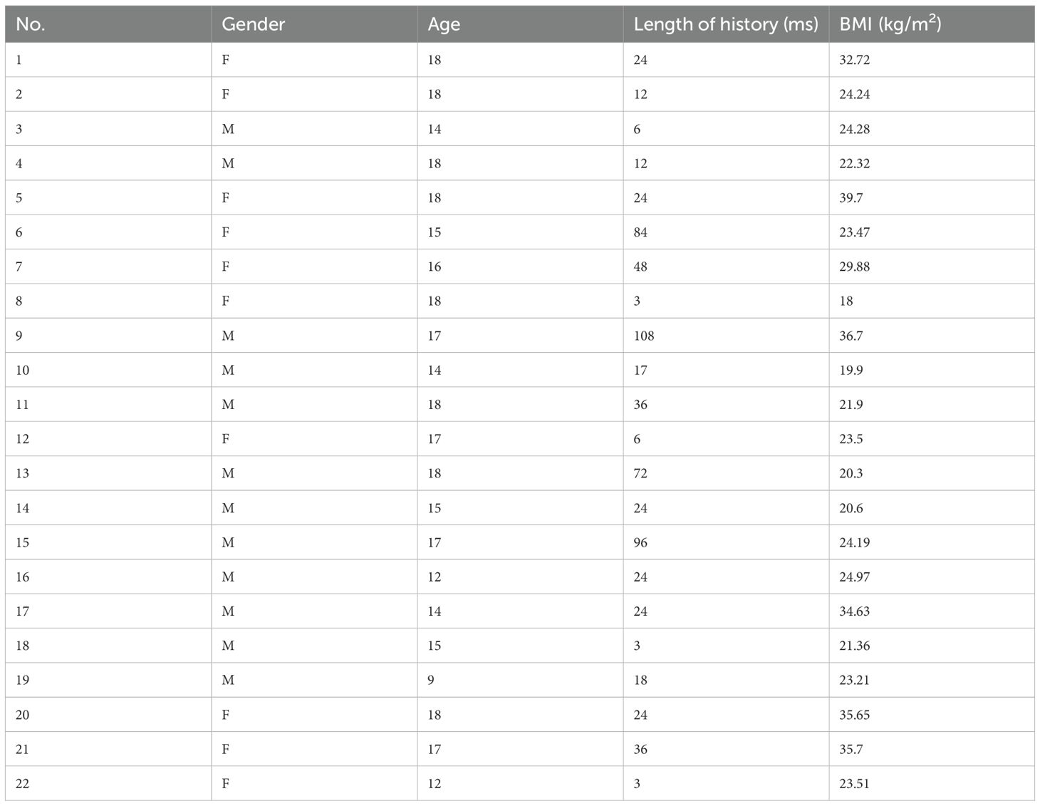
Table 1. The demographic information of 22 patients at diagnosis of CD.

Table 2

www.frontiersin.org

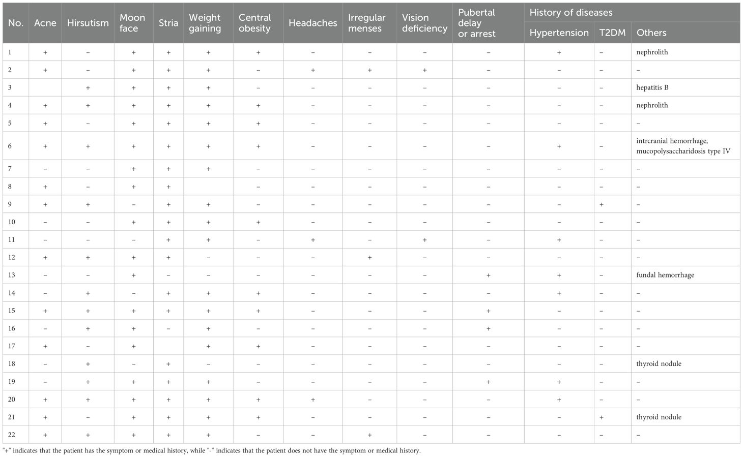
Table 2. Clinical signs and symptoms of 22 patients at diagnosis of CD.

Diagnosis of CD was confirmed by a multidisciplinary team comprising radiologists, endocrinologists, interventional radiologists, pediatricians, and neurosurgeons. Clinical manifestations, plasma cortisol circadian rhythm, low-dose dexamethasone suppression test (LDDST, 2 mg dexamethasone), and high-dose dexamethasone suppression test (HDDST, 8 mg dexamethasone) were assessed by pediatricians or endocrinologists. Following the 2mg LDDST, the 48-hour serum cortisol level exceeded 1.8 μg/dL, indicating inadequate suppression. In contrast, after the 8mg HDDST, the 48-hour cortisol level was suppressed to <50% of baseline, demonstrating significant suppression. Bilateral inferior petrosal sinus sampling (BIPSS) with or without desmopressin (DDAVP) stimulation was performed by experienced interventional radiologists. Samples were immediately placed on ice after collection. All biochemical analyses were conducted in a College of American Pathologists-accredited laboratory (No. 7217913).

Preoperative pituitary magnetic resonance imaging (MRI) was performed at 1.5 T or 3.0 T in all patients. T1-weighted and T2-weighted spin-echo images were obtained in coronal and sagittal planes (2-mm slice thickness) before and after gadolinium injection. A dynamic coronal sequence was also acquired within 2 minutes post-injection (Table 3).

Table 3

www.frontiersin.org

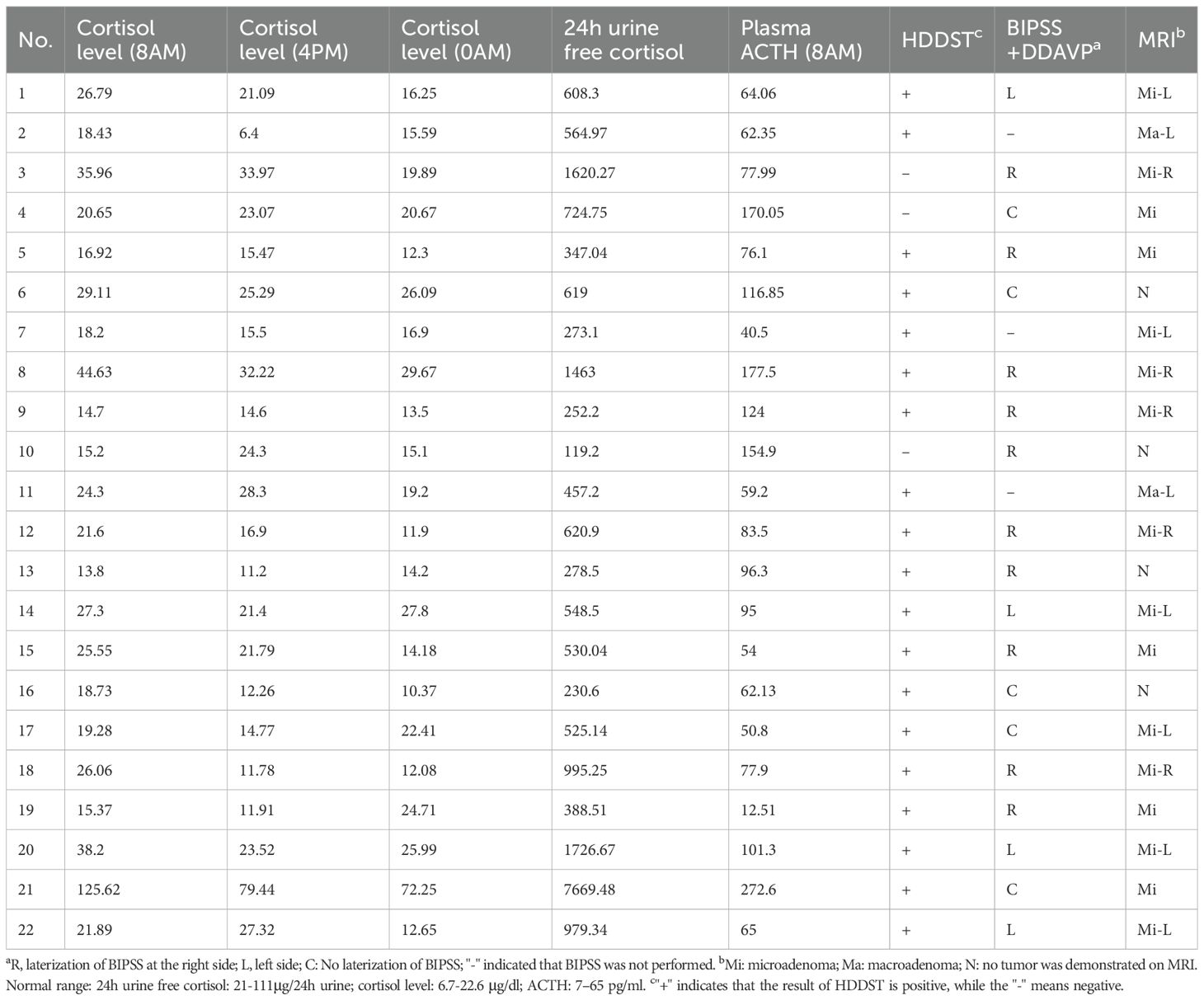
Table 3. Preoperative endocrinological evaluation and neuroimaging results of 22 patients at diagnosis of CD.

The same surgical team performed TSS on all patients using a mononostril approach. Microscopic TSS (MTSS) was utilized in 11 patients treated before 2016, while endoscopic TSS (ETSS) was employed in the subsequent 11 patients. For patients with concordant MRI-identified adenomas and BIPSS lateralization, exploration focused on the imaging-identified region, and a rim of pituitary tissue surrounding the tumor cavity was resected. If the tumor involved the cavernous sinus (CS), the inner CS wall was also inspected/explored. If BIPSS lateralization conflicted with MRI findings, the pituitary side indicated by BIPSS was explored first. For MRI-negative tumors, exploration commenced on the side with higher ACTH levels on BIPSS (when available) and proceeded to complete gland inspection. If no adenoma was identified intraoperatively, approximately half of the gland was resected, guided by BIPSS results.

Immediate remission was defined as a postoperative serum cortisol nadir <5 μg/dL or normal 24-hour UFC. Recurrent hypercortisolism was defined as the reappearance of biochemical hypercortisolism after a period of hypocortisolism or clinical adrenal insufficiency. The concordance of BIPSS lateralization with MRI localization refers to whether the tumor side indicated by BIPSS corresponds to the tumor side identified on MRI.

Patients were followed in the outpatient clinic at regular intervals. If endocrine evaluations were performed at local hospitals, results were communicated to the authors via WeChat. Mean follow-up duration was 29.4 months (range 2–129 months).

Results

Preoperative plasma cortisol levels measured at three time points were: mean 28.10 μg/dL at 8:00 AM (range 14.70-125.62 μg/dL), 22.39 μg/dL at 4:00 PM (range 6.4-79.44 μg/dL), and 20.62 μg/dL at midnight (range 11.9-72.25 μg/dL). Mean preoperative plasma ACTH level at 8:00 AM was 95.21 pg/mL (range 12.51-272.6 pg/mL), and mean 24-hour UFC was 979.18 μg/24h (range 119.20-7669.48 μg/24h). HDDST was positive in 19/22 patients. BIPSS with DDAVP was performed in 19 patients, demonstrating lateralization in 14 patients (4/14 left, 10/14 right).

MRI localized an adenoma in 18/22 patients (81.8%), comprising 16 microadenomas and 2 macroadenomas. Tumor location on MRI was: right sellar (n=5), left sellar (n=8), and central sellar (n=5). Concordance between BIPSS lateralization and MRI localization was 57.89% (11/19).

Immediate postoperative remission was achieved in 20 patients (90.9%). The two patients without immediate remission (Case 2: macroadenoma; Case 6: MRI- and pathology-negative) received additional treatments (Case2: gamma knife radiosurgery; Case 6: ketoconazole). Among the 20 patients with initial remission, 2 (10.0%) experienced recurrence (Case 3: microadenoma; Case 10: MRI-negative). Case3 received pasireotide after recurrence; Case 10 underwent repeat TSS, which did not achieve remission. Subsequent gamma knife treatment also ultimately failed. Ketoconazole therapy was then initiated. The sustained long-term remission rate for the cohort was 81.8% (18/22).

In these cases, intraoperative bleeding was controlled in all cases, and no patient required transfusion. Case 10 experienced a CSF leak following repeat transsphenoidal surgery (TSS). All patients who achieved postoperative remission were administered cortisone replacement therapy. The requirement for levothyroxine replacement differed between groups: one child in the ETSS group (1/11) versus five patients in the MTSS group (5/11). For diabetes insipidus, oral desmopressin was administered to three patients in the ETSS group and two in the MTSS group (Table 4).

Table 4

www.frontiersin.org

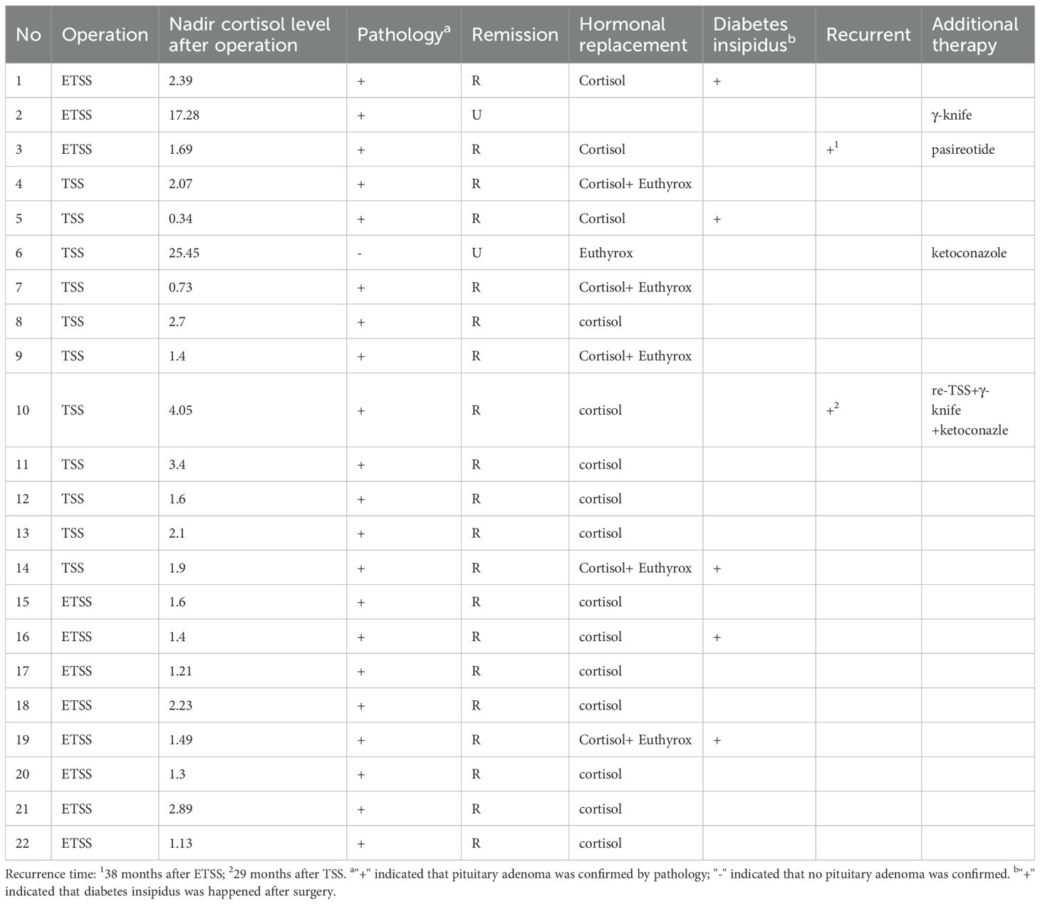
Table 4. The neurosurgical outcome and follow-up results of 22 patients of CD.

Discussion

This 20-year single-center experience represents one of the largest reported cohorts of surgically managed pediatric Cushing’s disease patients. Our findings demonstrate that transsphenoidal surgery (TSS), whether microscopic (MTSS) or endoscopic (ETSS), is a highly effective first-line treatment for pediatric CD, achieving an immediate remission rate of 90.9% and a sustained long-term remission rate of 81.8%.

The diagnostic complexity of pediatric CD is highlighted by the significant diagnostic delay observed (mean 32.0 months) and the occurrence of MRI-negative cases (4/22, 18.2%). This aligns with established literature emphasizing the challenges of pediatric CD diagnosis stemming from its rarity, atypical presentation, and the high proportion of microadenomas or radiologically occult tumors (3468). Our adherence to a rigorous multidisciplinary diagnostic protocol, incorporating standardized biochemical testing (LDDST, HDDST), BIPSS with DDAVP stimulation (performed in 19/22), and high-resolution dynamic pituitary MRI, reflects current best practices for confirming ACTH-dependent Cushing’s syndrome and tumor localization. The moderate concordance rate (57.89%) between BIPSS lateralization and MRI localization underscores their complementary roles, particularly in cases with equivocal imaging. BIPSS remains critical for guiding surgical exploration in MRI-negative or discordant cases, as evidenced by its use in our decision-making algorithm (910).

Our immediate remission rate (90.9%) compares favorably with contemporary pediatric CD surgical series, which typically report rates between 70% and 98% (1381113). The two immediate surgical failures occurred in patients with a macroadenoma (Case 2) or an MRI- and pathology-negative diagnosis (Case 6), profiles consistently associated with lower remission rates. The long-term remission rate of 81.8% (18/22) is robust, although the recurrence rate of 10% (2/20 initially remitted patients) merits attention. Both recurrences arose in patients with microadenomas, one of whom was MRI-negative (Case 10). This recurrence rate falls within the reported range (5-30%) for pediatric CD and reinforces the need for lifelong endocrine surveillance (11415). The relatively short mean follow-up (29.4 months) suggests that the true recurrence rate might be higher with extended observation, representing a limitation of this study.

Our experience reflects the evolution of surgical technique, with a transition from MTSS to ETSS after 2016. While the cohort size and follow-up duration preclude definitive conclusions regarding the comparative efficacy of MTSS versus ETSS in this specific pediatric population, both techniques yielded high success rates. In our group, no significant differences exist in remission or recurrence rates. However, regarding complications, ETSS demonstrates a lower incidence of hypopituitarism compared to MTSS, while the incidence of diabetes insipidus is similar. It should be noted, however, that this comparison remains limited by the small number of reported cases. The endoscopic approach offers theoretical advantages, such as wider panoramic visualization potentially aiding in the identification of small or laterally extending microadenomas, which are common in children. Larger, prospective studies with longer follow-up are warranted to directly compare outcomes between these surgical modalities in pediatric CD.

The spectrum of clinical manifestations observed (e.g., weight gain, moon facies, striae, hypertension, pubertal arrest/delay) demonstrates the profound multisystem impact of hypercortisolism in children. The notable prevalence of metabolic complications like hypertension (7/22) and type 2 diabetes mellitus (2/22), even in this young cohort, highlights the urgency of timely diagnosis and effective intervention to mitigate long-term morbidity (51618).

Limitations

This study shares the limitations inherent to retrospective, single-center designs. The modest sample size, though substantial for this rare condition, limits statistical power for subgroup analyses, such as rigorous comparison of MTSS vs. ETSS outcomes or identification of specific predictors of failure/recurrence. The mean follow-up period is relatively short for a disease with potential for late recurrence, long-term remission rates may be lower than reported, and the study could not capture long-term complications of TSS, particularly those affecting growth and development in pediatric patients. Detailed data on specific postoperative complications (e.g., diabetes insipidus, hypopituitarism) and pituitary function during follow-up would provide a more comprehensive assessment of treatment sequelae but were not the primary focus of this outcome report.

Conclusion

Despite the inherent diagnostic and therapeutic challenges of pediatric Cushing’s disease, transsphenoidal surgery performed in a specialized center achieves high rates of immediate and sustained remission. Our results support the efficacy of TSS as the primary treatment modality. A meticulous multidisciplinary diagnostic approach, including BIPSS when indicated, is crucial for success, particularly in MRI-negative cases. While recurrence remains a concern necessitating vigilant long-term follow-up, the majority of children with CD can attain durable disease control with appropriate surgical management. The transition to endoscopic techniques proved safe and effective, warranting further investigation in larger pediatric cohorts with extended follow-up.

Data availability statement

The original contributions presented in the study are included in the article/supplementary material. Further inquiries can be directed to the corresponding authors.

Ethics statement

The studies involving humans were approved by The ethics committee of Ruijin hospital. The studies were conducted in accordance with the local legislation and institutional requirements. The participants provided their written informed consent to participate in this study. Written informed consent was obtained from the individual(s), and minor(s)’ legal guardian/next of kin, for the publication of any potentially identifiable images or data included in this article.

Author contributions

BW: Methodology, Writing – original draft. HZ: Conceptualization, Data curation, Formal Analysis, Writing – original draft. TS: Methodology, Project administration, Writing – review & editing. JR: Data curation, Formal Analysis, Writing – original draft. QS: Resources, Supervision, Writing – review & editing. YS: Supervision, Writing – review & editing. LB: Supervision, Writing – review & editing.

Funding

The author(s) declare that no financial support was received for the research and/or publication of this article.

Conflict of interest

The authors declare that the research was conducted in the absence of any commercial or financial relationships that could be construed as a potential conflict of interest.

Generative AI statement

The author(s) declare that no Generative AI was used in the creation of this manuscript.

Any alternative text (alt text) provided alongside figures in this article has been generated by Frontiers with the support of artificial intelligence and reasonable efforts have been made to ensure accuracy, including review by the authors wherever possible. If you identify any issues, please contact us.

Publisher’s note

All claims expressed in this article are solely those of the authors and do not necessarily represent those of their affiliated organizations, or those of the publisher, the editors and the reviewers. Any product that may be evaluated in this article, or claim that may be made by its manufacturer, is not guaranteed or endorsed by the publisher.

References

1. Savage MO and Ferrigno R. Paediatric Cushing’s disease: long-term outcome and predictors of recurrence. Front Endocrinol (Lausanne). (2024) 15:1345174. doi: 10.3389/fendo.2024.1345174

PubMed Abstract | Crossref Full Text | Google Scholar

2. Tarcin G, Catli G, Cetinkaya S, Eren E, Kardelen AD, Akinci A, et al. Clinical features, diagnosis and treatment outcomes of Cushing’s disease in children: A multicenter study. Clin Endocrinol (Oxf). (2024) 100:19–28. doi: 10.1111/cen.14980

PubMed Abstract | Crossref Full Text | Google Scholar

3. Zheng X, Wang H, Zhang W, Feng S, Liu Y, Li S, et al. Diagnosis, manifestations, laboratory investigations, and prognosis in pediatric and adult cushing’s disease in a large center in China. Front Endocrinol (Lausanne). (2021) 12:749246. doi: 10.3389/fendo.2021.749246

PubMed Abstract | Crossref Full Text | Google Scholar

4. Joshi K, Taliou A, and Stratakis CA. Diagnostic and management challenges in paediatric Cushing’s syndrome. Clin Endocrinol (Oxf). (2024) 101:631–9. doi: 10.1111/cen.15096

PubMed Abstract | Crossref Full Text | Google Scholar

5. Ferrigno R, Hasenmajer V, Caiulo S, Minnetti M, Mazzotta P, Storr HL, et al. Paediatric Cushing’s disease: Epidemiology, pathogenesis, clinical management and outcome. Rev Endocr Metab Disord. (2021) 22:817–35. doi: 10.1007/s11154-021-09626-4

PubMed Abstract | Crossref Full Text | Google Scholar

6. Yanar EA, Makazan NV, Kareva MA, Vorontsov AV, Vladimirova VP, Bezlepkina OB, et al. Course of Cushing;s disease and treatment outcomes in correlation with pituitary MRI in children. Probl Endokrinol (Mosk). (2022) 68:93–104. doi: 10.14341/probl12854

PubMed Abstract | Crossref Full Text | Google Scholar

7. Maiter D, Chanson P, Constantinescu SM, and Linglart A. Diagnosis and management of pituitary adenomas in children and adolescents. Eur J Endocrinol. (2024) 191:R55–r69. doi: 10.1093/ejendo/lvae120

PubMed Abstract | Crossref Full Text | Google Scholar

8. Crock PA, Ludecke DK, Knappe UJ, and Saeger W. A personal series of 100 children operated for Cushing’s disease (CD): optimizing minimally invasive diagnosis and transnasal surgery to achieve nearly 100% remission including reoperations. J Pediatr Endocrinol Metab. (2018) 31:1023–31. doi: 10.1515/jpem-2018-0262

PubMed Abstract | Crossref Full Text | Google Scholar

9. Turan H, Catli G, Kardelen AD, Bober E, Akinci A, Cetinkaya S, et al. Diagnostic value of bilateral petrosal sinus sampling in children with cushing disease: A multi-center study. J Clin Res Pediatr Endocrinol. (2022) 14:29–36. doi: 10.4274/jcrpe.galenos.2021.2021.0152

PubMed Abstract | Crossref Full Text | Google Scholar

10. Chen S, Chen K, Lu L, Zhang X, Tong A, Pan H, et al. The effects of sampling lateralization on bilateral inferior petrosal sinus sampling and desmopressin stimulation test for pediatric Cushing’s disease. Endocrine. (2019) 63:582–91. doi: 10.1007/s12020-018-1779-x

PubMed Abstract | Crossref Full Text | Google Scholar

11. Lonser RR, Wind JJ, Nieman LK, Weil RJ, DeVroom HL, and Oldfield EH. Outcome of surgical treatment of 200 children with Cushing’s disease. J Clin Endocrinol Metab. (2013) 98:892–901. doi: 10.1210/jc.2012-3604

PubMed Abstract | Crossref Full Text | Google Scholar

12. Joshi SM, Hewitt RJ, Storr HL, Rezajooi K, Ellamushi H, Grossman AB, et al. Cushing’s disease in children and adolescents: 20 years of experience in a single neurosurgical center. Neurosurgery. (2005) 57:281–5. doi: 10.1227/01.NEU.0000166580.94215.53

PubMed Abstract | Crossref Full Text | Google Scholar

13. Kanter AS, Diallo AO, Jane JA Jr., Sheehan JP, Asthagiri AR, Oskouian RJ, et al. Single-center experience with pediatric Cushing’s disease. J Neurosurg. (2005) 103:413–20. doi: 10.3171/ped.2005.103.5.0413

PubMed Abstract | Crossref Full Text | Google Scholar

14. Pasternak-Pietrzak K, Moszczynska E, and Szalecki M. Treatment challenges in pediatric Cushing’s disease: Review of the literature with particular emphasis on predictive factors for the disease recurrence. Endocrine. (2019) 66:125–36. doi: 10.1007/s12020-019-02036-2

PubMed Abstract | Crossref Full Text | Google Scholar

15. Yordanova G, Martin L, Afshar F, Sabin I, Alusi G, Plowman NP, et al. Long-term outcomes of children treated for Cushing’s disease: a single center experience. Pituitary. (2016) 19:612–24. doi: 10.1007/s11102-016-0756-8

PubMed Abstract | Crossref Full Text | Google Scholar

16. Pasternak-Pietrzak K, Moszczyńska E, Jurkiewicz E, and Szalecki M. Paediatric Cushing’s disease – a literature review of epidemiology, pathogenesis, clinical symptoms, and diagnostics. Endokrynol Pol. (2020) 71:87–95. doi: 10.5603/EP.a2019.0040

PubMed Abstract | Crossref Full Text | Google Scholar

17. Storr HL and Savage MO. Management of endocrine disease: Paediatric Cushing’s disease. Eur J Endocrinol. (2015) 173:R35–45. doi: 10.1530/EJE-15-0013

PubMed Abstract | Crossref Full Text | Google Scholar

18. Tatsi C, Kamilaris C, Keil M, Saidkhodjaeva L, Faucz FR, Chittiboina P, et al. Paediatric Cushing syndrome: a prospective, multisite, observational cohort study. Lancet Child Adolesc Health. (2024) 8:51–62. doi: 10.1016/S2352-4642(23)00264-X

PubMed Abstract | Crossref Full Text | Google Scholar

Keywords: Cushing’s disease, pediatric, transsphenoidal surgery, surgical outcome, surgical strategy

Citation: Wang B, Zhang H, Su T, Ren J, Sun Q, Sun Y and Bian L (2025) The neurosurgical outcome of pediatric Cushing’s disease in a single center from China: a 20-year experience. Front. Endocrinol. 16:1663624. doi: 10.3389/fendo.2025.1663624

Received: 10 July 2025; Accepted: 22 August 2025;
Published: 03 September 2025.

Edited by:

Sadishkumar Kamalanathan, Jawaharlal Institute of Postgraduate Medical Education and Research (JIPMER), India

Reviewed by:

Aleksandra Zdrojowy-Wełna, Wroclaw Medical University, Poland
Medha Bhardwaj, Mahatma Gandhi University of Medical Sciences Technology, India

Copyright © 2025 Wang, Zhang, Su, Ren, Sun, Sun and Bian. This is an open-access article distributed under the terms of the Creative Commons Attribution License (CC BY). The use, distribution or reproduction in other forums is permitted, provided the original author(s) and the copyright owner(s) are credited and that the original publication in this journal is cited, in accordance with accepted academic practice. No use, distribution or reproduction is permitted which does not comply with these terms.

*Correspondence: Yuhao Sun, syh11897@rjh.com.cn; Liuguan Bian, Blg11118@rjh.com.cn

These authors have contributed equally to this work

Disclaimer: All claims expressed in this article are solely those of the authors and do not necessarily represent those of their affiliated organizations, or those of the publisher, the editors and the reviewers. Any product that may be evaluated in this article or claim that may be made by its manufacturer is not guaranteed or endorsed by the publisher.

Research Study for Patients Diagnosed with Cushing’s Disease and Their Caregivers

We’re looking for caregivers to loved ones diagnosed with Cushing’s Disease or patients diagnosed with Cushing’s Disease to participate in a research study.

✅ Who: Patients and caregivers of loved ones

⏳ What: 30-minute Online Survey

💰 Compensation: $60.00

Sign up here: https://rarepatientvoice.com/CushingsHelp/


The Outcome of Abnormal Glucose Metabolism and Its Clinical Features in Patients With Cushing’s Disease After Curative Surgery

Abstract

Objective

To investigate the outcomes of abnormal glucose metabolism and its clinical characteristics in patients with Cushing’s disease (CD) who achieved biochemical remission after surgery.

Methods

Patients diagnosed with CD who achieved biochemical remission and underwent regular follow-up after surgery were enrolled. Pre- and postoperative clinical data were collected and analyzed.

Result

151CD patients were included, of whom 80 (53 %) had preoperative abnormal glucose metabolism, including 56 with diabetes mellitus (DM) and 24 with impaired glucose regulation (IGR). At one year after surgery, 57 patients exhibited improved glucose metabolism, accompanied by a significant reduction in the homeostasis model assessment of insulin resistance (HOMA-IR). Improvements were mainly observed at 3 and 6 months after surgery. At one-year after surgery, there were 20 patients with diabetes and 16 with IGR. Compared to those with NGT, these individuals exhibited a higher prevalence of hypertension, hyperlipidemia, fatty liver, and abnormal bone metabolism.

Conclusion

CD patients demonstrated a high incidence of abnormal glucose metabolism. Notably, approximately two-thirds demonstrated improved glucose metabolism one year after curative surgery, with the greatest improvements observed at 3- to 6-month postoperative follow-up.

Introduction

Cushing’s disease (CD) is characterized by excessive endogenous cortisol production caused by pituitary adrenocorticotropic hormone adenoma and is the main cause of Cushing’s syndrome (CS). Surgical resection of the tumor is the preferred treatment. Prolonged exposure to hypercortisolism increases the risk of metabolic abnormalities, including obesity, hypertension, glucose and lipid abnormalities, osteoporosis, etc. Additionally, it significantly elevates the risk of infection, thrombosis, and hypokalemia. Abnormal glucose metabolism is a common complication of CS, with an incidence ranging from 13.1 % to 47 %[1], and diabetes is an independent risk factor for mortality in CD patients[2].
Previous clinical studies have found that metabolic abnormalities such as diabetes, hypertension, and hyperlipidemia improve in CS patients who achieve biochemical remission after surgical treatment. However, the concept of improvement in glucose metabolism, the incidence of improvement, and its related factors are inconsistent in various reports. Previous studies primarily assessed the outcome of glucose metabolism based on plasma glucose results at a single fixed follow-up time after surgery. The lack of regular follow-up data makes it difficult to clearly understand the trend of postoperative plasma glucose changes, and there are no clinical data on when glucose metabolism begins to improve or change. Therefore, this study retrospectively analyzed the follow-up data of patients with Cushing’s disease in our hospital before and after surgery, and monitored the changes in glucose metabolism, to explore the characteristics and clinical features of such changes in patients with Cushing’s disease who achieved remission from CD following surgery..

Access through your organization

Check access to the full text by signing in through your organization.

Access through your organization

Section snippets

Subjects

This study enrolled hospitalized patients with Cushing’s disease at Huashan Hospital, Fudan University from January 2014 to February 2020. Inclusion criteria were as follows: (1) Age ≥ 18 years; (2) diagnosis of Cushing’s disease according to the 2021 Consensus on the Diagnosis and Management of Cushing’s Disease, confirmed by pathology[3]; (3) biochemical remission after transsphenoidal surgery; (4) complete preoperative data and regular follow-up visits (including visits at 1, 3, 6, and

Patients’ baseline characteristics

A total of 168 patients with CD were admitted to Huashan Hospital from 2014 to 2020 with pathological diagnosis and regular postoperative follow-up; however, 17 patients were excluded due to no biochemical remission after surgery or relapse during follow-up (Fig. 1). Ultimately, 151 patients (32 males and 119 females) were included in this study. The baseline characteristics of the included patients were shown in Table 1. There were 80 cases (53 %) complicated with abnormal glucose metabolism

Discussion

CD was a rare disease often associated with abnormal glucose metabolism. Based on medical history and OGTT screening, we found that over half (53 %) of CD patients exhibited abnormal glucose metabolism before surgery, with 37.1 % being diagnosed with diabetes. Previous studies have shown that the prevalence of diabetes in CS patients ranged from 13.1 % to 47 %, and most reports falling between 35 % and 45 %, which is consistent with our findings [1,12,13]. However, it should be noted that CD

Author contributions

Q.C. analyzed the data and wrote the manuscript. Q.C., Y.L., X.L., Q.S., W.S., and H.Z. collected the data. Y.L., Z.Z., M.H., S.Z., and H.Y. recruited patients. J.Z., Y.S., and S.Z. conducted the study design and revised the manuscript. All authors read and approved the final manuscript.

CRediT authorship contribution statement

Qiaoli Cui: Writing – review & editing, Writing – original draft, Methodology, Investigation, Formal analysis, Data curation, Conceptualization. Yujia Li: Writing – original draft, Investigation, Formal analysis, Data curation. Xiaoyu Liu: Investigation, Formal analysis, Data curation. Quanya Sun: Investigation, Data curation. Wanwan Sun: Investigation, Formal analysis, Data curation. Min He: Project administration, Investigation. Jie Zhang: Writing – review & editing, Supervision, Funding

Declaration of competing interest

The authors declare that they have no known competing financial interests or personal relationships that could have appeared to influence the work reported in this paper.

Acknowledgments

We are indebted to the patients who participated in this study and all the doctors who contributed to the diagnosis and treatment of these patients. This work was supported by grants from the Multidisciplinary Diagnosis and Treatment (MDT) demonstration project in research hospitals (Shanghai Medical College, Fudan University, NO: DGF501069/017), National Science and Technology Major Project (NO: 2023ZD0506800,2023ZD0506802), 2023 Ningbo International Cooperation Program (NO: 2023H024).

References (16)

  • R.N. Clayton et al.

    Mortality in patients with Cushing’s disease more than 10 years after remission: a multicentre, multinational, retrospective cohort study

    Lancet Diabetes Endocrinol

    (2016)
  • M. Fleseriu et al.

    Consensus on diagnosis and management of Cushing’s disease: a guideline update

    Lancet Diabetes Endocrinol

    (2021)
  • C. Scaroni et al.

    Glucose Metabolism Abnormalities in Cushing Syndrome: from Molecular Basis to Clinical Management

    Endocr Rev

    (2017)
  • C. Dai et al.

    Surgical outcome of transsphenoidal surgery in Cushing’s disease: a case series of 1106 patients from a single center over 30 years

    Endocrine

    (2022)
  • E. Valassi et al.

    Delayed remission after transsphenoidal surgery in patients with Cushing’s disease

    J Clin Endocrinol Metab

    (2010)
  • X. He et al.

    Glucocorticoid Withdrawal Syndrome following treatment of endogenous Cushing Syndrome

    (2022)
  • E.V. Varlamov et al.

    Perioperative Management of a Patient with Cushing Disease

    J Endocr Soc

    (2022)
  • Q. Cui et al.

    The recovery time of hypothalamic-pituitary-adrenal axis after curative surgery in Cushing’s disease and its related factor

    Endocrine

    (2023)
There are more references available in the full text version of this article.

Improved Noninvasive Diagnostic Evaluations in Treatment-Naïve Adrenocorticotropic Hormone (ACTH)-Dependent Cushing’s Syndrome

Abstract

Background

Bilateral inferior petrosal sinus sampling (BIPSS) is important in the differential diagnosis of adrenocorticotropic hormone (ACTH)-dependent Cushing’s syndrome, but BIPSS is invasive and is not reliable on tumor lateralization. Thus, we evaluated the noninvasive diagnostic evaluations, high-dose dexamethasone suppression test (HDDST) combined with different pituitary MRI scans (conventional contrast-enhanced MRI [cMRI], dynamic contrast-enhanced MRI [dMRI], and high-resolution contrast-enhanced MRI [hrMRI]), by comparison with BIPSS.

Methods

We retrospectively analyzed 95 patients with ACTH-dependent Cushing’s syndrome who underwent HDDST, preoperative MRI scans (cMRI, dMRI and hrMRI) and BIPSS in our hospital between January 2016 and December 2021. The diagnostic performance of HDDST combined with cMRI (HDDST + cMRI), HDDST + dMRI and HDDST + hrMRI, and BIPSS was evaluated, including the sensitivity of identifying pituitary adenomas and the tumor lateralization accuracy.

Results

Compared with BIPSS (AUC, 0.98; 95%CI: 0.93, 1.00), the diagnostic performance of HDDST + hrMRI was comparable in both neuroradiologist 1 (AUC, 0.95; 95%CI: 0.89, 0.99; P = 0.129) and neuroradiologist 2 (AUC, 0.98; 95%CI: 0.92, 1.00; P = 0.707). For tumor lateralization accuracy, HDDST + hrMRI (90.6-95.3%) were significantly higher than that of BIPSS (24.7%, P < 0.001).

Conclusions

In patients with ACTH-dependent Cushing’s syndrome, HDDST + hrMRI, as noninvasive diagnostic evaluations, achieves high diagnostic performance comparable with gold standard (BIPSS), and it is superior to BIPSS in terms of tumor lateralization accuracy.

Peer Review reports

Background

Cushing’s syndrome is associated with debilitating morbidity and increased mortality [1]. Adrenocorticotropic hormone (ACTH)-dependent Cushing’s syndrome is characterized by ACTH hypersecretion. Bilateral inferior petrosal sinus sampling (BIPSS) is regarded as the gold standard to distinguish pituitary ACTH secretion (also known as Cushing’s disease) from ectopic ACTH syndrome (EAS) [12]. However, BIPSS is invasive and is not reliable on tumor lateralization [34]. Thus, it is important to improve the diagnostic performance of noninvasive evaluations with high sensitivity and tumor lateralization accuracy.

Current noninvasive evaluations in the differential diagnosis of ACTH-dependent Cushing’s syndrome include high-dose dexamethasone suppression test (HDDST), the CRH stimulation test and pituitary MRI. However, due to the non-availability of CRH for testing, the sensitivities of current available noninvasive evaluations in identifying ACTH-secreting pituitary adenomas cannot satisfy the clinical needs. Conventional contrast-enhanced MRI (cMRI) and dynamic contrast-enhanced MRI (dMRI) with two-dimensional (2D) fast spin echo (FSE) sequence is routinely used, and only 50–66% of the ACTH-secreting pituitary adenomas can be correctly detected [56]. Recently, by using 3D spoiled gradient recalled (SPGR) sequence, high-resolution contrast-enhanced MRI (hrMRI) has increased the sensitivity to up to 80% [7,8,9]. However, these noninvasive evaluations are still inferior to BIPSS, the sensitivity and specificity of which is about 90–95% [10,11,12,13]. With the development of 3D FSE sequence, superior image quality with diminished artifact has been achieved, providing a reliable alternative to detect pituitary adenomas [14]. Previous studies have shown that hrMRI using 3D FSE sequence has high diagnostic performance for identifying pituitary adenomas [1516]. To our knowledge, no study has investigated the diagnostic performance of HDDST combined with hrMRI using 3D FSE sequence (HDDST + hrMRI) in patients with Cushing’s syndrome, and whether it can avoid unnecessary BIPSS procedure.

The aim of this study is to evaluate the diagnostic performance of HDDST + hrMRI by comparison with BIPSS in patients with ACTH-dependent Cushing’s syndrome.

Methods

This study was performed in line with the principles of the Declaration of Helsinki. Approval was granted by the Institutional Review Board of Peking Union Medical College Hospital. Informed consent was waived in this study because it was a retrospective, non-interventional, and observational study. Clinical trial number is not applicable.

Study design and patient population

We retrospectively reviewed the medical records and imaging studies from January 2016 to December 2021, and 232 consecutive patients with ACTH-dependent Cushing’s syndrome, who underwent HDDST, cMRI, dMRI, hrMRI and BIPSS, were enrolled in the current study. A total of 137 patients were excluded from the study because of prior pituitary surgery (n = 122) or lack of histopathology due to no pituitary surgery in our hospital (n = 15). Finally, 95 patients were included in the current study (Fig. 1) and all the patients included were confirmed by histopathology or by clinical remission after surgical resection of the ACTH-secreting lesion. In the current study, all the patients with Cushing’s disease achieved clinical remission after surgical resection of the ACTH-secreting lesion. All the patients with EAS underwent contrast-enhanced thoracic and abdominal CT to identify the ACTH-secreting lesion. The clinical decision-making process was consistent with the previous study [1].

Fig. 1
figure 1

Flowchart of patient inclusion/exclusion process. ACTH = adrenocorticotropic hormone, BIPSS = bilateral inferior petrosal sinus sampling; cMRI = conventional contrast-enhanced MRI, dMRI = dynamic enhanced MRI, HDDST = high-dose dexamethasone suppression test, hrMRI = high-resolution contrast-enhanced MRI, NPV = negative predictive value, PPV = positive predictive value

HDDST

As previously described [17], the average 24-hour urinary free cortisol (24hUFC) level of 2 days before HDDST was recorded as baseline. Then, 2 mg dexamethasone was administered orally every 6 h for 2 days, and the 24hUFC level of the second day was measured. When the ratio of 24hUFC on the second day after HDDST to 24hUFC at baseline was less than 50%, the suppression in HDDST was marked as positive in the current study.

BIPSS

BIPSS was performed according to Doppman et al. [18]. Blood samples were collected from peripheral veins and bilateral inferior petrosal sinuses (IPSs) at multiple time points (0, 3, 5 and 10 min) after the introduction of 10 µg desmopressin [19]. An IPS to peripheral ACTH ratio of ≥ 2.0 at baseline or ≥ 3.0 after desmopressin stimulation at any time point [20] was marked as positive in the current study. Furthermore, tumor lateralization was predicted by an intersinus ratio of ≥ 1.4 [20].

Imaging

All the images were acquired on a 3.0 Tesla MR scanner (Discovery MR750w, GE Healthcare) using an 8-channel head coil. Detailed acquisition parameters and sequence order before and after contrast injection (gadopentetate dimeglumine [Gd-DTPA] at 0.05 mmol/kg [0.1 mL/kg] with a flow rate of 2 mL/s followed by a 10-mL saline solution flush) were as follows: coronal 2D FSE T2WI (field of view [FOV] = 20 cm × 20 cm, slice thickness = 4 mm, slice spacing = 1 mm, repetition time/echo time [TR/TE] = 4100/90 ms, number of excitation [NEX] = 1.2, matrix = 320 × 320, scan time = 49s), coronal 2D FSE T1WI (FOV = 18 cm × 16.2 cm, slice thickness = 3 mm, slice spacing = 0.6 mm, TR/TE = 400/12 ms, NEX = 2, matrix = 256 × 192, scan time = 49s), sagittal fat-saturated 3D FSE T1WI (FOV = 16.5 cm × 16.5 cm, slice thickness = 3 mm, slice spacing = 0, TR/TE = 460/16 ms, NEX = 2, matrix = 256 × 224, scan time = 60s), dynamic contrast-enhanced coronal 2D FSE T1WI (FOV = 19 cm × 17.1 cm, slice thickness = 2 mm, slice spacing = 0.5 mm, TR/TE = 375/14 ms, NEX = 1, matrix = 288 × 192, scan time = 23s/phase × 6 phases), contrast-enhanced coronal 2D FSE T1WI, contrast-enhanced sagittal fat-saturated 3D FSE T1WI, and contrast-enhanced coronal fat-saturated 3D FSE T1WI (FOV = 15.2 cm × 15.2 cm, slice thickness = 1.2 mm, slice spacing = -0.6 mm, TR/TE = 390/15 ms, NEX = 6, matrix = 256 × 256, scan time = 4 min 30s).

Images were independently evaluated by two experienced neuroradiologists (with 25 and 16 years of experience in neuroradiology, respectively). Both neuroradiologists were blinded to the clinical information of the patients. The image order of cMRI, dMRI and hrMRI was randomized. The detection of pituitary adenomas was scored using a 3-point scale (0 = poor, 1 = fair, 2 = excellent). Scores of 1 or 2 represented a successful pituitary adenoma detection. The gold standard was the histopathology, and the diameter and the location of lesions were recorded on the sequence where identified.

The signal-to-noise ratio (SNR) and contrast-to-noise ratio (CNR) were calculated as follows: SNR = SIadenoma / SDbackground, CNR = |SIpituitary – SIadenoma| / SDbackground. SIpituitary and SIadenoma were defined as the mean signal intensity of the pituitary gland and the pituitary adenoma, respectively. SDbackground was defined as the standard deviation of the signal intensity of the background. CNR was recorded as 0 when no pituitary adenoma was identified. Figure 2 showed the calculation of SNR and CNR using an operator defined region of interest.

Fig. 2

figure 2

The calculation of SNR and CNR using an operator defined region of interest. CNR = contrast-to-noise ratio, SD = standard deviation, SI = signal intensity, SNR = signal-to-noise ratio

Statistical analysis

The κ analysis was conducted to assess the interobserver agreements. The κ value was interpreted as follows: below 0.20, slight agreement; 0.21–0.40, fair agreement; 0.41–0.60, moderate agreement; 0.61–0.80, substantial agreement; greater than 0.80, almost perfect agreement.

To assess the diagnostic performance of different evaluations, the receiver operating characteristic curves were plotted and the area under curves (AUCs) were compared between noninvasive and invasive evaluations for each neuroradiologist by using the DeLong test. Sensitivity, specificity, positive predictive value, and negative predictive value were calculated. The Friedman’s test was used to evaluate the SNR and CNR measurements as well as conspicuity scores of pituitary adenomas between MR protocols, and the Wilcoxon signed-rank test was used for pairwise comparison. The McNemar’s test was used to evaluate the tumor lateralization accuracy. A P value of less than 0.05 was considered statistically significant. A stricter P value of less than 0.017 was considered statistically significant after Bonferroni correction. Statistical analysis was performed using MedCalc Statistical Software (version 23.0.2) and SPSS Statistics (version 22.0).

Results

Clinical characteristics

The clinical characteristics of the 95 patients with Cushing’s syndrome were shown in Table 1. There were 85 patients (median age, 38 years; interquartile range [IQR], 29–51 years; 55 females [65%]) with Cushing’s disease and 10 patients (median age, 39 years; IQR, 30–47 years; 5 females [50%]) with EAS. Of the 85 patients with Cushing’s disease, the median diameter of pituitary adenomas was 5 mm (IQR, 4–5 mm), ranging from 3 to 28 mm. Among them, 80 patients had microadenomas (less than 10 mm in size). Of the ten patients with EAS, one patient had an ovarian carcinoid tumor found by abdominal CT, others had pulmonary carcinoid tumors found by thoracic CT as the cause of Cushing’s syndrome. None of the patients with EAS had a lesion in the pituitary.

Table 1 Clinical characteristics of the patients

Diagnostic performance noninvasive and invasive evaluations

The inter-observer agreements between two neuroradiologists were moderate on cMRI (κ = 0.597), moderate on dMRI (κ = 0.595), and almost perfect on hrMRI (κ = 0.850), respectively.

The diagnostic performance of noninvasive and invasive evaluations was shown in Table 2. Compared with BIPSS (AUC, 0.98; 95%CI: 0.93, 1.00), the diagnostic performance of HDDST + hrMRI was comparable in both neuroradiologist 1 (AUC, 0.95; 95%CI: 0.89, 0.99; P = 0.129) and neuroradiologist 2 (AUC, 0.98; 95%CI: 0.92, 1.00; P = 0.707). However, the diagnostic performance of HDDST + cMRI and HDDST + dMRI was inferior to BIPSS (P ≤ 0.001 for all). No difference was found between HDDST + cMRI and HDDST + dMRI in neuroradiologist 1 (P = 0.050) and neuroradiologist 2 (P = 0.353).

Table 2 The diagnostic performance of noninvasive and invasive evaluations

Figures 3 and 4 showed that microadenomas were correctly diagnosed on hrMRI, but missed on cMRI or dMRI.

Fig. 3

figure 3

Images in a patient with Cushing’s disease. The lesion is missed on (a) coronal contrast-enhanced T1-weighted image and (b) coronal dynamic contrast-enhanced T1-weighted image obtained with two-dimensional (2D) fast spin echo (FSE) sequence. (c) Coronal contrast-enhanced T1-weighted image on high-resolution MRI obtained with 3D FSE sequence shows a round pituitary microadenoma measuring approximately 4 mm with delayed enhancement on the left side of the pituitary gland

Fig. 4

figure 4

Images in a patient with Cushing’s disease. The lesion is missed on (a) coronal contrast-enhanced T1-weighted image and (b) coronal dynamic contrast-enhanced T1-weighted image obtained with two-dimensional (2D) fast spin echo (FSE) sequence. (c) Coronal contrast-enhanced T1-weighted image on high-resolution MRI obtained with 3D FSE sequence shows a round pituitary microadenoma measuring approximately 5 mm with delayed enhancement on the left side of the pituitary gland

Further, subgroup analysis was conducted in 85 patients with Cushing’s disease. The conspicuity scores of pituitary adenomas on cMRI, dMRI and hrMRI were shown in Table 3. Significant differences between three MR protocols were found in neuroradiologist 1 and neuroradiologist 2 (P < 0.001 for both). Pairwise comparison showed no difference between cMRI and dMRI in neuroradiologist 1 (P = 0.732) and neuroradiologist 2 (P = 0.130). However, hrMRI had significantly higher scores than cMRI and dMRI in neuroradiologist 1 and neuroradiologist 2 (P < 0.001 for all). The SNR on cMRI, dMRI and hrMRI were 64.8 (IQR, 50.8–97.0), 42.4 (IQR, 30.2–57.0) and 65.1 (IQR, 51.9–92.4), respectively. The SNR on cMRI and hrMRI were similar (P = 0.759), but they were higher than that of dMRI (P < 0.001 for both). The CNR on cMRI, dMRI and hrMRI were27.0 (IQR, 17.8–43.8), 26.4 (IQR, 17.7–37.5), and 29.7 (IQR, 21.1–45.1), respectively. The CNR were comparable (P = 0.159).

Table 3 Conspicuity scores of pituitary adenomas on MRI

The comparison of tumor lateralization accuracy was shown in Table 4. Because HDDST has no role to identify the tumor lateralization, the tumor lateralization of noninvasive evaluations was only based on MRI. The sensitivity of BIPSS was 96.5% (82/85), comparable to those of hrMRI in neuroradiologist 1 (90.6%, P = 0.227) and neuroradiologist 2 (95.3%, P > 0.99). However, for tumor lateralization accuracy, 36 patients had BIPSS lateralization predicted by an intersinus ratio of ≥ 1.4 [20], and 21 patients had BIPSS lateralization that were concordant in laterality with surgery. The tumor lateralization accuracy was 58.3% (21/36).

Table 4 Tumor lateralization accuracy comparison

In the whole population, the tumor lateralization accuracy of BIPSS in total was 24.7% (21/85), which is significantly lower than those of hrMRI in neuroradiologist 1 (90.6%, P < 0.001) and neuroradiologist 2 (95.3%, P < 0.001).

Discussion

In patients with ACTH-dependent Cushing’s syndrome, it is crucial but challenging to distinguish pituitary secretion from ectopic ACTH secretion. In the current study, the diagnostic performance of noninvasive evaluations, HDDST + hrMRI, is comparable to BIPSS. Moreover, it is superior to BIPSS in terms of tumor lateralization.

No consensus agreement has been made that whether BIPSS should be performed in all the patients with suspected Cushing’s disease, although BIPSS is the gold standard with high sensitivity and specificity, which is about 90–95% [10,11,12,13]. On the one hand, about 10–40% of the population harbor nonfunctioning pituitary adenomas [1321], which may lead to false-positive results without centralizing BIPSS results. On the other hand, BIPSS is invasive and is not reliable on tumor lateralization. BIPSS will be bypassed when the tumor is greater than 6 mm in pituitary MRI and the patient has a classical presentation and dynamic biochemical results consistent with Cushing’s disease [13].

Noninvasive evaluations have comparable sensitivity to BIPSS for identifying pituitary adenomas in patients with Cushing’s disease. With the development of MRI technology, 3D FSE sequence provides a reliable alternative to detect pituitary adenomas [14]. The 3D FSE sequence overcomes the disadvantages of 3D SPGR sequence, such as bright blood and magnetic susceptibility [2223]. By using black blood in 3D FSE sequence, an obvious contrast between the pituitary and the cavernous sinus can be observed. By using fat saturation after enhancement, the hyperintensity of adjacent fat-containing tissue can be suppressed. All these mentioned above can facilitating the identification of pituitary adenomas. The sensitivity of hrMRI using 3D FSE sequence ranges from 87.7 to 93.8%, depending on radiologists with different experience levels [16]. Compared with traditional 2D FSE sequence acquiring images with 2- to 3-mm slice thickness, hrMRI using 3D FSE sequence acquiring images with 1.2-mm slice thickness can dramatically reduce the partial volume averaging effect, improving the identification of the microadenomas [15]. The trade-off between spatial resolution and image noise is challenging in pituitary MRI [24]. Previous studies have proved that hrMRI has high signal-to-noise ratio and contrast-to-noise ratio [1516], and sufficient contrast between pituitary adenomas and the pituitary gland could help to improve the identification of pituitary adenomas. In the current study, the conspicuity scores of hrMRI are significantly higher than those of cMRI and dMRI, supporting that hrMRI is reliable on identifying pituitary lesions. Besides, the diagnosis of Cushing’s disease cannot be made depending on the results of hrMRI alone. Given that there is a population with accidental adenomas when imaging, most of which are nonfunctioning pituitary adenomas, the results of HDDST will help rule out. In the current study, all the patients who underwent surgery had positive histopathology results, which means that no pituitary incidentalomas were found in this population. This might be caused by the relatively small sample size. Eighty patients with Cushing’s disease have microadenomas, and the median diameter at surgery is about 5 mm, consistent with previous studies [2526]. All these mentioned above makes it more difficult to identify the lesions in the current study. However, the sensitivity of HDDST + hrMRI in the current study is up to 95.3%, comparable to the gold standard.

Noninvasive evaluations have significantly higher tumor lateralization accuracy than BIPSS. According to the guideline, surgery is the first-line treatment [3]. Precise location of the pituitary adenoma before surgery can dramatically improve the postoperative remission rate [27]. However, the tumor lateralization accuracy of BIPSS, less than 80% in previous studies [192829], cannot satisfy the clinical need. According to previous studies, the cut-off value for tumor lateralization was set as an intersinus ratio of ≥ 1.4 [20], and the accuracy of lateralization by BIPSS ranged from 48.0 to 78.7% [192829]. In the current study, 36 patients had BIPSS lateralization and 21 patients had BIPSS lateralization that were concordant in laterality with surgery. The tumor lateralization accuracy was 58.3%, consistent with previous studies [192829]. However, the aim of our study is to evaluate the diagnostic performance of BIPSS in all the patients underwent BIPSS, therefore, the tumor lateralization accuracy of BIPSS in total was only 24.7% (21/85). In our study, many patients have positive BIPSS results with an intersinus ratio of < 1.4, resulting in the low tumor lateralization accuracy of BIPSS. One possible reason might be that desmopressin is not so effective. Another possible reason for low tumor lateralization accuracy of BIPSS is that IPSs have considerable anatomy variations. A previous study suggests that BIPSS results are much improved when venous drainage is symmetric [30]. Patients with asymmetric IPSs have dominant venous drainage, and when the dominant side of venous drainage is discordant with the side of the lesion, BIPSS will fail in tumor lateralization [30]. Failure in tumor lateralization will result in multiple incisions into the pituitary in search of adenoma or hemi- or subtotal hypophysectomy, increasing the risk of complications and reducing the remission rate [31]. In total, only 24.7% of the patients have a BIPSS lateralization that were concordant in laterality with surgery, whereas the tumor lateralization accuracy of HDDST + hrMRI is superior to BIPSS with statistical significance.

Limitations of the study included its retrospective nature. The bias may be introduced during the patient inclusion/exclusion process. Patients lack of any of preoperative MRI scans, HDDST, or BIPSS have not been included in the current study. Some patients will bypass hrMRI as well as BIPSS when they have obvious pituitary adenomas on cMRI and dMRI. The diagnostic performance of these evaluations might be better with the inclusion of these patients. Second, the sample size in our current study is relatively small. Because this is a single institutional study and Cushing’s syndrome is a rare disease. The relatively small sample size may limit the conclusions regarding the diagnostic performance of hrMRI for differentiating ectopic from pituitary sources of ACTH. A larger population from multicenter is needed for future study. Besides, a large portion of patients with prior pituitary surgery have been excluded. The imaging findings of these patients are more complicated and hrMRI may show more advantages than routine sequences in this population.

Conclusions

In conclusion, as noninvasive diagnostic evaluations, HDDST + hrMRI achieves high diagnostic performance comparable with gold standard (BIPSS), and it is superior to BIPSS in terms of tumor lateralization accuracy in patients with ACTH-dependent Cushing’s syndrome.

Data availability

The datasets used and analyzed during the current study are available from the corresponding author upon reasonable request.

Abbreviations

24hUFC:
24-hour urinary free cortisol
2D:
Two-dimensional
3D:
Three-dimensional
ACTH:
Adrenocorticotropic hormone
AUC:
Area under curve
BIPSS:
Bilateral inferior petrosal sinus sampling
cMRI:
Contrast-enhanced MRI
CNR:
Contrast-to-noise ratio
dMRI:
Dynamic contrast-enhanced MRI
EAS:
Ectopic adrenocorticotropic hormone syndrome
FSE:
Fast spin echo
HDDST:
High-dose dexamethasone suppression test
hrMRI:
High-resolution contrast-enhanced MRI
IPS:
Inferior petrosal sinus
IQR:
Interquartile range
SNR:
Signal-to-noise ratio
SPGR:
Spoiled gradient recalled

References

  1. Lacroix A, Feelders RA, Stratakis CA, Nieman LK. Cushing’s syndrome. Lancet (London England). 2015;386(9996):913–27.

    Article CAS PubMed Google Scholar

  2. Loriaux DL. Diagnosis and differential diagnosis of cushing’s syndrome. N Engl J Med. 2017;376(15):1451–9.

    Article CAS PubMed Google Scholar

  3. Nieman LK, Biller BM, Findling JW, Murad MH, Newell-Price J, Savage MO, et al. Treatment of cushing’s syndrome: an endocrine society clinical practice guideline. J Clin Endocrinol Metab. 2015;100(8):2807–31.

    Article CAS PubMed PubMed Central Google Scholar

  4. Wind JJ, Lonser RR, Nieman LK, DeVroom HL, Chang R, Oldfield EH. The lateralization accuracy of inferior petrosal sinus sampling in 501 patients with cushing’s disease. J Clin Endocrinol Metab. 2013;98(6):2285–93.

    Article CAS PubMed PubMed Central Google Scholar

  5. Boscaro M, Arnaldi G. Approach to the patient with possible cushing’s syndrome. J Clin Endocrinol Metab. 2009;94(9):3121–31.

    Article CAS PubMed Google Scholar

  6. Kasaliwal R, Sankhe SS, Lila AR, Budyal SR, Jagtap VS, Sarathi V, et al. Volume interpolated 3D-spoiled gradient echo sequence is better than dynamic contrast spin echo sequence for MRI detection of Corticotropin secreting pituitary microadenomas. Clin Endocrinol (Oxf). 2013;78(6):825–30.

    Article CAS PubMed Google Scholar

  7. Grober Y, Grober H, Wintermark M, Jane JA, Oldfield EH. Comparison of MRI techniques for detecting microadenomas in cushing’s disease. J Neurosurg. 2018;128(4):1051–7.

    Article PubMed Google Scholar

  8. Fukuhara N, Inoshita N, Yamaguchi-Okada M, Tatsushima K, Takeshita A, Ito J, et al. Outcomes of three-Tesla magnetic resonance imaging for the identification of pituitary adenoma in patients with cushing’s disease. Endocr J. 2019;66(3):259–64.

    Article PubMed Google Scholar

  9. Patronas N, Bulakbasi N, Stratakis CA, Lafferty A, Oldfield EH, Doppman J, Nieman LK. Spoiled gradient recalled acquisition in the steady state technique is superior to conventional Postcontrast spin echo technique for magnetic resonance imaging detection of adrenocorticotropin-secreting pituitary tumors. J Clin Endocrinol Metab. 2003;88(4):1565–9.

    Article CAS PubMed Google Scholar

  10. Pecori Giraldi F, Cavallo LM, Tortora F, Pivonello R, Colao A, Cappabianca P, et al. The role of inferior petrosal sinus sampling in ACTH-dependent cushing’s syndrome: review and joint opinion statement by members of the Italian society for endocrinology, Italian society for neurosurgery, and Italian society for neuroradiology. NeuroSurg Focus. 2015;38(2):E5.

    Article PubMed Google Scholar

  11. Biller BM, Grossman AB, Stewart PM, Melmed S, Bertagna X, Bertherat J, et al. Treatment of adrenocorticotropin-dependent cushing’s syndrome: a consensus statement. J Clin Endocrinol Metab. 2008;93(7):2454–62.

    Article CAS PubMed PubMed Central Google Scholar

  12. Newell-Price J, Bertagna X, Grossman AB, Nieman LK. Cushing’s syndrome. Lancet (London England). 2006;367(9522):1605–17.

    Article CAS PubMed Google Scholar

  13. Arnaldi G, Angeli A, Atkinson AB, Bertagna X, Cavagnini F, Chrousos GP, et al. Diagnosis and complications of cushing’s syndrome: a consensus statement. J Clin Endocrinol Metab. 2003;88(12):5593–602.

    Article CAS PubMed Google Scholar

  14. Sartoretti T, Sartoretti E, Wyss M, Schwenk A, van Smoorenburg L, Eichenberger B, et al. Compressed SENSE accelerated 3D T1w black blood turbo spin echo versus 2D T1w turbo spin echo sequence in pituitary magnetic resonance imaging. Eur J Radiol. 2019;120:108667.

    Article PubMed Google Scholar

  15. Liu Z, Hou B, You H, Lu L, Duan L, Li M, et al. High-resolution contrast-enhanced MRI with three-dimensional fast spin echo improved the diagnostic performance for identifying pituitary microadenomas in cushing’s syndrome. Eur Radiol. 2023;33(9):5984–92.

    Article PubMed PubMed Central Google Scholar

  16. Liu Z, Hou B, You H, Lu L, Duan L, Li M, et al. Three-Dimensional fast spin echo pituitary MRI in Treatment-Naive cushing’s disease: reduced impact of reader experience and increased diagnostic accuracy. J Magn Reson Imaging. 2024;59(6):2115–23.

    Article PubMed Google Scholar

  17. Liu Z, Zhang X, Wang Z, You H, Li M, Feng F, Jin Z. High positive predictive value of the combined pituitary dynamic enhanced MRI and high-dose dexamethasone suppression tests in the diagnosis of cushing’s disease bypassing bilateral inferior petrosal sinus sampling. Sci Rep. 2020;10(1):14694.

    Article CAS PubMed PubMed Central Google Scholar

  18. Doppman JL, Oldfield E, Krudy AG, Chrousos GP, Schulte HM, Schaaf M, Loriaux DL. Petrosal sinus sampling for Cushing syndrome: anatomical and technical considerations. Work in progress. Radiology. 1984;150(1):99–103.

    Article CAS PubMed Google Scholar

  19. Machado MC, de Sa SV, Domenice S, Fragoso MC, Puglia P Jr., Pereira MA, et al. The role of Desmopressin in bilateral and simultaneous inferior petrosal sinus sampling for differential diagnosis of ACTH-dependent cushing’s syndrome. Clin Endocrinol (Oxf). 2007;66(1):136–42.

    Article CAS PubMed Google Scholar

  20. Oldfield EH, Doppman JL, Nieman LK, Chrousos GP, Miller DL, Katz DA, et al. Petrosal sinus sampling with and without corticotropin-releasing hormone for the differential diagnosis of cushing’s syndrome. N Engl J Med. 1991;325(13):897–905.

    Article CAS PubMed Google Scholar

  21. Chong BW, Kucharczyk W, Singer W, George S. Pituitary gland MR: a comparative study of healthy volunteers and patients with microadenomas. AJNR Am J Neuroradiol. 1994;15(4):675–9.

    CAS PubMed PubMed Central Google Scholar

  22. Lien RJ, Corcuera-Solano I, Pawha PS, Naidich TP, Tanenbaum LN. Three-Tesla imaging of the pituitary and parasellar region: T1-weighted 3-dimensional fast spin echo cube outperforms conventional 2-dimensional magnetic resonance imaging. J Comput Assist Tomogr. 2015;39(3):329–33.

    PubMed Google Scholar

  23. Kakite S, Fujii S, Kurosaki M, Kanasaki Y, Matsusue E, Kaminou T, Ogawa T. Three-dimensional gradient echo versus spin echo sequence in contrast-enhanced imaging of the pituitary gland at 3T. Eur J Radiol. 2011;79(1):108–12.

    Article PubMed Google Scholar

  24. Kim M, Kim HS, Kim HJ, Park JE, Park SY, Kim YH, et al. Thin-Slice pituitary MRI with deep Learning-based reconstruction: diagnostic performance in a postoperative setting. Radiology. 2021;298(1):114–22.

    Article PubMed Google Scholar

  25. Vitale G, Tortora F, Baldelli R, Cocchiara F, Paragliola RM, Sbardella E, et al. Pituitary magnetic resonance imaging in cushing’s disease. Endocrine. 2017;55(3):691–6.

    Article CAS PubMed Google Scholar

  26. Jagannathan J, Smith R, DeVroom HL, Vortmeyer AO, Stratakis CA, Nieman LK, Oldfield EH. Outcome of using the histological pseudocapsule as a surgical capsule in Cushing disease. J Neurosurg. 2009;111(3):531–9.

    Article PubMed PubMed Central Google Scholar

  27. Yamada S, Fukuhara N, Nishioka H, Takeshita A, Inoshita N, Ito J, Takeuchi Y. Surgical management and outcomes in patients with Cushing disease with negative pituitary magnetic resonance imaging. World Neurosurg. 2012;77(3–4):525–32.

    Article PubMed Google Scholar

  28. Deipolyi A, Bailin A, Hirsch JA, Walker TG, Oklu R. Bilateral inferior petrosal sinus sampling: experience in 327 patients. J Neurointerv Surg. 2017;9(2):196–9.

    Article PubMed Google Scholar

  29. Castinetti F, Morange I, Dufour H, Jaquet P, Conte-Devolx B, Girard N, Brue T. Desmopressin test during petrosal sinus sampling: a valuable tool to discriminate pituitary or ectopic ACTH-dependent cushing’s syndrome. Eur J Endocrinol. 2007;157(3):271–7.

    Article CAS PubMed Google Scholar

  30. Lefournier V, Martinie M, Vasdev A, Bessou P, Passagia JG, Labat-Moleur F, et al. Accuracy of bilateral inferior petrosal or cavernous sinuses sampling in predicting the lateralization of cushing’s disease pituitary microadenoma: influence of catheter position and anatomy of venous drainage. J Clin Endocrinol Metab. 2003;88(1):196–203.

    Article CAS PubMed Google Scholar

  31. Castle-Kirszbaum M, Amukotuwa S, Fuller P, Goldschlager T, Gonzalvo A, Kam J, et al. MRI for Cushing disease: A systematic review. AJNR Am J Neuroradiol. 2023;44(3):311–6.

    Article CAS PubMed PubMed Central Google Scholar

Download references

Acknowledgements

We thank Dr. Kai Sun, Medical Research Center, Peking Union Medical College Hospital, for his guidance on the statistical analysis in this study. We thank all the patients who participated in this study.

Funding

This study was supported by the National Natural Science Foundation of China (grants 82371946 and 82071899), the Chinese Academy of Medical Sciences Innovation Fund for Medical Sciences (grant 2021-I2M-1-025), and the National High Level Hospital Clinical Research Funding (grants 2022-PUMCH-B-067 and 2022-PUMCH-B-114). The funding played no role in the design of the study and collection, analysis, and interpretation of data and in writing the manuscript.

Author information

Authors and Affiliations

  1. Department of Radiology, Peking Union Medical College Hospital, Chinese Academe of Medical Sciences and Peking Union Medical College, No.1 Shuaifuyuan Wangfujing Dongcheng Distinct, Beijing, 100730, China

    Zeyu Liu, Bo Hou, Hui You, Mingli Li & Feng Feng

  2. Department of Endocrinology, Peking Union Medical College Hospital, Chinese Academe of Medical Sciences and Peking Union Medical College, No.1 Shuaifuyuan Wangfujing Dongcheng Distinct, Beijing, 100730, China

    Lin Lu, Lian Duan & Huijuan Zhu

  3. Department of Neurosurgery, Peking Union Medical College Hospital, Chinese Academe of Medical Sciences and Peking Union Medical College, No.1 Shuaifuyuan Wangfujing Dongcheng Distinct, Beijing, 100730, China

    Kan Deng & Yong Yao

  4. State Key Laboratory of Complex Severe and Rare Disease, Peking Union Medical College Hospital, Chinese Academe of Medical Sciences and Peking Union Medical College, No.1 Shuaifuyuan Wangfujing Dongcheng Distinct, Beijing, 100730, China

    Yong Yao, Huijuan Zhu & Feng Feng

Contributions

All authors have participated sufficiently in this submission to take public responsibility for its content. H.Y. and F.F. proposed research ideas, revised the paper, and reviewed it academically. B.H. and Z.L. were responsible for literature review, data analysis and writing the manuscript. M.L. revised the paper. L.L., L.D. and H.Z. collected the clinical data. K.D. and Y.Y. collected the surgical and histopathology data. All authors reviewed the manuscript.

Corresponding authors

Correspondence to Hui You or Feng Feng.

Ethics declarations

Ethics approval and consent to participate

This study was performed in line with the principles of the Declaration of Helsinki. Approval was granted by the Institutional Review Board of Peking Union Medical College Hospital. Informed consent was waived by Institutional Review Board of Peking Union Medical College Hospital, because it was a retrospective, non-interventional, and observational study.

Consent for publication

Not applicable.

Competing interests

The authors declare no competing interests.

Additional information

Publisher’s note

Springer Nature remains neutral with regard to jurisdictional claims in published maps and institutional affiliations.

About this article

Cite this article

Liu, Z., Hou, B., You, H. et al. Improved noninvasive diagnostic evaluations in treatment-naïve adrenocorticotropic hormone (ACTH)-dependent Cushing’s syndrome. BMC Med Imaging 25, 252 (2025). https://doi.org/10.1186/s12880-025-01786-y

Download citation

https://bmcmedimaging.biomedcentral.com/articles/10.1186/s12880-025-01786-y

Postoperative Initiation of Thromboprophylaxis in Patients with Cushing’s Disease (PIT-CD):

Abstract

Background

Pituitary surgical intervention remains the preferred treatment for Cushing’s disease (CD) while postoperative venous thromboembolism (VTE) is a significant risk. Whether to prescribe pharmacological thromboprophylaxis presents a clinical dilemma, balancing the benefit of reducing VTE risk with the potential for increasing hemorrhagic events in these patients. Currently, strong evidence and established protocols for routine pharmacological thromboprophylaxis in this population are lacking. Therefore, a randomized, controlled trial is warranted to determine the efficacy and safety of combined pharmacological and mechanical thromboprophylaxis in reducing postoperative VTE risk in patients with CD.

Methods

This investigator-initiated, multi-center, prospective, randomized, open-label trial with blinded outcome assessment aims to evaluate the efficacy and safety of combined pharmacological and mechanical thromboprophylaxis compared to mechanical thromboprophylaxis alone in postoperative patients with CD. A total of 206 patients diagnosed with CD who will be undergoing transsphenoidal surgery will be randomized in a 1:1 ratio to receive either combined pharmacological and mechanical thromboprophylaxis (intervention) or mechanical thromboprophylaxis only (control). The primary outcome is the risk of VTE within 12 weeks following surgery.

Discussion

This trial represents a significant milestone in evaluating the efficacy of combined pharmacological and mechanical prophylaxis in reducing VTE events in postoperative CD patients.

Trial registration

ClinicalTrials.gov Identifier: NCT04486859, first registered on 22 July 2020.

Peer Review reports

Administrative information

Note: the numbers in curly brackets in this protocol refer to SPIRIT checklist item numbers. The order of the items has been modified to group similar items (see http://www.equator-network.org/reporting-guidelines/spirit-2013-statement-defining-standard-protocol-items-for-clinical-trials/).

Title {1} Postoperative Initiation of Thromboprophylaxis in patients with Cushing’s Disease (PIT-CD): a randomized control trial
Trial registration {2a and 2b} ClinicalTrials.gov Identifier: NCT04486859, first registered on 22 July 2020

WHO Trial Registration Data Set (Supplement)

Protocol version {3} Date: 1 July 2021, Version 5.0
Funding {4} The trial is supported by Clinical Research Plan of SHDC (SHDC2020CR2004A).
Author details {5a} Nidan Qiao, Min He, Zhao Ye, Wei Gong, Zengyi Ma, Yifei Yu, Zhenyu Wu, Lin Lu, Huijuan Zhu, Yong Yao, Zhihong Liao, Haijun Wang, Huiwen Tan, Bowen Cai, Yerong Yu, Ting Lei, Yan Yang, Changzhen Jiang, Xiaofang Yan, Yanying Guo, Yuan Chen, Hongying Ye, Yongfei Wang, Nicholas A. Tritos, Zhaoyun Zhang, Yao Zhao.
Name and contact information for the trial sponsor {5b} Investigator initiated trial, principal investigators, post-production correspondence:

Yao Zhao (YZ), Department of Neurosurgery, Huashan Hospital, Fudan University, 12 mid Wulumuqi Rd, Shanghai 200040, China. Email: zhaoyao@huashan.org.cn

Zhaoyun Zhang (ZZ), Department of Endocrinology, Huashan Hospital, Fudan University, 12 mid Wulumuqi Rd, Shanghai 200040, China. Email: zhangzhaoyun@fudan.edu.cn

Role of sponsor {5c} The trial sponsor holds responsibility for all key elements of the trial’s execution, including its design, data collection, management, analysis, interpretation of results, and reporting. An independent Data Safety Monitoring Board monitors data safety and participant protection to ensure the trial’s integrity and the safety of participants.

Introduction

Background and rationale {6a}

Cushing’s disease (CD) is characterized by hypercortisolism resulting from an adrenocorticotropic hormone-secreting pituitary adenoma [1]. Tumor-directed surgical intervention remains the preferred treatment for this condition. Patients with Cushing’s disease commonly experience a hypercoagulable state due to activation of the coagulation system [2], suppression of anticoagulation and fibrinolytic pathways, and enhanced platelet activation, significantly increasing their risk of venous thromboembolism (VTE). Postoperative VTE risk is further exacerbated by factors such as intravenous medications, blood loss, and prolonged bed rest. Multiple studies report postoperative VTE risks in patients with CD ranging from 3 to 20% [2,3,4,5].

The Endocrine Society and Pituitary Society recommends considering perioperative thromboprophylaxis as a strategy to reduce VTE risk in patients with CD [16]. However, this recommendation was based on a single study that investigated perioperative prophylactic anticoagulation in patients with Cushing’s syndrome [7]. The study was limited by its small sample size, single-center nature, and retrospective design. Crucial details such as the optimal timing for initiation, choice of anticoagulant, and duration of therapy were not established. Recent surveys of European and US centers indicate that thromboprophylaxis protocols are not routinely employed, and there is considerable heterogeneity in prophylactic practices across centers [89].

The primary risk associated with thromboprophylaxis is postoperative hemorrhage. In patients with CD, although the risk of bleeding is significantly lower than after a typical craniotomy, complications such as intrasellar hemorrhage and nasal bleeding may still occur. Due to its retrospective nature, the aforementioned study cannot conclusively determine whether the benefits of thromboprophylaxis outweigh its risks. Consequently, guidelines from hematology and neurosurgical societies have concluded that the current evidence is insufficient to support a standardized VTE prophylaxis regimen for neurosurgical patients [10,11,12]. Nevertheless, both the American Society of Hematology and European guidelines suggest that a combination of pharmacological and mechanical prophylaxis may be justified for higher-risk subgroups [1013].

Objectives {7}

Due to conflicting recommendations and lack of a definitive study to determine whether the benefits outweigh the risks regarding the use of pharmacological antithrombotic prophylaxis in patients with CD following pituitary surgery, we initiated this study, called Postoperative Initiation of Thromboprophylaxis in Patients with Cushing’s Disease (PIT-CD). The aim of this study is to evaluate whether the combined use of pharmacological and mechanical prophylaxis reduces VTE events compared to mechanical prophylaxis alone in postoperative CD patients.

Trial design {8}

Our hypothesis was that pharmacological prophylaxis in combination with intermittent pneumatic compression would be superior to intermittent pneumatic compression alone.

The PIT-CD study is an open-label, multicenter, prospective, randomized clinical trial with open-label treatment designed to assess the efficacy of combined pharmacological and mechanical prophylaxis compared to mechanical prophylaxis alone. Patients are randomized in a 1:1 ratio. The patient flow is illustrated in Fig. 1.

Fig. 1
figure 1

Patient flow

Methods: participants, interventions and outcomes

Study setting {9}

This study was initiated in tertiary centers across China with expertise in managing patients with CD. Currently, seven centers (see Supplements) are actively recruiting patients for the study.

Eligibility criteria {10}

Inclusion criteria

Patients are eligible for inclusion if they meet the following criteria:

  1. 1.Age between 18 and 65 years (inclusive)
  2. 2.Diagnosed with CD and scheduled to undergo transsphenoidal surgery
  3. 3.Either newly diagnosed or recurrent disease

A diagnosis of CD is confirmed based on the following criteria:

  1. A.Twenty-four-hour urine free cortisol > upper normal boundary and low-dose dexamethasone suppression test (overnight or over two days): serum cortisol > 1.8 µg/dL
  2. B.8 AM serum adrenocorticotropic hormone > 20 pg/mL
  3. C.High-dose dexamethasone suppression test: serum cortisol or 24-h urine cortisol suppression > 50%
  4. D.Inferior petrosal sinus sampling (IPSS) indicates elevated adrenocorticotropic hormone central gradient consistent with secretion from a central source

Patients are diagnosed with CD if both criteria A and B are met, in addition to either C or D. In patients with tumors smaller than 6 mm on MRI, IPSS indicating a central source is essential.

Exclusion criteria

Patients will be excluded from the study if they meet any of the following criteria:

  1. 1.History of VTE before surgery or within 24 h post-surgery
  2. 2.Acute bacterial endocarditis
  3. 3.Major bleeding events within the previous 6 months
  4. 4.Thrombocytopenia
  5. 5.Active gastrointestinal ulcers
  6. 6.History of stroke
  7. 7.High risk of bleeding due to clotting abnormalities
  8. 8.Participation in other clinical trials within the last three months
  9. 9.Contraindications to rivaroxaban (e.g., renal dysfunction with eGFR < 50 mL/min)
  10. 10.Presence of malignant diseases
  11. 11.Severe mental or neurological disorders
  12. 12.Presence of intracranial vascular abnormalities
  13. 13.Contraindications to mechanical prophylactic anticoagulation
  14. 14.Pregnancy
  15. 15.Any other condition that researchers deem inappropriate for study participation (e.g., oral contraceptive use, history of thrombophilia)

Who will obtain informed consent? {26a}

Patients with CD are provided with detailed information about the clinical trial, including known and foreseeable risks and potential adverse events. Investigators are required to thoroughly explain these details to the patients or their guardians if the patients lack capacity to provide consent. Following a comprehensive explanation and discussion, both the patients or their guardians and the investigators sign and date the informed consent form.

Additional consent provisions for collection and use of participant data and biological specimens {26b}

N/A. Biological specimens are unnecessary in this trial. Participant data was not intended to be included in any other ancillary studies.

Interventions

Explanation for the choice of comparators {6b}

Participants in the control arm of the study will be required to use a limb compression system twice daily, for 30 min each session, from the 2nd to the 7th day post-surgery. The intermittent pneumatic compression devices are the standard of care in the prevention deep vein thrombosis in many literatures [1415].

Intervention description {11a}

Participants in the intervention arm of the study will be required to use the same limb compression system, also for 30 min twice daily from the 2nd to the 7th day post-surgery. Additionally, participants will receive subcutaneous injections of low molecular weight heparin (4000 IU) once daily from the 2nd to the 4th day post-surgery. Starting on the 5th day and continuing through the 28th day post-surgery, participants will take oral rivaroxaban tablets (10 mg) once daily.

Criteria for discontinuing or modifying allocated interventions {11b}

Participants have the right to withdraw their consent at any time without providing a reason, thereby terminating their participation in the study. Any withdrawal and the reasons, if known, will be documented. Criteria for premature termination include the following: occurrence of the primary outcome (patients will still be monitored for safety for 12 weeks), failure to meet inclusion criteria, fulfillment of exclusion criteria, or loss of contact.

Strategies to improve adherence to interventions {11c}

Several strategies will be employed to maintain adherence to interventions in this trial. Participants will receive thorough preoperative education on the importance of pharmacological and mechanical prophylaxis in preventing VTE if they are assigned to the intervention arm or the importance of mechanical prophylaxis if they are assigned to the control arm. Detailed instructions on the use of the limb compression system and administration of rivaroxaban will be provided. Pill counts will be performed to document adherence in the intervention group.

Relevant concomitant care permitted or prohibited during the trial {11d}

N/A. Participants in both groups will receive treatment according to the current standard-of-care.

Provisions for post-trial care {30}

Participants experiencing adverse events will be followed until the events are resolved. Other participants will be regularly followed in accordance with clinical routine clinical practice. Participants in the trial are compensated in the event of trial-associated harms.

Outcomes {12}

Primary outcome

The primary outcome of the study is the risk of venous thromboembolism (VTE) within 12 weeks after surgery. VTE is defined as either deep vein thrombosis (DVT) or pulmonary embolism (PE), regardless of whether the cases are symptomatic or asymptomatic.

Secondary outcomes

The secondary outcomes are as follows: (1) risk of DVT within 12 weeks after surgery; (2) risk of PE within 12 weeks after surgery; (3) risk of symptomatic DVT, symptomatic PE, or symptomatic VTE within 12 weeks after surgery; (4) risk of VTE-associated mortality within 12 weeks after surgery; (5) risk of all-cause mortality within 12 weeks after surgery.

“Symptomatic” is defined as the presence of one or more of the following symptoms attributed to VTE: pain or swelling in the affected leg; chest pain, dyspnea, or decreased oxygen saturation.

Safety outcomes

Safety outcomes include the following: (1) major bleeding; (2) minor bleeding; (3) hemorrhage-associated surgery; (4) hemorrhage-associated readmission; (5) coagulation disorders (APTT or INR > 2.5 normal upper boundary); (6) thrombocytopenia; (7) increase in liver function tests.

Major bleeding is defined according to the Subcommittee on Control of Anticoagulation of the Scientific and Standardization Committee of the International Society on Thrombosis and Haemostasis [16]. This includes fatal bleeding; bleeding that is symptomatic and occurs in a critical area or organ; extrasurgical site bleeding causing a fall in hemoglobin level of 20 g/L or more, or leading to transfusion of two or more units of whole blood or red cells; surgical site bleeding that requires a second intervention.

Participant timeline {13}

A schema of all trial procedures and clinical visits is summarized in Table 1.

Table 1 Schedule of enrolment, interventions and assessments

Sample size {14}

Our estimates are based on a retrospective study examining the effects of preventive anticoagulation during the perioperative period in Cushing syndrome [7]. This study reported that the risk of postoperative VTE was lower in patients receiving preventive anticoagulants (6%) compared to those who did not (20%). Therefore, we assume that the risk of the primary outcome in the control group is 20%, while in the intervention group it is 5% within 12 weeks. Based on these assumptions, we calculated the required sample size for each group to be is 93 using PASS software, with an alpha level of 0.05 and a power of 0.9. Accounting for an estimated 10% dropout rate, the total number of patients required is 206.

Recruitment {15}

Clinical investigators will receive training on communicating with potential patients and their relatives, documenting screening logs, and other standard operating procedures during the kick-off meeting at each participating center. All centers will recruit patients competitively, and recruitment progress will be monitored to track the process. The estimated recruitment rate is 8 to 10 patients per month, with an expected recruitment period of 2 years.

Assignment of interventions: allocation

Sequence generation {16a}

The randomization procedure is computer- and web-based, and is stratified by age (≤ 35 years old vs. > 35 years old), sex (female vs. male) and disease duration (≤ 2 years vs. > 2 years).

Concealment mechanism {16b}

Participants are randomized using a web-based randomization system (edc.fudan.edu.cn). This system maintains allocation concealment by withholding the randomization code until screening is complete.

Implementation {16c}

Investigators will enroll participants, with the stratified block algorithms generating a random allocation sequence. Participant assignment through the randomization system is not subject to influence by the clinical investigators.

Assignment of interventions: blinding

Who will be blinded {17a}

This is an open-label trial, meaning that both the treating physicians and the participants are aware of the treatment allocation. However, a separate group of clinical outcome assessors (Clinical Event Committee, CEC), who are blinded to the treatment allocation, will determine the clinical outcomes. Similarly, lower limbs ultrasound and pulmonary computed tomography angiography (CTA) assessments will be adjudicated by an Independent Review Committee (IRC) that is blinded to the treatment allocation. Statisticians remain blinded to treatment allocation prior to the final analysis, and the interim analyses will be conducted by a separate team from the one undertaking the final analysis.

Procedure for unblinding if needed {17b}

N/A. The design is open label.

Data collection and management

Plans for assessment and collection of outcomes {18a}

Deep vein thrombosis (DVT) will be assessed using bilateral lower limb ultrasound. Asymptomatic participants will undergo evaluation at prespecified intervals (day 4, day 7, week 4, and week 12 post-intervention), while symptomatic individuals will receive immediate imaging upon presentation of clinical manifestations such as unilateral or bilateral lower extremity edema or pain. Pulmonary embolism (PE) screening will be performed via pulmonary computed tomography angiography (CTA) at day 7 in asymptomatic cases, with expedited assessment triggered by acute symptoms (e.g., chest pain, dyspnea) or radiographic evidence of DVT detected during lower limb ultrasonography. These events will be adjudicated by an Independent Review Committee (IRC). A CEC will be convened to assess other outcomes.

Plans to promote participant retention and complete follow-up {18b}

The initial intervention for participants takes place during the patient’s inpatient stay, during which researchers will provide detailed information about the required procedures. Participants will undergo routine follow-up at 4 weeks and 12 weeks post-surgery, with VTE-related follow-up arranged during these routine visits. Transportation and examination expenses for follow-up visits are reimbursable.

Data of those who discontinue will also be documented.

Data management {19}

Data will be kept, both on paper and in electronic databases, for at least 5 years. Data will be entered by clinical investigators using electronic case report forms (eCRFs) on a web-based platform (http://crip-ec.shdc.org.cn). The investigators will be introduced to the platform and trained in data entry during the initial kick-off meeting before the recruitment of the first study participant. Access to the study database will be restricted to authorized clinical investigators, who will use a personal ID and password to gain entry.

Confidentiality {27}

When adding a new participant to the database, identifying data (e.g., Chinese name) are entered on a form that is printed but not saved on the server. On this form, participants will be represented by a unique ID. The printed form is kept in a locked space accessible only to the principal investigator and may be used to unblind personal data if necessary.

Plans for collection, laboratory evaluation and storage of biological specimens for genetic or molecular analysis in this trial/future use {33}

N/A. There will be no biological specimens collected.

Statistical methods

Statistical methods for primary and secondary outcomes {20a}

The primary analysis will be conducted on the full analysis data set, adhering to the intention-to-treat principle, which includes all patients randomized in the study. Generalized linear models (GLMs) with binomial distribution will be employed to analyze primary, secondary, and safety outcomes. Treatment effects for these outcomes will be quantified as risk differences (RDs) with corresponding 95% confidence intervals (CIs). Additionally, odds ratios with 95% confidence intervals will be calculated using a logistic regression model, and hazard ratios with 95% confidence intervals will be calculated using a Cox Proportional model.

Safety analyses will be based on all randomized patients who have received the study treatment. The risk and percentages of adverse events (AEs) and serious adverse events (SAEs) will be summarized by treatment group. Instances of subject death will be summarized and listed. All analyses will be performed using the SAS system, version 9.4.

Interim analyses {21b}

The Data Safety Monitoring Board (DSMB) plans to convene the interim analysis meeting after randomization and 12-week follow-up visits are completed for 103 participants. The significance level for interim analysis (primary outcome) is set at 0.001 according to the Haybittle–Peto boundary principle.

Based on these analyses, the DSMB will advise the steering committee on whether the randomized comparisons in this study have demonstrated a clear benefit of the intervention. If the p-values from the interim analysis for both groups are less than 0.001, recruitment will be halted, and the study will meet the criteria for early termination. If the p-values are greater than or equal to 0.001, recruitment will continue until the planned sample size is achieved, with the final analysis significance level set at 0.049.

Methods for additional analyses (e.g., subgroup analyses) {20b}

For both primary and secondary outcomes, pre-specified subgroup analyses will be conducted based on sex, age, disease duration, and magnitude of urine free cortisol elevation.​

Methods in analysis to handle protocol non-adherence and any statistical methods to handle missing data {20c}

The primary analysis will be conducted on the intention-to-treat data set, which includes all randomized patients and is based on the treatment arm to which they were assigned, regardless of the therapy they actually received. A per-protocol analysis will also be performed to account for non-adherence. If appropriate, multiple imputation will be used to address any missing data in the dataset. The prespecified statistical analysis plan (SAP), developed by independent biostatisticians blinded to treatment allocation, will be prospectively registered on ClinicalTrials.gov prior to database lock.

Plans to give access to the full protocol, participant-level data and statistical code {31c}

The trial was prospectively registered in ClinicalTrials.gov with the Identifier NCT04486859. Updates to reflect significant protocol amendments will be submitted. The statistical analysis protocol will also be updated prior to database locking. The datasets and statistical code are available from the corresponding author upon reasonable request.

Oversight and monitoring

Composition of the coordinating centre and trial steering committee {5d}

The trial steering committee is composed of four Chinese experts and two international experts from outside of China. Investigators in participating centers are required to attend a training course during a kick-off event organized by the principal investigator. Each investigator must confirm that they have been properly introduced to trial-specific procedures. An IRC will adjudicate primary outcomes. An independent CEC will be responsible for ensuring high-quality outcomes and minimizing inconsistencies or bias in the clinical trial data.

Composition of the data monitoring committee, its role and reporting structure {21a}

The Data Safety Monitoring Board (DSMB) consists of three members, including one statistician. The DSMB will regularly receive blinded statistical reports and monitor serious adverse events throughout the trial to assess patient safety and determine if the trial should be terminated prematurely due to safety concerns.

An initial DSMB meeting will be conducted to ensure that DSMB members fully understand the research protocol, review and approve the DSMB charter, assess the monitoring plans for safety and efficacy data, and discuss the statistical methods, including stopping rules. A second DSMB meeting will be conducted to review the interim analysis. The interim analyses and the treatment allocation data will be provided by an independent trial statistician and provided confidentially to the DSMB chairman. An ad hoc DSMB meeting may be convened by either the principal investigators or the DSMB if imminent safety issues arise during the trial.

Adverse event reporting and harms {22}

Adverse events (AEs) and serious adverse events (SAEs) are defined according to the ICH GCP guidelines. All AEs and SAEs reported by study participants or observed by investigators within the study period must be documented in the eCRF and reported to the DSMB. Additionally, SAEs must be reported to the IRB.

Anticipated adverse events, including both major and minor bleeding events (e.g., epistaxis necessitating readmission), as well as coagulation disorders, thrombocytopenia, and elevated liver function tests, will be prospectively monitored in all trial participants. Unanticipated adverse events (not pre-specified in Section {12}) will be captured through spontaneous reporting. All adverse event data will be classified and graded according to the Common Terminology Criteria for Adverse Events (CTCAE) version 5.0 to ensure consistency. For reporting, we will disclose all protocol-specified adverse events from Section {12}, alongside any unanticipated events higher than Grade 3.

Frequency and plans for auditing the trial conduct {23}

The trial conduct will be regularly audited to ensure compliance with the study protocol and Good Clinical Practice guidelines. Audits will be conducted by independent monitors from Shanghai Shenkang Hospital Developing Centers. These audits will involve reviewing study documentation, informed consent forms, source data verification, and adherence to the protocol. Audits will also assess data entry accuracy and the overall management of the trial. The frequency of these audits will be determined based on the recruitment rate, safety concerns, and previous audit findings.

Plans for communicating important protocol amendments to relevant parties (e.g., trial participants, ethical committees) {25}

Any modifications to the study protocol will require protocol amendments, which will be promptly submitted for approval to the Institutional Review Board. These changes will only be implemented after receiving approval from the Institutional Review Board. Once approved, ClinicalTrials.gov will be updated to reflect any significant changes. If necessary, protocol training to implement the amendments will be provided by the study team to participating centers.

Dissemination plans {31a}

After database closure and data analysis, the trial statistician will prepare a report detailing the main study results. Following this, a meeting of the investigators will be convened to discuss the findings before drafting a scientific manuscript for peer review and publication in a major scientific journal. Additionally, efforts will be made to present the results at key international conferences of neuroendocrine societies.

Discussion

This trial represents a significant milestone in evaluating the efficacy of combined pharmacological and mechanical prophylaxis in reducing VTE events in postoperative CD patients. To date, no similar randomized controlled trials have addressed this specific clinical question.

Transnasal transsphenoidal pituitary tumor resection is the preferred surgical approach for patients with CD. Compared to craniotomy, transsphenoidal surgery has a significantly lower risk of bleeding. The published literature indicates a bleeding risk of 0.02% following transsphenoidal surgery [17], whereas the incidence of intracranial hemorrhage after craniotomy ranges from 1% to 1.5% [18]. Therefore, for clinical practicality and safety, this study will exclusively include patients undergoing transsphenoidal resection.

Early meta-analyses indicated that low molecular weight heparin is generally safer, with a relatively lower bleeding risk compared to rivaroxaban, particularly when used for thrombosis prevention after hip and knee replacement surgeries [19]. However, recent studies have shown that rivaroxaban may have no significant difference in major bleeding and non-major bleeding risks compared to enoxaparin in thromboprophylaxis following non-major orthopedic surgeries of the lower limbs [20]. Given the risk of postoperative bleeding and the potential bleeding side effects of oral medications, LMWH was chosen for initial postoperative treatment because of its relatively lower bleeding risk. As patients prepare for discharge, the more convenient oral medication was selected for ongoing prophylaxis.

Patients who develop early VTE on the first day after surgery or despite anticoagulant use will be included in a further post hoc analysis. This will help identify risk factors for VTE. This analysis will aim to determine why VTE occurred despite anticoagulant use and explore whether specific factors, such as hypertension, diabetes, body mass index, or disease duration, are associated with increased risk. Based on our findings, recommendations may include earlier initiation of prophylaxis, dosage adjustments, or extended duration of treatment for high-risk patients.

Trial status

This protocol is based on trial protocol version 5.0, dated July 1, 2021. The first patient was enrolled in December 2020, and the final patient is expected to be enrolled by the end of 2024. While the original plan anticipated completing recruitment by December 2022, the COVID-19 pandemic significantly impacted many districts and cities in China, leading to lockdowns that have severely delayed the implementation and recruitment for this trial.

Data availability {29}

Data will be made available from the corresponding author upon reasonable request.

Abbreviations

CD:
Cushing’s disease
VTE:
Venous thromboembolism
DVT:
Deep vein thrombosis
PE:
Pulmonary embolism
CEC:
Clinical events committee
IRC:
Independent Review Committee
CTA:
Computed tomography angiography
eCRFs:
Electronic case report forms
AE:
Adverse events
SAE:
Severe adverse events
DSMB:
Data Safety Monitoring Board

References

  1. Fleseriu M, Auchus R, Bancos I, Ben-Shlomo A, Bertherat J, Biermasz NR, et al. Consensus on diagnosis and management of Cushing’s disease: a guideline update. Lancet Diabetes Endocrinol. 2021;9(12):847–75. https://doi.org/10.1016/S2213-8587(21)00235-7.

    Article PubMed PubMed Central Google Scholar

  2. Feelders RA, Nieman LK. Hypercoagulability in Cushing’s syndrome: incidence, pathogenesis and need for thromboprophylaxis protocols. Pituitary. 2022;25(5):746–9. https://doi.org/10.1007/s11102-022-01261-9.

    Article CAS PubMed PubMed Central Google Scholar

  3. White AJ, Almeida JP, Filho LM, Oyem P, Obiri-Yeboah D, Yogi-Morren D, et al. Venous Thromboembolism and Prevention Strategies in Patients with Cushing’s Disease: A Systematic Review. World Neurosurg. 2024;S1878–8750(24):01460–8. https://doi.org/10.1016/j.wneu.2024.08.090.

    Article Google Scholar

  4. Waqar M, Chadwick A, Kersey J, Horner D, Kearney T, Karabatsou K, et al. Venous thromboembolism chemical prophylaxis after endoscopic trans-sphenoidal pituitary surgery. Pituitary. 2022;25(2):267–74. https://doi.org/10.1007/s11102-021-01195-8.

    Article CAS PubMed Google Scholar

  5. Wagner J, Langlois F, Lim DST, McCartney S, Fleseriu M. Hypercoagulability and Risk of Venous Thromboembolic Events in Endogenous Cushing’s Syndrome: A Systematic Meta-Analysis. Front Endocrinol (Lausanne). 2019;9:805. https://doi.org/10.3389/fendo.2018.00805.

    Article PubMed Google Scholar

  6. Nieman LK, Biller BM, Findling JW, Murad MH, Newell-Price J, Savage MO, et al. Treatment of Cushing’s Syndrome: An Endocrine Society Clinical Practice Guideline. J Clin Endocrinol Metab. 2015;100(8):2807–31. https://doi.org/10.1210/jc.2015-1818.

    Article CAS PubMed PubMed Central Google Scholar

  7. Boscaro M, Sonino N, Scarda A, Barzon L, Fallo F, Sartori MT, et al. Anticoagulant prophylaxis markedly reduces thromboembolic complications in Cushing’s syndrome. J Clin Endocrinol Metab. 2002;87(8):3662–6. https://doi.org/10.1210/jcem.87.8.8703.

    Article CAS PubMed Google Scholar

  8. White AJ, Almeida JP, Petitt JC, Yogi-Morren D, Recinos PF, Kshettry VR. Significant Variability in Postoperative Thromboprophylaxis in Cushing’s Disease Patients: A Survey of the North American Skull Base Society and the AANS/CNS Joint Tumor Section. J Neurol Surg B Skull Base. 2023;85(5):540–5. https://doi.org/10.1055/s-0043-1772698.

    Article PubMed PubMed Central Google Scholar

  9. Isand K, Feelders R, Brue T, Toth M, Deutschbein T, Reincke M, et al. High prevalence of venous thrombotic events in Cushing’s syndrome: data from ERCUSYN and details in relation to surgery. Eur J Endocrinol. 2024;190(1):75–85. https://doi.org/10.1093/ejendo/lvad176.

    Article PubMed Google Scholar

  10. Anderson DR, Morgano GP, Bennett C, Dentali F, Francis CW, Garcia DA, et al. American Society of Hematology 2019 guidelines for management of venous thromboembolism: prevention of venous thromboembolism in surgical hospitalized patients. Blood Adv. 2019;3(23):3898–944. https://doi.org/10.1182/bloodadvances.2019000975.

    Article PubMed PubMed Central Google Scholar

  11. Nyquist P, Bautista C, Jichici D, Burns J, Chhangani S, DeFilippis M, et al. Prophylaxis of Venous Thrombosis in Neurocritical Care Patients: An Evidence-Based Guideline: A Statement for Healthcare Professionals from the Neurocritical Care Society. Neurocrit Care. 2016;24(1):47–60. https://doi.org/10.1007/s12028-015-0221-y.

    Article CAS PubMed Google Scholar

  12. Raksin PB, Harrop JS, Anderson PA, Arnold PM, Chi JH, Dailey AT, et al. Congress of Neurological Surgeons Systematic Review and Evidence-Based Guidelines on the Evaluation and Treatment of Patients With Thoracolumbar Spine Trauma: Prophylaxis and Treatment of Thromboembolic Events. Neurosurgery. 2019;84(1):E39–42. https://doi.org/10.1093/neuros/nyy367.

From https://trialsjournal.biomedcentral.com/articles/10.1186/s13063-025-08923-6