Cardiometabolic Complications After Cushing’s Disease Remission

Abstract

Background and aim

Cushing’s disease (CD) is associated with phenotypic traits and comorbidities that may persist after the normalization of cortisol levels. Medical therapy is usually given in recurrent or persistent CD after transsphenoidal surgery. We aimed to investigate the impact of long-term normalization of daily cortisol secretion on clinical picture and cardiometabolic comorbidities, comparing surgical remission to medical treatment.

Methods

Monocentric retrospective study, two- and five-years observation. Sixty CD patients, with sustained normal 24-h urinary free cortisol (UFC) levels, divided group 1 (surgical remission, n = 36) and group 2 (medical remission, n = 24).

Results

Patients were different after achieving eucortisolism with surgery or medical treatment. Phenotypic traits: round face, dorsocervical fat pad, and bruisability persisted more prominently in the group 2, however abdominal obesity and muscle weakness persisted in both groups, especially in those patients with increased late-night salivary cortisol (LNSC). Hypertension: greater improvement was observed in group 1 (-31% vs. -5%, p = 0.04). Diabetes: less prevalent in group 1 after 2 years (2/36 vs. 9/24, p = 0.002), with a corresponding reduction in glucose-lowering treatments and persistence of impaired LNSC in diabetic patients (p < 0.001). Dyslipidemia: remained widespread in both groups, with minimal improvement over time (-22% in surgical and − 6% in medical cohort).

Conclusions

Surgical remission leads to faster and sustained improvements in clinical phenotype. However, obesity, arterial hypertension, and dyslipidemia do not completely revert in five years, especially during medical treatment. Most comorbidities persist despite UFC normalization, due to impaired LNSC: the recovery of cortisol rhythms confirms the remission of hypercortisolism.

Introduction

Cushing’s disease (CD) is caused by an adrenocorticotropic hormone (ACTH)-secreting pituitary tumor, resulting in persistent endogenous hypercortisolism. The cortisol excess leads to a typical clinical picture: round face, facial plethora, buffalo hump, cutaneous striae rubrae, easy bruising, proximal myopathy, weight gain with visceral obesity, hirsutism and acne [1,2,3]. Moreover, several comorbidities are cortisol-related: metabolic syndrome (visceral obesity, arterial hypertension, glucose intolerance or diabetes, and dyslipidemia), acquired thrombophilia, osteoporosis or vertebral fractures, immunological impairments with increased infection susceptibility, and psychiatric disorders [4]. The sum of physical changes and comorbidities leads to a reduced life expectancy and a worsening of the quality of life [5]. Pituitary trans-sphenoidal surgery (TSS) is the first-choice CD treatment [1]. Despite high remission rates (up to 90% in referral centers) [6], the risk of recurrence varies from 10 to 47% [7], especially in series with long-term follow-up. If surgery fails or is not feasible, cortisol excess can be managed with medical therapy. Not rarely, patients on cortisol-lowering therapy experience fluctuations of their cortisol levels, making outcome evaluations difficult and hardly standardized. The goals of CD treatment are to normalize cortisol levels, and to reduce the burden of comorbidities. The most used biochemical marker in clinical practice is urinary free cortisol (UFC), which estimates the cumulative daily secretion of cortisol, but does not offer information about cortisol rhythm [8].

In this study we compared two groups of CD patients with sustained normalization of 24-h UFC due either to post-surgical or medical cortisol-lowering therapy remission. The aim of the study was to analyze the impact of long-term normalization of hypercortisolism in terms of UFC, achieved with surgical or medical treatment, on endocrine parameters, cortisol-related clinical picture and comorbidities, in a five-years observation period of patients with CD.

Materials and methods

Subjects

Sixty CD patients were enrolled (75% female); the median age at diagnosis was 41 years (interquartile range [IQR] 32–52), followed at the Endocrinology Unit of Padua University Hospital from 2000 to 2021. This observational study was conducted in accordance with the STROBE (STrengthening the Reporting of OBservational studies in Epidemiology) guidelines [9]. The study, following the guidelines in the Declaration of Helsinki, was approved by the ethics committee of Padova University Hospital (PITACORA, protocol No. AOP3318, ethics committee registration 5938-AO-24), and all patients gave informed consent. All data are included in the Repository of the University of Padova [10].

The first normalized UFC is considered as the starting point of observation at follow-up (two or five years). The cohort was divided into two cohorts: group 1 achieved CD remission after surgery, and group 2 achieved long-term eucortisolism during medical therapy. The inclusion criterion was 24-h UFC levels (mean of two collections) below the upper limit of normality during the observational period. Postoperative long-term adrenal insufficiency requiring substitutive glucocorticoid treatment (with hydrocortisone or cortisone acetate tablets) 12 months after surgery or new-onset hypopituitarism were considered exclusion criteria. The group 1 was made of 36 patients (69% female) in remission after successful TSS. The second group consisted of 24 patients (83% female) on long-term medical treatment for CD persistence (n = 17) or relapse (n = 4) after surgery and three patients in primary medical therapy for poor surgical eligibility, as shown in Fig. 1. Within group 2, nine patients underwent previous radiotherapy without efficacy, at least 5 years before reaching adequate biochemical control with medical treatment; none developed hypopituitarism. 14/24 patients (58%) were treated with a monotherapy and 11/24 (46%) with combined therapies during the observation period. Details on medical therapies are shown in Table 1. In particular, 3 patients were treated with metyrapone + pasireotide s.c., 1 with metyrapone + ketoconazole, 2 with ketoconazole and cabergoline, 1 with metyrapone + cabergoline, 1 with metyrapone + ketoconazole + cabergoline, 1 with metyrapone + ketoconazole + pasireotide s.c., 1 with metyrapone + ketoconazole + pasireotide s.c. + cabergoline. Metyrapone and ketoconazole were administered two/three times a day, pasireotide s.c. twice daily and cabergoline once daily in the evening.

Fig. 1
figure 1

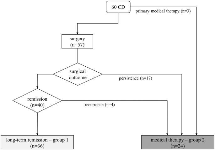
Treatment and outcome of the described cohort. Light gray box indicates those patients in group 1 (surgical remission, n = 36), dark gray box indicates the patients in group 2 that achieved normalization of UFC with medical therapy (n = 24, either primary or after surgical failure)

Table 1 Cortisol-lowering drugs, dose, and time in treatment of subjects treated with a single and combined lines of therapy

All 60 patients completed at least 2 years of follow-up; a long-term 5-years evaluation was available in 43 patients of the original cohort (32 after surgery and 11 with medical therapy). Baseline characteristics of the two cohorts are reported in Table 2.

Table 2 Baseline characteristics of the two groups and previous treatment modalities

Data collection and study design

Two researchers retrieved clinical and biochemical data independently from the local digital medical records. We considered as baseline visit the clinical and endocrine evaluation performed with active hypercortisolism. Therefore, the baseline visit consists in the pre-surgical evaluation in group 1, and in the post-surgical confirmation of active hypercortisolism in those in medical treatment (or diagnosis in case of primary treatment, group 2).

We considered clinical and biochemical outcomes during routine follow-up at two- and five-years in each group, starting from surgical remission or the beginning of a stable normalization of UFC under medical therapy. CD diagnosis was based on at least two parameters among 24-h UFC above the upper normal limit (ULN, at least two collections), unsuppressed cortisol levels (> 50 nmol/L) after 1 mg overnight dexamethasone test (1 mg-DST) or late-night salivary cortisol (LNSC) > ULN (at least two samples). In all subjects, CD diagnosis was considered in case of normal-high ACTH levels, positive response to dynamic tests (corticotropin-releasing hormone or desmopressin test, high-dose dexamethasone test), and, two cases, with petrosal sinus sampling (BIPSS) [11]. Long-term remission after TSS was defined through normal UFC, combined with serum cortisol levels < 50 nmol/L in the first month after surgery and need of glucocorticoid replacement therapy. A relapse of CD was defined as the reappearance of the typical signs and symptoms of CD associated with the alteration of at least two first-line screening tests. Presence/absence of clinical signs of CD (round face, facial rubor, buffalo hump, bruising, cutaneous red striae, acne, hirsutism and oligo/amenorrhea in females) were evaluated during outpatient visits by expert endocrinologists. The presence of hirsutism in females was measured according to the Ferriman–Gallwey score > 8 (extent of hair growth in 9 locations was rated 0–4). Proximal muscle strength was diagnosed if patients were not able to stand up from a low seated position with anteriorly extended arms. Bodyweight, body mass index (BMI), waist and hip circumference, systolic (SBP), and diastolic blood pressure (DBP) were assessed with calibrated tools. Overweight was diagnosed in patients with BMI 25–30 kg/m2, obesity with BMI > 30 kg/m2. Visceral obesity was diagnosed as waist circumference ≥ 94 cm in men and ≥ 80 cm in women, or with a waist/hip ratio (WHR) ≥ 1 according to International Diabetes Federation criteria. Arterial hypertension was diagnosed for SBP above 140 mm Hg and/or DBP above 90 mm Hg and/or in patients on antihypertensive drugs. Diabetes mellitus (DM) was diagnosed according to American Diabetes Association criteria or when patients were taking antidiabetic medication. Dyslipidemia was diagnosed when low-density lipoprotein (LDL) calculated cholesterol was ≥ 100 mg/dL and hypertriglyceridemia when triglycerides were ≥ 150 mg/dL or when patients were on lipid-lowering medication. The presence of carotid vascular disease (CVD) has been assessed by supra-aortic vessels duplex ultrasound. Cushing’s cardiomyopathy (CCM) was diagnosed by doppler echocardiography with evidence of impaired relaxation and left ventricular filling pattern. The medical history was checked for cardiovascular disease (acute coronary syndrome, ACS) in all cases. A shortened activated partial thromboplastin time (aPTT < 29 s) defined pro-thrombotic status.

Assays

All biochemical analyses were carried out in an ISO15189:2012-accredited clinical laboratory [12], cortisol levels have been measured in urine or saliva with a mass-spectrometry home-made validated method. UFC was determined by a home-brew liquid chromatography-mass spectrometry (LC-MS/MS) method (intra-assay/interassay coefficient of variation [CV] < 6%/< 8%) since 2011 [13], previously by a radio-immunometric assay (Radim, intra-assay/interassay CV < 3%/< 9%). The patients were instructed to discard the first morning urine void and to collect all urine for the next 24 h, so that the morning urine void on the second day was the final collection. The sample was kept refrigerated from collection time until it was analyzed: normal range for UFC is 16–168 nmol/24 h.

Salivary cortisol was measured by a radio-immunometric assay (Radim, intra-assay/interassay CV < 3%/< 9%) until 2014 [14], after then by LC-MS/MS method (intra-assay/interassay CV < 6%/< 8% [15]). In order to prevent food or blood contamination, samples were collected at least 30 min after subjects had eaten, brushed their teeth, smoked or assumed liquorice; undertaken using Salivette® devices containing a cotton swab with or without citric acid (Sarstedt, Nümbrecht, Germany). The sample was stored at − 80 °C, before analyses [15].

The 1-mg DST test was performed orally assuming 1 mg of dexamethasone between 11 P.M. and midnight, sampling serum cortisol the next morning at 8 A.M. Serum dexamethasone levels, routinely evaluated since 2017, were adequate in all cases [16]. Serum cortisol (RRID: AB_2810257) and ACTH (RRID: AB_2783635) were determined by immune-chemiluminescence assay (Immulite 2000, Siemens Healthcare). Dynamic second-line tests and BIPSS were performed according to international standards.

Statistical analysis

Data were analyzed using SPSS Software for Windows, version 24.0 (SPSS Inc). Data are reported as medians and interquartile range or as percentages. The comparison between continuous variables was performed by non-parametric Wilcoxon test or Mann–Whitney test, as appropriate. The comparison between categorical variables was performed by the χ2 test. The correlation between continuous variables was performed by linear regression analysis. The level of significance for the overall difference between the groups was tested with one-way ANOVA. A p value < 0.05 was considered statistically significant.

Results

Endocrine evaluation

At baseline the two groups were similar for morning serum/salivary cortisol, LNSC, cortisol after 1 mg DST and morning ACTH levels (Table 3); UFC levels were higher in the surgical cohort (p < 0.001). Endocrine parameters were not influenced by sex and BMI. At baseline, all patients had impaired salivary cortisol rhythm with increased LNSC and inadequate cortisol suppression after 1-mg DST. At two years the recovery of salivary cortisol rhythm was observed in 97% of patients after surgery and 50% of patients during medical therapy. The only patient who did not show recovery of cortisol rhythm in the surgical cohort had LNSC of 5.4 nmol/L (range 0.5–2.6 nmol/L), with adequate cortisol suppression after 1-mg DST and sustained normal UFC: it was considered a false-positive due to residual minor depression state.

Table 3 Biochemical pattern at baseline and during the follow-up

Adequate cortisol suppression after 1-mg DST (both with normal UFC and LNSC) was observed in 34 out of 36 patients (94%) in the surgical cohort; the two patients who did not show complete cortisol suppression after 1-mg DST had cortisol levels of 60 and 119 nmol/l, respectively. On the contrary, as per selection criteria, none of the patients in group 2 presented suppressed cortisol after 1-mg DST.

At 5 years follow-up, all cases in the surgical cohort had suppressed cortisol after 1-mg DST and normal salivary cortisol rhythm, whereas in group 2 9% had suppressed cortisol after 1-mg DST and 36% recovered salivary cortisol rhythm. At 5 years, UFC and salivary cortisol levels (either morning or late night) were similar in the two groups, while the median value of serum cortisol after 1-mg DST remained not adequately suppressed (median 75 nmol/L, from 18 to 257 nmol/L) during medical therapy (See Table 3). In group 2, patients on combined therapy had higher UFC (102 vs. 76 nmol/24h p = 0.03) and LNSC (2.4 vs. 1.9 p = 0.05) at 5 years, compared to patients on monotherapy.

Hirsutism, abdominal obesity, round face and facial rubor were prevalent in group 1 at baseline. On the contrary, the abdominal obesity, facial rubor and easy bruising were most commonly found in the medical cohort. The prevalence of facial rubor, buffalo hump and bruisability was higher after medical than surgical remission after 2 years of eucortisolism; at 5 years the prevalence of buffalo hump and bruisability was higher in patients under drug therapy as well (Table 4; Fig. 2). Higher levels of UFC at baseline were observed in all patients with proximal myopathy (p < 0.001).

Table 4 Two- and five-years changes in clinical phenotype from baseline in group 1 and group 2
Fig. 2

figure 2

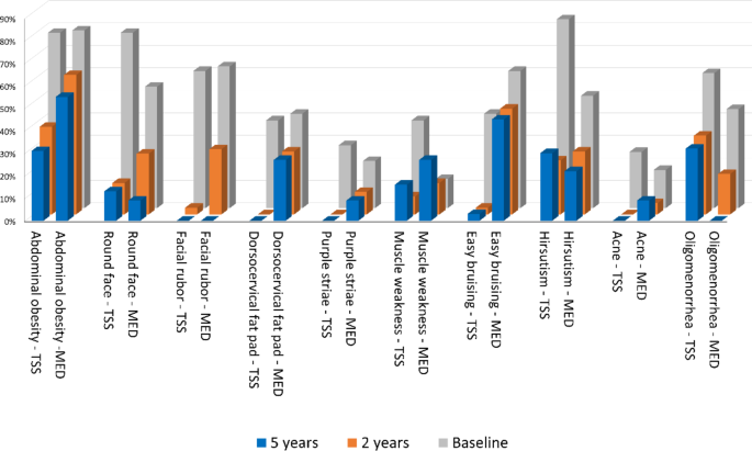
Signs and symptoms of hypercortisolism at baseline (grey bars), two-years (orange bars) and five-years (blue bars) follow up after surgical (TSS) or medical remission (MED)

Arterial hypertension

Arterial hypertension (AH) was the most frequent comorbidity in both groups at baseline, with similar distribution in the two groups (Table 5). The prevalence of AH decreased after two years in both groups, especially in the surgical cohort (64% vs. 44% in group 2, p < 0.001; 75% vs. 71% p = 0.003), with no further improvement after five years. Overall, hypertensive patients were older at diagnosis (45yrs vs. 31y; p < 0.001) and with larger BMI (29 vs. 25 kg/m2p = 0.03). Median UFC, morning salivary cortisol and LNSC, and 1-mg DST were not different in patients with/without AH at baseline and at 2 years. SBP and DBP values were similar in the two cohorts and were not correlated to UFC, LNSC or 1-mg DST throughout the follow-up. At 2 years, hypertensive patients had higher levels of morning salivary cortisol and LNSC with impaired rhythm (respectively 10.4 vs. 6 nmol/L, p = 0.01 and 3.2 vs. 1 nmol/l, p = 0.007). SBP and DBP values did not change during the five-years observation time in both groups; however, the number of anti-hypertensive drugs was higher in group 2 than in group 1 (p = 0.007). Overall patients treated with metyrapone showed higher values of DBP at 2 years (mean 89.4 vs. 81.7 mmHg, p = 0.01), the prevalence of AH did not differ from patients with other medical treatments.

Table 5 Two- and five-years changes in cardio-metabolic cortisol-related comorbidities of CD from baseline in group 1 and group 2

Glucose metabolism

DM prevalence at baseline did not show a correlation with BMI and age at CD diagnosis. DM prevalence was similar in group 1 and 2 after two and five years of follow-up. The follow-up analysis of DM was performed excluding patients in pasireotide, since its known impact in glucose metabolism. In both groups, median UFC, morning salivary and LNSC, and 1-mg DST were similar in patients with/without DM at baseline. At 5 years, patients with diabetes had higher levels of morning salivary cortisol and LNSC with impaired cortisol rhythm (respectively 15 vs. 7 nmol/L, p < 0.001 and 5.4 vs. 1.5 nmol/l, p < 0.001). None of the explored hormonal parameters was correlated with HbA1c levels in both groups at any time point considered. The number of antidiabetic drugs was higher after medical than surgical remission (Table 5).

As expected, patients treated with pasireotide had higher incidence of newly onset DM at 2- and 5 years (p = 0.02 and p = 0.05 respectively) and required more antidiabetic drugs at 2- and 5 years (p = 0.002, p = 0.05) or insulin units at 5 years (p = 0.03). HbA1c levels during pasireotide were higher than patients treated with other drugs (55.6 vs. 38 nmol/l, p = 0.002), requiring a higher number of antidiabetic drugs (p = 0.008). Patients on combined therapy with pasireotide had higher rates of DM at 2- and 5 years (p < 0.001 and p = 0.01) and used more antidiabetic drugs at 2- and 5 years (p = 0.004, p = 0.01) than those on monotherapy.

Lipid metabolism

The prevalence of dyslipidemia was similar in the two groups at baseline and after two years, and higher in the medical remission cohort after five years (p = 0.01). Overall, dyslipidemic patients were older at diagnosis (46y vs. 36y; p = 0.006) and had higher BMI (30 vs. 25 kg/m2p < 0.001). There was no correlation between hormone parameters and LDL or triglycerides levels. Lipid profile was similar between patients treated with different drugs.

Vascular disease and coagulative profile

There was no difference between the two groups, at baseline, in the prevalence of carotid vascular disease, history of ACS, and CCM; at 5 years, in both groups, no patient had a worsening of a previously diagnosed stenosis, or novel diagnosis of CVD, ACS and CCM.

The median aPTT value at baseline was in the pro-thrombotic range in both groups (25s), without sex and BMI differences. No correlation was observed between aPTT and UFC, LNSC and 1-mg DST levels. Patients who manifested easy bruising, had shorter aPTT at 2- and 5 years (median 24 vs. 27s, p = 0.03). aPTT does not increase within both groups at 2- and 5-years and aPTT was shorter during medical therapy compared to surgical remission both after 2 and 5 years (22.5s vs. 27s, p = 0.02 at 2y and 23.5s vs. 27.9s, p = 0.02 at 5y).

Discussion

The impact of CD remission on clinical picture and hypercortisolism-related comorbidities is still controversial. The current knowledge suggests that long-term CD surgical remission is associated with increased metabolic and vascular damage, not only if compared to active disease, but also even after long-term normalization of cortisol secretion [17]. If CD recurs after successful TSS, or if surgery fails/is not feasible, cortisol excess can be treated with medical therapy. Likewise, long-term studies (> 2 years) on the clinical effects of medical therapy on CD are lacking. Some prospective registry studies have been published [1], only one retrospective study on long-term use of ketoconazole described a multicentric cohort of CD patients without a control group [18].

In our study, we enrolled 60 patients with CD diagnosed and treated in a single tertiary care center, with sustained and long-term (2 and 5 years) UFC normalization after surgery or during medical therapy. As expected, UFC levels at baseline were different in the two groups, due to the distinct starting point of medical history: a patient with persistent-recurrent CD after pituitary surgery presents with lower UFC than the new diagnosis. After surgical remission, patients achieved the recovery of salivary cortisol rhythm and the complete suppression of cortisol after 1-mg DST (investigated after substitutive glucocorticoid treatment discontinuation) in almost all cases. On the contrary, if eucortisolism is achieved with long-term medical therapy the recovery of salivary cortisol rhythm was observed only in half of patients and only few of them showed cortisol suppression after 1-mg DST within the 5 years observation time. Patients who were more resistant to the recovery of cortisol rhythm were more likely to receive combined treatment, even if no treatment is superior to others in normalizing salivary cortisol rhythm, in line with previous reports [11819].

Within 2 years, patients in the surgical remission group showed a marked improvement of all phenotypic traits common at CD diagnosis compared to those in medical therapy. As observed also in other series of CD patients in remission [20], abdominal obesity persisted more than other clinical features over time, leading to an impaired body composition especially in the medically treated group [21]. Considering hyperandrogenism, acne improvement was more relevant at 2 and 5-years of follow up, probably due to a differential effect of ACTH-dependent adrenal androgens compared to hirsutism.

The impaired cortisol rhythm was a predictor of the long-lasting of most CD phenotypic features, as round face, buffalo hump, facial rubor, abdominal obesity, proximal myopathy and bruisability. A more severe clinical phenotype at baseline can explain a reduced control of hypercortisolism in monotherapy, requiring drug combination, and signs or symptoms are likely to persist despite the normalization of UFC [22]. In this study, no medication outperformed the others in terms of recovery from the CD phenotype.

The aetiology of hypertension and dyslipidemia is known to be heterogeneous, since both are influenced also by age at diagnosis and BMI, causing low rates of remission after UFC normalization [2324]. Arterial hypertension showed a decreasing trend with the best response within 2 years after UFC normalization only after surgical remission. Patients with disrupted salivary cortisol rhythm were more likely to remain hypertensive during the 5 years follow-up. Likewise, DM persistence during follow up correlates to impaired salivary cortisol rhythm and not with UFC. This finding is in contrast with the observations of Schernthaner-Reiter et al. [25]. on CD remission, and, on the contrary, supports data described by Guarnotta et al. [22]. Newell-Price et al.. recently found that when UFC and LSNC are both normal in patients treated with pasireotide, the rise in HbA1c levels is less evident than in patients with normal UFC but uncontrolled LNSC [26]. This observation underlines the importance of the impaired cortisol rhythm in the glucose impairment pathogenesis in CD. During the 5 years observation time, a worsening of previously diagnosed cardiovascular conditions, or novel acute vascular events, was not observed in both groups. This finding suggested that normalized UFC and intensive treatment of cardio-metabolic CD comorbidities play a fundamental role in reducing cardiovascular mortality [27]. A minor impact of CD therapy was observed in dyslipidemia, which persisted in both groups, with minimal improvement over time (−22% in surgical and − 6% in medical cohort). The criterion of 100 mg/dL LDL cut-off identifies a moderate CV risk reflecting the main focus of the study: the assessment of cardiometabolic complication after CD remission, assuming that they present a lower cardiovascular risk compared to patients with overt hypercortisolism.

Plasma hypercoagulability, with shortened aPTT, was found in all patients with active hypercortisolism. In the 5 years observation time, this parameter showed latency in increasing in both groups and in none achieved normality (> 28s). As previously observed in other studies, no correlation is observed between aPTT and any of the explored hormonal parameters [2228]. At 2- and 5 years, instead, shorter aPTT was observed during medical treatment than after surgical remission cohort. In both groups a shorter aPTT was associated with bruisability, which is related to impaired LNSC, strengthening the role of the impaired cortisol rhythm as a major driver of hypercoagulability. Also, Ferrante et al.. observed the long latency of plasma hypercoagulability, persisting for years after biochemical remission of CD: in that series thrombophilia appeared to be reversible within 5 years [29], while in our cohort the recovery takes longer.

Additionally, sexual differences characterize patients with patients with Cushing’s syndrome and hypogonadism in hypercortisolism is known to further increase the cardiovascular risk [3031]. However, it was not an interfering factor in our study population since hypopituitarism was considered an exclusion criterion, no case of new-onset hypogonadism was reported (even in male patients treated with ketoconazole), and the menopause transition in six women during the observation was not considered relevant.

The limits of the present study are its retrospective design, the variability of concomitant treatments, the heterogenous combinations of medical therapy used in clinical practice, the presence of treatment-specific adverse events that mimic the effects of hypercortisolism (such as pasireotide-induced DM and hypertension with metyrapone), the unpredictable effect of previous treatments, including radiotherapy. We considered UFC and LNSC as markers of hypercortisolism remission; nonetheless we acknowledge that both of them present some limitations, especially during medical treatment. The former considers the whole cortisol secretion during the day, and albeit UFC normalization is the main outcome of all trials for medical treatment [3233] it does not detect mild hypercortisolism. On the other hand, a normal LNSC does not fully reflect a normal circadian rhythm: only high cortisol levels in the morning with a decline in the night are able to restore clock-related activities [34].

Its strengths are the complete patient characterization in a single tertiary care center, the comparative study design, and the standardized protocols for diagnosis and long-term follow-up. In particular, samples have been processed within a single laboratory with accurate methods (LC-MS for urinary and salivary steroids), and all endocrine aspects of hypercortisolism were considered (overall daily cortisol production by UFC, circadian cortisol rhythm, and the recovery of the hypothalamic-pituitary axis by 1-mg DST overnight test).

To conclude, despite UFC normalization in both groups during follow-up, surgical remission results in more rapid and relevant improvements in CD phenotype and comorbidities. During medical therapy the UFC levels can be higher than after surgery, although in the normal range, and the normalization of LNSC is not always achieved: both conditions suggests that stricter criteria should be considered to define eucortisolism in patients with CD under medical treatment. Conditions such as obesity, hypertension, dyslipidemia, and hypercoagulability are not completely reversible in a 5-year observation time even in the surgical remission group. This observation underlines that all the comorbidities, independently of the normalization of UFC, must be intensively treated. Moreover, UFC normalization should not be considered the only biochemical goal to be reached, since the persistence of comorbidities seems to be more related to an impaired cortisol rhythm rather than to the cortisol secretory burden.

References

  1. Fleseriu M et al (2021) Consensus on diagnosis and management of Cushing’s disease: a guideline update, Dec. 01, Elsevier Ltd. https://doi.org/10.1016/S2213-8587(21)00235-7

  2. Gadelha M, Gatto F, Wildemberg LE, Fleseriu M Cushing’s syndrome. Dec 09 2023 Elsevier B V https://doi.org/10.1016/S0140-6736(23)01961-X

  3. Ceccato F et al (2024) Clinical and biochemical data for the diagnosis of endogenous hypercortisolism: the ‘cushingomic’ approach. J Clin Endocrinol Metab Jul. https://doi.org/10.1210/clinem/dgae517

    Article Google Scholar

  4. Pivonello R, Isidori AM, De Martino MC, Newell-Price J, Biller BMK, Colao A (2016) Complications of Cushing’s syndrome: state of the Art. Lancet Publishing Group. https://doi.org/10.1016/S2213-8587(16)00086-3

    Article Google Scholar

  5. Clayton RN et al (2016) Mortality in patients with Cushing’s disease more than 10 years after remission: A multicentre, multinational, retrospective cohort study. Lancet Diabetes Endocrinol 4(7):569–576. https://doi.org/10.1016/S2213-8587(16)30005-5

    Article PubMed Google Scholar

  6. Pivonello R, De Leo M, Cozzolino A, Colao A (2015) The treatment of Cushing’s disease. Endocr Soc. https://doi.org/10.1210/er.2013-1048

    Article Google Scholar

  7. Petersenn S et al (2015) Outcomes in patients with Cushing’s disease undergoing transsphenoidal surgery: Systematic review assessing criteria used to define remission and recurrence. BioScientifica Ltd. https://doi.org/10.1530/EJE-14-0883

    Article Google Scholar

  8. Broersen LHA, Jha M, Biermasz NR, Pereira AM, Dekkers OM (2018) Effectiveness of medical treatment for Cushing’s syndrome: a systematic review and meta-analysis. Pituitary 21(6):631–641. https://doi.org/10.1007/s11102-018-0897-z

    Article CAS PubMed PubMed Central Google Scholar

  9. von Elm E, Altman DG, Egger M, Pocock SJ, Gøtzsche PC, Vandenbroucke JP (2008) The Strengthening the Reporting of Observational Studies in Epidemiology (STROBE) statement: guidelines for reporting observational studies. J Clin Epidemiol 61(4): 344–349. https://doi.org/10.1016/j.jclinepi.2007.11.008

  10. Ceccato F (2024) The burden Of Cushing’s disease cardiometabolic comorbidities: comparison between surgical remission and long-term eucortisolism with medical treatment. Repository of the University of Padova. https://doi.org/10.25430/researchdata.cab.unipd.it.00001350

  11. Barbot M et al (2016) Second-line tests in the differential diagnosis of ACTH-dependent Cushing’s syndrome. Pituitary 19(5):488–495. https://doi.org/10.1007/s11102-016-0729-y

    Article CAS PubMed Google Scholar

  12. ISO 15189:2012 Medical laboratories — Requirements for quality and competence. Published 2022. Accessed October 10 (2023) https://www.iso.org/standard/56115.html

  13. Ceccato F et al (2014) The diagnostic performance of urinary free cortisol is better than the cortisol: cortisone ratio in detecting de Novo Cushing’s syndrome: the use of a LC-MS/MS method in routine clinical practice. Eur J Endocrinol 171(1):1–7. https://doi.org/10.1530/EJE-14-0061

    Article CAS PubMed Google Scholar

  14. Ceccato F et al (2012) Assessment of glucocorticoid therapy with salivary cortisol in secondary adrenal insufficiency. Eur J Endocrinol 167(6):769–776. https://doi.org/10.1530/EJE-12-0534

    Article CAS PubMed Google Scholar

  15. Antonelli G, Ceccato F, Artusi C, Marinova M, Plebani M (2015) Salivary cortisol and cortisone by LC-MS/MS: Validation, reference intervals and diagnostic accuracy in Cushing’s syndrome. Clinica Chimica Acta 451:247–251. https://doi.org/10.1016/j.cca.2015.10.004

    Article CAS Google Scholar

  16. Ceccato F et al (2020) Dexamethasone measurement during low-dose suppression test for suspected hypercortisolism: threshold development with and validation. J Endocrinol Invest 43(8):1105–1113. https://doi.org/10.1007/s40618-020-01197-6

    Article CAS PubMed Google Scholar

  17. Pivonello R, Faggiano A, Lombardi G, Colao A (2005) The metabolic syndrome and cardiovascular risk in Cushing’s syndrome. W.B. Saunders. https://doi.org/10.1016/j.ecl.2005.01.010

    Book Google Scholar

  18. Castinetti F et al (2014) Ketoconazole in Cushing’s disease: is it worth a try. J Clin Endocrinol Metab 99(5):1623–1630. https://doi.org/10.1210/jc.2013-3628

    Article CAS PubMed Google Scholar

  19. Barbot M et al (2014) Combination therapy for Cushing’s disease: Effectiveness of two schedules of treatment. Should we start with cabergoline or ketoconazole? Pituitary 17(2):109–117. https://doi.org/10.1007/s11102-013-0475-3

    Article CAS PubMed Google Scholar

  20. Colao A et al (1999) Persistence of Increased Cardiovascular Risk in Patients with Cushing’s Disease after Five Years of Successful Cure,., [Online]. Available: https://academic.oup.com/jcem/article/84/8/2664/2864186

  21. Ceccato F et al (2017) Sep., Body Composition is Different after Surgical or Pharmacological Remission of Cushing’s Syndrome: A Prospective DXA Study, Hormone and Metabolic Research, vol. 49, no. 9, pp. 660–666. https://doi.org/10.1055/s-0043-115008

  22. Guarnotta V et al (2017) The degree of urinary hypercortisolism is not correlated with the severity of cushing’s syndrome. Endocrine 55(2):564–572. https://doi.org/10.1007/s12020-016-0914-9

    Article CAS PubMed Google Scholar

  23. Giordano R et al (2011) Metabolic and cardiovascular outcomes in patients with Cushing’s syndrome of different aetiologies during active disease and 1 year after remission. Clin Endocrinol (Oxf) 75(3):354–360. https://doi.org/10.1111/j.1365-2265.2011.04055.x

    Article CAS PubMed Google Scholar

  24. Jha S, Sinaii N, McGlotten RN, Nieman LK (2020) Remission of hypertension after surgical cure of Cushing’s syndrome. Clin Endocrinol (Oxf), 92(20): 124–130. https://doi.org/10.1111/cen.14129

  25. Schernthaner-Reiter MH et al (2019) Factors predicting long-term comorbidities in patients with Cushing’s syndrome in remission. Endocrine 64(1):157–168. https://doi.org/10.1007/s12020-018-1819-6

    Article CAS PubMed Google Scholar

  26. Newell-Price J et al (2020) Use of late-night salivary cortisol to monitor response to medical treatment in Cushing’s disease. Eur J Endocrinol 182(2):207–217. https://doi.org/10.1530/EJE-19-0695

    Article CAS PubMed Google Scholar

  27. Mondin A et al (2023) Complications and mortality of Cushing’s disease: report on data collected over a 20-year period at a referral centre. Pituitary 26(5):551–560. https://doi.org/10.1007/s11102-023-01343-2

    Article PubMed PubMed Central Google Scholar

  28. Barbot M et al (2018) Effects of pasireotide treatment on coagulative profile: a prospective study in patients with Cushing’s disease. Endocrine 62(1):207–214. https://doi.org/10.1007/s12020-018-1669-2

    Article CAS PubMed Google Scholar

  29. Ferrante E et al (2022) Evaluation of procoagulant imbalance in Cushing’s syndrome after short- and long-term remission of disease. J Endocrinol Invest 45(1):9–16. https://doi.org/10.1007/s40618-021-01605-5

    Article CAS PubMed Google Scholar

  30. Zilio M, Barbot M, Ceccato F, Camozzi V, Bilora F, Casonato A, Frigo AC, Albiger N, Daidone V, Mazzai L, Mantero F, Scaroni C (2014) Diagnosis and complications of Cushing’s disease: gender-related differences. Clin Endocrinol (Oxf) 80(3):403–410. https://doi.org/10.1111/cen.12299

    Article CAS PubMed Google Scholar

  31. Detomas M, Deutschbein T, Tamburello M, Chifu I, Kimpel O, Sbiera S, Kroiss M, Fassnacht M, Altieri B (2024) Erythropoiesis in Cushing syndrome: sex-related and subtype-specific differences. Results from a monocentric study. J Endocrinol Invest 47(1):101–113. https://doi.org/10.1007/s40618-023-02128-x

    Article CAS PubMed Google Scholar

  32. Fleseriu M et al (2019) Long-term efficacy and safety of once-monthly pasireotide in Cushing’s disease: A Phase III extension study. Clin Endocrinol (Oxf) 91(6):776–785. https://doi.org/10.1111/cen.14081

    Article CAS PubMed Google Scholar

  33. Ceccato F et al (2018) Metyrapone treatment in Cushing’s syndrome: a real-life study. Endocrine 62(3):701–711. https://doi.org/10.1007/s12020-018-1675-4

    Article CAS PubMed Google Scholar

  34. Minnetti M, Hasenmajer V, Pofi R, Venneri MA, Alexandraki KI, Isidori AM (2020) Fixing the broken clock in adrenal disorders: Focus on glucocorticoids and chronotherapy. BioScientifica Ltd. https://doi.org/10.1530/JOE-20-0066

    Article Google Scholar

Download references

Funding

Open access funding provided by Università degli Studi di Padova within the CRUI-CARE Agreement.

All authors certify that they have no affiliations with or involvement in any organization or entity with any financial interest or non-financial interest in the subject matter or materials discussed in this manuscript.

Author information

Authors and Affiliations

  1. Department of Medicine-DIMED, University of Padova, Padova, Italy

    Irene Tizianel, Laura Lizzul, Alessandro Mondin, Giacomo Voltan, Pierluigi Mazzeo, Carla Scaroni, Mattia Barbot & Filippo Ceccato

  2. Endocrinology Unit, Department of Medicine DIMED, University Hospital of Padova, Via Ospedale Civile, 105, Padova, 35128, Italy

    Irene Tizianel, Laura Lizzul, Alessandro Mondin, Giacomo Voltan, Pierluigi Mazzeo, Carla Scaroni, Mattia Barbot & Filippo Ceccato

Corresponding author

Correspondence to Filippo Ceccato.

Ethics declarations

Conflict of interest

The authors declare that they have no conflict of interest.

Ethical approval

All procedures performed in studies involving human participants were in accordance with the ethical standards of the institutional and/or national research committee and with the 1964 Helsinki Declaration and its later amendments or comparable ethical standards.

Informed consent

Informed consent was obtained from all individual participants involved in the study.

Additional information

Publisher’s note

Springer Nature remains neutral with regard to jurisdictional claims in published maps and institutional affiliations.

Cite this article

Tizianel, I., Lizzul, L., Mondin, A. et al. Cardiometabolic complications after Cushing’s disease remission. J Endocrinol Invest (2025). https://doi.org/10.1007/s40618-025-02572-x

Download citation

Share this article

Anyone you share the following link with will be able to read this content:

Get shareable linkProvided by the Springer Nature SharedIt content-sharing initiative

Keywords

From https://link.springer.com/article/10.1007/s40618-025-02572-x

Ivory Tower: New PET Scan Molecule Offers Breakthrough in Detecting Cushing’s Syndrome

Asignificant advancement in medical imaging has been achieved by experts at the Postgraduate Institute of Medical Education and Research. Researchers from endocrinology and nuclear medicine departments have introduced a new PET scan molecule that enhances the detection of adenoma/small tumour causing Cushing’s syndrome. This development promises to improve surgical interventions and patient outcomes.

A collaborative effort between Dr Rama Walia from the endocrinology department and Dr Jaya Shukla from nuclear medicine department has resulted in the development of GA-68 MDEsMO, a specialised PET scan molecule. This innovative compound is designed to pinpoint tumors in the pituitary gland, which are often responsible for the excessive cortisol production characteristic of Cushing’s syndrome.

By providing a more precise visualisation of these tumours, the new technique enables neurosurgeons to operate with greater accuracy, preserving the normal functions of the pituitary gland and enhancing patients’ quality of life.

Recognising its potential, this pioneering technology was honoured at the institution’s recent Research Day.

Affects entire body

Cushing’s syndrome is a complex endocrine disorder that occurs due to an overproduction of cortisol, a hormone that influences multiple physiological processes. When cortisol levels surge beyond normal, it disrupts the body’s balance, leading to widespread health complications. The primary cause of this condition is a minuscule tumor within the pituitary gland, making diagnosis particularly challenging.

Currently, only 60 to 70 per cent of patients receive an accurate diagnosis due to the minuscule size of these tumors — often measuring less than a millimeter. The introduction of GA-68 MDEsMO is expected to bridge this diagnostic gap by facilitating early detection, thus enabling timely surgical intervention.

Hormonal disruptions

The pituitary gland, often referred to as the “master gland”, plays a crucial role in regulating hormone production. However, when affected by a tumor, it triggers an excessive release of hormones, leading to systemic damage.

Typical symptoms include unexplained weight gain, obesity and noticeable changes in skin texture. Many patients develop distinctive pink or purplish stretch marks on the abdomen, thighs and arms. Women may experience excessive hair growth, while men could suffer from reduced fertility and erectile dysfunction. Additionally, skin thinning, severe acne and heightened susceptibility to bruising are common indicators of the disease.

Understanding cortisol

Cortisol, a steroid hormone, is essential for stress regulation and overall metabolic balance. Produced by the adrenal glands, it influences numerous bodily functions through interactions with cortisol receptors present in most cells. The secretion of cortisol is managed by a complex system involving the hypothalamus, pituitary gland, and adrenal glands. Given its widespread presence, cortisol plays a vital role in multiple physiological processes, including immune response, metabolism, and blood pressure regulation.

However, any disruption in cortisol levels—whether an excess or deficiency—can lead to significant health challenges. This underscores the importance of precise diagnosis and timely treatment for disorders like Cushing’s syndrome. The introduction of GA-68 MDEsMO marks a crucial step in advancing medical science’s ability to manage and treat this condition effectively. —

https://www.tribuneindia.com/news/punjab/ivory-tower-new-pet-scan-molecule-offers-breakthrough-in-detecting-cushings-syndrome/

Ectopic CRH/ACTH-Co-Secreting Neuroendocrine Tumors Leading to Cushing’s Disease

Abstract

Adrenocorticotropic hormone (ACTH) and corticotropin-releasing hormone (CRH) are essential regulators of cortisol production within the hypothalamic-pituitary-adrenal (HPA) axis. Elevated cortisol levels, resulting from excessive ACTH, can lead to Cushing’s syndrome, a condition with significant morbidity. Neuroendocrine tumors (NETs) can ectopically produce both ACTH and CRH, contributing to this syndrome. This review discusses the pathophysiology, types, clinical presentation, diagnosis, and management of these tumors. Emphasis is placed on the importance of identifying dual CRH/ACTH secretion, which complicates diagnosis and necessitates tailored therapeutic strategies. Furthermore, the review highlights the prognosis, common complications, and future directions for research in this area.

We report the case of a 53-year-old female patient who presented with severe Cushing’s syndrome and was diagnosed with ectopic ACTH syndrome. Despite initial indications pointing towards pituitary-dependent hypercortisolism, further investigations revealed the presence of a highly differentiated atypically located tumor in the upper lobe of the left lung, adjacent to the mediastinum. Immunohistochemistry of the tumor tissue demonstrated not only ACTH but also CRH and CRH-R1 expression. The simultaneous expression of these molecules supports the hypothesis of the presence of a positive endocrine feedback loop within the NET, in which the release of CRH stimulates the expression of ACTH via binding to CRH-R1. This case report highlights the challenges in diagnosing and managing ectopic ACTH syndrome, emphasizing the importance of a comprehensive diagnostic approach to identify secondary factors impacting cortisol production, such as CRH production and other contributing neuroendocrine mechanisms.

Journal Section:

Review Article

This content is only available via PDF.

The Author(s). Published by S. Karger AG, Basel
Open Access License / Drug Dosage / Disclaimer
This article is licensed under the Creative Commons Attribution 4.0 International License (CC BY). Usage, derivative works and distribution are permitted provided that proper credit is given to the author and the original publisher.
Drug Dosage: The authors and the publisher have exerted every effort to ensure that drug selection and dosage set forth in this text are in accord with current recommendations and practice at the time of publication. However, in view of ongoing research, changes in government regulations, and the constant flow of information relating to drug therapy and drug reactions, the reader is urged to check the package insert for each drug for any changes in indications and dosage and for added warnings and precautions. This is particularly important when the recommended agent is a new and/or infrequently employed drug.
Disclaimer: The statements, opinions and data contained in this publication are solely those of the individual authors and contributors and not of the publishers and the editor(s). The appearance of advertisements or/and product references in the publication is not a warranty, endorsement, or approval of the products or services advertised or of their effectiveness, quality or safety. The publisher and the editor(s) disclaim responsibility for any injury to persons or property resulting from any ideas, methods, instructions or products referred to in the content or advertisements.

Treatment-Resistant Cushing Disease and Acromegaly in a Young Woman: A Case Of Functional Pituitary Macroadenoma

Abstract

Cushing disease and acromegaly are common endocrine disorders caused by excessive cortisol and growth hormone production, respectively. Both conditions can co-occur due to functioning pituitary adenomas, which are typically benign pituitary gland tumors. This report discusses a 30-year-old woman with hyperpituitarism leading to treatment-resistant Cushing disease and acromegaly caused by a functional pituitary macroadenoma.
A 30-year-old woman presented with a history of excessive weight gain, facial puffiness, fatigue, persistent headaches, and visual disturbances. Clinical examination revealed features consistent with Cushing disease and acromegaly, including a moon face, central obesity, and large hands and feet—the ophthalmologic evaluation identified bitemporal hemianopia, suggesting optic chiasm compression. Laboratory results showed elevated ACTH, IGF-1, and prolactin levels, alongside confirmed hypercortisolism. The patient also had secondary diabetes mellitus and galactorrhea—initial treatment with octreotide provided limited benefit, with persistent hormone elevations and insufficient symptom control. The patient underwent endonasal endoscopic transsphenoidal resection of the pituitary macroadenoma, leading to marked symptomatic and hormonal improvements. This underscores the diagnostic challenge and treatment complexity of such cases. Early diagnosis is critical for optimizing outcomes in patients with hyperpituitarism and mitigating complications. This case highlights the importance of multidisciplinary management and the necessity of long-term follow-up to monitor for recurrence and ensure sustained remission.

Introduction

Pituitary adenomas are benign tumors arising from the pituitary gland, often referred to as the “master gland” due to its central role in regulating key physiological processes such as growth, metabolism, and reproduction [1,2]. These tumors are classified by size into microadenomas (<10 mm) and macroadenomas (≥10 mm) and by hormonal activity into functioning and nonfunctioning adenomas. Functioning adenomas actively secrete hormones, leading to distinct syndromes such as prolactinomas, acromegaly (from growth hormone overproduction), and Cushing disease (from excess ACTH). In contrast, nonfunctioning adenomas do not secrete hormones but may cause symptoms due to mass effects, such as visual disturbances or hypopituitarism [[3][4][5]].
The simultaneous occurrence of Cushing disease and acromegaly is rare and presents a significant diagnostic and therapeutic challenge. Both conditions stem from hyperpituitarism, typically due to a functional pituitary adenoma [6,7]. Cushing disease results from ACTH hypersecretion, causing excessive cortisol production and features such as central obesity, hypertension, hyperglycemia, and muscle weakness [[8][9][10]]. Prolonged cortisol exposure can lead to severe complications, including cardiovascular diseases and osteoporosis. Acromegaly, on the other hand, arises from growth hormone overproduction, leading to elevated IGF-1 levels and characteristic features such as enlarged extremities, facial changes, and systemic complications like insulin resistance and joint abnormalities [[11][12][13]].
The coexistence of Cushing disease and acromegaly within the same affected person is extraordinarily rare, making this particular case record particularly noteworthy [14,15].
The simultaneous presentation of these 2 endocrine problems in a young lady because of a hormonally functioning pituitary macroadenoma presents a unique scientific venture [16,17]. The pituitary macroadenoma, defined as a tumor more than 10 mm in diameter, can compress adjoining structures within the sella turcica and enlarge into surrounding areas, leading to signs and symptoms with complications, visible disturbances, and hyperpituitarism. In this case, the patient presented with both Cushing disease and acromegaly, at the same time symptoms as a result of the mass impact of the macroadenoma.
The case of a 30-year-old female with hyperpituitarism, characterized with the aid of drug-resistant Cushing disease and acromegaly, highlights the complexities intricately associated with the analysis and control of a couple of endocrine issues bobbing up from a single pituitary macroadenoma. Her medical presentation changed into one marked by a history of noticeable weight gain, facial puffiness, fatigue, chronic complications, and visual disturbances. A thorough physical exam found traits consistent with each Cushing disorder and acromegaly, which include a moon face, vital weight problems, and enlarged arms and toes. The ophthalmologic exam confirmed bitemporal hemianopia, indicative of optic chiasm compression with the aid of the pituitary macroadenoma. Early recognition and multidisciplinary management are essential to mitigate the significant morbidity associated with these conditions. This case report highlights a rare instance of concurrent Cushing disease and acromegaly due to a functional pituitary macroadenoma, underscoring the importance of timely diagnosis and treatment.

Case presentation

This case of a 30-year-old female highlights the complexities of diagnosing and managing a functional pituitary macroadenoma presenting with overlapping features of Cushing disease and acromegaly, along with secondary diabetes mellitus.
The patient demonstrated classic signs of hypercortisolism, including central obesity with a “moon face” and “buffalo hump,” skin thinning, easy bruising, and muscle weakness. Cortisol’s catabolic effects were evident in her limb wasting and truncal obesity. Metabolic complications included hypertension and secondary diabetes mellitus, supported by elevated random blood sugar (22 mmol/L) and postprandial blood sugar levels (27 mmol/L). Laboratory findings showed significantly elevated ACTH levels (670 pg/mL; normal: 10–60 pg/mL) and increased morning urine cortisol levels.
The patient also exhibited hallmark features of acromegaly, including enlarged hands and feet, necessitating larger shoe and glove sizes, and distinct facial changes such as mandibular prognathism, frontal bossing, and nasal broadening. Soft tissue swelling and fatigue were also noted, alongside joint pain likely resulting from cartilage and bone overgrowth. Her IGF-1 levels were markedly elevated (798 ng/mL; normal: 100–300 ng/mL).
Hyperprolactinemia (643 ng/mL; normal: 5–25 ng/mL) caused galactorrhea, likely resulting from tumor compression of the pituitary stalk or direct prolactin secretion. Diabetes mellitus, secondary to insulin resistance driven by excess cortisol and growth hormone, further complicated her clinical picture (Table 1).

Table 1. Markedly elevated hormone levels preoperatively and their postoperative normalization.

Hormone Patient’s level (Preoperative) Postoperative levels Normal reference value
ACTH 670 pg/mL 90 pg/mL 10–60 pg/mL
IGF-1 798 ng/mL 280 ng/mL 100–300 ng/mL (age-dependent)
Prolactin 643 ng/mL 42 ng/mL 5–25 ng/mL
Morning Urine Cortisol Elevated Normal <50 mcg/24 h
Random Blood Sugar 22 mmol/L 6.5 mmol/L 4.0–7.8 mmol/L
2-Hour Postprandial Blood Sugar 27 mmol/L 7.0 mmol/L <7.8 mmol/L
TSH (Thyroid-Stimulating Hormone) 0.8 mIU/L 1.2 mIU/L 0.5–5.0 mIU/L
FT3 (Free Triiodothyronine) 4.5 pmol/L 4.0 pmol/L 3.5–7.7 pmol/L
FT4 (Free Thyroxine) 15 pmol/L 16 pmol/L 12–22 pmol/L
Secondary diabetes mellitus is a common trouble in sufferers with Cushing disease and acromegaly, stemming from the insulin resistance brought about by persistent hypercortisolism and hypersecretion of GH. This patient’s multiplied blood sugar also reflects tremendous impairment in glucose metabolism. Polyuria, polydipsia, and unexplained weight loss are classic signs of diabetes that could have been found in her clinical history but are frequently overshadowed by the traits of the more distinguished functions of her endocrine disorders. The affected person additionally experienced galactorrhea, an odd milk discharge from the breasts, that’s on account of her expanded prolactin levels (643 ng/mL, ordinary range: 2-29 ng/mL). Hyperprolactinemia inside the context of a pituitary macroadenoma can result from the tumor’s direct secretion of prolactin or from the stalk effect, where the tumor compresses the pituitary stalk, disrupting dopamine inhibition of prolactin secretion.
MRI was the primary imaging modality, revealing a large pituitary macroadenoma centered within the sella turcica and extending suprasellar. The tumor demonstrated homogeneous postcontrast enhancement and exerted mass effects, including optic chiasm compression correlating with bitemporal hemianopia. Other modalities, such as CT, were not considered due to MRI’s superior resolution for pituitary evaluation.
The MRI scans of the patient reveal a large, well-defined pituitary macroadenoma centered within the sella turcica, exhibiting significant suprasellar extension. On sagittal T1-weighted postcontrast imaging (Fig. 1), the lesion demonstrates homogeneous enhancement with clear, well-defined borders, expanding superiorly into the suprasellar region. Coronal T2-weighted images (Fig. 2) further delineate this suprasellar extension, with the mass exerting mass effect on adjacent structures.
Fig 1:

  1. Download: Download high-res image (309KB)
  2. Download: Download full-size image

Fig. 1. This sagittal T1-weighted postcontrast MRI of the brain, specifically focusing on the sella turcica region, reveals a large, homogeneously enhancing mass centered within the sella turcica, consistent with a pituitary macroadenoma. The mass exhibits clear, well-defined borders and appears to expand the sella, with extension into the suprasellar region (marked by circle).

Fig 2:

  1. Download: Download high-res image (522KB)
  2. Download: Download full-size image

Fig. 2. This image shows MRI scan of the brain in coronal T2-weighted images which reveals large suprasellar mass (marked by circles).

Additional sagittal T1-weighted postcontrast imaging (Fig. 3) confirms the uniform enhancement of the macroadenoma, filling the sella turcica and extending upward. Coronal T2-weighted MRI (Fig. 4) reveals the lesion as hyperintense, extending into the suprasellar region and displacing the optic chiasm. The imaging highlights the well-defined borders of the mass and the potential mass effect on adjacent structures.
Fig 3:

  1. Download: Download high-res image (618KB)
  2. Download: Download full-size image

Fig. 3. Sagittal T1-weighted postcontrast MRI depicting a large, homogeneously enhancing pituitary macroadenoma within the sella turcica, expanding into the suprasellar region with well-defined borders (marked by arrows).

Fig 4:

  1. Download: Download high-res image (1MB)
  2. Download: Download full-size image

Fig. 4. Coronal T2-weighted MRI demonstrating a large, hyperintense pituitary macroadenoma within the sella turcica, extending into the suprasellar region (marked by arrows). The lesion displaces the optic chiasm and exhibits well-defined borders, suggesting potential mass effect.

Axial T2-weighted MRI images (Fig. 5) depict a hyperintense lesion in the basal ganglia and thalamus, appearing as a bright, well-defined signal. This finding suggests a potential coexisting pathology affecting deep brain structures, which may or may not be related to the primary pituitary lesion. The characteristics and location of the pituitary macroadenoma correspond with the patient’s clinical presentation of bitemporal hemianopia, likely caused by compression of the optic chiasm.
Fig 5:

  1. Download: Download high-res image (537KB)
  2. Download: Download full-size image

Fig. 5. Axial T2-weighted MRI images of the brain showing a hyperintense lesion in the region of the basal ganglia and thalamus, indicated by white arrows. The lesion appears as a well-defined, bright signal, suggestive of a pathology affecting deep brain structure.

The overall imaging features, including homogeneous enhancement, well-defined borders, and suprasellar extension, are hallmark characteristics of pituitary macroadenomas. The potential lateral extension toward the cavernous sinus warrants further evaluation, while the hyperintense lesion in the basal ganglia and thalamus may indicate secondary effects or unrelated CNS pathology.
The imaging findings collectively support the diagnosis of a large, functioning pituitary macroadenoma, exceeding 10 mm in diameter. The mass’s size and anatomical impact align with the patient’s clinical presentation, which includes headaches, visual field deficits, and hormonal imbalances. The documented compression of the optic chiasm and possible involvement of the cavernous sinus provide a radiological explanation for the patient’s visual symptoms and hormonal disruptions. This MRI assessment substantiates the diagnosis of a pituitary macroadenoma with significant suprasellar extension and compression effects, consistent with the patient’s symptomatology and clinical findings.
The conglomeration of her clinical presentations, elevated hormone levels, and MRI findings of a big suprasellar mass pretty suggestive of a pituitary macroadenoma showed the analysis of a functioning pituitary adenoma. The preliminary treatment control with octreotide, a somatostatin analog, aimed to control both acromegaly and Cushing disorder by inhibiting GH and ACTH secretion. However, the suboptimal reaction highlighted the undertaking of achieving hormone manipulation in sufferers with massive, competitive adenomas.
Given the patient’s persistent symptoms and the insufficient biochemical response to medical therapy, surgical intervention was considered imperative. The patient underwent endonasal endoscopic transsphenoidal resection of the pituitary gland, a minimally invasive surgical approach targeting the tumor via the nasal passages. This approach was preferred over traditional craniotomy due to its demonstrated efficacy in reducing tumor size and lowering elevated hormone levels with fewer complications, reduced morbidity, shorter hospital stays, and faster recovery times. Additionally, the endoscopic technique offers superior visualization of the surgical field, which aids in precise tumor resection and preservation of normal pituitary tissue.
During the surgery, the tumor was noted to be soft and well-circumscribed, with no significant adherence to adjacent structures such as the cavernous sinus or optic chiasm. This facilitated a complete resection of the tumor, minimizing the risk of residual disease. There were no notable intraoperative complications, such as cerebrospinal fluid leakage or significant bleeding, underscoring the safety and efficacy of the chosen approach. Postoperatively, the patient demonstrated marked clinical improvement in her symptoms, accompanied by a significant reduction in hormone levels to within normal reference ranges. This confirmed the diagnosis and highlighted the effectiveness of the surgical intervention. Specifically, there was a substantial decrease in ACTH, IGF-1, and prolactin levels, leading to clinical remission of Cushing disease and acromegaly.
In the postoperative period, the patient did not require immediate hormone replacement therapy, as her endocrine functions remained stable. However, long-term monitoring is planned to assess for potential hormone deficiencies, disease recurrence, or other complications. The follow-up plan includes regular clinical evaluations, hormonal assays, and periodic imaging studies to ensure sustained remission and to promptly address any residual or recurrent tumor growth. This case highlights the crucial role of surgical intervention in managing functional pituitary macroadenomas, particularly when medical therapy fails. The successful outcome underscores the importance of a multidisciplinary approach and the need for lifelong surveillance to optimize long-term outcomes for such patients. This case scenario also underscores the complexities interwoven in diagnosing and coping with hyperpituitarism because of a pituitary macroadenoma, emphasizing the warrant for a complete and multidisciplinary approach. Early recognition of symptoms, correct diagnostic workup, and timely endocrine disorders.

Discussion

The case of this 30-year-old woman with concurrent refractory Cushing disease and acromegaly due to a functional pituitary macroadenoma highlights the challenges inherent in diagnosing and managing multiple endocrine disorders. Recognizing overlapping clinical features was central to reaching the diagnosis. Classic symptoms of Cushing disease, such as a moon face and central obesity, coupled with acromegalic features, including enlarged extremities, underscored the complexity of the case. The presence of bitemporal hemianopia further pointed to a large pituitary mass compressing the optic chiasm, necessitating imaging studies for confirmation. This case underscores the need for clinicians to remain vigilant when evaluating overlapping endocrine features to avoid delays in diagnosis and treatment [[18][19][20]].
Laboratory evaluations were pivotal, revealing markedly elevated ACTH, IGF-1, and prolactin levels, in addition to evidence of hypercortisolism and secondary diabetes mellitus. These findings highlighted the intricate interplay of hypersecreted pituitary hormones and the systemic consequences of unregulated hormone production. MRI findings of a large suprasellar pituitary tumor were instrumental in confirming the diagnosis of a functional macroadenoma and guided subsequent treatment decisions.
The patient’s suboptimal response to octreotide therapy underscored the limitations of medical treatments in addressing aggressive, hormone-secreting pituitary macroadenomas. While somatostatin analogs are effective in many cases of acromegaly and can provide symptomatic relief, their efficacy is limited in patients with large adenomas and significant hormonal hypersecretion. This case highlights the necessity of early consideration of definitive surgical intervention when medical therapy fails to achieve adequate biochemical control [[21][22][23]].
Endonasal endoscopic transsphenoidal surgery was selected for this patient due to its minimally invasive approach, superior visualization of the sellar region, and lower complication rates compared to traditional craniotomy. Intraoperatively, the tumor’s soft consistency and lack of adherence to adjacent structures facilitated a complete resection. Notably, the absence of significant complications, such as cerebrospinal fluid leakage or vascular injury, reflected the safety and precision of this surgical approach [[24][25][26]].
Postoperatively, the patient experienced substantial improvement in symptoms, with normalization of ACTH, IGF-1, and prolactin levels. This outcome underscores the efficacy of surgical intervention in achieving hormonal remission and alleviating symptoms in patients with functional macroadenomas. The resolution of her secondary diabetes mellitus and galactorrhea further reinforced the success of treatment [[27][28][29]].
Managing such complex endocrine disorders necessitates a multidisciplinary approach, with endocrinologists, radiologists, and neurosurgeons collaborating to ensure accurate diagnosis and effective treatment planning. Radiologists play a critical role in identifying and characterizing pituitary tumors, while endocrinologists monitor hormonal responses and guide perioperative management [[30][31][32]]. Neurosurgeons provide expertise in resecting these challenging lesions and optimizing patient outcomes.
The prognosis for patients undergoing surgical resection of functional pituitary macroadenomas is generally favorable when hormonal remission is achieved. However, long-term follow-up is critical to monitor for potential disease recurrence and manage any residual hormone deficiencies. Lifelong surveillance, including periodic hormonal assays and imaging studies, is recommended. Although the patient did not require immediate hormone replacement therapy, ongoing assessment of endocrine function remains essential to address emerging deficiencies promptly [[33][34][35][36]].
This case exemplifies the importance of integrating current evidence-based practices into patient care. Recent guidelines and studies emphasize the role of endoscopic surgery as the preferred approach for resecting pituitary tumors due to its high success rates and reduced morbidity compared to older techniques.

Conclusion

This case highlights the pivotal role of surgical intervention in managing hormone-resistant pituitary macroadenomas underscoring the role of a multidisciplinary approach involving endocrinology, radiology, and neurosurgery, demonstrating its effectiveness in resolving hormonal overproduction and alleviating symptoms. Long-term follow-up is indispensable to monitor for recurrence, address emerging complications, and ensure sustained remission, reinforcing the need for vigilance and specialized endocrine care in managing these complex disorders.

Patient consent

Written informed consent for publication of this case report was obtained from the patient(s). The patient(s) were provided with sufficient information regarding the nature of the publication, including the details to be disclosed and potential implications. The patient(s) have confirmed their understanding and voluntarily agreed to the publication of this case report.

References

Cited by (0)

Competing Interests: The authors declare that they have no known competing financial interests or personal relationships that could have appeared to influence the work reported in this paper.

Avascular Necrosis in Patients With Cushing Syndrome

Abstract

Cushing syndrome (CS) results from prolonged exposure to excess glucocorticoids, leading to a range of clinical manifestations including avascular necrosis (AVN), a rare complication of CS. Although AVN is often associated with exogenous glucocorticoid treatment, it can occur in endogenous CS but may be unrecognized because of its rarity and possibly from a subclinical presentation. We describe a case of a 71-year-old male with florid Cushing disease who initially presented with bilateral hip AVN and later developed bilateral shoulder AVN despite achieving biochemical remission following transsphenoidal surgery and adjuvant stereotactic photon radiosurgery. AVN in endogenous CS is underreported, and guidance on routine screening is lacking. Our case underscores the importance of considering AVN in patients with CS, especially in those with persistent or recurrent joint symptoms and markedly elevated cortisol levels. Early detection of AVN is crucial as it can lead to irreversible joint damage and disability if untreated. Screening strategies should be explored to identify high-risk patients who are diagnosed with CS for timely intervention, thereby preventing long-term morbidity associated with AVN.

Introduction

Cushing syndrome (CS) results from prolonged exposure to excess glucocorticoids, either from exogenous glucocorticoids or endogenous sources. In endogenous CS, hypercortisolism may be due to an ACTH-dependent process, most often from a corticotroph adenoma in Cushing disease (CD) or from ectopic ACTH secretion from neuroendocrine tumors or other solid tumors such as small cell lung carcinoma. On the other hand, ACTH-independent CS is mainly driven from adrenal pathology including adrenal adenomas, adrenocortical carcinomas, adrenal hyperplasia, and primary pigmented micronodular disease [1]. The presenting symptoms and signs of CS include hypertension, diabetes mellitus, weight gain, facial plethora, dorsocervical fat pads, muscle weakness, and osteoporosis, most of which may be detected on physical examination or diagnosed biochemically. A less common symptom is avascular necrosis (AVN) of bone tissue [12], which can present with pain or point tenderness of the hip or other joints as well as present subclinically [3].

AVN of the hip results from compromised blood supply to the bone tissue and usually impacts the hips and shoulders. This leads to necrosis of hematopoietic cells, adipocytes, and osteocytes. Subsequently, bone repair processes are activated, with differentiation of mesenchymal cells into osteoblasts to build new bone and hematopoietic stem cells into osteoclasts to remove necrotic tissue. However, because of impaired bone resorption and formation, subchondral fractures eventually occur [4]. Exogenous glucocorticoid treatment is 1 of the most common causes of AVN and may account for up to 38% of atraumatic AVN and is dose dependent [5]. Glucocorticoid treatment is theorized to cause AVN through increased systemic lipids, leading to compromised perfusion to the femoral head resulting from fat emboli or external lipocyte compression, as well as alterations in the inflammatory cytokines resulting in osteoclast activation and osteoblast apoptosis [46]. Compared to exogenous glucocorticoid treatment, AVN caused by endogenous hypercortisolism is not frequently reported nor is it screened for on diagnosis of CS.

We describe a patient who presented with bilateral hip AVN in the context of florid CD. We aim to highlight this presenting feature to heighten awareness for screening for this progressive condition, which can potentially lead to joint damage, loss of mobility, and long-term disability.

Case Presentation

A 71-year-old male with medical history of active tobacco use and obstructive sleep apnea was diagnosed with new-onset hypertension during an annual health visit. He was started on antihypertensive medications (losartan, hydrochlorothiazide, and spironolactone) by his primary care doctor, but the hypertension remained uncontrolled. Over the course of 2 months, the patient developed progressive lower extremity edema and was started on furosemide, which led to hypokalemia and was subsequently discontinued. He clinically deteriorated, with progressive anasarca and dyspnea, and then developed acute left eye ptosis and diplopia and was admitted to the hospital. The patient also endorsed irritability, mood swings, easy bruising, low libido, increased appetite, 30-lb weight gain, and bilateral hip pain.

Diagnostic Assessment

Physical examination was significant for oral candidiasis, dorsocervical fat pad, facial plethora, proximal muscle weakness, and bilateral hip tenderness. Testing confirmed ACTH-dependent CS with elevated 24-hour urine free cortisol of 1116 μg/24 hours (30788.21 nmol/24 hours) and 1171.9 μg/24 hours (32330.38 nmol/24 hours) (normal reference range, 3.5-45 μg/24 hours; 96.56-1241.46 nmol/24 hours) and ACTH of 173 pg/mL (38.06 pmol/L) and 112 pg/mL (24.64 pmol/L) (normal reference range, 7.2-63 pg/mL; 1.58-13.86 pmol/L) on 2 separate occasions. He had hypogonadotropic hypogonadism with total testosterone levels of 41 ng/dL (1.42 nmol/L) (normal reference range, 250-1100 ng/dL; 8.68-38.17 nmol/mL) and suppressed LH and FSH at <0.2 mIU/mL (<0.2 IU/L) (normal reference range, 0.6-12.1; 0.6-12/1.1 IU/L) and 0.2 mIU/mL (<0.2 IU/L) (normal reference range, 1.0-12.0 2 mIU/mL; 1.0-12.0 2 IU/L) respectively, whereas the remaining pituitary hormones were normal, although IGF-1 was low normal at 66 ng/mL (8.65 nmol/L) (normal reference range, 7.2-63 pg/mL; 1.58-13.86 pmol/L). He also had new-onset diabetes mellitus with glycated hemoglobin of 8% (<5.7%) (Table 1). Imaging of the lungs showed a 15-mm solid noncalcified nodule in the posterior right upper lobe concerning for neoplasm. Pituitary magnetic resonance imaging (MRI) revealed a 16 × 20 × 16 mm macroadenoma invading the left cavernous sinus (Fig. 1). Additionally, pelvis computed tomography (CT) scan demonstrated bilateral avascular necrosis of the capital femoral epiphysis without evidence of fracture or subchondral collapse (Fig. 2A and 2B).

Pituitary magnetic resonance imaging (MRI) with gadolinium, using T1-weighted, turbo spin-echo revealed sequence revealed a 16 × 20 × 16 mm macroadenoma invading the left cavernous sinus (white arrow).

Figure 1.

Pituitary magnetic resonance imaging (MRI) with gadolinium, using T1-weighted, turbo spin-echo revealed sequence revealed a 16 × 20 × 16 mm macroadenoma invading the left cavernous sinus (white arrow).

Coronal inversion recovery image bilateral hips demonstrates geographic lesions bilateral femoral heads with serpentine borders consistent with bilateral femoral head bone infarcts. No subchondral collapse or arthritic changes identified (A). Axial proton density with fat saturation image bilateral hips demonstrates geographic lesions bilateral femoral heads with serpentine borders consistent with bilateral femoral head bone infarcts. No subchondral collapse or arthritic changes identified (B). Coronal T1 image of the right shoulder demonstrates geographic lesion medial humeral head with serpentine border consistent with bone infarct. No subchondral collapse or arthritic changes identified (C). Coronal T1 image of the left shoulder demonstrates geographic lesion medial humeral head with serpentine border consistent with bone infarct. No subchondral collapse or arthritic changes identified (D) (white arrows).

Figure 2.

Coronal inversion recovery image bilateral hips demonstrates geographic lesions bilateral femoral heads with serpentine borders consistent with bilateral femoral head bone infarcts. No subchondral collapse or arthritic changes identified (A). Axial proton density with fat saturation image bilateral hips demonstrates geographic lesions bilateral femoral heads with serpentine borders consistent with bilateral femoral head bone infarcts. No subchondral collapse or arthritic changes identified (B). Coronal T1 image of the right shoulder demonstrates geographic lesion medial humeral head with serpentine border consistent with bone infarct. No subchondral collapse or arthritic changes identified (C). Coronal T1 image of the left shoulder demonstrates geographic lesion medial humeral head with serpentine border consistent with bone infarct. No subchondral collapse or arthritic changes identified (D) (white arrows).

Table 1.

Laboratory evaluation of the patient at presentation

Lab Value Reference Range
Conventional units (Système International units)
ACTH 173 pg/mL (38.06 pmol/L) 7.2-63 pg/mL (1.58-13.86 pmol/L)
24-h urine free cortisol 1116 μg/24 h (30,788.21 nmol/24 h) 4.0-55.0 μg/24 h (110.35-1517.34 nmol/24 h)
Total testosterone 41 ng/mL (1.42 nmol/L) 250-1100 ng/mL (8.68-38.17 nmol/L)
Free testosterone 12.3 pg/mL (0.07 nmol/L) 30.0-135.0 pg/mL (0.17-0.79 nmol/L)
LH <0.2 mIU/mL (<0.2 IU/L) 0.6-12.1 mIU/mL (0.6-12.1 IU/L)
FSH 0.2 mIU/mL (0.2 IU/L) 1-12 mIU/mL (1-12 IU/L)
Prolactin 9.6 ng/mL (9.6 μg/L) 3.5-19.4 ng/mL (3.5-19.4 μg/L)
TSH 0.746 mIU/L 0.450-5.330 mIU/L
Free T4 0.66 ng/dL (8.49 pmol/L) 0.61-1.60 ng/dL (7.85-20.59 pmol/L
IGF-1
Z score
66 ng/mL (8.65 nmol/L)
−0.9
34-245 ng/mL (4.45-32.09 nmol/L)
−2.0 to +2.0
HbA1c 8.2% <5.7%

Abbreviations: Hb A1c, hemoglobin A1C.

Treatment

Prophylactic treatment was started with subcutaneous heparin for anticoagulation and trimethoprim-sulfamethoxazole for opportunistic infections. Orthopedic evaluation did not recommend acute intervention for the hip AVN. Given the pituitary macroadenoma on imaging and left cranial nerve VI palsy, it was determined that the patient likely had CD, so he underwent transsphenoidal surgery. Surgical pathology confirmed the adenoma was ACTH positive, sparsely granulated, with Ki-67 index of 4%, and without increased mitotic activity (Fig. 3).

Hematoxylin and eosin (A) and adrenocorticotropic hormone (B) stained sections show oval nuclei with “salt and pepper” chromatin and granular, ACTH-positive cytoplasm. Original magnification 250×.

Figure 3.

Hematoxylin and eosin (A) and adrenocorticotropic hormone (B) stained sections show oval nuclei with “salt and pepper” chromatin and granular, ACTH-positive cytoplasm. Original magnification 250×.

Outcome and Follow-up

Due to ongoing hypercortisolism (Table 2) and residual tumor in the left cavernous sinus, the patient underwent adjuvant treatment with stereotactic photon radiosurgery at a dose of 13 Gy targeted to the left cavernous sinus and was started on osilodrostat, an oral, reversible inhibitor of 11β-hydroxylase that drives the final step of cortisol synthesis and aldosterone synthase, which converts 11-deoxycorticosterone to aldosterone [7]. The starting dose of osilodrostat was 2 mg twice per day. As the patient developed nausea, lack of appetite, and malaise with decreasing cortisol levels, osilodrostat was reduced to 1 mg daily, and he was started on hydrocortisone replacement therapy on week 11 postoperatively (Table 3). Ultimately, both osilodrostat and hydrocortisone were discontinued following normalization of cortisol levels. Regarding the rest of the hormonal deficiencies, his total testosterone and IGF-1 levels improved to levels of 483 ng/dL (16.76 nmol/L) and 99 (12.97 nmol/L), respectively, and he did not require hormone replacement therapy. Clinically, the patient improved with resolution of his hypertension and diabetes and achieved a 38-lb weight loss. Additionally, his diplopia improved and his hip pain resolved without any restriction in mobility. However, 1 year postoperatively, the patient developed bilateral shoulder pain. MRI of the shoulders demonstrated subchondral changes in the right humeral head (Fig. 2C) and a linear area of subchondral change involving the left humeral head (Fig. 2D) consistent with AVN, as well as a bilateral high-grade supraspinatus tear and acromioclavicular joint osteoarthritis. He was treated with an intraarticular methylprednisolone 40-mg injection to both shoulders, with subsequent improvement of the pain and joint mobility. He also underwent a coronary artery bypass graft surgery for 3-vessel disease. The patient has otherwise maintained normal urine and salivary cortisol levels off osilodrostat or hydrocortisone, and 1 year after surgery, the ACTH (cosyntropin) stimulation test was normal. The pulmonary nodule has remained stable on serial imaging.

Table 2.

Postoperative cortisol and ACTH levels

Postoperative day
Lab Reference Range Conventional units (Système International units) 1 2 2 3 4 5
Morning cortisol 3.7-19.4 μg/dL (102.08- 535.21 nmol/L) 26 μg/dL (717.29 nmol/L) 21.5 μg/dL (593.14 nmol/L) 6 μg/dL (165.53 nmol/L) 8.1 μg/dL (223.46 nmol/L) 16.4 μg/dL (452.44 nmol/L) 21.5 μg/dL (593.14 nmol/L)
ACTH 7.2-63.3 pg/mL (1.58- 13.93 pmol/L) 72 pg/mL (15.84 pmol/L) 62 pg/mL (13.64 pmol/L)

Table 3.

Titration of osilodrostat treatment based on cortisol levels

Postoperative week
Lab Reference range Conventional units (Système International units) 8 9 11 13 15 18 22 24
ACTH 7.2-63.3 pg/mL (1.58-13.93 pmol/L) 95.6 pg/mL (21.03 pmol/L) 131 pg/mL (28.82 pmol/L) 58.8 pg/mL (12.94 pmol/L) 79.3 pg/mL (17.45 pmol/L) 79.9 pg/mL (17.58 pmol/L) 73.4 pg/mL (16.15 pmol/L) 62 pg/mL (13.64 pmol/L) 71.5 pg/mL (15.73 pmol/L)
Morning cortisol 3.7-19.4 μg/dL (102.08-535.21 nmol/L) 23.9 μg/dL (659.35 nmol/L) 18.8 μg/dL (518.65 nmol/L) 6.6 μg/dL (182.08 nmol/L) 4.5 μg/dL (124.15 nmol/L) 3.3 μg/dL (91.04 nmol/L) 2.4 μg/dL (66.21) nmol/L 8.2 μg/dL (226.22. nmol/L) 4.1 μg/dL (113.11 nmol/L)
LNSC <0.010-0.090 μg/dL (0.28-2.48 nmol/L) 0.615 μg/dL (16.97 nmol/L) 0.058 μg/dL (1.60 nmol/L) 0.041 μg/dL (1.13 nmol/L) 0.041 μg/dL (1.13 nmol/L)
UFC, 24-h 5-64 μg/24 h (137.94-1765.63 nmol/24 h) 246 μg/24 h (6786.65 nmol/24 h) 226 μg/24 h (6234.89 nmol/24 h) 2 μg/24 h (55.18. nmol/24 h)
Osilodrostat dose 2 mg BID 2 mg BID 2 mg AM
3 mg PM
2 mg BID 2 mg AM
1 mg PM
1 mg BID 1 mg daily Oslidrostat discontinued

Abbreviations: BID, twice per day; LNSC, late night salivary cortisol; UFC, urine free cortisol.

Discussion

Our patient exhibited pronounced hypercortisolism secondary to CD, with bilateral hip AVN as 1 of the presenting symptoms. Despite achieving biochemical remission of the disease and resolution of other associated symptoms, the patient was later diagnosed with bilateral shoulder AVN.

AVN caused by endogenous hypercortisolism is seldom documented, and routine screening for it is not typically conducted during the diagnosis of CS. However, AVN has been reported to be a presenting symptom in several case reports or may manifest years after the initial diagnosis [8]. Reported causes of AVN in endogenous CS include pituitary adenomas, adrenal adenomas or carcinomas, adrenal hyperplasia, or neuroendocrine tumors [8‐23] (Table 4), with some cases of AVN associated with severe hypercortisolism [1015]. Other risk factors associated with AVN include hip trauma, femoral fractures, hip dislocation, systemic lupus erythematosus in the setting of concomitant corticosteroid treatments, or vasculitis, sickle cell disease, hypercoagulability, Gaucher disease, hyperlipidemia or hypertriglyceridemia, hyperuricemia, hematological malignancies, antiretroviral medications, alcohol use, and exogenous steroid treatment [4]. Our patient had no history of hip trauma or other aforementioned comorbidities. Furthermore, during presentation, his lipid levels were normal, with low-density lipoprotein cholesterol of 89 mg/dL (<130 mg/dL) and triglycerides of 97 mg/dL (<150 mg/dL). Therefore, it is likely that his bilateral hip and shoulder AVN was caused by severe endogenous hypercortisolism.

Table 4.

Published cases of avascular necrosis in patients with endogenous hypercortisolism

First author, year Age (y)/sex Time of diagnosis in relation to CS diagnosis AVN related symptoms Imaging modality Imaging description Diagnosis Treatment
Salazar D, 2021 [15] 38 F 3 y prior to diagnosis Right hip pain MRI
  • Right hip joint effusion and synovitis
  • Flattening of the femoral head-Subcortical edema
Adrenal adenoma Right hip arthroplasty
Madell SH, 1964 [16] 41 F 1 month before diagnosis Right shoulder pain X-ray
  • Increased density of the right humeral head with spotty areas of radiolucency
  • Early flattening and beginning of fragmentation
Adrenal adenoma Osteotomy
Anand A, 2022 [21] 47 M Bilateral hip pain MRI
  • Necrosis of bilateral femur heads
adrenocortical carcinoma
Belmahi N, 2018 [9] 28 F Progressive limping and right hip pain MRI
  • Right femoral head AVN
Pituitary adenoma Right total hip replacement
Wicks I, 1987 [10] 39 M 18 months before diagnosis Progressive hip pain and stiffens X-ray
Bone scan
  • Lucent and sclerotic regions within flattened femoral heads
  • Some loss of articular cartilage
Pituitary adenoma Conservative management
Koch C, 1999 [11] 30 F Sudden onset of severe left hip pain MRI
  • Abnormal high intensity signal changes in the bone marrow of the left femoral head
  • Joint effusion
  • Stage 2 AVN
Pituitary adenoma Immediate core decompression surgery with decongestion of the left femoral head
Premkumar M, 2013 [12] 26 F 2 y after pituitary surgery for Cushing, while on replacement steroid therapy Progressive bilateral hip pain resulting in difficulty in walking MRI
  • Bilateral multiple bony infarcts in the proximal femur and distal femur
  • Femoral head collapse fractures -Stage 2 avascular necrosis
Pituitary adenoma
Bauddh N, 2022 [13] 24 M 2 y prior to diagnosis Progressive left hip pain and difficulty in walking X-ray
MRI
  • Left femoral head AVN
Pituitary adenoma Planned for surgery of hip AVN
Joseph A, 2022 [14] 21 F 1 y prior to diagnosis Bilateral hip joint pain X-ray
MRI
  • Ill-defined mixed sclerotic and lytic pattern of the femoral heads
  • Cortical disruption of the round contour
  • Low signal intensity in the subchondral region of the femoral necks on T1-weighted images
Pituitary adenoma Planned for total hip replacement.
Bisphosphonates.
Pazderska A, 2016 [19] 36 F Right leg pain MRI
  • Bilateral AVN of the femoral heads
  • Left femoral head with early bone fragmentation
Bilateral primary pigmented micronodular adrenal disease Spontaneous healing of AVN after adrenalectomy.
Papadakis G, 2017 [22] 55 F MRI
PET/CT 68Ga-DOTATATE
  • Bilateral AVN
  • Bone marrow edema extending to the intertrochanteric area
  • Mild subchondral femoral head collapse of the left hip
  • Increased activity in bilateral femoral heads and in the bone marrow consistent with edema
  • Mild left femoral head collapse
Ectopic ACTH- secreting tumor
Phillips K, 1986 [8] 24 F 4.5 y after diagnosis Right femoral AVN X-ray
  • Flattening and sclerosis of femoral head
Cushing disease
25 F 4 y after diagnosis Right femoral AVN
  • Subchondral lucency
43 F 8 mo after diagnosis Right humeral AVN
  • Sclerosis and flattening of articular surface of humeral head
61 F 11 y after diagnosis Left femoral AVN and bilateral humeral heads
  • Cortical indistinctness and subchondral lucency
  • Left humeral head flattening and sclerosis
Cerletty J, 1973 [20] 54 M 3 mo before diagnosis Right femoral head fracture X-ray
  • Bilateral subchondral sclerosis of the femoral heads
  • Some narrowing of the joint space on the left
  • Infraction of the margin of the right femoral head
  • Femoral neck fracture.
Bilateral adrenal cortical hyperplasia Total hip joint arthroplasty
Ha J-S, 2019 [18] 36 F 2 y before diagnosis 2 mo left hip restricted range of motion X-ray
MRI
  • Right femoral head with areas of hyperlucency and surrounding sclerosis
  • Subtle changes in the shape of the articular surface
  • Bilateral femoral head osteonecrosis -Increased amount of joint fluid and bone marrow edema in the left hip
  • Right femoral head necrosis
Adrenal cortical adenoma Total hip replacement
Takada, J, 2004 [17] 55 F Intense right hip pain and a limp MRI
  • Low-intensity band on T1-weighted images
  • Stage 2 AVN.
Adrenal adenoma Total hip arthroplasty
Modlinger RS, 1972 [23] 69 F Increased pain of right shoulder X-ray
  • Bilateral shoulders with aseptic necrosis of the humeral heads
Ectopic ACTH secretion NET form pancreatic tumor

Abbreviations: AVN, avascular necrosis; F, female; M, male; MRI, magnetic resonance imaging; NET, neuroendocrine tumor.

AVN can result in irreversible femoral head collapse, leading to severe limitation in movement, reduced joint functionality, and decreased quality of life [24]. Initially, patients may be asymptomatic or endorse nonspecific pain when presenting with AVN and may not be diagnosed until an advanced stage when they develop more severe pain and disability [25]. In a meta-analysis assessing the prevalence of AVN in patients with systemic lupus erythematosus, including those who received corticosteroid treatment, asymptomatic AVN was detected in 29% of patients and symptomatic disease was noted in 9% [26]. AVN can diagnosed with MRI or CT imaging. Although noncontrast MRI has higher sensitivity and specificity in detecting early stages of the disease, CT is comparable to MRI in more advanced stages. Ancillary imaging modalities include plain radiography, positron emission tomography, and bone scan [27].

Staging of AVN relies on radiologic features and size of lesions. In earlier stages, imaging can be normal (stage 0) or with subtle abnormalities on MRI or bone scan and normal radiography (stage 1). As the disease progresses, structural changes, including cystic and sclerotic changes (stage 2), subchondral collapse (stage 3), flattening of the femoral head (stage 4), joint narrowing and acetabular changes (stage 5), and, finally, advanced degenerative changes (stage 6) can be detected on most imaging modalities.

Management of early stages of AVN includes observation or conservative weight-bearing management, medical therapy with bisphosphonates, anticoagulation therapy, statins, and vasodilators. Invasive procedures such as mesenchymal stem cells implantation, osteotomy, surgical joint decompression, and total hip replacement are reserved for more advanced stages [28]. Indeed, AVN accounts for approximately 10% of total hip replacements in the United States [29]. Staging has prognostic implications for treatment options and disease outcomes. Early-stage disease, when diagnosed and treated, can often regress, and be cured. Conservative measures, medical treatment, biophysical stimulation, extracorporeal shockwave therapy, or core decompression, can prevent femoral head collapse and further hip arthroplasty. On the other hand, late-stage disease, characterized by joint collapse, is irreversible and often requires joint replacement [30].

Although actual prevalence rates of AVN in endogenous CS is unknown, one should consider screening for AVN in this high-risk population, particularly in patients showing markedly elevated cortisol levels, as in our case. Such an approach would facilitate the early identification of individuals who would benefit from earlier medical or surgical interventions, thereby preventing permanent joint destruction and chronic disability.

Learning Points

  • AVN can be a complication of endogenous hypercortisolism.
  • AVN may present asymptomatically or with nonspecific symptoms such as joint pain.
  • AVN can affect multiple joints, including hips and shoulders, and its early diagnosis relies on MRI or CT imaging.
  • Early detection and intervention for AVN are crucial to prevent irreversible joint damage and disability.
  • Screening for AVN in patients with CS should be considered to enable timely intervention and prevent long-term complications, particularly in patients with hip or shoulder pain and severe hypercortisolism.

Contributors

All authors made individual contributions to authorship. N.T. and O.C. were involved in the diagnosis and management of the patient and manuscript submission. S.B. was involved in the histopathology section and preparation of histology images. T.L. was involved in the interpretation and preparation of the radiology images. A.N.M. was responsible for the patient’s surgery and treatment plan. All authors reviewed and approved the final draft.

Funding

No public or commercial funding.

Disclosures

Dr. Odelia Cooper is an Editorial Board member for JCEM Case Reports and played no role in the journal’s evaluation of the manuscript. There are no other disclosures to declare.

Informed Patient Consent for Publication

Signed informed consent obtained directly from patient.

Data Availability Statement

Data sharing is not applicable to this article as no datasets were generated or analyzed during the current study.

Abbreviations

  • AVN

    avascular necrosis

  • CD

    Cushing disease

  • CS

    Cushing syndrome

  • CT

    computed tomography

  • MRI

    magnetic resonance imaging

© The Author(s) 2025. Published by Oxford University Press on behalf of the Endocrine Society.
This is an Open Access article distributed under the terms of the Creative Commons Attribution License (https://creativecommons.org/licenses/by/4.0/), which permits unrestricted reuse, distribution, and reproduction in any medium, provided the original work is properly cited. See the journal About page for additional terms.