A Case of Recurrent Cushing’s Disease With Optimised Perinatal Outcomes

Abstract

Summary

This is a case of a patient with a 10-year history of Cushing’s disease (CD) that was previously treated with transsphenoidal pituitary tumour resection. Conception occurred spontaneously, and during early pregnancy recurrent CD became apparent both clinically and biochemically. Repeat transsphenoidal surgery took place during the second trimester, and the high-risk pregnancy resulted in a live neonate. Despite evidence of hypercortisolism and recurrent CD at 6 months postpartum, the patient had a second successful, uncomplicated pregnancy, further adding to the rarity and complexity of this case. Pregnancy in CD is rare because hypercortisolism seen in CD suppresses gonadotropin release, leading to menstrual irregularities and infertility. Diagnosis of CD is particularly challenging because many clinical and biochemical features of normal pregnancy overlap considerably with those seen in CD. Diagnosis and treatment are extremely important to reduce rates of perinatal morbidity and mortality.

Learning points

  • Hypercortisolism suppresses gonadotropin release, leading to menstrual irregularities and infertility. In CD, hypersecretion of both androgens and cortisol further contributes to higher rates of amenorrhoea and infertility.
  • Pregnancy itself is a state of hypercortisolism, with very few studies detailing normal ranges of cortisol in each trimester of pregnancy for midnight salivary cortisol and urinary free cortisol testing.
  • Treatment of CD reduces maternal morbidity and rates of foetal loss, and can be either surgical (preferred) or medical.
  • CD can relapse, often many years after initial surgery.
  • There are a limited number of cases of Cushing’s syndrome in pregnancy, therefore, the best possible treatment is difficult to determine and should be individualised to the patient.

Background

CD is rare in the general population. It is even rarer to present as a clinical conundrum during pregnancy. Diagnosis is challenging due to the overlap of physiological hormonal changes during pregnancy with features of Cushing’s syndrome, and it is further complicated by limited data for cortisol reference ranges in a pregnant state. The prognostic benefits of treatment of CD in pregnancy in reducing perinatal morbidity and mortality must be carefully weighed up against the risks of surgery and/or medical management in pregnancy.

Case presentation

The patient was a 31-year-old female diagnosed with Cushing’s disease at age 20 years. Initial clinical features were oligomenorrhoea, weight gain, hypertension, and impaired glucose tolerance. She had markedly elevated 24 h urinary free cortisol (UFC) of 1,984 nmol/day, which was six times the upper limit of normal (ULN). Results of a 1 mg dexamethasone suppression test (DST) showed failure to suppress cortisol levels, with an elevated morning cortisol of 695 nmol/L (reference range (RR): 100–690). ACTH levels remained inappropriately normal at 7.3 pmol/L (RR: < 12.1), suggesting ACTH-dependent hypercortisolism. A 5 mm by 4.4 mm microadenoma was identified on magnetic resonance imaging (MRI) scan of the pituitary gland, and she underwent initial transsphenoidal pituitary adenectomy. Histopathological examination demonstrated positive staining for adrenocorticotrophic hormone (ACTH). Immediately after surgery, she required hydrocortisone and levothyroxine replacement for several months, which was gradually weaned and eventually ceased. She had routine MRI with gadolinium and biochemical surveillance for 5 years, which showed cortisol levels within the normal ranges and no visible pituitary lesion on imaging, and she was then lost to follow-up. Results of 1 mg DST and 24 h UFC measurements were not available from this time period. Other medical history was significant for mild depression. The patient was a non-smoker and did not drink alcohol.

At age 30 years, the patient experienced weight gain and facial rounding, prompting an endocrinology referral. While awaiting review, she spontaneously achieved conception and was confirmed to be 6 weeks’ gestation at time of the first visit. An early diagnosis of gestational diabetes mellitus was made, and she commenced insulin therapy. Gestational hypertension was also confirmed, treated with methyldopa 500 mg mane and 250 mg midi. Other medications included folic acid 5 mg daily, cholecalciferol, and ferrous sulphate.

The patient was referred to a tertiary hospital high-risk pregnancy service for ongoing care. She was initially reviewed at 8 + 5 weeks’ gestation and was noted to have plethora, round facies, and prominent dorsocervical fat pads. Central adiposity with violaceous striae over the lower abdomen was evident. Visual fields were normal to gross confrontation, and formal visual field assessment was confirmed to be normal. Weight was 70 kg, with BMI 26.7 kg/m2.

As pregnancy progressed, insulin and antihypertensive requirements increased, with an additional methyldopa 250 mg nocte required to keep blood pressure at target.

Investigation

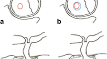
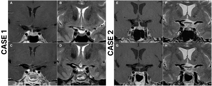
The 24 h UFC was 450 nmol/24 h (1.5× ULN of non-pregnant reference range). Late-night salivary cortisol (LNSC) was 17 nmol/L (non-pregnant reference range <8 nmol/L). Serum pathology results are shown in Table 1. MRI brain performed at 6 weeks’ gestation revealed a possible 6 by 4 mm nodule in the left lateral aspect of the sella (Fig. 1). IV contrast was not used as the patient was within the first trimester.

Table 1Laboratory investigations at initial consultation (8 + 5 weeks gestation). Bold values indicate abnormal results.

Investigation Result Reference range
Fasting glucose, mmol/L 5.2
HbA1c, % 5.4
24 h urinary cortisol, nmol/d 450 54–319
Cortisol (08:22), nmol/L 521 138–650
Midnight salivary cortisol, nmol/L 17 <8
ACTH, pmol/L 10 <12.1
IGF-1, nmol/L 31 12–42
Growth hormone, mIU/L 2.9 0–15
TSH, mIU/L 2.34 0.4–3.2
FT4, pmol/L 11.9 11–17
Figure 1
Figure 1
MRI brain without IV contrast performed in the first trimester of the patient’s first pregnancy, demonstrating a T2 hypointense lesion in the left lateral aspect of the sella, which is most likely consistent with a pituitary adenoma.

Citation: Endocrinology, Diabetes & Metabolism Case Reports 2025, 4; 10.1530/EDM-25-0092

At 14 weeks’ gestation, the repeat 24 h UFC was 542 nmol/L and LNSC was 17 nmol. There is a lack of pregnancy-specific reference ranges for 24 h UFC or LNSC measurements, making it difficult to make a definitive biochemical diagnosis. After careful discussion in a multidisciplinary team meeting, she proceeded with bilateral inferior petrosal sinus sampling (IPSS), which demonstrated a central to peripheral gradient with values presented in Table 2.

Table 2Results of bilateral inferior petrosal sinus sampling. ACTH (ng/L) at different timepoints are presented.

0 2 min 5 min 10 min 15 min
Right 258 823 1,040 864 728
Left 73 196 228 263 234
Peripheral 12 41 56 81 86
Right: peripheral 21.50 20.07 18.57 10.67 8.46
Left: peripheral 6.08 4.78 4.07 3.25 2.72

Treatment

The patient underwent transsphenoidal resection of her adenoma at 17+ weeks’ gestation. She recovered uneventfully.

Day 1 postoperative cortisol level remained elevated at 706 nmol/L, falling to 587 nmol/L by Day 3. Postoperative steroid treatment was not required.

Histopathological examination demonstrated a pituitary adenoma with mild nuclear atypia and infrequent positive ACTH staining (Fig. 2). In addition to the tumour and normal pituitary tissue, there was also abundant eosinophilic proteinaceous material present, which may have suggested contents of an associated cyst, although presence of cyst lining was not present to confirm this diagnosis. A small fragment of included bone appeared invaded by the adenoma within the resected tissue.

Figure 2
Figure 2
Positive ACTH staining in pituitary adenoma.

Citation: Endocrinology, Diabetes & Metabolism Case Reports 2025, 4; 10.1530/EDM-25-0092

Outcome and follow-up

The patient’s insulin and antihypertensive requirements plateaued postoperatively. Serial ultrasound showed that the fetal size was consistently in the 15th percentile. There were no features of preeclampsia throughout gestation.

At 35 + 5 weeks’ gestation, she had premature rupture of membranes and delivered a healthy live male infant weighing 2,250 g via normal vaginal delivery. Diabetes and hypertension resolved promptly after delivery, with cessation of insulin and antihypertensive medications.

At 5 weeks postpartum morning cortisol was within normal range at 265 nmol/L, with ACTH 6.8 pmol/L. At 10 weeks postpartum, the 24 h UFC was within normal limits at 136 nmol/day, and a 1 mg DST showed a detectable, equivocal cortisol level of 98 nmol/L. Repeat MRI pituitary was performed 2 months postpartum, which did not show any residual pituitary adenoma. No pituitary hormone replacement was required.

By 6 months postpartum, repeat 1 mg DST showed failure to suppress cortisol, with cortisol level at 154 nmol/L (RR without dexamethasone: 138–650 nmol/L), suggesting residual CD. Ambulatory blood pressure monitoring revealed essential hypertension, with average BP 141/101 mmHg across 24 h, requiring treatment with methyldopa. Despite evidence of residual CD, the patient desired a second pregnancy. Reassuringly, her cortisol burden was low, with LNSC 5 nmol/L (RR: < 8) and 24 h UFC 143 nmol/day (non-pregnant RR: 54–319), both within reference range. No definite lesion was identified on MRI brain with intravenous contrast. Extensive discussions between the endocrinologist, maternal–foetal medicine specialist, neurosurgeon, and the patient were held. The pros and cons of pursuing further treatment such as radiotherapy versus proceeding with pregnancy despite suggestion of active Cushing’s disease were explicitly discussed.

The patient confirmed her second pregnancy 11 months after the birth of her first child, and this proceeded without complications. There was no evidence of gestational diabetes on 75 g glucose tolerance tests performed at 16 and 26 weeks’ gestation. Blood pressure was well managed on methyldopa alone. She delivered a healthy male infant via normal vaginal delivery at 38 weeks’ gestation and breastfed successfully. MRI was performed at 16 weeks postpartum and did not show an appreciable sella/suprasellar mass. Repeat 24 h UFC was 275 nmol/day, consistent with ongoing CD. Clinical features of CD returned, included central adiposity, liver function test derangement, and raised HbA1c with fasting hyperinsulinaemia. Pituitary radiation therapy was discussed, including the possibility of more than one dose being required, the strong likelihood of inducing panhypopituitarism, and the unknown duration of time between radiation and remission (1). The alternative option of medical management with osilodrostat was discussed, given its recent availability and government subsidy in Australia. The patient was recently commenced on osilodrostat 1 mg twice daily after ECG attendance to exclude prolonged QTc, and patient education regarding the potential risk of hypoadrenalism and when to seek medical attention.

Discussion

Managing Cushing’s disease (CD) in pregnancy is complex and requires a multidisciplinary approach, as recurrence can occur years after initial remission. Suspected Cushing’s syndrome (CS) requires careful assessment. In cases where active disease poses significant maternal and foetal risks, transsphenoidal pituitary surgery can be safely performed in the second trimester. CD increases the risk of gestational diabetes and hypertension, requiring close monitoring to optimise outcomes. Postpartum, persistent hypercortisolism may indicate recurrence, highlighting the need for long-term endocrine follow-up. Despite mild residual disease, successful pregnancies are possible with appropriate monitoring and management, emphasising the importance of thorough family planning discussions.

UFC values are twice as high in pregnant patients compared to non-pregnant controls (2). In the first trimester of normal pregnancy, UFC values are normal, but by the third trimester, they increase three-fold up to values seen in CS (3). Therefore, CS should only be suspected when UFC values in the second and third trimesters are greater than three times the upper limit of normal (3). LNSC is a useful screening test because in CS, the usual circadian nadir of cortisol secretion is lost. At least 2–3 UFC and/or NSC screening tests are recommended (4). Lopes et al. (5) established reference values for LNSC with suggested normal ranges of 0.8–6.9 nmol/L in the first trimester, 1.1–7.2 nmol/L in the second trimester, and 1.9–9.1 nmol/L in the third trimester (5). The use of a 1 mg DST in pregnancy is not recommended because the hypothalamus–pituitary–adrenal (HPA) axis response to exogenous glucocorticoids is blunted, making it difficult to interpret test results (3).

Adrenal adenomas are responsible for 40–50% of CS cases in pregnancy, while CD causes 33% (3). In non-pregnant patients, ACTH levels are useful to classify the likely cause of CS. Undetectable ACTH levels cannot be relied upon for diagnosis in pregnancy because ACTH levels are elevated in pregnancy (3). Using high-dose dexamethasone suppression testing (HDST) as an initial test in pregnant patients has been recommended (3). A lack of suppression of ACTH with administration of high-dose dexamethasone suggests adrenal aetiology. However, HDST is not always definitive (3). Ultrasound imaging of the adrenal glands is also recommended as an initial investigation because most adrenal lesions can be visualised (35). Pregnancy complicates visualisation of a pituitary mass by MRI imaging because physiologic enlargement of the pituitary gland during pregnancy may mask small tumours (6). Non-contrast MRI has reduced sensitivity for detection of CD. However, gadolinium contrast is not recommended in pregnant women (7).

Inferior petrosal sinus sampling (IPSS) is the gold standard for diagnosing CD in the non-pregnant population (4). The most recent guidelines for diagnosis of CS suggest that IPSS is not necessary for diagnosis if MRI clearly shows a tumour >10 mm in the context of dynamic test results and clinical features that also strongly suggest CD (4). Lindsay and colleagues (3) caution the use of IPSS unless prior non-invasive testing remains equivocal due to risks of thromboembolism and exposure to radiation posed by IPSS (3). However, these risks can be mitigated with extra precautions during pregnancy, including use of lead barrier protection, a direct jugular approach, and with the procedure occurring at a specialised centre (3).

Treatment of CS in pregnancy should be individualised depending on the patient presentation and gestational age (4). Active treatment of CS, by either medical or surgical intervention, reduces maternal morbidity and rates of foetal loss (4). Surgery is usually preferred because there are fewer complications at delivery and it has high rates of remission (8). Surgery reduces rates of perinatal and maternal morbidity but does not reduce rates of preterm birth and intrauterine growth restriction (IUGR) (9). Pituitary or adrenal surgery should ideally be done in the second trimester, before week 24 of pregnancy, in a high-volume centre with multidisciplinary team input (8). There is a risk of spontaneous abortion with anaesthesia given in the first trimester and an increased risk of premature labour with anaesthesia given in the third trimester (7).

Unfortunately, CD can recur, and 50% of recurrence occurs within 50 months of pituitary surgery (14). Recurrence is defined as ongoing clinical and biochemical evidence of hypercortisolism after an initial period of remission. Factors that increase the likelihood of postoperative remission included the identification of a tumour on MRI pre-surgery, no invasion of the sinus cavernous by the adenoma, older age (greater than 35 years), low postoperative cortisol and ACTH levels, and long-term hypocortisolism (greater than 1 year) (1). A second pituitary surgery is often the first-line treatment option in recurrence, which has overall lower rates of remission compared to first surgery and increased risk of hypopituitarism due to scar tissue in the pituitary and often the need for more aggressive surgical technique (1). Both fractionated radiotherapy and stereotactic radiosurgery are therapeutic options and achieve high rates of remission (1).

There are no medications that are approved for treatment of CD in pregnancy, although the latest guidelines suggest consideration of metyrapone, ketoconazole, or cabergoline (46). The newer agent, osilodrostat inhibits the enzymes 11-beta-hydroxylase and 18-hydroxylase, reducing production of cortisol and aldosterone respectively, thereby normalising UFC values, reducing systolic and diastolic blood pressure, fasting blood glucose levels, and improving body weight in clinical trials (10). There is no information on osilodrostat use and safety in pregnancy, but it is an effective agent in patients who are unsuitable for surgery and patients with recurrent disease after surgery (10). It is associated with risk of hypoadrenalism, prolongation of the QTc interval, and increased serum testosterone levels, particularly at higher doses (10). Each medication poses its own risk of side effects and therefore treatment must be individualised. Overall, medical treatment should only be used in pregnancy when surgical treatment is contraindicated (6).

Our case demonstrates a rare case of CD in pregnancy with no significant adverse perinatal outcomes for mother or baby, albeit late preterm delivery in the first pregnancy. Ongoing endocrinology surveillance is essential to monitor for recurrent CD.

Declaration of interest

The authors declare that there is no conflict of interest that could be perceived as prejudicing the impartiality of the research reported.

Funding

This research did not receive any specific grant from any funding agency in the public, commercial, or not-for-profit sector.

Patient consent

Written informed consent for publication of their clinical details was obtained from the patient.

Author contribution statement

Several case details and timeline of events were gathered by EW. This is a patient of SG.

References

Global Longitudinal Strain Reduction With Apical Sparing in Cushing Syndrome-Related Heart Failure With Preserved Ejection Fraction (HFpEF): A Case Report

Abstract

We describe a case of a 56-year-old woman with a history of recurrent pituitary adenoma, not well followed, and known comorbidities of coronary artery disease, hypertension, and type 2 diabetes mellitus. She arrived with severely high blood pressure and signs pointing to hypercortisolism. Further evaluation revealed left ventricular hypertrophy, reduced global longitudinal strain, and preserved left ventricular ejection fraction, consistent with heart failure with preserved ejection fraction (HFpEF). Workup for amyloidosis was negative. This case highlights that chronic hypercortisolism may cause pathophysiological changes in the heart, leading to HFpEF, and may induce myocardial fibrosis and impaired myocardial mechanics, producing an echocardiographic pattern that can mimic infiltrative cardiomyopathy. Recognition of this overlap is crucial to avoid misdiagnosis and to ensure timely endocrine and cardiovascular management.

Introduction

Hypercortisolism is defined as a clinical condition resulting from excessive tissue exposure to cortisol or other glucocorticoids. Sustained exposure ultimately leads to Cushing syndrome (CS), a well-established constellation of clinical manifestations arising from chronic endogenous or exogenous cortisol excess [1]. CS is associated with profound metabolic derangements that significantly increase cardiovascular risk, not only during the active phase of the disease but also persisting long after biochemical remission [2,3]. Cardiovascular complications, including premature atherosclerosis, coronary artery disease (CAD), heart failure, and cerebrovascular events, are major contributors to the excess mortality observed in CS compared with the general population [1,3]. Among these complications, arterial hypertension remains the most frequent cardiovascular disorder in patients with Cushing disease (CD) [4].

Although left ventricular (LV) systolic function is generally preserved in patients with CS, several studies have demonstrated that chronic cortisol excess induces structural and functional cardiac alterations, predisposing to major adverse cardiac events and the development of heart failure [5] In the broader context of chronic congestive heart failure, disease progression is tightly coupled with activation of neuroendocrine stress pathways, most notably the hypothalamic-pituitary-adrenal axis, which governs cortisol secretion [6]. Cortisol, a pivotal stress hormone, increases in response to physiological strain, and its sustained elevation contributes to adverse myocardial remodeling.

Heart failure with preserved ejection fraction (HFpEF), a chronic and progressive syndrome, exemplifies the deleterious effects of persistent myocardial stress. While overt heart failure is an uncommon complication of CS, when it does occur, it most often presents with preserved LV ejection fraction (LVEF) or with subclinical LV dysfunction [7]. Prior evidence has also linked CS to LV hypertrophy, diastolic dysfunction, and subtle systolic impairment, with many of these changes demonstrating reversibility upon normalization of cortisol levels [8].

This case is unique as it highlights the interplay between CS and cardiac amyloidosis, emphasizing their overlapping yet distinct echocardiographic features. Global longitudinal strain (GLS), a measure of myocardial deformation, is particularly useful for differentiating these conditions and reveals subtle differences in strain patterns between the two.

Case Presentation

A 56-year-old woman with a significant past medical history of recurrent pituitary macroadenoma, treated with two prior surgical resections, the most recent five years earlier without subsequent follow-up, CAD, long-standing hypertension, and type 2 diabetes mellitus, presented to the emergency department with hypertensive urgency.

On arrival, she presented with a hypertensive crisis, with blood pressure measured at 200/110 mmHg, associated with severe cephalalgia, without syncope, visual changes, or focal neurological deficits. An MRI Brain demonstrated no evidence of acute intracranial hemorrhage or mass effect (Video 1). Initial laboratory testing showed normal complete blood count, renal function, and serum electrolytes. On physical examination, she exhibited characteristic Cushingoid stigmata, including rounded moon facies, central adiposity, and bilateral lower-extremity pitting edema.

She was commenced on intensive antihypertensive therapy, including spironolactone, clonidine, telmisartan, carvedilol, amlodipine, and intravenous furosemide (20 mg, subsequently escalated to 40 mg). Given her clinical appearance and history of pituitary disease, an endocrine evaluation was undertaken. An overnight dexamethasone suppression test revealed an unsuppressed morning cortisol of 360 nmol/L, consistent with hypercortisolism.

Cardiac assessment supported a diagnosis of HFpEF. Transthoracic echocardiography demonstrated preserved left ventricular ejection fraction (60%), impaired GLS (-10%), and mild concentric left ventricular hypertrophy (Figure 1; Video 2).

Transthoracic-echocardiography-demonstrating-reduced-global-longitudinal-strain-(-10%)-consistent-with-preserved-EF-(60%)
Figure 1: Transthoracic echocardiography demonstrating reduced global longitudinal strain (-10%) consistent with preserved EF (60%)

EF: Ejection Fraction

Workup for alternative causes of HFpEF, including renal impairment and infiltrative cardiomyopathy, was unremarkable; both serum and urine protein electrophoresis with immunofixation excluded amyloidosis.

Magnetic resonance imaging of the pituitary revealed recurrence of the macroadenoma. The patient was referred to neurosurgery for consideration of repeat resection, and glucocorticoid-sparing medical therapy was initiated. During hospitalization, her blood pressure was gradually stabilized, diuretic therapy improved signs of congestion, and her functional status returned to near baseline with restored mobility (Video 3).

Discussion

Epidemiology and clinical significance

CD is a severe endocrine disorder characterized by chronic exposure to excess glucocorticoids. Patients with CD have a two- to fivefold higher mortality compared with the general population, predominantly due to cardiovascular complications [4]. Chronic hypercortisolism is associated with systemic hypertension, left ventricular hypertrophy (LVH), diastolic dysfunction, and accelerated atherosclerosis, increasing the risk of myocardial ischemia and heart failure. While these cardiovascular manifestations are common, the development of isolated dilated cardiomyopathy (DCM) in the absence of other major comorbidities is rare but clinically noteworthy [9].

Pathophysiology of cardiac involvement

Chronic glucocorticoid excess contributes to cardiovascular remodeling via multiple mechanisms. Persistent hypertension and metabolic disturbances promote LVH and diastolic dysfunction. Additionally, glucocorticoid excess induces endothelial dysfunction, insulin resistance, and myocardial fibrosis, impairing ventricular compliance and predisposing to HFpEF [1,6]. Advanced echocardiographic techniques, such as GLS, can detect subclinical systolic dysfunction before overt reductions in LVEF [6]. In our patient, preserved LVEF (60%) coupled with markedly reduced GLS (-10%) and concentric LVH was consistent with HFpEF secondary to chronic cortisol excess, further supported by clinical signs of volume overload such as edema and severe hypertension [7].

Apical sparing and mimicking amyloidosis

An important observation in this case was relative apical sparing despite markedly reduced GLS, a strain pattern classically associated with cardiac amyloidosis [10]. Although infiltrative disease was excluded (negative serum and urine protein electrophoresis with immunofixation), this overlap illustrates how hypercortisolism-induced remodeling can phenocopy amyloidosis on imaging. Recent work has shown that hypercortisolism, beyond metabolic derangements, impairs myocardial mechanics and contractile efficiency [11]. Thus, patients with atypical strain findings should undergo careful endocrine evaluation to avoid misdiagnosis. Ultimately, the recognition that hypercortisolism may produce amyloid-like echocardiographic signatures has both diagnostic and management implications. It broadens the differential diagnosis of HFpEF and stresses the need for a multidisciplinary approach involving endocrinology and cardiology to prevent misdiagnosis and ensure tailored therapy.

Dilated cardiomyopathy in CS

Although uncommon, DCM with severe LV systolic dysfunction has been described in CS. Frustaci et al. reported eight cases of hypercortisolism due to adrenal adenoma among 473 patients with DCM (1.7%), all presenting with LVEF <30% and symptomatic heart failure. Endomyocardial biopsy revealed cardiomyocyte hypertrophy, interstitial fibrosis, and myofibrillolysis, distinct from idiopathic DCM and valvular disease controls. Follow-up biopsies in three patients one year post-adrenalectomy demonstrated substantial regression of these changes, highlighting the reversibility of glucocorticoid-induced myocardial injury [12].

Although not assessed in our patient, prior studies have implicated atrogin-1 in CS-related myocardial remodeling. At the molecular level, upregulation of atrogin-1, an E3 ubiquitin ligase expressed in skeletal, smooth, and cardiac muscle, was observed in CS-associated DCM compared with idiopathic DCM and controls [13]. Atrogin-1, implicated in skeletal muscle atrophy and sarcopenia, facilitates proteasomal degradation of intracellular proteins. Its overexpression in cardiomyocytes contributes to glucocorticoid-mediated myocardial remodeling. Importantly, atrogin-1 expression declined significantly following surgical correction of cortisol excess, paralleling improvements in cardiac structure and function. This reversibility mirrors recovery seen in glucocorticoid-induced skeletal myopathy and underscores the unique potential for cardiac recovery in CS-related DCM [9].

Clinical implications and differential diagnosis

This case underscores the multisystem burden of endogenous hypercortisolism, with particular cardiovascular susceptibility [1,6]. Chronic cortisol excess should be considered in the differential diagnosis of HFpEF, particularly when conventional risk factors coexist with systemic features such as central obesity, moon facies, and proximal myopathy [8]. Secondary causes of HFpEF, including cardiac amyloidosis, were excluded, supporting hypercortisolism as the primary etiology. Recognizing CS as a reversible contributor to myocardial dysfunction has important clinical implications, as timely endocrine intervention can improve cardiac function, lower blood pressure, and potentially prevent progression to irreversible myocardial remodeling.

Left ventricular hypertrophy and structural remodeling

Electrocardiographic and echocardiographic studies have characterized the cardiac phenotype in patients with CS. In a cohort of 12 consecutive patients, most had concomitant hypertension (11/12) and diabetes mellitus (7/12). Preoperative ECGs commonly demonstrated high-voltage QRS complexes (10 patients) and T-wave inversions (7 patients), indicative of LV strain. Echocardiography revealed LVH in nine patients, all exhibiting asymmetric septal hypertrophy. Interventricular septal thickness ranged from 16 to 32 mm, with septal-to-posterior wall ratios from 1.33 to 2.67. Compared with essential hypertension or primary aldosteronism, CS patients exhibited more pronounced LVH and a higher prevalence of asymmetric septal hypertrophy, suggesting a unique glucocorticoid-mediated remodeling pattern [13].

Postoperative follow-up in nine patients demonstrated normalization of ECG abnormalities, decreased septal thickness, and resolution of asymmetric septal hypertrophy in all but one patient, highlighting the partial reversibility of LVH following correction of hypercortisolism. The pronounced septal thickening relative to the posterior wall implies that excessive cortisol exposure, beyond hemodynamic effects of hypertension, contributes significantly to myocardial remodeling [13].

Impact of disease duration on concentric remodeling

Fallo et al. evaluated 18 patients with CS compared with 18 matched controls, adjusting for sex, age, body size, blood pressure, and duration of hypertension. Eleven participants in each group were hypertensive. Echocardiography revealed elevated relative wall thickness (RWT >0.45) in 11 patients with CS (five normotensive, six hypertensive) versus two hypertensive controls. Left ventricular mass index was abnormal in three CS patients and in four hypertensive controls, while systolic function was preserved in all participants [14].

No correlation was observed between RWT and either blood pressure or urinary cortisol levels in patients with CS. Instead, RWT correlated significantly with disease duration, indicating that prolonged exposure to glucocorticoid excess, rather than hormone levels or hemodynamic load, is the primary determinant of concentric LV remodeling. Postoperative echocardiography showed normalization of RWT in five of six patients previously affected, reinforcing the concept of reversible myocardial structural changes following correction of hypercortisolism [14].

Conclusions

CS represents a rare but clinically important etiology of heart failure with preserved ejection fraction and, less commonly, dilated cardiomyopathy. Chronic hypercortisolism promotes systemic hypertension, LVH, diastolic dysfunction, myocardial fibrosis, and remodeling that may mimic infiltrative cardiomyopathies such as amyloidosis on echocardiography. GLS with apical sparing, although typically associated with amyloidosis, may also occur in cortisol-induced cardiomyopathy. Advanced imaging, including GLS, can detect subclinical myocardial impairment before overt systolic dysfunction develops. Notably, cardiac structural and functional abnormalities may partially or completely reverse following normalization of cortisol levels, highlighting the importance of early recognition and timely endocrine intervention. Clinicians should maintain a high index of suspicion for hypercortisolism in patients presenting with unexplained LVH, HFpEF, or atypical DCM, particularly when systemic features of CS are present. Future studies are needed to better characterize strain patterns in endocrine cardiomyopathies and to refine imaging-based algorithms for early detection.

References

  1. Uwaifo GI, Hura DE: Hypercortisolism. StatPearls [Internet]. StatPearls Publishing, Treasure Island (FL); 2024.
  2. De Leo M, Pivonello R, Auriemma RS, et al.: Cardiovascular disease in Cushing’s syndrome: heart versus vasculature. Neuroendocrinology. 2010, 92 Suppl 1:50-4. 10.1159/000318566
  3. Graversen D, Vestergaard P, Stochholm K, Gravholt CH, Jørgensen JO: Mortality in Cushing’s syndrome: a systematic review and meta-analysis. Eur J Intern Med. 2012, 23:278-82. 10.1016/j.ejim.2011.10.013
  4. Uzie Bło-Życzkowska B, Krzesinński P, Witek P, Zielinński G, Jurek A, Gielerak G, Skrobowski A: Cushing’s disease: subclinical left ventricular systolic and diastolic dysfunction revealed by speckle tracking echocardiography and tissue Doppler imaging. Front Endocrinol (Lausanne). 2017, 8:222. 10.3389/fendo.2017.00222
  5. Brosolo G, Catena C, Da Porto A, Bulfone L, Vacca A, Verheyen ND, Sechi LA: Differences in regulation of cortisol secretion contribute to left ventricular abnormalities in patients with essential hypertension. Hypertension. 2022, 79:1435-44. 10.1161/HYPERTENSIONAHA.122.19472
  6. Gladden JD, Linke WA, Redfield MM: Heart failure with preserved ejection fraction. Pflugers Arch. 2014, 466:1037-53. 10.1007/s00424-014-1480-8
  7. Owan TE, Redfield MM: Epidemiology of diastolic heart failure. Prog Cardiovasc Dis. 2005, 47:320-32. 10.1016/j.pcad.2005.02.010
  8. Pereira AM, Delgado V, Romijn JA, Smit JW, Bax JJ, Feelders RA: Cardiac dysfunction is reversed upon successful treatment of Cushing’s syndrome. Eur J Endocrinol. 2010, 162:331-40. 10.1530/EJE-09-0621
  9. Gill A, Dean N, Al-Agha R: Cushing’s, dilated cardiomyopathy and stroke: case report and literature review. Can J Gen Intern Med. 2016, 11:46-9.
  10. Klein AL, Oh J, Miller FA, Seward JB, Tajik AJ: Two-dimensional and Doppler echocardiographic assessment of infiltrative cardiomyopathy. J Am Soc Echocardiogr. 1988, 1:48-59. 10.1016/s0894-7317(88)80063-4
  11. Sahiti F, Detomas M, Cejka V, et al.: The impact of hypercortisolism beyond metabolic syndrome on left ventricular performance: a myocardial work analysis. Cardiovasc Diabetol. 2025, 24:132. 10.1186/s12933-025-02680-1
  12. Frustaci A, Letizia C, Verardo R, Grande C, Calvieri C, Russo MA, Chimenti C: Atrogin-1 pathway activation in Cushing syndrome cardiomyopathy. J Am Coll Cardiol. 2016, 67:116-7. 10.1016/j.jacc.2015.10.040
  13. Sugihara N, Shimizu M, Kita Y, et al.: Cardiac characteristics and postoperative courses in Cushing’s syndrome. Am J Cardiol. 1992, 1:1475-80.
  14. Fallo F, Budano S, Sonino N, Muiesan ML, Agabiti-Rosei E, Boscaro M: Left ventricular structural characteristics in Cushing’s syndrome. J Hum Hypertens. 1994, 8:509-13.

From https://www.cureus.com/articles/413845-global-longitudinal-strain-reduction-with-apical-sparing-in-cushing-syndrome-related-heart-failure-with-preserved-ejection-fraction-hfpef-a-case-report?score_article=true#!/

Reconstructive Liposuction for Residual Lipodystrophy After Remission of Cushing’s Disease

Abstract

Cushing’s syndrome (CS) is often presented due to an adrenocorticotropic hormone (ACTH)-secreting pituitary adenoma, characterized by high chronic cortisol levels. Surgical resection of the pituitary adenoma is the primary treatment, but long-term metabolic and physical sequelae can persist, affecting psychological well-being and social functioning. Glucocorticoids are directly involved in alterations of fat metabolism, favoring centripetal adiposity. Even after hormonal normalization, patients may experience residual lipodystrophy. Impairment of body image may cause psychological distress and social isolation. The objective is to illustrate the potential therapeutic value of reconstructive liposuction in restoring body image and psychological well-being in a patient with persistent lipodystrophy after Cushing’s disease remission.

We report a case of a 16-year-old female with recurrent Cushing’s disease secondary to a pituitary microadenoma, confirmed by elevated urinary free cortisol and magnetic resonance imaging (MRI). It was initially treated with transsphenoidal resection in 2019; disease recurrence was confirmed and again treated in 2024. Despite intervention, the prolonged hypercortisolism developed into secondary lipodystrophy, leading to severe body image dissatisfaction and social withdrawal. Thyroid function remained euthyroid, ruling out metabolic contributors. Because of the psychological distress caused by persistent fat redistribution, the patient underwent elective liposuction in 2025. Postoperative follow-up revealed reduced psychological distress and improved well-being and self-esteem. Reconstructive liposuction can play a key role in the treatment and management of persistent post-CS lipodystrophy, contributing significantly to psychological recovery. Prospective studies evaluating surgical criteria and long-term psychosocial outcomes are needed to define eligibility criteria and assess outcomes, leading to the development of clinical guidelines for aesthetic interventions in post-CS recovery.

Introduction

Corticotroph pituitary adenomas (corticotropinomas) are pituitary tumors that secrete excess adrenocorticotropic hormone (ACTH), causing endogenous Cushing’s syndrome (CS). Most of these adenomas are sporadic and monoclonal, although in some rare cases, they are associated with germline mutations (e.g., in USP8) or genetic syndromes [1,2]. Clinically, excess ACTH causes a classic presentation with centripetal obesity, purple striae, muscle asthenia, hypertension, and emotional disturbances such as depression or anxiety [3-5]. Chronically elevated cortisol levels promote fat deposition in central body regions – face, neck, torso, and abdomen – at the expense of relative thinning of the limbs [3], leading to lipodystrophy that can seriously affect the patient’s quality of life.

At the molecular level, glucocorticoids stimulate the differentiation of preadipocytes into mature adipocytes and enhance lipoprotein lipase activity in peripheral fat tissues [6], thereby increasing the uptake of circulating fatty acids and the storage of triglycerides. At the same time, they increase hepatic lipogenesis and modulate cortisol receptor homeostasis (e.g., 11β-HSD1 in adipose tissue), favoring visceral fat distribution [6]. Although glucocorticoids can induce acute lipolysis, they exert chronic lipogenic effects – especially in subcutaneous adipose tissue – which promotes fat accumulation in the face, neck, and trunk [6]. This central adiposity, characteristic of CS, is further enhanced by increased hepatic lipogenesis and the overexpression of 11β-HSD1 in adipose tissue, which amplifies the local action of cortisol [6].

Case Presentation

In 2019, a 16-year-old female patient was initially diagnosed with a 4 × 3 mm pituitary microadenoma (Figure 1), following clinical suspicion of Cushing’s disease. The diagnosis was confirmed through imaging studies and endocrinological testing, which revealed consistently elevated urinary free cortisol levels ranging from 459 to 740.07 µg/24 hours (normal range: <50 µg/24 hours), indicative of endogenous hypercortisolism. No dynamic load tests (such as dexamethasone suppression or ACTH stimulation) were performed, as the diagnosis was supported by the clinical context and laboratory findings. Moreover, no clinical or biochemical evidence of adrenal insufficiency was observed during follow-up.

T1-weighted-sagittal-MRI-scan-showing-a-corticotroph-pituitary-microadenoma-(4-×-3-mm)-circled-in-red
Figure 1: T1-weighted sagittal MRI scan showing a corticotroph pituitary microadenoma (4 × 3 mm) circled in red

The lesion is localized within the anterior pituitary gland, consistent with an ACTH-secreting adenoma causing Cushing’s disease in the patient.

MRI, magnetic resonance imaging; ACTH, adrenocorticotropic hormone

The patient underwent transsphenoidal endonasal resection of the pituitary tumor in 2019. Although initially successful, disease recurrence was confirmed, and a second endonasal transsphenoidal surgery was performed in 2024. Despite these interventions, the prolonged hypercortisolism led to the development of secondary lipodystrophy, manifesting as centripetal fat accumulation, a dorsal fat pad, and disproportionate truncal adiposity (Figure 2). These physical alterations had a significant psychosocial impact, as reported by the patient during follow-up visits, resulting in body image dissatisfaction, low self-esteem, and social withdrawal. No formal psychometric scales were administered.

Preoperative-and-intraoperative-images-of-the-patient
Figure 2: Preoperative and intraoperative images of the patient

A and B panels show the anterior and posterior views prior to liposuction, demonstrating centripetal adipose accumulation characteristic of Cushing’s syndrome. The C panel shows the intraoperative stage following abdominal and flank liposuction, with placement of drainage tubes, and visible reduction in subcutaneous fat volume.

A thyroid function panel revealed a slightly elevated thyroid-stimulating hormone (TSH) level (4.280 μUI/mL; reference range: 0.270-4.200), with total and free T3 and T4 values within normal limits, ruling out clinically significant hypothyroidism as a confounding factor for her phenotype. The biochemical profile suggested a euthyroid state, despite borderline TSH elevation, which was interpreted as a subclinical or adaptive response to chronic cortisol excess (Table 1).

Parameter Normal Range Patient’s Value
Cortisol (µg/24 hour) 58.0 – 403.0 459.5 – 740.07
TSH (µUI/mL) 0.270 – 4.200 4.280
Total T3 (ng/mL) 0.80 – 2.00 1.02
Free T3 (pg/mL) 2.00 – 4.40 3.33
Total T4 (µg/dL) 4.50 – 12.00 8.63
Free T4 (ng/dL) 0.92 – 1.68 1.36
Table 1: Comparison between the patient’s hormone levels and standard reference ranges

A persistently elevated 24-hour urinary cortisol range is observed, consistent with endogenous hypercortisolism. The thyroid profile remains within normal limits, with a mildly elevated TSH in the absence of overt thyroid dysfunction. These findings support the functional and metabolic profile characteristic of Cushing’s syndrome.

TSH, thyroid-stimulating hormone

The procedure targeted lipodystrophic regions identified through clinical examination and patient concerns, rather than formal imaging or anthropometric measurements. It aimed to restore body contour, alleviate somatic distress, and improve her overall self-perception and quality of life. Postoperative follow-up revealed patient-reported improvements in body image and psychological well-being. While these outcomes were not evaluated with formal instruments, the clinical improvement was evident and significant from the patient’s perspective, highlighting the role of plastic surgery not only as a reconstructive tool, but also as a therapeutic strategy for restoring dignity and social functioning in patients recovering from CS.

Discussion

After successful treatment of the pituitary adenoma, many metabolic parameters improve; however, fat distribution usually only partially reverses. Longitudinal studies show that, in the medium term, weight and abdominal circumference decrease, and there is some redistribution of fat toward the limbs following cortisol remission [3].

For example, Bavaresco et al. (2024) observed that, after hormone levels normalized, total fat was reduced and part of it shifted from the visceral area to the legs [3]. Nevertheless, their review highlights that a significant proportion of patients continue to present with residual visceral adiposity and moderate obesity (body mass index, or BMI >25), despite hormonal control [7]. In our case, truncal adiposity persisted based on clinical assessment, though no formal anthropometric measurements were performed.

Although liposuction is not traditionally considered first-line therapy for cortisol-induced lipodystrophy secondary to Cushing’s disease, increasing evidence from related lipodystrophic syndromes supports its clinical utility. For instance, in human immunodeficiency virus (HIV)-associated cervicodorsal lipodystrophy, Barton et al. (2021) conducted a 15-year retrospective analysis comparing liposuction and excisional lipectomy, finding that 80% of patients undergoing liposuction alone experienced recurrence, while none of the patients treated with excisional lipectomy showed recurrence – albeit with a higher risk of postoperative seroma formation [7]. These findings underscore that, while liposuction may be less durable than excision, it remains a viable option for selected cases, especially when used for contouring or as an adjunct [7]. Similarly, the Endocrine Society guidelines on lipodystrophy management emphasize the importance of personalized approaches, particularly when localized adipose accumulation contributes to persistent metabolic dysfunction or psychological distress [8]. Akinci et al. (2024) also highlight that, even in partial or atypical lipodystrophy syndromes, patients often report substantial impairment in quality of life due to disfiguring fat redistribution [9]. In this context, liposuction should not be dismissed as merely cosmetic but considered part of a functional and psychosocial rehabilitation strategy. The present case exemplifies this rationale, as the patient – despite biochemical remission of Cushing’s disease – continued to experience debilitating body image disturbances and emotional distress, which were ameliorated following targeted liposuction. This supports the integration of body-contouring procedures into multidisciplinary care protocols for endocrine-related lipodystrophies, especially when residual physical stigma persists after hormonal normalization [7-9].

Body image disorders, such as those secondary to CS or lipodystrophy, significantly impact self-perception, self-esteem, and social functioning. For example, a study by Alcalar et al. (2013) reported that patients with active Cushing’s disease had significantly lower SF-36 scores – particularly in emotional role functioning and mental health domains – compared to controls [10]. Similarly, Akinci et al. (2024) described that patients with partial lipodystrophy demonstrated marked reductions in EQ-5D index values and visual analog scale (VAS) scores, indicating impaired health-related quality of life [9]. These findings underscore that fat redistribution disorders can substantially compromise psychosocial well-being, even after endocrine remission.

This is especially relevant in women, where sociocultural stereotypes surrounding female physical appearance reinforce thinness, symmetry, and youthfulness as standards of personal value and social acceptance [1]. This societal context amplifies body dissatisfaction when visible physical changes occur, even after the clinical remission of endocrine diseases, often leading to social withdrawal, anxiety, or depression [3,10]. Within this framework, plastic surgery – such as reconstructive liposuction – has proven to be a valuable therapeutic tool, offering physical restoration that can enhance self-confidence and promote social reintegration [4]. Postoperative follow-up in our case revealed patient-reported improvements in body image and psychological well-being. While these outcomes were not assessed using formal psychometric tools, the clinical benefit was evident from the patient’s perspective. This aligns with prior findings demonstrating the psychosocial value of reconstructive surgery, which can enhance self-esteem and social reintegration after physical disfigurement [11,12]. These observations underscore the role of plastic surgery not only as a reconstructive intervention, but also as a therapeutic strategy for restoring dignity and quality of life in patients recovering from CS.

Although validated psychometric instruments such as the Body Image Quality of Life Inventory (BIQLI) and the Dysmorphic Concern Questionnaire (DCQ) are available to assess body image disturbances, these were not applied in our case. Nonetheless, they represent useful tools for evaluating subjective impact in both clinical practice and research settings. The BIQLI evaluates the effect of body image on various aspects of life – social interactions, self-worth, sexuality, and emotional well-being – using a Likert scale ranging from -3 (very negative impact) to +3 (very positive impact), providing a quantifiable assessment of its influence on quality of life [5]. The DCQ, on the other hand, identifies dysfunctional concerns about perceived physical flaws by assessing behaviors such as avoidance, mirror checking, and concealment; higher scores are associated with suspected body dysmorphic disorder (BDD) [6]. These tools are useful for initial diagnosis, surgical candidate selection, and postoperative follow-up, as they objectively measure subjective changes related to body image. Their advantages include ease of use, clinical validity, and applicability in research settings. However, they also have limitations: they do not replace comprehensive psychological evaluation, may be influenced by cultural context, and do not detect deeper psychiatric comorbidities. Therefore, a multidisciplinary and ethically grounded approach – integrating plastic surgery, endocrinology, and psychology – is essential to ensure safe and patient-centered treatment planning.

Aesthetic liposuction is associated with significant improvements in perceived body image and patient quality of life [11]. For example, Papadopulos et al. (2019) observed statistically significant increases in perception of one’s own body appearance and high satisfaction with postoperative results [12]. These aesthetic gains were accompanied by psychological improvements: the same study documented an increase in emotional stability and a reduction in postoperative anxiety [12]. Similarly, Kamundi (2023) found that nearly all assessed dimensions of quality of life improved after liposuction (p < 0.05 in most of them). Altogether, these findings suggest that liposuction not only corrects physical alterations typical of CS, but also strengthens self-esteem and psychological well-being by substantially improving satisfaction with one’s body image [11].

Moreover, self-esteem influences adherence to medical treatments and lifestyle changes. By improving self-image through reconstructive surgery, it is plausible that the patient feels more motivated to maintain healthy habits, such as diet and regular exercise, that prevent metabolic relapse [12,13].

Nonetheless, it is important to emphasize that liposuction, in this context, should be viewed as a reconstructive complement, not a primary treatment. There are no established protocols or formal guidelines that explicitly include plastic surgery in the care of cured CS; the decision is personalized, based on the residual functional and psychological impact.

Conclusions

Reconstructive plastic surgery, though not a primary therapeutic approach for CS, plays a key role in enhancing patients’ quality of life following remission. Liposuction, in particular, offers a safe and effective solution for persistent lipodystrophy, providing aesthetic benefits with minimal scarring, rapid recovery, and low complication rates in properly selected patients.

This case underscores the importance of addressing both physical and psychosocial sequelae after endocrine stabilization. A multidisciplinary approach – encompassing endocrinology, neurosurgery, and plastic surgery – not only restores physical appearance but also contributes to emotional recovery, self-esteem, and overall patient satisfaction.

References

  1. Tatsi 😄 Cushing syndrome/disease in children and adolescents. Endotext [Internet]. Feingold KR, Ahmed SF, Anawalt B, et al. (ed): MDText.com, Inc., South Dartmouth (MA); 2000.
  2. Mir N, Chin SA, Riddell MC, Beaudry JL: Genomic and non-genomic actions of glucocorticoids on adipose tissue lipid metabolism. Int J Mol Sci. 2021, 22:8503. 10.3390/ijms22168503
  3. Bavaresco A, Mazzeo P, Lazzara M, Barbot M: Adipose tissue in cortisol excess: what Cushing’s syndrome can teach us?. Biochem Pharmacol. 2024, 223:116137. 10.1016/j.bcp.2024.116137
  4. Nieman LK: Molecular derangements and the diagnosis of ACTH-dependent Cushing’s syndrome. Endocr Rev. 2022, 43:852-77. 10.1210/endrev/bnab046
  5. Patni N, Chard C, Araujo-Vilar D, Phillips H, Magee DA, Akinci B: Diagnosis, treatment and management of lipodystrophy: the physician perspective on the patient journey. Orphanet J Rare Dis. 2024, 19:263. 10.1186/s13023-024-03245-3
  6. Peckett AJ, Wright DC, Riddell MC: The effects of glucocorticoids on adipose tissue lipid metabolism. Metabolism. 2011, 60:1500-10. 10.1016/j.metabol.2011.06.012
  7. Barton N, Moore R, Prasad K, Evans G: Excisional lipectomy versus liposuction in HIV-associated lipodystrophy. Arch Plast Surg. 2021, 48:685-90. 10.5999/aps.2020.02285
  8. Brown RJ, Araujo-Vilar D, Cheung PT, et al.: The diagnosis and management of lipodystrophy syndromes: a multi-society practice guideline. J Clin Endocrinol Metab. 2016, 101:4500-11. 10.1210/jc.2016-2466
  9. Akinci B, Celik Gular M, Oral EA: Lipodystrophy syndromes: presentation and treatment. Endotext [Internet]. Feingold KR, Anawalt B, Boyce A, et al. (ed): MDText.com, Inc., South Dartmouth (MA); 2024.
  10. Alcalar N, Ozkan S, Kadioglu P, Celik O, Cagatay P, Kucukyuruk B, Gazioglu N: Evaluation of depression, quality of life and body image in patients with Cushing’s disease. Pituitary. 2013, 16:333-40. 10.1007/s11102-012-0425-5
  11. Kamundi RK: Determining the Impact of Liposuction on Patient Satisfaction of Quality of Life and Body Image: A Prospective Study in Nairobi, Kenya. University of Nairobi, Nairobi; 2023.
  12. Papadopulos NA, Kolassa MJ, Henrich G, Herschbach P, Kovacs L, Machens HG, Klöppel M: Quality of life following aesthetic liposuction: a prospective outcome study. J Plast Reconstr Aesthet Surg. 2019, 72:1363-72. 10.1016/j.bjps.2019.04.008
  13. Saariniemi KM, Salmi AM, Peltoniemi HH, Charpentier P, Kuokkanen HOM: Does liposuction improve body image and symptoms of eating disorders?. Plast Reconstr Surg Glob Open. 2015, 3:461. 10.1097/GOX.0000000000000440

From https://www.cureus.com/articles/376886-reconstructive-liposuction-for-residual-lipodystrophy-after-remission-of-cushings-disease-a-case-report#!/

The Role of Late-Night Salivary Cortisol in the Assessment of Endocrine Remission in Patients With Cushing’s Disease After Pituitary Radiotherapy

ABSTRACT

Context

Pituitary radiotherapy (RT) is used for persistent/recurrent Cushing’s disease (CD) after pituitary surgery. The utility of late-night salivary cortisol (LNSC) in evaluating endocrine remission after RT is unclear.

Objective

To identify the clinical characteristics and outcomes of patients with CD after RT, in endocrine remission based on normal LNSC (group 1) or 24 h urinary free cortisol (UFC; group 2).

Design and Setting

Retrospective cohort, observational study in academic medical centre. Patients (16−86 years old, n = 75) with CD who underwent RT were studied; group 1 (n = 16), group 2 (n = 18), and 41 patients not in remission. Outcome measures included within-group and between-group changes (group 1 vs. group 2) in clinical characteristics, endocrine data and time to remission.

Results

Seventy-five patients with CD, aged (median [range]: 50 years [16, 86], 71% female, BMI: 34.7 kg/m2 [19.1, 62.5], 63% with macroadenomas) underwent RT and 34 (45.3%) entered endocrine remission, including 16 (21.3%) in group 1 and 18 (24%) in group 2 (median: 56 months). From RT to remission, there were decreases in BMI (group 1: −3.9 ± 0.7 kg/m2p = 0.0001; group 2: −5.2 ± 1.9 kg/m2p = 0.0123) and systolic blood pressure (group 1: −7.9 ± 3.9 mmHg, p = 0.03; group 2: −10.1 ± 4.5 mmHg (p = 0.008). There were no between-group differences in BMI, blood pressure, HbA1c, number of antihypertensive or antihyperglycemic agents, UFC, median time to remission, recurrence risk.

Conclusion

In patients with CD following RT, LNSC appears to be clinically equivalent to UFC in the assessment of endocrine remission. These patients can be followed with LNSC, which is easier for patients to collect.

Conflicts of Interest

Helen A Shih has no conflict of interest related to this study. Other financial disclosures include: UpToDate (section editor, writer), MedLink Neurology (writer), AbbVie (grant support to institution), Advanced Accelerator Applications (advisory board), Servier Pharmaceuticals (consultant, advisory board, educational material development). Beverly M. K. Biller has served as an occasional consultant to Recordati and Xeris. The other authors declare no conflicts of interest.

Repeat Endoscopic Endonasal Transsphenoidal Surgery for Residual or Recurrent Cushing’s Disease: Safety, Feasibility, And Success

Abstract

Purpose

The success and outcomes of repeat endoscopic transsphenoidal surgery (ETS) for residual or recurrent Cushing’s disease (CD) are underreported in the literature. This study aims to address this gap by assessing the safety, feasibility, and efficacy of repeat ETS in these patients.

Methods

A retrospective analysis was conducted on 56 patients who underwent a total of 65 repeat ETS performed by a single neurosurgeon between January 2006 and December 2020. Data including demographic, clinical, laboratory, radiological, and operative details were collected from electronic medical records. Logistic regression was utilized to identify potential predictors associated with sustained remission.

Results

Among the cases, 40 (61.5%) had previously undergone microscopic surgery, while 25 (38.5%) had prior endoscopic procedures. Remission was achieved in 47 (83.9%) patients after the first repeat ETS, with an additional 9 (16.1%) achieving remission after the second repeat procedure. During an average follow-up period of 97.25 months, the recurrence rate post repeat surgery was 6.38%. Sustained remission was achieved in 48 patients (85.7%), with 44 after the first repeat ETS and 4 following the second repeat ETS. Complications included transient diabetes insipidus (DI) in 5 (7.6%) patients, permanent (DI) in 2 (3%) patients, and one case (1.5%) of panhypopituitarism. Three patients (4.6%) experienced rhinorrhea necessitating reoperation. A serum cortisol level > 5 µg/dL on postoperative day 1 was associated with a reduced likelihood of sustained remission.

Conclusion

Repeat ETS is a safe and effective treatment option for residual or recurrent CD with satisfactory remission rates and low rates of complications.

Introduction

Cushing’s disease (CD) arises from an adrenocorticotropic hormone (ACTH)-secreting pituitary adenoma, leading to excessive endogenous glucocorticoid production [1]. The reported incidence of CD varies from 0.7 to 2.4 cases per million individuals annually [2,3,4,5,6]. Hypercortisolism impacts every bodily system and is linked to elevated morbidity and mortality risks [78]. Therefore, prompt CD diagnosis and management are crucial to enhance patient outcomes.

Transsphenoidal surgery remains the primary treatment for CD, and have been associated with satisfactory remission rates ranging from 65 to 94% [2359,10,11]. Two surgical techniques are utilized: microscopic and endoscopic approaches. While both methods are effective, studies indicate that endoscopic transsphenoidal surgery (ETS) offers higher rates of complete tumor removal and lower complication rates [12,13,14]. ETS holds advantages over microscopic transsphenoidal surgery (MTS) due to superior tumor visualization, especially for laterally invasive tumors and macroadenomas [15]. Since its introduction in 1997, ETS has gained popularity and is now the standard surgical approach for managing CD [16].

Remission rates post-ETS for CD treatment range from 77 to 90% [17,18,19,20,21,22]. Despite ETS’s technical benefits and favorable outcomes, recurrence rates for Cushing’s disease after successful ETS range between 5.6% and 22.8% [17182223]. Reoperating for residual or recurrent CD presents challenges due to altered surgical landmarks and scar tissue formation from previous surgeries, potentially elevating morbidity, and mortality risks [2425]. Limited literature exists on the success and outcomes of repeat endoscopic transsphenoidal surgery for residual or recurrent CD. This study aims to address this gap by assessing the safety, feasibility, and efficacy of repeat ETS in patients with residual or recurrent Cushing’s disease.

Methods

Study design

This is a retrospective cohort study of repeat endoscopic transsphenoidal surgery for residual or recurrent Cushing’s disease. All patients underwent endoscopic endonasal transsphenoidal surgery by the senior author between 2006 and 2020. The study protocol was approved by the local ethics committee for clinical studies.

Patient selection

The study participants were selected based on specific inclusion and exclusion criteria. Inclusion criteria were as follows: (i) a confirmed diagnosis of Cushing’s disease, (ii) prior transsphenoidal surgery, and (iii) confirmation of residual or recurrent CD through clinical, laboratory, and/or imaging assessments. Exclusion criteria included: (i) prior craniotomy without transsphenoidal surgery, (ii) previous radiotherapy before reoperation, (iii) inaccessible clinical, laboratory, or radiological data, and (iv) follow-up duration of less than 6 months.

Diagnostic criteria

Each patient underwent thorough screening for active Cushing’s disease. An increased 24-hour urine cortisol level > 45 µg/day or a serum fasting cortisol level exceeding 1.8 µg/dl following a low-dose (2 mg) dexamethasone suppression test was deemed abnormal. Subsequently, a high-dose (8 mg) dexamethasone test was administered, and a reduction of 50% or more from the baseline value was indicative of active Cushing’s disease. Due to the technical limitations of the institution that the research has been done, late-night salivary cortisol tests were not performed. Early remission was characterized by a fasting serum cortisol level below 5 µg/dl on the 1st and 7th postoperative days. Patients displaying a serum cortisol level below 1.8 µg/dl after the low-dose dexamethasone suppression test or those requiring continued corticosteroid replacement post-surgery were considered to maintain remission. The presence of a residual adenoma on postoperative magnetic resonance imaging (MRI) confirmed residual disease.

Routine follow-up protocol

Patients were evaluated for Cushing’s disease symptoms before surgery and monitored at 6 months after surgery, as well as during yearly check-ups for any changes in their condition. Fasting serum ACTH and cortisol levels were measured in the morning before surgery, on the 1st and 7th days after surgery, at the 1st, 3rd, and 6th months, and during yearly follow-up appointments. Prior to surgery, all patients underwent contrast-enhanced pituitary MRI and paranasal sinus CT scans. Follow-up pituitary MRI scans were conducted on the 1st day, at 3 and 12 months after surgery, and then annually thereafter.

Data collection

Data from electronic medical records were gathered, encompassing demographic, clinical, laboratory, radiological, and operative details. Laboratory assessments comprised an anterior pituitary hormone panel (Follicle-stimulating hormone [FSH], Luteinizing hormone [LH], Thyroid-stimulating hormone [TSH], Prolactin [PRL], Growth hormone [GH]), serum electrolytes, preoperative and postoperative serum ACTH, and cortisol levels. Patient records, along with CT and MRI scans, were scrutinized to document preoperative tumor characteristics such as size, multifocality, relationship with the cavernous sinus, Hardy-Wilson classification of sellar destruction, and suprasellar extension. Tumors larger than 10 mm were classified as macroadenomas. The operative database was examined to collect data on previous surgeries, including the number and dates of prior procedures, as well as the surgical techniques utilized. Outcome measures comprised remission rates and surgical complications.

Statistical analysis

Statistical analysis was conducted utilizing SPSS 23.0 software (IBM, New York). Two-group comparisons were performed using Chi-square and Fisher’s exact tests for categorical variables and Student’s t-test for continuous variables. Categorical variables were presented as numbers and percentages, while continuous variables were presented as means ± SD or median [IQR]. Logistic regression was performed to investigate potential predictors linked to sustained remission. A p-value of < 0.05 was deemed statistically significant.

Results

Baseline characteristics

Supplementary File 1 displays the demographic characteristics of the patient cohort.

A retrospective analysis was conducted on 190 patients who underwent a total of 212 operations for CD at our department between January 2006 and December 2020. Among them, 56 patients, comprising 65 repeat endonasal transsphenoidal surgeries due to either recurrence (n = 18, 27.7%) or residual disease (n = 47, 72.3%), were identified. The majority of patients were female (n = 48, 85.7%), with a mean age of 37.6 ± 12.4 years. Of the 56 patients, 43 (76.8%) were referred from another institution. Most patients (n = 42, 75%) had undergone only one prior surgery, while 12 patients (21.4%) had a history of two previous surgeries, and 2 patients (3.6%) had undergone three prior surgeries before referral to our center. The average follow-up duration since the first repeat ETS was 97.2 ± 36.8 months. The mean time to recurrence was 80.2 ± 61.1 months (median 75 months, range 23.2 to 103.5 months).

Hormonal data

Table 1 depicts the preoperative and postoperative serum ACTH and cortisol levels. The average preoperative serum cortisol levels for the entire patient cohort stood at 18.7 ± 11.1 µg/dL (median 17, range 12-24.6). The median preoperative 24-hour urine free cortisol level was 237 µg /day [188.5–425.5]. On the initial postoperative day, the mean serum cortisol levels for all patients were 13.4 ± 13.8 µg/dL (median 6.4, range 1.7–21). In 46.2% of cases (n = 30), cortisol levels on the first postoperative day were below 5 µg/dL (< 2 µg/dL in 33.8%). A comparison of the mean preoperative and postoperative serum ACTH and cortisol levels between the groups with residual disease and recurrence is detailed in Table 1.

Table 1 Cohort overview and comparison of recurrence and residual disease groups

Radiological findings

In the entire case cohort, there were 41 microadenomas (63.1%) and 24 macroadenomas (36.9%). Fifteen cases (23.1%) exhibited bifocal adenomas. Adenoma extension into the cavernous sinuses, indicated by cavernous sinus wall displacement, was present in 21 cases (32.3%), while invasion into the cavernous sinuses was observed in 10 cases (15.4%). Based on the Hardy-Wilson Classification, there were 38 Grade I adenomas (58.5%), 16 Grade II adenomas (24.6%), 6 Grade III adenomas (9.2%), and 5 Grade IV adenomas (7.7%). Thirty patients (46.2%) presented with Stage A adenoma, 7 (10.8%) with Stage B adenoma, 2 (3.1%) with Stage C adenoma, 1 (1.5%) with Stage D adenoma, and 25 (38.5%) with Stage E adenoma. As indicated in Table 1, there were no statistically significant differences between patients with residual disease and recurrence concerning radiological findings.

Surgical characteristics

A single surgeon conducted all 65 reoperations. Among these, 47 patients (72.3%) underwent repeat ETS due to residual disease, while 18 (27.7%) did so due to recurrence. The previous surgical technique was microscopic in 40 cases (61.5%) and endoscopic in 25 cases (38.5%). Microscopic transsphenoidal surgeries were exclusively performed at other institutions. There was a notable disparity between patients with residual disease and recurrence regarding the technique of the previous surgery. Residual disease occurrence following endoscopic transsphenoidal surgery was less frequent (n = 11/25, 44%) compared to after microscopic transsphenoidal surgery (n = 36/40, 90%; p < 0.001) (Table 1). Immunohistochemical staining of the specimens indicated that 55 cases (85%) exhibited ACTH-positive adenoma. Nevertheless, all patients with a negative pathology at the repeat surgery had a confirmed ACTH-adenoma at the first surgery. Of the 10 patients (15%) with a negative ACTH-positive adenoma pathology, two patients underwent inferior petrosal sinus sampling (IPSS) previously and were confirmed to have CD. Remaining patients did not undergo an additional inferior petrosal sinus sampling (IPSS) because all functional test results indicated a central source and MRI confirmed pituitary microadenoma in all cases. Notably, there are studies reporting that IPSS may not be required in patients with a sellar mass and a biochemical testing suggestive of CD [2627]. Additionally, we also explored both sides of the pituitary and confirmed the adenoma intraoperatively. Therefore, negative pathology in the repeat surgery is most likely due to sampling error.

Outcomes

As depicted in Fig. 1, among the 56 patients, 47 (83.9%) experienced initial remission following the first repeat ETS, while 9 (16.1%) still had residual adenoma. Within the group achieving initial remission, 44 patients (93.6%) maintained remission without the need for further surgeries, while 3 (6.4%) experienced recurrence during follow-up and required a second repeat ETS.

Fig. 1
figure 1

Outcomes of repeat endoscopic transsphenoidal surgery for residual or recurrent Cushing’s disease

Among the 9 patients with residual disease after the first repeat ETS, 1 (11.1%) opted to defer further treatment, 1 (11.1%) received radiotherapy, 1 (11.1%) chose adrenalectomy, and 6 (66.7%) underwent a second repeat ETS. Of the 9 patients who underwent a second repeat ETS due to residual disease or recurrence, 4 (44.4%) sustained remission, 5 (55.6%) still had residual disease, but 3 of them deferred further treatment, 1 received radiotherapy, while 1 achieved remission after adrenalectomy. Overall, 78.5% (n = 51) of the entire case cohort achieved remission following repeat ETS. Representative cases are presented in Fig. 2.

Fig. 2
figure 2

Case 1: Preoperative and postoperative magnetic resonance imaging (MRI) scans of a 49-year-old female who underwent repeat endoscopic transsphenoidal surgery (ETS) due to recurrent Cushing’s disease and achieved remission. The patient underwent initial surgery 14 years ago at an outside institution. Preoperative T2 (A), and T1 contrast-enhanced (B) MRI scans demonstrate a right-sided pituitary adenoma. Postoperative T2 (C), and T1 contrast-enhanced (D) MRI scans demonstrate total resection of the adenoma. Case 2: Preoperative and postoperative magnetic resonance imaging (MRI) scans of a 53-year-old female who underwent repeat endoscopic transsphenoidal surgery (ETS) due to recurrent Cushing’s disease and achieved remission. The patient underwent initial surgery 3 years ago at an outside institution. Preoperative T2 (E), and T1 contrast-enhanced (F) MRI scans demonstrate a left-sided pituitary adenoma, in close relation to ICA. Postoperative T2 (G), and T1 contrast-enhanced (H) MRI scans demonstrate total resection of the adenoma

Transient diabetes insipidus (DI) developed in 5 patients (7.6%), while 2 (3%) experienced permanent DI following repeat ETS. Intraoperative cerebrospinal fluid (CSF) leak occurred in 20 operations (30.7%). Three patients (4.6%) developed rhinorrhea and required reoperation. Five patients (7.6%) developed prolactin deficiency, 3 patients (4.6%) had GH deficiency, and another 3 patients (4.6%) had TSH deficiency requiring thyroxine replacement. Four patients (6.2%) had combined deficiencies in TSH, FSH, LH and prolactin, while one patient (1.5%) developed panhypopituitarism following the second repeat ETS.

Factors predisposing to unsuccessful repeat endoscopic transsphenoidal surgery

Among the 42 patients who underwent repeat ETS for residual disease, 9 (21.4%) still had residual disease after the first repeat ETS. We conducted a multivariable logistic regression analysis to explore potential risk factors for unsuccessful repeat ETS. However, the analysis did not reveal any significant association between the success of repeat ETS and factors such as extension or invasion into cavernous sinuses, sellar or parasellar extension, or tumor size (Supplementary File 1).

Potential predictors of sustained remission

We conducted a multivariable logistic regression analysis to investigate possible predictors of sustained remission. The variables included in the analysis are detailed in Table 5. The results indicated that having a serum cortisol level exceeding 5 µg/dL on postoperative day 1 was linked to a decreased likelihood of achieving sustained remission (Odds ratio [OR] 0.09, 95% confidence interval [CI] 0.01–0.52, p = 0.006) (Table 2).

Table 2 Logistic regression analysis of potential predictors for continued remission

Discussion

Transsphenoidal surgery remains the established standard for treating Cushing’s disease, with demonstrated remission rates ranging from 65 to 94%, contingent upon the surgeon’s expertise and remission criteria [2359,10,11]. The advent of endoscopic techniques has notably augmented this approach, offering wider visibility, reduced nasal trauma, and shorter hospital stays [16252829]. While the effectiveness of ETS in managing CD is well-documented, literature on its efficacy in treating residual or recurrent cases is limited. Our study addresses this gap by assessing the safety, feasibility, and outcomes of repeat ETS for patients with persistent or recurrent Cushing’s disease.

In our study, 56 patients underwent 65 repeat ETS procedures for residual or recurrent Cushing’s disease. Mean follow-up duration was 97.2 ± 36.8 months, which is one of the longest follow-up durations that has been reported following repeat endoscopic transsphenoidal surgery [530,31,32]. Of these patients, 40 (61.5%) had previously undergone microscopic surgery, while 25 (38.5%) had undergone prior endoscopic procedures. Importantly, a notable difference emerged between patients with residual disease and those experiencing recurrence regarding the prior surgical approach, with residual disease being less frequent after endoscopic surgery compared to microscopic surgery (p < 0.001). This variance was expected, as numerous studies have indicated that ETS yields a higher rate of complete resection compared to MTS [12,13,14].

After the first repeat ETS, 47 patients (83.9%) achieved remission, and 78.5% (n = 44) of them maintained remission at a mean follow-up of 97.2 months without requiring additional surgery. Limited data exists regarding the remission rates of CD following repeat transsphenoidal surgery, with reported rates ranging from 28.9 to 73% [333435]. Burke et al. reported an immediate remission rate of 86.7% and a continued remission rate of 73.3% at follow-up after repeat ETS [36]. Among our patients who achieved remission after successful repeat ETS, 3 individuals (6.38%, n = 3/47) experienced recurrence after the first repeat ETS, with a mean time to recurrence of 45.6 months. The rates of CD recurrence following reoperation vary, with documented rates ranging between 22% and 63.2% [3738]. In our study, 9 patients required a second repeat ETS due to residual disease or recurrence. Of these, 4 (44.4%) achieved continued remission following the second repeat ETS, while 5 (55.6%) had residual disease; however, 4 of them deferred further treatment, and 1 achieved remission after adrenalectomy. In total, 47 patients (83.9%) in the entire patient cohort achieved remission following endoscopic transsphenoidal surgery and did not require further intervention.

Within our case cohort, among the 42 patients who underwent repeat ETS for residual disease, 9 individuals (21.4%) continued to exhibit residual disease following the first repeat ETS. We did not establish a significant association between the success of repeat ETS and factors such as extension or invasion into cavernous sinuses, sellar or parasellar extension, or tumor size.

The degree of hypocortisolism following transsphenoidal surgery is considered a potential indicator of remission in the postoperative period [3]. Numerous studies have indicated that patients with subnormal postoperative cortisol levels tend to experience a lower recurrence rate compared to those with normal or supranormal levels, although consensus on the precise cutoff level remains elusive [30,31,3239]. In a retrospective study involving 52 patients with CD, researchers reported a 100% positive predictive value of a postoperative nadir cortisol level < 2 µg/dL for achieving remission [5]. Additionally, Esposito et al. observed that a morning serum cortisol level ≤ 5 µg/dL on postoperative day 1 or 2 appears to serve as a reliable predictor of remission [11]. In our investigation, logistic regression analysis revealed that patients with a serum cortisol level > 5 µg/dL on postoperative day 1 were less inclined to achieve continued remission compared to those with a serum cortisol level < 5 µg/dL on postoperative day 1.

Repeat transsphenoidal surgery presents unique challenges due to distorted surgical landmarks and the presence of scar tissue from prior procedures, often resulting in lower cure rates and increased morbidity risk [242528]. Non-surgical options such as radiotherapy and radiosurgery have been considered as an effective treatment option for recurrent or residual CD due to low rates of morbidity and acceptable remission rates [2840]. However, our findings suggest that the outcomes and complication rates associated with repeat ETS are comparable to primary ETS for CD and superior to other non-surgical options for residual or recurrent CD. Within our patient cohort, 5 (7.6%) individuals experienced transient diabetes insipidus (DI), while 2 (3%) developed permanent DI. Additionally, one patient (1.5%) experienced panhypopituitarism following the second repeat ETS. Similarly, various studies have reported DI rates ranging from 2 to 13% and panhypopituitarism rates between 2% and 9.7% [252841,42,43]. In our series, 3 (5.3%) patients developed rhinorrhea and required reoperation, consistent with reported rates of postoperative CSF leak ranging from 1 to 5% following repeat endoscopic transsphenoidal surgery for residual or recurrent pituitary tumors [252844]. While radiotherapy and radiosurgery are options for patients who have failed transsphenoidal surgery or experienced recurrence, the literature suggests remission rates ranging from 46 to 84%, with several studies indicating high recurrence rates (25-50%) following radiotherapy [4045,46,47]. In our study, among 56 patients, 47 (83.9%) achieved remission following the first repeat ETS, while 4 (17.8%) achieved remission after the second repeat ETS. Over a mean follow-up duration of 97.25 months, our recurrence rate following repeat ETS was 27.7%, with a mean time to recurrence of 45.62 months.

At our institution, we adhere to a specific algorithm (Fig. 3) for managing Cushing’s disease patients and implement a meticulous protocol for individuals undergoing repeat ETS for residual or recurrent CD. A thorough clinical and radiological assessment is conducted for all patients before surgery. Detailed radiological evaluation is particularly essential to identify any distortions in surgical landmarks from prior procedures, such as the course of sphenoidal septa and the location of the sellar floor opening, as well as other potential aberrations like internal carotid artery and optic nerve dehiscence. Imaging techniques should encompass dynamic pituitary MRI with and without contrast and paranasal CT scans. Our objective is to achieve extensive exposure during surgery, which is especially critical for managing bifocal adenomas or adenomas with cavernous sinus invasion or extension. The expanded visual field also facilitates the visualization of concealed parts of the adenoma, allowing the surgeon to achieve complete resection, which may be challenging or even impossible with limited exposure. We employ a multilayer closure technique to prevent CSF leaks, and if necessary, utilize a vascularized pedicled nasoseptal flap (Hadad-Bassagasteguy flap).

Fig. 3
figure 3

Specific algorithm for the management of Cushing’s disease patients

In summary, our findings suggest that in the hands of experienced surgeons, repeat ETS represents a safe and effective treatment option for managing residual or recurrent Cushing’s disease.

Strengths and limitations

Our study represents one of the largest case series in the literature examining the safety, feasibility, and efficacy of repeat ETS for managing recurrent or residual CD. Our findings underscore the safety and efficacy of repeat ETS in experienced centers, showcasing satisfactory remission rates and minimal complications. However, it is important to acknowledge the retrospective nature of our study, which inherently introduces potential biases such as selection bias. Lastly, our study exclusively focuses on patients undergoing surgical intervention for recurrent or residual CD, limiting our ability to compare the effectiveness of surgical treatment with alternative modalities like radiotherapy or radiosurgery.

Conclusion

Our study underscores the efficacy and safety of repeat endoscopic transsphenoidal surgery in managing residual or recurrent Cushing’s disease. Remarkably, 82.1% of patients achieved remission after their first reoperation, aligning closely with reported remission rates following primary endoscopic transsphenoidal surgery. Furthermore, the complication rates observed in our cohort were consistent with documented rates for both primary and repeat transsphenoidal surgeries. Notably, patients with serum cortisol levels < 5 µg/dL are more likely to maintain remission. Overall, our findings emphasize that in the hands of experienced surgeons, repeat endoscopic transsphenoidal surgery emerges as a reliable and safe treatment modality for residual or recurrent Cushing’s disease, offering satisfactory remission rates and minimal complications.

Data availability

No datasets were generated or analysed during the current study.

Abbreviations

ACTH:
adrenocorticotropic hormone
CD:
Cushing’s disease
CT:
computed tomography
DI:
diabetes insipidus
ETS:
endoscopic endonasal transsphenoidal surgery
MRI:
magnetic resonance imaging
MTS:
microscopic transsphenoidal surgery

References

  1. Cushing H (1994) The basophil adenomas of the pituitary body and their clinical manifestations (pituitary basophilism). 1932. Obes Res 2(5):486–508

    Article CAS PubMed Google Scholar

  2. Aranda G, Enseñat J, Mora M, Puig-Domingo M, Martínez de Osaba MJ, Casals G et al (2015) Long-term remission and recurrence rate in a cohort of Cushing’s disease: the need for long-term follow-up. Pituitary 18(1):142–149

    Article CAS PubMed Google Scholar

  3. Pendharkar AV, Sussman ES, Ho AL, Hayden Gephart MG, Katznelson L (2015) Cushing’s disease: predicting long-term remission after surgical treatment. Neurosurg Focus 38(2):E13

    Article PubMed Google Scholar

  4. Etxabe J, Vazquez JA (1994) Morbidity and mortality in Cushing’s disease: an epidemiological approach. Clin Endocrinol (Oxf) 40(4):479–484

    Article CAS PubMed Google Scholar

  5. Hameed N, Yedinak CG, Brzana J, Gultekin SH, Coppa ND, Dogan A et al (2013) Remission rate after transsphenoidal surgery in patients with pathologically confirmed Cushing’s disease, the role of cortisol, ACTH assessment and immediate reoperation: a large single center experience. Pituitary 16(4):452–458

    Article CAS PubMed Google Scholar

  6. Acebes JJ, Martino J, Masuet C, Montanya E, Soler J (2007) Early post-operative ACTH and cortisol as predictors of remission in Cushing’s disease. Acta Neurochir (Wien). ;149(5):471-7; discussion 7–9

  7. Fernandez-Rodriguez E, Stewart PM, Cooper MS (2009) The pituitary-adrenal axis and body composition. Pituitary 12(2):105–115

    Article CAS PubMed Google Scholar

  8. van Haalen FM, Broersen LH, Jorgensen JO, Pereira AM, Dekkers OM (2015) Management of endocrine disease: mortality remains increased in Cushing’s disease despite biochemical remission: a systematic review and meta-analysis. Eur J Endocrinol 172(4):R143–R149

    Article PubMed Google Scholar

  9. Locatelli M, Vance ML, Laws ER (2005) Clinical review: the strategy of immediate reoperation for transsphenoidal surgery for Cushing’s disease. J Clin Endocrinol Metab 90(9):5478–5482

    Article CAS PubMed Google Scholar

  10. Blevins LS Jr., Christy JH, Khajavi M, Tindall GT (1998) Outcomes of therapy for Cushing’s disease due to adrenocorticotropin-secreting pituitary macroadenomas. J Clin Endocrinol Metab 83(1):63–67

    CAS PubMed Google Scholar

  11. Esposito F, Dusick JR, Cohan P, Moftakhar P, McArthur D, Wang C et al (2006) Clinical review: early morning cortisol levels as a predictor of remission after transsphenoidal surgery for Cushing’s disease. J Clin Endocrinol Metab 91(1):7–13

    Article CAS PubMed Google Scholar

  12. Guo S, Wang Z, Kang X, Xin W, Li X (2021) A Meta-analysis of endoscopic vs. microscopic transsphenoidal surgery for non-functioning and functioning pituitary adenomas: comparisons of efficacy and safety. Front Neurol. ;12

  13. Gao Y, Zhong C, Wang Y, Xu S, Guo Y, Dai C et al (2014) Endoscopic versus microscopic transsphenoidal pituitary adenoma surgery: a meta-analysis. World J Surg Oncol 12:94

    Article PubMed PubMed Central Google Scholar

  14. Yu SY, Du Q, Yao SY, Zhang KN, Wang J, Zhu Z, Jiang XB (2018) Outcomes of endoscopic and microscopic transsphenoidal surgery on non-functioning pituitary adenomas: a systematic review and meta-analysis. J Cell Mol Med 22(3):2023–2027

    Article PubMed PubMed Central Google Scholar

  15. Broersen LHA, van Haalen FM, Biermasz NR, Lobatto DJ, Verstegen MJT, van Furth WR et al (2019) Microscopic versus endoscopic transsphenoidal surgery in the Leiden cohort treated for Cushing’s disease: surgical outcome, mortality, and complications. Orphanet J Rare Dis 14(1):64

    Article PubMed PubMed Central Google Scholar

  16. Jho HD, Carrau RL (1997) Endoscopic endonasal transsphenoidal surgery: experience with 50 patients. J Neurosurg 87(1):44–51

    Article CAS PubMed Google Scholar

  17. Berker M, Işikay I, Berker D, Bayraktar M, Gürlek A (2014) Early promising results for the endoscopic surgical treatment of Cushing’s disease. Neurosurg Rev 37:105-114

  18. Netea-Maier RT, van Lindert EJ, den Heijer M, van der Eerden A, Pieters GF, Sweep CG et al (2006) Transsphenoidal pituitary surgery via the endoscopic technique: results in 35 consecutive patients with Cushing’s disease. Eur J Endocrinol 154(5):675–684

    Article CAS PubMed Google Scholar

  19. Dehdashti AR, Gentili F (2007) Current state of the art in the diagnosis and surgical treatment of cushing disease: early experience with a purely endoscopic endonasal technique. Neurosurg Focus 23(3):E9

    Article PubMed Google Scholar

  20. Starke RM, Reames DL, Chen CJ, Laws ER, Jane JA (2013) Jr. Endoscopic transsphenoidal surgery for cushing disease: techniques, outcomes, and predictors of remission. Neurosurgery 72(2):240–247 discussion 7

    Article PubMed Google Scholar

  21. Sarkar S, Rajaratnam S, Chacko G, Mani S, Hesargatta AS, Chacko AG (2016) Pure endoscopic transsphenoidal surgery for functional pituitary adenomas: outcomes with Cushing’s disease. Acta Neurochir (Wien) 158(1):77–86 discussion

    Article PubMed Google Scholar

  22. Cebula H, Baussart B, Villa C, Assié G, Boulin A, Foubert L et al (2017) Efficacy of endoscopic endonasal transsphenoidal surgery for Cushing’s disease in 230 patients with positive and negative MRI. Acta Neurochir (Wien) 159(7):1227–1236

    Article PubMed Google Scholar

  23. Wagenmakers MA, Boogaarts HD, Roerink SH, Timmers HJ, Stikkelbroeck NM, Smit JW et al (2013) Endoscopic transsphenoidal pituitary surgery: a good and safe primary treatment option for Cushing’s disease, even in case of macroadenomas or invasive adenomas. Eur J Endocrinol 169(3):329–337

    Article CAS PubMed Google Scholar

  24. Esquenazi Y, Essayed WI, Singh H, Mauer E, Ahmed M, Christos PJ, Schwartz TH (2017) Endoscopic endonasal Versus Microscopic Transsphenoidal surgery for recurrent and/or residual pituitary adenomas. World Neurosurg 101:186–195

    Article PubMed PubMed Central Google Scholar

  25. Cavallo LM, Solari D, Tasiou A, Esposito F, de Angelis M, D’Enza AI, Cappabianca P (2013) Endoscopic endonasal transsphenoidal removal of recurrent and regrowing pituitary adenomas: experience on a 59-patient series. World Neurosurg 80(3–4):342–350

    Article PubMed Google Scholar

  26. Perlman JE, Johnston PC, Hui F et al (2021) Pitfalls in performing and interpreting Inferior Petrosal Sinus Sampling: personal experience and literature review. J Clin Endocrinol Metab 106(5):e1953–e1967. https://doi.org/10.1210/clinem/dgab012

    Article PubMed PubMed Central Google Scholar

  27. Yogi-Morren D, Habra MA, Faiman C et al (2015) Pituitary MRI findings in patients with pituitary and ectopic ACTH-dependent cushing syndrome: does a 6-mm pituitary tumor size cut-off value exclude ectopic ACTH syndrome? Endocr Pract 21(10):1098–1103

    Article Google Scholar

  28. Negm HM, Al-Mahfoudh R, Pai M, Singh H, Cohen S, Dhandapani S et al (2017) Reoperative endoscopic endonasal surgery for residual or recurrent pituitary adenomas. J Neurosurg 127(2):397–408

    Article PubMed Google Scholar

  29. Cooke RS, Jones RA (1994) Experience with the direct transnasal transsphenoidal approach to the pituitary fossa. Br J Neurosurg 8(2):193–196

    Article CAS PubMed Google Scholar

  30. Chee GH, Mathias DB, James RA, Kendall-Taylor P (2001) Transsphenoidal pituitary surgery in Cushing’s disease: can we predict outcome? Clin Endocrinol (Oxf) 54(5):617–626

    Article CAS PubMed Google Scholar

  31. Atkinson AB, Kennedy A, Wiggam MI, McCance DR, Sheridan B (2005) Long-term remission rates after pituitary surgery for Cushing’s disease: the need for long-term surveillance. Clin Endocrinol (Oxf) 63(5):549–559

    Article PubMed Google Scholar

  32. Yap LB, Turner HE, Adams CB, Wass JA (2002) Undetectable postoperative cortisol does not always predict long-term remission in Cushing’s disease: a single centre audit. Clin Endocrinol (Oxf) 56(1):25–31

    Article CAS PubMed Google Scholar

  33. Liu X, Dai C, Bao X, Deng K, Yao Y, Sun B et al (2021) Treatment and outcomes of recurrent/persistent Cushing’s disease: a single-center experience. Ann Palliat Med 10(3):2494–2504

    Article PubMed Google Scholar

  34. Valderrábano P, Aller J, García-Valdecasas L, García-Uría J, Martín L, Palacios N, Estrada J (2014) Results of repeated transsphenoidal surgery in Cushing’s disease. Long-term follow-up. Endocrinol Nutr 61(4):176–183

    Article PubMed Google Scholar

  35. Wang B, Zheng S, Ren J, Zhong Z, Jiang H, Sun Q et al (2022) Reoperation for Recurrent and Persistent Cushing’s Disease without Visible MRI Findings. J Clin Med 11(22): 6848

  36. Burke WT, Penn DL, Repetti CS, Iuliano S, Laws ERJ (2019) Outcomes after repeat transsphenoidal surgery for recurrent Cushing Disease: updated. Neurosurgery 85(6):E1030–E6

    Article PubMed Google Scholar

  37. Bakiri F, Tatai S, Aouali R, Semrouni M, Derome P, Chitour F, Benmiloud M (1996) Treatment of Cushing’s disease by transsphenoidal, pituitary microsurgery: prognosis factors and long-term follow-up. J Endocrinol Invest 19(9):572–580

    Article CAS PubMed Google Scholar

  38. Dickerman RD, Oldfield EH (2002) Basis of persistent and recurrent cushing disease: an analysis of findings at repeated pituitary surgery. J Neurosurg 97(6):1343–1349

    Article PubMed Google Scholar

  39. Patil CG, Prevedello DM, Lad SP, Vance ML, Thorner MO, Katznelson L, Laws ER (2008) Jr. Late recurrences of Cushing’s disease after initial successful transsphenoidal surgery. J Clin Endocrinol Metab 93(2):358–362

    Article CAS PubMed Google Scholar

  40. Castinetti F, Brue T, Ragnarsson O (2019) Radiotherapy as a tool for the treatment of Cushing’s disease. Eur J Endocrinol 180(5):D9–d18

    Article PubMed Google Scholar

  41. Tajudeen BA, Mundi J, Suh JD, Bergsneider M, Wang MB (2015) Endoscopic endonasal surgery for recurrent pituitary tumors: technical challenges to the surgical approach. J Neurol Surg B Skull Base 76(1):50–56

    Article PubMed Google Scholar

  42. Hwang JM, Kim YH, Kim JW, Kim DG, Jung HW, Chung YS (2013) Feasibility of endoscopic endonasal approach for recurrent pituitary adenomas after microscopic trans-sphenoidal approach. J Korean Neurosurg Soc 54(4):317–322

    Article PubMed PubMed Central Google Scholar

  43. Berker M, Hazer DB, Yücel T, Gürlek A, Cila A, Aldur M, Onerci M (2012) Complications of endoscopic surgery of the pituitary adenomas: analysis of 570 patients and review of the literature. Pituitary 15(3):288–300

    Article PubMed Google Scholar

  44. Rudnik A, Zawadzki T, Wojtacha M, Bazowski P, Zubgałuszka-Ignasiak B, Duda I (2005) [Endoscopic transsphenoidal treatment of pituitary adenomas]. Neurol Neurochir Pol 39(1):17–23 discussion 4–5

    PubMed Google Scholar

  45. Minniti G, Osti M, Jaffrain-Rea ML, Esposito V, Cantore G, Maurizi Enrici R (2007) Long-term follow-up results of postoperative radiation therapy for Cushing’s disease. J Neurooncol 84(1):79–84

    Article PubMed Google Scholar

  46. Estrada J, Boronat M, Mielgo M, Magallón R, Millan I, Díez S et al (1997) The long-term outcome of pituitary irradiation after unsuccessful transsphenoidal surgery in Cushing’s disease. N Engl J Med 336(3):172–177

    Article CAS PubMed Google Scholar

  47. Littley MD, Shalet SM, Beardwell CG, Ahmed SR, Sutton ML (1990) Long-term follow-up of low-dose external pituitary irradiation for Cushing’s disease. Clin Endocrinol (Oxf) 33(4):445–455

    Article CAS PubMed Google Scholar

Download references

Acknowledgements

Not applicable.

Funding

The authors declare that no funds, grants, or other support were received during the preparation of this manuscript.

Open access funding provided by the Scientific and Technological Research Council of Türkiye (TÜBİTAK).

Author information

Authors and Affiliations

  1. Department of Neurosurgery, Faculty of Medicine, Hacettepe University, Ankara, Turkey

    Sahin Hanalioglu, Muhammet Enes Gurses, Neslihan Nisa Gecici, Baylar Baylarov & Ilkay Isikay

  2. Department of Neurosurgery, Miller School of Medicine, University of Miami, Miami, FL, USA

    Muhammet Enes Gurses

  3. Department of Endocrinology and Metabolism, Faculty of Medicine, Hacettepe University, Ankara, Turkey

    Alper Gürlek

  4. Department of Neurosurgery, Hacettepe University School of Medicine, Sihhiye, Ankara, 06230, Turkey

    Mustafa Berker

Contributions

Conceptualization: S.H, M.B; Methodology: S.H, M.E.G, N.N.G; Formal analysis and investigation: M.E.G, N.N.G, B.B; Writing – original draft preparation: N.N.G; Writing – review and editing: S.H, M.E.G, B.B, I.I, A.G, M.B; Supervision: S.H, I.I, A.G, M.B.

Corresponding author

Correspondence to Mustafa Berker.

Ethics declarations

Ethical approval

This study is approved by the ethics committee of the hospital where the research was conducted and informed consent is obtained from patients.

Competing interests

The authors declare no competing interests.

Additional information

Publisher’s Note

Springer Nature remains neutral with regard to jurisdictional claims in published maps and institutional affiliations.

Electronic supplementary material

Below is the link to the electronic supplementary material.

About this article

Cite this article

Hanalioglu, S., Gurses, M.E., Gecici, N.N. et al. Repeat endoscopic endonasal transsphenoidal surgery for residual or recurrent cushing’s disease: safety, feasibility, and success. Pituitary (2024). https://doi.org/10.1007/s11102-024-01396-x

Download citation