The following is the summary of “Increased Risk of Ocular Hypertension in Patients With Cushing’s Disease” published in the December 2022 issue of Glaucoma by Ma, et al.
Ocular hypertension was more common in people with Cushing’s illness. The usage of steroids in the body is a major contributor to high intraocular pressure (IOP). Topical or systemic glucocorticoid use may increase the prevalence of ocular hypertension in the general population from 30–40%. The prevalence of ocular hypertension in endogenous hypercortisolemia and the ophthalmological consequences following endocrine remission after surgical resection are unknown. During the period of January 2019 through July 2019, all patients with Cushing’s disease (CD) who were hospitalized at a tertiary pituitary facility for surgical intervention had their intraocular pressure (IOP), vision field, and peripapillary retinal nerve fiber layer thickness recorded.
Nonfunctioning pituitary adenoma (NFPA) patients and acromegaly patients from the same time period were used as comparison groups. Researchers showed postoperative changes in IOP, estimated the odds ratio (OR), and identified risk variables for the development of ocular hypertension. About 52 patients with CD were included in the study (mean age 38.4±12.4 years). Patients with CD had an IOP that was 19.4±5.4 mm Hg in the left eye and 20.0±7.1 mm Hg in the right eye, which was significantly higher than that of patients with acromegaly (17.5±2.3 mm Hg in the left eye and 18.6±7.0 mm Hg in the right eye, P=0.033) and NFPA (17.8±2.6 mm Hg in the left eye and 17.4±2.4 mm Hg in the right eye, Ocular hypertension was diagnosed in 21 eyes (20.2%) of CD patients, but only 4 eyes (4.7%) of acromegaly patients and 4 eyes (4.5%) of NFPA patients. Patients with CD had an odds ratio (OR) of 5.1 [95% CI, 1.3-25.1, P=0.029] and 6.6 [95% CI, 1.8-30.3, P=0.007] for developing ocular hypertension compared with the 2 control groups.
Higher levels of urine-free cortisol were associated with an increased risk of ocular hypertension in CD patients (OR=19.4, 95% CI, 1.7-72.6). Patients with CD saw a decrease in IOP at 1 month following surgery, and this improvement was maintained for another 2 months. Researchers conclude that endogenous hypercortisolemia should be included as part of the glaucoma assessment due to the increased risk of ocular hypertension in CD. Ophthalmologists and neuroendocrinologists should use their judgment in light of this finding.
The study covered in this summary was published on Research Square as a preprint and has not yet been peer reviewed.
Key Takeaways
Adding a corticotropin-releasing hormone (CRH) stimulation test immediately following a 2-day low-dose dexamethasone suppression test (LDDST) ― what’s known as a Dex-CRH test and was first introduced in 1993 ― identified Cushing disease in 5 of 65 people (7.7%) with a confirmed diagnosis but who had previously shown normal cortisol levels on a conventional LDDST.
However, the Dex-CRH test also resulted in one (2.5%) false positive case compared with an LDDST alone.
Measuring serum dexamethasone levels further improved the diagnostic accuracy of the Dex-CRH test.
Why This Matters
It can be challenging to diagnose Cushing syndrome and to differentiate Cushing disease from nonneoplastic physiologic hypercortisolism caused by conditions that can present with Cushing syndrome–like clinical features, such as diabetes and obesity.
The Dex-CRH test, first described in 1993, initially appeared superior to an LDDST alone for ruling out nonneoplastic hypercortisolism, with a report of 100% sensitivity, specificity, and diagnostic accuracy. However, subsequent studies that used different protocols and in which dexamethasone was not measured had results that called into question the accuracy, sensitivity, and specificity of the Dex-CRH test.
This study reports the accuracy, sensitivity, and specificity of the Dex-CRH test for diagnosing Cushing disease, performed as first described, in 107 patients, including 74 for whom dexamethasone was also measured.
Study Design
The researchers analyzed data from 107 patients with suspected Cushing disease who underwent a Dex-CRH test during 2002–2014 at the Cleveland Clinic.
Key Results
Sixty-five people received a confirmed diagnosis of Cushing disease and underwent follow-up for a median of 66 months. Cushing disease was not confirmed in 42 patients who were followed for a median of 52 months.
The median age of the 107 patients was 40 years, and 82% to 88% were women. The median body mass index for these patients was 34–37 kg/m2.
Among the 65 patients with confirmed Cushing disease, five patients (7.7%) had a suppressed cortisol level no greater than 1.4 μg/dL after the LDDST but were appropriately classified as having Cushing disease with a cortisol level that surpassed 1.4 μg/dL by 15 minutes after CRH stimulation.
In contrast, 3 of 42 patients (7.1%) in the group without confirmed Cushing disease had an abnormal Dex-CRH test result. For one of these three patients, the LDDST result was borderline normal, with a cortisol level post-DEX of 1.4 μg/dL that increased to 3.1 μg/dL by 15 minutes after CRH stimulation, which resulted in this patient receiving a false positive diagnosis.
A cortisol threshold value of more than 1.4 μg/dL during the Dex-CRH test was diagnostic of Cushing disease with sensitivity of 100%, specificity of 93%, and diagnostic accuracy of 97%.
Among the 74 patients with dexamethasone measurements, the sensitivity of the Dex-CRH test was unchanged, but the specificity and diagnostic accuracy increased to 97% and 99%, respectively.
Limitations
The study was retrospective.
Not all patients underwent measurement of dexamethasone level.
No uniform protocol existed for the diagnostic work-up and follow-up of patients suspected of having Cushing disease.
Disclosures
The study did not receive commercial funding.
The authors had no financial disclosures.
This is a summary of a preprint research study, “The Addition of Corticotropin-Releasing Hormone to 2-Day Low Dose Dexamethasone,” written by researchers primarily from the Cleveland Clinic and Johns Hopkins University School of Medicine, published on Research Square, and provided to you by Medscape. This study has not yet been peer reviewed. The full text of the study can be found on research square.com.
Hypertension is one of the most common clinical features of patients with overt and subclinical hypercortisolism. Although previous studies have shown the coexistence of autonomous cortisol and aldosterone secretion, it is unclear whether aldosterone plays a role in hypertension among patients with hypercortisolism. Therefore, we examined the associations of plasma aldosterone concentrations (PACs) with hypertension among patients with overt and subclinical hypercortisolism.
Methods
This single-center retrospective cohort study included patients with adrenal tumor and serum cortisol levels after 1-mg dexamethasone suppression test >1.8 µg/dL (50 nmol/L). Using multivariable regression models adjusting for baseline characteristics, we investigated the association of PACs with systolic blood pressure and postoperative improvement of hypertension after the adrenalectomy.
Results
Among 89 patients enrolled in this study (median age, 51 years), 21 showed clinical signs of Cushing syndrome (overt hypercortisolism) and 68 did not show clinical presentations (subclinical hypercortisolism). We found that higher PACs were significantly associated with elevated systolic blood pressure among patients with subclinical hypercortisolism (adjusted difference [95% CI] = +0.59 [0.19-0.99], P = 0.008) but not among those with overt hypercortisolism. Among 33 patients with subclinical hypercortisolism and hypertension who underwent adrenalectomy, the postoperative improvement of hypertension was significantly associated with higher PACs at baseline (adjusted risk difference [95% CI] = +1.45% [0.35-2.55], P = 0.01).
Conclusion
These findings indicate that aldosterone may contribute to hypertension among patients with subclinical hypercortisolism. Further multi-institutional and population-based studies are required to validate our findings and examine the clinical effectiveness of the intervention targeting aldosterone for such patients.
Cortisol production in the adrenal gland is regulated by the hypothalamus-pituitary-adrenal (HPA) axis. Subclinical hypercortisolism is a status characterized by the alteration of HPA axis secretion without typical signs or symptoms of overt hypercortisolism (eg, moon face, truncal obesity, easy bruising, thin extremities, proximal myopathy, cutaneous purple striae) [1, 2]. Although overt hypercortisolism can be detected by its clinical presentations or severe complications, it is sometimes challenging for clinicians to appropriately diagnose subclinical hypercortisolism because of the absence of such clinical presentations [2]. The 1-mg overnight dexamethasone suppression test (1-mg DST) measures the response of the adrenal glands to ACTH through the HPA axis and therefore has been widely used for screening and diagnosis of subclinical hypercortisolism [1, 3]. The European Society of Endocrinology Guideline has defined a partial suppression of the HPA axis (ie, serum cortisol levels after 1-mg DST [F-DST] > 1.8 µg/dL [50 nmol/L]) without clinical signs of overt cortisol hypersecretion as “possible autonomous cortisol secretion” and recommended screening these patients for metabolic disorders including hypertension and type 2 diabetes mellitus to offer appropriate treatment of these comorbidities [4].
Hypertension is one of the most common and distinguishing clinical features in patients with subclinical hypercortisolism [2] as well as overt hypercortisolism [5]. Although hypertension can be triggered by excess cortisol levels [5, 6], it is still unclear whether even slightly elevated cortisol levels among individuals with subclinical hypercortisolism contribute to the occurrence of hypertension. This raises another potential mechanism to cause hypertension such as the coexistence of hyperaldosteronism (ie, excess aldosterone that is an essential steroid hormone for sodium reabsorption, water retention, and blood pressure control) [7]. Previous studies have reported that 10% to 20% of primary aldosteronism is accompanied by cortisol-producing adenoma [8-10], and autonomous cortisol secretion was decreased after the resection of the aldosterone-producing adenoma (a subtype of primary aldosteronism) [11]. Furthermore, a previous mass spectrometry-based analysis revealed that cortisol secretion was frequently found in patients with primary aldosteronism [12]. Although these studies have examined cortisol biosynthesis in primary aldosteronism [13], evidence about whether aldosterone plays a role in the occurrence of hypertension among people with subclinical hypercortisolism is limited.
To address this knowledge gap, we performed a cohort study examining the association between aldosterone and hypertension among patients with adrenal tumor and F-DST >1.8 µg/dL, stratified by whether patients had clinical signs of Cushing syndrome or not. We first analyzed the cross-sectional association between aldosterone and blood pressure at baseline. Then, we analyzed the longitudinal association between aldosterone at baseline and the improvement rate of hypertension after the adrenalectomy. Last, to further clarify the role of aldosterone in the regulation of blood pressure in subclinical hypercortisolism, we described the difference in aldosterone response to ACTH after the adrenalectomy according to the postoperative improvement of hypertension.
Materials and Methods
Data Sources and Study Participants
A retrospective cohort study was designed to assess the clinical characteristics (focusing on aldosterone) among patients with hypercortisolism at the Yokohama Rosai Hospital from 2008 to 2017. We enrolled 89 patients with adrenal tumor and F-DST > 1.8 µg/dL (50 nmol/L) [3, 4, 14]. We then categorized them into 2 groups: (1) overt hypercortisolism (F-DST > 5.0 µg/dL [138 nmol/L]) and having clinical signs of Cushing syndrome (moon face, central obesity, dorsocervical fat pad [buffalo hump], purple striae, thin skin, easy bruising, and proximal myopathy] [15]) and (2) subclinical hypercortisolism (not having such clinical signs). All patients with overt hypercortisolism in this study showed F-DST > 5.0 µg/dL (138 nmol/L). The study was approved by the research ethics committee of the Yokohama Rosai Hospital, and all participants provided written informed consent.
Measurements
Demographic characteristics were self-reported, and body mass index (BMI) was calculated using measured weight and height. Systolic blood pressure was measured in the sitting position using a standard upper arm blood pressure monitor after a 5-minute rest in a quiet place [16]. The mean of 2 measurements was recorded. If the measurement was done only once on a given occasion, the level obtained was recorded. When the patients were already taking antihypertensives at enrollment, they were asked to report their blood pressure levels at the diagnosis of hypertension (ie, systolic blood pressure before starting antihypertensives). Blood samples were collected at 8:00 AM after the patient had rested in the supine position for 30 minutes. We measured F (µg/dL, × 27.6 for nmol/L) and ACTH (pg/mL, × 0.220 for pmol/L) using chemiluminescent enzyme immunoassay and electrochemiluminescent immunoassay, respectively. Plasma aldosterone concentrations (PACs; ng/dL, × 27.7 for pmol/L) and plasma renin activities (PRAs; ng/mL/h) were measured using radioimmunoassay. Any antihypertensive drugs were replaced with calcium channel antagonists (including dihydropyridine calcium channel antagonists) and/or α blocker several weeks before the measurement of PACs and PRAs according to the clinical guideline of the Japan Endocrine Society [17]. We also measured urine aldosterone (µg/day × 2.77 for nmol/d) and urine cortisol (µg/day, × 2.76 for nmol/d) using radioimmunoassay. The tumor size was estimated using contrast-enhanced thin-section computed tomography scans of the adrenal glands.
To evaluate whether the patients had autonomous cortisol secretion, we performed 1-mg DST, in which dexamethasone (1 mg) was administered at 11:00 PM, and blood samples were drawn at 8:00 AM the following morning. F and ACTH were measured in 1-mg DST.
The total or partial adrenalectomy was performed in all cases with overt hypercortisolism. For patients with subclinical hypercortisolism, the adrenalectomy was recommended to those who showed F-DST > 5.0 µg/dL (138 nmol/L) accompanying metabolic disorders [3]. It was also recommended to those who were expected to improve their clinical symptoms and/or metabolic disorders by the tumor resection, which included patients with hypertension possibly resulting from autonomous aldosterone secretion as well as autonomous cortisol secretion from the adrenal gland. The adrenalectomy was conducted when patients agreed with the treatment plan through informed consent. To evaluate whether patients had autonomous aldosterone secretion, we used the screening criterion of primary aldosteronism (ie, PAC/PRA ratio; aldosterone-to-renin ratio [ARR] > 20), followed by the confirmatory tests of primary aldosteronism that included the saline infusion test, captopril challenge, and/or furosemide stimulation test [17].
For patients who were considered to receive a benefit by the adrenalectomy and who agreed with the examination, we performed the segment-selective adrenal venous sampling to assess the laterality of hyperaldosteronism [18-20]. First, blood samples were collected from the bilateral central adrenal veins before ACTH stimulation. Then, we collected samples from the superior, lateral, and inferior tributaries of the right central adrenal vein and the superior and lateral tributaries of the left central adrenal vein after ACTH stimulation. Aldosterone excess (ie, hyperaldosteronism) was considered when the effluent aldosterone concentrations were > 250 ng/dL before ACTH stimulation and 1400 ng/dL after ACTH stimulation, respectively [18-20]. We used the absolute value instead of the lateralization index because individuals included in our study had elevated cortisol concentrations given the inclusion criteria (ie, F-DST >1.8 µg/dL [50 nmol/L]). For 9 patients with subclinical hypercortisolism who showed bilateral adrenal nodules, the side of adrenalectomy was determined by the nodule size and the results of adrenal venous sampling (ie, laterality of hyperaldosteronism). The adrenalectomy was conducted when patients agreed with the treatment plan through informed consent. Immunohistochemical evaluation of aldosterone synthase cytochrome P450 (CYP11B2) was conducted for some resected nodules.
To evaluate the postoperative cortisol responsiveness to ACTH, we performed an ACTH stimulation test a year after the adrenalectomy, in which blood samples were collected and PAC and F were measured 30 and 60 minutes after ACTH administration. Postoperative improvement of hypertension was defined as blood pressure <140/90 mmHg without antihypertensives or the reduction of the number of antihypertensives to maintain blood pressure <140/90 mmHg after the adrenalectomy.
Statistical Analyses
We describe the demographic characteristics and endocrine parameters at baseline comparing patients with overt hypercortisolism and those with subclinical hypercortisolism using the Fisher exact test for categorical variables and Mann-Whitney U test for continuous variables. Second, for each group, we investigated the association between the baseline characteristics and systolic blood pressure using ordinary least-squares regression models. The model included age, sex, BMI, serum potassium levels, estimated glomerular filtration rate, tumor size, and F and PAC at 8:00 AM. Third, we estimated the risk difference and 95% CI of the improvement rate of hypertension after the adrenalectomy according to these baseline characteristics (including systolic blood pressure) using a modified least-squares regression model with a Huber-White robust standard error [21]. Last, to evaluate whether the improvement of hypertension is related to postoperative cortisol and aldosterone secretion, we compared PAC and F responsiveness to ACTH from peripheral blood samples between patients who improved hypertension and those who did not using the Mann-Whitney U test. The longitudinal and postoperative analyses were performed among patients with subclinical hypercortisolism because only 2 cases with overt hypercortisolism failed to show the improvement of hypertension after the adrenalectomy.
To assess the robustness of our findings, we conducted the following 2 sensitivity analyses. First, we replaced F at 8:00 AM with F after DST in our regression models. Second, we estimated the risk difference of the improvement rate of hypertension after the adrenalectomy according to the postoperative F and PAC levels after ACTH stimulation, adjusting for the baseline characteristics included in our main model.
We also conducted several additional analyses. First, to investigate the relationship of change in PAC after adrenalectomy with the improvement rate of hypertension, we included decrease in PAC between before and after adrenalectomy instead of PAC at baseline in the model. Second, to assess the relationship between aldosterone and hypertension among patients with subclinical hypercortisolism without primary aldosteronism, we reran the analyses excluding patients who met the diagnostic criteria of primary aldosteronism. Third, to understand the overall association, we reran the analyses using all samples as a single group to assess the relationship among people with overall (ie, overt and subclinical) hypercortisolism. Last, we compared PAC and F responsiveness with ACTH during adrenal venous sampling between patients with and without postoperative improvement of hypertension. All statistical analyses were performed using Stata, version 15.
Results
Among the 89 enrolled patients, 21 showed clinical signs of overt Cushing syndrome and 68 did not. The flow of the study population is shown in Fig. 1. Among 21 patients with overt hypercortisolism, 19 patients had hypertension. All patients underwent adrenalectomy, and 16 patients showed improved hypertension levels after the surgery (1 patient was referred to another hospital; therefore, no information is available). Among 68 patients with subclinical hypercortisolism, 63 had hypertension. After the evaluation of autonomous aldosterone secretion as well as autonomous cortisol secretion, of 33 patients who underwent adrenalectomy, 23 (70%) showed improved hypertension levels after the adrenalectomy (10 patients in the surgery group decided not to undergo adrenalectomy). Patients with subclinical hypercortisolism who underwent adrenalectomy showed lower PRA and higher ARR than those without adrenalectomy (Supplementary Table S1) [22].
Enrollment and follow-up of the study population after the adrenalectomy. aThe prevalence of patients with overt hypercortisolism and hypertension among this study population may be higher than in the general population and therefore needs to be carefully interpreted given that the study institute is one of the largest centers for adrenal diseases in Japan. bAll patients in this category showed autonomous cortisol secretion (ie, serum cortisol levels >5.0 µg/dL [138 nmol/L] after a 1-mg dexamethasone suppression test). cOne case underwent adrenalectomy at another hospital and therefore no information was available after the adrenalectomy. dThe adrenalectomy was performed for 33 patients who were expected to improve their clinical symptoms and/or metabolic disorders, including hypertension. This assessment was mainly based on autonomous cortisol secretion evaluated by a 1-mg dexamethasone suppression test, complicated metabolic disorders, and autonomous aldosterone secretion evaluated by adrenal venous sampling for patients who were positive for the screening and confirmatory tests of primary aldosteronism. Details in the assessment can be found in the Methods section or elsewhere [18-20].
Demographic Characteristics and Endocrine Parameters Among Patients With Overt and Subclinical Hypercortisolism
The median age (interquartile range) was 51 years (46, 62 years), and 72% were female. Patients with overt hypercortisolism were relatively younger and showed a higher estimated glomerular filtration rate and larger tumor size compared with patients with subclinical hypercortisolism (Table 1). Other demographic characteristics were similar between these groups. Patients with overt hypercortisolism showed higher F with undetected low ACTH, higher F after DST, and higher urine cortisol levels compared with those with subclinical hypercortisolism who instead showed higher PAC and ARR. Among patients with subclinical hypercortisolism, 9/68 (13.2%) showed undetectable ACTH levels and 25/68 (36%) were positive for PA screening criterion (ie, ARR > 20) followed by at least 1 positive confirmatory test. Based on the results of adrenal venous sampling of these cases, 9 showed aldosterone excess in the right nodules, 6 showed aldosterone excess in the left nodules, and 7 showed aldosterone excess on both sides, respectively (3 cases did not show aldosterone excess on both sides). Immunohistochemical evaluation of CYP11B2 was examined for 6 resected adrenal glands, and all of them showed positive expression.
Association of Demographic Characteristics and Endocrine Parameters With Systolic Blood Pressure
Among patients with overt hypercortisolism, we did not find a significant association of demographic characteristics and endocrine parameters with systolic blood pressure (Table 2). However, among patients with subclinical hypercortisolism, we found that higher PACs at 8:00 AM were significantly associated with systolic blood pressure (adjusted coefficient [95% CI] = +0.59 [0.19-0.99], P = 0.008). The results did not change when we used F after DST instead of F at 8:00 AM (Supplementary Table S2) [22].
Table 2.
Cross-sectional association of demographic characteristics and endocrine parameters with systolic blood pressure among patients with overt and subclinical hypercortisolism
aACTH and PRA were not included in the main model because they have strong correlation with F and PAC, respectively (ie, multicollinearity). The results did not change when additionally adjusting for ACTH and PRA.
bThe results did not change when we replaced F at 8:00 AM with F after DST (Supplementary Table S2).
Association of Demographic Characteristics and Endocrine Parameters With Hypertension Improvement After the Adrenalectomy Among Patients With Subclinical Hypercortisolism
Among 33 patients with subclinical hypercortisolism and hypertension who underwent the adrenalectomy, we found that age and higher PAC were significantly associated with a higher improvement rate of hypertension after the adrenalectomy (age, adjusted risk difference [95% CI] = +2.36% [1.08-3.64], P = 0.001; PAC, adjusted risk difference [95% CI] = +1.45% [0.35-2.55], P = 0.01; Table 3). The results did not change when we used F after DST instead of F at 8:00 AM (Supplementary Table S3) [22]. Patients with improved hypertension after the surgery showed significantly lower PACs 60 minutes after a postoperative ACTH stimulation test than those without the improvement of hypertension (P = 0.05), although F and PAC/F ratio were not significantly different between these 2 groups (Table 4). The association between lower PACs after postoperative ACTH stimulation and higher improvement rate of hypertension was also found in the multivariable regression analysis adjusting for baseline characteristics (adjusted risk difference [95% CI] = −1.08% [−1.92 to −0.25], P = 0.01; Supplementary Table S4) [22].
Table 3.
Longitudinal association of demographic characteristics and endocrine parameters with hypertension improvement after the adrenalectomy among patients with subclinical hypercortisolisma
aAnalysis was not performed for patients with overt hypercortisolism because only 2/18 cases failed to show improved hypertension after the adrenalectomy.
bACTH and PRA were not included in the main model because they have strong correlation with F and PAC, respectively (ie, multicollinearity). The results did not change when additionally adjusting for ACTH and PRA.
cThe results did not change when we replaced F at 8:00 AM with F after DST (Supplementary Table S3).
Aldosterone and cortisol response to ACTH a year after the adrenalectomy according to hypertension improvement status among patients with subclinical hypercortisolisma
Outcome: hypertension improvement status after the adrenalectomy
aAnalysis was not performed for patients with overt hypercortisolism because only 2/18 cases failed to show improved hypertension after the adrenalectomy.
bThe association was also observed after adjusting for baseline characteristics (eg, age, sex, body mass index, systolic blood pressure, serum potassium, estimated glomerular filtration rate, tumor size) and F 60 min after ACTH stimulation a year after the adrenalectomy (Supplementary Table S4).
Decreased PAC between before and after adrenalectomy was significantly associated with hypertension improvement (Supplementary Table S5) [22]. When we restricted samples to those without primary aldosteronism, PACs at baseline tended to be associated with systolic blood pressure but the 95% CI included the null (Supplementary Table S6) [22]. Decreased PAC after adrenalectomy was associated with hypertension improvement after the adrenalectomy, whereas PAC at baseline was not associated with that outcome (Supplementary Table S7) [22]. When we analyzed the entire sample (ie, both overt and subclinical hypercortisolism), PAC at baseline was associated with systolic blood pressure at baseline (Supplementary Table S8) [22] and hypertension improvement after the adrenalectomy (Supplementary Table S9) [22]. We also found the higher median value of PAC response to ACTH during adrenal venous sampling at the remained (ie, not resected by the adrenalectomy) side of adrenal gland among patients whose hypertension did not improve compared with those whose hypertension improved after the surgery, but the difference was not statistically significant (Supplementary Table S10) [22].
Discussion
In this retrospective cohort study, we found that higher aldosterone levels were associated with higher systolic blood pressure among patients with possible autonomous cortisol secretion and without clinical signs of overt Cushing syndrome (ie, subclinical hypercortisolism). In this group, higher aldosterone before the adrenalectomy was associated with the postoperative improvement of hypertension. Moreover, we found that patients with postoperative improvement of hypertension showed lower aldosterone response to ACTH after the adrenalectomy compared with those without the improvement of hypertension. Decrease in PACs after the adrenalectomy was associated with improved hypertension even among patients with subclinical hypercortisolism who did not have primary aldosteronism at baseline, whereas baseline PAC was not associated with that outcome. We found no evidence that aldosterone is associated with systolic blood pressure among patients with overt hypercortisolism. These findings indicate that elevated aldosterone may contribute to the presence of hypertension and its improvement rate after the adrenalectomy for patients with subclinical hypercortisolism.
To the best of our knowledge, this is one of the first studies to assess the potential role of aldosterone in hypertension among patients with overt and subclinical hypercortisolism, during both pre- and postoperative phases. Since aldosterone- and cortisol-producing adenoma was reported in 1979 [23, 24], several studies have assessed the cortisol production in aldosterone-producing adenoma clinically and histologically [8-10, 25] and showed the correlation between the degree of glucocorticoid excess levels and metabolic markers including BMI, waist circumference, blood pressure, insulin resistance, and high-density lipoprotein [12]. Prior research suggested that aldosterone-producing adenoma might produce cortisol as well as aldosterone even when serum cortisol levels after DST is less than 1.8 µg/dL (50 nmol/L) [11]. Although these studies have focused on cortisol synthesis among patients with aldosterone-producing adenoma, little is known about aldosterone synthesis among patients with cortisol-producing adenoma. Given that patients with hypercortisolism tend to have therapy-resistant hypertension and electrolyte disorders [8], our findings may generate the hypothesis that aldosterone contributes to the incidence and severity of hypertension in patients with possible autonomous cortisol secretion; this warrants further investigation.
There are several mechanisms by which cortisol excess leads to hypertension, such as regulating endothelial nitric oxide synthase expression modulated by 11β-hydroxysteroid dehydrogenases [26], activating the mineralocorticoid receptor [27] and upregulating vascular endothelin-1 [28]. Moreover, hypercortisolism impairs the production of endothelial vasodilators, including prostacyclin, prostaglandins, and kallikreins [29]. Despite these potential mechanisms, the direct effect of cortisol may not be sufficient to explain hypertension in patients with hypercortisolism, particularly subclinical hypercortisolism, and the presence of cortisol and aldosterone coproducing adenoma indicates another potential pathway to induce hypertension through aldosterone excess. Aldosterone is a steroid hormone not only promoting sodium reabsorption and volume expansion but also activating the mineralocorticoid receptor in the kidney and nonepithelial tissues (eg, adipose tissue, heart, endothelial cells, and vascular smooth muscle cells) [30]. It also induces oxidative stress, inflammation, fibrosis, vascular tone, and endothelial dysfunction [31]; therefore, aldosterone excess could induce hypertension even when it is slightly elevated [32]. A recent multiethnic study showed that aldosterone levels within the reference range were associated with subclinical atherosclerosis partially mediated through elevated blood pressure [33]. These mechanisms support our results indicating the potential contribution of aldosterone to hypertension among patients with subclinical hypercortisolism.
This study had several limitations. First, we did not have information on the duration of cortisol excess and therefore the estimated effect of cortisol on hypertension in our study might have been underestimated. The duration of exposure to mild hypercortisolism may be one of the important drivers of cardiovascular and metabolic disorders including irreversible vasculature remodeling in patients with subclinical hypercortisolism [2]. Second, we did not have the genetic information of adrenal tumors including aldosterone-producing adenoma. Given the heterogeneity of aldosterone responsiveness to ACTH [34] and postoperative hypertension resolution rate across genetic mutations (eg, KCNJ5, ATP1A1, ATP2B3, CACNA1D, CTNNB1) [35], such information might affect our findings. Third, because of the nature of an observational study, we cannot rule out the unmeasured confounding. Fourth, because aldosterone and cortisol levels were measured at a single point, we may have a risk of mismeasurement. Moreover, when evaluating aldosterone levels, we used dihydropyridine calcium channel blockers to control hypertension based on the clinical guideline of primary aldosteronism in Japan; this might lower serum aldosterone levels. Fifth, because the present study was conducted at a single center, selection bias is inevitable [13]. Given that primary aldosteronism—one of the major causes of secondary hypertension—has still been underdiagnosed, partially because of insufficient recognition of clinical guidelines [36], our findings may indicate the importance of considering aldosterone when evaluating patients with subclinical hypercortisolism accompanied by hypertension. However, we need to carefully interpret the observed “prevalence” in this study because individuals potentially having subclinical hypercortisolism were likely to come to our hospital, which specializes the adrenal disorders, and thus the numbers do not reflect the prevalence in general population. The small number of resected adrenal glands with the evaluation of CYP11B2 expression in this study cohort also limits the prevalence estimation of primary aldosteronism. Finally, as we only followed up 1 year after the adrenalectomy, we could not evaluate the long-term resolution rate of hypertension. To overcome these limitations and generalize our findings, future molecular studies and multicenter longitudinal studies with sufficient individual datasets and longer follow-up are required.
In conclusion, plasma aldosterone concentrations were associated with systolic blood pressure and improvement rate of hypertension after the adrenalectomy among patients with subclinical hypercortisolism—possible autonomous cortisol secretion without clinical signs of overt Cushing syndrome. Our findings underscore the importance of considering aldosterone when patients have an adrenal tumor with possible autonomous cortisol secretion complicated with hypertension. Future molecular and epidemiological studies are warranted to identify the potential role of aldosterone in hypertension among patients with subclinical hypercortisolism, clarify how often these patients also have primary aldosteronism, and examine the clinical effectiveness of the intervention targeting aldosterone for such patients.
Funding
K.I. was supported by the Japan Society for the Promotion of Science (JSPS; 21K20900 and 22K17392) and The Japan Endocrine Society. Study sponsors were not involved in study design, data interpretation, writing, or the decision to submit the article for publication. The funders had no role in the design and conduct of the study; collection, management, analysis, and interpretation of the data; preparation, review, or approval of the manuscript; and decision to submit the manuscript for publication.
Conflicts of Interest
All of authors confirm that there is no conflict of interest in relation to this work.
Data Availability
Restrictions apply to the availability of some data generated or analyzed during this study to preserve patient confidentiality or because they were used under license. The corresponding author will on request detail the restrictions and any conditions under which access to some data may be provided.
This is an Open Access article distributed under the terms of the Creative Commons Attribution-NonCommercial-NoDerivs licence (https://creativecommons.org/licenses/by-nc-nd/4.0/), which permits non-commercial reproduction and distribution of the work, in any medium, provided the original work is not altered or transformed in any way, and that the work is properly cited. For commercial re-use, please contact journals.permissions@oup.com
Purpose: Transsphenoidal surgery is the first-line treatment for Cushing’s disease (CD), even with negative preoperative magnetic resonance imaging (MRI) results. Some patients with persistent or recurring hypercortisolism have negative MRI findings after the initial surgery. We aimed to analyze the efficacy of repeat surgery in two groups of patients and determine if there is an association between positive MRI findings and early remission. Patients and Methods: Clinical, imaging, and biochemical information of 42 patients who underwent repeat surgery by a single neurosurgeon between 2002 and 2021 was retrospectively analyzed. We compared the endocrinological, histopathological, and surgical outcomes before and after repeat surgery among 14 CD patients with negative MRI findings and 28 patients with positive MRI findings. Results: Immediate remission was achieved in 29 patients (69.0%) who underwent repeat surgery. Among all patients, 28 (66.7%) had MRI findings consistent with solid lesions. There was no significant difference in remission rates between the recurrence and persistence groups (77.8% vs. 57.1%, odds ratio = 2.625, 95% confidence interval = 0.651 to 10.586). Patients in remission after repeat surgery were not associated with positive MRI findings (odds ratio = 3.667, 95% confidence interval = 0.920 to 14.622). Conclusions: In terms of recurrence, repeat surgery in patients with either positive or negative MRI findings showed reasonable remission rates. For persistent disease with positive MRI findings, repeat surgery is still an option; however, more solid evidence is needed to determine if negative MRI findings are predictors for failed reoperations for persistent hypercortisolism.
Transsphenoidal pituitary surgery is the primary treatment choice for patients with Cushing’s Disease (CD), which has a reported remission rate of 70% to 90% [1,2]. However, hypercortisolism persists in some of these surgical patients and recurs in 3–29% of patients, even in those who have benefited from remission for more than a decade [3,4].
In cases in which the primary surgery failed, serval treatments are considered, including reoperation, medication, conventional radiotherapy, radiosurgery, and bilateral adrenalectomy [4]. With remission rates as high as 87% [5], reoperation is a feasible option worth considering. Although some studies have concentrated on the risk factors and long-term outcomes of repeated transsphenoidal surgery [6,7], the necessity of reoperation in patients with varied clinical, imaging, and pathological characteristics has not been adequately discussed. Reoperation is considered when lesions remain visible on magnetic resonance imaging (MRI), given that tumor removal will likely lead to remission, even if it is located in the cavernous sinus [8]. Nevertheless, the incidence of positive MRI findings is typically low in CD patients with either recurrent or persistent disease [5,9,10,11]. Furthermore, MRI has limitations in revealing the accurate structures of the operated area due to distorted anatomy related to the formation of granulation tissue and inflammatory changes after the initial surgery [12]. Unlike the considerable remission rate achieved after the first operation despite negative MRI findings [1], the decision to perform a second operation without visible lesions detected on MRI is challenging for neurosurgeons. These uncertainties emphasize the importance of discussing the risk factors and the necessity of repeat surgery, especially for patients with negative radiological results.
Our retrospective study aimed to ascertain the treatment preference for reoperation in patients with persistent and recurrent CD and evaluate the significance of MRI findings for selecting patients that are likely to benefit from reoperation. Furthermore, we aimed to provide a reference for surgeons in making decisions on repeat surgical intervention for patients who are most likely to benefit, thereby improving the remission rates associated with reoperation.
2. Patients and Methods
We retrospectively identified patients with CD treated with repeated transsphenoidal surgery between 2002 and 2021 at our institution. Patients with three or more pituitary surgeries were excluded from the present study. The preoperative and postoperative evaluations of the first surgeries are shown in Table 1. All patients fulfilled the following inclusion criteria: persistent hypercortisolism after initial surgery or recurrence after remission with a period of normocortisolism or adrenal insufficiency.
Table 1. Preoperative characteristics of the initial surgery.
This study included 42 patients aged 44.4 ± 14.6 years at the time of the repeat operation (Table S1). The median interval between the two operations was 43 months (interquartile range [IQR] = 18–90). The median follow-up duration after the second operation was 15.5 months (IQR = 4–59).
2.1. Diagnosis
The diagnostic criteria for recurrence in the present study included new onset or recurrence of symptoms, clinical features, serum cortisol level, 24 h urinary-free cortisol (UFC) level, and biochemical tests (low-dose dexamethasone suppression test and high-dose dexamethasone suppression test (HDDST)), which are frequently used to define CD remission, recurrence, and persistence. An algorithm that is currently used in biochemical assessment and management of recurrent and persistent disease is shown in Figure 1. All tests were performed in a College of American Pathologists-accredited laboratory (No. 7217913). Serum cortisol and UFC were examined using an Access Immunoassay System (Beckman Coulter Inc., Fullerton, CA, USA). The normal ranges were 6.7–22.6 µg/dL and 21–111 µg/24 h, respectively. Plasma adrenocorticotropic hormone (ACTH) levels were measured using an ELSA-ACTH immunoradiometric method (Cisbio Bioassays, Codolet, France). The normal range was 12–78 pg/mL. A serum cortisol value of less than 5 μg/dL was considered to indicate remission. Patients who were not considered to be in remission were discharged and routinely evaluated 6 months after surgery for possible delayed remission. Patients were administered oral cortisone and gradually withdrawn to a physiologic replacement dose after 1 month. The yearly follow-up visit included physical examinations and serum cortisol, UFC, and plasma ACTH assessments. MRI was not performed routinely after surgery unless persistent or recurrent hypercortisolism was confirmed biochemically, as postoperative imaging may not be reliably interpreted for hormone-active pituitary adenoma.
Figure 1. Algorithm of the biochemical assessment and treatment of persistent and recurrent Cushing’s disease.
Contrast-enhanced pituitary MRI at our center was conducted to facilitate diagnosis and surgical planning using a superconducting magnet 1.5/3.0 Tesla scanner (SIGNA; GE Healthcare, Chicago, IL, USA). Before gadolinium injection (0.01 mmol/kg gadopentetate dimeglumine; Magnevist, Berlex Laboratories, Inc., Montville, NJ, USA), T1-weighted spin echo and T2-weighted turbo spin echo images were obtained in the coronal and sagittal planes. Beginning simultaneously with gadolinium injection, coronal and sagittal T1-weighted spin echo images were obtained 2 min after the injection. Imaging studies were independently reviewed by a neuroradiologist, endocrinologist, and the patient’s neurosurgeon. Pituitary imaging prior to the first surgery performed outside of our center was acquired and re-interpreted by the same team. Full agreement was reached on the positive nature of the MRI findings. Otherwise, when MRI findings appeared normal or interpretation was ambiguous, the MRI findings were considered negative.
Meanwhile, bilateral inferior petrosal sinus sampling (BIPSS) with or without vasopressin (available after 2015) stimulation was performed in nine patients who experienced recurrence but lacked initially positive ACTH staining on the first histological examination to reconfirm whether the Cushing’s syndrome diagnosis was pituitary-dependent. Two patients were evaluated by BIPSS, although the initial pathology was positive. Regarding persistent disease, among eight patients without positive ACTH staining in their first pathological assessment, five were confirmed by positive BIPSS results and five were confirmed by visible radiological lesions. Only one patient with negative ACTH-staining adenoma underwent repeat surgery with either negative BIPSS results or negative imaging findings.
2.2. Surgical Procedure
The same surgeon performed surgery on all patients via the mononostril transsphenoidal approach under a microscope or endoscope (available from December 2015). The initial location prior to the first operation did not guide the resection during repeat surgery. For each patient with positive MRI results, the imaging-identified areas for adenoma were biopsied as frozen sections for the initial pathological evaluation. Subsequent resection with a rim of pituitary tissue around the tumor cavity was conducted to confirm neoplasm-free margins. No further exploration was performed before frozen pathology confirmation was available unless the BIPSS result showed an increased ACTH level on the other side.
For invisible tumors on MRI, the dura mater was opened widely to facilitate exploration of the whole gland, starting from the initial location on MRI before the first surgery or the side with the higher ACTH level in the BIPSS, if available. If no obvious tumor was identified on this side by the neurosurgeon intraoperatively, half of the gland was resected using the guidance of BIPSS lateralization.
If a tumor was frozen pathologically and identified after half of the gland was removed, the residual gland remained unresected and was only gently explored and sampled in the most suspected area. In some circumstances in which the frozen section was negative, it was subjected to a subtotal adenohypophysectomy of the intermediate lobe and neurohypophysis.
If invasive adenoma characteristics were also identified, the involved dura and medial wall of the cavernous sinus were resected or coagulated. A sample was collected for postoperative pathological confirmation, if available.
2.3. Outcome
Patients were defined as being in remission with an immediate postoperative serum cortisol nadir <5 μg/dL or 24 h UFC at a normal level [13]. Persistent hypercortisolism was defined as an increased postoperative UFC level, while recurrent hypercortisolism was defined as a reappearance of hypercortisolism after a period of normocortisolism or adrenal insufficiency.
2.4. Statistical Analysis
Descriptive statistics are presented as means ± standard deviations when normally distributed or medians and ranges when not normally distributed to describe patient outcome measures and incidence of remission among the study population. Statistical significance was set at a p value < 0.05. Fisher’s exact test was used to compare proportions of categorical measures between groups. All analyses were conducted using Instat (GraphPad Software, San Diego, CA, USA).
3. Results
3.1. Patient Characteristics
The basic information and perioperative evaluations of the two operations are shown in Table 1 and Table S1. Among all 27 recurrent cases, the preoperative MRI before the first operation showed a definite pituitary adenoma. The other 12 patients with persistent hypercortisolism had positive MRI findings before the first surgery. The remaining three patients with negative radiographic findings were diagnosed with CD and underwent the first transsphenoidal surgery (TSS) based on their endocrinological results.
For patients with confirmed persistent or recurrent CD, the imaging findings prior to the second operation of 14 individuals were negative (no solid evidence of tumors), and 28 clearly had positive results for the presence of a solid lesion. All patients who underwent a second surgery for recurrent or persistent hypercortisolism after the initial surgery were endocrinologically re-evaluated before the repeat surgery. There were 38 cases with positive HDDST results among 42 patients. BIPSS was performed in 18 patients with only one that did not reach the criteria of pituitary origin.
3.2. Outcome
In our study, 29 of 42 patients (69.0%, 22 recurrent and 7 persistent cases of CD) were in remission after the repeat operation without additional therapy during follow-up (Table S1). At follow-up, compared with patients with persistent disease, the recurrence group had a higher remission rate, although the difference was not significant (77.8% [21/27] vs. 57.1% [8/15]; p > 0.05; odds ratio = 2.625, 95% confidence interval = 0.651 to 10.586). Negative preoperative MRI findings were not associated with lower odds of immediate remission after repeat surgery (p > 0.05; odds ratio = 3.667, 95% confidence interval = 0.920 to 14.622; Table 2).
Table 2. The remission rate of the recurrent and persistent hypercortisolism patients with or without positive MRI findings.
3.3. Association between Outcomes and MRI Findings
The remission rates of the persistent and recurrent disease groups with positive and negative MRI findings prior to the second procedure are shown in Table 2. Twenty-nine patients whose MRI findings revealed the existence of pituitary adenomas achieved successful outcomes after reoperation (Representative case, #19, Figure 2). The other seven patients who experienced recurrent or persistent hypercortisolism without clear imaging evidence of tumor appearance also benefited from reoperation (Representative case, #11, Figure 3).
Figure 2. Preoperative and postoperative MR images of the two operations (A–D) demonstrate an in situ relapsed intrasellar mass (yellow arrow). Biochemical results obtained before and after the operations (E) show the tumor-related hormone change. KCZ, ketoconazole; MR, magnetic resonance.
Figure 3. MR images (A) demonstrated a pituitary microadenoma on the left side (yellow arrow) before the first operation but not at the subsequent follow-ups (B,C). The biochemical results obtained before the second operation (D) revealed hypercortisolism indicating relapse without obvious MRI confirmation. MR, magnetic resonance; MRI, magnetic resonance imaging.
3.4. Pathology
Respectively, 15/27 (55.6%) and 7/15 (46.7%) patients with recurrent and persistent hypercortisolism had ACTH-positive staining in the first pathological findings. Among patients who achieved remission after the second operation, 20 of 29 patients had confirmed adenoma with positive ACTH pathological staining, while 3 patients with adenoma were ACTH-negative. There were five patients that did not achieve remission even though they had positive ACTH-staining adenoma in the second pathological examination. Meanwhile, five patients achieved remission, although no adenomas were found in their pathological specimens. Overall, positive pathology after either the initial or repeated surgery was not a significant predictor for remission after the second surgery.
3.5. Complications
Four of forty-two patients experienced major postoperative complications and underwent medical or surgical interventions. Most patients recovered well after the second operation, except in one case with persistent hypercortisolism, where a severe intracranial infection led to death. Another three cases with cerebral spinal fluid leakage related to the second operation were successfully surgically repaired afterwards.
Hypopituitarism was a common complication in this subgroup of CD. All of the patients in remission after the second TSS underwent glucocorticoid replacement therapy (hydrocortisone or cortisone), adjusted according to the 24 h UFC. A total of 20 patients (20/29, 68.9%) underwent thyroxine replacement therapy. Three patients (3/29, 10.3%) had permanent diabetes insipidus. In the non-remission group, five patients (5/13, 38.5%) experienced hypothyroidism, and two patients (2/13, 15.4%) had permanent diabetes insipidus.
4. Discussion
In the present study, we reported outcomes for 42 patients undergoing repeat TSS for recurrent and persistent disease in which an overall remission rate of 69.0% was achieved. Immediate remission rates after reoperation for recurrence have been reported in the literature up to 87% [13,14], which is similar to those of other second-line therapies such as radiation therapy and medical treatment. The CD recurrence rate after the initial TSS is reportedly 10–25% with a follow-up time of 10 years [15,16,17]. Ram et al. reported that surgeons performed a second TSS immediately after the first TSS when the postoperative serum cortisol level did not meet the standard level of remission. With an interval time of 1 to 6 weeks, 71% of patients with persistent disease achieved immediate remission, and 53% (9/17) achieved long-term remission [13]. Another study showed a remission rate of 70% with reoperation performed within 10 days [18]. A second TSS reportedly leads an additional 8% of patients to long-term CD remission [3]. Recurrence groups had slightly higher remission rates, which are insignificant when compared with persistent groups in the present study. Similar findings are demonstrated in the study by Ram et al. implicating that failure of the initial surgery suggested that the patient was more difficult to treat successfully with surgery than most patients with recurrence [13]. Therefore, the selection criteria for potential patients and reoperation strategies require further discussion.
4.1. Surgical Strategy
The surgical strategy for the initial CD surgery varies depending on the major concerns of different pituitary surgeons. Some surgeons intend to preserve more normal gland tissue during surgery while others chase higher remission rates. Selective adenectomy is a reasonable choice for visible tumors. Several authors adopted a slightly extended resection with a rim or sometimes 2–3 mm of like-normal tissue around the tumor, which could be considered a partial hypophysectomy [19,20]. A hemi-hypophysectomy is more common in cases in which no tumor was identified during the operation, and the MRI or BIPSS results indicated remarkable lateralization of the tumor origin [21]. Wide exploration of the contralateral side should also be conducted in cases in which BIPSS results are inconsistent with the MRI findings, which may help identify tiny tumors. More extensive procedures, including subtotal or sometimes total pituitary gland resection, have been performed to maximize remission rates up to 75.9–81.8% [20,22], which may be a reasonable recommendation when imaging/intraoperative findings are not definitive, considering the negative impacts on reoperated patients with persistent hypercortisolism rather than hypopituitarism. Interestingly, pathological confirmation rates are fairly low in cases with extended resection even though they show high remission rates. There seems to be a current trend of surgeons performing a partial hypophysectomy, as a total hypophysectomy can lead to hypopituitarism [5,22,23], given that it may not obviously increase remission rates and may decrease quality of life [24].
4.2. MRI Findings
Regarding radiological findings, we emphasize that negative MRI findings do not necessarily indicate the inexistence of pituitary adenomas or negative pathological results. A number of cases in the study by Wagenmakers et al. showed that remission achieved after repeated transsphenoidal surgery was not predictable by positive MRI findings before the first or second operation [10]. Preoperative MRI provides a reference for the diagnosis of pituitary adenomas, although it has a limited predictive function for patient prognosis [9], especially for the repeat operation in which the original anatomical structure was more or less destroyed in the initial surgery. A positive MRI finding before the second operation should promote confidence in surgeons. The remission rate after reoperation with positive MRI findings was reportedly as high as 72.7% [10]. According to our study, the two positive-MRI groups with different initial surgical outcomes showed higher remission rates, albeit insignificantly. Positive MRI findings suggest better endocrinological outcomes may be achieved by a second operation in both recurrent and persistent disease groups compared with patients with negative imaging findings. An excellent remission rate (more than 80%) was achieved in the recurrent group with positive MRI findings, thus encouraging a repeat TSS. An acceptable remission rate (over 60%) close to those of alternative treatment options was observed in the recurrent group with negative MRI findings, as well as the persistent group with positive MRI findings. We noted that one patient with persistent CD and negative MRI findings achieved remission after reoperation. Therefore, whether a second surgical treatment is beneficial for these patients should be carefully considered.
Regarding the recurrent or persistent cases of CD, patients underwent an initial surgery, and we regarded the MRI findings as a possible method to assist in decision making. A second operation is considered when visible lesions remain on MRI under the assumption that removal of the residual tumor leads to remission of the disease. Meanwhile, some recurrent and persistent patients with negative MRI findings also benefited from reoperation. Furthermore, MRI has its limitations in revealing the accurate structures of the originally operated area. The distortion and cicatrization from the previous operation and material packing in the sellar region lead to confusion [12,25]. Unlike the considerable remission rate achieved after the initial operation despite negative MRI findings, reoperation without certain lesion detection on MRI is associated with dissatisfactory remission rates [1], similar to the results of our study. Nevertheless, Knappe and Lüdecke [9] presented a different opinion regarding the significance of MRI findings and reported that it was not usually helpful for determining therapeutic strategies due to its low incidence of detecting existing microadenomas (missed diagnosis in 38–70% of cases). However, the BIPSS results in these cases in which MRI revealed no definitive information on tumors are therefore critical for surgeons to ascertain the pituitary origin of the disease, although another study suggested that MRI and BIPSS do not help locate recurrent tumors [10]. MRI may not help identify tumors in the cavernous sinus or other parasellar regions.
4.3. Pathology
We compared the pathological results and remission situations of recurrent patients and persistent patients and failed to find any relationship between pathological results and remission expectations. These findings are supported by the findings of Ram et al. [13], in which no tumors were found in 11 of 17 patients during the second procedure, and 6 of 11 patients achieved remission. In a series by Locatelli et al. [11], no tumors were found in 8 of 12 patients during the second operation, and 5 had surgical remissions. Even in cases of remission, the positive rate of pathological exams was not as high as expected. There was no significant difference in remission rates between patients grouped by pathological results or one-to-one correspondence between histopathological confirmation and surgical outcomes [11]. To date, little evidence supports the prediction of reoperation outcomes by either of the two pathology results.
4.4. Other Considerations and Factors
In patients with recurrent and persistent hypercortisolism after their first operation, it was difficult to identify solid lesions on MRI compared with the initial preoperative scans. Notably, BIPSS may provide more information, especially for patients who did not undergo this test before the first operation. Moreover, it may help avoid unnecessary repeat TSS in patients with persistent hypercortisolism by revealing false positives for pituitary ACTH overproduction. BIPSS results have the potential to not only confirm the pituitary origin of the condition (despite the fact that the first histological examination did not show ACTH-positive staining) but also to guide exploration and decision making for a hemi-hypophysectomy or accessing the cavernous sinus, especially for patients without obvious tumors identified intraoperatively. Careful dissection is highly recommended on the side of the obviously lateralized BIPSS results, which sometimes also indicate cavernous sinus invasion not shown on MRI and the necessity of opening the medical wall to achieve extended exploration. The predictive value of BIPSS lateralization in repeated surgery requires further investigation, although it is not optimal in native patients with CD [26].
According to a study by Lonser et al. [27], over 20% of CD patients had cavernous sinus invasion that was confirmed histologically. The authors advocated for complete resection, including the invaded sella dura and medial cavernous sinus wall by an experienced surgeon’s hands. Notably, endoscopy with magnification and lighting provides a panoramic view to facilitate extended exploration of the sella, including the cavernous sinus, compared with the microscope-based approach. Micko et al. demonstrated that an endoscope allows for a radical inspection of the entire medial wall of the cavernous sinus [28] and increases the lateral angle of visualizations to facilitate differentiation between tumor tissues and other tissues. These advantages over the microscopic transsphenoidal approach are critical for recurrent and unremitted cases; however, further studies with larger sample sizes are needed to verify this conclusion.
4.5. Other Adjunctive Treatments to Repeat Surgery
Previous studies have noted that ketoconazole may contribute to enhanced tumor appearance on MRI to facilitate pituitary resection in some circumstances [29]. Castinetti et al. reported that visible lesions may be identified on MRI in one-third of patients who were administered ketoconazole [30].
In the literature, reoperation for persistent cases without visible lesions on MRI is rarely satisfactory [31], although these patients may benefit from radiosurgery using the entire sellar region as the therapeutic target [32]. The hormonal normalization was achieved after radiosurgery in half of the cases, including those with negative MRI findings [33]. In general, the radiosurgery outcomes and the less commonly used radiotherapy are more favorable, particularly in MRI-negative cases with persistent hypercortisolism compared with repeat surgery, with potentially fewer complications and a shorter length of hospital stay [34,35]. Salvage TSS for refractory CD after radiation therapy has rarely been reported [36] owing to the difficulty of disrupting surgical landmarks, the formation of scar tissue, and the effects of preoperative radiotherapy [34].
Bilateral adrenalectomy is generally considered the ultima ratio in patients who fail to respond to other treatment options. However, patients who undergo bilateral adrenalectomy will require lifelong surveillance of the corticotroph tumor’s progression, which may lead to Nelson’s syndrome, via MRI and ACTH measurements. Most experts agree that selective transsphenoidal adenomectomy should be recommended as the first-line therapy in patients with Nelson’s syndrome before extrasellar expansion of the tumor occurs [37].
4.6. Limitations
Similar to previous studies, our sample size was not large enough to conduct powerful statistical analyses. Some patients lost during follow-up limited the evaluation of long-term outcomes in the current study. We observed a trend in the predictable values of positive preoperative MRI findings, which is not enough evidence to support an apparent relationship. A potential weakness of the present study is that the outcome was only focused on the biochemical benefits of remission after surgical intervention, possibly leading to an underestimation of the risks of hypopituitarism and decreased quality of life. Indeed, larger case series are needed to further investigate the potential predictive factors and best surgical strategy.
5. Conclusions
Patients with initial surgical treatment may experience hypercortisolism without positive MRI findings in both recurrent and persistent disease. Our findings suggest that for most patients who experience recurrent or persistent CD, reoperation should be an option even with negative MRI findings. However, further comprehensive investigation on recurrent or persistent CD patients is required. Larger groups of surgically treated CD patients with long follow-up periods should be evaluated to improve reoperation outcomes and determine the appropriate selection criteria for repeat surgery, especially for persistent CD patients.
B.W. and Y.S. contributed to the study’s conception and design. S.Z. drafted the manuscript. J.R., Z.Z., H.J., Q.S., T.S. and W.W. contributed to data acquisition, analysis, and interpretation. B.W. and Y.S. critically revised the manuscript for important intellectual content. Y.S. and L.B. accept final responsibility for this article. All authors have read and agreed to the published version of the manuscript.
Funding
This work was supported in part by the National Natural Science Foundation of China (82000751) and the Shanghai Sailing Program (20YF1438900).
Institutional Review Board Statement
This study involving human participants was conducted in accordance with the 1964 Helsinki declaration and its later amendments or comparable ethical standards and was approved by the Ruijin Hospital Ethics Committee of Shanghai Jiao Tong University School of Medicine (approval number 2020-64).
Informed Consent Statement
The need for individual consent was waived by the Ethics Committee owing to the retrospective nature of the study.
Data Availability Statement
All data generated or analyzed during this study are included in this article. Further enquiries may be directed to the corresponding authors.
Conflicts of Interest
The authors have no relevant financial or non-financial interests to disclose.
References
Sun, Y.; Sun, Q.; Fan, C.; Shen, J.; Zhao, W.; Guo, Y.; Su, T.; Wang, W.; Ning, G.; Bian, L. Diagnosis and therapy for Cushing’s disease with negative dynamic MRI finding: A single-centre experience. Clin. Endocrinol. 2012, 76, 868–876. [Google Scholar] [CrossRef]
Tritos, N.A.; Biller, B.M.K. Current management of Cushing’s disease. J. Intern. Med. 2019, 286, 526–541. [Google Scholar] [CrossRef] [PubMed]
Dimopoulou, C.; Schopohl, J.; Rachinger, W.; Buchfelder, M.; Honegger, J.; Reincke, M.; Stalla, G.K. Long-term remission and recurrence rates after first and second transsphenoidal surgery for Cushing’s disease: Care reality in the Munich Metropolitan Region. Eur. J. Endocrinol. 2014, 170, 283–292. [Google Scholar] [CrossRef] [PubMed]
Rutkowski, M.J.; Flanigan, P.M.; Aghi, M.K. Update on the management of recurrent Cushing’s disease. Neurosurg. Focus 2015, 38, E16. [Google Scholar] [CrossRef] [PubMed]
Burke, W.T.; Penn, D.L.; Repetti, C.S.; Iuliano, S.; Laws, E.R. Outcomes After Repeat Transsphenoidal Surgery for Recurrent Cushing Disease: Updated. Neurosurgery 2019, 85, E1030-e6. [Google Scholar] [CrossRef] [PubMed]
Alexandraki, K.I.; Kaltsas, G.A.; Isidori, A.M.; Storr, H.L.; Afshar, F.; Sabin, I.; Akker, S.A.; Chew, S.L.; Drake, W.M.; Monson, J.P.; et al. Long-term remission and recurrence rates in Cushing’s disease: Predictive factors in a single-centre study. Eur. J. Endocrinol. 2013, 168, 639–648. [Google Scholar] [CrossRef]
Espinosa-de-Los-Monteros, A.L.; Sosa-Eroza, E.; Espinosa, E.; Mendoza, V.; Arreola, R.; Mercado, M. Long-term outcome of the different treatment alternatives for recurrent and persistent cushing disease. Endocr. Pract. 2017, 23, 759–767. [Google Scholar] [CrossRef] [PubMed]
Mastorakos, P.; Taylor, D.G.; Chen, C.-J.; Buell, T.; Donahue, J.H.; Jane, J.A. Prediction of cavernous sinus invasion in patients with Cushing’s disease by magnetic resonance imaging. J. Neurosurg. 2018, 130, 1593–1598. [Google Scholar] [CrossRef]
Knappe, U.J.; Lüdecke, D.K. Persistent and recurrent hypercortisolism after transsphenoidal surgery for Cushing’s disease. Acta Neurochir. Suppl. 1996, 65, 31–34. [Google Scholar] [CrossRef]
Wagenmakers, M.A.; Netea-Maier, R.T.; van Lindert, E.J.; Timmers, H.J.; Grotenhuis, J.A.; Hermus, A.R. Repeated transsphenoidal pituitary surgery (TS) via the endoscopic technique: A good therapeutic option for recurrent or persistent Cushing’s disease (CD). Clin. Endocrinol. 2009, 70, 274–280. [Google Scholar] [CrossRef]
Locatelli, M.; Vance, M.L.; Laws, E.R. Clinical review: The strategy of immediate reoperation for transsphenoidal surgery for Cushing’s disease. J. Clin. Endocrinol. Metab. 2005, 90, 5478–5482. [Google Scholar] [CrossRef] [PubMed]
Wang, F.; Zhou, T.; Wei, S.; Meng, X.; Zhang, J.; Hou, Y.; Sun, G. Endoscopic endonasal transsphenoidal surgery of 1166 pituitary adenomas. Surg. Endosc. 2015, 29, 1270–1280. [Google Scholar] [CrossRef]
Ram, Z.; Nieman, L.K.; Cutler, G.B., Jr.; Chrousos, G.P.; Doppman, J.L.; Oldfield, E.H. Early repeat surgery for persistent Cushing’s disease. J. Neurosurg. 1994, 80, 37–45. [Google Scholar] [CrossRef] [PubMed]
Aranda, G.; Enseñat, J.; Mora, M.; Puig-Domingo, M.; Martínez de Osaba, M.J.; Casals, G.; Verger, E.; Ribalta, M.T.; Hanzu, F.A.; Halperin, I. Long-term remission and recurrence rate in a cohort of Cushing’s disease: The need for long-term follow-up. Pituitary 2015, 18, 142–149. [Google Scholar] [CrossRef] [PubMed]
Swearingen, B.; Biller, B.M.; Barker, F.G., 2nd; Katznelson, L.; Grinspoon, S.; Klibanski, A.; Zervas, N.T. Long-term mortality after transsphenoidal surgery for Cushing disease. Ann. Intern. Med. 1999, 130, 821–824. [Google Scholar] [CrossRef]
Atkinson, A.B.; Kennedy, A.; Wiggam, M.I.; McCance, D.R.; Sheridan, B. Long-term remission rates after pituitary surgery for Cushing’s disease: The need for long-term surveillance. Clin. Endocrinol. 2005, 63, 549–559. [Google Scholar] [CrossRef]
Patil, C.G.; Prevedello, D.M.; Lad, S.P.; Vance, M.L.; Thorner, M.O.; Katznelson, L.; Laws, E.R., Jr. Late recurrences of Cushing’s disease after initial successful transsphenoidal surgery. J. Clin. Endocrinol. Metab. 2008, 93, 358–362. [Google Scholar] [CrossRef]
Trainer, P.J.; Lawrie, H.S.; Verhelst, J.; Howlett, T.A.; Lowe, D.G.; Grossman, A.B.; Savage, M.O.; Afshar, F.; Besser, G.M. Transsphenoidal resection in Cushing’s disease: Undetectable serum cortisol as the definition of successful treatment. Clin. Endocrinol. 1993, 38, 73–78. [Google Scholar] [CrossRef]
Guilhaume, B.; Bertagna, X.; Thomsen, M.; Bricaire, C.; Vila-Porcile, E.; Olivier, L.; Racadot, J.; Derome, P.; Laudat, M.H.; Girard, F. Transsphenoidal pituitary surgery for the treatment of Cushing’s disease: Results in 64 patients and long term follow-up studies. J. Clin. Endocrinol. Metab. 1988, 66, 1056–1064. [Google Scholar] [CrossRef]
Bakiri, F.; Tatai, S.; Aouali, R.; Semrouni, M.; Derome, P.; Chitour, F.; Benmiloud, M. Treatment of Cushing’s disease by transsphenoidal, pituitary microsurgery: Prognosis factors and long-term follow-up. J. Endocrinol. Investig. 1996, 19, 572–580. [Google Scholar] [CrossRef] [PubMed]
Carr, S.B.; Kleinschmidt-DeMasters, B.K.; Kerr, J.M.; Kiseljak-Vassiliades, K.; Wierman, M.E.; Lillehei, K.O. Negative surgical exploration in patients with Cushing’s disease: Benefit of two-thirds gland resection on remission rate and a review of the literature. J. Neurosurg. 2018, 129, 1260–1267. [Google Scholar] [CrossRef] [PubMed]
Rees, D.A.; Hanna, F.W.; Davies, J.S.; Mills, R.G.; Vafidis, J.; Scanlon, M.F. Long-term follow-up results of transsphenoidal surgery for Cushing’s disease in a single centre using strict criteria for remission. Clin. Endocrinol. 2002, 56, 541–551. [Google Scholar] [CrossRef] [PubMed]
Santos, A.; Resmini, E.; Gómez-Ansón, B.; Crespo, I.; Granell, E.; Valassi, E.; Pires, P.; Vives-Gilabert, Y.; Martínez-Momblán, M.A.; de Juan, M.; et al. Cardiovascular risk and white matter lesions after endocrine control of Cushing’s syndrome. Eur. J. Endocrinol 2015, 173, 765–775. [Google Scholar] [CrossRef]
Abe, T.; Tanioka, D.; Sugiyama, K.; Kawamo, M.; Murakami, K.; Izumiyama, H. Electromagnetic field system for transsphenoidal surgery on recurrent pituitary lesions—Technical note. Surg. Neurol. 2007, 67, 40–44; discussion 44–45. [Google Scholar] [CrossRef]
Deipolyi, A.; Bailin, A.; Hirsch, J.A.; Walker, T.G.; Oklu, R. Bilateral inferior petrosal sinus sampling: Experience in 327 patients. J. Neurointerventional Surg. 2017, 9, 196–199. [Google Scholar] [CrossRef]
Lonser, R.R.; Ksendzovsky, A.; Wind, J.J.; Vortmeyer, A.O.; Oldfield, E.H. Prospective evaluation of the characteristics and incidence of adenoma-associated dural invasion in Cushing disease. J. Neurosurg. 2012, 116, 272–279. [Google Scholar] [CrossRef]
Micko, A.S.; Wöhrer, A.; Wolfsberger, S.; Knosp, E. Invasion of the cavernous sinus space in pituitary adenomas: Endoscopic verification and its correlation with an MRI-based classification. J. Neurosurg. 2015, 122, 803–811. [Google Scholar] [CrossRef] [PubMed]
Lau, D.; Rutledge, C.; Aghi, M.K. Cushing’s disease: Current medical therapies and molecular insights guiding future therapies. Neurosurg. Focus 2015, 38, E11. [Google Scholar] [CrossRef]
Castinetti, F.; Morange, I.; Jaquet, P.; Conte-Devolx, B.; Brue, T. Ketoconazole revisited: A preoperative or postoperative treatment in Cushing’s disease. Eur. J. Endocrinol. 2008, 158, 91–99. [Google Scholar] [CrossRef]
Valderrábano, P.; Aller, J.; García-Valdecasas, L.; García-Uría, J.; Martín, L.; Palacios, N.; Estrada, J. Results of repeated transsphenoidal surgery in Cushing’s disease. Long-term follow-up. Endocrinol. Nutr. 2014, 61, 176–183. [Google Scholar] [CrossRef] [PubMed]
Sheehan, J.M.; Vance, M.L.; Sheehan, J.P.; Ellegala, D.B.; Laws, E.R., Jr. Radiosurgery for Cushing’s disease after failed transsphenoidal surgery. J. Neurosurg. 2000, 93, 738–742. [Google Scholar] [CrossRef] [PubMed]
Jagannathan, J.; Sheehan, J.P.; Jane, J.A. Evaluation and management of Cushing syndrome in cases of negative sellar magnetic resonance imaging. Neurosurg. Focus 2007, 23, E3. [Google Scholar] [CrossRef] [PubMed]
Benveniste, R.J.; King, W.A.; Walsh, J.; Lee, J.S.; Delman, B.N.; Post, K.D. Repeated transsphenoidal surgery to treat recurrent or residual pituitary adenoma. J. Neurosurg. 2005, 102, 1004–1012. [Google Scholar] [CrossRef]
Jahangiri, A.; Wagner, J.; Han, S.W.; Zygourakis, C.C.; Han, S.J.; Tran, M.T.; Miller, L.M.; Tom, M.W.; Kunwar, S.; Blevins, L.S., Jr.; et al. Morbidity of repeat transsphenoidal surgery assessed in more than 1000 operations. J. Neurosurg. 2014, 121, 67–74. [Google Scholar] [CrossRef]
McCollough, W.M.; Marcus, R.B., Jr.; Rhoton, A.L., Jr.; Ballinger, W.E.; Million, R.R. Long-term follow-up of radiotherapy for pituitary adenoma: The absence of late recurrence after greater than or equal to 4500 cGy. Int. J. Radiat. Oncol. Biol. Phys. 1991, 21, 607–614. [Google Scholar] [CrossRef]
Reincke, M.; Albani, A.; Assie, G.; Bancos, I.; Brue, T.; Buchfelder, M.; Chabre, O.; Ceccato, F.; Daniele, A.; Detomas, M.; et al. Corticotroph tumor progression after bilateral adrenalectomy (Nelson’s syndrome): Systematic review and expert consensus recommendations. Eur. J. Endocrinol. 2021, 184, P1–P16. [Google Scholar] [CrossRef]
Publisher’s Note: MDPI stays neutral with regard to jurisdictional claims in published maps and institutional affiliations.
Wang, B.; Zheng, S.; Ren, J.; Zhong, Z.; Jiang, H.; Sun, Q.; Su, T.; Wang, W.; Sun, Y.; Bian, L. Reoperation for Recurrent and Persistent Cushing’s Disease without Visible MRI Findings. J. Clin. Med.2022, 11, 6848. https://doi.org/10.3390/jcm11226848
AMA Style
Wang B, Zheng S, Ren J, Zhong Z, Jiang H, Sun Q, Su T, Wang W, Sun Y, Bian L. Reoperation for Recurrent and Persistent Cushing’s Disease without Visible MRI Findings. Journal of Clinical Medicine. 2022; 11(22):6848. https://doi.org/10.3390/jcm11226848Chicago/Turabian Style
Wang, Baofeng, Shuying Zheng, Jie Ren, Zhihong Zhong, Hong Jiang, Qingfang Sun, Tingwei Su, Weiqing Wang, Yuhao Sun, and Liuguan Bian. 2022. “Reoperation for Recurrent and Persistent Cushing’s Disease without Visible MRI Findings” Journal of Clinical Medicine 11, no. 22: 6848. https://doi.org/10.3390/jcm11226848
Find Other Styles
Note that from the first issue of 2016, MDPI journals use article numbers instead of page numbers. See further details here.
Article Metrics
Citations
No citations were found for this article, but you may check on Google Scholar
Corcept Therapeutics (NASDAQ:CORT) announced an agreement with Hikma Pharmaceuticals (OTCPK:HKMPF) USA on Thursday to resolve the ongoing patent lawsuit related to Korlym, an oral therapy indicated for patients with Cushing’s syndrome.
The litigation was filed in the New Jersey district court in 2021, shortly after Hikma (OTCPK:HKMPF) informed Corcept (CORT) about its submission of an Abbreviated New Drug Application (ANDA) seeking FDA approval for a generic version of Korlym.
Per the terms of the settlement, Corcept (CORT) has allowed Hikma (OTCPK:HKMPF) the rights to market a generic version of Korlym from Oct. 01, 2034, or earlier subject to certain conditions.
The companies plan to submit the agreement for the review of the U.S. Federal Trade Commission (FTC) and the United States Department of Justice (DOJ).
A similar patent lawsuit filed by Corcept (CORT) against the U.S. unit of Teva Pharmaceutical (TEVA) remains pending.
Thanks mainly to higher sales volumes of Korlym, Corcept (CORT) added $366.0M net product revenue in 2021 with ~3% YoY growth.


![Enrollment and follow-up of the study population after the adrenalectomy. aThe prevalence of patients with overt hypercortisolism and hypertension among this study population may be higher than in the general population and therefore needs to be carefully interpreted given that the study institute is one of the largest centers for adrenal diseases in Japan. bAll patients in this category showed autonomous cortisol secretion (ie, serum cortisol levels >5.0 µg/dL [138 nmol/L] after a 1-mg dexamethasone suppression test). cOne case underwent adrenalectomy at another hospital and therefore no information was available after the adrenalectomy. dThe adrenalectomy was performed for 33 patients who were expected to improve their clinical symptoms and/or metabolic disorders, including hypertension. This assessment was mainly based on autonomous cortisol secretion evaluated by a 1-mg dexamethasone suppression test, complicated metabolic disorders, and autonomous aldosterone secretion evaluated by adrenal venous sampling for patients who were positive for the screening and confirmatory tests of primary aldosteronism. Details in the assessment can be found in the Methods section or elsewhere [18-20].](https://oup.silverchair-cdn.com/oup/backfile/Content_public/Journal/jes/7/1/10.1210_jendso_bvac167/2/m_bvac167f1.jpeg?Expires=1673226229&Signature=lH6Fo2svnZ4zLJ1vgDEWEShEHMnJYbw7JNP1Va6L~hR1SEu38yoBriwSZN9YHqMv3KcleFNJHIJun5Q0YYsBF9KclE-FpKJ0hmyzTCE6ZPxtRFpYNYDFSPY~4HTccGaYS6NvslM7g4mleOc1ZalfFRPRAzNZAeIvsLeaKXeF4pXP5dqL0W1jT-SgLyVkPbVhteCGMSm4fLytXM85yaB0AhExA3WwQuihmQU1sYbI0BeOE5tneusxGAFbxZDx2CbCT1UuYvaYHNAYQ6Q7NHMF2cpE0fW9HSkUpejOb5zgycvPAFaPiWmDZxoHDDhCIVzuUFpr7ngYe7MoS2O-QKd6tA__&Key-Pair-Id=APKAIE5G5CRDK6RD3PGA)

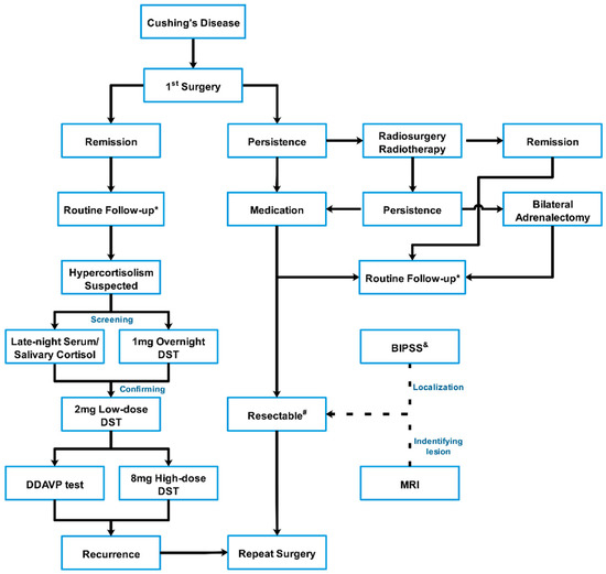
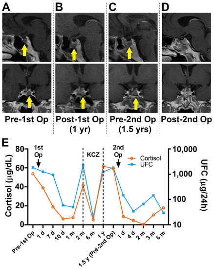
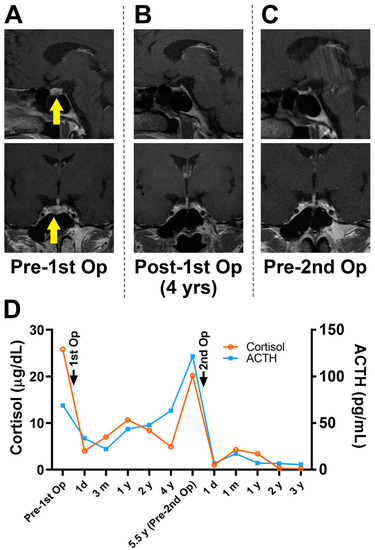
