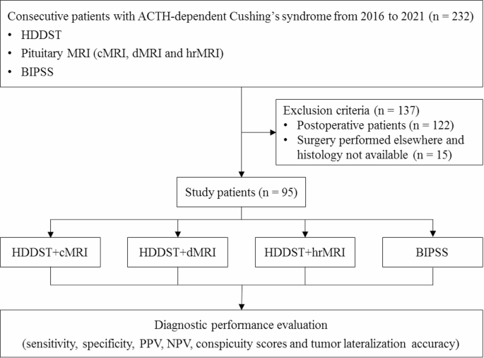
Cushing’s Syndrome in a Young Woman Due to Prolonged Betamethasone Nasal Drop Use

Abstract

Background

Cushing’s syndrome is an uncommon but serious condition caused by long-term exposure to elevated cortisol levels, which is usually iatrogenic in origin. Although systemic corticosteroids are the most frequent agents, the association of intranasal corticosteroids with this condition is remarkably rare.

Case presentation

This report is about a 21-year-old Iranian woman using betamethasone nasal drops for nasal obstruction. The patient presented with weight gain, Amenorrhea, mood disturbances, red purplish striae, and mild hirsutism. Hormonal assessments revealed suppression of the hypothalamic–pituitary–adrenal axis.

Conclusion

This case demonstrates the underappreciated systemic effects of intranasal betamethasone to induce Cushing’s syndrome. It serves as a pivotal reminder of the need for vigilance in prescribing practices and reinforces the importance of early diagnosis to ensure favorable patient outcomes.

Peer Review reports

Background

Iatrogenic Cushing’s syndrome (CS) is an endocrine disease caused by long-term or high-dose glucocorticoid use [1]. Although iatrogenic cases are commonly associated with oral or injectable glucocorticoids [2], few reports described CS after the use of intranasal steroid sprays (INS) such as betamethasone in adults [3,4,5,6,7]. Currently, INS is widely used for managing conditions such as allergic rhinitis, nasal polyposis, and other upper airway disorders owing to their localized effects and limited systemic absorption [89]. However, prolonged use, high doses, or using potent formulations can lead to significant systemic absorption, resulting in Hypothalamic–pituitary–adrenal (HPA) axis suppression, and frank CS [10]. Betamethasone nasal spray, a cornerstone in the treatment of nasal congestion, has the potential for systemic absorption by the nasal mucosa, particularly with prolonged or excessive use [11].

This report presents the case of a young woman who developed CS following the overuse of betamethasone nasal drops. It also highlights the importance of detailed patient histories when diagnosing CS and highlights the critical need to educate patients on the proper use and potential risks of steroid therapies to prevent complications. This case report adheres to the case report (CARE) guidelines [12].

Case presentation

This is the case of a 21-year-old Iranian female who presented with a history of rapid weight gain (30 kg in 8 months), irregular menstrual cycles, and significant mood changes. Her body mass index (BMI) was calculated at 40.07 kg/m2, classifying her as obese, and her blood pressure was recorded at 115/75 mmHg. In addition, she exhibited red–purple striae on her abdomen and limbs and mild hirsutism (modified Ferriman–Gallwey Score (FGS) score = 10), prompting admission for further evaluation after multiple outpatient visits yielded no definitive diagnosis.

Figure 1 is a clinical photograph (with patient consent) or an illustration of the red–purple striae.

Fig. 1

figure 1

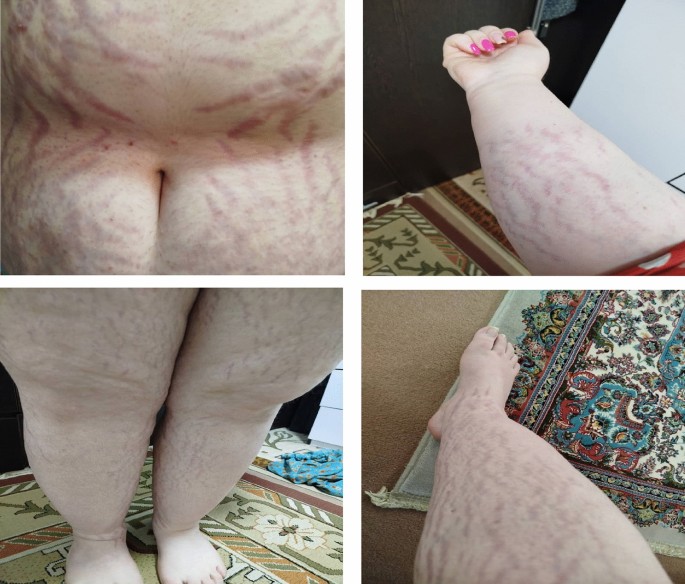
Clinical photograph showcasing the red–purplish striae on the patient’s abdomen, arms, and lower limbs

Upon admission, the patient’s history revealed prolonged use of betamethasone 0.1% 1 mg/mL nasal drops, administered at a daily dosage of 5 cc, in combination with oxymetazoline (a sympathomimetic nasal preparation) at a daily dosage of 1 cc, over approximately 12 months, to address nasal obstruction. Her symptoms began 6 months after starting the nasal drops. Further medication history revealed no other corticosteroid use. Notably, the patient had a past diagnosis of polycystic ovary (PCO) syndrome made on the basis of Rotterdam 2003 criteria (oligomenorrhea since menarche and clinically androgen excess) but did not undergo treatment or maintain laboratory records.

A detailed hormonal evaluation was undertaken. Morning plasma cortisol less than 0.05 µg/dL and adrenocorticotropic hormone (ACTH) less than 5 (10–56 pg/mL) measurements were abnormally low. Her 24-hour urine-free cortisol concentrations of 1.04 µg/24 h were significantly reduced, indicating suppression of the HPA axis secondary to prolonged exogenous corticosteroid exposure. All tests were repeated several times by endocrinologists during the time course of disease manifestations.

Table 1 summarizes the hormonal test results to clearly display the abnormalities.

Table 1 Hormonal and biochemical test results with reference values

Imaging studies before admission included a computed tomography (CT) scan of the adrenal glands, which showed that both adrenal glands were of normal size. However, a dynamic pituitary magnetic resonance imaging (MRI) revealed an 11 mm pituitary gland, despite there being no rationale for imaging studies in this scenario.

The patient was counseled extensively about the condition, and betamethasone nasal drops were discontinued immediately. Ear, nose, and throat (ENT) consultation revealed normal findings and the psychiatric team diagnosed her with major depressive disorder (MDD). She was discharged on 15 mg prednisolone with a structured tapering plan to allow for gradual recovery of adrenal function and to prevent acute adrenal insufficiency. Follow-up appointments were scheduled to monitor her clinical progress and re-evaluate her HPA axis recovery.

Discussion

This case highlights the rare but significant occurrence of iatrogenic CS secondary to prolonged use of intranasal betamethasone. Although oral corticosteroids are well-known to cause HPA axis suppression, INS is generally considered safer owing to their localized effects and lowering systemic absorption side effects. However, the associated potential of systemic absorption in INS remains a concern [13]. As demonstrated in this case, prolonged use of potent formulations such as betamethasone can lead to significant systemic effects, particularly when administered inappropriately or at high doses.

Betamethasone nasal drops, although effective for treating nasal congestion and inflammation [1415], carry a potential risk of systemic absorption through the nasal mucosa. Factors, such as prolonged use [61617], and high potency [18], can significantly increase systemic bioavailability. R. J. Perry et al. [19] in study of seven children highlights that even patients receiving doses within conventional safety ranges may exhibit varying sensitivity to glucocorticoids, leading to symptomatic adrenal suppression or glucocorticoid excess. Unlike newer corticosteroid compounds, such as fluticasone or mometasone, which undergo extensive first-pass metabolism in the liver, betamethasone exhibits minimal hepatic metabolism, contributing to its prolonged systemic activity [2021]. This pharmacokinetic profile underscores the need for careful regulation and monitoring of its use, even in ostensibly localized therapies.

The clinical manifestations in this patient, including central obesity, striae, hirsutism, and mood changes, were classic features of CS and guided the diagnostic process [22]. Scutelnicu et al. [23] reported a case of a patient in the second trimester of pregnancy who, owing to chronic sinusitis, underwent intranasal betamethasone spray therapy. The patient manifested extensive striae on the lower limbs, as well as edema in the legs, arms, and face, accompanied by a weight gain of 22 kg over 3 months. After switching the patient’s treatment to an alpha-1 adrenergic agonist spray, the condition was managed uneventfully without any symptoms of adrenal insufficiency.

Requesting imaging assessments, including a CT scan and MRI, as a first step further complicated the diagnostic process. This highlights a common diagnostic pitfall: the use of imaging as an initial approach can lead to the discovery of incidentalomas, which may misdirect clinical attention. Such findings risk overshadowing the primary etiology of the condition, potentially resulting in misdiagnosis or delayed treatment. This emphasizes the importance of prioritizing functional assessments over imaging in the early diagnostic workup to avoid unwarranted diagnostic confusion and ensure accurate identification of the underlying pathology.

Management involved the immediate cessation of betamethasone nasal drops and initiation of a structured tapering regimen with prednisolone to support adrenal recovery. The importance of stress-dose precautions during intercurrent illnesses was emphasized, alongside comprehensive patient education to prevent future misuse of corticosteroids. The gradual improvement in adrenal function during follow-up highlights the reversibility of glucocorticoid-induced adrenal suppression with appropriate intervention.

Conclusion

This case underscores several critical lessons. First, it emphasizes the importance of heightened awareness among healthcare providers regarding the potential systemic effects of topical corticosteroids, particularly potent formulations such as betamethasone. Second, it highlights the need for thorough history-taking and detailed patient education to prevent corticosteroid misuse. This report contributes to the limited body of literature on iatrogenic CS from intranasal corticosteroids, particularly in adults. Documenting the clinical presentation, diagnostic challenges, and successful management of this case, provides valuable insights into preventing, recognizing, and treating similar cases. It serves as a reminder of the delicate balance between therapeutic benefit and potential harm in corticosteroid therapy and advocates for ongoing research to establish safer prescribing practices.

Data availability

The data analyzed and generated in this study can be accessed through the corresponding author upon reasonable request.

Abbreviations

CS:
Cushing’s syndrome
INS:
Intranasal corticosteroids
HPA axis:
Hypothalamic–pituitary–adrenal axis
BMI:
Body mass index
FGS:
Ferriman–Gallwey Score
PCO:
Polycystic ovary
ACTH:
Adrenocorticotropic hormone
CT:
Computed tomography
MRI:
Magnetic resonance imaging
ENT:
Ear, nose, and throat
MDD:
Major depressive disorder

References

  1. Cristante J, Chabre O. Factitious, or iatrogenic but unexpected Cushing’s syndrome. Ann Endocrinol (Paris). 2023;84(3):370–2.

    PubMed Google Scholar

  2. Lacroix A, Feelders RA, Stratakis CA, Nieman LK. Cushing’s syndrome. Lancet. 2015;386(9996):913–27.

    CAS PubMed Google Scholar

  3. Scutelnicu A, Panaitescu AM, Ciobanu AM, Gica N, Botezatu R, Peltecu G, et al. Iatrogenic cushing’s syndrome as a consequence of nasal use of betamethasone spray during pregnancy. Acta Endocrinol. 2020;16(4):511–7.

    CAS Google Scholar

  4. Sakamoto M, Morita K, Okamura E, Uchino T, Okamoto K, Ozawa Y, et al. A case of iatrogenic cushing syndrome due to overuse of nasal steriod and concurrent administration of clarithromycin. Teikyo Med J. 2018;41(4):161–8.

    Google Scholar

  5. Nutting CM, Page SR. Iatrogenic Cushing’s syndrome due to nasal betamethasone: a problem not to be sniffed at! Postgrad Med J. 1995;71(834):231–2.

    CAS PubMed PubMed Central Google Scholar

  6. Stevens DJ. Cushing’s syndrome due to the abuse of betamethasone nasal drops. J Laryngol Otol. 1988;102(3):219–21.

    CAS PubMed Google Scholar

  7. Dow A, Yu R, Carmichael J. Too little or too much corticosteroid? Coexisting adrenal insufficiency and Cushing’s syndrome from chronic, intermittent use of intranasal betamethasone. Endocrinol Diabetes Metab Case Rep. 2013;2013:13–0036.

    Google Scholar

  8. Bachert C, Desrosiers MY, Hellings PW, Laidlaw TM. The role of biologics in chronic rhinosinusitis with nasal polyps. J Allergy Clin Immunol Pract. 2021;9(3):1099–106.

    CAS PubMed Google Scholar

  9. Bachert C, Han JK, Wagenmann M, Hosemann W, Lee SE, Backer V, et al. EUFOREA expert board meeting on uncontrolled severe chronic rhinosinusitis with nasal polyps (CRSwNP) and biologics: definitions and management. J Allergy Clin Immunol. 2021;147(1):29–36.

    CAS PubMed Google Scholar

  10. Quddusi S, Browne P, Toivola B, Hirsch IB. Cushing syndrome due to surreptitious glucocorticoid administration. Arch Intern Med. 1998;158(3):294–6.

    CAS PubMed Google Scholar

  11. Grayson JW, Harvey RJ. Topical corticosteroid irrigations in chronic rhinosinusitis. Int Forum Allergy Rhinol. 2019;9(S1):S9–15.

    PubMed Google Scholar

  12. Gagnier JJ, Kienle G, Altman DG, Moher D, Sox H, Riley D. The CARE guidelines: consensus-based clinical case reporting guideline development. Global Adv Health Med. 2013;2(5):38–43.

    Google Scholar

  13. McDonnell J, Weller K, Pien LC. Safety of intranasal steroids: an updated perspective. Curr Allergy Asthma Rep. 2020;20(11):69.

    PubMed Google Scholar

  14. Scadding GK. Other anti-inflammatory uses of intranasal corticosteroids in upper respiratory inflammatory diseases. Allergy. 2000;55(s62):19–23.

    PubMed Google Scholar

  15. Chong LY, Head K, Hopkins C, Philpott C, Burton MJ, Schilder AG. Different types of intranasal steroids for chronic rhinosinusitis. Cochrane Database Syst Rev. 2016;4(4):Cd011993.

    PubMed Google Scholar

  16. Findlay CA, Macdonald JF, Wallace AM, Geddes N, Donaldson MD. Childhood Cushing’s syndrome induced by betamethasone nose drops, and repeat prescriptions. BMJ. 1998;317(7160):739–40.

    CAS PubMed PubMed Central Google Scholar

  17. Reynolds C, Agrawal P, McCann A, O’Sullivan T, Chroinin MN, O’Riordan SMP. Cushing syndrome and adrenal insufficiency induced by high dose prolonged intranasal betamethasone. Arch Dis Child. 2019;104:A275.

    Google Scholar

  18. Oluwayemi IO, Oduwole AO, Oyenusi E, Onyiriuka AN, Abdullahi M, Fakeye-Udeogu OB, et al. Iatrogenic Cushing’s syndrome in children following nasal steroid. Pan Afr Med J. 2014;17:237.

    PubMed PubMed Central Google Scholar

  19. Perry RJ, Findlay CA, Donaldson MDC. Cushing’s syndrome, growth impairment, and occult adrenal suppression associated with intranasal steroids. Arch Dis Child. 2002;87(1):45–8.

    CAS PubMed PubMed Central Google Scholar

  20. Matera MG, Rinaldi B, Calzetta L, Rogliani P, Cazzola M. Pharmacokinetics and pharmacodynamics of inhaled corticosteroids for asthma treatment. Pulm Pharmacol Ther. 2019;58: 101828.

    CAS PubMed Google Scholar

  21. Krzyzanski W, Milad MA, Jobe AH, Peppard T, Bies RR, Jusko WJ. Population pharmacokinetic modeling of intramuscular and oral dexamethasone and betamethasone in Indian women. J Pharmacokinet Pharmacodyn. 2021;48(2):261–72.

    CAS PubMed PubMed Central Google Scholar

  22. Savas M, Mehta S, Agrawal N, van Rossum EFC, Feelders RA. Approach to the patient: diagnosis of Cushing syndrome. J Clin Endocrinol Metab. 2022;107(11):3162–74.

    PubMed PubMed Central Google Scholar

  23. Scutelnicu A, Panaitescu AM, Ciobanu AM, Gica N, Botezatu R, Peltecu G, et al. Iatrogenic Cushing’s syndrome as a consequence of nasal use of betamethasone spray during pregnancy. Acta Endocrinol (Buchar). 2020;16(4):511–7.

    CAS PubMed Google Scholar

Download references

Acknowledgements

Not applicable.

Funding

Not Applicable.

Author information

Authors and Affiliations

  1. Gastrointestinal and Liver Diseases Research Center, Iran University of Medical Sciences, Tehran, Iran

    Mohammadsadra Shamohammadi

  2. M.D., Endocrinologist Assistant Professor of Internal Medicine Assistant Professor of Internal Medicine, Iran University of Medical Sciences at Rasool Akram General Hospital, Tehran, Iran

    Delaram Eskandari

  3. Professor of Endocrinology Department of Endocrinology, Rasool Akram Medical Complex, School of Medicine, Iran University of Medical Sciences, Tehran, Iran

    Amir Ziaee

  4. Assistant Professor of Endocrinology & Metabolism Department of Internal Medicine, School of Medicine Hazrat-e Rasool General Hospital Iran University of Medical Sciences Medical Doctor at Iran University of Medical Sciences, Tehran, Iran

    Seyed Hossein Samadanifard

  5. Assistant Professor of Endocrinology & Metabolism Department of Internal Medicine, School of Medicine, Iran University of Medical Sciences, Tehran, Iran

    Haleh Chehrehgosha

  6. M.D., Endocrinologist Assistant Professor of Internal Medicine Assistant Professor of Internal Medicine, Iran University of Medical Sciences at Rasool Akram General Hospital, Tehran, Iran

    Amir Hossein Ghanooni

Contributions

MS and DE wrote the original draft; AZ and SHS collected the data. DE and HC were the patient’s doctors; MS and AHG reviewed, edited, and supervised the manuscript. All authors have read and approved the final version of the manuscript.

Corresponding author

Correspondence to Delaram Eskandari.

Ethics declarations

Ethics approval and consent to participate

This study was conducted in accordance with ethical guidelines and was approved by the Research Ethics Committee of Iran University of Medical Sciences under approval number IR.IUMS.REC.1404.208.

Consent for publication

Written informed consent was obtained from the patient for publication of this case report and any accompanying images. A copy of the written consent is available for review by the Editor-in-Chief of this journal.

Competing interests

The authors declare that they have no competing interests.

Additional information

Publisher’s Note

Springer Nature remains neutral with regard to jurisdictional claims in published maps and institutional affiliations.

Cushing Syndrome Leaves Lasting Health Effects

TOPLINE:

Compared with a matched population-based control group, patients with Cushing syndrome continued to exhibit elevated systolic and diastolic blood pressures along with reduced kidney function at least 14 years after biochemical remission.

METHODOLOGY:

  • Researchers in Germany conducted a retrospective cohort study to assess the long-term trajectory of blood pressure and kidney function in patients who achieved remission of Cushing syndrome.
  • They included 81 patients with Cushing syndrome (median age at baseline, 44 years; 75.3% women) and compared them with 243 matched control individuals from a population-based cohort.
  • Data were collected before treatment at baseline and at median follow-up intervals of 7.1 and 14 years after biochemical remission, with assessments of blood pressure, glomerular filtration rate, the prevalence of chronic kidney disease, and the use of antihypertensives.

TAKEAWAY:

  • Patients with Cushing syndrome had a significant reduction in blood pressure and required fewer antihypertensives at both 7 and 14 years vs baseline.
  • However, when compared with the control group, patients with Cushing syndrome had significantly elevated systolic and diastolic pressures at baseline and 7 and 14 years post-remission (P ≤ .0002 for all).
  • Although the proportion of patients on antihypertensive medications decreased in the Cushing syndrome group after remission was achieved, the prevalence of uncontrolled hypertension remained higher than in the control group at all follow-up points. In fact, reducing the use of these medications was associated with an increased risk for uncontrolled hypertension.
  • Kidney function assessed via glomerular filtration rate remained consistently lower among patients with Cushing syndrome than among control individuals at baseline and 7 and 14 years post-remission (P = .005, P < .0001, and P = .0359, respectively).

IN PRACTICE:

“Our findings provide further evidence that cardiovascular effects of hypercortisolism are not entirely reversible with the normalization of cortisol levels and enhance our understanding of the deteriorative long-term cardiovascular consequences of chronic hypercortisolism,” the authors wrote.

SOURCE:

This study was led by Katrin Ritzel, Ludwig-Maximilians-Universität München (LMU Munich), LMU University Hospital in Munich, Germany. It was published online on July 29, 2025, in Journal of Endocrinological Investigation.

LIMITATIONS:

The retrospective design and single-centre nature of this study could have been considered limitations.

DISCLOSURES:

This study was supported by Else Kröner-Fresenius Stiftung. Some authors reported being supported by Deutsche Forschungsgemeinschaft, the Munich Clinician Scientist Program, the Clinician Scientist Pro­gramme on Rare Important Syndromes in Endocrinology, and other sources. All authors reported having no conflicts of interest.

https://www.medscape.com/viewarticle/cushing-syndrome-leaves-lasting-health-effects-2025a1000kj0

Patient Finds Relief After Adrenal Gland Tumor Removed by Baylor Endocrine Surgeon

Hanna Pierce didn’t expect to learn she had a tumor on her adrenal gland during a CT scan. Just two weeks after delivering her second child and recovering from COVID-19, she went to urgent care with concerns about a possible blood clot. Instead, imaging revealed a tumor in her adrenal gland. “I didn’t have symptoms,” she said. “They were checking for something else and just happened to find it.”

That unexpected discovery in 2021 launched Pierce into a years-long journey that ultimately led to robotic surgery at Baylor Medicine with Dr. Feibi Zheng, an endocrine surgeon who specializes in treating adrenal tumors.

“Many people haven’t heard much about the adrenal glands,” said Zheng, assistant professor in the Division of Surgical Oncology. “They sit on top of the kidneys and produce hormones like cortisol that regulate everything from metabolism to the body’s stress response. If a tumor is overproducing cortisol, it can silently wreak havoc on the body over time.”

Doctors told Pierce that her tumor was consistently producing slightly elevated cortisol, a red flag. “My doctor told me if we left it alone, it could develop into diabetes or full-blown Cushing’s syndrome. At first, we just monitored it,” she said.

In the months and years that followed, Pierce did experience symptoms but attributed them to the demands of motherhood. “After my second child, I couldn’t lose weight no matter what I did. I had anxiety, constant fatigue in the afternoons, and I wasn’t sleeping well,” she recalled. “But I just chalked it up to being a mom of two.”

By 2024, her endocrinologist said it was time to act and referred her to Zheng, who confirmed the tumor was still producing excess cortisol. “Dr. Zheng told me I was going to feel so much better and explained what she was going to do,” Pierce said. “When I went to see her for the consultation, she was very informative. She didn’t pressure me to have surgery but explained everything to me.”

The Baylor Medicine endocrine surgery team, including Zheng and supported by physician assistant Holly Clayton, provided a seamless and collaborative care experience. “Our team-based model allows for better coordination and patient support,” said Clayton, who helped manage Pierce’s preoperative workup and performed her postop visit via telemedicine. “It was clear she wanted answers and a plan, and we were glad to be able to guide her through this process together.”

Zheng performed the adrenalectomy robotically, using a posterior approach — an advanced technique that involves going through the back instead of the front of the abdomen. “It’s a less common approach, but for the right patients, it can reduce pain and speed up recovery,” Zheng said.

Pierce said she felt calm going into the procedure. “Usually, I have white coat syndrome and feel anxious, but this time I didn’t. Everyone gave me step-by-step instructions, and Dr. Zheng explained everything clearly. I really felt like I was in good hands.”

Within a week or two of her June surgery, Pierce noticed changes. “I dropped four pounds almost immediately,” she said. “My face wasn’t as puffy. I felt less anxious and more like myself. Even though I was still recovering, I had more energy, and my body felt like it had reset.”

“Surgery to correct cortisol-producing tumors can make a major difference in quality of life, even if patients don’t meet the full criteria for a Cushing’s diagnosis,” Zheng said. “Mrs. Pierce’s case is a perfect example. She didn’t feel well, but she didn’t know why. Her endocrinologist saw [that] her metabolic parameters were getting worse. Now that the tumor is gone, her symptoms are improving, and her health trajectory is back on track.”

Just a month after surgery, Pierce says she has more energy and is continuing to lose weight. She’s relieved that a straightforward procedure made such a noticeable difference in how she feels.

From https://blogs.bcm.edu/2025/07/23/patient-finds-relief-after-adrenal-gland-tumor-removed/

The Outcome of Abnormal Glucose Metabolism and Its Clinical Features in Patients With Cushing’s Disease After Curative Surgery

Abstract

Objective

To investigate the outcomes of abnormal glucose metabolism and its clinical characteristics in patients with Cushing’s disease (CD) who achieved biochemical remission after surgery.

Methods

Patients diagnosed with CD who achieved biochemical remission and underwent regular follow-up after surgery were enrolled. Pre- and postoperative clinical data were collected and analyzed.

Result

151CD patients were included, of whom 80 (53 %) had preoperative abnormal glucose metabolism, including 56 with diabetes mellitus (DM) and 24 with impaired glucose regulation (IGR). At one year after surgery, 57 patients exhibited improved glucose metabolism, accompanied by a significant reduction in the homeostasis model assessment of insulin resistance (HOMA-IR). Improvements were mainly observed at 3 and 6 months after surgery. At one-year after surgery, there were 20 patients with diabetes and 16 with IGR. Compared to those with NGT, these individuals exhibited a higher prevalence of hypertension, hyperlipidemia, fatty liver, and abnormal bone metabolism.

Conclusion

CD patients demonstrated a high incidence of abnormal glucose metabolism. Notably, approximately two-thirds demonstrated improved glucose metabolism one year after curative surgery, with the greatest improvements observed at 3- to 6-month postoperative follow-up.

Introduction

Cushing’s disease (CD) is characterized by excessive endogenous cortisol production caused by pituitary adrenocorticotropic hormone adenoma and is the main cause of Cushing’s syndrome (CS). Surgical resection of the tumor is the preferred treatment. Prolonged exposure to hypercortisolism increases the risk of metabolic abnormalities, including obesity, hypertension, glucose and lipid abnormalities, osteoporosis, etc. Additionally, it significantly elevates the risk of infection, thrombosis, and hypokalemia. Abnormal glucose metabolism is a common complication of CS, with an incidence ranging from 13.1 % to 47 %[1], and diabetes is an independent risk factor for mortality in CD patients[2].
Previous clinical studies have found that metabolic abnormalities such as diabetes, hypertension, and hyperlipidemia improve in CS patients who achieve biochemical remission after surgical treatment. However, the concept of improvement in glucose metabolism, the incidence of improvement, and its related factors are inconsistent in various reports. Previous studies primarily assessed the outcome of glucose metabolism based on plasma glucose results at a single fixed follow-up time after surgery. The lack of regular follow-up data makes it difficult to clearly understand the trend of postoperative plasma glucose changes, and there are no clinical data on when glucose metabolism begins to improve or change. Therefore, this study retrospectively analyzed the follow-up data of patients with Cushing’s disease in our hospital before and after surgery, and monitored the changes in glucose metabolism, to explore the characteristics and clinical features of such changes in patients with Cushing’s disease who achieved remission from CD following surgery..

Access through your organization

Check access to the full text by signing in through your organization.

Access through your organization

Section snippets

Subjects

This study enrolled hospitalized patients with Cushing’s disease at Huashan Hospital, Fudan University from January 2014 to February 2020. Inclusion criteria were as follows: (1) Age ≥ 18 years; (2) diagnosis of Cushing’s disease according to the 2021 Consensus on the Diagnosis and Management of Cushing’s Disease, confirmed by pathology[3]; (3) biochemical remission after transsphenoidal surgery; (4) complete preoperative data and regular follow-up visits (including visits at 1, 3, 6, and

Patients’ baseline characteristics

A total of 168 patients with CD were admitted to Huashan Hospital from 2014 to 2020 with pathological diagnosis and regular postoperative follow-up; however, 17 patients were excluded due to no biochemical remission after surgery or relapse during follow-up (Fig. 1). Ultimately, 151 patients (32 males and 119 females) were included in this study. The baseline characteristics of the included patients were shown in Table 1. There were 80 cases (53 %) complicated with abnormal glucose metabolism

Discussion

CD was a rare disease often associated with abnormal glucose metabolism. Based on medical history and OGTT screening, we found that over half (53 %) of CD patients exhibited abnormal glucose metabolism before surgery, with 37.1 % being diagnosed with diabetes. Previous studies have shown that the prevalence of diabetes in CS patients ranged from 13.1 % to 47 %, and most reports falling between 35 % and 45 %, which is consistent with our findings [1,12,13]. However, it should be noted that CD

Author contributions

Q.C. analyzed the data and wrote the manuscript. Q.C., Y.L., X.L., Q.S., W.S., and H.Z. collected the data. Y.L., Z.Z., M.H., S.Z., and H.Y. recruited patients. J.Z., Y.S., and S.Z. conducted the study design and revised the manuscript. All authors read and approved the final manuscript.

CRediT authorship contribution statement

Qiaoli Cui: Writing – review & editing, Writing – original draft, Methodology, Investigation, Formal analysis, Data curation, Conceptualization. Yujia Li: Writing – original draft, Investigation, Formal analysis, Data curation. Xiaoyu Liu: Investigation, Formal analysis, Data curation. Quanya Sun: Investigation, Data curation. Wanwan Sun: Investigation, Formal analysis, Data curation. Min He: Project administration, Investigation. Jie Zhang: Writing – review & editing, Supervision, Funding

Declaration of competing interest

The authors declare that they have no known competing financial interests or personal relationships that could have appeared to influence the work reported in this paper.

Acknowledgments

We are indebted to the patients who participated in this study and all the doctors who contributed to the diagnosis and treatment of these patients. This work was supported by grants from the Multidisciplinary Diagnosis and Treatment (MDT) demonstration project in research hospitals (Shanghai Medical College, Fudan University, NO: DGF501069/017), National Science and Technology Major Project (NO: 2023ZD0506800,2023ZD0506802), 2023 Ningbo International Cooperation Program (NO: 2023H024).

References (16)

  • R.N. Clayton et al.

    Mortality in patients with Cushing’s disease more than 10 years after remission: a multicentre, multinational, retrospective cohort study

    Lancet Diabetes Endocrinol

    (2016)
  • M. Fleseriu et al.

    Consensus on diagnosis and management of Cushing’s disease: a guideline update

    Lancet Diabetes Endocrinol

    (2021)
  • C. Scaroni et al.

    Glucose Metabolism Abnormalities in Cushing Syndrome: from Molecular Basis to Clinical Management

    Endocr Rev

    (2017)
  • C. Dai et al.

    Surgical outcome of transsphenoidal surgery in Cushing’s disease: a case series of 1106 patients from a single center over 30 years

    Endocrine

    (2022)
  • E. Valassi et al.

    Delayed remission after transsphenoidal surgery in patients with Cushing’s disease

    J Clin Endocrinol Metab

    (2010)
  • X. He et al.

    Glucocorticoid Withdrawal Syndrome following treatment of endogenous Cushing Syndrome

    (2022)
  • E.V. Varlamov et al.

    Perioperative Management of a Patient with Cushing Disease

    J Endocr Soc

    (2022)
  • Q. Cui et al.

    The recovery time of hypothalamic-pituitary-adrenal axis after curative surgery in Cushing’s disease and its related factor

    Endocrine

    (2023)
There are more references available in the full text version of this article.

Improved Noninvasive Diagnostic Evaluations in Treatment-Naïve Adrenocorticotropic Hormone (ACTH)-Dependent Cushing’s Syndrome

Abstract

Background

Bilateral inferior petrosal sinus sampling (BIPSS) is important in the differential diagnosis of adrenocorticotropic hormone (ACTH)-dependent Cushing’s syndrome, but BIPSS is invasive and is not reliable on tumor lateralization. Thus, we evaluated the noninvasive diagnostic evaluations, high-dose dexamethasone suppression test (HDDST) combined with different pituitary MRI scans (conventional contrast-enhanced MRI [cMRI], dynamic contrast-enhanced MRI [dMRI], and high-resolution contrast-enhanced MRI [hrMRI]), by comparison with BIPSS.

Methods

We retrospectively analyzed 95 patients with ACTH-dependent Cushing’s syndrome who underwent HDDST, preoperative MRI scans (cMRI, dMRI and hrMRI) and BIPSS in our hospital between January 2016 and December 2021. The diagnostic performance of HDDST combined with cMRI (HDDST + cMRI), HDDST + dMRI and HDDST + hrMRI, and BIPSS was evaluated, including the sensitivity of identifying pituitary adenomas and the tumor lateralization accuracy.

Results

Compared with BIPSS (AUC, 0.98; 95%CI: 0.93, 1.00), the diagnostic performance of HDDST + hrMRI was comparable in both neuroradiologist 1 (AUC, 0.95; 95%CI: 0.89, 0.99; P = 0.129) and neuroradiologist 2 (AUC, 0.98; 95%CI: 0.92, 1.00; P = 0.707). For tumor lateralization accuracy, HDDST + hrMRI (90.6-95.3%) were significantly higher than that of BIPSS (24.7%, P < 0.001).

Conclusions

In patients with ACTH-dependent Cushing’s syndrome, HDDST + hrMRI, as noninvasive diagnostic evaluations, achieves high diagnostic performance comparable with gold standard (BIPSS), and it is superior to BIPSS in terms of tumor lateralization accuracy.

Peer Review reports

Background

Cushing’s syndrome is associated with debilitating morbidity and increased mortality [1]. Adrenocorticotropic hormone (ACTH)-dependent Cushing’s syndrome is characterized by ACTH hypersecretion. Bilateral inferior petrosal sinus sampling (BIPSS) is regarded as the gold standard to distinguish pituitary ACTH secretion (also known as Cushing’s disease) from ectopic ACTH syndrome (EAS) [12]. However, BIPSS is invasive and is not reliable on tumor lateralization [34]. Thus, it is important to improve the diagnostic performance of noninvasive evaluations with high sensitivity and tumor lateralization accuracy.

Current noninvasive evaluations in the differential diagnosis of ACTH-dependent Cushing’s syndrome include high-dose dexamethasone suppression test (HDDST), the CRH stimulation test and pituitary MRI. However, due to the non-availability of CRH for testing, the sensitivities of current available noninvasive evaluations in identifying ACTH-secreting pituitary adenomas cannot satisfy the clinical needs. Conventional contrast-enhanced MRI (cMRI) and dynamic contrast-enhanced MRI (dMRI) with two-dimensional (2D) fast spin echo (FSE) sequence is routinely used, and only 50–66% of the ACTH-secreting pituitary adenomas can be correctly detected [56]. Recently, by using 3D spoiled gradient recalled (SPGR) sequence, high-resolution contrast-enhanced MRI (hrMRI) has increased the sensitivity to up to 80% [7,8,9]. However, these noninvasive evaluations are still inferior to BIPSS, the sensitivity and specificity of which is about 90–95% [10,11,12,13]. With the development of 3D FSE sequence, superior image quality with diminished artifact has been achieved, providing a reliable alternative to detect pituitary adenomas [14]. Previous studies have shown that hrMRI using 3D FSE sequence has high diagnostic performance for identifying pituitary adenomas [1516]. To our knowledge, no study has investigated the diagnostic performance of HDDST combined with hrMRI using 3D FSE sequence (HDDST + hrMRI) in patients with Cushing’s syndrome, and whether it can avoid unnecessary BIPSS procedure.

The aim of this study is to evaluate the diagnostic performance of HDDST + hrMRI by comparison with BIPSS in patients with ACTH-dependent Cushing’s syndrome.

Methods

This study was performed in line with the principles of the Declaration of Helsinki. Approval was granted by the Institutional Review Board of Peking Union Medical College Hospital. Informed consent was waived in this study because it was a retrospective, non-interventional, and observational study. Clinical trial number is not applicable.

Study design and patient population

We retrospectively reviewed the medical records and imaging studies from January 2016 to December 2021, and 232 consecutive patients with ACTH-dependent Cushing’s syndrome, who underwent HDDST, cMRI, dMRI, hrMRI and BIPSS, were enrolled in the current study. A total of 137 patients were excluded from the study because of prior pituitary surgery (n = 122) or lack of histopathology due to no pituitary surgery in our hospital (n = 15). Finally, 95 patients were included in the current study (Fig. 1) and all the patients included were confirmed by histopathology or by clinical remission after surgical resection of the ACTH-secreting lesion. In the current study, all the patients with Cushing’s disease achieved clinical remission after surgical resection of the ACTH-secreting lesion. All the patients with EAS underwent contrast-enhanced thoracic and abdominal CT to identify the ACTH-secreting lesion. The clinical decision-making process was consistent with the previous study [1].

Fig. 1
figure 1

Flowchart of patient inclusion/exclusion process. ACTH = adrenocorticotropic hormone, BIPSS = bilateral inferior petrosal sinus sampling; cMRI = conventional contrast-enhanced MRI, dMRI = dynamic enhanced MRI, HDDST = high-dose dexamethasone suppression test, hrMRI = high-resolution contrast-enhanced MRI, NPV = negative predictive value, PPV = positive predictive value

HDDST

As previously described [17], the average 24-hour urinary free cortisol (24hUFC) level of 2 days before HDDST was recorded as baseline. Then, 2 mg dexamethasone was administered orally every 6 h for 2 days, and the 24hUFC level of the second day was measured. When the ratio of 24hUFC on the second day after HDDST to 24hUFC at baseline was less than 50%, the suppression in HDDST was marked as positive in the current study.

BIPSS

BIPSS was performed according to Doppman et al. [18]. Blood samples were collected from peripheral veins and bilateral inferior petrosal sinuses (IPSs) at multiple time points (0, 3, 5 and 10 min) after the introduction of 10 µg desmopressin [19]. An IPS to peripheral ACTH ratio of ≥ 2.0 at baseline or ≥ 3.0 after desmopressin stimulation at any time point [20] was marked as positive in the current study. Furthermore, tumor lateralization was predicted by an intersinus ratio of ≥ 1.4 [20].

Imaging

All the images were acquired on a 3.0 Tesla MR scanner (Discovery MR750w, GE Healthcare) using an 8-channel head coil. Detailed acquisition parameters and sequence order before and after contrast injection (gadopentetate dimeglumine [Gd-DTPA] at 0.05 mmol/kg [0.1 mL/kg] with a flow rate of 2 mL/s followed by a 10-mL saline solution flush) were as follows: coronal 2D FSE T2WI (field of view [FOV] = 20 cm × 20 cm, slice thickness = 4 mm, slice spacing = 1 mm, repetition time/echo time [TR/TE] = 4100/90 ms, number of excitation [NEX] = 1.2, matrix = 320 × 320, scan time = 49s), coronal 2D FSE T1WI (FOV = 18 cm × 16.2 cm, slice thickness = 3 mm, slice spacing = 0.6 mm, TR/TE = 400/12 ms, NEX = 2, matrix = 256 × 192, scan time = 49s), sagittal fat-saturated 3D FSE T1WI (FOV = 16.5 cm × 16.5 cm, slice thickness = 3 mm, slice spacing = 0, TR/TE = 460/16 ms, NEX = 2, matrix = 256 × 224, scan time = 60s), dynamic contrast-enhanced coronal 2D FSE T1WI (FOV = 19 cm × 17.1 cm, slice thickness = 2 mm, slice spacing = 0.5 mm, TR/TE = 375/14 ms, NEX = 1, matrix = 288 × 192, scan time = 23s/phase × 6 phases), contrast-enhanced coronal 2D FSE T1WI, contrast-enhanced sagittal fat-saturated 3D FSE T1WI, and contrast-enhanced coronal fat-saturated 3D FSE T1WI (FOV = 15.2 cm × 15.2 cm, slice thickness = 1.2 mm, slice spacing = -0.6 mm, TR/TE = 390/15 ms, NEX = 6, matrix = 256 × 256, scan time = 4 min 30s).

Images were independently evaluated by two experienced neuroradiologists (with 25 and 16 years of experience in neuroradiology, respectively). Both neuroradiologists were blinded to the clinical information of the patients. The image order of cMRI, dMRI and hrMRI was randomized. The detection of pituitary adenomas was scored using a 3-point scale (0 = poor, 1 = fair, 2 = excellent). Scores of 1 or 2 represented a successful pituitary adenoma detection. The gold standard was the histopathology, and the diameter and the location of lesions were recorded on the sequence where identified.

The signal-to-noise ratio (SNR) and contrast-to-noise ratio (CNR) were calculated as follows: SNR = SIadenoma / SDbackground, CNR = |SIpituitary – SIadenoma| / SDbackground. SIpituitary and SIadenoma were defined as the mean signal intensity of the pituitary gland and the pituitary adenoma, respectively. SDbackground was defined as the standard deviation of the signal intensity of the background. CNR was recorded as 0 when no pituitary adenoma was identified. Figure 2 showed the calculation of SNR and CNR using an operator defined region of interest.

Fig. 2

figure 2

The calculation of SNR and CNR using an operator defined region of interest. CNR = contrast-to-noise ratio, SD = standard deviation, SI = signal intensity, SNR = signal-to-noise ratio

Statistical analysis

The κ analysis was conducted to assess the interobserver agreements. The κ value was interpreted as follows: below 0.20, slight agreement; 0.21–0.40, fair agreement; 0.41–0.60, moderate agreement; 0.61–0.80, substantial agreement; greater than 0.80, almost perfect agreement.

To assess the diagnostic performance of different evaluations, the receiver operating characteristic curves were plotted and the area under curves (AUCs) were compared between noninvasive and invasive evaluations for each neuroradiologist by using the DeLong test. Sensitivity, specificity, positive predictive value, and negative predictive value were calculated. The Friedman’s test was used to evaluate the SNR and CNR measurements as well as conspicuity scores of pituitary adenomas between MR protocols, and the Wilcoxon signed-rank test was used for pairwise comparison. The McNemar’s test was used to evaluate the tumor lateralization accuracy. A P value of less than 0.05 was considered statistically significant. A stricter P value of less than 0.017 was considered statistically significant after Bonferroni correction. Statistical analysis was performed using MedCalc Statistical Software (version 23.0.2) and SPSS Statistics (version 22.0).

Results

Clinical characteristics

The clinical characteristics of the 95 patients with Cushing’s syndrome were shown in Table 1. There were 85 patients (median age, 38 years; interquartile range [IQR], 29–51 years; 55 females [65%]) with Cushing’s disease and 10 patients (median age, 39 years; IQR, 30–47 years; 5 females [50%]) with EAS. Of the 85 patients with Cushing’s disease, the median diameter of pituitary adenomas was 5 mm (IQR, 4–5 mm), ranging from 3 to 28 mm. Among them, 80 patients had microadenomas (less than 10 mm in size). Of the ten patients with EAS, one patient had an ovarian carcinoid tumor found by abdominal CT, others had pulmonary carcinoid tumors found by thoracic CT as the cause of Cushing’s syndrome. None of the patients with EAS had a lesion in the pituitary.

Table 1 Clinical characteristics of the patients

Diagnostic performance noninvasive and invasive evaluations

The inter-observer agreements between two neuroradiologists were moderate on cMRI (κ = 0.597), moderate on dMRI (κ = 0.595), and almost perfect on hrMRI (κ = 0.850), respectively.

The diagnostic performance of noninvasive and invasive evaluations was shown in Table 2. Compared with BIPSS (AUC, 0.98; 95%CI: 0.93, 1.00), the diagnostic performance of HDDST + hrMRI was comparable in both neuroradiologist 1 (AUC, 0.95; 95%CI: 0.89, 0.99; P = 0.129) and neuroradiologist 2 (AUC, 0.98; 95%CI: 0.92, 1.00; P = 0.707). However, the diagnostic performance of HDDST + cMRI and HDDST + dMRI was inferior to BIPSS (P ≤ 0.001 for all). No difference was found between HDDST + cMRI and HDDST + dMRI in neuroradiologist 1 (P = 0.050) and neuroradiologist 2 (P = 0.353).

Table 2 The diagnostic performance of noninvasive and invasive evaluations

Figures 3 and 4 showed that microadenomas were correctly diagnosed on hrMRI, but missed on cMRI or dMRI.

Fig. 3

figure 3

Images in a patient with Cushing’s disease. The lesion is missed on (a) coronal contrast-enhanced T1-weighted image and (b) coronal dynamic contrast-enhanced T1-weighted image obtained with two-dimensional (2D) fast spin echo (FSE) sequence. (c) Coronal contrast-enhanced T1-weighted image on high-resolution MRI obtained with 3D FSE sequence shows a round pituitary microadenoma measuring approximately 4 mm with delayed enhancement on the left side of the pituitary gland

Fig. 4

figure 4

Images in a patient with Cushing’s disease. The lesion is missed on (a) coronal contrast-enhanced T1-weighted image and (b) coronal dynamic contrast-enhanced T1-weighted image obtained with two-dimensional (2D) fast spin echo (FSE) sequence. (c) Coronal contrast-enhanced T1-weighted image on high-resolution MRI obtained with 3D FSE sequence shows a round pituitary microadenoma measuring approximately 5 mm with delayed enhancement on the left side of the pituitary gland

Further, subgroup analysis was conducted in 85 patients with Cushing’s disease. The conspicuity scores of pituitary adenomas on cMRI, dMRI and hrMRI were shown in Table 3. Significant differences between three MR protocols were found in neuroradiologist 1 and neuroradiologist 2 (P < 0.001 for both). Pairwise comparison showed no difference between cMRI and dMRI in neuroradiologist 1 (P = 0.732) and neuroradiologist 2 (P = 0.130). However, hrMRI had significantly higher scores than cMRI and dMRI in neuroradiologist 1 and neuroradiologist 2 (P < 0.001 for all). The SNR on cMRI, dMRI and hrMRI were 64.8 (IQR, 50.8–97.0), 42.4 (IQR, 30.2–57.0) and 65.1 (IQR, 51.9–92.4), respectively. The SNR on cMRI and hrMRI were similar (P = 0.759), but they were higher than that of dMRI (P < 0.001 for both). The CNR on cMRI, dMRI and hrMRI were27.0 (IQR, 17.8–43.8), 26.4 (IQR, 17.7–37.5), and 29.7 (IQR, 21.1–45.1), respectively. The CNR were comparable (P = 0.159).

Table 3 Conspicuity scores of pituitary adenomas on MRI

The comparison of tumor lateralization accuracy was shown in Table 4. Because HDDST has no role to identify the tumor lateralization, the tumor lateralization of noninvasive evaluations was only based on MRI. The sensitivity of BIPSS was 96.5% (82/85), comparable to those of hrMRI in neuroradiologist 1 (90.6%, P = 0.227) and neuroradiologist 2 (95.3%, P > 0.99). However, for tumor lateralization accuracy, 36 patients had BIPSS lateralization predicted by an intersinus ratio of ≥ 1.4 [20], and 21 patients had BIPSS lateralization that were concordant in laterality with surgery. The tumor lateralization accuracy was 58.3% (21/36).

Table 4 Tumor lateralization accuracy comparison

In the whole population, the tumor lateralization accuracy of BIPSS in total was 24.7% (21/85), which is significantly lower than those of hrMRI in neuroradiologist 1 (90.6%, P < 0.001) and neuroradiologist 2 (95.3%, P < 0.001).

Discussion

In patients with ACTH-dependent Cushing’s syndrome, it is crucial but challenging to distinguish pituitary secretion from ectopic ACTH secretion. In the current study, the diagnostic performance of noninvasive evaluations, HDDST + hrMRI, is comparable to BIPSS. Moreover, it is superior to BIPSS in terms of tumor lateralization.

No consensus agreement has been made that whether BIPSS should be performed in all the patients with suspected Cushing’s disease, although BIPSS is the gold standard with high sensitivity and specificity, which is about 90–95% [10,11,12,13]. On the one hand, about 10–40% of the population harbor nonfunctioning pituitary adenomas [1321], which may lead to false-positive results without centralizing BIPSS results. On the other hand, BIPSS is invasive and is not reliable on tumor lateralization. BIPSS will be bypassed when the tumor is greater than 6 mm in pituitary MRI and the patient has a classical presentation and dynamic biochemical results consistent with Cushing’s disease [13].

Noninvasive evaluations have comparable sensitivity to BIPSS for identifying pituitary adenomas in patients with Cushing’s disease. With the development of MRI technology, 3D FSE sequence provides a reliable alternative to detect pituitary adenomas [14]. The 3D FSE sequence overcomes the disadvantages of 3D SPGR sequence, such as bright blood and magnetic susceptibility [2223]. By using black blood in 3D FSE sequence, an obvious contrast between the pituitary and the cavernous sinus can be observed. By using fat saturation after enhancement, the hyperintensity of adjacent fat-containing tissue can be suppressed. All these mentioned above can facilitating the identification of pituitary adenomas. The sensitivity of hrMRI using 3D FSE sequence ranges from 87.7 to 93.8%, depending on radiologists with different experience levels [16]. Compared with traditional 2D FSE sequence acquiring images with 2- to 3-mm slice thickness, hrMRI using 3D FSE sequence acquiring images with 1.2-mm slice thickness can dramatically reduce the partial volume averaging effect, improving the identification of the microadenomas [15]. The trade-off between spatial resolution and image noise is challenging in pituitary MRI [24]. Previous studies have proved that hrMRI has high signal-to-noise ratio and contrast-to-noise ratio [1516], and sufficient contrast between pituitary adenomas and the pituitary gland could help to improve the identification of pituitary adenomas. In the current study, the conspicuity scores of hrMRI are significantly higher than those of cMRI and dMRI, supporting that hrMRI is reliable on identifying pituitary lesions. Besides, the diagnosis of Cushing’s disease cannot be made depending on the results of hrMRI alone. Given that there is a population with accidental adenomas when imaging, most of which are nonfunctioning pituitary adenomas, the results of HDDST will help rule out. In the current study, all the patients who underwent surgery had positive histopathology results, which means that no pituitary incidentalomas were found in this population. This might be caused by the relatively small sample size. Eighty patients with Cushing’s disease have microadenomas, and the median diameter at surgery is about 5 mm, consistent with previous studies [2526]. All these mentioned above makes it more difficult to identify the lesions in the current study. However, the sensitivity of HDDST + hrMRI in the current study is up to 95.3%, comparable to the gold standard.

Noninvasive evaluations have significantly higher tumor lateralization accuracy than BIPSS. According to the guideline, surgery is the first-line treatment [3]. Precise location of the pituitary adenoma before surgery can dramatically improve the postoperative remission rate [27]. However, the tumor lateralization accuracy of BIPSS, less than 80% in previous studies [192829], cannot satisfy the clinical need. According to previous studies, the cut-off value for tumor lateralization was set as an intersinus ratio of ≥ 1.4 [20], and the accuracy of lateralization by BIPSS ranged from 48.0 to 78.7% [192829]. In the current study, 36 patients had BIPSS lateralization and 21 patients had BIPSS lateralization that were concordant in laterality with surgery. The tumor lateralization accuracy was 58.3%, consistent with previous studies [192829]. However, the aim of our study is to evaluate the diagnostic performance of BIPSS in all the patients underwent BIPSS, therefore, the tumor lateralization accuracy of BIPSS in total was only 24.7% (21/85). In our study, many patients have positive BIPSS results with an intersinus ratio of < 1.4, resulting in the low tumor lateralization accuracy of BIPSS. One possible reason might be that desmopressin is not so effective. Another possible reason for low tumor lateralization accuracy of BIPSS is that IPSs have considerable anatomy variations. A previous study suggests that BIPSS results are much improved when venous drainage is symmetric [30]. Patients with asymmetric IPSs have dominant venous drainage, and when the dominant side of venous drainage is discordant with the side of the lesion, BIPSS will fail in tumor lateralization [30]. Failure in tumor lateralization will result in multiple incisions into the pituitary in search of adenoma or hemi- or subtotal hypophysectomy, increasing the risk of complications and reducing the remission rate [31]. In total, only 24.7% of the patients have a BIPSS lateralization that were concordant in laterality with surgery, whereas the tumor lateralization accuracy of HDDST + hrMRI is superior to BIPSS with statistical significance.

Limitations of the study included its retrospective nature. The bias may be introduced during the patient inclusion/exclusion process. Patients lack of any of preoperative MRI scans, HDDST, or BIPSS have not been included in the current study. Some patients will bypass hrMRI as well as BIPSS when they have obvious pituitary adenomas on cMRI and dMRI. The diagnostic performance of these evaluations might be better with the inclusion of these patients. Second, the sample size in our current study is relatively small. Because this is a single institutional study and Cushing’s syndrome is a rare disease. The relatively small sample size may limit the conclusions regarding the diagnostic performance of hrMRI for differentiating ectopic from pituitary sources of ACTH. A larger population from multicenter is needed for future study. Besides, a large portion of patients with prior pituitary surgery have been excluded. The imaging findings of these patients are more complicated and hrMRI may show more advantages than routine sequences in this population.

Conclusions

In conclusion, as noninvasive diagnostic evaluations, HDDST + hrMRI achieves high diagnostic performance comparable with gold standard (BIPSS), and it is superior to BIPSS in terms of tumor lateralization accuracy in patients with ACTH-dependent Cushing’s syndrome.

Data availability

The datasets used and analyzed during the current study are available from the corresponding author upon reasonable request.

Abbreviations

24hUFC:
24-hour urinary free cortisol
2D:
Two-dimensional
3D:
Three-dimensional
ACTH:
Adrenocorticotropic hormone
AUC:
Area under curve
BIPSS:
Bilateral inferior petrosal sinus sampling
cMRI:
Contrast-enhanced MRI
CNR:
Contrast-to-noise ratio
dMRI:
Dynamic contrast-enhanced MRI
EAS:
Ectopic adrenocorticotropic hormone syndrome
FSE:
Fast spin echo
HDDST:
High-dose dexamethasone suppression test
hrMRI:
High-resolution contrast-enhanced MRI
IPS:
Inferior petrosal sinus
IQR:
Interquartile range
SNR:
Signal-to-noise ratio
SPGR:
Spoiled gradient recalled

References

  1. Lacroix A, Feelders RA, Stratakis CA, Nieman LK. Cushing’s syndrome. Lancet (London England). 2015;386(9996):913–27.

    Article CAS PubMed Google Scholar

  2. Loriaux DL. Diagnosis and differential diagnosis of cushing’s syndrome. N Engl J Med. 2017;376(15):1451–9.

    Article CAS PubMed Google Scholar

  3. Nieman LK, Biller BM, Findling JW, Murad MH, Newell-Price J, Savage MO, et al. Treatment of cushing’s syndrome: an endocrine society clinical practice guideline. J Clin Endocrinol Metab. 2015;100(8):2807–31.

    Article CAS PubMed PubMed Central Google Scholar

  4. Wind JJ, Lonser RR, Nieman LK, DeVroom HL, Chang R, Oldfield EH. The lateralization accuracy of inferior petrosal sinus sampling in 501 patients with cushing’s disease. J Clin Endocrinol Metab. 2013;98(6):2285–93.

    Article CAS PubMed PubMed Central Google Scholar

  5. Boscaro M, Arnaldi G. Approach to the patient with possible cushing’s syndrome. J Clin Endocrinol Metab. 2009;94(9):3121–31.

    Article CAS PubMed Google Scholar

  6. Kasaliwal R, Sankhe SS, Lila AR, Budyal SR, Jagtap VS, Sarathi V, et al. Volume interpolated 3D-spoiled gradient echo sequence is better than dynamic contrast spin echo sequence for MRI detection of Corticotropin secreting pituitary microadenomas. Clin Endocrinol (Oxf). 2013;78(6):825–30.

    Article CAS PubMed Google Scholar

  7. Grober Y, Grober H, Wintermark M, Jane JA, Oldfield EH. Comparison of MRI techniques for detecting microadenomas in cushing’s disease. J Neurosurg. 2018;128(4):1051–7.

    Article PubMed Google Scholar

  8. Fukuhara N, Inoshita N, Yamaguchi-Okada M, Tatsushima K, Takeshita A, Ito J, et al. Outcomes of three-Tesla magnetic resonance imaging for the identification of pituitary adenoma in patients with cushing’s disease. Endocr J. 2019;66(3):259–64.

    Article PubMed Google Scholar

  9. Patronas N, Bulakbasi N, Stratakis CA, Lafferty A, Oldfield EH, Doppman J, Nieman LK. Spoiled gradient recalled acquisition in the steady state technique is superior to conventional Postcontrast spin echo technique for magnetic resonance imaging detection of adrenocorticotropin-secreting pituitary tumors. J Clin Endocrinol Metab. 2003;88(4):1565–9.

    Article CAS PubMed Google Scholar

  10. Pecori Giraldi F, Cavallo LM, Tortora F, Pivonello R, Colao A, Cappabianca P, et al. The role of inferior petrosal sinus sampling in ACTH-dependent cushing’s syndrome: review and joint opinion statement by members of the Italian society for endocrinology, Italian society for neurosurgery, and Italian society for neuroradiology. NeuroSurg Focus. 2015;38(2):E5.

    Article PubMed Google Scholar

  11. Biller BM, Grossman AB, Stewart PM, Melmed S, Bertagna X, Bertherat J, et al. Treatment of adrenocorticotropin-dependent cushing’s syndrome: a consensus statement. J Clin Endocrinol Metab. 2008;93(7):2454–62.

    Article CAS PubMed PubMed Central Google Scholar

  12. Newell-Price J, Bertagna X, Grossman AB, Nieman LK. Cushing’s syndrome. Lancet (London England). 2006;367(9522):1605–17.

    Article CAS PubMed Google Scholar

  13. Arnaldi G, Angeli A, Atkinson AB, Bertagna X, Cavagnini F, Chrousos GP, et al. Diagnosis and complications of cushing’s syndrome: a consensus statement. J Clin Endocrinol Metab. 2003;88(12):5593–602.

    Article CAS PubMed Google Scholar

  14. Sartoretti T, Sartoretti E, Wyss M, Schwenk A, van Smoorenburg L, Eichenberger B, et al. Compressed SENSE accelerated 3D T1w black blood turbo spin echo versus 2D T1w turbo spin echo sequence in pituitary magnetic resonance imaging. Eur J Radiol. 2019;120:108667.

    Article PubMed Google Scholar

  15. Liu Z, Hou B, You H, Lu L, Duan L, Li M, et al. High-resolution contrast-enhanced MRI with three-dimensional fast spin echo improved the diagnostic performance for identifying pituitary microadenomas in cushing’s syndrome. Eur Radiol. 2023;33(9):5984–92.

    Article PubMed PubMed Central Google Scholar

  16. Liu Z, Hou B, You H, Lu L, Duan L, Li M, et al. Three-Dimensional fast spin echo pituitary MRI in Treatment-Naive cushing’s disease: reduced impact of reader experience and increased diagnostic accuracy. J Magn Reson Imaging. 2024;59(6):2115–23.

    Article PubMed Google Scholar

  17. Liu Z, Zhang X, Wang Z, You H, Li M, Feng F, Jin Z. High positive predictive value of the combined pituitary dynamic enhanced MRI and high-dose dexamethasone suppression tests in the diagnosis of cushing’s disease bypassing bilateral inferior petrosal sinus sampling. Sci Rep. 2020;10(1):14694.

    Article CAS PubMed PubMed Central Google Scholar

  18. Doppman JL, Oldfield E, Krudy AG, Chrousos GP, Schulte HM, Schaaf M, Loriaux DL. Petrosal sinus sampling for Cushing syndrome: anatomical and technical considerations. Work in progress. Radiology. 1984;150(1):99–103.

    Article CAS PubMed Google Scholar

  19. Machado MC, de Sa SV, Domenice S, Fragoso MC, Puglia P Jr., Pereira MA, et al. The role of Desmopressin in bilateral and simultaneous inferior petrosal sinus sampling for differential diagnosis of ACTH-dependent cushing’s syndrome. Clin Endocrinol (Oxf). 2007;66(1):136–42.

    Article CAS PubMed Google Scholar

  20. Oldfield EH, Doppman JL, Nieman LK, Chrousos GP, Miller DL, Katz DA, et al. Petrosal sinus sampling with and without corticotropin-releasing hormone for the differential diagnosis of cushing’s syndrome. N Engl J Med. 1991;325(13):897–905.

    Article CAS PubMed Google Scholar

  21. Chong BW, Kucharczyk W, Singer W, George S. Pituitary gland MR: a comparative study of healthy volunteers and patients with microadenomas. AJNR Am J Neuroradiol. 1994;15(4):675–9.

    CAS PubMed PubMed Central Google Scholar

  22. Lien RJ, Corcuera-Solano I, Pawha PS, Naidich TP, Tanenbaum LN. Three-Tesla imaging of the pituitary and parasellar region: T1-weighted 3-dimensional fast spin echo cube outperforms conventional 2-dimensional magnetic resonance imaging. J Comput Assist Tomogr. 2015;39(3):329–33.

    PubMed Google Scholar

  23. Kakite S, Fujii S, Kurosaki M, Kanasaki Y, Matsusue E, Kaminou T, Ogawa T. Three-dimensional gradient echo versus spin echo sequence in contrast-enhanced imaging of the pituitary gland at 3T. Eur J Radiol. 2011;79(1):108–12.

    Article PubMed Google Scholar

  24. Kim M, Kim HS, Kim HJ, Park JE, Park SY, Kim YH, et al. Thin-Slice pituitary MRI with deep Learning-based reconstruction: diagnostic performance in a postoperative setting. Radiology. 2021;298(1):114–22.

    Article PubMed Google Scholar

  25. Vitale G, Tortora F, Baldelli R, Cocchiara F, Paragliola RM, Sbardella E, et al. Pituitary magnetic resonance imaging in cushing’s disease. Endocrine. 2017;55(3):691–6.

    Article CAS PubMed Google Scholar

  26. Jagannathan J, Smith R, DeVroom HL, Vortmeyer AO, Stratakis CA, Nieman LK, Oldfield EH. Outcome of using the histological pseudocapsule as a surgical capsule in Cushing disease. J Neurosurg. 2009;111(3):531–9.

    Article PubMed PubMed Central Google Scholar

  27. Yamada S, Fukuhara N, Nishioka H, Takeshita A, Inoshita N, Ito J, Takeuchi Y. Surgical management and outcomes in patients with Cushing disease with negative pituitary magnetic resonance imaging. World Neurosurg. 2012;77(3–4):525–32.

    Article PubMed Google Scholar

  28. Deipolyi A, Bailin A, Hirsch JA, Walker TG, Oklu R. Bilateral inferior petrosal sinus sampling: experience in 327 patients. J Neurointerv Surg. 2017;9(2):196–9.

    Article PubMed Google Scholar

  29. Castinetti F, Morange I, Dufour H, Jaquet P, Conte-Devolx B, Girard N, Brue T. Desmopressin test during petrosal sinus sampling: a valuable tool to discriminate pituitary or ectopic ACTH-dependent cushing’s syndrome. Eur J Endocrinol. 2007;157(3):271–7.

    Article CAS PubMed Google Scholar

  30. Lefournier V, Martinie M, Vasdev A, Bessou P, Passagia JG, Labat-Moleur F, et al. Accuracy of bilateral inferior petrosal or cavernous sinuses sampling in predicting the lateralization of cushing’s disease pituitary microadenoma: influence of catheter position and anatomy of venous drainage. J Clin Endocrinol Metab. 2003;88(1):196–203.

    Article CAS PubMed Google Scholar

  31. Castle-Kirszbaum M, Amukotuwa S, Fuller P, Goldschlager T, Gonzalvo A, Kam J, et al. MRI for Cushing disease: A systematic review. AJNR Am J Neuroradiol. 2023;44(3):311–6.

    Article CAS PubMed PubMed Central Google Scholar

Download references

Acknowledgements

We thank Dr. Kai Sun, Medical Research Center, Peking Union Medical College Hospital, for his guidance on the statistical analysis in this study. We thank all the patients who participated in this study.

Funding

This study was supported by the National Natural Science Foundation of China (grants 82371946 and 82071899), the Chinese Academy of Medical Sciences Innovation Fund for Medical Sciences (grant 2021-I2M-1-025), and the National High Level Hospital Clinical Research Funding (grants 2022-PUMCH-B-067 and 2022-PUMCH-B-114). The funding played no role in the design of the study and collection, analysis, and interpretation of data and in writing the manuscript.

Author information

Authors and Affiliations

  1. Department of Radiology, Peking Union Medical College Hospital, Chinese Academe of Medical Sciences and Peking Union Medical College, No.1 Shuaifuyuan Wangfujing Dongcheng Distinct, Beijing, 100730, China

    Zeyu Liu, Bo Hou, Hui You, Mingli Li & Feng Feng

  2. Department of Endocrinology, Peking Union Medical College Hospital, Chinese Academe of Medical Sciences and Peking Union Medical College, No.1 Shuaifuyuan Wangfujing Dongcheng Distinct, Beijing, 100730, China

    Lin Lu, Lian Duan & Huijuan Zhu

  3. Department of Neurosurgery, Peking Union Medical College Hospital, Chinese Academe of Medical Sciences and Peking Union Medical College, No.1 Shuaifuyuan Wangfujing Dongcheng Distinct, Beijing, 100730, China

    Kan Deng & Yong Yao

  4. State Key Laboratory of Complex Severe and Rare Disease, Peking Union Medical College Hospital, Chinese Academe of Medical Sciences and Peking Union Medical College, No.1 Shuaifuyuan Wangfujing Dongcheng Distinct, Beijing, 100730, China

    Yong Yao, Huijuan Zhu & Feng Feng

Contributions

All authors have participated sufficiently in this submission to take public responsibility for its content. H.Y. and F.F. proposed research ideas, revised the paper, and reviewed it academically. B.H. and Z.L. were responsible for literature review, data analysis and writing the manuscript. M.L. revised the paper. L.L., L.D. and H.Z. collected the clinical data. K.D. and Y.Y. collected the surgical and histopathology data. All authors reviewed the manuscript.

Corresponding authors

Correspondence to Hui You or Feng Feng.

Ethics declarations

Ethics approval and consent to participate

This study was performed in line with the principles of the Declaration of Helsinki. Approval was granted by the Institutional Review Board of Peking Union Medical College Hospital. Informed consent was waived by Institutional Review Board of Peking Union Medical College Hospital, because it was a retrospective, non-interventional, and observational study.

Consent for publication

Not applicable.

Competing interests

The authors declare no competing interests.

Additional information

Publisher’s note

Springer Nature remains neutral with regard to jurisdictional claims in published maps and institutional affiliations.

About this article

Cite this article

Liu, Z., Hou, B., You, H. et al. Improved noninvasive diagnostic evaluations in treatment-naïve adrenocorticotropic hormone (ACTH)-dependent Cushing’s syndrome. BMC Med Imaging 25, 252 (2025). https://doi.org/10.1186/s12880-025-01786-y

Download citation

https://bmcmedimaging.biomedcentral.com/articles/10.1186/s12880-025-01786-y