From Weight Gain To Diabetes

Cushing’s syndrome happens when the body has too much cortisol, the stress hormone. It can cause weight gain, high blood pressure, and diabetes. So how to keep your health in check and what are the treatment options available? In an exclusive interview with Times Now, an Endocrinologist explains its symptoms, causes, and treatments.
We often blame stress for everything—from sleepless nights to stubborn weight gain. But did you know your body’s stress hormone, cortisol, could be at the root of more serious health issues like high blood pressure and diabetes? Yes, you read that right! But how? We got in touch with Dr Pranav A Ghody, Endocrinologist at Wockhardt Hospital, Mumbai Central, who explains how excessive cortisol levels can lead to a condition known as Cushing’s Syndrome.
What Exactly is Cortisol, and Why is it Important?
Hormones are the body’s chemical messengers, travelling through the bloodstream to regulate essential functions. Among them, cortisol, produced by the adrenal glands (tiny glands sitting above the kidneys), plays a crucial role in controlling blood pressure, blood sugar, energy metabolism, and inflammation. The pituitary gland, located at the base of the brain, regulates cortisol through another hormone called Adrenocorticotropic Hormone (ACTH).
Often referred to as the “stress hormone,” cortisol spikes when we’re under stress. However, when levels remain high for too long, it can lead to Cushing’s Syndrome, a disorder first identified in 1912 by Dr Harvey Cushing.

What Causes Cushing’s Syndrome?

Dr Ghody explains that Cushing’s Syndrome occurs when the body is exposed to excessive cortisol, which can happen in two ways:

1. Exogenous (External) Cushing’s Syndrome
This is the most common form and results from prolonged use of steroid medications (such as prednisone) to treat conditions like asthma, rheumatoid arthritis, and lupus, or to prevent transplant rejection. Since steroids mimic cortisol, long-term use can disrupt the body’s hormone balance.
2. Endogenous (Internal) Cushing’s Syndrome
This occurs when the body produces too much cortisol due to a tumour in the pituitary gland, adrenal glands, or other organs (lungs, pancreas, thymus). While rare—affecting about 10 to 15 people per million annually—it’s more common in women between 20 and 50 years old. When caused by a pituitary tumour, it’s specifically called Cushing’s Disease.

Symptoms: How To Recognize Signs Of Cushing’s Syndrome

Excess cortisol affects multiple organs, leading to a variety of symptoms. This includes:

– Weight gain around the belly (central obesity)
– Rounded, puffy face (moon face)
– Excess facial and body hair (hirsutism)
– Fat accumulation on the upper back (buffalo hump)
– Thin arms and legs
– Dark red-purple stretch marks on the chest and abdomen
– Extreme fatigue and muscle weakness
– Depression or anxiety
– Easily bruising with minimal trauma
– Irregular menstrual cycles in women
– Reduced fertility or low sex drive
– Difficulty sleeping
High blood pressure and newly diagnosed or worsening diabetes are also common red flags.

Why is Cushing’s Syndrome Often Misdiagnosed?

Dr Ghody explains that while severe cases of Cushing’s Syndrome are easier to identify, milder forms can often be missed or mistaken for conditions like obesity, diabetes, or polycystic ovary syndrome (PCOS).

Diagnosing Cushing’s Syndrome involves:
1. Measuring cortisol levels in the blood, urine, or saliva.
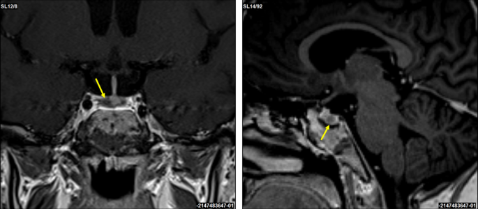
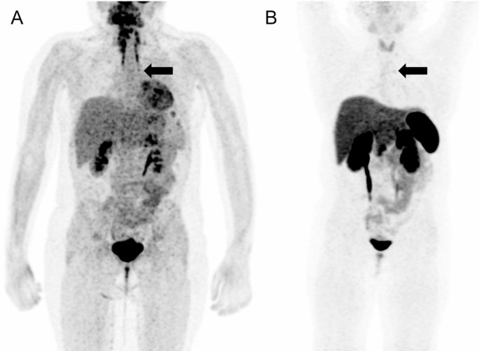
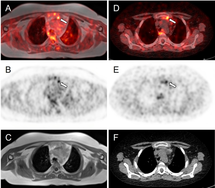
2. Identifying the source through ACTH hormone testing, MRI/CT scans, and advanced techniques like Inferior Petrosal Sinus Sampling (IPSS) or nuclear medicine scans
Treatment Options: How is Cushing’s Syndrome Managed?
Once diagnosed, the treatment depends on the cause:
– If due to steroid medication, the dosage is gradually reduced under medical supervision.
– If caused by a tumour, surgery is the primary treatment. Some patients, especially those with pituitary tumours, may require repeat surgery, gamma knife radiosurgery, or medications to control cortisol levels.

Can You Prevent Cushing’s Syndrome?

While complete prevention isn’t always possible, Dr Ghody shares some key strategies to reduce risk:

– Use steroids cautiously – If prescribed, take the lowest effective dose for the shortest time. Never stop abruptly without consulting a doctor.
– Genetic screening for people at risk – If you have a family history of pituitary or adrenal tumours, regular monitoring can help with early detection.
– Maintain a healthy lifestyle – A diet rich in fresh vegetables, and fruits, low sodium intake, adequate calcium, and vitamin D can help manage the metabolic effects of excess cortisol.
– Avoid alcohol and tobacco – These can further disrupt hormone balance and overall health.
“Cushing’s Syndrome can be life-threatening if left untreated, but early diagnosis and proper management can significantly improve quality of life. So if you experience unexplained weight gain, blood pressure spikes, or other symptoms, consult an endocrinologist to manage hormonal imbalances,” he said.

Older Adults With Cushing’s Disease Present With Fewer Symptoms Than Younger Patients

Key takeaways:

  • Older age was tied to a higher prevalence of 10 comorbidities among a group of 608 people with Cushing’s disease.
  • Younger age was associated with most hallmark features of Cushing’s disease.

The presentation of Cushing’s disease varies by age, with older adults having fewer hallmark features of the condition and more comorbidities, according to study findings published in The Journal of Clinical Endocrinology & Metabolism.

Researchers assessed data from 608 people diagnosed with Cushing’s disease and treated with a transsphenoidal tumor resection at 11 academic pituitary centers in the U.S. from 2003 to 2023 (82% women; 77.3% white). Patients were divided into 10-year age interval groups, with the youngest group consisting of those aged 10 to 19 years and the oldest containing adults aged 70 to 79 years. Researchers found Cushing’s disease presents differently as adults age, with older adults experiencing more comorbidities and complications, but fewer hallmark features such as weight gain, facial rounding and hirsutism.

“The diagnosis of Cushing’s disease remains challenging, particularly with age,” Won Kim, MD, associate clinical professor of neurosurgery and radiation oncology at the David Geffen School of Medicine at UCLA, told Healio. “The older a patient is, the more likely that he or she may have a slower-growing tumor with fewer classic manifestations of the disease.”

Kim and colleagues obtained data from the Registry of Adenomas of the Pituitary and Related Disorders. Hallmark features of Cushing’s disease were identified by consensus opinion.

The number of comorbidities increased with patient age (beta = 0.0466; P < .001), according to the researchers.

Older age was associated with several comorbidities for patients with Cushing’s disease, including hypertension (P < .001), diabetes (P < .001), hyperlipidemia (P < .001), cancer (P < .001), coronary artery disease (P < .001), chronic obstructive pulmonary disease (P = .044), cardiac arrhythmia (P = .023), hepatitis (P = .038), anxiety (P = .039) and osteopenia (P = .024). The most common comorbidity was hypertension, which was prevalent in 67.2% of participants.

In an analysis of presenting hallmark features of Cushing’s disease, younger age was positively associated with weight gain (P < .001), facial rounding (P < .001), abdominal striae (P < .001), hirsutism (P < .001), menstrual irregularities (P < .001) and acne (P < .001). Older age was positively tied to obstructive sleep apnea (P = .007). The most common hallmark feature of Cushing’s disease was weight gain, prevalent in 80.2% of patients.

“Our work highlights that we must lower our threshold for suspecting Cushing’s disease in patients without the classic physical manifestations as the age of the patient increases,” Kim said in an interview. “Subtle clues, such as increasingly difficult to control medical conditions such as hypertension and diabetes, may be the only things we see.”

Older age was associated with lower preoperative 24-hour urinary free cortisol levels (beta = –0.0256; P = 6.89 x 10⁻⁷), but higher postoperative nadir cortisol (beta = 0.0342; P = 1.03 x 10⁻⁴) and higher adrenocorticotropin (beta = 0.0204; P = 5.22 x 10⁻⁴).

In an assessment of tumor characteristics, older age was tied to having a higher Knosp grade tumor (beta = 0.011; P = .00435), greater tumor volume (beta = 0.0261; P = .0233) and higher maximum tumor dimension (beta = 0.009; P = 3.82 x 10⁻⁴). Older age was inversely associated with Ki-67 index, which is a measure of tumor’s proliferation (beta = –0.0459; P = 1.39 x 10⁻⁴).

Age was not associated with a patient’s number of surgical complications. Older age was linked to a greater prevalence of deep vein thrombosis or venous thromboembolism (beta = 0.07; P = .014). Younger age was tied to a higher prevalence of postoperative arginine vasopressin (beta = –0.02; P = .048).

Kim said the study’s findings should encourage health care professionals to adjust their methods for screening for Cushing’s disease in older adults.

“Improving our diagnostic sensitivity through our standardized assessments for the disease should account for these new findings,” Kim told Healio.

For more information:

Won Kim, MD, can be reached at wonkim@mednet.ucla.edu.

Published by:endocrine today logo

Cushing Disease Clinical Phenotype and Tumor Behavior Vary With Age

Abstract

Context

Little is known about presenting clinical characteristics, tumor biology, and surgical morbidity of Cushing disease (CD) with aging.

Objective

Using a large multi-institutional data set, we assessed diagnostic and prognostic significance of age in CD through differences in presentation, laboratory results, tumor characteristics, and postoperative outcomes.

Methods

Data from the Registry of Adenomas of the Pituitary and Related Disorders (RAPID) were reviewed for patients with CD treated with transsphenoidal tumor resection at 11 centers between 2003 and 2023. Outcomes assessed included comorbidities, presenting features, preoperative endocrine evaluations, perioperative characteristics, postoperative endocrine laboratory values, and complications.

Results

Of the 608 patients evaluated, 496 (81.6%) were female; median age at surgery was 44 years (range, 10-78 years). Increasing age was associated with increasing comorbidities, frailty, rates of postoperative thromboembolic disease, Knosp grade, tumor size, and postoperative cortisol and adrenocorticotropin nadirs. Conversely, increasing age was associated with decreased hallmark CD features, preoperative 24-hour urinary free cortisol, Ki-67 indices, and arginine vasopressin deficiency. Younger patients presented more frequently with weight gain, facial rounding/plethora, abdominal striae, hirsutism, menstrual irregularities, dorsocervical fat pad, and acne. Obstructive sleep apnea and infections were more common with increasing age.

Conclusion

There are age-dependent differences in clinical presentation, tumor behavior, and postoperative outcomes in patients with CD. Compared to younger patients, older patients present with a less classic phenotype characterized by fewer hallmark features, more medical comorbidities, and larger tumors. Notably, age-related differences suggest a more indolent tumor behavior in older patients, potentially contributing to delayed diagnosis and increased perioperative risk. These findings underscore the need for tailored diagnostic and therapeutic approaches across age groups, with a focus on managing long-term comorbidities and optimizing surgical outcomes.

Can We Predict the Risk of Venous Thromboembolism in Patients With Cushing’s Syndrome

Purpose

Patients with Cushing’s syndrome (CS) have an increased venous thromboembolism (VTE) risk with most studies focusing on the perioperative period. The purpose of this study was to assess the 5-year VTE risk and identify predictors of VTE at CS diagnosis.

Methods

A comparative nationwide retrospective cohort study of 609 patients (mean age 48.1 ± 17.2 years, 65.0% women) with CS, and 3018 age-, sex-, body mass index-, and socioeconomic status-individually matched controls. Ectopic CS and adrenal cancer were excluded. The time-to-event of pulmonary embolism (PE) or deep vein thrombosis (DVT) within 5 years of CS diagnosis was examined. VTE risk was calculated with death as competing event.

Results

VTE occurred in 16 cases (2.6%), compared to 17 (0.56%) controls (hazard ratio [HR] 4.71, 95% CI, 2.38–9.33). The 5-year HRs for PE and DVT were 7.47 (95% CI, 2.66–20.98) and 3.32 (95% CI, 1.36–8.12), respectively. After excluding patients and controls with current or prior malignancy the risk for VTE was 7.57 (95% CI, 2.98–19.20). Patients with CS ≥ 60 years at diagnosis (HR, 3.49; 95% CI, 1.30–9.35), with hypertension (HR, 5.53; 95% CI, 1.26–24.27), ischemic heart disease (HR, 3.60; 95% CI, 1.25–10.36), kidney disease (HR, 4.85; 95% CI, 1.39–16.90), or VTE event prior to CS diagnosis (HR, 33.65; 95% CI, 10.07–112.42) had an increased risk of VTE within five years.

Conclusions

In this large cohort of patients with CS, the 5-year VTE risk was 5 times higher compared with matched controls. Key baseline predictors included age ≥ 60, hypertension, heart/kidney disease, and prior VTE.

From https://link.springer.com/article/10.1007/s11102-024-01482-0

 This is a preview of subscription content, log in via an institution  to check access.

Ectopic Adrenocorticotrophic Hormone Syndrome in a 10-Year-Old Girl With a Thymic Neuroendocrine Tumor

Abstract

Background

Thymic neuroendocrine tumor as a cause of Cushing syndrome is extremely rare in children.

Case presentation

We report a case of a 10-year-old girl who presented with typical symptoms and signs of hypercortisolemia, including bone fractures, growth retardation, and kidney stones. The patient was managed with oral ketoconazole, during which she experienced adrenal insufficiency, possibly due to either cyclic adrenocorticotropic hormone (ACTH) secretion or concurrent COVID-19 infection. The patient underwent a diagnostic work-up which indicated the possibility of an ACTH-secreting pituitary neuroendocrine tumor. However, after a transsphenoidal surgery, the diagnosis was not confirmed on histopathological examination. Subsequent bilateral inferior petrosal sinus sampling showed strong indications of the presence of ectopic ACTH syndrome. Detailed rereading of functional imaging studies, including 18F-FDG PET/MRI and 68Ga DOTATOC PET/CT, ultimately identified a small lesion in the thymus. The patient underwent videothoracoscopic thymectomy that confirmed a neuroendocrine tumor with ACTH positivity on histopathological examination.

Conclusion

This case presents some unique challenges related to the diagnosis, management, and treatment of thymic neuroendocrine tumor in a child. We can conclude that ketoconazole treatment was effective in managing hypercortisolemia in our patient. Further, a combination of functional imaging studies can be a useful tool in locating the source of ectopic ACTH secretion. Lastly, in cases of discrepancy in the results of stimulation tests, bilateral inferior petrosal sinus sampling is highly recommended to differentiate between Cushing disease and ectopic ACTH syndrome.

Peer Review reports

Background

In children above seven years of age, the majority of pediatric Cushing syndrome (CS) cases are caused by a pituitary neuroendocrine tumors (PitNET). However, a differential diagnosis of hypercortisolemia in children is often challenging concerning the interpretation of stimulation tests and the fact that up to 50% of PitNET may not be detected on magnetic resonance imaging (MRI) [1]. An ectopic adrenocorticotropic hormone (ACTH) syndrome (EAS) is extremely rare in children. Its diagnosis is often missed or confused with Cushing disease (CD) [2]. Most ACTH-secreting tumors originate from bronchial or thymic neuroendocrine tumors (NETs), or less commonly, from NETs in other locations. To diagnose EAS, specific functional imaging studies are often indicated to elucidate the source of ACTH production.

Pharmacotherapy may be used before surgery to control hypercortisolemia and its symptoms/signs, or in patients in whom the source of hypercortisolism has not been found (e.g., EAS), or surgery failed. Ketoconazole or metyrapone, as adrenal steroidogenesis blockers, were found to be very efficient, although they exhibit side effects [3].

Furthermore, cyclic secretion of ACTH followed by fluctuating plasma cortisol levels is extremely rare in children, including those with EAS [45]. Therefore, in cyclic EAS, the use of steroid inhibitors or acute illness or trauma can be associated with adrenal insufficiency, which can be life-threatening. Here we describe the clinical features, laboratory and radiological investigations, results, management, and clinical outcome of a 10-year-old girl with a thymic NET presenting with ACTH secretion.

Case presentation

A 10-year-old girl was acutely admitted to our university hospital for evaluation of facial edema and macroscopic hematuria in May 2021. A day before admission, she presented to the emergency room for dysuria, pollakiuria, nausea, and pain in her right lower back. Over the past year she had experienced excessive weight gain with increased appetite and growth retardation (Fig. 1). Her height over three years had shifted from the 34th to the 13th centile (Fig. 1). Her parents noticed facial changes, pubic hair development, increased irritability, and moodiness.

Fig. 1

figure 1

Body weight, body height, and body mass index development of the case patient. The black arrow indicates the first presentation, the blue arrow indicates the start of ketoconazole treatment and the yellow arrow indicates the time of thymectomy. Mid-parental height is indicated by the green line

At admission, she was found to have a moon face with a plethora, few acne spots on forehead, as well as facial puffiness. In contrast to slim extremities, an abnormal fat accumulation was observed in the abdomen. Purple striae were present on abdomen and thighs. She did not present with any bruising, proximal myopathy, or edema. On physical examination, she was prepubertal, height was 135 cm (13th centile), and weight was 37 kg (69th centile) with a BMI of 20.4 kg/m2 (90th centile). She developed persistent hypertension. Her past medical history was uneventful except for two fractures of her upper left extremity after minimal trips one and three years ago, both treated with a caste. Apart from hypothyroidism on the maternal side, there was no history of endocrine abnormalities or tumors in the family.

In the emergency room, the patient was started on sulfonamide, pain medication, and intravenous (IV) fluids. Her hypertensive crises were treated orally with angiotensin-converting enzyme inhibitor or with a combination of adrenergic antagonists and serotonin agonists administered IV. Hypokalemia had initially been treated with IV infusion and then with oral potassium supplements. A low serum phosphate concentration required IV management. The initial investigation carried out in the emergency room found hematuria with trace proteinuria. Kidney ultrasound showed a 5 mm stone in her right ureter with a 20 mm hydronephrosis. She did not pass any kidney stones, however, fine white sand urine analysis reported 100% brushite stone.

Hypercortisolemia was confirmed by repeatedly increased 24-hour urinary free cortisol (UFC), (5011.9 nmol/day, normal range 79.0-590.0 nmol/day). Her midnight cortisol levels were elevated (961 nmol/l, normal range 68.2–537 nmol/l). There was no suppression of serum cortisol after 1 mg overnight dexamethasone suppression test (DST) or after low-dose DST (LDDST). An increased morning plasma ACTH (30.9 pmol/l, normal range 1.6–13.9 pmol/) suggested ACTH-dependent hypercortisolemia. There was no evidence of a PitNET on a 1T contrast-enhanced MRI. The high-dose DST (HDDST) did not induce cortisol suppression (cortisol 1112 nmol/l at 23:00, cortisol 1338 nmol/l at 8:00). Apart from the kidney stone, a contrast-enhanced computed tomography (CT) of her neck, chest, and abdomen/pelvis did not detect any lesion. Various tumor markers were negative and the concentration of chromogranin A was also normal.

A corticotropin-releasing hormone (CRH) stimulation test induced an increase in serum cortisol by 32% at 30 min and ACTH concentration by 67% at 15 min (Table 1). A 3T contrast-enhanced MRI scan of the brain identified a 3 × 2 mm lesion in the lateral right side of the pituitary gland (Fig. 2). An investigation of other pituitary hormones was unremarkable. Apart from low serum potassium (minimal level of 2.8 mmol/l; normal range 3.3–4.7 mmol/l) and phosphate (0.94 mmol/l; normal range 1.28–1.82 mmol/l) concentrations, electrolytes were normal. The bone mineral density assessed by whole dual-energy X-ray absorptiometry was normal.

Fig. 2

figure 2

Coronal and sagittal 3T contrast-enhanced brain MRI scans. A suspected 3 × 2 mm lesion in the lateral right side of the pituitary gland (yellow arrows)

The patient was presented at the multidisciplinary tumor board and it was decided that she undergoes transsphenoidal surgery for the pituitary lesion. No PitNET was detected on histopathological examination and no favorable biochemical changes were noted after surgery. After the patient recovered from surgery, subsequent bilateral inferior petrosal sinus sampling (BIPSS) confirmed EAS as the maximum ratio of central to peripheral ACTH concentrations was only 1.7. During the investigation for tumor localization, she was started on ketoconazole treatment (300 mg/day) to alleviate symptoms and signs of hypercortisolism. Treatment with ketoconazole had a beneficial effect on patient health (Fig. 1). There was a weight loss of 2 kg in a month, a disappearance of facial plethora, and a decrease in vigorous appetite. Her liver function tests remained within the normal range.

Table 1 Result of corticotropin-releasing hormone stimulation test

The 24-hour UFC excretion normalized three weeks after ketoconazole initiation. However, six weeks after continuing ketoconazole therapy (400 mg/day), the patient complained of nausea, vomiting, and diarrhea. She was found to have adrenal insufficiency with a low morning serum cortisol of 10.70 nmol/l (normal range 68.2–537 nmol/l) and salivary cortisol concentrations < 1.5 nmol/l (normal range 1.7–29 nmol/l). She was also found to be positive for COVID-19 infection. Ketoconazole treatment was stopped and our patient was educated to take stress steroids in case of persisting or worsening symptoms. Her clinical status gradually improved and steroids were not required.

Meanwhile, whole-body fluorine-18 fluorodeoxyglucose positron emission tomography (18F-FDG PET)/MRI was performed with no obvious hypermetabolic lesion suspicious of a tumor. No obvious accumulation was detected on 68Ga-DOTATOC PET/CT images (Fig. 3). However, a subsequent careful and detailed re-review of the images detected a discrete lesion on 18F-FDG PET/MRI and 68Ga-DOTATOC PET/CT scans in the left anterior mediastinum, in the thymus (Fig. 4).

Fig. 3

figure 3

18F-FDG PET/MRI (A) and 68Ga-DOTATOC (B) PET/CT scans. Whole body MIP reconstructions. Subtle correspondent focal hyperactivity in the left mediastinum (black arrow). The 18F-FDG PET/MRI image courtesy of Prof. Jiri Ferda, MD, PhD, Clinic of the Imaging Methods, University Hospital Plzen, Czech Republic

Fig. 4

figure 4

Axial slices of PET/MRI (AC) and 68Ga-DOTATOC (DF) PET/CT scans. Subtle correspondent focal hyperactivity in the left mediastinum (white arrow). No obvious finding on MRI (C) and CT (F) scans. The FDG PET/MRI image courtesy of Prof. Jiri Ferda, MD, PhD, Clinic of the Imaging Methods, University Hospital Plzen, Czech Republic

Three weeks after the episode of adrenal insufficiency and being off ketoconazole treatment, our patient´s pre-surgery laboratory tests showed slightly low morning cortisol 132 nmol/l with surprisingly normal ACTH 2.96 pmol/l (normal range 1.6–13.9 pmol/). Given the upcoming surgery, she was initiated on a maintenance dose of hydrocortisone (15 mg daily = 12.5 mg/m2/day). Further improvement of cushingoid characteristics (improvement of facial plethora and moon face, weight loss) was noticed. Our patient underwent videothoracoscopic surgery, and a hyperplastic thymus of 80 × 70 × 15 mm with a 4 mm nodule was successfully removed. Tumor immunohistochemistry was positive for ACTH, chromogranin A, CD56, and synaptophysin. Histopathological findings were consistent with a well-differentiated NET grade 1. A subsequent genetic screening did not detect any pathogenic variant in the MEN1 gene.

After surgery, hydrocortisone was switched to a stress dose and gradually decreased to a maintenance dose. Antihypertensive medication was stopped and further weight loss was observed after thymectomy. Within a few weeks after the thoracic surgery, the patient entered puberty, her mood improved significantly, and potassium supplements were stopped. Finally, hydrocortisone treatment was stopped ten months after thymectomy.

Discussion and conclusions

The case presented here demonstrates a particularly challenging work-up of the pediatric patient with the diagnosis of CS caused by EAS due to thymic NET. Differentiating CD and EAS can sometimes be difficult, including the use of various laboratory and stimulation tests and their interpretation, as well as proper, often challenging, reading of functional imaging modalities, especially if a discrete lesion is present at an unusual location [1]. When using established criteria for Cushing disease (for the CRH test an increase of cortisol and/or ACTH by ≥ 20% or ≥ 35%, respectively, and a ≥ 50% suppression of cortisol for the HDDST) our patient presented discordant results. The CRH stimulation test induced an increase in cortisol by 32% and ACTH by 67% and the 3T MRI pointed to the right-side pituitary lesion, both to yield false positive results. The HDDST, on the other hand, did not induce cortisol suppression and was against characteristic findings for CD. We did not proceed with desmopressin testing, which also induces an excess ACTH and cortisol response in CD patients and has rarely been used in pediatric patients, except in those with extremely difficult venous access [6]. Recently published articles investigated the reliability of CRH stimulation tests and HDDST and both concluded that the CRH test has greater specificity than HDDST [78]. Elenius et al. suggested optimal response criteria as a ≥ 40% increase of ACTH and/or cortisol (cortisol as the most specific measure of CD) during the CRH test and a ≥ 69% suppression of serum cortisol during HDDST [7]. Using these criteria, the CD would be excluded in our patient. To demonstrate that the proposed thresholds for the test interpretation widely differ, Detomas et al. proposed a ≥ 12% cortisol increase and ≥ 31% ACTH increase during the CRH test to confirm CD [8].

The fact that up to 50% of PitNET may not be detected on MRI [1] and that more than 20% of patients with EAS are reported to have pituitary incidentalomas [9] makes MRI somewhat unreliable in differentiating CD and EAS. However, finally, well-established and generally reliable BIPSS in our patient supported the diagnosis of EAS. Thus, BIPSS is considered a gold standard to differentiate between CD and EAS; however, it can still provide false negative results in cyclic CS if performed in the trough phase [10] or in vascular anomalies or false positive results as in a recent case of orbital EAS [11].

In children, the presence of thymus tissue may be misinterpreted as normal. Among other reports of thymic NET [12], Hanson et al. reported a case of a prepubertal boy in whom a small thymic NET was initially treated as normal thymus tissue on CT [13]. In our case, initially, the lesion was not detected on the 18F-FDG and 68Ga-DOTATOC PET scans. A small thymic NET was visible only after a detailed and careful re-reading of both PET scans. Although somatostatin receptor (SSR) PET imaging may be helpful in identifying ectopic CRH- or ACTH-producing tumors, there are still some limitations [13]. For example, in the study by Wannachalee et al., 68Ga-DOTATATE identified suspected primary lesions causing ECS in 65% of patients with previously occult tumors and was therefore concluded as a sensitive method for primary as well as metastatic tumors [14]. In our patient, the final correct diagnosis was based on the results of both PET scans. This is in full support of the article published by Liu et al. who concluded that 18F-FDG and SSR PET scans are complementary in determining the proper localization of ectopic ACTH production [15]. Additionally, it is worth noting that not all NETs stain positively for ACTH which may present a burden in its identification.

To control hypercortisolemia, both ketoconazole and metyrapone were considered in our patient. Due to the side effects of metyrapone on blood pressure, ketoconazole was started as a preferred option in our pediatric patient. A retrospective multicenter study concluded that ketoconazole treatment is effective with acceptable side effects, with no fatal hepatitis and adrenal insufficiency in 5.4% of patients [3]. During ketoconazole treatment, our patient developed adrenal insufficiency; however, it is impossible to conclude whether this was solely due to ketoconazole treatment or whether an ongoing COVID-19 infection contributed to the adrenal insufficiency or whether this was caused by a phase of lower or no ACTH secretion from the tumor often seen in patients with cyclic ACTH secretion. The patient’s cyclic ACTH secretion is highly probable since her morning cortisol was slightly lower and ACTH was normal, even after being off ketoconazole treatment for 3 weeks.

When retrospectively and carefully reviewing all approaches to the diagnostic and management care of our pediatric patient, it would be essential to proceed to BIPSS before any pituitary surgery, especially when obtaining discrepant results from stimulation tests, as well as detecting a discrete pituitary lesion ( 6 mm) as recommended by the current guidelines [16]. This was our first experience using ketoconazole in a young child, and although this treatment was associated with very good outcomes in treating hypercortisolemia, close monitoring, and family education on signs and symptoms of adrenal insufficiency are essential to recognizing adrenal insufficiency promptly in any patient with EAS, especially those presenting also with some other comorbidities or stress, here COVID-19 infection.

In conclusion, the pediatric patient here presenting with EAS caused by thymic NET needs very careful assessment including whether cyclic CS is present, the outline of a good management plan to use all tests appropriately and in the correct sequence, monitoring carefully for any signs or symptoms of adrenal insufficiency, and apply appropriate imaging studies, with experienced radiologists providing accurate readings. Furthermore, ketoconazole treatment was found to be effective in reducing the symptoms and signs of CS in this pediatric patient. Finally, due to the rarity of this disease and the challenging work-up, we suggest that a multidisciplinary team of experienced physicians in CS management is highly recommended.

Data availability

No datasets were generated or analysed during the current study.

Abbreviations

ACTH:
Adrenocorticotrophic hormone
BIPSS:
Bilateral inferior petrosal sinus sampling
CD:
Cushing disease
CRH:
Corticotropin-releasing hormone
CS:
Cushing syndrome
CT:
Computed tomography
DST:
Dexamethasone suppression test
EAS:
Ectopic adrenocorticotropic hormone syndrome
18F-FDG PET:
Fluorine-18 fluorodeoxyglucose positron emission tomography
HDDST:
High-dose dexamethasone suppression test
IV:
Intravenous
LDDST:
Low-dose dexamethasone suppression test
NET:
Neuroendocrine tumor
PitNET:
Pituitary neuroendocrine tumor
UFC:
Urinary free cortisol

References

  1. Streuli R. A rare case of an ACTH/CRH co-secreting midgut neuroendocrine tumor mimicking Cushing’s disease. Endocrinol Diabetes Metab Case Rep. 2017;2017:17–58. ,Krull I, Brändle M, et al.

    PubMed PubMed Central Google Scholar

  2. Karageorgiadis AS, Papadakis GZ, Biro J, et al. Ectopic adrenocorticotropic hormone and corticotropin-releasing hormone co-secreting tumors in children and adolescents causing cushing syndrome: a diagnostic dilemma and how to solve it. J Clin Endocrinol Metab. 2015;100(1):141–8.

    Article CAS PubMed Google Scholar

  3. Castinetti F, Guignat L, Giraud P, et al. Ketoconazole in Cushing’s disease: is it worth a try? J Clin Endocrinol Metab. 2014;99(5):1623–30.

    Article CAS PubMed Google Scholar

  4. Mi Q, Yin M-Z, Gao Y-J et al. Thymic atypical carcinoid with cyclical Cushing’s syndrome in a 7-year-old boy: a case report and review of the literature. Intern Med. 2014;4(5).

  5. Moszczyńska E, Pasternak-Pietrzak K, Prokop-Piotrkowska M, et al. Ectopic ACTH production by thymic and appendiceal neuroendocrine tumors – two case reports. J Pediatr Endocrinol Metab. 2020;34(1):141–6.

    Article PubMed Google Scholar

  6. Crock PA, Ludecke DK, Knappe UJ, et al. A personal series of 100 children operated for Cushing’s disease (CD): optimizing minimally invasive diagnosis and transnasal surgery to achieve nearly 100% remission including reoperations. J Pediatr Endocrinol Metab. 2018;31(9):1023–31.

    Article CAS PubMed Google Scholar

  7. Elenius H, McGlotten R, Nieman LK. Ovine CRH stimulation and 8 mg dexamethasone suppression tests in 323 patients with ACTH-dependent Cushing’s syndrome. J Clin Endocrinol Metab. 2023;109(1):e189–189.

    Article Google Scholar

  8. Detomas M, Ritzel K, Nasi-Kordhishti I, et al. Outcome of CRH stimulation test and overnight 8 mg dexamethasone suppression test in 469 patients with ACTH-dependent Cushing’s syndrome. Front Endocrinol (Lausanne). 2022;13:955945.

    Article PubMed Google Scholar

  9. Yogi-Morren D, Habra MA, Faiman C, et al. Pituitary MRI findings in patients with pituitary and ectopic ACTH-dependent Cushing syndrome: does a 6-mm pituitary tumor size cut-off value exclude ectopic ACTH syndrome? Endocr Pract. 2015;21(10):1098–103.

    Article PubMed Google Scholar

  10. Albani A, Berr CM, Beuschlein F, et al. A pitfall of bilateral inferior petrosal sinus sampling in cyclic Cushing’s syndrome. BMC Endocr Disord. 2019;19(1):105.

    Article PubMed PubMed Central Google Scholar

  11. Tan H, Chen D, Yu Y, et al. Unusual ectopic ACTH syndrome in a patient with orbital neuroendocrine tumor, resulted false-positive outcome of BIPSS: a case report. BMC Endocr Disord. 2020;20(1):116.

    Article CAS PubMed PubMed Central Google Scholar

  12. Ahmed MF, Ahmed S, Abdussalam A, et al. A rare case of ectopic adrenocorticotropic hormone syndrome (EAS) in an adolescent girl with a thymic neuroendocrine tumour. Cureus. 2024;16(8):e66615.

    PubMed PubMed Central Google Scholar

  13. Hanson JA, Sohaib SA, Newell-Price J, et al. Computed tomography appearance of the thymus and anterior mediastinum in active Cushing’s syndrome. J Clin Endocrinol Metab. 1999;84:602–5.

    CAS PubMed Google Scholar

  14. Wannachalee T, Turcu AF, Bancos I, et al. The clinical impact of [68 Ga]-DOTATATE PET/CT for the diagnosis and management of ectopic adrenocorticotropic hormone – secreting Tumours. Clin Endocrinol (Oxf). 2019;91(2):288–94.

    Article PubMed Google Scholar

  15. Liu Q, Zang J, Yang Y, et al. Head-to-head comparison of 68Ga-DOTATATE PET/CT and 18F-FDG PET/CT in localizing tumors with ectopic adrenocorticotropic hormone secretion: a prospective study. Eur J Nucl Med Mol Imaging. 2021;48(13):4386–95.

    Article CAS PubMed Google Scholar

  16. Flesiriu M, Auchus R, Bancos I, et al. Consensus on diagnosis and management of Cushing’s disease: a guideline update. Lancet Diabetes Endocrinol. 2021;9(12):847–75.

    Article Google Scholar

Download references

Acknowledgements

The authors thank all the colleagues from the Thomayer University Hospital and Military University Hospital who were involved in the inpatient care of this patient.

Funding

This work was supported by the Charles University research program Cooperatio Pediatrics, Charles University, Third Faculty of Medicine, Prague.

Author information

Authors and Affiliations

  1. Department of Children and Adolescents, Third Faculty of Medicine, Charles University, University Hospital Kralovske Vinohrady, Šrobárova 50, Prague, 100 34, Czech Republic

    Irena Aldhoon-Hainerová

  2. Department of Pediatrics, Thomayer University Hospital, Prague, Czech Republic

    Irena Aldhoon-Hainerová

  3. Department of Medicine, Military University Hospital, Prague, Czech Republic

    Mikuláš Kosák

  4. Third Department of Medicine, First Faculty of Medicine, Charles University, Prague, Czech Republic

    Michal Kršek

  5. Institute of Nuclear Medicine, First Faculty of Medicine, Charles University, General University Hospital, Prague, Czech Republic

    David Zogala

  6. Developmental Endocrinology, Metabolism, Genetics and Endocrine Oncology Affinity Group, Eunice Kennedy Shriver NICHD, NIH, Bethesda, MD, USA

    Karel Pacak

Contributions

All authors made individual contributions to the authorship. IAH, MK, MK, and DZ were involved in the diagnosis and management of this patient. DZ was responsible for the patient´s imaging studies. IAH wrote the first draft of the manuscript. KP revised the manuscript critically. All authors reviewed and approved the final draft.

Corresponding author

Correspondence to Irena Aldhoon-Hainerová.

Ethics declarations

Ethics approval and consent to participate

Not applicable.

Consent for publication

Signed informed consent was obtained from the patient and the patient´s parents for the publication of this case report and accompanying images.

Competing interests

The authors declare no competing interests.

Additional information

Publisher’s note

Springer Nature remains neutral with regard to jurisdictional claims in published maps and institutional affiliations.

https://bmcendocrdisord.biomedcentral.com/articles/10.1186/s12902-024-01756-5