Cardiac Magnetic Resonance Reveals Biventricular Impairment In Cushing’s Syndrome

Purpose

Cushing’s syndrome (CS) is associated with severe cardiovascular (CV) morbidity and mortality. Cardiac magnetic resonance (CMR) is the non-invasive gold standard for assessing cardiac structure and function; however, few CMR studies explore cardiac remodeling in patients exposed to chronic glucocorticoid (GC) excess. We aimed to describe the CMR features directly attributable to previous GC exposure in patients with cured or treated endogenous CS.

Methods

This was a prospective, multicentre, case-control study enrolling consecutive patients with cured or treated CS and patients harboring non-functioning adrenal incidentalomas (NFAI), comparable in terms of sex, age, CV risk factors, and BMI. All patients were in stable condition and had a minimum 24-month follow-up.

Results

Sixteen patients with CS and 15 NFAI were enrolled. Indexed left ventricle (LV) end-systolic volume and LV mass were higher in patients with CS (p = 0.027; p = 0.013); similarly, indexed right ventricle (RV) end-diastolic and end-systolic volumes were higher in patients with CS compared to NFAI (p = 0.035; p = 0.006). Morphological alterations also affected cardiac function, as LV and RV ejection fractions decreased in patients with CS (p = 0.056; p = 0.044). CMR features were independent of metabolic status or other CV risk factors, with fasting glucose significantly lower in CS remission than NFAI (p < 0.001) and no differences in lipid levels or blood pressure.

Conclusion

CS is associated with biventricular cardiac structural and functional impairment at CMR, likely attributable to chronic exposure to cortisol excess independently of known traditional risk factors.

Introduction

Cushing’s syndrome (CS), or chronic hypercortisolism, is associated with increased mortality mostly due to cardiovascular disease [12], with infectious diseases and coexisting comorbidities also playing a role [1,2,3,4,5,6,7,8,9]. Older age at diagnosis, longer disease activity, uncontrolled hypertension, and diabetes mellitus are the main factors increasing mortality in CS [12].

The higher cardiovascular risk in CS has traditionally been attributed to chronic hypertension, vascular atherosclerosis, and increased thromboembolism [210,11,12,13,14], ultimately leading to an increased risk for myocardial infarction, cardiac failure, and stroke [215].

Prompt and effective treatment of cortisol excess is crucial for reversing comorbidities and reducing the mortality risk associated with CS [12]. However, concomitant treatment for cardiovascular comorbidities should also be provided to mitigate cardiovascular damage [1216]. Despite improved treatment modalities, comorbidities can persist in a significant proportion of patients even after remission of CS [217], suggesting that the consequence of prolonged exposure to glucocorticoid (GC) excess can produce irreversible alteration in cardiac structure.

Alterations in cardiac kinetics and structure include abnormal relaxation patterns (decreased systolic strain and impaired diastolic filling) and concentric left ventricle hypertrophy [218,19,20], the latter being more severe in CS patients when compared to hypertensive controls [220]. Increased myocardial fibrosis, caused by enhanced responsiveness to angiotensin II and activation of the mineralocorticoid receptor in response to cortisol excess, further complicates the scenario [2].

Albeit myocardial fibrosis and cardiac abnormalities might improve [21], cardiovascular alterations can persist for up to 5 years since remission of GC excess [22223], underscoring the importance of prompt diagnosis and treatment, but also monitoring of increased risk.

Most studies rely on 2D echocardiography to characterize cardiac alterations in patients with CS [24]. Still, cardiac magnetic resonance (CMR) is now the established non-invasive gold standard method for measuring left ventricle (LV) volume, LV mass (LVM), and cardiac function due to its higher accuracy, reproducibility, and lower variability [25]. Few controlled studies assess cardiac dysfunction in CS patients by CMR, with preliminary data confirming the 2D-echocardiography observation of altered LV function and structure [182426,27,28,29].

Therefore, our study aims to provide a detailed characterization of cardiac alterations in patients who have been exposed to chronic GC excess using a CMR-based approach and help clarify which are directly attributable to GC excess by matching the CS cohort with randomly selected adrenal patients with proven intact hypothalamic-pituitary-adrenal-axis, but similar traditional cardio-metabolic risk factors.

Materials and methods

Study design and population

We performed a prospective, multicentric, case-control study. From September 2014 to January 2020, consecutive adult (>18 years) patients diagnosed with CS as per current criteria [30] (either cured or with an active drug-treated disease) were recruited from the endocrinology outpatient clinics of the Department of Experimental Medicine at “Sapienza” University of Rome and the Department of Clinical Medicine and Surgery at “Federico II” University of Naples. Disease remission following surgery was defined by urinary free cortisol (UFC) levels per upper limit of normal (ULN) < 1.0 and by serum morning cortisol levels <50 nmol/L following overnight 1 mg dexamethasone suppression, in the absence of any cortisol-lowering treatment. Disease control under chronic medical therapy was defined by UFC xULN <1.0 [1631]. Patients with contraindications (or unwilling to undergo) to CMR were excluded from the study. The control group consisted of randomly selected patients with non-functioning adrenal incidentalomas (NFAI) diagnosed according to current criteria [32] undergoing follow-up imaging for the adrenal lesion, comparable with patients in terms of sex, age, BMI, and traditional cardiovascular risk factors. Sixteen patients with CS and 15 NFAI entered the study. All patients must have been in stable condition, including hormonal control, for at least 6 months before entering the study. All patients provided written informed consent after fully explaining the purpose and nature of all procedures used. The study was approved by the Ethical Committee of Policlinico Umberto I (ref. number 4245). The study has been performed according to the ethical standards of the 1964 Declaration of Helsinki and its later amendments. This study adhered to the Strengthening the Reporting of Observational Studies in Epidemiology (STROBE) guidelines for reporting.

Study procedures

Clinical and laboratory assessment

All patients underwent an accurate medical history review, including drugs used, hormonal assessment at diagnosis (UFC xULN, serum cortisol after dexamethasone suppression test), comorbidities (hypertension, glucose metabolism impairment, dyslipidemia, obesity), and cardiovascular risk factors (e.g., smoking habit). Subsequently, they were submitted to physical examination with measurement of anthropometric parameters and vital signs. Blood sampling for the assessment of biochemistry and hormones was performed at the local laboratory of each participating center; to better standardize results about disease activity, UFC levels were normalized by the upper limit of normal of each center’s laboratory. Clinical and laboratory findings and the prevalence of cardiometabolic complications have been compared between patient groups (CS vs NFAI) and cured and drug-treated patients (cured CS vs controlled CS). All patients were followed up for a minimum of 24-month timeframe.

Cardiac evaluation

All subjects underwent cardiac evaluation with CMR imaging performed as previously described [33] with a 1.5-T clinical magnetic resonance imaging system (Avanto, Siemens, Healthcare Solutions, Erlangen, Germany). During the examination, an ECG device was used for cardiac gating, and all acquisitions were made in apnea at the end of inspiration. In all cases, CMR imaging was performed by the same radiologist expert in cardiac imaging (N.G.) using the same acquisition protocol. CMR was performed at study entry, but not earlier than 6 months from any previous severe acute disease, event, or procedure.

T1-mapping for the evaluation of fibrosis

The T1-mapping technique has been used to quantify the degree of myocardial fibrosis non-invasively. The measured extracellular volume fraction (ECV) is highly sensitive and indicates diffuse myocardial fibrosis [34]. T1-mapping is automatically calculated as the average of the intensity of the individual pixels with and without contrast medium in T1 and expressed in msec (CMR 42 SW). The calculation of the ECV has been performed using the mathematical formula using the hematocrit value [DR1 myocardium: (1/T1 myocardial-post) − (1/T1 myocardial-pre); DR1 blood: (1/T1 blood-post) − (1/T1 blood-pre); Myocardial partition coefficient (λ) = (DR1 myocardial/DR1 blood); ECV = (1 − hematocrit) × (λ)]. A cut-off of ECV > 30% was used to identify increased interstitial fibrosis [35].

CMR findings have been compared between patient groups (CS vs NFAI). Moreover, the comparison of cardiac parameters has also been performed in CS patients according to cardiometabolic comorbidities, disease status, and sex.

The main steps of CMR image acquisition are shown in Fig. 1.

Fig. 1

figure 1

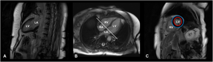
Cardiac magnetic resonance image acquisition protocol. T1-weighted, late gadolinium enhancement cardiac MR images of a 71-year-old female patient with Cushing’s disease, cured after successful neurosurgery. A Vertical long axis slice, coronal plane, two-chamber view. B Horizontal long axis slice, axial plane, four-chamber view. C Short-axis slice at the end of the diastole, sagittal plane. Red circle: endocardium; Blue circle: epicardium. LA left atrium, LV left ventricle, RA right atrium, RV right ventricle

Statistical analysis

Continuous variables are expressed as standard deviation (SD), median and 95% confidence interval (95%CI) as per data distribution, assessed through the Shapiro–Wilk test. Dichotomous variables are expressed as frequencies and percentages when relevant. According to variable distribution, the Student’s t-test or the non-parametric Mann–Whitney U test was performed to compare continuous variables between CS and NFAI and between cured and drug-controlled patients. Differences between groups regarding qualitative variables were evaluated by χ2 statistics. Bivariate correlations between numerical variables were analyzed using Pearson’s or Spearman’s correlation test, as appropriate. The statistical significance was set at p < 0.05. Statistical analyses were performed using SPSS 20.0 for MacOS (SPSS Inc.).

Results

Patient characteristics

The cohort characteristics are summarized in Table 1. Sixteen patients with CS (12 females, mean age 47 ± 12 years) and 15 with NFAI (7 females, mean age 55 ± 10 years) were enrolled during the study period. Twelve patients (75%) had been diagnosed with Cushing’s disease (CD), while four (25%) had ACTH-independent CS due to a cortisol-secreting adrenal adenoma.

Table 1 Baseline characteristics of patients with CS

At enrollment, eleven (69%) patients were cured and five (31%) had drug-treated CD. Among patients with CD, nine had previously undergone pituitary surgery, seven (58%) were cured, and five (42%) presented with a biochemically persistent disease. In the latter group, 3 patients had adequate biochemical control under medical therapy, whereas 2 patients were not entirely on target, because of low compliance and intolerance to medical treatments.

All patients with a cortisol-secreting adrenal adenoma had undergone unilateral adrenalectomy and were cured at the time of enrollment.

Table 2 details comorbidities and their therapies for CS and NFAI patients.

Table 2 Biochemical and clinical parameters in CS patients and NFAI

Biochemical and clinical evaluation

The main clinical and biochemical parameters are reported in Table 2. Sex, age, and BMI did not differ between the CS patients and NFAI. The two groups were similar concerning HbA1c, fasting insulin or homeostatic model assessment for insulin resistance, and lipid levels; fasting glucose levels were marginally lower in CS than in NFAI (p < 0.001). No differences were found in systolic and diastolic blood pressure, the prevalence of cardiometabolic complications or drugs (i.e., diagnosis of hypertension, dyslipidemia, obesity, and diabetes or prescriptions needed to control such comorbidities), suggesting that GC excess was resolved (or adequately controlled) at the time of enrollment for the great majority of patients.

Subgroup analysis of cardiac parameters in patients with Cushing’s syndrome

A subgroup analysis was performed to assess possible differences in cardiac parameters when the CS cohort was stratified according to disease status and cardiometabolic comorbidities (i.e., between patients with or without hypertension, glucose metabolism impairment, dyslipidemia, obesity), smoking, sex, and disease status.

Firstly, pharmacologically treated CS exhibited higher systolic and diastolic blood pressure levels than cured CS (p = 0.027), but no other differences were found regarding CMR parameters. Subgroup analysis according to the presence/absence of comorbidities revealed no significant effect on cardiac parameters, except for CS patients with impaired glucose tolerance, who showed a lower RV-EF compared with the remaining CS patients (p = 0.017).

Analyzing sex differences, male CS patients displayed higher RV-EDVi (p = 0.035) and RV-ESVi (p = 0.044), as well as a trend toward higher LV-EDVi (p = 0.067), LV-ESVi (p = 0.053) and LVMi (p = 0.066) compared to females. However, male CS patients only exhibited higher interventricular septum (IVS) thickness (p = 0.001) when compared to male and female reference ranges for the general population, age and sex-matched [36].

Comparison of cardiac parameters between patients and controls

A comparison of the main morphostructural and functional cardiac parameters between CS and NFAI in the left and the right ventricle is reported in Fig. 2 and Fig. 3, respectively. CMR cardiac morphology revealed an increased left ventricle-end systolic volume index (LV-ESVi) (31.0 ± 8.7 vs 24.1 ± 7.4, p = 0.027) in CS compared to NFAI (Fig. 2). Left ventricle mass index (LVMi) was also higher in CS (51.0 ± 11.8 vs 41.8 ± 6.9, p = 0.013) (Fig. 2), albeit none matched the criteria for left ventricular hypertrophy [37]. Regarding cardiac function, a trend toward lower left ventricle-ejection fraction (LV-EF) was measured in CS (57.1 ± 6.3 vs 61.9 ± 7.0, p = 0.056). Mirroring the alterations found in the left ventricle, higher indexed right ventricle-end systolic volume (RV-ESVi) (34.0 ± 7.7 vs 26.3 ± 6.0, p = 0.006) and right ventricle-end diastolic volume (RV-EDVi) (74.8 ± 14.2 vs 64.9 ± 9.3, p = 0.035), as well as lower right ventricle-ejection fraction (RV-EF) (54.6 ± 5.5 vs 59.5 ± 7.1, p = 0.044) were measured in patients with CS (Fig. 3). Dedicated T1 mapping technique did not reveal any difference between CS and NFAI, either before or after contrast administration. No patient had ECV greater than 30%, and no difference in ECV values was observed between groups.

Fig. 2
figure 2

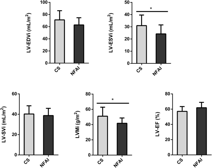
Comparative analysis of left ventricle parameters in Cushing’s syndrome and NFAI patients. Left ventricle morphological and functional cardiac parameters in patients with Cushing’s syndrome (gray bars) and patients with NFAI (black bars). Data are expressed as mean ± SD. *p < 0.05. CS Cushing’s syndrome, CNT NFAI, LV-EDVi Left Ventricle End-Diastolic Volume index, LV-ESVi Left Ventricle End-Systolic Volume index, LV-SVi Left Ventricle Stroke Volume index, LVMi Left Ventricular Mass index, LV-EF Left Ventricle Ejection Fraction

Fig. 3
figure 3

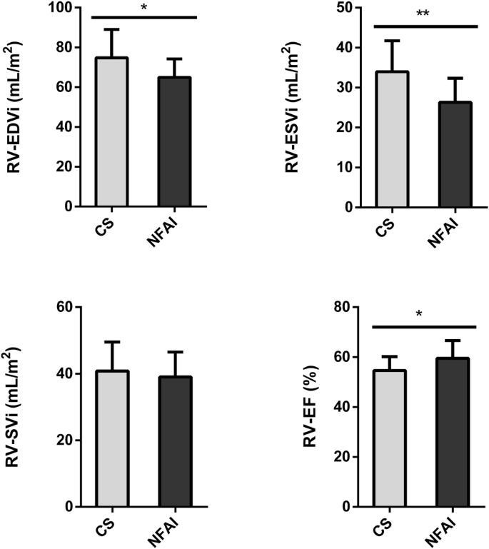
Comparative analysis of right ventricle parameters in Cushing’s syndrome and NFAI patients. Right ventricle morphological and functional cardiac parameters in patients with Cushing’s syndrome (gray bars) and patients with NFAI (black bars). Data are expressed as mean ± SD. *p < 0.05; **p < 0.01; CS Cushing’s syndrome, CNT NFAI, RV-EDVi Right Ventricle End-Diastolic Volume index, RV-ESVi Right Ventricle End-Systolic Volume index, RV-SVi Right Ventricle Stroke Volume index, RV-EF Right Ventricle Ejection Fraction

No significant correlations were found between CMR parameters and UFC x ULN (assessed at diagnosis or date of CMR evaluation), serum cortisol after dexamethasone suppression test (at diagnosis) or disease duration from diagnosis. An explicative summary of cardiac parameters is shown in Table 3.

Table 3 Cardiac parameters in CS patients and NFAI

Discussion

The current study reveals that exposure to endogenous GC excess induces a peculiar early remodeling of affected patients’ left and right ventricles, which can persist after CS remission and is independent of traditional cardiometabolic risk factors. Namely, the higher LV and RV ESVi and EDVi observed in patients exposed to GC excess is accompanied by higher LVMi. However, LV and RV ejection fractions are only mildly reduced, suggesting that a morphological impairment anticipates a performance dysfunction. The fact that such alterations occur rapidly in CS and are partially irreversible after remission advocates the use of CMR to improve the management of fatal cardiac complications in this rare endocrine disease.

Several echocardiographic studies have evaluated cardiac structure and function in patients with CS and found LV systolic and diastolic dysfunction [192138,39,40,41]. Albeit cardiac echocardiography is more practical in everyday clinical practice, CMR allows an evaluation of ventricular mass and volumes free of cardiac geometric assumption, ensuring a higher accuracy and reproducibility [2942].

Few controlled studies evaluating small cohorts have analyzed patients with CS using CMR [1826,27,28]. Kamenicky and coworkers compared 18 patients with active CS with 18 controls matched for age, sex, and BMI and found that patients had lower LV, RV, and left atrium ejection fractions, along with increased left and right ESVi and end-diastolic LV segmental thickness. Of note, successful treatment of CS was associated with an improvement in ventricular and atrial systolic performance [18]. A later study from the same group evaluated 23 patients with active CS and compared them with 27 controls matched for age, sex, and BMI, reporting increased left ventricular wall thickness, and reduced ventricular stroke volumes in patients [28]. A CMR study comparing CS patients with age and sex-matched controls showed that patients with active disease had higher LVMi than controls, as opposed to those in disease remission [27]. In all the studies mentioned above, patients and controls significantly differed in cardiovascular risk factors, with a worse cardiovascular profile in patients than controls. Conversely, our cohorts were largely homogeneous, without any significant difference between patients with CS and NFAI in glycometabolic profile, except for a surprisingly marginally lower fasting glucose levels in CS than in NFAI. This is likely because CS patients were either cured or drug-treated, and NFAI were comparable in terms of BMI and known CV risk factors.

As a result, the two groups did not significantly differ either in systolic and diastolic blood pressure levels or in the overall prevalence of cardiometabolic complications or the drugs prescribed to treat them. Nevertheless, our results confirmed the CMR findings of previous studies regarding higher cardiac volumes and mass and lower ejection fractions in patients with CS than in NFAI, advocating a direct effect of GC excess exposure in cardiac impairment beyond the known cardiovascular risk factors. Moreover, the results of the current study highlight the importance of a biventricular evaluation in this context, as opposed to most 2D-echocardiographic studies. Ultrasound measurement of RV volumes is challenging; therefore, most CS echocardiographic studies have mainly focused on the LV [192138,39,40,41], whereas CMR studies suggest an impairment in both left and right ventricles. The RV is anatomically and functionally different from the LV. In the absence of clear alterations in pulmonary resistance, our findings suggest RV involvement is a direct effect of GC excess on cardiomyocytes, whose receptors are equally expressed in left and right ventricles in donor hearts and dilated cardiomyopathy [43].

Cardiac morphological alterations in our cohort were not related to increased myocardial fibrosis, as we did not find any difference between patients and controls in T1 mapping evaluation, probably also due to the superimposable cardiometabolic profile of the two study groups. Albeit patients had non-significantly higher postcontrast T1 values, none had ECV values compatible with fibrosis. Similarly, Roux and coworkers evaluated 10 patients with active CD matched with 10 hypertensive and 10 healthy controls and performed a CMR study using the T1 mapping technique and found increased native myocardial T1 in CD, independently from hypertension, without differences in myocardial partition coefficient (λ) between groups. These results support the hypothesis of a potential role of T1 mapping in identifying early biomarkers of subclinical myocardial fibrosis in this disease [26].

Even though we didn’t find any significant correlation between indicators of hypercortisolism severity (UFC x ULN, disease duration) and CMR parameters, the independency of cardiac alterations from traditional cardiometabolic risk factors, claims a direct role of hypercortisolism on cardiac impairment, acting as a fingerprint of GC excess exposure. Our data point toward a persistent toxic effect on the heart, mediated directly through GC and/or mineralocorticoid receptors [21144], that produces changes in cardiac structure that are clinically silent but long-lasting, as if the heart retained a memory of GC excess exposure. The mineralocorticoid pathway increases collagen secretion by activating fibroblasts [45]. In addition, stimulating mineralocorticoid receptors decreases myocyte contractility and stimulates mitosis, resulting in myocardial hypertrophy and dysfunction [46]. However, previous data on mineralocorticoid antagonism in GC-induced hypertension did not prove convincing [47], disclosing the need for direct control of GC receptors (for example, via selective GC receptor antagonists such as relacorilant). Indeed, there is evidence supporting the role of GCs in driving alterations in vasoactive substances, thus impacting the balance between vasoconstriction and vasodilation (including catecholamines, nitric oxide, and atrial natriuretic peptide), as well as the activation of the renin-angiotensin system, leading to cardiac hypercontractility [1148].

The present study has shown more structural rather than functional changes at CMR in CS patients without evidence of fibrosis, thus suggesting the latter probably as a late phenomenon.

Additive to the direct role of cortisol, almost 60% of our patients presented hypertension, which could have contributed to the development of cardiac impairment. Similarly, the impact of other CS-related cardiovascular risk factors, such as visceral obesity, glucose intolerance and dyslipidemia, cannot be entirely ruled out.

Finally, our study showed for the first time that sex might affect cardiac morphological changes induced by GC excess. Male CS patients exhibited higher IVS thickness compared to females after adjusting for population age and sex reference ranges, independently from the prevalence of hypertension, supporting sex-related differences as observed in other cardiovascular diseases [4950]. Recently, a study by Wolf et al. showed that male sex was an independent predictor of increased epicardial and pericardial fat [28], which may play a role in the pathogenesis of CS cardiomyopathy. However, among CS patients, we did not find any differences in the prevalence of male and female hypogonadism (50% vs 42%, p = 1.000). Still, we can not exclude that estrogen exposure could have protected GC-related cardiomyopathy [51].

Very few studies have evaluated cardiac structure and function in CS using CMR, and this is a strength of the current study. However, it does have some limitations. The cross-sectional design and the lack of sample size in such a small and heterogeneous study population with different etiologies of endogenous CS, including both cured and well-controlled patients, might have underestimated the cardiac impairment. Anyway, considering that CS is a rare disease, we opted for a study design closer to a CS clinic’s real-life setting; this aspect represents a strength of this study. Nevertheless, according to the published evidence, as well as to our results, it is likely that cardiac dysfunction might persist in CS even after disease remission. Indeed, although our CS patients had higher biventricular volumes, the subgroup comparison between surgically cured and drug-treated patients revealed no differences in cardiac morphology or biochemical or cardiometabolic complications prevalence, althoghut the lack of standardization of the evaluation period. A previous paper found a significantly higher LVMi in active patients than in remission [27]. Anyway, longitudinal studies (baseline versus post-treatment) with larger population are needed to better clarify the reversibility of cardiac changes after treatment.

We propose a novel approach to cardiac disease in CS, going beyond the traditional cardiometabolic risk factors and evaluating both ventricles, preferably with CMR. Moreover, the present study highlights the importance of a sex-oriented approach in the management of CS complications, taking into account the sex-related differences in cardiac damage of these patients, for whom cardiac complications still represent the major cause of death, very often occurring during remission [2].

Conclusions

In CS biventricular cardiac remodeling associated with functional impairment, has been ascribed to a multifactorial pathogenesis. Our findings highlight the greater contribution of direct effect of GC excess exposure on myocardium than on cardiovascular risk factors, suggesting a sex-related differences in cardiac impairment. More importantly, the maladaptive change triggered by chronic exposure to GC excess, even if the latter is resolved, is persistent and clinically silent and could be detected though a more sensitive and precise approach with CMR.

Data availability

The datasets used and/or analyzed during the current study are available from the corresponding author upon reasonable request.

References

  1. R. Pivonello, M. De Leo, A. Cozzolino, A. Colao, The Treatment of Cushing’s Disease. Endocr. Rev. 36, 385–486 (2015). https://doi.org/10.1210/er.2013-1048

    Article CAS PubMed PubMed Central Google Scholar

  2. R. Pivonello, A.M. Isidori, M.C. De Martino, J. Newell-Price, B.M. Biller, A. Colao, Complications of Cushing’s syndrome: state of the art. Lancet Diabetes Endocrinol. 4, 611–629 (2016). https://doi.org/10.1016/S2213-8587(16)00086-3

    Article CAS PubMed Google Scholar

  3. J. Newell-Price, X. Bertagna, A.B. Grossman, L.K. Nieman, Cushing’s syndrome. Lancet 367, 1605–1617 (2006). https://doi.org/10.1016/S0140-6736(06)68699-6

    Article CAS PubMed Google Scholar

  4. R. Pivonello, M.C. De Martino, M. De Leo, G. Lombardi, A. Colao, Cushing’s Syndrome. Endocrinol. Metab. Clin. North Am. 37, 135–149 (2008). https://doi.org/10.1016/j.ecl.2007.10.010. ix

    Article CAS PubMed Google Scholar

  5. C. Steffensen, A.M. Bak, K.Z. Rubeck, J.O. Jorgensen, Epidemiology of Cushing’s syndrome. Neuroendocrinology 92, 1–5 (2010). https://doi.org/10.1159/000314297

    Article CAS PubMed Google Scholar

  6. R.N. Clayton, P.W. Jones, R.C. Reulen et al. Mortality in patients with Cushing’s disease more than 10 years after remission: a multicentre, multinational, retrospective cohort study. Lancet Diabetes Endocrinol. 4, 569–576 (2016). https://doi.org/10.1016/S2213-8587(16)30005-5

    Article PubMed Google Scholar

  7. E. Valassi, A. Tabarin, T. Brue et al. High mortality within 90 days of diagnosis in patients with Cushing’s syndrome: results from the ERCUSYN registry. Eur. J. Endocrinol. 181, 461–472 (2019). https://doi.org/10.1530/EJE-19-0464

    Article CAS PubMed Google Scholar

  8. V. Hasenmajer, E. Sbardella, F. Sciarra, M. Minnetti, A.M. Isidori, M.A. Venneri, The Immune System in Cushing’s Syndrome. Trends Endocrinol. Metab. 31, 655–669 (2020). https://doi.org/10.1016/j.tem.2020.04.004

    Article CAS PubMed Google Scholar

  9. M. Minnetti, V. Hasenmajer, E. Sbardella et al. Susceptibility and characteristics of infections in patients with glucocorticoid excess or insufficiency: the ICARO tool. Eur. J. Endocrinol. 187, 719–731 (2022). https://doi.org/10.1530/EJE-22-0454

    Article CAS PubMed PubMed Central Google Scholar

  10. M. De Leo, R. Pivonello, R.S. Auriemma et al. Cardiovascular disease in Cushing’s syndrome: heart versus vasculature. Neuroendocrinology 92, 50–54 (2010). https://doi.org/10.1159/000318566

    Article CAS PubMed Google Scholar

  11. A.M. Isidori, C. Graziadio, R.M. Paragliola et al. The hypertension of Cushing’s syndrome: controversies in the pathophysiology and focus on cardiovascular complications. J. Hypertens. 33, 44–60 (2015). https://doi.org/10.1097/HJH.0000000000000415

    Article CAS PubMed PubMed Central Google Scholar

  12. T. Mancini, B. Kola, F. Mantero, M. Boscaro, G. Arnaldi, High cardiovascular risk in patients with Cushing’s syndrome according to 1999 WHO/ISH guidelines. Clin. Endocrinol. 61, 768–777 (2004). https://doi.org/10.1111/j.1365-2265.2004.02168.x

    Article Google Scholar

  13. A.M. Isidori, M. Minnetti, E. Sbardella, C. Graziadio, A.B. Grossman, Mechanisms in endocrinology: The spectrum of haemostatic abnormalities in glucocorticoid excess and defect. Eur. J. Endocrinol. 173, R101–R113 (2015). https://doi.org/10.1530/EJE-15-0308

    Article CAS PubMed Google Scholar

  14. F. Fallo, G. Di Dalmazi, F. Beuschlein et al. Diagnosis and management of hypertension in patients with Cushing’s syndrome: a position statement and consensus of the Working Group on Endocrine Hypertension of the European Society of Hypertension. J. Hypertens. 40, 2085–2101 (2022). https://doi.org/10.1097/HJH.0000000000003252

    Article CAS PubMed Google Scholar

  15. O.M. Dekkers, E. Horvath-Puho, J.O. Jorgensen et al. Multisystem morbidity and mortality in Cushing’s syndrome: a cohort study. J. Clin. Endocrinol. Metab. 98, 2277–2284 (2013). https://doi.org/10.1210/jc.2012-3582

    Article CAS PubMed Google Scholar

  16. L.K. Nieman, B.M. Biller, J.W. Findling et al. Treatment of Cushing’s Syndrome: An Endocrine Society Clinical Practice Guideline. J. Clin. Endocrinol. Metab. 100, 2807–2831 (2015). https://doi.org/10.1210/jc.2015-1818

    Article CAS PubMed PubMed Central Google Scholar

  17. F.M. van Haalen, L.H. Broersen, J.O. Jorgensen, A.M. Pereira, O.M. Dekkers, Management of endocrine disease: Mortality remains increased in Cushing’s disease despite biochemical remission: a systematic review and meta-analysis. Eur. J. Endocrinol. 172, R143–R149 (2015). https://doi.org/10.1530/EJE-14-0556

    Article CAS PubMed Google Scholar

  18. P. Kamenicky, A. Redheuil, C. Roux et al. Cardiac structure and function in Cushing’s syndrome: a cardiac magnetic resonance imaging study. J. Clin. Endocrinol. Metab. 99, E2144–E2153 (2014). https://doi.org/10.1210/jc.2014-1783

    Article CAS PubMed PubMed Central Google Scholar

  19. M.L. Muiesan, M. Lupia, M. Salvetti et al. Left ventricular structural and functional characteristics in Cushing’s syndrome. J. Am. Coll. Cardiol. 41, 2275–2279 (2003). https://doi.org/10.1016/s0735-1097(03)00493-5

    Article PubMed Google Scholar

  20. A.M. Pereira, V. Delgado, J.A. Romijn, J.W. Smit, J.J. Bax, R.A. Feelders, Cardiac dysfunction is reversed upon successful treatment of Cushing’s syndrome. Eur. J. Endocrinol. 162, 331–340 (2010). https://doi.org/10.1530/EJE-09-0621

    Article CAS PubMed Google Scholar

  21. K.H. Yiu, N.A. Marsan, V. Delgado et al. Increased myocardial fibrosis and left ventricular dysfunction in Cushing’s syndrome. Eur. J. Endocrinol. 166, 27–34 (2012). https://doi.org/10.1530/EJE-11-0601

    Article CAS PubMed Google Scholar

  22. A. Colao, R. Pivonello, S. Spiezia et al. Persistence of increased cardiovascular risk in patients with Cushing’s disease after five years of successful cure. J. Clin. Endocrinol. Metab. 84, 2664–2672 (1999). https://doi.org/10.1210/jcem.84.8.5896

    Article CAS PubMed Google Scholar

  23. A. Faggiano, R. Pivonello, S. Spiezia et al. Cardiovascular risk factors and common carotid artery caliber and stiffness in patients with Cushing’s disease during active disease and 1 year after disease remission. J Clin Endocrinol. Metab. 88, 2527–2533 (2003). https://doi.org/10.1210/jc.2002-021558

    Article CAS PubMed Google Scholar

  24. A. Kanzaki, M. Kadoya, S. Katayama, H. Koyama, Cardiac Hypertrophy and Related Dysfunctions in Cushing Syndrome Patients-Literature Review. J. Clin. Med. 11, 7035 (2022). https://doi.org/10.3390/jcm11237035

    Article CAS PubMed PubMed Central Google Scholar

  25. M. Salerno, B. Sharif, H. Arheden et al. Recent Advances in Cardiovascular Magnetic Resonance: Techniques and Applications. Circ. Cardiovasc. Imaging 10, e003951 (2017). https://doi.org/10.1161/CIRCIMAGING.116.003951

    Article PubMed PubMed Central Google Scholar

  26. C. Roux, N. Kachenoura, Z. Raissuni et al. Effects of cortisol on the heart: characterization of myocardial involvement in cushing’s disease by longitudinal cardiac MRI T1 mapping. J. Magn. Reson. Imaging 45, 147–156 (2017). https://doi.org/10.1002/jmri.25374

    Article PubMed Google Scholar

  27. F. Maurice, B. Gaborit, C. Vincentelli et al. Cushing Syndrome Is Associated With Subclinical LV Dysfunction and Increased Epicardial Adipose Tissue. J. Am. Coll. Cardiol. 72, 2276–2277 (2018). https://doi.org/10.1016/j.jacc.2018.07.096

    Article PubMed Google Scholar

  28. P. Wolf, B. Marty, K. Bouazizi et al. Epicardial and Pericardial Adiposity Without Myocardial Steatosis in Cushing Syndrome. J. Clin. Endocrinol. Metab. 106, 3505–3514 (2021). https://doi.org/10.1210/clinem/dgab556

    Article PubMed Google Scholar

  29. M. Moustaki, G. Markousis-Mavrogenis, A. Vryonidou, S.A. Paschou, S. Mavrogeni, Cardiac disease in Cushing’s syndrome. Emphasis on the role of cardiovascular magnetic resonance imaging. Endocrine 83, 548–558 (2024). https://doi.org/10.1007/s12020-023-03623-0

    Article CAS PubMed Google Scholar

  30. M. Fleseriu, R. Auchus, I. Bancos et al. Consensus on diagnosis and management of Cushing’s disease: a guideline update. Lancet Diabetes Endocrinol. 9, 847–875 (2021). https://doi.org/10.1016/S2213-8587(21)00235-7

    Article PubMed PubMed Central Google Scholar

  31. J.M. Hinojosa-Amaya, D. Cuevas-Ramos, The definition of remission and recurrence of Cushing’s disease. Best. Pract. Res. Clin. Endocrinol. Metab. 35, 101485 (2021). https://doi.org/10.1016/j.beem.2021.101485

    Article PubMed Google Scholar

  32. M. Fassnacht, S. Tsagarakis, M. Terzolo et al. European Society of Endocrinology clinical practice guidelines on the management of adrenal incidentalomas, in collaboration with the European Network for the Study of Adrenal Tumors. Eur. J. Endocrinol. 189, G1–G42 (2023). https://doi.org/10.1093/ejendo/lvad066

    Article PubMed Google Scholar

  33. R. Pofi, E. Giannetta, N. Galea et al. Diabetic Cardiomiopathy Progression is Triggered by miR122-5p and Involves Extracellular Matrix: A 5-Year Prospective Study. JACC Cardiovasc. Imaging. 14, 1130–1142 (2021). https://doi.org/10.1016/j.jcmg.2020.10.009

    Article PubMed Google Scholar

  34. J.C. Moon, D.R. Messroghli, P. Kellman et al. Myocardial T1 mapping and extracellular volume quantification: a Society for Cardiovascular Magnetic Resonance (SCMR) and CMR Working Group of the European Society of Cardiology consensus statement. J. Cardiovasc. Magn. Reson. 15, 92 (2013). https://doi.org/10.1186/1532-429X-15-92

    Article PubMed PubMed Central Google Scholar

  35. P. Haaf, P. Garg, D.R. Messroghli, D.A. Broadbent, J.P. Greenwood, S. Plein, Cardiac T1 Mapping and Extracellular Volume (ECV) in clinical practice: a comprehensive review. J. Cardiovasc. Magn. Reson. 18, 89 (2016). https://doi.org/10.1186/s12968-016-0308-4

    Article PubMed PubMed Central Google Scholar

  36. N. Kawel-Boehm, S.J. Hetzel, B. Ambale-Venkatesh et al. Reference ranges (“normal values”) for cardiovascular magnetic resonance (CMR) in adults and children: 2020 update. J. Cardiovasc. Magn. Reson. 22, 87 (2020). https://doi.org/10.1186/s12968-020-00683-3

    Article PubMed PubMed Central Google Scholar

  37. R. Janardhanan, C.M. Kramer, Imaging in hypertensive heart disease. Expert Rev. Cardiovasc. Ther. 9, 199–209 (2011). https://doi.org/10.1586/erc.10.190

    Article CAS PubMed PubMed Central Google Scholar

  38. M. Baykan, C. Erem, O. Gedikli et al. Assessment of left ventricular diastolic function and Tei index by tissue Doppler imaging in patients with Cushing’s Syndrome. Echocardiography 25, 182–190 (2008). https://doi.org/10.1111/j.1540-8175.2007.00572.x

    Article PubMed Google Scholar

  39. N.A. Bayram, R. Ersoy, C. Aydin et al. Assessment of left ventricular functions by tissue Doppler echocardiography in patients with Cushing’s disease. J. Endocrinol. Invest. 32, 248–252 (2009). https://doi.org/10.1007/BF03346461

    Article CAS PubMed Google Scholar

  40. P.M. Toja, G. Branzi, F. Ciambellotti et al. Clinical relevance of cardiac structure and function abnormalities in patients with Cushing’s syndrome before and after cure. Clin. Endocrinol. 76, 332–338 (2012). https://doi.org/10.1111/j.1365-2265.2011.04206.x

    Article CAS Google Scholar

  41. F. Tona, M. Boscaro, M. Barbot et al. New insights to the potential mechanisms driving coronary flow reserve impairment in Cushing’s syndrome: A pilot noninvasive study by transthoracic Doppler echocardiography. Microvasc. Res. 128, 103940 (2020). https://doi.org/10.1016/j.mvr.2019.103940

    Article CAS PubMed Google Scholar

  42. A.C. Armstrong, S. Gidding, O. Gjesdal, C. Wu, D.A. Bluemke, J.A. Lima, LV mass assessed by echocardiography and CMR, cardiovascular outcomes, and medical practice. JACC Cardiovasc. Imaging 5, 837–848 (2012). https://doi.org/10.1016/j.jcmg.2012.06.003

    Article PubMed PubMed Central Google Scholar

  43. C. Sylven, E. Jansson, P. Sotonyi, F. Waagstein, T. Barkhem, M. Bronnegard, Cardiac nuclear hormone receptor mRNA in heart failure in man. Life Sci. 59, 1917–1922 (1996). https://doi.org/10.1016/s0024-3205(96)00539-5

    Article CAS PubMed Google Scholar

  44. A.S. Mihailidou, T.Y. Loan Le, M. Mardini, J.W. Funder, Glucocorticoids activate cardiac mineralocorticoid receptors during experimental myocardial infarction. Hypertension 54, 1306–1312 (2009). https://doi.org/10.1161/HYPERTENSIONAHA.109.136242

    Article CAS PubMed Google Scholar

  45. G. Fujisawa, R. Dilley, M.J. Fullerton, J.W. Funder, Experimental cardiac fibrosis: differential time course of responses to mineralocorticoid-salt administration. Endocrinology 142, 3625–3631 (2001). https://doi.org/10.1210/endo.142.8.8339

    Article CAS PubMed Google Scholar

  46. X. Feng, S.A. Reini, E. Richards, C.E. Wood, M. Keller-Wood, Cortisol stimulates proliferation and apoptosis in the late gestation fetal heart: differential effects of mineralocorticoid and glucocorticoid receptors. Am. J. Physiol. Regul. Integr. Comp. Physiol. 305, R343–R350 (2013). https://doi.org/10.1152/ajpregu.00112.2013

    Article CAS PubMed PubMed Central Google Scholar

  47. P.M. Williamson, J.J. Kelly, J.A. Whitworth, Dose-response relationships and mineralocorticoid activity in cortisol-induced hypertension in humans. J. Hypertens. Suppl. 14, S37–S41 (1996)

    CAS PubMed Google Scholar

  48. S.L. Ong, J.A. Whitworth, How do glucocorticoids cause hypertension: role of nitric oxide deficiency, oxidative stress, and eicosanoids. Endocrinol. Metab. Clin. North. Am. 40, 393–407 (2011). https://doi.org/10.1016/j.ecl.2011.01.010

    Article CAS PubMed Google Scholar

  49. A. De Bellis, G. De Angelis, E. Fabris, A. Cannata, M. Merlo, G. Sinagra, Gender-related differences in heart failure: beyond the “one-size-fits-all” paradigm. Heart Fail. Rev. 25, 245–255 (2020). https://doi.org/10.1007/s10741-019-09824-y

    Article PubMed Google Scholar

  50. R. Pofi, E. Giannetta, T. Feola et al. Sex-specific effects of daily tadalafil on diabetic heart kinetics in RECOGITO, a randomized, double-blind, placebo-controlled trial. Sci. Transl. Med. 14, eabl8503 (2022). https://doi.org/10.1126/scitranslmed.abl8503

    Article CAS PubMed Google Scholar

  51. D. Gianfrilli, R. Pofi, T. Feola, A. Lenzi, E. Giannetta, The Woman’s Heart: Insights into New Potential Targeted Therapy. Curr. Med. Chem. 24, 2650–2660 (2017). https://doi.org/10.2174/0929867324666161118121647

    Article CAS PubMed Google Scholar

Download references

Funding

This work was supported by the PRecisiOn Medicine to Target Frailty of Endocrine-metabolic Origin (PROMETEO) project (NET-2018-12365454) by the Ministry of Health and the European Union – NextGenerationEU through the Italian Ministry of University and Research under PNRR – M4C2-I1.3 Project PE_00000019 “HEAL ITALIA” to Andrea Isidori CUP B53C22004000006. Open access funding provided by Università degli Studi di Roma La Sapienza within the CRUI-CARE Agreement.

Author information

Author notes

  1. These authors contributed equally: Tiziana Feola, Alessia Cozzolino
  2. These authors jointly supervised this work: Andrea M. Isidori, Elisa Giannetta

Authors and Affiliations

  1. Department of Experimental Medicine, Sapienza University of Rome, Rome, Italy

    Tiziana Feola, Alessia Cozzolino, Dario De Alcubierre, Federica Campolo, Andrea M. Isidori & Elisa Giannetta

  2. Neuroendocrinology, Neuromed Institute, IRCCS, Pozzilli, Italy

    Tiziana Feola & Dario De Alcubierre

  3. Oxford Centre for Diabetes, Endocrinology, and Metabolism, Churchill Hospital, Oxford University Hospitals, NHS Trust, Oxford, UK

    Riccardo Pofi

  4. Department of Radiological Sciences, Oncology and Pathology, Sapienza University of Rome, Rome, Italy

    Nicola Galea & Carlo Catalano

  5. Dipartimento di Medicina Clinica e Chirurgia, Università Federico II di Napoli, Naples, Italy

    Chiara Simeoli, Nicola Di Paola & Rosario Pivonello

  6. Centre for Rare Diseases (ENDO-ERN accredited), Policlinico Umberto I, Rome, Italy

    Andrea M. Isidori

Contributions

A.M.I., E.G., T.F., A.C. contributed to the idea and design of the study, analysis of the data, and writing of the manuscript. D.D.A., R.P., C.S., N.D.P., F.C. and R.P.i. contributed to the design of the study, analysis of the data, and revision of the report. T.F., A.C., D.D.A., N.G. and C.C. analyzed the data. All authors contributed to the interpretation of the data and the writing of the paper. All authors read and approved the final manuscript.

Corresponding authors

Correspondence to Andrea M. Isidori or Elisa Giannetta.

Ethics declarations

Conflict of interest

RPi has received research support to Università Federico II di Napoli as a principal investigator for clinical trials from Novartis Pharma, Recordati, Strongbridge Biopharma, Corcept Therapeutics, HRA Pharma, Shire, Takeda, Neurocrine Biosciences, Camurus AB, and Pfizer, has received research support to Università Federico II di Napoli from Pfizer, Ipsen, Novartis Pharma, Strongbridge Biopharma, Merk Serono, and Ibsa, and received occasional consulting honoraria from Novartis Pharma, Recordati, Strongbridge Biopharma, HRA Pharma, Crinetics Pharmaceuticals, Corcept Therapeutics, Pfizer, and Bresmed Health Solutions. AMI has been a consultant for Novartis, Takeda, Recordati, and Sandoz companies and has received unconditional research grants from Shire, IPSEN, and Pfizer. All the other authors have nothing to disclose.

Consent to publish

The authors affirm that human research participants provided informed consent for publication of the images in Fig. 1.

Ethics approval and conset to participate

All patients provided written informed consent after fully explaining the purpose and nature of all procedures used. The study was approved by the Ethical Committee of Policlinico Umberto I (ref. number 4245). The study has been performed according to the ethical standards of the 1964 Declaration of Helsinki and its later amendments.

Additional information

Publisher’s note Springer Nature remains neutral with regard to jurisdictional claims in published maps and institutional affiliations.

About this article

Cite this article

Feola, T., Cozzolino, A., De Alcubierre, D. et al. Cardiac magnetic resonance reveals biventricular impairment in Cushing’s syndrome: a multicentre case-control study. Endocrine (2024). https://doi.org/10.1007/s12020-024-03856-7

Download citation

A More Accurate Diagnosis of Cushing’s Syndrome

Cushing’s syndrome is associated with excessive cortisol production and, if left untreated, can result in severe complications, such as heart attacks, strokes, and type 2 diabetes. To diagnose this condition, a dexamethasone suppression test is commonly performed.

Various factors, such as metabolic rate and interactions with other medications, can affect test efficacy. Therefore, it is crucial to measure the concentration of dexamethasone concurrently with cortisol to avoid false-positive results.

To address this issue, a team of researchers at the University of Turin, led by Professor Giulio Mengozzi in the Department of Medical Sciences, has developed a liquid chromatography-tandem mass spectrometry method.

This new method enables the simultaneous quantification of cortisol, cortisone, dexamethasone, and six additional exogenous corticosteroids, leading to a more accurate diagnosis of Cushing’s syndrome.

The symptoms of Cushing’s syndrome

Cushing’s syndrome is a medical condition characterized by an abnormal and prolonged increase in cortisol production, typically affecting females between the ages of 30 and 50.1

While the issue may originate from within the body (endogenous), it is more commonly caused by external factors, such as the use of glucocorticoid medications.

Visible symptoms of Cushing’s syndrome include weight gain, an accumulation of fat around the base of the neck, a fatty hump between the shoulders, the appearance of a “moon face”, and easy bruising. However, not all individuals with the syndrome exhibit these symptoms, rendering diagnosis challenging. Without timely treatment, Cushing’s syndrome can lead to severe complications, including heart attack, stroke, blood clots in the legs and lungs, increased susceptibility to infections, memory loss, and type 2 diabetes.

Dexamethasone testing

A commonly used method for diagnosing Cushing’s syndrome is the dexamethasone suppression test (DST), which measures the adrenal gland’s response to adrenocorticotropic hormone (ACTH).

ACTH regulates cortisol levels in the blood plasma and stimulates the adrenal cortex to produce cortisol. When cortisol levels increase, ACTH secretion is suppressed. Dexamethasone, a synthetic steroid similar to cortisol, is administered during the DST to lower ACTH levels.

DSTs are available in low-dose (LDDST) and high-dose (HDDST). They can be performed overnight or over two days.

LDDSTs are used initially to diagnose Cushing’s syndrome. If the result is positive, HDDSTs help classify the disease as ACTH-dependent or independent. These tests are typically conducted in the following manner.2

A more accurate diagnosis of Cushing’s syndrome

Cortisol is a steroid hormone of the glucocorticoid class made by the adrenal glands.

Image Credit: Shutterstock/Kateryna Kon

LDDST

  • Overnight protocol: 1 mg of dexamethasone is administered at 11:00 pm, and the serum cortisol levels are measured at 8:00 am the following morning.
  • Two-day protocol: serum cortisol levels are measured at 8:00 am and 0.5 mg of dexamethasone is administered every six hours (9:00 am, 3:00 pm, 9:00 pm, 3:00 am) for two days, totalling 4 mg. Serum cortisol levels are then measured at 9:00 am, six hours after the last dose has been delivered.

HDDST

  • Overnight protocol: baseline serum cortisol or 24-hour urinary free cortisol (UFC) is measured in the morning, and 8 mg of dexamethasone is given at 11.00 pm. Cortisol level in blood is then measured at 8.00 am the following morning.
  • Two-day protocol: Baseline serum cortisol or 24-hour UFC is measured at 8:00 am; 2 mg of dexamethasone is administered every six hours (9:00 am, 3:00 pm, 9:00 pm, 3:00 am) for two days, totaling 16 mg, in tandem with the collection of urine for UFC measurements. Serum cortisol levels are measured at 9:00 am, six hours after the last dose.

Patients whose pituitary glands produce excessive amounts of ACTH will exhibit an abnormal response to the low-dose test but a normal reaction to the high-dose test.

During the LDDST, cortisol levels should decrease following the administration of dexamethasone, and a cut-off value of below 18 ng/mL is recommended to distinguish a healthy response from an unhealthy one.

For the HDDST, a decrease in urine-free cortisol (UFC) or serum cortisol greater than 50% indicates the presence of ACTH-dependent Cushing’s syndrome. This rule applies to both the overnight LDDST and the two-day HDDST methods.

Measuring cortisol levels

Chemiluminescence immunoassay (CLIA) is a widely used method for measuring cortisol and other steroids due to its simplicity, automation, and good sensitivity.

However, it has some drawbacks, including cross-reactivity leading to overestimation of target analyte levels, non-standardization of kits, and the inability to measure more than one analyte per analysis. This is particularly problematic since studies indicate that measuring dexamethasone in combination with cortisol can reduce the number of false-positive DST results and improve interpretability. 3,4,5

Liquid chromatography-tandem mass spectrometry (LC-MS/MS) has emerged as a popular alternative to CLIA for DSTs due to its ability to measure multiple analytes simultaneously and its superior specificity.

Analytes are separated via LC, and their concentrations are measured by MS, with triple quadrupole MS configurations commonly used for this purpose. This technique provides the ability to measure multiple analytes simultaneously, along with higher accuracy and sensitivity than CLIA.

Increased ease of use and accuracy

In the Division of Endocrinology, Diabetes, and Metabolism at the University of Turin, a team has developed an LC-MS/MS technique for simultaneous quantifying cortisol, cortisone, dexamethasone, and six other exogenous corticosteroids in serum/plasma samples.6

This method can be readily applied in any clinical laboratory equipped with a mass spectrometer and is effective in DSTs, enabling precise measurements of the target analytes in a single chromatographic run (Figure 1).

Sample preparation (1 Hour)

  1. Dilute 200 μL of the serum/plasma sample with 200 μL of water.
  2. Perform supported liquid extraction, manually transferring 400 μL of sample to a microplate.
  3. Apply positive pressure using Tecan Resolvex® A200 automated positive pressure processor.
  4. Elute with 700 μL of methyl tert-butyl ether.
  5. Evaporate and reconstitute in H2 O/MeOH (1:1, v/v).
  6. Agitate.

LC-MS/MS analysis (10 Minutes)

  • LC column: C18 (100 × 2.1 mm, 1.7 μm)
  • Flow rate: 400 μL/min
  • Temperature: 30 °C
  • Injection volume: 20 μL
  • Mobile phase A: H2O + 0.2 mM ammonium fluoride
  • Mobile phase B: acetonitrile
  • Elution programme: Table 1.

The study demonstrated a strong correlation between the results obtained from the newly developed LC-MS/MS method and those obtained using a commercially available CE IVD-marked Steroid Panel LC-MS* kit (Tecan).

The Tecan kit enables simultaneous dexamethasone, cortisol, and cortisone measurement and includes all the necessary components for easy implementation, such as calibrators and controls. The samples are prepared using solid-phase extraction (SPE), which can be semi-automatically performed on a Resolvex® A200 positive pressure processor (Tecan). The kit can measure 15 other steroids in the core steroid metabolism pathway due to the effectiveness of the SPE process.

Table 1. LC gradient elution programme. Source: Tecan

Time (min) Mobile phase A (%) Mobile phase B (%)
0 90 10
0.5 65 35
4.5 65 35
4.51 35 65
6.0 2 98
8.0 2 98
8.01 90 10
10.0 90 10

* In USA: for research use only. Not for use in diagnostic procedures. Product availability and regulatory status may vary from country to country. Consult with your Tecan associate for further information.

A more accurate diagnosis of Cushing’s syndrome

Figure 1. Example chromatogram of the Steroid Panel LC-MS internal standard – run 1. ESI, electrospray ionization; 1, aldosterone; 2, cortisone; 3, dehydro-epiandrosterone sulfate; 4, cortisol; 5, 21-deoxycortisol; 6, corticosterone; 7, dexamethasone; 8, 11-deoxycortisol; 9, androstenedione; 10, 11-deoxycorticosterone; 11, testosterone; 12, dehydroepiandrosterone; 13, 17-hydroxyprogesterone; 14, dihydrotestosterone; 15, progesterone.

Image Credit: Tecan

Summary

Early diagnosis of Cushing’s syndrome is critical to prevent potentially fatal complications. A reliable method for reducing the number of false positives in DSTs involves the simultaneous measurement of cortisol and dexamethasone levels, which can be accurately achieved using LC-MS/MS.

The LC-MS/MS method described in this article enables the simultaneous measurement of multiple analytes, such as cortisol, cortisone, and dexamethasone, in serum or plasma.

This analytical approach can provide clinical laboratories with a straightforward method for performing DSTs, and the commercially available kit can ensure consistent and dependable results.

References and further reading

  1. Cushing’s syndrome [website]. National Institute of Diabetes and Digestive and Kidney Diseases 2018 (https://www.niddk.nih.gov/health-information/endocrine-diseases/cushings-syndrome).
  2. Dogra P, Vijayashankar NP. Dexamethasone suppression test. StatPearls 2022, 8 August (https://www.ncbi.nlm.nih.gov/books/NBK542317 ).
  3. Ceccato F, Artusi C, Barbot M, et al. Dexamethasone measurement during low-dose suppression test for suspected hypercortisolism: threshold development with and validation. J Endocrinol Invest 2020;43(8):1105–1113. doi: 10.1007/s40618-020-01197-6.
  4. Roper SM. Yield of serum dexamethasone measurement for reducing false-positive results of low-dose dexamethasone suppression testing. J Appl Lab Med 2021;6(2):480–485. doi: 10.1093/jalm/jfaa193.
  5. Fleseriu M, Auchus R, Bancos I, et al. Consensus on diagnosis and management of Cushing’s disease: a guideline update. Lancet Diabetes Endocrinol 2021;9(12):847–875. doi: 10.1016/S2213- 8587(21)00235-7.
  6. Ponzetto F, Parasiliti-Caprino M, Settanni F, et al. Simultaneous measurement of cortisol, cortisone, dexamethasone and additional exogenous corticosteroids by rapid and sensitive LC-MS/MS analysis. Molecules 2022;28(1):248. doi: 10.3390/molecules28010248.

From https://www.news-medical.net/whitepaper/20240524/A-more-accurate-diagnosis-of-Cushinge28099s-syndrome.aspx

Venous Thromboembolism in Cushing Syndrome

Abstract

Background

Patients with Cushing syndrome (CS) are at increased risk of venous thromboembolism (VTE).

Objective

The aim was to evaluate the current management of new cases of CS with a focus on VTE and thromboprophylaxis.

Design and methods

A survey was conducted within those that report in the electronic reporting tool (e-REC) of the European Registries for Rare Endocrine Conditions (EuRRECa) and the involved main thematic groups (MTG’s) of the European Reference Networks for Rare Endocrine Disorders (Endo-ERN) on new patients with CS from January 2021 to July 2022.

Results

Of 222 patients (mean age 44 years, 165 females), 141 patients had Cushing disease (64%), 69 adrenal CS (31%), and 12 patients with ectopic CS (5.4%). The mean follow-up period post-CS diagnosis was 15 months (range 3–30). Cortisol-lowering medications were initiated in 38% of patients. One hundred fifty-four patients (69%) received thromboprophylaxis (including patients on chronic anticoagulant treatment), of which low-molecular-weight heparins were used in 96% of cases. VTE was reported in six patients (2.7%), of which one was fatal: two long before CS diagnosis, two between diagnosis and surgery, and two postoperatively. Three patients were using thromboprophylaxis at time of the VTE diagnosis. The incidence rate of VTE in patients after Cushing syndrome diagnosis in our study cohort was 14.6 (95% CI 5.5; 38.6) per 1000 person-years.

Conclusion

Thirty percent of patients with CS did not receive preoperative thromboprophylaxis during their active disease stage, and half of the VTE cases even occurred during this stage despite thromboprophylaxis. Prospective trials to establish the optimal thromboprophylaxis strategy in CS patients are highly needed.

Significance statement

The incidence rate of venous thromboembolism in our study cohort was 14.6 (95% CI 5.5; 38.6) per 1000 person-years. Notably, this survey showed that there is great heterogeneity regarding time of initiation and duration of thromboprophylaxis in expert centers throughout Europe.

Introduction

Endogenous hypercortisolism (Cushing syndrome, CS) is a rare disorder with an estimated incidence of 0.2–5.0 cases per million inhabitants per year in various populations, whereas its prevalence is close to 39–79 cases per million (12). The majority of cases are adrenocorticotropic hormone (ACTH) dependent, of which a pituitary corticotrope adenoma (Cushing disease, CD) is the most prevalent cause, whereas ACTH-secreting non-pituitary tumors (ectopic ACTH and corticotropin-releasing hormone syndrome secretion) are responsible for about 5–10% of cases. ACTH-independent cases of CS (adrenal adenomas or uni- or bilateral adrenal hyperplasia) account for the remaining 20% of cases (13).

It is well-known that endogenous hypercortisolism is associated with increased morbidity and mortality (456). This increased risk is mainly driven by cardiovascular events, including venous thromboembolic events (VTEs) such as pulmonary embolism (PE) and deep vein thrombosis (DVT). It has been demonstrated that the primary risk factors associated with VTE include older age (>69 years), reduced mobility, acute severe infections, previous cardiovascular events, higher midnight plasma cortisol levels, and shorter activated partial thromboplastin time (7). Additionally, a recent analysis of the ERCUSYN database found a higher prevalence of VTE among male patients, patients with a history of multiple surgeries, and those with high urinary cortisol levels (8). Several studies have observed an increased risk of VTE in patients with endogenous hypercortisolism even long after successful treatment. A study showed that the VTE incidence is almost seven times higher in the years before diagnosing endogenous hypercortisolism and almost 17 times higher in the first year after diagnosis; this incidence remains increased in the initial months following successful treatment (9). This results in an increased incidence rate of 14.6 per 1000 person-years for VTE in patients with endogenous hypercortisolism compared to the general population (10). The cortisol-induced hypercoagulability is thought to be partially caused by activation of the coagulation cascade with an increase in, e.g. von Willebrand factor, fibrinogen, and factor VIII concentrations (1112), impaired fibrinolysis (4) and endothelial dysfunction (13). Changes in pro- and anticoagulant factors may persist after successful surgery or medical therapy for at least several months (1415).

Given the lack of evidence from clinical trials, there is a large practice variation regarding thromboprophylaxis management and perioperative medical treatment in patients with endogenous hypercortisolism, even among reference centers that have obtained specific national and international accreditation for the diagnosis and treatment of CS (16). To further map local practice patterns and associated VTE complications in CS, we performed a study across the European Reference Network on Rare Endocrine Conditions (Endo-ERN) expert centers using the European Registries for Rare Endocrine Conditions (EuRRECa), and the contributors to the relevant main thematic groups (MTGs), i.e. Adrenal (one) and Pituitary (six) of the Endo-ERN.

Methods

The main objective of this study was to collect epidemiological and routine clinical data on new CS cases reported on the EuRRECa electronic reporting tool (e-REC) and Endo-ERN with a focus on VTE and thromboprophylaxis.

EuRRECa was constructed to support the needs of Endo-ERN, maximizing the opportunity for all patients, healthcare professionals, and researchers to participate and use high-quality, patient-centered registries for these rare conditions. The two platforms of the EuRRECa project encompass the Core registry, which collects a common dataset and clinician- and patient-reported outcomes, and an electronic surveillance system, the e-Reporting on Rare Endocrine Conditions (e-REC) program (17).

e-REC is a program that monthly captures the number of new cases of rare endocrine conditions seen at the participating centers.

e-REC is used for continuous monitoring of the expert centers of ERNs (Endo-ERN, ERN BOND), for mapping expert centers not only within European Union, for understanding the occurrence of the rare endocrine and bone conditions, and for conducting secondary surveys.

Because e-REC only provides a number of cases with a specific diagnosis without any personal data, there is no informed consent needed. e-REC is open to Endo-ERN and other centers involved in the care of patients with rare endocrine conditions.

Secondary survey

Secondary surveys (https://eurreb.eu/registries/e-rec/secondary-survey/) on e-REC-reported cases allow for the collection of well-defined routine clinical data for quality assurance and for understanding the clinical presentation of the reported condition. No personally identifiable data, such as date of birth, date of surgery, date of VTE, or exact laboratory tests, were collected.

First, the e-REC team sorted e-REC IDs of patients with endogenous hypercortisolism (ORPHA443287, ORPHA1501, ORPHA99408, ORPHA96253) reported between January 2021 and July 2022. Then the centers were provided with the list of IDs and queried to revisit these cases and to add clinical data to the online questionnaire. The survey questionnaire utilized Webropol survey, a secure online tool endorsed and supported by NHS Greater Glasgow & Clyde and NHS Scotland. The use of e-REC and secondary surveys was approved by the institutional board of the Leiden University Medical Center, and participating centers were advised to seek local approval if needed.

In addition, healthcare providers (not reporting in e-REC) of the relevant main thematic groups (‘Adrenal’ and ‘Pituitary’) of Endo-ERN were queried regarding any of their reported new encounters with a confirmed diagnosis of CS from January 2021 to July 2022. Patients with suspected but not confirmed CS were excluded (according to the current guideline) (18).

VTE in CS survey

The survey was open for entry from October 2022 to June 2023. Follow-up started on the date of initial CS diagnosis (within the period of interest – January 2021 till July 2022) and ended when an endpoint of interest occurred (VTE, bleeding, death) or on the date of filling in the questionnaire, whichever came first.

A survey was designed consisting of questions on the occurrence of VTE, and if so, additional questions assessed risk factors of VTE, treatment regimens, and VTE complications. Questions included data about relevant co-morbidities and the different items of the Cushing severity index (CSI) – a validated score for reliable clinometric evaluation of severity in endogenous hypercortisolism (19) using eight different parameters (fat distribution, skin lesions, muscle weakness, mood disorders, hypertension, diabetes mellitus, hypokalemia, and sex-related disturbances), each one graded from 0 to 2 with a maximum score of 16. These components enabled the calculation of the CSI score of all subjects. For the full questionnaire, see Annex 1 (see section on supplementary materials given at the end of this article).

Statistical analyses

Continuous data are presented as mean ± s.d. (range) and were compared using ANOVA. All the other values, if not normally distributed, are expressed as median with interquartile range (IQR) and compared using ANCOVA. Statistical analysis was performed using SPSS version 25.0.

The individual person-time was calculated based on the dates of reporting in e-Rec and filling in the survey and on the date of VTE. Incidence rates for VTE were calculated by dividing the observed number of VTE cases within the study period by the sum of individual person-years and were presented with accompanying 95% CI. Any VTE occurring before diagnosis was ignored in the estimation of the incidence rate.

Results

Patient characteristics

The survey was completed by 35 clinicians in 20 centers from six countries (Fig. 1). Within the 18-month study period, a total of 222 new patients were reported with endogenous hypercortisolism. The mean follow-up period was 15 ± 8 months (range 3–30). The total number of person-years was 274. Table 1 shows the clinical and demographic characteristics of patients with CS.

Figure 1View Full Size
Figure 1

Overview of countries responding to the survey.

Citation: Endocrine Connections 13, 6; 10.1530/EC-24-0046

Table 1Clinical and demographic characteristics of patients with Cushing syndrome of different origin.

Demographic/clinical variable Cushing disease Adrenal Cushing syndrome Ectopic Cushing syndrome Total
Number of patients: n (%) 141 (63.5%) 69 (31.1%) 12 (5.4%) 222 (100%)
Age (years): median (IQR) (range) 43 (22.5) (7–79) 46 (25.5) (3–80) 48 (37) (22–77) 43 (25) (3–80)
Female: n (%) 105 (74.4%) 54 (78.2%) 6 (50%) 165 (74.3%)
СSI: mean ± s.d. 5.77 ± 2.88 4.81 ± 2.72 8.5 ± 2.87 5.6 ± 2.9
Number of comorbidities: mean ± s.d. 1.9 ± 1.58 1.97 ± 1.39 2.17 ± 1.7 1.93 ± 1.53
Obesity: n (%) 49 (34.8%) 23 (33.3%) 4 (33.3%) 76 (34.2%)
Hypertension: n (%) 90 (63.8%) 49 (71%) 9 (75%) 148 (66.7%)
Diabetes: n (%) 30 (21.3%) 17 (24.6%) 5 (41.7%) 52 (23.4%)
Previous VTE: n (%) 9 (6.4%) 2 (2.9%) 0 11 (4.9%)
VTE: n (%) 4 (2.8%) 1 (1.4%) 1 (8.3%) 6 (2.7%)
Cortisol-lowering treatment: n (%) 60 (42.6%) 14 (20.2%) 10 (83.3%) 84 (37.8%)
Thromboprophylaxis: n (%) 103 (73%) 41 (59.4%) 10 (83.3%) 154 (69.3%)
Surgery: n (%) 133 (94.3%) 64 (92.8%) 7 (58.3%) 204 (91.9%)

CSI, Cushing severity index; VTE, venous thromboembolism.


One hundred forty-one patients had Cushing’s disease (64%), 69 had ACTH-independent CS (31%), and 12 patients had ectopic CS (5.4%). One hundred sixty-five (74%) were female with a mean age of 44 ± 16 years (range 3–80). Ninety-one patients (41%) were overweight (BMI 25–30 kg/m2), and 76 (34%) were obese (BMI ≥ 30 kg/m2). A previous VTE (not related to CS based on the clinical judgment of the reporters, information on the time of occurrence was unavailable) was reported in 11 (4.9%) patients, and other cardiovascular events (e.g. myocardial infarction, myocarditis, cerebrovascular disease, and stroke) in 11 patients (4.9%). Most patients underwent surgery (n = 204, 92%), pituitary (n = 130, 64%), adrenal surgery (n = 68, 33%), and other surgery (n = 6, 3%); 47 (23%) of them had repeated surgery.

The mean number of comorbidities was 2 ± 1.5 (range 0–10). In 36 (16.2%) patients, no relevant comorbidities were reported, and 25 had more than 4 (11%). Mean CSI was 5.6 ± 2.9 (0–13), patients with CD had higher scores compared to patients with adrenal CS 5.8 ± 2.9 vs 4.8 ± 2.7 (MD 1.0; 95% CI 0.2; 1.8). Patients with ectopic CS had the highest scores (8.5 ± 2.9), with a mean difference of 3.7 (95% CI 2.0; 5.4) compared to adrenal CS, and a mean difference of 2.7 (95% CI 1.0; 4.4) when compared to CD.

Cortisol-lowering medical treatment

Eighty-four patients (38%) received pre-surgical cortisol-lowering medical treatment, the majority receiving metyrapone (68%) or ketoconazole (30%). Other used agents were osilodrostat (8%), mitotane (1%), and levoketoconazole (1%). Of the pre-treated patients, 60 had CD (43% of the total CD group), 14 had adrenal CS (20% of the total adrenal CS group), and 10 had ectopic CS (83% of the total ectopic CS group). Patients with CD and ectopic CS were treated more often in comparison with patients with adrenal CS, with OR 2.9 (1.5; 5.7), P = 0.0019 and OR 19.6 (3.9; 100), P = 0.0003, respectively.

There were no major differences in patient characteristics between pre-treated and non-pre-treated patients in terms of age (44 ± 17 vs 43 ± 15 years; MD 1.0; 95% CI −3.4; 5.4), sex distribution (65/83 vs 101/138, OR 1.3; 95% CI 0.7; 2.5), number of comorbidities (1.8 ± 1.2 vs 2.0 ± 1.8; MD 0.2; 95% CI −0.2; 0.6), and CSI (6.2 ± 3.0 vs 5.4 ± 2.8; MD 0.8; 95% CI 0.01; 1.6).

Medical cortisol-lowering treatment was initiated at the time of diagnosis in 59 cases (70%) and usually discontinued 1 day before or after surgery (91%). Hypercortisolism was completely controlled in 43 patients (21%) and partially controlled in 40 (20%) before surgery, irrespective of disease origin (based on the cortisol levels).

VTE prophylaxis

Protocolled and unprotocolled initiation of thromboprophylaxis

A thromboprophylaxis protocol specific for patients with CS was present in 6 out of 20 centers (30%), while three centers (15%) had no thromboprophylaxis protocol, and 11 out of 20 (55%) had a protocol not specific for CS. Thromboprophylaxis was given to 154 out of 222 patients (69%); in 15 cases (9.7%), this was a therapeutic treatment due to a previous event/condition. Thromboprophylaxis was initiated from CS diagnosis onward in 43 cases (28%): thirty-one patients (31/43, 72%) were from centers (n = 3) with specific thromboprophylaxis protocols for patients with CS, and consequently, the treatment was initiated at the time of diagnosis. The remaining 12 patients (28%) started thromboprophylaxis due to the presence of risk factors such as severe CS, older age, limited mobility, active malignancy, or additional cardiovascular comorbidities. Thromboprophylaxis was initiated 2−6 weeks before surgery – in nine cases (5.8%), 1 week before surgery – in eight cases (5.2%), the day before/of surgery in 50 cases (33%), and after surgery – in 26 cases (19%). The remaining 30% of patients did not receive any thromboprophylaxis. In three cases (1.9%), data about the initiation of thromboprophylaxis were missing. In patients with CD, therapy was started more often on the day before/of surgery (40%) compared to adrenal CS patients (20%), OR 2.7 (95% CI 1.1; 6.5). At the same time, thromboprophylaxis was more often prescribed after surgery in patients with adrenal CS (12/41 vs 13/103; OR 2.86 (95% CI 1.1; 7.0)). The use of elastic compressive stockings was reported in 83 (37%) of patients.

Thromboprophylactic agents and duration of treatment

Low-molecular-weight heparins (LMWHs) were prescribed in the vast majority of cases, with n = 147 (96%). Nadroparine was used in 57 patients (39%), with a dose ranging from 2850 to 5700 IU per day depending on BMI. Enoxaparin, ranging from 4000 to 6000 IU per day, was prescribed in 52 patients (35%), while dalteparin, ranging from 2500 to 5000 IU per day, was used in 32 patients (22%). Other drugs included tinzaparin and fondaparinux. Direct oral anticoagulants (DOACs) were used in only six patients (3.9%) (with dosages ranging from 10 to 20 mg/day for rivaroxaban and 2.5–10 mg/day for apixaban), and warfarin was prescribed in one patient (0.6%).

Thromboprophylaxis was discontinued during the first week after surgery in 55 patients (36%), during 2–4 weeks in 28 patients (18%), 6–12 weeks in 26 patients (17%), and was continued longer in 17 patients (11%). The median pre- and postoperative duration of thromboprophylaxis was 14 days (IQR = Q3–Q1 = 28–7 = 21).

Differences between patients that received and those that did not receive thromboprophylaxis

The 68 patients not receiving any thromboprophylaxis had lower CSI scores 4.3 ± 2.5 vs 6.2 ± 2.9 (MD 1.9; 95% CI 1.1; 2.8), and more often did not undergo surgery, 12/68 vs 6/154 (OR 5.3 (95% CI 1.9; 14.8)). Within the cohort of patients with CD, thromboprophylaxis was prescribed more often to older patients (45 ± 15 vs 37 ± 15 years) and to patients with higher CSI (6.1 ± 2.8 vs 4.7 ± 2.7, MD 1.4, 95% CI 0.4; 2.4). Among the patients with adrenal CS, thromboprophylaxis was initiated more often with higher CSI (5.8 ± 2.9 vs 3.6 ± 1.9, MD 2.2, 95% CI 0.9; 3.5), but no differences were observed in age and number of comorbidities (MD 4.6, 95% CI (−4.0; 13.2) and MD 0.1 (−0.5; 0.8), respectively).

Bleeding complications

No major bleeding was reported; two patients reported epistaxis, not related to pituitary surgery.

Venous thromboembolic event

Six cases of VTE were reported (2.7%, 95% CI 1; 6), (Table 2): four patients with CD, one patient with adrenal CS, and one patient with ectopic CS. At the time of VTE, 5 out of 6 had uncontrolled hypercortisolemia.

Table 2Clinical and demographic characteristics of patients with Cushing syndrome of different origin and VTE.

Demographic/clinical variable Case 1 Case 2 Case 3 Case 4 Case 5 Case 6
Type of CS CD CD CD CD Benign adrenal CS Ectopic CS
Sex F F F M M F
Age 48 55 33 54 35 39
Risk factors Overweight

Hypertension

Osteoporosis with fractures

Obesity

Hypertension

Previous VTE

Obesity

Hypertension

Repeated pituitary surgery

Obesity

Hypertension

Previous VTE

Diabetes

Overweight

Hypertension

Osteoporosis with fractures

Previous VTE

Hypertension
CSI 7 5 7 5 1 11
Medical treatment No No Yes (controlled CS) No No Yes (uncontrolled CS)
TPX start 1 week pre-op The day of surgery 1 week pre-op Before Dz of CS Before Dz of CS From diagnosis
TPX stop 2 weeks post-op 1 week post-op 6 weeks post-op Ongoing DOAC Ongoing LMWH Ongoing LMWH
TPX type Nadroparine Nadroparine Nadroparine Rivaroxaban Fondaparinux Tinzaparin
VTE type Central retinal vein occlusion PE Thrombophlebitis with thrombus v. cephalica PE + DVT PE Inferior vena cava thrombosis resulting to death
VTE timing 12 weeks pre-op 6 weeks post-op 9 days post-op 24 months before diagnosis 4 weeks before diagnosis Was not operated

CSI, Cushing severity index; CS, Cushing syndrome; CD, Cushing disease; DVT, deep vein thrombosis; DOAC, direct oral anticoagulants; LMWH, low-molecular-weight heparin; PE, pulmonary embolism; TPX, thromboprophylaxis; VTE, venous thromboembolism.


Three patients (3/6) had a previous VTE, and most of them had several additional risk factors for thrombosis. There were three cases of PE (one combined with DVT), one case of central retinal vein thrombosis, and one case of thrombophlebitis with thrombus of the vena cephalica. The patient with ectopic CS died because of thrombosis of the vena cava inferior despite cortisol-lowering treatment with four different agents and thromboprophylaxis with LWMH treatment. VTE episodes were registered during a very wide time frame: from 2 years before the diagnosis of CS to 6 weeks after surgery. One VTE episode was reported in the group of patients with elastic stockings usage (1/83), three in group without stockings (3/121), and two in the group with unknown status (OR 0.7 (95% CI 0.1; 8.1)).

The incidence rate of VTE after CS diagnosis in this survey was 14.6 (95% CI 5.5; 38.6) per 1000 person-years (four events for 274 person-years).

The incidence rate of VTE in CS of different origins in patients receiving thromboprophylaxis was 10.2 (95% CI 2.6; 40.5) vs 25.6 (95% CI 6.5; 100.7) cases per 1000 person-years without thromboprophylaxis (two events for 196 person-years vs two events for 78 person-years), which was an incidence rate ratio between the two groups of 2.5 (95% CI 0.18; 34.7), P > 0.05.

Discussion

The results of this study, which represent real-world clinical data of patients treated for CS in European reference centers, are consistent with previous cohort studies and demonstrate similar rates. In the presence of heterogeneous policies on thromboprophylaxis in expert centers throughout Europe, our study also provides better insight into the various policies on pre-surgery cortisol-lowering treatment. We found that the incidence rate of VTE in patients with CS was 14.6 (95% CI 5.5; 38.6) per 1000 person-years, and VTE occurred even in patients on cortisol-lowering medication and anticoagulants.

A specific thromboprophylaxis protocol for patients with CS was not available in the vast majority of centers, despite the fact that retrospective cohort studies have shown a decrease in VTE-associated mortality and morbidity in patients with endogenous hypercortisolism on anticoagulant treatment (2021). Thromboprophylaxis in CS patients has been reported to be associated with low bleeding rates (2223), which is confirmed in the present study.

The optimal timing for initiation of thromboprophylaxis probably depends on the risk profile of individual patients (especially patient’s mobility) and remains unclear, which is reflected by the diverse start dates in our study: 28% of patients started at the time of CS diagnosis, 33% the day before/of surgery, and 19% directly after surgery. The duration of thromboprophylaxis is also unclear and differed greatly among the study population. At present, different studies have confirmed that the risk of VTE remains increased at least until 3 months after successful surgery and may normalize after 6 months (924). Prolonging thromboprophylaxis with LMWH until 30 days after surgery appears to reduce the VTE incidence in patients with CD without any significant side effects (91420). Of note, in our study, half of the VTE events (n = 3) occurred despite active thromboprophylaxis, highlighting the fact that thromboprophylaxis (or dosages which were used) may be insufficient in the highest risk categories, such as previous VTE and ectopic CS. Unfortunately, the design of the secondary survey does not allow us to answer the question of whether the doses were adapted accordingly to glomerular filtration rate and weight. Nowadays, it is generally accepted that hypercortisolism per se is an important risk factor for VTE, although a relation between the severity of hypercortisolism and changes in coagulation factors has not been demonstrated (11). Consequently, it seems beneficial to start cortisol-lowering treatment in patients with CS while awaiting curative surgery regardless of thromboprophylaxis, to decrease the risk of postoperative withdrawal syndrome. This might be beneficial for the postoperative VTE risk as the corticosteroid withdrawal syndrome is a pro-inflammatory, and thus a pro-thrombotic, state in itself, thereby theoretically reducing the risk of VTE (11). Unfortunately, no clinical guidance exists on this topic, which is reflected by the real-world outcome data of this study. Initiation of cortisol-lowering medication varies from center to center and between countries and also depends on the origin of the underlying disease. As observed in this study, only 20% of patients with adrenal CS were treated with cortisol-lowering medication vs 83% of patients with ectopic CS and 43% of patients with Cushing’s disease. It is plausible to assume that this reflects both differences in disease severity and differences in the pre- and peri-operative management of adrenal and neurosurgical surgeries and the availability or lack of surgical procedures. In agreement with this, it has been suggested that in patients pretreated with cortisol-lowering medication before surgery, VTE risk was lower than patients not receiving cortisol-lowering medication before surgery (10). However, a recent larger study of the European Registry on Cushing syndrome (ERCUSYN) did not observe differences in post-surgical morbidities including thromboembolism within 180 days of surgery (6), although the proportion of patients receiving thromboprophylaxis in their study was lower, which may have influenced the results. Similar data were published in a more recent analysis of the ERCUSYN database (8). However, it has been reported that patients with higher cortisol levels (blood samples measured at midnight and free cortisol measured in urine) also had a higher VTE risk (7825). The present study did not detect a difference in VTE risk between the different types of endogenous hypercortisolism, as in other studies, probably due to the small number of events. Also, other preventive measures, such as early mobilization after surgery and the use of elastic compressive stocking until mobilization, may have a role in the management of thromboprophylaxis, but we have not found difference within the groups in our survey (20).

Our study has some limitations as it was a retrospective survey, which may have introduced selection and detection bias. The secondary survey design limits the access to exact data (as precise date of VTE, surgery, details on previous VTE, adjustment of LMWH dosage for weight and others), so the dataset is rather different from a single-center chart review. Even with the use of e-REC, we cannot be sure that all new cases of CS have been included in the registry and in the survey. Also, several centers have reported less than five cases. Additionally, the date of e-REC registration is probably not the exact date of diagnosis, since there could be referral delay before patients are seen in a tertiary center. This might affect the VTE incidence rate.

Moreover, the total number of patients and events related to VTE is comparatively smaller than in previous studies. This limited dataset poses challenges in drawing robust conclusions regarding predisposing factors, subgroupings, optimal dosages, and clinical strategies for preventing VTEs. All these factors should be taken into account when designing a prospective observational study on the incidence of VTEs in patients with Cushing syndrome. However, we do feel that considering the similarities of our data with previously reported studies, the findings of the survey are consistent with current daily clinical practice throughout different expert centers in Europe. Additionally, the unique setup of this real-world multiple tertiary expert center collaborative study can be a starting point for the prospective registry on the EuRRECa platform aimed at improving best practice.

Conclusion

The incidence rate of VTE in patients after CS diagnosis in our study cohort was 14.6 (95% CI 5.5; 38.6) per 1000 person-years.

Of patients with CS, 30% did not receive preoperative thromboprophylaxis, and at the same time, half of the VTE cases occurred despite active thromboprophylaxis. Prospective clinical trials are needed to develop evidence-based guidelines on thromboprophylaxis and harmonized local protocols throughout the Endo-ERN.

Supplementary materials

This is linked to the online version of the paper at https://doi.org/10.1530/EC-24-0046.

Declaration of interest

NMA-D the LUMC funding (EuRRECa is funded through ENDO ERN within the European Union within the framework of the EU4H Programme, grant agreement no. 101084921). FAK has received research funding from Bayer, BMS, BSCI, AstraZeneca, MSD, Leo Pharma, Actelion, Farm-X, The Netherlands Organisation for Health Research and Development, the Dutch Thrombosis Foundation, the Dutch Heart Foundation and the Horizon Europe Program, all outside this work and paid to his institution. FG has received funding from research purposes from Pfizer, Ipsen, and Camurus. EN is supported by the Clinician Scientist Program RISE (Rare Important Syndromes in Endocrinology), supported by the Else-Kröner-Fresenius Stiftung and Eva Luise und Horst Köhler Stiftung. RP has received research funding from Recordati AG., Corcept Therapeutics, Strongbridge Biopharma, Neurocrine Biosciences; and served as a consultant for Corcept Therapeutics, Recordati AG., Crinetics Pharmaceuticals, H. Lundbeck A/S. SFA (EuRRECa is funded through ENDO ERN within the European Union within the framework of the EU4H Programme, grant agreement no. 101084921). AMP (Endo-ERN is funded by the European Union within the framework of the EU4H Programme, grant agreement no. 101084921). Other co-authors – none. SFA is Editor-in-Chief of Endocrine Connections. SFA was not involved in the review or editorial process for this paper, on which he is listed as an author.

Funding

This publication is supported by Endo-ERN. Endo-ERN is funded by the European Union within the framework of the EU4H Programme, grant agreement no. 101084921.

Acknowledgements

L Bakker (Department of Medicine, Division of Endocrinology, Leiden University Medical Centre, Leiden, Netherlands); S Bensing, K Berinder, M Petersson (Department of Endocrinology, Karolinska University Hospital, Stockholm, Sweden); and C Brachet, P Chausseur, B Corvilain, N Driessens, R Fishler (Department of Endocrinology, Hôpital Universitaire de Bruxelles, Hôpital Erasme, Brussels, Belgium).

References

Unveiling the Uncommon: Cushing’s Syndrome (CS) Masquerading as Severe Hypokalemia

Abstract

Cushing’s syndrome (CS) arises from an excess of endogenous or exogenous cortisol, with Cushing’s disease specifically implicating a pituitary adenoma and exaggerated adrenocorticotropic hormone (ACTH) production. Typically, Cushing’s disease presents with characteristic symptoms such as weight gain, central obesity, moon face, and buffalo hump.

This case report presents an unusual manifestation of CS in a 48-year-old male with a history of hypertension, where severe hypokalemia was the primary presentation. Initial complaints included bilateral leg swelling, muscle weakness, occasional shortness of breath, and a general feeling of not feeling well. Subsequent investigations revealed hypokalemia, metabolic alkalosis, and an abnormal response to dexamethasone suppression, raising concerns about hypercortisolism. Further tests, including 24-hour urinary free cortisol and ACTH testing, confirmed significant elevations. Brain magnetic resonance imaging (MRI) identified a pituitary macroadenoma, necessitating neurosurgical intervention.

This case underscores the rarity of CS presenting with severe hypokalemia, highlighting the diagnostic challenges and the crucial role of a collaborative approach in managing such intricate cases.

Introduction

Cushing’s syndrome (CS), characterized by excessive cortisol production, is well-known for its diverse and often conspicuous clinical manifestations. Cushing’s disease is a subset of CS resulting from a pituitary adenoma overproducing adrenocorticotropic hormone (ACTH), leading to heightened cortisol secretion. The classic presentation involves a spectrum of symptoms such as weight gain, central obesity, muscle weakness, and mood alterations [1].

Despite its classic presentation, CS can demonstrate diverse and atypical features, challenging conventional diagnostic paradigms. This case report sheds light on a rare manifestation of CS, where severe hypokalemia was the primary clinical indicator. Notably, instances of CS prominently manifesting through severe hypokalemia are scarce in the literature [1,2].

Through this exploration, we aim to provide valuable insights into the diagnostic intricacies of atypical CS presentations, underscore the significance of a comprehensive workup, and emphasize the collaborative approach essential for managing such uncommon hormonal disorders.

Case Presentation

A 48-year-old male with a history of hypertension presented to his primary care physician with complaints of bilateral leg swelling, occasional shortness of breath, dizziness, and a general feeling of malaise persisting for 10 days. The patient reported increased water intake and urinary frequency without dysuria. The patient was diagnosed with hypertension eight months ago. He experienced progressive muscle weakness over two months, hindering his ability to perform daily activities, including using the bathroom. The primary care physician initiated a blood workup that revealed severe hypokalemia with a potassium level of 1.3 mmol/L (reference range: 3.6 to 5.2 mmol/L), prompting referral to the hospital.

Upon admission, the patient was hypertensive with a blood pressure of 180/103 mmHg, a heart rate of 71 beats/minute, a respiratory rate of 18 breaths/minute, and an oxygen saturation of 96% on room air. Physical examination revealed fine tremors, bilateral 2+ pitting edema in the lower extremities up to mid-shin, abdominal distension with normal bowel sounds, and bilateral reduced air entry in the bases of the lungs on auscultation. The blood work showed the following findings (Table 1).

Parameter Result Reference Range
Potassium (K) 1.8 mmol/L 3.5-5.0 mmol/L
Sodium (Na) 144 mmol/L 135-145 mmol/L
Magnesium (Mg) 1.3 mg/dL 1.7-2.2 mg/dL
Hemoglobin (Hb) 15.5 g/dL 13.8-17.2 g/dL
White blood cell count (WBC) 13,000 x 103/µL 4.5 to 11.0 × 10⁹/L
Platelets 131,000 x 109/L 150-450 x 109/L
pH 7.57 7.35-7.45
Bicarbonate (HCO3) 46 mmol/L 22-26 mmol/L
Lactic acid 4.2 mmol/L 0.5-2.0 mmol/L
Table 1: Blood work findings

In order to correct the electrolyte imbalances, the patient received intravenous (IV) magnesium and potassium replacement and was later transitioned to oral. The patient was also started on normal saline at 100 cc per hour. To further investigate the complaint of shortness of breath, the patient underwent a chest X-ray, which revealed bilateral multilobar pneumonia (Figure 1). He was subsequently treated with ceftriaxone (1 g IV daily) and clarithromycin (500 mg twice daily) for seven days.

A-chest-X-ray-revealing-(arrows)-bilateral-multilobar-pneumonia
Figure 1: A chest X-ray revealing (arrows) bilateral multilobar pneumonia

With persistent abdominal pain and lactic acidosis, a computed tomography (CT) scan abdomen and pelvis with contrast was conducted, revealing a psoas muscle hematoma. Subsequent magnetic resonance imaging (MRI) depicted an 8×8 cm hematoma involving the left psoas and iliacus muscles. The interventional radiologist performed drainage of the hematoma involving the left psoas and iliacus muscles (Figure 2).

Magnetic-resonance-imaging-(MRI)-depicting-an-8x8-cm-hematoma-(arrow)-involving-the-left-psoas-and-iliacus-muscles
Figure 2: Magnetic resonance imaging (MRI) depicting an 8×8 cm hematoma (arrow) involving the left psoas and iliacus muscles

In light of the concurrent presence of hypokalemia, hypertension, and metabolic alkalosis, there arose concerns about Conn’s syndrome, prompting consultation with endocrinology. Their recommended workup for Conn’s syndrome included assessments of the aldosterone-renin ratio and random cortisol levels. The results unveiled an aldosterone level below 60 pmol/L (reference range: 190 to 830 pmol/L in SI units) and a plasma renin level of 0.2 pmol/L (reference range: 0.7 to 3.3 mcg/L/hr in SI units). Notably, the aldosterone-renin ratio was low, conclusively ruling out Conn’s syndrome. The random cortisol level was notably elevated at 1334 nmol/L (reference range: 140 to 690 nmol/L).

Furthermore, a low-dose dexamethasone suppression test was undertaken due to the high cortisol levels. Following the administration of 1 mg of dexamethasone at 10 p.m., cortisol levels were measured at 9 p.m., 3 a.m., and 9 a.m. the following day. The results unveiled a persistently elevated cortisol level surpassing 1655 nmol/L, signaling an abnormal response to dexamethasone suppression and raising concerns about a hypercortisolism disorder, such as CS.

In the intricate progression of this case, the investigation delved deeper with a 24-hour urinary free cortisol level, revealing a significant elevation at 521 mcg/day (reference range: 10 to 55 mcg/day). Subsequent testing of ACTH portrayed a markedly elevated level of 445 ng/L, distinctly exceeding the normal reference range of 7.2 to 63.3 ng/L. A high-dose 8 mg dexamethasone test was performed to ascertain the source of excess ACTH production. The baseline serum cortisol levels before the high-dose dexamethasone suppression test were 1404 nmol/L, which decreased to 612 nmol/L afterward, strongly suggesting the source of excess ACTH production to be in the pituitary gland.

A CT scan of the adrenal glands ruled out adrenal mass, while an MRI of the brain uncovered a 1.3×1.3×3.2 cm pituitary macroadenoma (Figure 3), leading to compression of adjacent structures. Neurosurgery was consulted, and they recommended surgical removal of the macroadenoma due to the tumor size and potential complications. The patient was referred to a tertiary care hospital for pituitary adenoma removal.

Magnetic-resonance-imaging-(MRI)-of-the-brain-depicting-a-1.3x1.3x3.2-cm-pituitary-macroadenoma-(star)
Figure 3: Magnetic resonance imaging (MRI) of the brain depicting a 1.3×1.3×3.2 cm pituitary macroadenoma (star)

Discussion

CS represents a complex endocrine disorder characterized by excessive cortisol production. While the classic presentation of CS includes weight gain, central obesity, and muscle weakness, our case highlights an uncommon initial manifestation: severe hypokalemia. This atypical presentation underscores the diverse clinical spectrum of CS and the challenges it poses in diagnosis and management [1,2].

While CS typically presents with the classic symptoms mentioned above, severe hypokalemia as the initial manifestation is exceedingly rare. Hypokalemia in CS often results from excess cortisol-mediated activation of mineralocorticoid receptors, leading to increased urinary potassium excretion and renal potassium wasting. Additionally, metabolic alkalosis secondary to cortisol excess further exacerbates hypokalemia [3,4].

Diagnosing a case of Cushing’s disease typically commences with a thorough examination of the patient’s medical history and a comprehensive physical assessment aimed at identifying characteristic manifestations such as central obesity, facial rounding, proximal muscle weakness, and increased susceptibility to bruising. Essential to confirming the diagnosis are laboratory examinations, which involve measuring cortisol levels through various tests, including 24-hour urinary free cortisol testing, late-night salivary cortisol testing, and dexamethasone suppression tests. Furthermore, assessing plasma ACTH levels aids in distinguishing between pituitary-dependent and non-pituitary causes of CS. Integral to the diagnostic process are imaging modalities such as MRI of the pituitary gland, which facilitate the visualization of adenomas and the determination of their size and precise location [1-4].

Treatment for Cushing’s disease primarily entails surgical removal of the pituitary adenoma via transsphenoidal surgery, with the aim of excising the tumor and restoring normal pituitary function. In cases where surgical intervention is unsuitable or unsuccessful, pharmacological therapies employing medications such as cabergoline (a dopamine receptor agonist) or pasireotide (a somatostatin analogue) may be considered to suppress ACTH secretion and regulate cortisol levels. Additionally, radiation therapy, whether conventional or stereotactic radiosurgery, serves as a supplementary or alternative treatment approach to reduce tumor dimensions and mitigate ACTH production [5,6]. To assess the effectiveness of treatment, manage any problem, and assure long-term illness remission, diligent long-term follow-up and monitoring are essential. Collaborative multidisciplinary care involving specialists such as endocrinologists, neurosurgeons, and other healthcare professionals is pivotal in optimizing patient outcomes and enhancing overall quality of life [2,4].

The prognosis of CS largely depends on the underlying cause, stage of the disease, and efficacy of treatment. Early recognition and prompt intervention are essential for improving outcomes and minimizing long-term complications. Surgical resection of the adrenal or pituitary tumor can lead to remission of CS in the majority of cases. However, recurrence rates vary depending on factors such as tumor size, invasiveness, and completeness of resection [2,3]. Long-term follow-up with endocrinologists is crucial for monitoring disease recurrence, assessing hormonal function, and managing comorbidities associated with CS.

Conclusions

In conclusion, our case report highlights the rarity of severe hypokalemia as the initial presentation of CS. This unique presentation underscores the diverse clinical manifestations of CS and emphasizes the diagnostic challenges encountered in clinical practice. A multidisciplinary approach involving endocrinologists, neurosurgeons, and radiologists is essential for the timely diagnosis and management of CS. Early recognition, prompt intervention, and long-term follow-up are essential for optimizing outcomes and improving the quality of life for patients with this endocrine disorder.

References

  1. Nieman LK, Biller BM, Findling JW, Newell-Price J, Savage MO, Stewart PM, Montori VM: The diagnosis of Cushing’s syndrome: an Endocrine Society Clinical Practice Guideline. J Clin Endocrinol Metab. 2008, 93:1526-40. 10.1210/jc.2008-0125
  2. Newell-Price J, Bertagna X, Grossman AB, Nieman LK: Cushing’s syndrome. Lancet. 2006, 367:1605-17. 10.1016/S0140-6736(06)68699-6
  3. Torpy DJ, Mullen N, Ilias I, Nieman LK: Association of hypertension and hypokalemia with Cushing’s syndrome caused by ectopic ACTH secretion: a series of 58 cases. Ann N Y Acad Sci. 2002, 970:134-44. 10.1111/j.1749-6632.2002.tb04419.x
  4. Elias C, Oliveira D, Silva MM, Lourenço P: Cushing’s syndrome behind hypokalemia and severe infection: a case report. Cureus. 2022, 14:e32486. 10.7759/cureus.32486
  5. Fleseriu M, Petersenn S: Medical therapy for Cushing’s disease: adrenal steroidogenesis inhibitors and glucocorticoid receptor blockers. Pituitary. 2015, 18:245-52. 10.1007/s11102-014-0627-0
  6. Pivonello R, De Leo M, Cozzolino A, Colao A: The treatment of Cushing’s disease. Endocr Rev. 2015, 36:385-486. 10.1210/er.2013-1048

From Knee Pain Consultation to Pituitary Surgery: The Challenge of Cushing Disease Diagnosis

Abstract

Cushing syndrome (CS) is a rare endocrinological disorder resulting from chronic exposure to excessive cortisol. The term Cushing disease is used specifically when this is caused by excessive secretion of adrenocorticotropic hormone (ACTH) by a pituitary tumor, usually an adenoma. This disease is associated with a poor prognosis, and if left untreated, it has an estimated 5-year survival rate of 50%. We present the case of a 66-year-old female patient who received a referral to endocrinology for an evaluation of obesity due to right knee arthropathy. Taking into consideration her age, she was screened for osteoporosis, with results that showed diminished bone density. Considering this, combined with other clinical features of the patient, suspicion turned toward hypercortisolism. Laboratory findings suggested that the CS was ACTH-dependent and originated in the pituitary gland. After a second look at the magnetic resonance imaging results, a 4-mm lesion was identified on the pituitary gland, prompting a transsphenoidal resection of the pituitary adenoma.

Introduction

Chronic excessive exposure to glucocorticoids leads to the diverse clinical manifestations of Cushing syndrome (CS), which has an annual incidence ranging from 1.8 to 3.2 cases per million individuals [1]. The syndrome’s signs and symptoms are not pathognomonic, and some of its primary manifestations, such as obesity, hypertension, and glucose metabolism alterations, are prevalent in the general population [2], making diagnosis challenging. Endogenous CS falls into 2 categories: adrenocorticotropic hormone (ACTH)-dependent (80%-85% of cases), mostly due to a pituitary adenoma, or ACTH-independent (15%-20% of cases), typically caused by adrenal adenomas or hyperplasia [3]. Cushing disease (CD) represents a specific form of CS, characterized by the presence of an ACTH-secreting pituitary tumor [1]. Untreated CD is associated with high morbidity and mortality compared to the general population [1], with a 50% survival rate at 5 years [2]. However, surgical removal of a pituitary adenoma can result in complete remission, with mortality rates similar to those of the general population [2]. This article aims to highlight the challenges of suspecting and diagnosing CD and to discuss the current management options for this rare condition.

Case Presentation

A 66-year-old woman received a referral to endocrinology for an evaluation of obesity due to right knee arthropathy. During physical examination, she exhibited a body mass index of 34.3 kg/m2, blood pressure of 180/100, a history of non-insulin-requiring type 2 diabetes mellitus with glycated hemoglobin (HbA1c) of 6.9% (nondiabetic: < 5.7%; prediabetic: 5.7% to 6.4%; diabetic: ≥ 6.5%) and hypertension. Additionally, the patient complained of proximal weakness in all 4 limbs.

Diagnostic Assessment

Upon admission, densitometry revealed osteoporosis with T scores of −2.7 in the lumbar spine and −2.8 in the femoral neck. Hypercortisolism was suspected due to concomitant arterial hypertension, central obesity, muscle weakness, and osteoporosis. Physical examination did not reveal characteristic signs of hypercortisolism, such as skin bruises, flushing, or reddish-purple striae. Late-night salivary cortisol (LNSC) screening yielded a value of 8.98 nmol/L (0.3255 mcg/dL) (reference value [RV] 0.8-2.7 nmol/L [0.029-0.101 mcg/dL]) and ACTH of 38.1 pg/mL (8.4 pmol/L) (RV 2-11 pmol/L [9-52 pg/mL]). A low-dose dexamethasone suppression test (LDDST) was performed (cutoff value 1.8 mcg/dL [49 nmol/L]), with cortisol levels of 7.98 mcg/dL (220 nmol/L) at 24 hours and 20.31 mcg/dL (560 nmol/L) at 48 hours. Subsequently, a high-dose dexamethasone suppression test (HDDST) was conducted using a dose of 2 mg every 6 hours for 2 days, for a total dose of 16 mg, revealing cortisol levels of 0.0220 nmol/L (0.08 ng/mL) at 24 hours and 0.0560 nmol/L (0.0203 ng/mL) at 48 hours, alongside 24-hour urine cortisol of 0.8745 nmol/L (0.317 ng/mL) (RV 30-145 nmol/24 hours [approximately 11-53 μg/24 hours]) [4].

These findings indicated the presence of endogenous ACTH-dependent hypercortisolism of pituitary origin. Consequently, magnetic resonance imaging (MRI) was requested, but the results showed no abnormalities. Considering ectopic ACTH production often occurs in the lung, a high-resolution chest computed tomography scan was performed, revealing no lesions.

Treatment

Upon reassessment, the MRI revealed a 4-mm adenoma, prompting the decision to proceed with transsphenoidal resection of the pituitary adenoma.

Outcome and Follow-Up

The histological analysis revealed positive staining for CAM5.2, chromogranin, synaptophysin, and ACTH, with Ki67 staining at 1%. At the 1-month follow-up assessment, ACTH levels were 3.8 pmol/L (17.2 pg/mL) and morning cortisol was 115.8621 nmol/L (4.2 mcg/dL) (RV 5-25 mcg/dL or 140-690 nmol/L). Somatomedin C was measured at 85 ng/mL (RV 70-267 ng/mL) and prolactin at 3.5 ng/mL (RV 4-25 ng/mL). At the 1-year follow-up, the patient exhibited a satisfactory postoperative recovery. However, she developed diabetes insipidus and secondary hypothyroidism. Arterial hypertension persisted. Recent laboratory results indicated a glycated hemoglobin (HbA1c) level of 5.4%. Medications at the time of follow-up included prednisolone 5 milligrams a day, desmopressin 60 to 120 micrograms every 12 hours, losartan potassium 50 milligrams every 12 hours, and levothyroxine 88 micrograms a day.

Discussion

CD is associated with high mortality, primarily attributable to cardiovascular outcomes and comorbidities such as metabolic and skeletal disorders, infections, and psychiatric disorders [1]. The low incidence of CD in the context of the high prevalence of chronic noncommunicable diseases makes early diagnosis a challenge [2]. This case is relevant for reviewing the diagnostic approach process and highlighting the impact of the availability bias, which tends to prioritize more common diagnoses over rare diseases. Despite the absence of typical symptoms, a timely diagnosis was achieved.

Once exogenous CS is ruled out, laboratory testing must focus on detecting endogenous hypercortisolism to prevent misdiagnosis and inappropriate treatment [5]. Screening methods include 24-hour urinary free cortisol (UFC) for total cortisol load, while circadian rhythm and hypothalamic-pituitary-adrenal (HPA) axis function may be evaluated using midnight serum cortisol and LNSC [5]. An early hallmark of endogenous CS is the disruption of physiological circadian cortisol patterns, characterized by a constant cortisol level throughout the day or no significant decrease [2]. Measuring LNSC has proven to be useful in identifying these patients. The LNSC performed on the patient yielded a high result.

To assess HPA axis suppressibility, tests such as the overnight and the standard 2-day LDDST [5] use dexamethasone, a potent synthetic corticosteroid with high glucocorticoid receptor affinity and prolonged action, with minimal interference with cortisol measurement [6]. In a normal HPA axis, cortisol exerts negative feedback, inhibiting the secretion of corticotropin-releasing-hormone (CRH) and ACTH. Exogenous corticosteroids suppress CRH and ACTH secretion, resulting in decreased synthesis and secretion of cortisol. In pathological hypercortisolism, the HPA axis becomes partially or entirely resistant to feedback inhibition by exogenous steroids [56]. The LDDST involves the administration of 0.5 mg of dexamethasone orally every 6 hours for 2 days, with a total dose of 4 mg. A blood sample is drawn 6 hours after the last administered dose [6]. Following the LDDST, the patient did not demonstrate suppression of endogenous corticosteroid production.

After diagnosing CS, the next step in the diagnostic pathway involves categorizing it as ACTH-independent vs ACTH-dependent. ACTH-independent cases exhibit low or undetectable ACTH levels, pointing to adrenal origin. The underlying principle is that excess ACTH production in CD can be partially or completely suppressed by high doses of dexamethasone, a response not observed in ectopic tumors [6]. In this case, the patient presented with an ACTH of 38.1 pg/mL (8.4 pmol/L), indicative of ACTH-dependent CD.

Traditionally, measuring cortisol levels and conducting pituitary imaging are standard practices for diagnosis. Recent advances propose alternative diagnostic methods such as positron emission tomography (PET) scans and corticotropin-releasing factor (CRF) tests [7]. PET scans, utilizing radioactive tracers, offer a view of metabolic activity in the adrenal glands and pituitary region, aiding in the identification of abnormalities associated with CD. Unfortunately, the availability of the aforementioned tests in the country is limited.

Once ACTH-dependent hypercortisolism is confirmed, identifying the source becomes crucial. A HDDST is instrumental in distinguishing between a pituitary and an ectopic source of ACTH overproduction [26]. The HDDST involves administering 8 mg of dexamethasone either overnight or as a 2-day test. In this case, the patient received 2 mg of dexamethasone orally every 6 hours for 2 days, totaling a dose of 16 mg. Simultaneously, a urine sample for UFC is collected during dexamethasone administration. The HDDST suppressed endogenous cortisol production in the patient, suggesting a pituitary origin.

In ACTH-dependent hypercortisolism, CD is the predominant cause, followed by ectopic ACTH syndrome and, less frequently, an ectopic CRH-secreting tumor [35]. With the pretest probability for pituitary origin exceeding 80%, the next diagnostic step is typically an MRI of the pituitary region. However, the visualization of microadenomas on MRI ranges from 50% to 70%, requiring further testing if results are negative or inconclusive [5]. Initial testing of our patient revealed no pituitary lesions. Following a pituitary location, ACTH-secreting tumors may be found in the lungs. Thus, a high-resolution chest computed tomography scan was performed, which yielded negative findings. Healthcare professionals must keep these detection rates in mind. In instances of high clinical suspicion, repeating or reassessing tests and imaging may be warranted [3], as in our case, ultimately leading to the discovery of a 4-mm pituitary adenoma.

It is fundamental to mention that the Endocrine Society Clinical Practice Guideline on Treatment of CS recommends that, when possible, all patients presenting with ACTH-dependent CS and lacking an evident causal neoplasm should be directed to an experienced center capable of conducting inferior petrosal sinus sampling to differentiate between pituitary and nonpituitary or ectopic cause [8]. However, in this instance, such a referral was regrettably hindered by logistical constraints.

Regarding patient outcomes and monitoring in CD, there is no consensus on defining remission criteria following tumor resection. Prolonged hypercortisolism results in suppression of corticotropes, resulting in low levels of ACTH and cortisol after surgical intervention. Typically, remission is identified by morning serum cortisol values below 5 µg/dL (138 nmol/L) or UFC levels between 28 and 56 nmol/d (10-20 µg/d) within 7 days after surgical intervention. In our case, the patient’s morning serum cortisol was 115.8621 nmol/L (4.2 µg/dL), indicating remission. Remission rates in adults are reported at 73% to 76% in selectively resected microadenomas and at 43% in macroadenomas [8], highlighting the need for regular follow-up visits to detect recurrence.

Following the surgery, the patient experienced diabetes insipidus, a relatively common postoperative occurrence, albeit usually transient [8]. It is recommended to monitor serum sodium levels during the first 5 to 14 days postsurgery for early detection and management. Additionally, pituitary deficiencies may manifest following surgery. In this patient, prolactin levels were compromised, potentially impacting sexual response. However, postoperative somatomedin levels were normal, and gonadotropins were not measured due to the patient’s age group, as no additional clinical decisions were anticipated based on those results. Secondary hypothyroidism was diagnosed postoperatively.

Moving forward, it is important to emphasize certain clinical signs and symptoms for diagnosing CD. The combination of low bone mineral density (Likelihood Ratio [LR] +21.33), central obesity (LR +3.10), and arterial hypertension (LR + 2.29) [9] has a higher positive LR than some symptoms considered “characteristic,” such as reddish-purple striae, plethora, proximal muscle weakness, and unexplained bruising [210]. It is essential to give relevance to the signs the patient may present, emphasizing signs that have been proven to have an increased odds ratio (OR) such as osteoporosis (OR 3.8), myopathies (OR 6.0), metabolic syndrome (OR 2.7) and adrenal adenoma (OR 2.4) [9‐11]. The simultaneous development and worsening of these conditions should raise suspicion for underlying issues. Understanding the evolving nature of CD signs highlights the importance of vigilance during medical examinations, prioritizing the diagnostic focus, and enabling prompt initiation of treatment.

Recognizing the overlap of certain clinical features in CS is fundamental to achieving a timely diagnosis.

Learning Points

  • CS diagnosis is challenging due to the absence of pathognomonic signs and symptoms and the overlap of features present in many pathologies, such as metabolic syndrome.
  • Early detection of CS is crucial, given its association with high morbidity and mortality resulting from chronic exposure to glucocorticoids.
  • Recognizing the combination of low bone mineral density, obesity, hypertension, and diabetes as valuable clinical indicators is key in identifying CS.
  • Interdisciplinary collaboration is essential to achieve a comprehensive diagnostic approach.

Acknowledgments

We extend our gratitude to Pontificia Universidad Javeriana in Bogotá for providing essential resources and facilities that contributed to the successful completion of this case report. Special acknowledgment is reserved for the anonymous reviewers, whose insightful feedback significantly enhanced the quality of this manuscript during the peer-review process. Their contributions are sincerely appreciated.

Contributors

All authors made individual contributions to authorship. A.B.O. was involved in the diagnosis and management of this patient. M.A.G., J.M.H., and A.B.O. were involved in manuscript drafting and editing. All authors reviewed and approved the final draft.

Funding

This research received no public or commercial funding.

Disclosures

The authors declare that they have no conflicts of interest related to the current study.

Informed Patient Consent for Publication

Signed informed consent could not be obtained from the patient or a proxy but has been approved by the treating institution.

Data Availability Statement

Restrictions apply to the availability of some or all data generated or analyzed during this study to preserve patient confidentiality or because they were used under license. The corresponding author will on request detail the restrictions and any conditions under which access to some data may be provided.

References

1

Hakami
OA

,

Ahmed
S

,

Karavitaki
N

.

Epidemiology and mortality of Cushing’s syndrome

.

Best Pract Res Clin Endocrinol Metab

.

2021

;

35

(

1

):

101521

.

2

Nieman
LK

,

Biller
BMK

,

Findling
JW

, et al.

The diagnosis of Cushing’s syndrome: an endocrine society clinical practice guideline

.

J Clin Endocrinol Metab

.

2008

;

93

(

5

):

1526

1540

.

3

Gutiérrez Restrepo
J

,

Latorre Sierra
G

,

Campuzano Maya
G

.

Síndrome de cushing

.

Med Lab

.

2009

;

15

:

411

430

.

4

Petersenn
S

,

Newell-Price
J

,

Findling
JW

, et al.

High variability in baseline urinary free cortisol values in patients with Cushing’s disease

.

Clin Endocrinol (Oxf)

.

2014

;

80

(

2

):

261

269

.

5

Lila
AR

,

Sarathi
V

,

Jagtap
VS

,

Bandgar
T

,

Menon
P

,

Shah
NS

.

Cushing’s syndrome: stepwise approach to diagnosis

.

Indian J Endocrinol Metab

.

2011

;

15

(

Suppl4

):

S317

S321

.

6

Dogra
P

,

Vijayashankar
NP

.

Dexamethasone suppression test. In: StatPearls StatPearls Publishing; 2024

. Accessed January 29, 2024. http://www.ncbi.nlm.nih.gov/books/NBK542317/

7

Müller
OA

,

Dörr
HG

,

Hagen
B

,

Stalla
GK

,

von Werder
K

.

Corticotropin releasing factor (CRF)-stimulation test in normal controls and patients with disturbances of the hypothalamo-pituitary-adrenal axis

.

Klin Wochenschr

.

1982

;

60

(

24

):

1485

1491

.

8

Nieman
LK

,

Biller
BMK

,

Findling
JW

, et al.

Treatment of Cushing’s syndrome: an endocrine society clinical practice guideline

.

J Clin Endocrinol Metab

.

2015

;

100

(

8

):

2807

2831

.

9

Aron
DC

.

Cushing’s syndrome: why is diagnosis so difficult?
Rev Endocr Metab Disord

.

2010

;

11

(

2

):

105

116

.

10

Braun
LT

,

Vogel
F

,

Zopp
S

, et al.

Whom should we screen for cushing syndrome? the Endocrine Society practice guideline recommendations 2008 revisited

.

J Clin Endocrinol Metab

.

2022

;

107

(

9

):

e3723

e3730

.

11

Schneider
HJ

,

Dimopoulou
C

,

Stalla
GK

,

Reincke
M

,

Schopohl
J

.

Discriminatory value of signs and symptoms in Cushing’s syndrome revisited: what has changed in 30 years?
Clin Endocrinol (Oxf)

.

2013

;

78

(

1

):

153

154

.

Abbreviations


  • ACTH

    adrenocorticotropic hormone

  • CD

    Cushing disease

  • CRH

    corticotropin-releasing hormone

  • CS

    Cushing syndrome

  • HDDST

    high-dose dexamethasone suppression test

  • HPA

    hypothalamic-pituitary-adrenal

  • LDDST

    low-dose dexamethasone suppression test

  • LNSC

    late-night salivary cortisol

  • MRI

    magnetic resonance imaging

  • OR

    odds ratio

  • RV

    reference value

  • UFC

    urinary free cortisol

© The Author(s) 2024. Published by Oxford University Press on behalf of the Endocrine Society.
This is an Open Access article distributed under the terms of the Creative Commons Attribution License (https://creativecommons.org/licenses/by/4.0/), which permits unrestricted reuse, distribution, and reproduction in any medium, provided the original work is properly cited.