What Do *You* Think? Smartwatch Measures Cortisone

Share your thoughts here.

The human body responds to stress, from the everyday to the extreme, by producing a hormone called cortisol.

To date, it has been impractical to measure cortisol as a way to potentially identify conditions such as depression and post-traumatic stress, in which levels of the hormone are elevated. Cortisol levels traditionally have been evaluated through blood samples by professional labs, and while those measurements can be useful for diagnosing certain diseases, they fail to capture changes in cortisol levels over time.

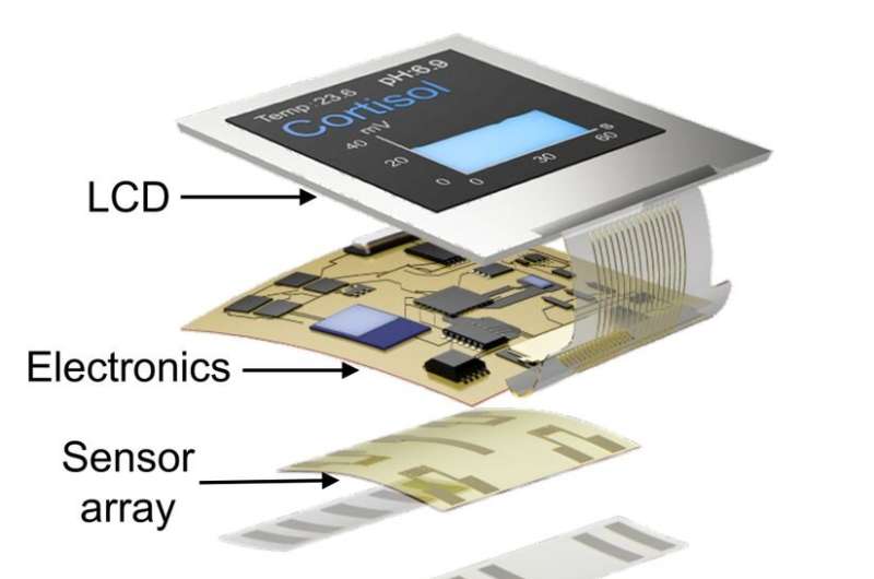
Now, a UCLA research team has developed a device that could be a major step forward: A smartwatch that assesses cortisol levels found in sweat—accurately, noninvasively and in real time. Described in a study published in Science Advances, the technology could offer wearers the ability to read and react to an essential biochemical indicator of stress.

“I anticipate that the ability to monitor variations in cortisol closely across time will be very instructive for people with psychiatric disorders,” said co-corresponding author Anne Andrews, a UCLA professor of psychiatry and biobehavioral sciences, member of the California NanoSystems Institute at UCLA and member of the Semel Institute for Neuroscience and Human Behavior. “They may be able to see something coming or monitor changes in their own personal patterns.”

Cortisol is well-suited for measurement through sweat, according to co-corresponding author Sam Emaminejad, an associate professor of electrical and computer engineering at the UCLA Samueli School of Engineering, and a member of CNSI.

“We determined that by tracking cortisol in sweat, we would be able to monitor such changes in a wearable format, as we have shown before for other small molecules such as metabolites and pharmaceuticals,” he said. “Because of its small molecular size, cortisol diffuses in sweat with concentration levels that closely reflect its circulating levels.”

The technology capitalizes on previous advances in wearable bioelectronics and biosensing transistors made by Emaminejad, Andrews and their research teams.

In the new smartwatch, a strip of specialized thin adhesive film collects tiny volumes of sweat, measurable in millionths of a liter. An attached sensor detects cortisol using engineered strands of DNA, called aptamers, which are designed so that a cortisol molecule will fit into each aptamer like a key fits a lock. When cortisol attaches, the aptamer changes shape in a way that alters electric fields at the surface of a transistor.

The invention—along with a 2021 study that demonstrated the ability to measure key chemicals in the brain using probes—is the culmination of a long scientific quest for Andrews. Over more than 20 years, she has spearheaded efforts to monitor molecules such as serotonin, a chemical messenger in the brain tied to mood regulation, in living things, despite transistors’ vulnerability to wet, salty biological environments.

Sweating the small stuff: Smartwatch developed at UCLA measures key stress hormone
The technology capitalizes on previous work by Sam Emaminejad, Anne Andrews and their UCLA research teams. Credit: Emaminejad Lab and Andrews Lab/UCLA

In 1999, she proposed using nucleic acids—rather than proteins, the standard mechanism—to recognize specific molecules.

“That strategy led us to crack a fundamental physics problem: how to make transistors work for electronic measurements in biological fluids,” said Andrews, who is also a professor of chemistry and biochemistry.

Meanwhile, Emaminejad has had a vision of ubiquitous personal health monitoring. His lab is pioneering wearable devices with biosensors that track the levels of certain molecules that are related to specific health measures.

“We’re entering the era of point-of-person monitoring, where instead of going to a doctor to get checked out, the doctor is basically always with us,” he said. “The data are collected, analyzed and provided right on the body, giving us real-time feedback to improve our health and well-being.”

Emaminejad’s lab had previously demonstrated that a disposable version of the specialized adhesive film enables smartwatches to analyze chemicals from sweat, as well as a technology that prompts small amounts of sweat even when the wearer is still. Earlier studies showed that sensors developed by Emaminejad’s group could be useful for diagnosing diseases such as cystic fibrosis and for personalizing drug dosages.

One challenge in using cortisol levels to diagnose depression and other disorders is that levels of the hormone can vary widely from person to person—so doctors can’t learn very much from any single measurement. But the authors foresee that tracking individual cortisol levels over time using the smartwatch may alert wearers, and their physicians, to changes that could be clinically significant for diagnosis or monitoring the effects of treatment.

Among the study’s other authors is Janet Tomiyama, a UCLA associate professor of psychology, who has collaborated with Emaminejad’s lab over the years to test his wearable devices in clinical settings.

“This work turned into an important paper by drawing together disparate parts of UCLA,” said Paul Weiss, a UCLA distinguished professor of chemistry and biochemistry and of materials science and engineering, a member of CNSI, and a co-author of the paper. “It comes from us being close in proximity, not having ego problems and being excited about working together. We can solve each other’s problems and take this technology in new directions.”

The paper’s co-first authors are UCLA postdoctoral scholar Bo Wang and Chuanzhen Zhao, a former UCLA graduate student. Other co-authors are Zhaoqing Wang, Xuanbing Cheng, Wenfei Liu, Wenzhuo Yu, Shuyu Lin, Yichao Zhao, Kevin Cheung and Haisong Lin, all of UCLA; and Milan Stojanović and Kyung-Ae Yang of Columbia University.

From https://techxplore.com/news/2022-02-small-newly-smartwatch-key-stress.html

What Is Facial Plethora?

What does it mean to have facial plethora? 

Facial plethora involves facial swelling and redness. It’s a symptom of another condition, rather than a condition itself.

It occurs when blood flow to your face increases. It can also happen when the amount of blood in your body increases.

The underlying causes of facial plethora vary greatly, and treatment depends on the specific cause.

 What can cause a plethoric face? 

Several possible conditions can cause facial plethora. They include:

Cushing syndrome

Facial plethora is a main symptom of  Cushing syndrome , a rare condition that’s caused by high levels of cortisol, known as the stress hormone.

The hormone cortisol is produced by your adrenal gland. It’s involved in bodily functions like the stress response, metabolism, and inflammation.

 Too much cortisol  can happen if you have a tumor in the pituitary or adrenal gland. The tumor causes your body to overproduce cortisol, leading to high levels. It can also happen after taking  corticosteroid  drugs, like prednisone.

High levels of cortisol can increase blood pressure. This increases blood flow in the skin on your face, resulting in facial plethora.

Superior vena cava syndrome

T he superior vena cava (SVC) is a major vein in your body. It brings blood to your heart from your chest, head, neck, and arms.

Some types of cancer, like lung cancer, can partially or completely block the SCV. A blood clot can also block the SCV. If this happens, a group of symptoms called superior vena cava syndrome (SCVS) may develop.

In SCVS, the blockage of blood flow causes upper body swelling. This can lead to symptoms like facial plethora. 

Carcinoid syndrome

Neuroendocrine cells are involved in basic bodily functions. They work by sending information via hormones.

If neuroendocrine cells develop into a tumor, it’s called a carcinoid tumor. Carcinoid syndrome happens when the tumor spreads to your liver and makes too much serotonin.

The tumor releases chemicals into the bloodstream, causing a range of symptoms. Some chemicals widen the blood vessels and increase blood flow, resulting in facial swelling.

Polycythemia vera

Polycythemia vera is a rare blood disorder that causes excess production of red blood cells. This increases red blood cell mass, causing thickened blood and swelling.

The swelling can lead to plethora of the face and palms.

Rosacea

Rosacea is a chronic inflammatory skin disorder. The inflammation can cause facial flushing and swelling.

In people with lighter skin tones, the flushing appears red. In those with darker skin tones, it may look like brown discoloration.

Other symptoms of rosacea include:

  • acne-like breakouts
  • thickening skin
  • burning
  • warm skin

Sunburn

sunburn happens when ultraviolet rays from the sun damage skin cells. This causes your body to release inflammatory substances, resulting in dilated blood vessels and increased blood flow.

The increased blood flow leads to swelling and redness, or facial plethora.

 Is facial plethora a symptom of a syndrome? 

In some cases, facial plethora may indicate a syndrome.

According to a 2015 study, facial plethora is one of the first identified symptoms of Cushing syndrome. It’s also one of the first symptoms to get better as Cushing syndrome is treated.

Facial plethora may also be caused by SVCS or carcinoid syndrome.

 What are the features of facial plethora? 

Facial plethora involves a group of features, rather than a single characteristic. It generally involves facial symptoms like:

  • swelling and increased roundness
  • increased puffiness in your cheeks
  • redness (on lighter skin tones)
  • brown discoloration (on darker skin tones)

VIEW GALLERY2
 Does facial plethora pose other health risks? 

The symptoms of facial plethora may be uncomfortable. But there’s no research stating that facial plethora itself is linked to complications.

However, if the underlying condition goes untreated, it may lead to complications. The condition may get worse or cause other side effects.

That’s why it’s important to contact a healthcare professional if you think you have facial plethora.

 How is facial plethora treated? 

Treatment for a plethoric face depends on the condition causing it. Treatment may include:

Medication

A healthcare professional may suggest using medication for the following conditions:

  • Cushing syndrome. A doctor can prescribe medications that lower your cortisol levels.
  • SVCS. If this condition is caused by a blood clot, a therapy called thrombolysis can be used to break down the clot. This procedure allows medication to get to the site of a blood clot and dissolve the blockage.
  • Carcinoid syndrome. Some medications can block the chemicals produced by a carcinoid tumor.
  • Polycythemia vera. If you have polycythemia vera, you might need medication to reduce red blood cell production.
  • Rosacea. A dermatologist can prescribe topical medication to manage rosacea symptoms.

Chemotherapy or radiation

Chemotherapy or radiation treatment may be used for tumors that cause:

  • Cushing syndrome
  • SVCS
  • carcinoid syndrome

Surgery

In certain cases, you might need surgery. This option may be used for tumors that cause:

  • Cushing syndrome
  • SVCS
  • carcinoid syndrome

If SCVS is caused by a blood clot, surgery may be used to insert a stent or remove the blood clot.

 Takeaway 

Facial plethora is characterized by facial swelling and puffiness. It can cause redness in lighter complexions and brown discoloration in darker skin tones.

It’s a main symptom of Cushing syndrome, but it may also be a symptom of SVCS and carcinoid syndrome. Other causes include polycythemia vera, rosacea, and sunburn.

Treatment depends on the specific cause. If you experience facial swelling and discoloration, reach out to a healthcare professional for a diagnosis.

Capillary Microarchitectural Changes in Cushing’s Syndrome

This article was originally published here

Microvasc Res. 2022 Jan 21:104323. doi: 10.1016/j.mvr.2022.104323. Online ahead of print.

ABSTRACT

PURPOSE: Macrovascular alterations are prominent in Cushing’s syndrome (CS). Microvascular abnormalities are yet to be established. This cross-sectional observational study aimed to evaluate microvascular changes in nailfold capillaries and their association with disease status and carotid intima-media thickness (CIMT) as a marker of atherosclerosis.

METHODS: A total of 70 patients with CS [46 (65.7%) ACTH-dependent pituitary adenoma and 24 (34.3%) adrenocortical adenomas] and 100 healthy controls were enrolled. The microvascular structure was evaluated using nailfold video-capillaroscopy (NVC).

RESULTS: The median number of capillaries was less [10 mm (IQR: 2, min-max:7-14) vs. 11 mm (IQR: 2, min-max:9-19) (p < 0.001)], the median limb diameter and capillary width were wider in the CS group than in the controls (p = 0.016 and p = 0.002, respectively). Microhemorrhages within limited areas were more frequent in the CS group than in the controls (p = 0.046). Observed capillary changes were similar among the patients with CS with remission or active disease. CIMT levels were higher in the CS group than in the controls and similar in subjects with active disease and remission. Univariate logistic regression analyses revealed that the number of capillaries and capillary widths were associated with body mass index (BMI), the presence of type 2 diabetes mellitus, HbA1c, and CIMT.

CONCLUSION: Morphologic alterations present similarly in nailfold capillaries in subjects with CS regardless of disease status, resembling changes in chronic atherosclerotic diseases. Microvascular changes in nailfold capillaries measured using NVC can be used as a marker in the assessment of cardiovascular risk in patients with CS.

PMID:35074338 | DOI:10.1016/j.mvr.2022.104323

From https://www.docwirenews.com/abstracts/rheumatology-abstracts/capillary-microarchitectural-changes-in-cushings-syndrome/

Synergistic Cortisol Suppression by Ketoconazole–Osilodrostat Combination Therapy

Abstract

Summary

Here, we describe a case of a patient presenting with adrenocorticotrophic hormone-independent Cushing’s syndrome in a context of primary bilateral macronodular adrenocortical hyperplasia. While initial levels of cortisol were not very high, we could not manage to control hypercortisolism with ketoconazole monotherapy, and could not increase the dose due to side effects. The same result was observed with another steroidogenesis inhibitor, osilodrostat. The patient was finally successfully treated with a well-tolerated synergitic combination of ketoconazole and osilodrostat. We believe this case provides timely and original insights to physicians, who should be aware that this strategy could be considered for any patients with uncontrolled hypercortisolism and delayed or unsuccessful surgery, especially in the context of the COVID-19 pandemic.

Learning points

  • Ketoconazole–osilodrostat combination therapy appears to be a safe, efficient and well-tolerated strategy to supress cortisol levels in Cushing syndrome.
  • Ketoconazole and osilodrostat appear to act in a synergistic manner.
  • This strategy could be considered for any patient with uncontrolled hypercortisolism and delayed or unsuccessful surgery, especially in the context of the COVID-19 pandemic.
  • Considering the current cost of newly-released drugs, such a strategy could lower the financial costs for patients and/or society.

Background

Untreated or inadequately treated Cushing’s syndrome (CS) is a morbid condition leading to numerous complications. The latter ultimately results in an increased mortality that is mainly due to cardiovascular events and infections. The goal of the treatment with steroidogenesis inhibitors is normalization of cortisol production allowing the improvement of comorbidities (1). Most studies dealing with currently available steroidogenesis inhibitors used as monotherapy reported an overall antisecretory efficacy of roughly 50% in CS. Steroidogenesis inhibitors can be combined to better control hypercortisolism. To the best of our knowledge, we report here for the first time a patient treated with a ketoconazole–osilodrostat combination therapy.

Case presentation

Here, we report the case of Mr D.M., 53-years old, diagnosed with adrenocorticotrophic hormone (ACTH)-independent CS 6 months earlier. At diagnosis, he presented with resistant hypertension, hypokalemia, diabetes mellitus, easy bruising, purple abdominal striae and major oedema of the lower limbs.

Investigations

A biological assessment was performed, and the serum cortisol levels are depicted in Table 1. ACTH levels were suppressed (mean levels 1 pg/mL). Mean late-night salivary cortisol showed a four-fold increase (Table 2), and mean 24 h-urinary cortisol showed a two-fold increase. Serum cortisol was 1000 nmol/L at 08:00 h after 1 mg dexamethasone dose at 23:00 h. The rest of the adrenal hormonal workup was within normal ranges (aldosterone: 275 pmol/L and renin: 15 mIU/L). An adrenal CT was performed (Fig. 1) and exhibited a 70-mm left adrenal mass (spontaneous density: 5 HU and relative washout: 65%) and a 45-mm right adrenal mass (spontaneous density: −2 HU and relative washout: 75%). The case was discussed in a multidisciplinary team meeting, which advised to perform 18F-FDG PET-CT and 123I-Iodocholesterol scintigraphy before considering surgery. A genetic screening was performed, testing for ARMC5 and PRKAR1A pathogenic variants.

Figure 1View Full Size
Figure 1
Adrenal CT depicting the bilateral macronodular adrenocortical hyperplasia.

Citation: Endocrinology, Diabetes & Metabolism Case Reports 2021, 1; 10.1530/EDM-21-0071

Table 1Serum cortisol levels at diagnosis (A), using ketoconazole monotherapy (B), using osilodrostat monotherapy (C) and using osilodrostat–ketoconazole combination therapy (D).

Serum cortisol (nmol/L) 08:00 h 24:00 h 16:00 h 20:00 h 12:00 h 16:00 h
A. At diagnosis 660 615 716 566 541 561
B. Ketoconazole monotherapy 741 545 502 224 242 508
C. Osilodrostat monotherapy 658 637 588 672 486 692
D. Osilodrostat–ketoconazole combination 436 172 154 103 135 274
Table 2Salivary cortisol levels at diagnosis (A), using ketoconazole monotherapy (B), using osilodrostat monotherapy (C) and using osilodrostat-ketoconazole combination therapy (D).

Salivary cortisol (nmol/L) 23:00 h 12:00 h 13:00 h Mean
A. At diagnosis 47 62 38 49
B. Ketoconazole monotherapy 20 15 21 18
C. Osilodrostat monotherapy 85 90 56 77
D. Osilodrostat–ketoconazole combination 10 14 9 11

Treatment

As this condition occurred during the COVID-19 pandemic, it was decided to first initiate steroidogenesis inhibitors to lower the patient’s cortisol levels. Initially, ketoconazole was initiated and uptitrated up to 1000 mg per day based on close serum cortisol monitoring, with a three-fold increase of liver enzymes and poor control of cortisol levels (Table 1). In the absence of biological efficacy, ketoconazole was replaced by osilodrostat, which was gradually increased up to 30 mg per day (10 mg at 08:00 h and 20 mg at 20:00 h) without reaching normal cortisol levels (Table 1) and with slightly increased blood pressure levels. Considering the lack of efficacy of anticortisolic drugs used as monotherapy, we combined osilodrostat (30 mg per day) to ketoconazole (600 mg per day), that is, at the last maximal tolerated dose as monotherapy of each drug.

Outcome

This combination of steroidogenesis inhibitors achieved a good control in cortisol levels, mimicking a physiological circadian rhythm (Table 1D). The patient did not exhibit any side effect and the control of cortisol levels resulted in a rapid improvement of hypertension, kalemia, diabetes control and disappearance of lower limbs oedema. The patient underwent a 18F-FDG PET-CT that did not exhibit any increased uptake in both adrenal masses and a 123I-Iodocholesterol scintigraphy exhibiting a highly increased uptake in both adrenal masses, predominating in the left adrenal mass (70 mm). Unilateral adrenalectomy of the larger mass was then performed, and as the immediate post-operative serum cortisol level was 50 nmol/L, hydrocortisone was administered at a dose of 30 mg per day, with a stepwise decrease to 10 mg per day over 3 months. Pathological examination exhibited macronodular adrenal hyperplasia with a 70-mm adreno cortical adenoma (WEISS score: 1 and Ki67: 1%). The genetic screening exhibited a c.1908del p.(Phe637Leufs*6) variant of ARMC5 (pathogenic), located in exon 5. The patient has no offspring and is no longer in contact with the rest of his family.

Discussion

The goal of the treatment with steroidogenesis inhibitors is normalization of cortisol production allowing the improvement of comorbidities (1). Most studies dealing with currently available steroidogenesis inhibitors used as monotherapy reported an overall antisecretory efficacy of roughly 50% in CS. This rate of efficacy was probably underestimated in retrospective studies due to the lack of adequate uptitration of the dose; For example, the median dose reported in the French retrospective study on ketoconazole was only 800 mg/day, while 50% of the patients were uncontrolled at the last follow-up (2).

Steroidogenesis inhibitors can be combined to better control hypercortisolism. Up to now, such combinations, mainly ketoconazole and metyrapone, were mainly reported in patients with severe CS (median urinary-free Cortisol (UFC) 30- to 40-fold upper-limit norm (ULN)) and life-threatening comorbidities (34). Normal UFC was reported in up to 86% of these patients treated with high doses of ketoconazole and metyrapone. Expected side effects (such as increased liver enzymes for ketoconazole or worsened hypertension and hypokalemia for metyrapone) were reported in the majority of the patients. The fear of these side effects probably explains the lack of uptitration in previous reports. Combination of steroidogenesis inhibitors has previously been described by Daniel et al. in the largest study reported on the use of metyrapone in CS; 29 patients were treated with metyrapone and ketoconazole or mitotane, including 22 in whom the second drug was added to metyrapone monotherapy because of partial efficacy or adverse effects. The final median metyrapone dose in patients controlled with combination therapy was 1500 mg per day (5).

Combination of adrenal steroidogenesis inhibitors should not be reserved to patients with severe hypercortisolism. In the case shown here, the association was highly effective in terms of secretion, using lower doses than those applied as a single treatment, but without the side effects previously observed with higher doses of each treatment used as a monotherapy. To our knowledge, the association of ketoconazole and osilodrostat had never been reported. Ketoconazole blocks several enzymes of the adrenal steroidogenesis such as CYP11A1, CYP17, CYP11B2 (aldosterone synthase) and CYP11B1 (11-hydroxylase), leading to decreased cortisol and occasionally testosterone concentrations. Though liver enzymes increase is not dose-dependent, it usually happens at doses exceeding 400–600 mg per day (2). Osilodrostat blocks CYP11B1 and CYP11B2; a combination should thus allow for a complete blockade of these enzymes that are necessary for cortisol secretion. Short-term side effects such as hypokalemia and hypertension are similar to those observed with metyrapone, due to increased levels of the precursor deoxycorticosterone, correlated with the dose of osilodrostat (6). As for our patient, the occurrence of side effects should not lead to immediately switch to another drug, but rather to decrease the dose and add another cortisol-lowering drug. Moreover, considering the current cost of newly-released drugs such a strategy could lower financial costs for patients and/or society.

Another point to take into account is the current COVID-19 pandemic, for which, as recently detailed in experts’ opinion (7), the main aim is to reach eucortisolism, whatever the way. Indeed patients presenting with CS usually also present with comorbidities such as obesity, hypertension, diabetes mellitus and immunodeficiency (8). Surgery, which represents the gold standard strategy in the management of CS (19), might be delayed to reduce the hospital-associated risk of COVID-19, with post-surgical immunodepression and thromboembolic risks (7). Because immunosuppression and thromboembolic diathesis are common CS features (910), during the COVID-19 pandemic, the use of steroidogenesis inhibitors appears of great interest. In these patients, combing steroidogenesis inhibitors at intermediate doses might allow for a rapid control of hypercortisolism without risks of major side effects if a single uptitrated treatment is not sufficient. Obviously, the management of associated comorbidities would also be crucial in this situation (11).

To conclude, we report for the first time a case of CS, in the context of primary bilateral macronodular adrenocortical hyperplasia successfully treated with a well-tolerated combination of ketoconazole and osilodrostat. While initial levels of cortisol were not very high, we could not manage to control hypercortisolism with ketoconazole monotherapy, and could not increase the dose due to side effects. The same result was observed with another steroidogenesis inhibitor, osilodrostat. This strategy could be considered for any patient with uncontrolled hypercortisolism and delayed or unsuccessful surgery, especially in the context of the COVID-19 pandemic.

Declaration of interest

F C and T B received research grants from Recordati Rare Disease and HRA Pharma Rare Diseases. Frederic Castinetti is on the editorial board of Endocrinology, Diabetes and Metabolism case reports. Frederic Castinetti was not involved in the review or editorial process for this paper, on which he is listed as an author.

Funding

This work did not receive any specific grant from any funding agency in the public, commercial or not-for-profit sector.

Patient consent

Informed written consent has been obtained from the patient for publication of the case report.

Author contribution statement

V A was the patient’s physician involved in the clinical care and collected the data. T B and F C supervised the management of the patient. F C proposed the original idea of this case report. V A drafted the manuscript. F C critically reviewed the manuscript. T B revised the manuscript into its final version.

References

From This Month’s Reader’s Digest

From the February, 2022 issue of Reader’s Digest:

readers-digest.jpg

Read the original article at readers-digest-misdiagnosed