Corcept Completes Enrollment in Phase 3 Gradient Trial of Relacorilant in Patients With Adrenal Cushing’s Syndrome

Corcept Therapeutics Incorporated (NASDAQ: CORT), a commercial-stage company engaged in the discovery and development of medications to treat severe endocrinologic, oncologic, metabolic and neurologic disorders by modulating the effects of the hormone cortisol, today announced completion of enrollment in GRADIENT, a Phase 3 trial of its proprietary selective cortisol modulator relacorilant in patients with Cushing’s syndrome (hypercortisolism) caused by an adrenal adenoma or adrenal hyperplasia.

“Hypercortisolism with adrenal etiology affects many patients and is associated with serious cardiometabolic comorbidities, including hypertension and hyperglycemia, and increased risk of premature death,” said Bill Guyer, PharmD, Corcept’s Chief Development Officer. “GRADIENT is the first prospective placebo-controlled study to be conducted exclusively in these patients with Cushing’s syndrome. We expect data from GRADIENT in the fourth quarter of this year.”

GRADIENT is a randomized, double-blind, placebo-controlled trial conducted at sites in the United States, Europe and Israel. One-hundred thirty-seven patients were randomized 1:1 to receive relacorilant or placebo for 22 weeks. Primary endpoints are improvement in glucose metabolism and hypertension.

About Cushing’s Syndrome (Hypercortisolism)
Cushing’s syndrome is caused by excessive activity of the hormone cortisol. Endogenous Cushing’s syndrome is an orphan disease that most often affects adults aged 20-50. Symptoms vary, but most patients experience one or more of the following manifestations: high blood sugar, diabetes, high blood pressure, upper-body obesity, rounded face, increased fat around the neck, thinning arms and legs, severe fatigue and weak muscles. Irritability, anxiety, cognitive disturbances and depression are also common. Cushing’s syndrome can affect every organ system and can be lethal if not treated effectively.

About Relacorilant
Relacorilant is a selective cortisol modulator that binds to the glucocorticoid receptor (GR), but does not bind to the body’s other hormone receptors. Corcept is studying relacorilant in a variety of serious disorders, including ovarian, adrenal and prostate cancer and Cushing’s syndrome. Relacorilant is proprietary to Corcept and is protected by composition of matter, method of use and other patents. Relacorilant has orphan drug designation in the United States and the European Union for the treatment of Cushing’s syndrome.

About Corcept Therapeutics
For over 25 years, Corcept’s focus on cortisol modulation and its potential to treat patients across a wide variety of serious disorders has led to the discovery of more than 1,000 proprietary selective cortisol modulators. Corcept’s advanced clinical trials are being conducted in patients with hypercortisolism, solid tumors, amyotrophic lateral sclerosis (ALS) and liver disease (NASH). In February 2012, the company introduced Korlym, the first medication approved by the U.S. Food and Drug Administration for the treatment of patients with Cushing’s syndrome. Corcept is headquartered in Menlo Park, California. For more information, visit Corcept.com.

Forward-Looking Statements
Statements in this press release, other than statements of historical fact, are forward-looking statements based on our current plans and expectations that are subject to risks and uncertainties that might cause our actual results to differ materially from those such statements express or imply. These risks and uncertainties include, but are not limited to, our ability to operate our business; risks related to the study and development of Korlym as well as relacorilant, miricorilant, dazucorilant and our other product candidates, including their clinical attributes, regulatory approvals, mandates, oversight and other requirements; and the scope and protective power of our intellectual property. These and other risks are set forth in our SEC filings, which are available at our website and the SEC’s website.

In this press release, forward-looking statements include those concerning the development of relacorilant as a treatment for Cushing’s syndrome, and design, timing and expectations regarding our GRADIENT trial. We disclaim any intention or duty to update forward-looking statements made in this press release.

From https://finance.yahoo.com/news/corcept-completes-enrollment-phase-3-120000179.html

Clinical Features and Treatment Options for Pediatric Adrenal Incidentalomas

Abstract

Background

The aim of this study was to investigate the clinical features and treatment options for pediatric adrenal incidentalomas(AIs) to guide the diagnosis and treatment of these tumors.

Methods

The clinical data of AI patients admitted to our hospital between December 2016 and December 2022 were collected and retrospectively analyzed. All patients were divided into neonatal and nonneonatal groups according to their age at the time of the initial consultation.

Results

In the neonatal group, 13 patients were observed and followed up, and the masses completely disappeared in 8 patients and were significantly reduced in size in 5 patients compared with the previous findings. Four patients ultimately underwent surgery, and the postoperative pathological diagnosis was neuroblastoma in three patients and teratoma in one patient. In the nonneonatal group, there were 18 cases of benign tumors, including 9 cases of ganglioneuroma, 2 cases of adrenocortical adenoma, 2 cases of adrenal cyst, 2 cases of teratoma, 1 case of pheochromocytoma, 1 case of nerve sheath tumor, and 1 case of adrenal hemorrhage; and 20 cases of malignant tumors, including 10 cases of neuroblastoma, 9 cases of ganglioneuroblastoma, and 1 case of adrenocortical carcinoma.

Conclusions

Neuroblastoma is the most common type of nonneonatal AI, and detailed laboratory investigations and imaging studies are recommended for aggressive evaluation and treatment in this population. The rate of spontaneous regression of AI is high in neonates, and close observation is feasible if the tumor is small, confined to the adrenal gland and has no distant metastasis.

Peer Review reports

Background

The incidence of adrenal incidentaloma (AI) is increasing due to the increased frequency of imaging and improved imaging sensitivity [1]. AI is relatively common in adults, and several organizations, such as the American Association of Clinical Endocrinologists/American Association of Endocrine Surgeons and the European Society Endocrinology, have proposed specific protocols to guide the evaluation, treatment, and follow-up management of AI in adults [2]. Although AI, a nonfunctioning adrenocortical adenoma, is most common in adults, neuroblastoma is the most common incidental tumor of the adrenal gland in children. In addition, in the neonatal period, which is a more complex stage of childhood, the biology of adrenal masses found in this age group is also more specific, and the nature of these masses can range from spontaneous regression to rapid progression to aggressive disease with metastatic dissemination and even death. Given that AI is the most common malignant tumor, the management of AI in children cannot be simply based on the measurements used in adult AI. In this study, we retrospectively analyzed the clinical data of pediatric AI patients in a single center to investigate the clinical characteristics and management of AI in children.

Methods

A total of 66 children with adrenal tumors were diagnosed and treated at the Department of Urology of the Children’s Hospital of Nanjing Medical University from December 2016 to December 2022. A total of 55 cases were detected during physical examination, or the patients were diagnosed and received treatment for diseases other than adrenal disease after excluding adrenal tumors detected due to typical clinical manifestations or signs such as centripetal obesity and precocious puberty. Research protocols involving human materials were approved by the Medical Ethics Committee of the Children’s Hospital of Nanjing Medical University. All clinical information, radiological diagnosis, laboratory test results, intervention results, and follow-up data were collected from the department’s database.

All the children underwent ultrasonography and CT scanning, and 11 children underwent MRI. In addition to routine tests such as blood routine and biochemical indexes, the examination and evaluation of adrenal endocrine hormones and tumor markers included (1) plasma cortisol and ACTH levels, (2) plasma catecholamine and metabolite determination, (3) plasma renin and plasma aldosterone, (4) urinary vanillylmandelic acid/homovanillic acid(VMA/HVA), and (5) AFP, CEA, NSE, and CA19-9. Five patients underwent a low-dose dexamethasone suppression test. Seventeen of the 55 patients were treated with watch-waiting therapy, 4 of the 17 ultimately underwent surgery, 4 of the 38 patients underwent tumor biopsy, and 34 underwent adrenalectomy.

The data were analyzed using Graph Pad Prism 8. The measurement data are expressed as ‾x ± sd. The maximum diameter of the tumors, age of the patients with benign and malignant tumors, and maximum diameter of the tumors between the laparoscopic surgery group and the open surgery group were compared using paired t tests, and the percentages of the count data were compared using Fisher’s exact test.

Results

In this study, all patients were divided into two groups according to their age at the time of consultation: the neonate group and the nonneonate group.

Neonate group:

There were 7 male and 10 female patients, 7 of whom were diagnosed via prenatal examination and 10 of whom were diagnosed after birth. Five patients were diagnosed with lesions on the left side, 12 patients were diagnosed with lesions on the right side, and the maximal diameters of the masses ranged from 16 to 48 mm. The characteristics of the AIs in the neonate group are presented in Table 1.

Table 1 Characteristics of AI in the neonates group

Among the 17 patients, 8 had cystic masses with a maximum diameter of 1648 mm, 5 had cystic-solid masses with a maximum diameter of 3339 mm, and 4 had solid masses with a maximum diameter of 1845 mm. Two patients with solid adrenal gland masses suggested by CT scan had obvious elevations in serum NSE and maximum diameters of 44 and 45 mm, respectively. These patients underwent adrenal tumor resection, and the pathology diagnosed that they had neuroblastomas(NB). In one patient, the right adrenal gland was 26 × 24 × 27 mm in size with slightly elevated echogenicity at 38 weeks after delivery, and the mass increased to a size of 40 × 39 × 29 mm according to the 1-month postnatal review. MRI suggested that the adrenal gland tumor was associated with liver metastasis, and the pathology of the tumor suggested that it was NB associated with liver metastasis after surgical resection (stage 4 S, FH). One child was found to have 25 × 24 × 14 mm cystic echoes in the left adrenal region during an obstetric examination, and ultrasound revealed 18 × 11 mm cystic solid echoes 5 days after birth. Ultrasound revealed 24 × 15 mm cystic solid echoes at 2 months. Serum NSE and urinary VMA were normal, and the tumor was excised due to the request of the parents. Pathology suggested a teratoma in the postoperative period. A total of 13 children did not receive surgical treatment or regular review via ultrasound, serum NSE or urine VMA. The follow-up time ranged from 1 to 31 months, with a mean of 9.04 ± 7.61 months. Eight patients had complete swelling, and 5 patients were significantly younger than the previous patients. Nonneonate group:

There were 24 male and 14 female patients in the nonneonate group; 24 patients had lesions on the left side, 14 patients had lesions on the right side, and the maximal diameters of the masses ranged from 17 to 131 mm. Most of these tumors were found during routine physical examinations or incidentally during examinations performed for various complaints, such as gastrointestinal symptoms, respiratory symptoms, or other related conditions. As shown in Table 2, abdominal pain was the most common risk factor (44.7%) for clinical onset, followed by routine physical examination and examination for respiratory symptoms.

Table 2 Clinical presentations leading to discovery of AI in non-neonate group

Among the 38 patients, 10 had NBs with maximum diameters ranging from 20 to 131 mm, 9 had ganglion cell neuroblastomas with maximum diameters ranging from 33.6 to 92 mm, 9 had ganglion cell neuromas with maximum diameters ranging from 33 to 62 mm, 2 had adrenal adenomas with maximum diameters ranging from 17 to 70 mm, 1 had a cortical carcinoma with a maximum diameter of 72 mm, 2 had adrenal cysts with maximum diameters ranging from 26 to 29 mm, 2 had mature teratomas with maximum diameters of 34 and 40 mm, 1 had a pheochromocytoma with a diameter of 29 mm, 1 had a nerve sheath tumor with a diameter of 29 mm, and 1 patient with postoperative pathological confirmation of partial hemorrhagic necrosis of the adrenal gland had focal calcification with a maximum diameter of 25 mm (Table 3).

Table 3 Distribution of different pathologies among AI with various sizes in non-neonate group

The mean age of children with malignant tumors was significantly lower than that of children with benign tumors (57.95 ± 37.20 months vs. 105.0 ± 23.85 months; t = 4.582, P < 0.0001). The maximum diameter of malignant tumors ranged from 20 to 131 mm, while that of benign tumors ranged from 17 to 72 mm, and the maximum diameter of malignant tumors was significantly greater than that of benign tumors (65.15 ± 27.61 mm v 37.59 ± 12.98 mm; t = 3.863, P = 0.0004). Four biopsies, 5 laparoscopic adrenal tumor resections and 11 open adrenal tumor resections were performed for malignant tumors, and 16 laparoscopic adrenal tumor resections and 2 open procedures were performed for benign tumors. The maximum diameter of the tumors ranged from 17 to 62 mm in 21 children who underwent laparoscopic surgery and from 34 to 99 mm in 13 children who underwent open resection; there was a statistically significant difference in the maximum diameter of the tumors between the laparoscopic surgery group and the open surgery group (35.63 ± 10.36 mm v 66.42 ± 20.60 mm; t = 5.798, P < 0.0001).

Of the 42 children with definitive pathologic diagnoses at surgery, 23 had malignant tumors, and 19 had benign tumors. There were 15 malignant tumors with calcification on imaging and 5 benign tumors. The percentage of malignant tumors with calcifications in was significantly greater than that of benign tumors (65.22% v 26.32%; P = 0.0157). In the present study, all the children underwent CT, and 31 patients had postoperative pathological confirmation of NB. A total of 26 patients were considered to have neurogenic tumors according to preoperative CT, for a diagnostic compliance rate of 83.97%. Three children were considered to have cortical adenomas by preoperative CT, and 1 was ultimately diagnosed by postoperative pathology, for a diagnostic compliance rate of 33.33%. For 4 patients with teratomas and adrenal cysts, the CT findings were consistent with the postoperative pathology. According to our research, NB 9-110HU, GNB 15-39HU, GB 19-38HU, ACA 8HU, adrenal cyst 8HU, and cellular achwannoma 17HU.

Discussion

According to the clinical practice guidelines developed by the European Society of Endocrinology and European Network for the Study of Adrenal Tumors, AI is an adrenal mass incidentally detected on imaging not performed for a suspected adrenal disease [3]. The prevalence of AI is approximately 4%, and the incidence increases with age [4]. Most adult AIs are nonfunctioning benign adrenal adenomas (up to 75%), while others include functioning adrenal adenomas, pheochromocytomas, and adrenocortical carcinomas [5]. In contrast to the disease spectrum of adult AI cases, NB is the most common tumor type among children with AI, and benign cortical adenomas, which account for the vast majority of adult AI, accounting for less than 0.5% of cases in children [6]. According to several guidelines, urgent assessment of an AI is recommended in children because of a greater likelihood of malignancy [37].

When an adult patient is initially diagnosed with AI, it should be clear whether the lesion is malignant and functional. In several studies, the use of noncontrast CT has been recommended as the initial imaging method for adrenal incidentaloma; a CT attenuation value ≤ 10 HU is used as the diagnostic criterion for benign adenomas; and these methods have a specificity of 71-79% and a sensitivity of 96-98% [89]. A CT scan of tumors with diameters greater than 4 to 6 cm, irregular margins or heterogeneity, a CT attenuation value greater than 10 HU, or a relative contrast enhancement washout of less than 40% 10 or 15 min after administration of contrast media on enhanced CT is considered to indicate potential malignancy [7]. As the most common AI in children, NB often appears as a soft tissue mass with uneven density on CT, often accompanied by high-density calcified shadows, low-density cystic lesions or necrotic areas. CT scans can easily identify more typical NBs, and for those AIs that do not show typical calcified shadows on CT, it is sometimes difficult to differentiate neurogenic tumors from adenomas. In these patients, except for the 1 patient with adrenal cysts who had a CT value of 8 HU, very few of the remaining AI patients had a CT value less than 10 HU. Therefore, the CT value cannot be used simply as a criterion for determining the benign or malignant nature of AI, and additional imaging examinations, such as CT enhancement, MRI, and FDG-PET if necessary, should be performed immediately for AI in children.

Initial hormonal testing is also needed for functional assessment, and aldosterone secretion should also be assessed when the patient is hypertensive or hypokalemic [7]. Patients with AI who are not suitable for surgery should be observed during the follow-up period, and if abnormal adrenal secretion is detected or suggestive of malignancy during this period, prompt adrenal tumor resection is needed. For adult patients with AI, laparoscopic adrenal tumor resection is one of the most effective treatments that has comparative advantages in terms of hospitalization time and postoperative recovery speed; however, there is still some controversy over whether to perform laparoscopic surgery for some malignant tumors with large diameters, especially adrenocortical carcinomas, and some studies have shown that patients who undergo laparoscopic surgery are more prone to peritoneal seeding of tumors [10].

The maximum diameter of an adult AI is a predictor of malignancy, and a study by the National Italian Study Group on Adrenal Tumors, which included 887 AIs, showed that adrenocortical carcinoma was significantly correlated with the size of the mass, and the sensitivity of detecting adrenocortical carcinoma with a threshold of 4 cm was 93% [11]. According to the National Institutes of Health, patients with tumors larger than 6 cm should undergo surgical treatment, while patients with tumors smaller than 4 cm should closely monitored; for patients with tumors between 4 and 6 cm, the choice of whether to be monitored or surgically treated can be based on other indicators, such as imaging [12]. A diameter of 4 cm is not the initial threshold for determining the benign or malignant nature of a mass in children.

In a study of 26 children with AI, Masiakos et al. reported that 9 of 18 benign lesions had a maximal diameter less than 5 cm, 4 of 8 malignant lesions had a maximal diameters less than 5 cm, and 2 had a diameter less than 3 cm. The mean maximal diameter of benign lesions was 4.2 ± 1.7 cm, whereas the mean maximum diameter of malignant lesions was 5.1 ± 2.3 cm. There was no statistically significant difference between the two comparisons; therefore, this study concluded that children with AI diameters less than 5 cm cannot be treated expectantly [6]. Additionally, this study revealed that malignant lesions occurred significantly more frequently than benign lesions in younger children (mean age 1.7 ± 1.8 years v 7.8 ± 5.9 years; P = 0.02).

In the nonneonatal group of this study, 20 patients with malignant tumors had maximum diameters ranging from 20 to 131 mm, 10 had malignant tumors larger than 60 mm, and 3 had tumors smaller than 40 cm; 18 patients with benign tumors had maximum diameters ranging from 17 to 70 mm, 5 had diameters ranging from 40 to 60 mm, and 5 had diameters larger than 60 mm. Therefore, it is not recommended to use the size of the largest diameter of the tumor to decide whether to wait and observe or intervene surgically for children with AI. Instead, it is necessary to consider the age of the child; laboratory test results, such as whether the tumor indices are elevated or not; whether the tumor has an endocrine function; etc.; and imaging test results to make comprehensive judgments and decisions. Preoperative aggressive evaluation and prompt surgical treatment are recommended for nonneonatal pediatric AI patients.

Adrenal hematoma and NBs are the most common types of adrenal area masses in children, while pheochromocytoma, adrenal cyst, and teratoma are rarer masses [13]. In clinical practice, adrenal hematoma and NB are sometimes difficult to differentiate, especially when adrenal masses are found during the prenatal examination and neonatal period, and such children need to be managed with caution. The Children’s Oncology Group (COG ANBL00B1) implemented the watchful waiting treatment for children under 6 months of age with a solid adrenal mass < 3.1 cm in diameter (or a cystic mass < 5 cm) without evidence of distant metastasis, and if there is a > 50% increase in the adrenal mass volume, there is no return to the baseline VMA or HVA levels, or if there is a > 50% increase in the urinary VMA/HVA ratio or an inversion, surgical resection should be performed [14]. Eighty-three children in this study underwent expectant observation, 16 of whom ultimately underwent surgical resection (8 with INSS stage 1 NB, 1 with INSS stage 2B, 1 with INSS stage 4 S, 2 with low-grade adrenocortical neoplasm, 2 with adrenal hemorrhage, and 2 with extralobar pulmonary sequestration). Most of the children who were observed had a reduced adrenal mass volume. Of the 56 patients who completed the final 90 weeks of expectant observation, 27 (48%) had no residual mass, 13 (23%) had a residual mass volume of 0–1 ml, 8 (14%) had a mass volume of 1–2 ml, and 8 (14%) had a volume of > 2 ml; ultimately, 71% of the residual masses had a volume ≤ 1 ml and 86% had a residual volume ≤ 2 ml. In this study, a total of 16 patients were included in the watchful waiting treatment group; 3 patients underwent surgical treatment during the follow-up period, and 13 patients ultimately completed watchful waiting treatment. After 1–31 months of follow-up, 8 patients’ swelling completely disappeared, and 5 patients’ swelling significantly decreased. After strict screening for indications and thorough follow-up review, AIs in the neonatal period can be subjected to watchful waiting treatment, and satisfactory results can be achieved.

For benign adrenal tumors, laparoscopic surgery is superior to open surgery in terms of successful resection, whereas the feasibility of minimally invasive surgery for AI with preoperative suspicion of malignancy is controversial. The European Cooperative Study Group for Pediatric Rare Tumors recommends that minimally invasive surgery be considered only for early childhood tumors and should be limited to small, localized tumors; additionally, imaging should suggest no invasion of surrounding tissue structures or lymph nodes; and this strategy requires surgeons with extensive experience in oncologic and adrenal surgery [15]. NB is the most common pediatric AI, and open tumor resection remains the mainstay of treatment. For small, early tumors without evidence of invasion on preoperative examination, laparoscopic resection may be considered if the principles of oncologic surgery can be adhered to. If the patient responds to chemotherapy, the decision to perform laparoscopic tumor resection can also be re-evaluated after chemotherapy. According to the current study, the recurrence and mortality rates of laparoscopic surgery are comparable to those of open surgery [1617]. The relative contraindications for laparoscopic NB resection include a tumor diameter greater than 6 cm, venous dilatation, and the involvement of adjacent organs or blood vessels [18]. Patients who undergo open adrenalectomy have higher overall survival and recurrence-free survival rates than patients who undergo laparoscopic adrenalectomy [19]. Open adrenalectomy remains the gold standard for surgical resection of adrenocortical carcinoma, whereas laparoscopic adrenalectomy should be reserved for highly selected patients and performed by surgeons with appropriate expertise [20].

Cortical tumors are particularly rare among children with AIs and are sometimes not clearly distinguishable from neurogenic tumors on preoperative imaging; in such patients, the presence of subclinical Cushing’s syndrome needs to be carefully evaluated preoperatively; otherwise, a perioperative adrenal crisis may occur [21]. In patients in whom the possibility of an adrenocortical tumor was considered preoperatively, the assessment for subclinical Cushing’s syndrome mainly involved assessing the serum dehydroepiandrosterone sulfate level and performing an overnight dexamethasone suppression test.

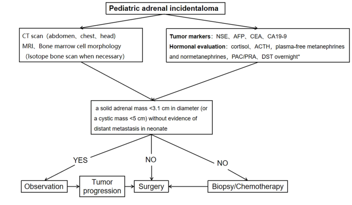
A procedure for evaluating pediatric AI is shown in Fig. 1. Imaging is the first step in the evaluation of AI in children. CT can be used to clarify the nature of most tumors. MRI can be used to evaluate imaging risk factors (IDRFs) for NB. Bone marrow cytomorphology is recommended for all children with AI, along with microscopic residual neuroblastoma testing and further bone scanning if the bone marrow examination is positive. In addition, serum tumor marker levels and other relevant tests should be performed, and hormone levels should be evaluated. If adrenal adenomas cannot be completely excluded during the preoperative examination, a 1 mg overnight dexamethasone suppression test should be performed to exclude subclinical Cushing’s syndrome. In patients with hypertensive hypokalemia, the presence of aldosteronism should be evaluated by testing plasma aldosterone concentrations and plasma renin activity. Adrenal masses found in the neonatal period can be observed if the tumor is small, confined to the adrenal gland and shows no evidence of distant metastasis, while tumors that increase significantly in size during the follow-up period or that are associated with persistently elevated tumor markers require aggressive surgical treatment.

Fig. 1

figure 1

Algorithm for the evaluation and management of a pediatric adrenal incidentaloma. *DST overnight :20µg/kg dexamethasoneweight ˂40 kg,1 mg dexamethasone if ≥ 40 kg. CT = computed tomographic;MRI = magnetic resonance imaging;NSE = neuron-specific enolase;AFP = alpha-fetoprotein;CEA = carcinoembryonic antigen;CA19-9 = cancerantigen19-9;ACTH = adrenocorticotropic hormone;PAC = plasma aldosterone concentration; PRA = plasma renin activity;DST = dexamethasone suppression test

Data availability

The datasets analyzed during the current study are not public, but are available from the corresponding author on reasonable request.

Abbreviations

CT:
computed tomographic
MRI:
magnetic resonance imaging
ACTH:
adrenocorticotropic hormone
VMA:
vanillylmandelic acid
HVA:
homovanillic Acid
AFP:
alpha-fetoprotein
CEA:
carcinoembryonic antigen
NSE:
neuron-specific enolase
CA19-9:
cancerantigen19-9
FH:
favorable histology
HU:
Hounsfiled Unit
COG:
Children’s Oncology Group
INSS:
International Neuroblastoma Staging System

References

  1. Barzon L, Sonino N, Fallo F, Palu G, Boscaro M. Prevalence and natural history of adrenal incidentalomas. Eur J Endocrinol. 2003;149(4):273–85.

    Article CAS PubMed Google Scholar

  2. Maas M, Nassiri N, Bhanvadia S, Carmichael JD, Duddalwar V, Daneshmand S. Discrepancies in the recommendedmanagement of adrenalincidentalomas by variousguidelines. J Urol. 2021;205(1):52–9.

    Article PubMed Google Scholar

  3. Fassnacht M, Tsagarakis S, Terzolo M, et al. European Society of Endocrinology clinical practice guidelines on the management of adrenal incidentalomas, in collaboration with the European network for the study of adrenal tumors. Eur J Endocrinol. 2023;189(1):G1–42.

    Article PubMed Google Scholar

  4. Young WFJr. Clinical practice. The incidentally discovered adrenal mass. N Engl J Med. 2007;356(6):601–10.

    Article Google Scholar

  5. Rowe NE, Kumar R, Schieda N, et al. Diagnosis, management, and follow-up of the incidentallydiscoveredadrenalmass: CUAguidelineendorsed by the AUA. J Urol. 2023;210(4):590–9.

    Article PubMed Google Scholar

  6. Masiakos PT, Gerstle JT, Cheang T, Viero S, Kim PC, Wales P. Is surgery necessary for incidentally discovered adrenal masses in children?J. Pediatr Surg. 2004;39(5):754–8.

    Article Google Scholar

  7. Lee JM, Kim MK, Ko SH et al. Clinical guidelines for the management of adrenal incidentaloma. Endocrinol Metab. 2017;32(2).

  8. Terzolo M, Stigliano A, Chiodini I, et al. AME position statement on adrenal incidentaloma. Eur J Endocrinol. 2011;164(6):851–70.

    Article CAS PubMed Google Scholar

  9. Boland GW, Blake MA, Hahn PF, Mayo-Smith WW. Incidental adrenal lesions: principles, techniques, and algorithms for imaging characterization. Radiology. 2008;249(3):756–75.

    Article PubMed Google Scholar

  10. Payabyab EC, Balasubramaniam S, Edgerly M, et al. Adrenocortical cancer: a molecularlycomplexdiseasewheresurgerymatters. Clin Cancer Res. 2016;22(20):4989–5000.

    Article CAS PubMed Google Scholar

  11. Angeli A, Osella G, Alì A, Terzolo M. Adrenal incidentaloma: an overview of clinical and epidemiological data from the National Italian Study Group. Horm Res. 1997;47(4–6):279–83.

    Article CAS PubMed Google Scholar

  12. Grumbach MM, Biller BM, Braunstein GD, et al. Management of the clinically inapparent adrenal mass (incidentaloma). Ann Intern Med. 2003;138(5):424–9.

    Article PubMed Google Scholar

  13. Zhang K, Zhang Y, Zhang Y, Chao M. A retrospective analysis of the clinical characteristics of 207 hospitalized children with adrenal masses. Front Pediatr. 2023;11:1215095.

    Article PubMed PubMed Central Google Scholar

  14. Nuchtern JG, London WB, Barnewolt CE, et al. A prospective study of expectant observation as primary therapy for neuroblastoma in young infants: a Children‘s oncology group study. Ann Surg. 2012;256(4):573–80.

    Article PubMed Google Scholar

  15. Virgone C, Roganovic J, Vorwerk P, et al. Adrenocortical tumours in children and adolescents: the EXPeRT/PARTNER diagnostic and therapeutic recommendations. Pediatr Blood Cancer. 2021;68(suppl 4):e29025.

    Article PubMed Google Scholar

  16. Chang S, Lin Y, Yang S, et al. Safety and feasibility of laparoscopic resection of abdominal neuroblastoma without image-defined risk factors: a single-center experience. World J Surg Oncol. 2023;21(1):113.

    Article PubMed PubMed Central Google Scholar

  17. Zenitani M, Yoshida M, Matsumoto S, et al. Feasibility and safety of laparoscopic tumor resection in children with abdominal neuroblastomas. Pediatr Surg Int. 2023;39(1):91.

    Article PubMed Google Scholar

  18. International Pediatric Endosurgery Group. IPEG guidelines for the surgical treatment of adrenal masses in children. J Laparoendosc Adv Surg Tech A. 2010;20(2):vii–ix.

    Google Scholar

  19. Nakanishi H, Miangul S, Wang R, et al. Open versuslaparoscopicsurgery in the management of adrenocorticalcarcinoma: a systematicreview and meta-analysis. Ann Surg Oncol. 2023;30(2):994–1005.

    Article PubMed Google Scholar

  20. Gaillard M, Razafinimanana M, Challine A, et al. Laparoscopic or openadrenalectomy for stage I-IIadrenocorticalcarcinoma: a retrospectivestudy. J Clin Med. 2023;12(11):3698.

    Article PubMed PubMed Central Google Scholar

  21. Utsumi T, Iijima S, Sugizaki Y, et al. Laparoscopic adrenalectomy for adrenal tumors with endocrine activity: perioperative management pathways for reduced complications and improved outcomes. Int J Urol. 2023;30(10):818–26.

    Article CAS PubMed Google Scholar

Download references

Acknowledgements

We would like to express our deepest gratitude to all the patients and their parents who participated in this study. Their patience and cooperation were instrumental to the success of this research. We thank our colleagues in the Department of Radiology for their invaluable contributions in diagnosing and monitoring the progression of adrenal incidentalomas. We sincerely appreciate the hard work of the pathologists in diagnosing and classifying tumors, which laid the foundation for our study. Finally, we would like to thank our institution for providing the necessary resources and an enabling environment to conduct this research.

Funding

Not applicable.

Author information

Authors and Affiliations

  1. Department of Urology, Children’s Hospital of Nanjing Medical University, 72 Guangzhou Road, Nanjing, 210008, Jiangsu, China

    Xiaojiang Zhu, Saisai Liu, Yimin Yuan, Nannan Gu, Jintong Sha, Yunfei Guo & Yongji Deng

Contributions

X.J.Z. and Y.J.D designed the study; S.S.L., Y.M.Y., N.N.G., and J.T.S. carried out the study and collected important data; X.J.Z. analysed data and wrote the manuscript; Y.F.G. and Y.J.D.gave us a lot of very good advices and technical support; All authors read and approved the final manuscript.

Corresponding author

Correspondence to Yongji Deng.

Ethics declarations

Competing interests

The authors declare no competing interests.

Ethics approval and consent to participate

Ethics approval for this study was granted by the Ethics Committee of Children’s Hospital of Nanjing Medical University. Informed written consent was obtained from all the guardians of the children and we co-signed the informed consent form with their parents before the study. We confirmed that all methods were performed in accordance with relevant guidelines and regulations.

Conflict of interest

There are no conflicts of interest.

Additional information

Publisher’s Note

Springer Nature remains neutral with regard to jurisdictional claims in published maps and institutional affiliations.

Bilateral Co-Secretory Lesions Presenting with Coexisting Cushing Syndrome and Primary Aldosteronism


Abstract

Background

There is an increasing number of cases of aldosterone- and cortisol-producing adenomas (A/CPAs) reported in the context of primary aldosteronism (PA). Most of these patients have PA complicated with subclinical Cushing’s syndrome; cases of apparent Cushing’s syndrome (CS) complicated with aldosteronism are less reported. However, Co-secretory tumors were present in the right adrenal gland, a cortisol-secreting adenoma and an aldosterone-producing nodule (APN) were present in the left adrenal gland, and aldosterone-producing micronodules (APMs) were present in both adrenal glands, which has not been reported. Here, we report such a case, offering profound insight into the diversity of clinical and pathological features of this disease.

Case presentation

The case was a 45-year-old female from the adrenal disease diagnosis and treatment centre in West China Hospital of Sichuan University. The patient presented with hypertension, moon-shaped face, central obesity, fat accumulation on the back of the neck, disappearance of cortisol circadian rhythm, ACTH < 5 ng/L, failed elevated cortisol inhibition by dexamethasone, orthostatic aldosterone/renin activity > 30 (ng/dL)/(ng/mL/h), and plasma aldosterone concentration > 10 ng/dL after saline infusion testing. Based on the above, she was diagnosed with non-ACTH-dependent CS complicated with PA. Adrenal vein sampling showed no lateralization for cortisol and aldosterone secretion in the bilateral adrenal glands. The left adrenocortical adenoma was removed by robot-assisted laparoscopic resection. However, hypertension, fatigue and weight gain were not alleviated after surgery; additionally, purple striae appeared in the lower abdomen, groin area and inner thigh, accompanied by systemic joint pain. One month later, the right adrenocortical adenoma was also removed. CYP11B1 were expressed in the bilateral adrenocortical adenomas, and CYP11B2 was also expressed in the right adrenocortical adenomas. APN existed in the left adrenal gland and APMs in the adrenal cortex adjacent to bilateral adrenocortical adenomas. After another surgery, her serum cortisol and plasma aldosterone returned to normal ranges, except for slightly higher ACTH.

Conclusions

This case suggests that it is necessary to assess the presence of PA, even in CS with apparent symptoms. As patients with CS and PA may have more complicated adrenal lesions, more data are required for diagnosis.

Peer Review reports

Background

Because both adrenal Cushing’s syndrome and primary aldosteronism (PA) can manifest as adrenocortical adenomas, it is difficult to distinguish between them on the sole basis of adrenal computed tomography (CT). There may also be multiple adenomas with different functions in the same adrenal gland [1], which also leads to the difficulty in the interpretation of adrenal vein blood collection results. With the increased reports on cases of PA complicated with subclinical Cushing’s syndrome in clinical practice, increasing attention is being given to the screening of PA complicated with subclinical Cushing’s syndrome. However, PA screening may be ignored in the diagnosis and treatment of adrenal Cushing’s syndrome.

Although it has been reported that PA with a diameter > 2 cm may be complicated with aldosterone- and cortisol-producing adenomas (A/CPAs) [2], cases of apparent Cushing’s syndrome complicated with PA are less well known.

Recently, Y. Fushimi et al. [3] reported a case of apparent Cushing’s syndrome complicated with PA. The cortisol-producing enzyme cytochrome P450 (CYP) 11B1 was diffusely expressed in the adenoma, but based on staining, the aldosterone synthase CYP11B2 was significantly expressed in the adjacent adrenal cortex. This finding indicated that aldosterone-producing micronodules (APMs) in the adjacent adrenal cortex may be the pathological basis of PA.

Here, a case of bilateral co-secretory lesions presenting with coexisting Cushing syndrome and primary aldosteronism detected by AVS and confirmed by immunohistochemical analysis after surgical resection is reported. Moreover, APMs were found in the adrenal cortex adjacent to bilateral adrenocortical adenomas; an aldosterone-producing nodule was detected adjacent to the unilateral adenoma.

Case presentation

A 45-year-old female patient was admitted to the adrenal disease diagnosis and treatment centre in West China Hospital of Sichuan University due to “increased blood pressure, weight gain for one year and facial oedema for half a year”. After nifedipine controlled-release tablets 30 mg daily and terazosin 2 mg daily were applied, the blood pressure of this patient was still as high as 179/113 mmHg. She had no family history of endocrine disease or malignant tumour. Her body mass index (BMI) was 25.6 kg/m2 at admission, with a moon-shaped face, fat accumulation on the back of the neck and thin skin. Hormonal, glucose, renal function, lipid, and blood electrolyte tests were completed, and the physiological rhythm of cortisol had disappeared. Aldosterone-renin-angiotensin system (RAAS) results showed a significant decrease in renin activity and a significantly higher aldosterone/renin ratio (ARR) (as provided in Table 1). Dynamic testing for hormones was conducted, and the results were as follows: (i) in terms of the saline infusion test (SIT) in supine position, the before and after aldosterone level was 17.03 ng/dL and 15.45 ng/dL, respectively; (ii) in terms of the captopril challenge test (CCT), the before and after aldosterone level was 18.49 ng/dl and 15.25 ng/mL, respectively, with an inhibition rate of 17.52%; (iii) in terms of the standard low-dose dexamethasone suppression test, the before and after serum cortisol level was 467.9 nmol/L and 786.3 nmol/L, respectively; the before and after 24-h urine free cortisol (24-h UFC) level was 332.3 µg/24 and 480.4 µg/24, respectively. An enhanced CT scan revealed adenoma lesions in both adrenal glands (Fig. 1a and b). Bone mineral density measurement with dual-energy X-ray absorptiometry indicated osteoporosis. Chest CT showed old fractures of the 9th rib on the left side and the 2nd rib on the right side.

Table 1 Peripheral blood laboratory data for this case
Fig. 1


figure 1

Adrenal CT of the patient: A nodule with a size of approximately 1.6 × 1.5 cm was found in the left adrenal gland, and a nodule with a size of approximately 2.2 × 1.8 cm was found in the right adrenal gland. Irregular mild to moderate enhancement was on enhanced CT, and the surrounding fat gap was clear

Based on the above clinical features, the patient was diagnosed with “non-ACTH-dependent Cushing’s syndrome complicated with PA”. To assess lateralization, adrenal vein sampling (AVS) stimulated by ACTH was performed after obtaining informed consent. The results showed no lateralization of cortisol and aldosterone secretion (Table 2).

Table 2 Results of AVS

After communicating with the patient, the left adrenocortical adenoma was first removed by robot-assisted laparoscopic resection; the thickened adrenal cortex near the left adrenocortical adenoma was also resected during the surgery. The pathological report revealed adrenocortical adenoma, the Weiss score was 1, and immunohistochemistry showed weak CYP11B1 expression in the adenoma and positive CYP11B2 expression in an adjacent nodule. Hypertension was not alleviated after surgery. One month later, purple lines appeared on both sides of the lower abdomen, groin area and inner thigh, accompanied by weight gain, apparent systemic joint pain and fatigue in both lower limbs. The patient was readmitted to the hospital, and examination revealed orthostatic ALD at 11.99 ng/dL, PRA at 0.08 ng/mL/h, angiotensin II at 39.38 ng/L (reference range: 55.3–115.3 ng/L) and ARR at 149.88 (ng/dL)/(ng/mL/h). In addition, ACTH was 2.37 ng/L, serum cortisol was 352.30–353.50–283.90 nmol/L at 8 h-16 h-24 h, 24-h UFC was 112.8 µg, and serum cortisol was 342.10 nmol/L in the morning after the 1 mg dexamethasone suppression test. Enhanced CT of the kidneys and adrenal glands showed no solid nodules or masses in the left adrenal gland, though a nodule with a size of approximately 2.2*1.8 cm was detected in the right adrenal gland. Enhanced CT showed irregular mild to moderate enhancement. Therefore, the diagnosis was still “non-ACTH-dependent Cushing’s syndrome complicated with PA”. Subsequently, the right adrenocortical adenoma and the thickened adrenal cortex near the right adrenocortical adenoma were removed by robot-assisted laparoscopic resection. The pathological report indicated adrenocortical adenoma, and immunohistochemistry showed diffuse homogeneous expression of CYP11B1 and CYP11B2. Antibodies against CYP11B1 (MABS502) and CYP11B1 (MABS1251) were purchased from the Millipore Corporation. There were APMs in the adrenal cortex adjacent to the bilateral cortical adenomas. The fluorescence staining image of the left cortical adenoma is shown in Fig. 2. The immunohistochemistry image of the left adrenal gland is given in Fig. 3 and that of the right adrenal gland in Fig. 4. The immunofluorescence method used in this study was indirect immunofluorescence double staining procedure. Paraffin-embedded human adrenal tissues were prepared using heat-induced epitope retrieval after deparaffinization. Tissue sections were blocked with 5% goat serum in PBS, pH 7.4, containing 0.5% SDS, for 1 h. The slides were incubated with individual primary antibodies at 4℃ overnight, followed by incubation with Alexa Fluor 488-, and Alexa Fluor 647-conjugated secondary antibodies specific to the species of the primary antibodies with DAPI for immunofluorescence staining. Antibodies used included anti-CYP11B1 (Millipore, Cat. No. MABS502, 1:100), anti-CYP11B2(Millipore, Cat. No. MABS1251, 1:100), Alexa Fluor 488-conjugated anti-rat IgG secondary antibody (CYP11B1; Green) and Alexa Fluor 647-conjugated anti-mouse IgG secondary antibody (CYP11B2; Red). Nuclei were stained with DAPI.

Fig. 2

figure 2

Routine hematoxylin and eosin (H&E) staining and immunofluorescence of the left adrenocortical adenoma (green represents expression of CYP11B1 and red that of CYP11B2). This adrenocortical adenoma and the surrounding cortex was cut into three parts. A and C show the overall appearance of the resected portion, with a nodule adjacent to the adenoma. B shows a neoplastic lesion formed by clear cells (aldosterone-producing cell) within nodules, lacking a fibrous envelope. C clearly shows the weak and diffuse expression of CYP11B1 in adrenocortical adenoma and CYP11B2 expression in a nodule in the cortex adjacent to the adenoma. D shows local enlargement of the aldosterone-producing nodule and three aldosterone-producing micronodules adjacent to it

Fig. 3

figure 3

Resected adrenocortical adenoma and part of the adrenal cortex on the left side. A shows expression of Aldosterone-producing micronodule CYP11B2 in the cortex adjacent to the adenoma. B shows an aldosterone-producing nodule with a diameter of approximately 2 mm. C shows weak positive expression of CYP11B1 in the adenoma and D negative expression of CYP11B1 in the aldosterone-producing nodule

Fig. 4

figure 4

Resected adrenocortical adenoma and part of the adrenal cortex on the right side. A and B show several Aldosterone-producing micronodules (positive expression of CYP11B2) in the cortex adjacent to the adenoma. C shows diffuse expression of CYP11B1 in the adenoma. D shows diffuse expression of CYP11B2 in the adenoma

The Cushing’s syndrome in this patient disappeared after surgery, and glucocorticoids were discontinued after 15 months according to medical advice. Follow-up was conducted for half a year after drug discontinuance, and the patient had no fatigue or dizziness; she was satisfied with the outcomes. Her systolic and diastolic blood pressure remained at 100–120 mmHg and 70–80 mmHg, respectively. During the most recent re-examination, the following results were obtained: (1) orthostatic ALD of 19.1 ng/dL and orthostatic renin concentration of 12.59 µIU/mL, with an aldosterone/renin ratio (ARR) of 1.52; (2) PTC at 8 AM of 247 nmol/L, ACTH of 93.55 ng/L and 24-h UFC of 26.8 µg; (3) parathyroid hormone of 3.86 pmol/L; (4) 25-OH-VitD of 119.5 nmol/L; (5) serum creatinine of 60 µmol/L; (6) serum sodium of 140.4 nmol/L, serum potassium of 3.87 mmol/L and serum calcium of 2.27 mmol/L.

Discussion and conclusions

Adrenal Cushing’s syndrome is caused by excessive autonomic secretion of cortisol induced by adrenal cortical tumours or adrenal cortical hyperplasia; primary aldosteronism (PA) is caused by excessive autonomic secretion of aldosterone induced by adrenal cortical tumours or adrenal cortical hyperplasia. More adverse symptoms occur if aldosterone and cortisol-producing adenomas are present. Specifically, (1) it is more difficult to control hypertension; (2) the incidence of major adverse cardiovascular and cerebrovascular events would increase [4]; (3) glucose intolerance and other metabolic complications would be aggravated [56]; (4) patients would be prone towards osteoporosis [78]; (5) adrenal vein sampling results may be misinterpreted [9]; and (6) adrenal insufficiency may occur after surgery. Therefore, it is of great clinical significance to avoid missed diagnosis of A/CPAs.

Despite many reports on A/CPAs, the majority of these patients may have subclinical Cushing’s syndrome (SCS), and cases of apparent Cushing’s syndrome complicated with PA are rarely reported. In the present case, the clinical manifestation of Cushing’s syndrome were more apparent, and it would be appropriate to call it cortisol-aldosterone cosecretoma. Naoyoshi Onoda et al. [10] reported a case of Cushing’s syndrome caused by a left adrenocortical adenoma (30 mm in diameter) and PA caused by a right adrenocortical adenoma (20 mm in diameter), and Fushimi et al. [3] reported a case of right A/CPA (25 mm*22 mm in size). Interestingly, in the present report, the patient had bilateral A/CPAs, and the clinical manifestations of Cushing’s syndrome became more apparent after unilateral resection was performed. Similar to the above two cases, APMs were found in the adrenal cortex adjacent to the A/CPAs, but aldosterone-producing nodules were found near the cortisol-producing adenoma on the left side.

The biochemical phenotype of APM-inducing autonomic aldosterone secretion has not been clarified. APMs can also be found in the adrenal tissue of 30% of individuals with normal blood pressure [11] and surrounding areas of APA [1213]. APMs do not express CYP11B1 or CYP17A1, which are necessary for the generation of cortisol [1214]. In our patient, the aldosterone-producing nodule in the left adrenal gland may have developed from APM. More than one-third of APMs carry known mutations in CACNA1D and ATP1A1, promoting the generation of aldosterone [1415]. Unfortunately, we did not perform whole-exome sequencing on the DNA of the peripheral blood and adenoma tissues of this patient. Due to the existence of APMs adjacent to the adenoma, it remains unclear whether there is a risk of the relapse of PA in these cases after resection of adrenal the adenoma. Therefore, it was necessary to conduct medical follow-up for this patient.

Remi Goupil et al. performed AVS on 8 patients with cortisol-producing adenoma (CPA), and the results showed that cortisol on the CPA side was higher than that on the contralateral side (median, 6.7 times [range: 2.4–27.2]); P = 0.012]) [16]. There was no significant difference in bilateral cortisol and aldosterone concentrations after AVS in this patient, which is consistent with bilateral A/CPA. Although immunohistochemical results revealed weak expression of CYP11B1 for the first time, expression of cortisol in bilateral adrenal venous blood samples increased significantly after ACTH stimulation. Hence, cortisol was over-synthesized on both sides, and bilateral A/CPAs was definitively diagnosed.

In summary, this case highlights the need for A/CPA screening. The complicated pathological features of these cases impose challenges to our understanding of this disease. Due to the presence of APMs in the adrenal cortex near bilateral adrenocortical adenomas, more clinical data are required to identify whether the disease might relapse after simple resection of the adenoma in these patients. Therefore, further medical follow-up of these patient is needed.

Availability of data and materials

Not applicable.

Abbreviations

CS:
Cushing’s syndrome
PA:
Primary aldosteronism
ACTH:
Adrenocorticotropic hormone
UFC:
Urinary free cortisol
AVS:
Adrenal vein sampling
A/CPA:
Aldosterone-and cortisol producing adenoma
APN:
Aldosterone-producing nodules
APM:
Aldosterone-producing micronodule
CYP:
Cytochrome P450
CT:
Computed tomography
PAC:
Plasma aldosterone concentration
PRA:
Plasma renin activity
ARR:
Aldosterone /renin ratio

References

  1. Stenman A, Shabo I, Ramström A, Zedenius J, Juhlin CC: Synchronous aldosterone- and cortisol-producing adrenocortical adenomas diagnosed using CYP11B immunohistochemistry. SAGE open medical case reports. 2019, 7:2050313×19883770.

  2. Hiraishi K, Yoshimoto T, Tsuchiya K, Minami I, Doi M, Izumiyama H, Sasano H, Hirata YJ. Clinicopathological features of primary aldosteronism associated with subclinical Cushing’s syndrome. Endocr J. 2011;58(7):543–51.

    Article CAS PubMed Google Scholar

  3. Fushimi Y, Tatsumi F, Sanada J, Shimoda M, Kamei S, Nakanishi S, Kaku K, Mune T, Kaneto H. Concurrence of overt Cushing’s syndrome and primary aldosteronism accompanied by aldosterone-producing cell cluster in adjacent adrenal cortex: case report. BMC Endocr Disord. 2021;21(1):163.

    Article PubMed PubMed Central Google Scholar

  4. Araujo-Castro M, BengoaRojano N, FernándezArgüeso M, Pascual-Corrales E, Jiménez Mendiguchía L. García Cano AMCardiometabolic risk in patients with primary aldosteronism and autonomous cortisol secretion. Case-control study. Med Clin (Barc). 2021;157(10):473–9.

    Article CAS PubMed Google Scholar

  5. Petramala L, Olmati F, Concistrè A, Russo R, Mezzadri M, Soldini M, De Vincentis G, Iannucci G, De Toma G, Letizia C. Cardiovascular and metabolic risk factors in patients with subclinical Cushing. Endocrine. 2020;70(1):150–63.

    Article CAS PubMed Google Scholar

  6. Akehi Y, Yanase T, Motonaga R, Umakoshi H, Tsuiki M, Takeda Y, Yoneda T, Kurihara I, Itoh H, Katabami T, et al. High Prevalence of Diabetes in Patients With Primary Aldosteronism (PA) Associated With Subclinical Hypercortisolism and Prediabetes More Prevalent in Bilateral Than Unilateral PA: A Large Multicenter Cohort Study in Japan. Diabetes Care. 2019;42(5):938–45.

    Article CAS PubMed Google Scholar

  7. Shi S, Lu C, Tian H, Ren Y, Chen T. Primary Aldosteronism and Bone Metabolism: A Systematic Review and Meta-Analysis. Front Endocrinol (Lausanne). 2020;11:574151.

    Article PubMed Google Scholar

  8. Petramala L, Zinnamosca L, Settevendemmie A, Marinelli C, Nardi M, Concistrè A, Corpaci F, Tonnarini G, De Toma G, Letizia C. Bone and mineral metabolism in patients with primary aldosteronism. Int J Endocrinol. 2014;2014:836529.

    Article PubMed PubMed Central Google Scholar

  9. Späth M, Korovkin S, Antke C, Anlauf M, Willenberg HS. Aldosterone- and cortisol-co-secreting adrenal tumors: the lost subtype of primary aldosteronism. Eur J Endocrinol. 2011;164(4):447–55.

    Article PubMed Google Scholar

  10. Onoda N, Ishikawa T, Nishio K, Tahara H, Inaba M, Wakasa K, Sumi T, Yamazaki T, Shigematsu K, Hirakawa K. Cushing’s syndrome by left adrenocortical adenoma synchronously associated with primary aldosteronism by right adrenocortical adenoma: report of a case. Endocr J. 2009;56(3):495–502.

    Article CAS PubMed Google Scholar

  11. Williams T, Gomez-Sanchez C, Rainey W, Giordano T, Lam A, Marker A, Mete O, Yamazaki Y, Zerbini M, Beuschlein F, et al. International Histopathology Consensus for Unilateral Primary Aldosteronism. J Clin Endocrinol Metab. 2021;106(1):42–54.

    Article PubMed Google Scholar

  12. Nishimoto K, Tomlins SA, Kuick R, Cani AK, Giordano TJ, Hovelson DH, Liu CJ, Sanjanwala AR, Edwards MA, Gomez-Sanchez CE, et al. Aldosterone-stimulating somatic gene mutations are common in normal adrenal glands. Proc Natl Acad Sci U S A. 2015;112(33):E4591-4599.

    Article CAS PubMed PubMed Central Google Scholar

  13. Omata K, Anand SK, Hovelson DH, Liu CJ, Yamazaki Y, Nakamura Y, Ito S, Satoh F, Sasano H, Rainey WE, et al. Aldosterone-producing cell clusters frequently harbor somatic mutations and accumulate with age in normal adrenals. J Endocr Soc. 2017;1(7):787–99.

    Article CAS PubMed PubMed Central Google Scholar

  14. Omata K, Tomlins SA, Rainey WE. Aldosterone-producing cell clusters in normal and pathological States. Horm Metab Res. 2017;49(12):951–6.

    Article CAS PubMed PubMed Central Google Scholar

  15. Lalli E, Barhanin J, Zennaro MC, Warth R. Local Control of Aldosterone Production and Primary Aldosteronism. Trends Endocrinol Metab. 2016;27(3):123–31.

    Article CAS PubMed Google Scholar

  16. Goupil R, Wolley M, Ahmed AH, Gordon RD, Stowasser M. Does concomitant autonomous adrenal cortisol overproduction have the potential to confound the interpretation of adrenal venous sampling in primary aldosteronism? Clin Endocrinol (Oxf). 2015;83(4):456–61.

    Article CAS PubMed Google Scholar

Download references

Acknowledgements

Not applicable

Funding

This study was supported by the Discipline Excellence Development 1.3.5 Project of West China Hospital, Sichuan University (No. ZYGD18022).

Author information

Authors and Affiliations

  1. Department of Endocrinology and Metabolism, Adrenal Center, West China Hospital of Sichuan University, Chengdu, 610041, Sichuan, ChinaHongjiao Gao, Yan Ren, Tao Chen & Haoming Tian
  2. Department of Endocrinology and Metabolism, The Third Affiliated Hospital of Zunyi Medical University (The First People’s Hospital of Zunyi), Zunyi, Guizhou, ChinaHongjiao Gao
  3. Institute of Clinical Pathology, West China Hospital of Sichuan University, Chengdu, Sichuan, ChinaLi Li & Fei Chen

Contributions

HG, TC researched data and/or wrote the manuscript. LL, FC contributed to immumohistochemical staining. HT, TC, YR contributed to discussion. All authors have read and approved the manuscript.

Corresponding authors

Correspondence to Tao Chen or Haoming Tian.

Ethics declarations

Ethics approval and consent to participate

Not applicable.

Consent for publication

Written informed consent was obtained from the patient for publication of this Case report and any accompanying images. A copy of the written consent is available for review by the Editor of this journal.

Competing interests

We do not have any potential conflicts of interest relevant to this article.

Additional information

Publisher’s Note

Springer Nature remains neutral with regard to jurisdictional claims in published maps and institutional affiliations.

About this article

Cite this article

Gao, H., Li, L., Chen, F. et al. Bilateral co-secretory lesions presenting with coexisting Cushing syndrome and primary aldosteronism: a case report. BMC Endocr Disord 23, 263 (2023). https://doi.org/10.1186/s12902-023-01454-8

Download citation

Share this article

Anyone you share the following link with will be able to read this content:

Get shareable linkProvided by the Springer Nature SharedIt content-sharing initiative

Keywords

Cushing’s Syndrome Presenting as Non-Atherosclerotic Myocardial Infarction and Heart Failure

Abstract

Cushing’s syndrome is a rare cause of myocardial infarction and heart failure. Herein, we report a female patient who presented acute myocardial infarction and heart failure with reduced ejection fraction. The patient was found to have hypercortisolism secondary to adrenocortical adenoma and responded well to therapy. This case underlines the effects of hypercortisolism on the cardiovascular system. The clinical presentation of this patient is unique because non-atherosclerotic myocardial infarction is rarely reported in Cushing’s syndrome patients.

Introduction

Cushing’s syndrome is an endocrine condition associated with excessive secretion of cortisol. Hypertension, vascular atherosclerosis, and chronic cardiac remodelling and dysfunction are commonly recognized cardiovascular complications in Cushing’s syndrome patients.1 Herein, we report a rare case of Cushing’s syndrome patient with a primary diagnosis of non-atherosclerotic myocardial infarction and heart failure (HF).

Case Report

A 61-year-old female with a past medical history of chronic obstructive pulmonary disease was admitted with sudden onset chest pain on 6 February 2018. Electrocardiogram showed ST-segment elevation in leads V3–V5. Blood biochemical results of 1 h after the onset of chest pain: cardiac troponin I (cTnI) 0.06 ug/L↑, creatine kinase (CK) 63 U/L, creatine phosphokinase-MB (CK-MB) 22 U/L, aspartate transferase (AST) 19 U/L, and lactic dehydrogenase (LDH) 482 U/L. Myocardial injury markers were markedly elevated at the time point of 18 h after onset: cTnI 13.9 ug/L↑, CK 613 U/L↑, CK-MB 102 U/L↑, AST 112 U/L↑, and LDH 833 U/L↑. Due to the acute ECG changes and elevated myocardial injury markers, the patient was preliminarily diagnosed as ST-segment elevation myocardial infarction (STEMI) and underwent coronary angiography, which showed no stenosis, occlusion or dissection of coronary arteries (Figure 1). Echocardiography showed enlarged left atrial dimension (LAD, 55 mm) and left ventricular end diastolic dimension (LVDd, 57 mm), and reduced ejection fraction (EF, 33%). The patient was treated for STEMI and HF, and was started on aspirin, statin, diuretic of furosemide and spirolactone, metoprolol, and Sacubitril/valsartan (SV, initiated June, 2020). The patient was strictly adherent to the medication prescribed (Table 1).

Details are in the caption following the image

Coronary angiogram demonstrating no significant obstruction in coronary artery circulation.
Table 1. Echocardiography results
2020-06-22 2020-09-02 2021-03-29 2021-06-02 2021-09-01 2021-10-22 2021-12-21
LAD (mm) 55 55 46 52 47 44 41
LVDd (mm) 57 57 53 55 54 51 55
IVS (mm) 10 10 11 10 10 10 11
LVPW (mm) 11 11 11 10 11 9 10
EF (%) 33 30 31 39 47 49 52.5
  • EF, ejection fraction; IVS, interventricular septum; LAD, left atrium dimension; LVDd, left ventricular end diastolic dimension; LVPW, left ventricular posterior wall.

However, the patient’s condition was not improved despite optimized medication. On 26 January 2021, the patient was re-admitted with recurrent chest distress and oedema, with new symptoms of facial plethora, centripetal obesity, and hyperglycaemia (Figure S1). Abdominal CT scan showed a right adrenal adenoma (Figure 2). Cardiac magnetic resonance imaging revealed enlarged LVDd (62 mm), and reduced EF, with delayed myocardial enhancement and evidence of myocardial fibrosis and fatty deposits (Figure 3). Laboratory findings showed hypokalaemia: potassium 3.0 mmol/L, elevated serum cortisol level, low plasma ACTH level, and positive 1-mg overnight dexamethasone suppression test. Based on the above findings, the patient was diagnosed with Cushing’s syndrome and started treatment with the glucocorticoid receptor inhibitor mifepristone on 5 February 2021.

Details are in the caption following the image

Abdominal CT scan showed adrenal adenoma at the right.

Details are in the caption following the image

Cardiac magnetic resonance imaging revealed enlarged LVDd, reduced EF, with delayed myocardial enhancement, evidence of myocardial fibrosis and fatty deposits.

With mifepristone added to the previous medical therapy (aspirin, statin, sacubitril/valsartan, metoprolol and diuretic of furosemide and spirolactone, and mifepristone), the patient’s condition and cardiac function improved, and echocardiography (21 December 2021) showed increased EF (52.5%). The patient underwent partial adrenalectomy on 22 December 2021. Postoperative pathology confirmed adrenal cortical adenoma. At last follow-up on 29 May 2023, the patient showed marked improvement in face and body shape, with no complaints of chest distress or oedema (Figure S2).

Discussion

In this case, the patient was first evaluated for STEMI due to her symptoms of chest pain, and the elevated ST-segment on ECG, along with the moderately elevated troponin I and other cardiac enzyme levels. However, coronary atherosclerotic heart disease was ruled out by the normal cardiac catheterization. We presume that a possible reason for acute myocardial infarction (AMI) might be vasospastic angina due to abnormal hormone levels with Cushing’s syndrome, leading to increased excessive myocardial metabolic demand and relative myocardial hypoxia, which eventually induced myocardial infarction. Although coronary atherosclerotic heart disease is the main cause of AMI, many non-atherosclerotic processes can lead to an imbalance between decreased coronary blood flow and increased myocardial metabolic demand. To date, non-atherosclerotic myocardial infarction has rarely been reported in Cushing’s syndrome patients. Vieira JT et al. reported that a patient with Cushing’s disease was considered to have spontaneous coronary artery dissection, which is a rare reason for AMI.2

Cushing’s syndrome is associated with an increased risk of cardiac failure,3 with both structural alterations and functional impairment. In our case, the patient’s CMR imaging showed typical features of cardiac geometry, function, and fibrosis, in accordance with previous reports.4 The underlying mechanisms may be the enhanced responsiveness to angiotensin II and activation of the mineralocorticoid receptor in direct response to cortisol excess.5

Our patient responded well to the therapy of conventional anti-HF medication of sacubitril/valsartan, metoprolol, and diuretic, once mifepristone was added. This favourable response to the pharmacological regimen supports the benefits of the agents for the normalization of excess cortisol. This case indicates that early diagnosis and effective treatment of Cushing’s syndrome may be crucial in preventing irreversible cardiac dysfunction secondary to cardiovascular events and heart failure.

Acknowledgements

This work was financially supported by the National Natural Science Foundation of China (81900409 and 82172182) and the PLA Youth Training Project for Medical Science (19QNP037).

Conflict of interest

The authors declares that there is no conflict of interest.

From https://onlinelibrary.wiley.com/doi/10.1002/ehf2.14548

First-of-its Kind Hormone Replacement Treatment Shows Promise in Patient Trials

Cortisol
Ball-and-stick model of the cortisol (hydrocortisone) molecule. Credit: Public Domain

A first-of-its kind hormone replacement therapy that more closely replicates the natural circadian and ultradian rhythms of our hormones has shown to improve symptoms in patients with adrenal conditions. Results from the University of Bristol-led clinical trial are published today in the Journal of Internal Medicine.

Low levels of a key hormone called cortisol is typically a result of conditions such as Addison’s and congenital adrenal hyperplasia. The hormone regulates a range of vital processes, from cognitive processes such as memory formation, metabolism and immune responses, through to  and blood sugar levels. When low, it can trigger symptoms of debilitating fatigue, nausea, , dangerously  and depression. Although rare, these adrenal conditions require lifelong daily hydrocortisone replacement therapy.

Although existing oral hormone replacement treatment can restore , it is still associated with an impaired quality of life for patients. Scientists believe this is because the current treatment does not mimic the body’s normal physiological timing, missing cortisol’s anticipatory rise and lacking its underlying ultradian and .

The new “pulsatility” therapy, the culmination of ten years’ research by the Bristol team, is designed to deliver standard hydrocortisone replacement to patients via a pump which replicates more closely cortisol’s natural rhythmic secretion pattern. The pulsatile subcutaneous pump has now revealed promising results in its first clinical trial.

Twenty participants aged 18 to 64 years with adrenal insufficiency conditions were assessed during the double-blinded PULSES six-week trial and treated with usual dose hydrocortisone replacement therapy administered either via the pump or the standard three times daily oral treatment.

While only psychological and metabolic symptoms were assessed during the trial, results revealed the pump therapy decreased fatigue by approximately 10%, improved mood and increased patient energy levels by 30% first thing in the morning—a key time frame when many patients struggle. Patient MRI scans also revealed alteration in the way that the brain processes emotional information.

Dr. Georgina Russell, Honorary Lecturer at the University’s Bristol Medical School, and the lead author, explained, “Patients on cortisol replacement therapy often have side effects which makes it difficult for them to lead normal lives. We hope this  will offer greater hope for the thousands of people living with hormone insufficiency conditions.”

Stafford Lightman, a neuroendocrinology expert and Professor of Medicine at Bristol Medical School: Translational Health Sciences (THS), and the study’s joint lead author, added, “Besides reduction in dosage, cortisol replacement has remained unchanged for many decades. It is widely recognized that current replacement therapy is unphysiological due to its lack of pre-awakening surge, ultradian rhythmicity, and post dose supraphysiological peaks. The new therapy clearly shows that the timing of cortisol delivery- in line with the body’s own rhythmic pattern of cortisol secretion—is important for normal cognition and behavior.

“Our findings support the administration of hormone  that mimics natural physiology, and is one of the first major advances in adrenal insufficiency treatment to date.”

Joe Miles, a participant on the PULSES trial, explained, “The Crono P pump has been life-changing. I noticed a very quick improvement compared to tablets when I was on the PULSES study. I went from feeling tired all the time to having sudden energy.

“When the PULSES study ended and I had to return the pump, I simply couldn’t cope with going back to how I used to be, so I made it my mission to write to as many doctors to have it prescribed privately.

“I’ve now been on it for six years and have introduced a number of other people with Addison’s disease to the pump, and all of them have said it’s life changing. Some have gone from being seriously ill to feeling better than they have done for years.”

Dr. Russell said, “Approximately 1% of the UK population is taking steroids at any moment in time; these individuals can experience debilitating psychological side effects. This trial has shown that even at physiological levels, brain functioning is disrupted and that we need to explore not only the dose but the pattern of steroids delivery when considering any type of steroid treatment.”

More information: Ultradian hydrocortisone replacement alters neuronal processing, emotional ambiguity, affect and fatigue in adrenal insufficiency: The PULSES trial, Journal of Internal Medicine (2023). DOI: 10.1111/joim.13721

Journal information: Journal of Internal Medicine