Evaluation of Ketoconazole As a Treatment for Cushing’s Disease in a Retrospective Cohort

Objective: The first-line treatment for Cushing’s disease is transsphenoidal surgery, after which the rates of remission are 60 to 80%, with long-term recurrence of 20 to 30%, even in those with real initial remission. Drug therapies are indicated for patients without initial remission or with surgical contraindications or recurrence, and ketoconazole is one of the main available therapies. The objective of this study was to evaluate the safety profile of and the treatment response to ketoconazole in Cushing’s disease patients followed up at the endocrinology outpatient clinic of a Brazilian university hospital.

Patients and methods: This was a retrospective cohort of Cushing’s disease patients with active hypercortisolism who used ketoconazole at any stage of follow-up. Patients who were followed up for less than 7 days, who did not adhere to treatment, or who were lost to follow-up were excluded.

Results: Of the 172 Cushing’s disease patients who were followed up between 2004 and 2020, 38 received ketoconazole. However, complete data was only available for 33 of these patients. Of these, 26 (78%) underwent transsphenoidal surgery prior to using ketoconazole, five of whom (15%) had also undergone radiotherapy; seven used ketoconazole as a primary treatment. Ketoconazole use ranged from 14 days to 14.5 years. A total of 22 patients had a complete response (66%), three patients had a partial response (9%), and eight patients had no response to treatment (24%), including those who underwent radiotherapy while using ketoconazole. Patients whose hypercortisolism was controlled or partially controlled with ketoconazole had lower baseline 24-h urinary free cortisol levels than the uncontrolled group [times above the upper limit of normal: 0.62 (SD, 0.41) vs. 5.3 (SD, 8.21); p < 0.005, respectively] in addition to more frequent previous transsphenoidal surgery (p < 0.04). The prevalence of uncontrolled patients remained stable over time (approximately 30%) despite ketoconazole dose adjustments or association with other drugs, which had no significant effect. One patient received adjuvant cabergoline from the beginning of the follow-up, and it was prescribed to nine others due to clinical non-response to ketoconazole alone. Ten patients (30%) reported mild adverse effects, such as nausea, vomiting, dizziness, and loss of appetite. Only four patients had serious adverse effects that warranted discontinuation. There were 20 confirmed episodes of hypokalemia among 10/33 patients (30%).

Conclusion: Ketoconazole effectively controlled hypercortisolism in 66% of Cushing’s disease patients, being a relatively safe drug for those without remission after transsphenoidal surgery or whose symptoms must be controlled until a new definitive therapy is carried out. Hypokalemia is a frequent metabolic effect not yet described in other series, which should be monitored during treatment.

Introduction

Cushing’s disease (CD) results from a pituitary tumor that secretes adrenocorticotropic hormone (ACTH), which leads to chronic hypercortisolism. It is a potentially fatal disease with high morbidity and a mortality rate of up to 3.7 times than that of the general population (14) associated to several clinical–metabolic disorders caused by excess cortisol and/or loss of circadian rhythm (5). In general, its management is a challenge even in reference centers (67).

Transsphenoidal surgery (TSS), the treatment of choice for CD, results in short-term remission in 60 to 80% of patients (8). However, recurrence rates of 20 to 30% are found in long-term follow-up, even in those with clear initial remission (9). Drug therapies can help control excess cortisol in patients without initial remission, in cases of recurrence, and in those with contraindications or high initial surgical risk (10).

Nevertheless, specific drugs that act on the pituitary adenoma, which could directly treat excess ACTH, have a limited effect, and only pasireotide is approved for this purpose in Brazil (1112). In this scenario, adrenal steroidogenesis blockers are important. One such off-label medication is the antifungal drug ketoconazole, a synthetic imidazole derivative that inhibits the enzymes CYP11A1, CYP17, CYP11B2, and CYP11B1. Because of its hepatotoxicity and the availability of other drugs, it has been withdrawn from the market in several countries (13). In Europe, it is still approved for use in CD, although in the United States, it is recommended for off-label use almost in CD (1416). Due to the potential benefits for hypercortisolism, ketoconazole has been replaced by levoketoconazole, which the European Union has recently approved for CD with a lower expected hepatotoxicity (17).

Thus, when adrenal inhibitors are used as an alternative treatment for CD, information about the outcomes of drugs such as ketoconazole are important. Clinical studies on these effects in CD are scarce, mostly retrospective, multicenter, or from developed countries (1418). A recent meta-analysis on the therapeutic modalities for CD included only four studies (246 patients) that evaluated urinary cortisol response as a treatment outcome and eight studies (366 patients) describing the prevalence of some side effects: change in transaminase activity, digestive symptoms, skin rash, and adrenal insufficiency. Hypokalemia was not mentioned in this meta-analysis (19).

The objective of this study was to evaluate the safety profile of and treatment response to ketoconazole in CD patients followed during a long term in the endocrinology outpatient clinic of a Brazilian university hospital.

Patients and methods

Patients

We retrospectively evaluated 38 patients (27 women) diagnosed with CD. These patients, whose treatment included ketoconazole at any time between 2004 and 2020, are part of a prospective cohort series from the Hospital de Clínicas de Porto Alegre neuroendocrinology outpatient clinic.

The diagnostic criteria for hypercortisolism were based on high 24-h urinary free cortisol levels (24-h UFC) in at least two samples, non-suppression of serum cortisol after low-dose dexamethasone testing (>1.8 µg/dl), and/or loss of cortisol rhythm (midnight serum cortisol >7.5 µg/dl or midnight salivary cortisol >0.208 nmol/L). CD was diagnosed by normal or elevated ACTH levels, evidence of pituitary adenoma >0.6 cm on magnetic resonance image (MRI), and ACTH central/periphery gradient on inferior petrosal sinus catheterization when MRI was normal or showed an adenoma <0.6 cm.

CD was considered to be in remission after the improvement of hypercortisolism symptoms or clinical signs of adrenal insufficiency, associated with serum cortisol within reference values, normalization of 24-h UFC and/or serum cortisol <1.8 μg/dl at 8 am after 1 mg dexamethasone overnight, and/or normalization of midnight serum or salivary cortisol. In patients with active disease, to evaluate the ketoconazole treatment response, 24-h UFC was used as a laboratory parameter, as recommended in similar publications (14162021), but in some cases, we considered elevated late night salivary cortisol and/or 1 mg dexamethasone overnight cortisol (even with normal 24-h UFC), given the greater assessment sensitivity seen through these two methods in the detection of early recurrence when compared with 24-h UFC (22).

Inclusion criteria

We included patients with CD and active hypercortisolism who used ketoconazole either as primary treatment, after TSS without hypercortisolism remission, or after a recurrence.

Exclusion criteria

We excluded patients with CD and active hypercortisolism who used ketoconazole but had <7 days of follow-up, irregular outpatient follow-up, treatment non-adherence, and incomplete medical records or those who were lost to follow-up.

Evaluated parameters

Prior to ketoconazole treatment, all patients underwent an assessment of pituitary function and hypercortisolism, including serum cortisol, ACTH, 24-hour UFC, cortisol suppression after 1 mg dexamethasone overnight, midnight serum cortisol, and/or midnight salivary cortisol. The evaluated parameters were sex, age at diagnosis, weight, height, prevalence and severity of hypertension and DM, pituitary tumor characteristics, prior treatment (surgery, radiotherapy, or other medications), symptoms at disease onset, biochemical tests (renal function, hepatic function, and lipid profile), number of medications used to treat associated comorbidities, data on medication tolerance, and reasons for discontinuation, when necessary.

The clinical parameters observed during treatment were control of blood pressure and hyperglycemia, anthropometric measurements (weight, height, and body mass index), jaundice, and any other symptoms or adverse effects reported by patients.

The biochemical evaluation included fasting glucose, glycated hemoglobin, lipid profile (total cholesterol, high-density lipoprotein, low-density lipoprotein, and triglycerides), markers of liver damage (transaminases, bilirubin, gamma-glutamyl transferase, and alkaline phosphatase), electrolytes (sodium and potassium), and renal function (creatinine and urea). Hypecortisolism was accessed preferentially by 24-h UFC, however, late-night salivary cortisol and cortisol after 1 mg overnight dexamethasone could also be used.

Study design

This retrospective cohort study included patients with CD who were followed up at the Hospital de Clínicas de Porto Alegre Endocrinology Division, with their medical records from the first outpatient visit and throughout clinical follow-up collected. This study was approved by the Hospital de Clínicas de Porto Alegre Research Ethics Committee (number 74555617.0.0000.5327).

Outcomes

Hypercortisolism was considered controlled when the 24-h UFC and/or late-night salivary cortisol (LNSC) and/or overnight 1 mg dexamethasone suppression test (DST) levels were normalized in at least two consecutive assessments. Hypercortisolism was considered partially controlled when there was a 50% over-reduction in 24-h UFC and/or LNSC and/or DST levels but still above normal. A reduction lower than 50% in these parameters was considered as non-response.

We also assessed the ketoconazole doses that resulted in 24-h UFC normalization, maximum dose, medication tolerance, adverse effects, and changes in liver, kidney, and biochemical function. Due to the characteristics of this study, these outcomes were periodically evaluated in all patient consultations, which occurred usually every 2 to 4 months.

Data collection

This retrospective cohort evaluated outpatient medical records and any tests indicated by the attending physician as a pragmatic study. Ketoconazole use followed the department’s care protocol, which is based on national and international guidelines (4), and all patients received a similar care routine: the recommended initial prescription was generally taken in two to six doses at 100 to 300 mg/day. It was then increased by 200 mg every 2 to 4 months until hypercortisolism was controlled or side effects developed, especially those related to liver function. The maximum prescription was 1,200 mg/day. Clinical follow-up of these patients was performed 30 days after starting the medication and every 2–4 months thereafter (23).

Clinical, anthropometric, laboratory, and other exam data were collected through a review of the hospital’s electronic medical records for the entire follow-up period. Data from the first and last consultation were considered in the final analysis of all parameters.

Statistical analysis

Baseline population characteristics were described as mean and standard deviation (SD) or median with interquartile ranges (25–75) for continuous variables. The chi-square test was used to compare qualitative variables, and Student’s t-test or ANOVA was used to compare the quantitative variables. The Mann–Whitney U-test was used for unpaired data. P-values <0.05 were considered significant. Statistical analysis was performed in SPSS 18.0 (SPSS Inc., Chicago, IL, USA) and R package geepack 1.3-1.

Results

Treatment with ketoconazole was indicated for 41 of the 172 CD patients. In 3/41 patients, ketoconazole was unallowed due to concomitant liver disease, and 38 received ketoconazole during CD treatment between 2004 and 2020. Of these, five were excluded due to insufficient data to determine the response to ketoconazole (short treatment time, irregular follow-up, incomplete medical records, or lost to follow-up). The baseline characteristics of every sample are shown in Table 1. Thus, 33/41 patients were included in the final analysis. The patients were predominantly women (84.2%) and white (89.5%); 11 had microadenoma, 15 had macroadenoma, and 11 had no adenoma visualized. In 12/33 patients, pituitary imaging was not performed immediately before starting ketoconazole. Hypertension was observed in 26 patients (78%) and DM in 12 patients (36%). The mean age at CD diagnosis was 31.7 years.

Table 1
www.frontiersin.org
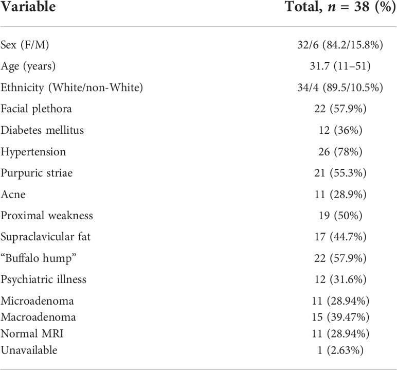
TABLE 1 Baseline clinical data of Cushing’s disease patients treated with ketoconazole.

Of the 33 patients with complete data, 26 (78%) underwent TSS prior to starting ketoconazole, five of whom (15%) had also undergone radiotherapy. Thus, seven patients used ketoconazole as primary treatment since performing a surgical procedure was impossible at that time. Of these, four had no response to ketoconazole, one had a partial response, and two had a complete response. At follow-up, four of these patients underwent their first TSS, and three continued the ketoconazole therapy, achieving full UFC control. Among those who used ketoconazole after TSS (n = 26), 20 had a complete response, two had a partial response, and four had no response. Figure 1 shows the study flow chart and patient distribution throughout the treatment.

Figure 1
www.frontiersin.org
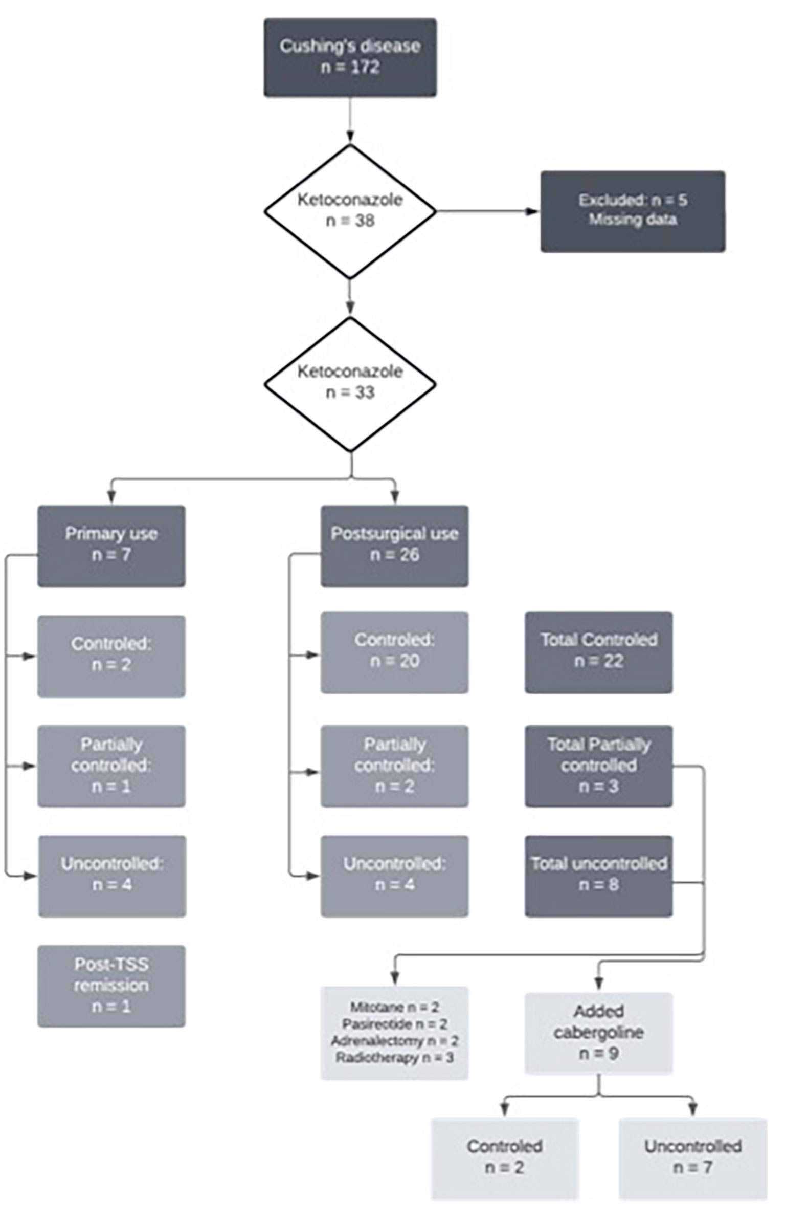
FIGURE 1 Flowchart of ketoconazole treatment in Cushing’s disease patients.

Individual patient data are described in Table 2. The duration of ketoconazole use ranged from 14 days (in one patient who used it pre-TSS) to 14.5 years. The total follow-up time of the 22 patients with controlled CD ranged from 3 months to 14.5 years, with a mean of 5.33 years and a median of 4.8 years.

Table 2
www.frontiersin.org
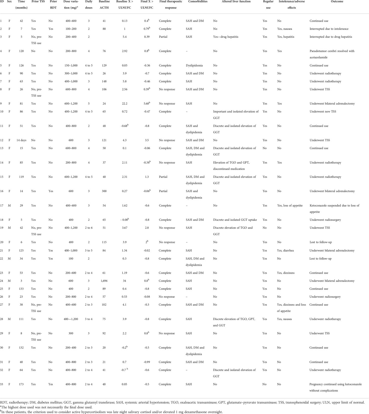
TABLE 2 Individual data.

Therapeutic response

Relative therapeutic response data are described in Table 3. Patients whose hypercortisolism was controlled or partially controlled with ketoconazole had lower baseline 24-h UFC than the uncontrolled group [times above the upper limit of normal: 0.62 (SD, 0.41) vs. 5.3 (SD, 8.21); p < 0.005, respectively], in addition to more frequent prior TSS (p < 0.04). In some patients (4/33), 24-h UFC was in the normal range at the beginning of ketoconazole therapy, but they were prescribed with the medication due to the clinical recurrence of CD associated to cortisol non-suppression after 1 mg dexamethasone overnight and/or abnormal midnight salivary or serum cortisol.

Table 3
www.frontiersin.org
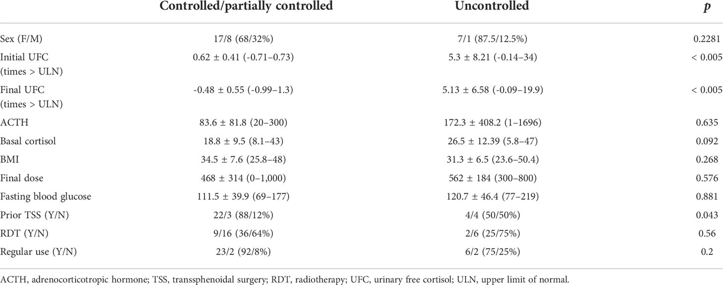
TABLE 3 Baseline characteristics of Cushing’s disease patients according to therapeutic response to ketoconazole.

Figure 2 shows that the prevalence of uncontrolled patients remained stable over time (approximately 30%) despite dose adjustments or association with other drugs, which led to no differences. When analyzing only the results of the last follow-up visit (eliminating fluctuations during follow-up), 22 patients had a complete response (66%), three patients had a partial response (9%), and eight patients had no response to ketoconazole treatment (24%), which includes patients who underwent radiotherapy during ketoconazole treatment.

Figure 2
www.frontiersin.org
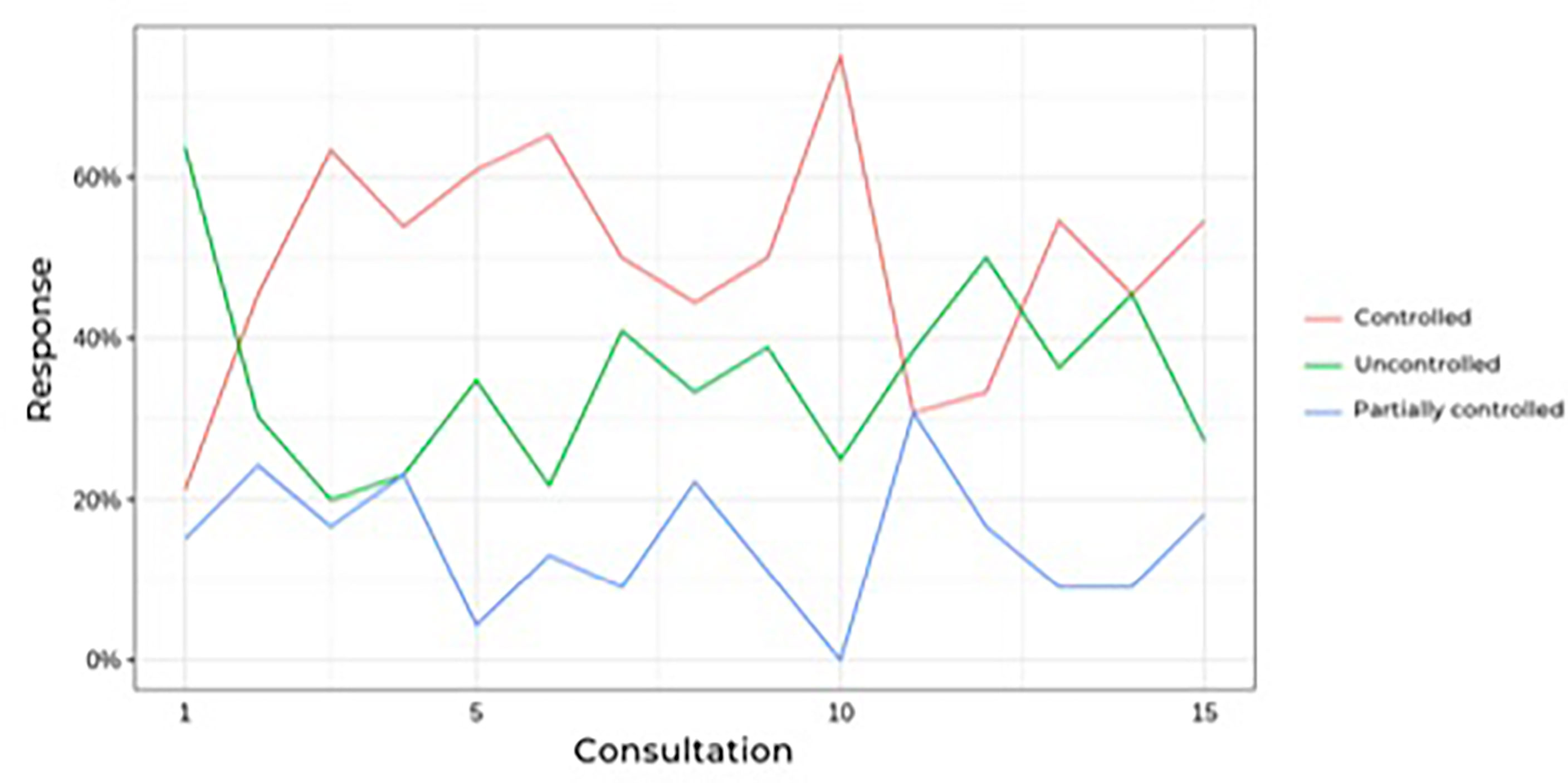
FIGURE 2 Prevalence of controlled hypercortisolism during follow-up of Cushing’s disease patients treatesd with ketoconazole.

During follow-up, no significant differences were found in blood pressure control or in dehydroepiandrosterone sulfate, cortisol, ACTH, or glucose levels. Worsening of hypertension control was observed in association with hypokalemia in some cases, as described in side effects. The ketoconazole doses ranged from 100 to 1,200 mg per day, and there were no significant dose or response differences between the groups (Table 4). Figure 3 shows the patients, their dosages, and 24-h UFC control at the first and last consultation, showing a trend toward hypercortisolism reduction in approximately 70% of the cohort (25 of 33). Only four patients used doses lower than 300 mg at the end of follow-up. One of them used before TSS and suspended its use after surgery. One patient, who has already undergone radiotherapy, discontinued ketoconazole due to intolerance, despite adequate control of hypercortisolism. Another one, who had also undergone radiotherapy, was lost to follow-up when it was controlled using 100 mg daily, and one remained controlled using 200 mg, without previous radiotherapy.

Table 4
www.frontiersin.org
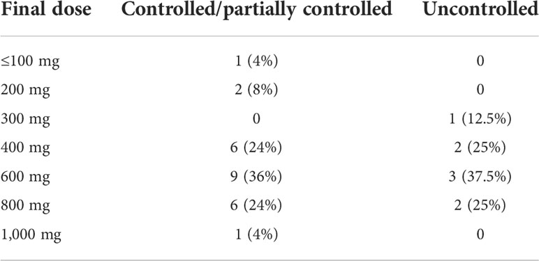
TABLE 4 Final dose of ketoconazole used in patients with Cushing’s disease.

Figure 3
www.frontiersin.org
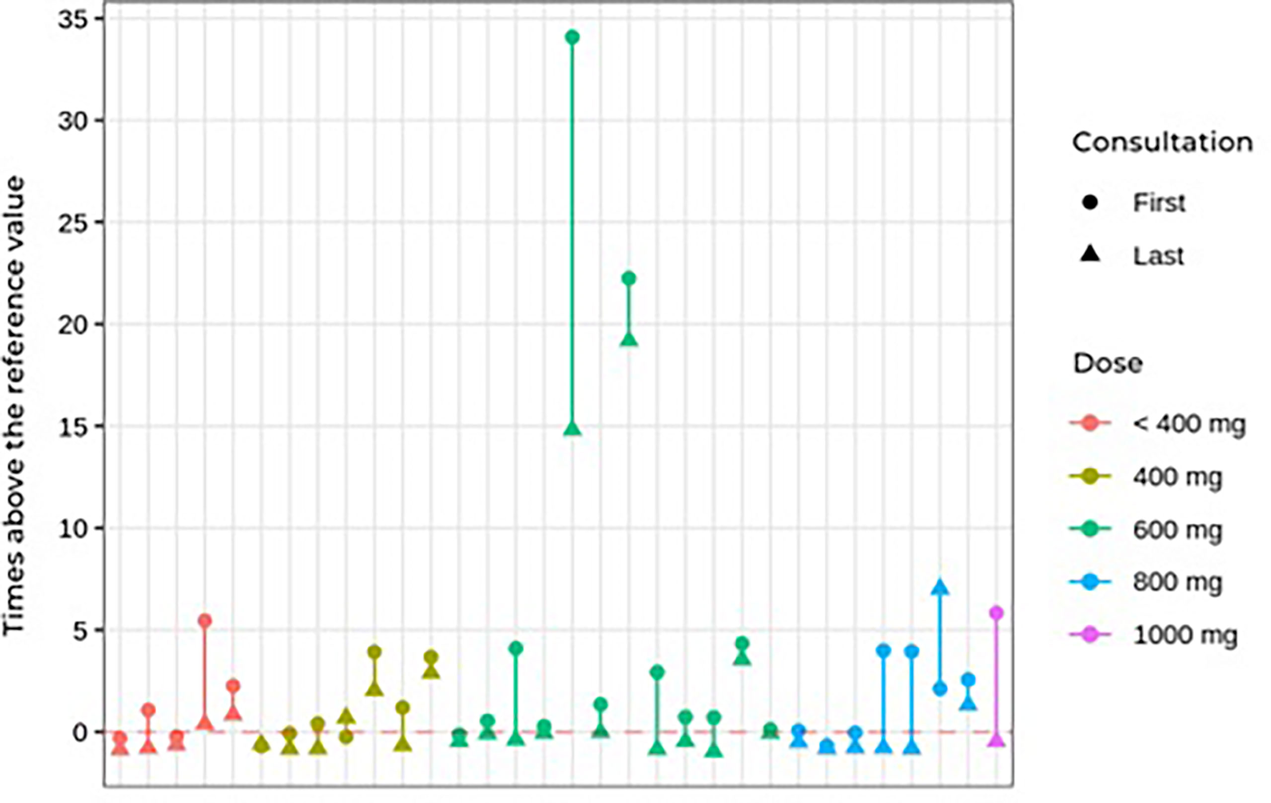
FIGURE 3 First and last consultation 24çhour UFC results vs. ketoconazole dosage in Cushing’s disease patients.

Side effects

Regarding adverse effects (Table 5), there was no significant difference between the controlled/partially controlled group and the uncontrolled group regarding liver enzyme changes or drug intolerance. Mild adverse effects, including nausea, vomiting, dizziness, and loss of appetite, occurred in 10 patients (30%). Only four patients had serious adverse effects that warranted discontinuing the medication. In two cases, ketoconazole was discontinued due to a significantly acute increase in liver enzymes (drug-induced hepatitis) during the use of 400 and 800 mg of ketoconazole. Non-significant elevation of transaminases (up to three times the normal value) was observed in three cases. A slight increase in gamma-glutamyltransferase occurred in six patients. In these nine patients with elevated liver markers, the daily dose ranged from 400 to 1,200 mg. None of those with mild increases in liver markers needed to discontinue ketoconazole.

Table 5
www.frontiersin.org
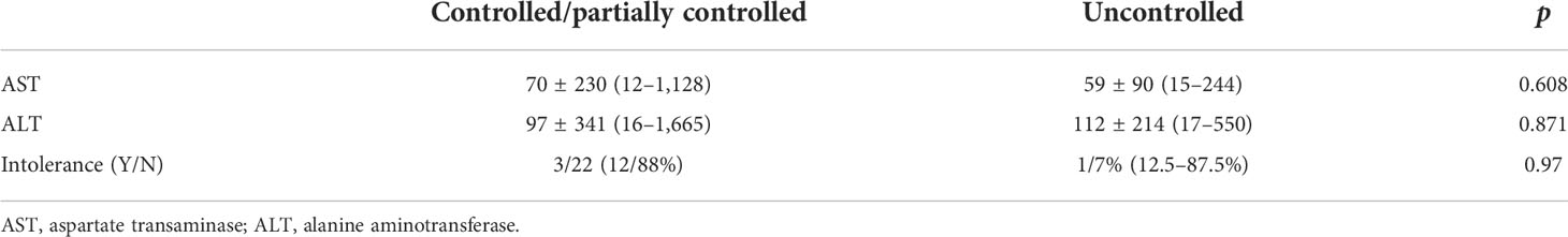
TABLE 5 Adverse effects of ketoconazole in Cushing’s disease patients treated with ketoconazole.

One female patient developed pseudotumor cerebri syndrome, which was treated with acetazolamide. She did not need to discontinue ketoconazole, having used it for more than 10 years without new side effects and achieving complete control of hypercortisolism (24). Another patient became pregnant during follow-up while using the medication, but no maternal or fetal complications occurred (25).

Hypokalemia was also observed during follow-up. Twenty episodes of reduced potassium levels occurred in 10 patients over the course of treatment. Of these episodes, six occurred in controlled patients, three in partially controlled patients, and 11 in uncontrolled patients (Table 6). The hypokalemia was managed with spironolactone (25 to 100 mg) and oral potassium supplementation.

Table 6
www.frontiersin.org
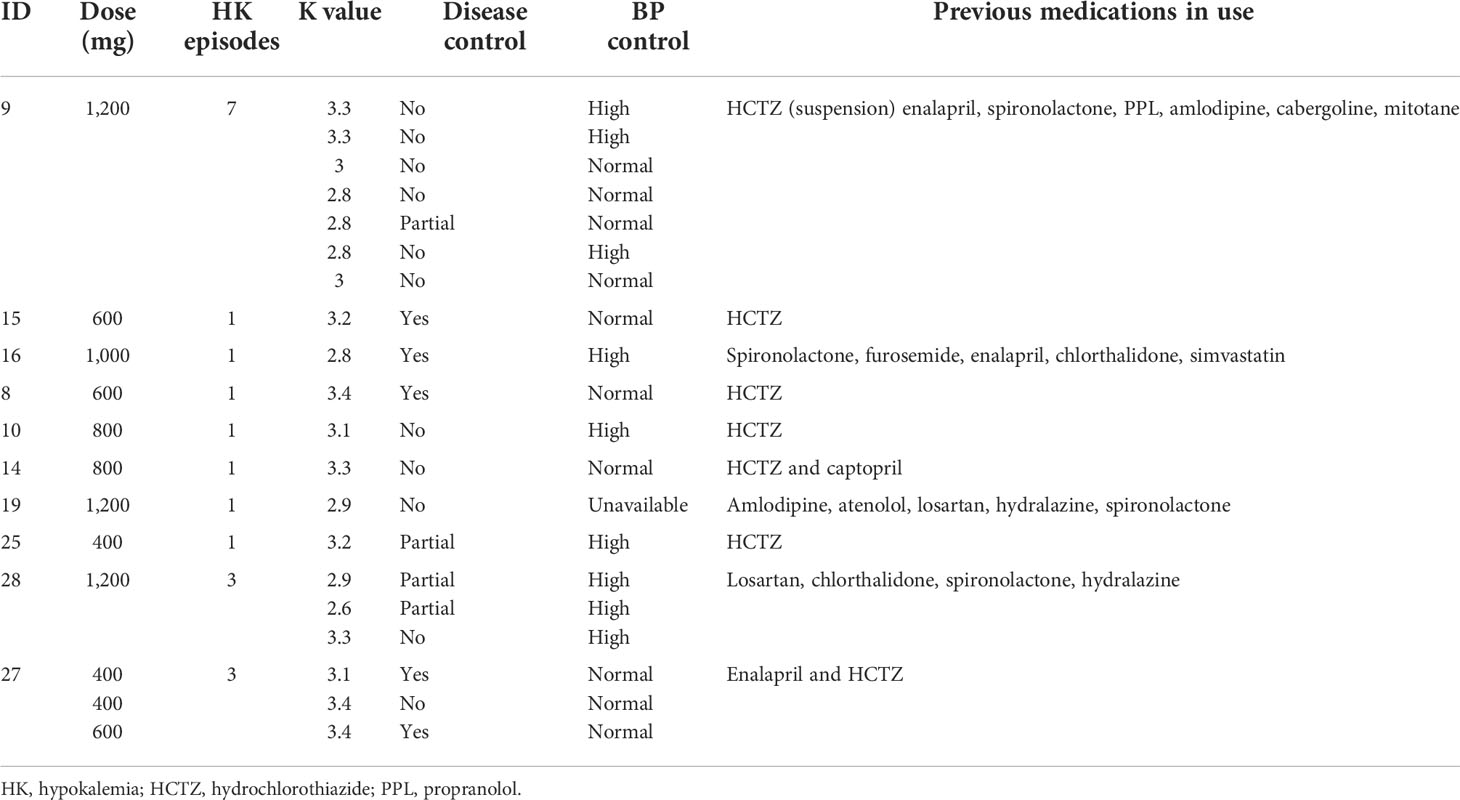
TABLE 6 Characteristics of Cushing’s disease patients who developed hypokalemia during ketoconazole treatment.

Ketoconazole and associations

Of the patients who used an association of cabergoline and ketoconazole, one did so since the beginning of follow-up, while another nine were prescribed cabergoline during follow-up due to non-response to ketoconazole alone. Of these 10 patients, two did not start the medication due to problems in obtaining the drug. Thus, in two of the nine patients on the maximum tolerated dose of ketoconazole or who could not tolerate a higher dose due to hepatic enzymatic changes, 1.5–4.5 mg of cabergoline per week was associated. In patients not controlled with ketoconazole plus cabergoline, mitotane (two patients) or pasireotide (two patients) was added. Only two of nine patients responded to the combination of cabergoline and ketoconazole. Data on these associations are shown in Table 7.

Table 7
www.frontiersin.org
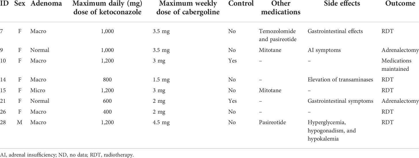
TABLE 7 Effects of associating cabergoline with ketoconazole in Cushing’s disease patients.

Considering that one of the indications for the treatment of hypercortisolism may be complementary to radiotherapy, we analyzed the eight patients who underwent radiotherapy after transsphenoidal surgery. In these patients, doses of ketoconazole from 200 to 1,200 mg were used, and in six patients there was a normalization of the UFC in 1 to 60 months of treatment. Thus, the association of ketoconazole with radiotherapy was effective in normalizing the 24-h UFC in 75% of cases.

Clinical follow-up

New therapeutic approaches were attempted in some patients during follow-up: radiotherapy (eight patients), new TSS (five patients), and bilateral adrenalectomy (four patients). At the end of this analysis, 11 patients remained on ketoconazole, all with controlled hypercortisolism. Among the 11 patients who were not fully controlled by the last visit, five were using ketoconazole as pre-TSS therapy and underwent TSS as soon as possible, while three others underwent radiotherapy and two underwent bilateral adrenalectomy. One patient was lost to follow-up.

Discussion

According to the current consensus about CD, drug treatment should be reserved for patients without remission after TSS, those who cannot undergo surgical treatment, or those awaiting the effects of radiotherapy (416). Drugs available in this context may act as adrenal steroidogenesis blockers (ketoconazole, osilodrostat, metyrapone, mitotane, levoketoconazole, and etomidate), in pituitary adenoma (somatostatinergic receptor ligands—pasireotide), dopamine receptor agonists (cabergoline), or glucocorticoid receptor blockers (mifepristone) (1626). Among these alternatives, the drug of choice still cannot be determined. Thus, the best option must be established individually, considering aspects such as remission potential, safety profile, availability, cost, etc. (162728).

For over 30 years, ketoconazole has been prescribed off-label for CD patients with varied rates of remission of hypercortisolism, and it can be used in monotherapy or associated with other drugs (2930). The Brazilian public health system does not provide drugs for the treatment of CD, and among medications with a better profile for controlling hypercortisolism, such as osilodrostat, levoketoconazole, and pasireotide, only pasireotide has been approved by the national regulatory authority (ANVISA). Due to such pragmatic considerations, ketoconazole is among the most commonly used drugs in our health system, whether recently associated or not with cabergoline (7).

In this cohort, the most prevalent response type was complete (66%). Since 75% of the CD patients who used ketoconazole had a complete or partial response, there was a clear trend towards improvement in hypercortisolism. When only those who used ketoconazole post-TSS were evaluated, the rate of control increased to 76%. We found that patients with a higher initial 24-h UFC tended to have less control of excess cortisol, a difference that was not observed when analyzing ketoconazole dose or follow-up time.

In our series and at the prescribed doses, the combination of cabergoline and ketoconazole was not effective in the management of hypercortisolism since only two of nine patients (22%) had their 24-hour UFC normalized. However, it should be observed that this association was used in patients who had more severe CD and, consequently, were less likely to have a favorable response. The effects of cabergoline in CD patients remain controversial, although some studies have shown promising responses (3132).

Previous reviews found that the efficacy of ketoconazole for hypercortisolism control was quite heterogeneous, ranging from 14 to 100% in 99 patients (3334). Our cohort’s response rate was lower than that of Sonino et al. (89%) (20) but higher than that of a multicenter cohort by Castinetti et al. (approximately 50%) (14). Regarding other smaller series (3537) our results reinforce some findings that demonstrate a percentage of control greater than 50% of the cases.

Our analyses showed a trend toward a response that continued, with some oscillations, over time. The rate of uncontrolled patients remained stable over time (approximately 30%), regardless of association with other drugs (cabergoline, mitotane, or pasireotide) or dose adjustments. Speculatively, it would appear that patients who respond to ketoconazole treatment would show some type of response as soon as therapy begins.

Our cohort has the longest follow-up time of any study on ketoconazole use in CD, nearly 15 years. Our results demonstrate that patients who benefit from ketoconazole (i.e., control of hypercortisolism and associated comorbidities) can safely use it for a long term since those who did not experience liver enzyme changes at the beginning of treatment also had no long-term changes.

Another relevant information for clinical practice is the result of treatment with ketoconazole associated with radiotherapy, which demonstrated normalizing the 24-h UFC in 75% of cases, a finding that reinforces the use of this therapeutic combination, especially in cases that are more resistant to different treatment modalities.

As described in the literature, adverse effects, such as nausea, vomiting, dizziness, headache, loss of appetite, and elevated transaminases, are relatively frequent (38). In our cohort, 10 patients (30%) had mild adverse effects, and four (12%) had more serious adverse effects requiring discontinuation. In other studies, up to 20% of patients required discontinuation due to side effects (14). We documented 20 episodes of hypokalemia during ketoconazole treatment, some with worsening blood pressure control. In most cases, hypokalemia has occurred in association with the use of diuretic drugs, which may have potentiated potassium spoliation, reinforcing the need of stringent surveillance in hypertensive Cushing’s disease patients using this combination. It can also result from the enzymatic blockade that could lead to the elevation of adrenal mineralocorticoid precursors (pex. deoxycorticosterone), with consequent sodium retention and worsening hypertension. Although it has not been analyzed in other series with ketoconazole, this side effect has been observed in patients who received other adrenal-blocking drugs, such as osilodrostat and metyrapone (16). This alteration seems to be transient in some patients; in our series, it was managed by suspending drugs that could worsen hypokalemia and introducing spironolactone and/or potassium supplementation. Hypokalemia may also result from continuing intense adrenal stimulation by ACTH and changes in the activity of the 11-beta-hydroxysteroid dehydrogenase enzyme, which increase the mineralocorticoid activity of cortisol, as observed in patients with severe hypercortisolism in uncontrolled CD (39). Hypogonadism occurred in one male patient. In two adolescent patients (one female and one male), hypercortisolism was effectively controlled without altering the progression of puberty. As described in other cohorts, this effect was expected due to the high doses, which block adrenal and testicular androgen production (20).

Thus, our findings confirm previous reports in the literature and add important information about the side effects and safety of long-term ketoconazole use in CD treatment. Our data reinforce the current recommendations about ketoconazole for recurrent cases or those refractory to surgery, including proper follow-up by an experienced team specializing in evaluating clinical and biochemical responses and potential adverse effects (71840). Despite the severity of many of our CD patients, no ketoconazole-related death occurred during follow-up, including long-term observation. On the other hand, no patient progressed to definitive remission of hypercortisolism, even after many years of treatment with ketoconazole.

Conclusions

In our cohort of patients, ketoconazole proved to be an effective and safe alternative for CD treatment, although it can produce side effects that require proper identification and management, allowing effective long-term treatment. We found side effects that have been rarely described in the literature, including hypokalemia and worsening hypertension, which require specific care and management. Thus, ketoconazole is an effective alternative for CD patients who cannot undergo surgery, who do not achieve remission after pituitary surgery, or who have recurrent hypercortisolism.

Data availability statement

The raw data supporting the conclusions of this article will be made available by the authors without undue reservation.

Ethics statement

The studies involving human participants were reviewed and approved by the Hospital de Clínicas de Porto Alegre Research Ethics Committee. Written informed consent for participation was not required for this study in accordance with the national legislation and the institutional requirements.

Author contributions

CV and MAC created the research format. CV, RBM, and MCBC realized the search on medical records. CV performed the statistical analysis. MAC, ACVM, and TCR participated in the final data review and discussion. ACVM participated in the final data review and discussion as volunteer collaborator. All authors contributed to the article and approved the submitted version.

Funding

This work was supported by the “Coordenação de Aperfeiçoamento de Pessoal de Nı́vel Superior” (CAPES), Ministry of Health – Brazil, through a PhD scholarship; and the Research Incentive Fund (FIPE) of Hospital de Clı́nicas de Porto Alegre.

Acknowledgments

The authors would like to thank the HCPA Research and Graduate Studies Group (GPPG) for the statistical technical support provided by Rogério Borges. We also thank the Research Incentive Fund of Hospital de Clínicas de Porto Alegre and Coordenação de Aperfeiçoamento de Pessoal de Nível Superior (CAPES), by funds applied. We also thank the Graduate Program in Endocrinology and Metabolism (PPGEndo UFRGS) for all the support in the preparation of this research.

Conflict of interest

The authors declare that the research was conducted in the absence of any commercial or financial relationships that could be construed as a potential conflict of interest.

Publisher’s note

All claims expressed in this article are solely those of the authors and do not necessarily represent those of their affiliated organizations, or those of the publisher, the editors and the reviewers. Any product that may be evaluated in this article, or claim that may be made by its manufacturer, is not guaranteed or endorsed by the publisher.

References

1. Fleseriu M, Castinetti F. Updates on the role of adrenal steroidogenesis inhibitors in cushing’s syndrome: a focus on novel therapies. Pituitary (2016) 19(6):643–53. doi: 10.1007/s11102-016-0742-1

PubMed Abstract | CrossRef Full Text | Google Scholar

2. Pivonello R, De Leo M, Cozzolino A, Colao A. The treatment of cushing’s disease. Endocr Rev (2015) 36(4):385–486. doi: 10.1210/er.2013-1048

PubMed Abstract | CrossRef Full Text | Google Scholar

3. Nieman LK, Biller BMK, Findling JW, Newell-Price J, Savage MO, Stewart PM, et al. The diagnosis of cushing’s syndrome: An endocrine society clinical practice guideline. J Clin Endocrinol Metab (2008) 93(5):1526–40. doi: 10.1210/jc.2008-0125

PubMed Abstract | CrossRef Full Text | Google Scholar

4. Nieman LK, Biller BMK, Findling JW, Murad MH, Newell-Price J, Savage MO, et al. Treatment of cushing’s syndrome: An endocrine society clinical practice guideline. J Clin Endocrinol Metab (2015) 100(8):2807–31. doi: 10.1210/jc.2015-1818

PubMed Abstract | CrossRef Full Text | Google Scholar

5. Pivonello R, De Martino M, De Leo M, Lombardi G, Colao A. Cushing’s syndrome. Endocrinol Metab Clin North (2008) 37(1):135–49. doi: 10.1016/j.ecl.2007.10.010

CrossRef Full Text | Google Scholar

6. Alexandraki KI, Grossman AB. Therapeutic strategies for the treatment of severe cushing’s syndrome. Drugs (2016) 76(4):447–8. doi: 10.1007/s40265-016-0539-6

PubMed Abstract | CrossRef Full Text | Google Scholar

7. Machado MC, Fragoso MCBV, Moreira AC, Boguszewski CL, Neto LV, Naves LA, et al. A review of cushing’s disease treatment by the department of neuroendocrinology of the Brazilian society of endocrinology and metabolism. Arch Endocrinol Metab (2018) 62(1):87–105. doi: 10.20945/2359-3997000000014

PubMed Abstract | CrossRef Full Text | Google Scholar

8. Rollin G, Ferreira NP, Czepielewski MA. Prospective evaluation of transsphenoidal pituitary surgery in 108 patients with Cushing’s disease. Arq Bras Endocrinol Metabol. (2007) 51(8):1355–61. doi: 10.1590/s0004-27302007000800022

PubMed Abstract | CrossRef Full Text | Google Scholar

9. Patil CG, Prevedello DM, Lad SP, Lee Vance M, Thorner MO, Katznelson L, et al. Late recurrences of cushing’s disease after initial successful transsphenoidal surgery. J Clin Endocrinol Metab (2008) 93(2):358–62. doi: 10.1210/jc.2007-2013

PubMed Abstract | CrossRef Full Text | Google Scholar

10. Rubinstein G, Osswald A, Zopp S, Ritzel K, Theodoropoulou M, Beuschlein F, et al. Therapeutic options after surgical failure in cushing’s disease: A critical review. Best Pract Res Clin Endocrinol Metab (2019) 33(2):101270. doi: 10.1016/j.beem.2019.04.004

PubMed Abstract | CrossRef Full Text | Google Scholar

11. Zhao N, Yang X, Li C, Yin X. Efficacy and safety of pasireotide for Cushing’s disease: A protocol for systematic review and meta-analysis. Medicine (Baltimore). (2020) 99(51):e23824. doi: 10.1097/MD.0000000000023824

PubMed Abstract | CrossRef Full Text | Google Scholar

12. Pivonello R, Fleseriu M, Newell-Price J, Bertagna X, Findling J, Shimatsu A, et al. Efficacy and safety of osilodrostat in patients with cushing’s disease (LINC 3): a multicentre phase III study with a double-blind, randomised withdrawal phase. Lancet Diabetes Endocrinol (2020) 8(9):748–61. doi: 10.1016/S2213-8587(20)30240-0

PubMed Abstract | CrossRef Full Text | Google Scholar

13. Yan JY, Nie XL, Tao QM, Zhan SY, De Zhang Y. Ketoconazole associated hepatotoxicity: A systematic review and meta-analysis. Biomed Environ Sci (2013) 26(7):605–10. doi: 10.3967/0895-3988.2013.07.013

PubMed Abstract | CrossRef Full Text | Google Scholar

14. Castinetti F, Guignat L, Giraud P, Muller M, Kamenicky P, Drui D, et al. Ketoconazole in cushing’s disease: Is it worth a try. J Clin Endocrinol Metab (2014) 99(5):1623–30. doi: 10.1210/jc.2013-3628

PubMed Abstract | CrossRef Full Text | Google Scholar

15. Castinetti F, Nieman LK, Reincke M, Newell-Price J. Approach to the patient treated with steroidogenesis inhibitors. J Clin Endocrinol Metab (2021) 106(7):2114–23. doi: 10.1210/clinem/dgab122

PubMed Abstract | CrossRef Full Text | Google Scholar

16. Fleseriu M, Auchus R, Bancos I, Bem-Shlomo A, Bertherat J, Biermasz NR, et al. Consensus on diagnosis and management of cushing’s disease: a guideline update. Lancet Diabetes Endocrinol (2021) 9(12):847–75. doi: 10.1016/s2213-8587(21)00235-7

PubMed Abstract | CrossRef Full Text | Google Scholar

17. Fleseriu M, Pivonello R, Elenkova A, Salvatori R, Auchus RJ, Feelders RA, et al. Efficacy and safety of levoketoconazole in the treatment of endogenous cushing’s syndrome (SONICS): a phase 3, multicentre, open-label, single-arm trial. Lancet Diabetes Endocrinol (2019) 7(11):855–65. doi: 10.1016/S2213-8587(19)30313-4

PubMed Abstract | CrossRef Full Text | Google Scholar

18. Tritos NA. Adrenally directed medical therapies for cushing syndrome. J Clin Endocrinol Metab (2021) 106(1):16–25. doi: 10.1210/clinem/dgaa778

PubMed Abstract | CrossRef Full Text | Google Scholar

19. Simões Corrêa Galendi J, Correa Neto ANS, Demetres M, Boguszewski CL, dos S V. N. nogueira, “Effectiveness of medical treatment of cushing’s disease: A systematic review and meta-analysis,”. Front Endocrinol (Lausanne) (2021) 12:732240(September). doi: 10.3389/fendo.2021.732240

PubMed Abstract | CrossRef Full Text | Google Scholar

20. Sonino N, Boscaro M, Paoletta A, Mantero F, Zillotto D. Ketoconazole treatment in cushing’s syndrome: experience in 34 patients. Clin Endocrinol (Oxf) (1991) 35(4):347–52. doi: 10.1111/j.1365-2265.1991.tb03547.x

PubMed Abstract | CrossRef Full Text | Google Scholar

21. Costenaro F, Rodrigues TC, Rollin GAF, Czepielewski MA. Avaliação do eixo hipotálamohipófise adrenal no diagnóstico e na remissão da doença de cushing. Arquivos Brasileiros Endocrinologia e Metabologia (2012). doi: 10.1590/S0004-27302012000300002

CrossRef Full Text | Google Scholar

22. Amlashi FG, Swearinger B, Faje AT, Nachtigall LB, Miller KK, Klibanski A, et al. Accuracy of late-night salivary cortisol in evaluating postoperative remission and recurrence in cushing’s disease. J Clin Endocrinol Metab (2015) 100(10):3770–7. doi: 10.1210/jc.2015-2107

PubMed Abstract | CrossRef Full Text | Google Scholar

23. Silveiro SP, Satler F. Rotinas em endocrinologia. (Porto Alegre: Artmed) (2015).

Google Scholar

24. Costenaro F, Rodrigues TC, Ferreira NP, da Costa TG, Schuch T, Boschi V, et al. Pseudotumor cerebri during cushing’s disease treatment with ketoconazole. Arq. Bras Endocrinol Metabol (2011). doi: 10.1590/s0004-27302011000400008

CrossRef Full Text | Google Scholar

25. Costenaro F, Rodrigues TC, De Lima PB, Ruszczyk J, Rollin G, Czepielewski MA. A successful case of cushing’s disease pregnancy treated with ketoconazole. Gynecol Endocrinol (2015) 31(3):176–8. doi: 10.3109/09513590.2014.995615

PubMed Abstract | CrossRef Full Text | Google Scholar

26. Gadelha MR, Neto LV. Efficacy of medical treatment in cushing’s disease: A systematic review. Clin Endocrinol (Oxf) (2014) 80(1):1–12. doi: 10.1111/cen.12345

PubMed Abstract | CrossRef Full Text | Google Scholar

27. Fleseriu M, Petersenn S. New avenues in the medical treatment of cushing’s disease: Corticotroph tumor targeted therapy. J Neurooncol (2013) 114(1):1–11. doi: 10.1007/s11060-013-1151-1

PubMed Abstract | CrossRef Full Text | Google Scholar

28. Fleseriu M, Petersenn S. Medical management of cushing’s disease: What is the future? Pituitary (2012) 15(3):330–41. doi: 10.1007/s11102-012-0397-5

PubMed Abstract | CrossRef Full Text | Google Scholar

29. Feelders RA, De Bruin C, Pereira AM, Romijn JÁ, Netea-Maier RT, Hermus AR, et al. Pasireotide alone or with cabergoline and ketoconazole in cushing’s disease. N Engl J Med (2010) 362(19):1846–8. doi: 10.1056/NEJMc1000094

PubMed Abstract | CrossRef Full Text | Google Scholar

30. Barbot M, Albiger N, Ceccato F, Zilio M, Frigo AC, Denaro Lc, et al. Combination therapy for cushing’s disease: Effectiveness of two schedules of treatment. should we start with cabergoline or ketoconazole? Pituitary (2014) 17(2):109–17. doi: 10.1007/s11102-013-0475-3

PubMed Abstract | CrossRef Full Text | Google Scholar

31. Vilar L, Naves LA, Azevedo MF, Arruda MJ, Arahata CM, Silva LM, et al. Effectiveness of cabergoline in monotherapy and combined with ketoconazole in the management of cushing’s disease. Pituitary (2010) 13(2):123–9. doi: 10.1007/s11102-009-0209-8

PubMed Abstract | CrossRef Full Text | Google Scholar

32. Pivonello R, De Martino MC, Cappabianca P, De Leo M, Faggiano A, Lombardi G, et al. The medical treatment of cushing’s disease: Effectiveness of chronic treatment with the dopamine agonist cabergoline in patients unsuccessfully treated by surgery. J Clin Endocrinol Metab (2009) 94(1):223–30. doi: 10.1210/jc.2008-1533

PubMed Abstract | CrossRef Full Text | Google Scholar

33. Castinetti F, Morange I, Jaquet P, Conte-Devolx B, Brue T. Ketoconazole revisited: A preoperative or postoperative treatment in cushing’s disease. Eur J Endocrinol (2008). doi: 10.1530/EJE-07-0514

PubMed Abstract | CrossRef Full Text | Google Scholar

34. Loli P, Berselli ME, Tagliaferri M. Use of ketoconazole in the treatment of cushing’s syndrome. J Clin Endocrinol Metab (1986) 63(6):1365–71. doi: 10.1210/jcem-63-6-1365

PubMed Abstract | CrossRef Full Text | Google Scholar

35. Kakade HR, Kasaliwal R, Khadilkar KS, Jadhav S, Bukan A, Khare Sc, et al. Clinical, biochemical and imaging characteristics of cushing’s macroadenomas and their long-term treatment outcome. Clin Endocrinol (Oxf) (2014) 81(3):336–42. doi: 10.1111/cen.12442

PubMed Abstract | CrossRef Full Text | Google Scholar

36. Luisetto G, Zangari M, Camozzi V, Boscaro M, Sonino N, Fallo F. Recovery of bone mineral density after surgical cure, but not by ketoconazole treatment, in cushing’s syndrome. Osteoporos Int (2001) 12(11):956–60. doi: 10.1007/s001980170025

PubMed Abstract | CrossRef Full Text | Google Scholar

37. Huguet I, Aguirre M, Vicente A, Alramadan M, Quiroga I, Silva J, et al. Assessment of the outcomes of the treatment of cushing’s disease in the hospitals of castilla-la mancha. Endocrinol y Nutr (2015) 62(5):217–23. doi: 10.1016/j.endonu.2015.02.007

CrossRef Full Text | Google Scholar

38. Tritos NA, Biller BMK. Advances in the medical treatment of cushing disease. Endocrinol Metab Clin North Am (2020) 49(3):401–12. doi: 10.1016/j.ecl.2020.05.003

PubMed Abstract | CrossRef Full Text | Google Scholar

39. Torpy D, Mullen N, Ilias I, Nieman L. Association of hypertension and hypokalemia with cushing’s syndrome caused by ectopic ACTH secretion. Ann N Y Acad Sci (2002) 970:134–44. doi: 10.1111/j.1749-6632.2002.tb04419.x

PubMed Abstract | CrossRef Full Text | Google Scholar

40. Varlamov EV, Han AJ, Fleseriu M. “Updates in adrenal steroidogenesis inhibitors for cushing’s syndrome – a practical guide,”. Best Pract Res Clin Endocrinol Metab (2021) 35(1):101490. doi: 10.1016/j.beem.2021.101490

PubMed Abstract | CrossRef Full Text | Google Scholar

Keywords: Cushing’s disease, Cushing’s syndrome, hypercortisolism, treatment, ketoconazole

Citation: Viecceli C, Mattos ACV, Costa MCB, Melo RBd, Rodrigues TdC and Czepielewski MA (2022) Evaluation of ketoconazole as a treatment for Cushing’s disease in a retrospective cohort. Front. Endocrinol. 13:1017331. doi: 10.3389/fendo.2022.1017331

Received: 11 August 2022; Accepted: 06 September 2022;
Published: 07 October 2022.

Edited by:

Luiz Augusto Casulari, University of Brasilia, Brazil

Reviewed by:

Juliana Drummond, Federal University of Minas Gerais, Brazil
Monalisa Azevedo, University of Brasilia, Brazil

Copyright © 2022 Viecceli, Mattos, Costa, Melo, Rodrigues and Czepielewski. This is an open-access article distributed under the terms of the Creative Commons Attribution License (CC BY). The use, distribution or reproduction in other forums is permitted, provided the original author(s) and the copyright owner(s) are credited and that the original publication in this journal is cited, in accordance with accepted academic practice. No use, distribution or reproduction is permitted which does not comply with these terms.

*Correspondence: Mauro Antonio Czepielewski, maurocze@terra.com.br

Disclaimer: All claims expressed in this article are solely those of the authors and do not necessarily represent those of their affiliated organizations, or those of the publisher, the editors and the reviewers. Any product that may be evaluated in this article or claim that may be made by its manufacturer is not guaranteed or endorsed by the publisher.

From https://www.frontiersin.org/articles/10.3389/fendo.2022.1017331/full

TP53 Mutations in Functional Corticotroph Tumors Are Linked to Invasion and Worse Clinical Outcome

Abstract

Corticotroph macroadenomas are rare but difficult to manage intracranial neoplasms. Mutations in the two Cushing’s disease mutational hotspots USP8 and USP48 are less frequent in corticotroph macroadenomas and invasive tumors. There is evidence that TP53 mutations are not as rare as previously thought in these tumors. The aim of this study was to determine the prevalence of TP53 mutations in corticotroph tumors, with emphasis on macroadenomas, and their possible association with clinical and tumor characteristics. To this end, the entire TP53 coding region was sequenced in 86 functional corticotroph tumors (61 USP8 wild type; 66 macroadenomas) and the clinical characteristics of patients with TP53 mutant tumors were compared with TP53/USP8 wild type and USP8 mutant tumors. We found pathogenic TP53 variants in 9 corticotroph tumors (all macroadenomas and USP8 wild type). TP53 mutant tumors represented 14% of all functional corticotroph macroadenomas and 24% of all invasive tumors, were significantly larger and invasive, and had higher Ki67 indices and Knosp grades compared to wild type tumors. Patients with TP53 mutant tumors had undergone more therapeutic interventions, including radiation and bilateral adrenalectomy. In conclusion, pathogenic TP53 variants are more frequent than expected, representing a relevant amount of functional corticotroph macroadenomas and invasive tumors. TP53 mutations associated with more aggressive tumor features and difficult to manage disease.

Introduction

Pituitary neuroendocrine tumors are the second most common intracranial neoplasm [1]. They are usually benign, but when aggressive they may be particularly difficult to manage, accompanied by high comorbidity and increased mortality [2]. Corticotroph tumors constitute 6–10% of all pituitary tumors, but they represent up to 45% of aggressive pituitary tumors and pituitary carcinomas [2]. Functional corticotroph tumors cause Cushing’s disease (CD), a debilitating condition accompanied by increased morbidity and mortality due to glucocorticoid excess [3]. Pituitary surgery is the first line treatment, but recurrence is observed in 15–20% of cases of whom most are macroadenomas (with a size of ≥ 10 mm) [4]. Treatment options include repeated pituitary surgery, radiation therapy, medical treatment and bilateral adrenalectomy (BADX) [3]. With respect to the latter, corticotroph tumor progression after bilateral adrenalectomy/Nelson’s syndrome (CTP-BADX/NS) is a frequent severe complication and may present with aggressive tumor behavior [5,6,7].

Corticotroph tumors (including CTP-BADX/NS) carry recurrent somatic mutations in the USP8 gene in ~ 40–60% of cases [8,9,10,11,12,13]. These USP8 mutant tumors are usually found in female patients and are generally less invasive [8,9,10,11]. Additional genetic studies identified a second mutational hotspot in the USP48 gene, but no other driver mutations [14,15,16,17,18]. Focusing on USP8 wild type corticotroph tumors, we recently discovered TP53 mutations in 6 out of 18 cases (33%) [17]. Subsequent reports documented TP53 mutations in small series of mainly aggressive corticotroph tumors and carcinomas [1920].

TP53 is the most commonly mutated gene in malignant neoplasms [2122], including brain and neuroendocrine tumors [2324]. Until our previous report [17], TP53 mutations were only described in isolated cases of aggressive pituitary tumors and carcinomas, and were therefore considered very rare events [81625,26,27,28]. A link between TP53 mutations and an aggressive corticotroph tumor phenotype has been hypothesized, but the heterogeneity and small size of the studies reported did not support significant clinical associations [1719].

To address this, we determined the prevalence of TP53 variants in a cohort of 86 patients with functional corticotroph tumors, including 61 with USP8 wild type tumors, and studied the associations between TP53 mutational status and clinical features.

Methods

Patients and samples

We analyzed tumor samples of 86 adult patients: 61 USP8 wild type and 25 USP8 mutant. Sixty-six patients (46 females, 20 males) were diagnosed with CD between 1994 and 2020 in Germany (Hamburg, Munich, Erlangen, and Tübingen) and Luxembourg. Twenty additional patients (16 females, 4 males) were diagnosed with CTP-BADX/NS, operated and followed up in 7 different international centers (Nijmegen, Munich, Erlangen, Hamburg, Paris, Rio de Janeiro, and Würzburg). Twenty-three out of 86 samples were collected prospectively between 2018 and 2021, and 63 were retrospective cases (of which 42 were investigated in the context of USP8 and USP48 screenings and published elsewhere) [9121317]. Seventy-one tumors were fresh frozen and 15 were formalin fixed paraffin embedded. Paired blood was available for 12 cases. The median follow-up time after initial diagnosis was 44 months (range 2–384 months).

Endogenous Cushing’s syndrome was diagnosed according to typical clinical signs and symptoms and established biochemical procedures suggesting glucocorticoid excess. Clinical features included central obesity, moon face, buffalo hump, muscle weakness, easy bruising, striae, acne, low-impact bone fractures, mood changes, irregular menstruation, infertility and impotency. Biochemical diagnosis was based on increased 24 h urinary free cortisol (UFC) and late-night salivary cortisol levels, and lack of serum cortisol suppression after low-dose dexamethasone test. A pituitary ACTH source was confirmed by > 2.2 pmol/l (10 pg/ml) basal plasma ACTH, > 50% suppression of serum cortisol during an 8 mg dexamethasone test, and ACTH and cortisol response to corticotrophin releasing hormone stimulation.

The clinical and pathological features of our study cohort are summarized in Additional file 1: Supplementary Table 1. All patients underwent pituitary surgery. The presence of an ACTH-producing pituitary tumor was confirmed histologically after surgical resection. Biochemical remission after surgery was defined as postoperative 24 h-UFC levels below or within the normal range, or serum cortisol levels < 5 µg/dl after low-dose (1 or 2 mg) dexamethasone suppression test. Tumor control was achieved when there was no evidence of regrowth or disease recurrence. Tumor invasion was defined as radiological or intraoperative evidence of tumor within the sphenoid and/or cavernous sinuses [29]. CTP-BADX/NS was defined as an expanding pituitary tumor after bilateral adrenalectomy (BADX) following expert consensus recommendations [5].

DNA extraction, TP53 amplification and sequencing

Genomic DNA was extracted using the Maxwell Tissue DNA Kit (Promega), Maxwell Blood DNA kit (Promega) or the FFPE DNA mini kit (Qiagen), depending on the type of sample, as described previously [912]. The entire coding sequence of TP53 (including exons 9β and 9γ) as well as noncoding regions adjacent to each exon were amplified using the GoTaq DNA polymerase (Promega) and specific primers (Additional file 1: Supplementary Table 2). Amplification of USP8 hotspot region and Sanger sequencing were performed as described previously [912]. Chromatograms were analyzed using the Mutation Surveyor v4.0.9 (Soft Genetics). Samples were examined for TP53 coding and splicing variants. Variant position and pathogenicity was investigated in ENSEMBL (www.ensembl.org), the UCSC Genome Browser (http://genome-euro.ucsc.edu), the IARC TP53 database (https://p53.iarc.fr/TP53GeneVariations.aspx), the Catalogue Of Somatic Mutations in Cancer (COSMIC; https://cancer.sanger.ac.uk/cosmic), ClinVar (https://www.ncbi.nlm.nih.gov/clinvar/), PHANTM (http://mutantp53.broadinstitute.org/), the Human Splicing Finder (HSF; http://www.umd.be/HSF3/) and VarSEAK splicing predictor (https://varseak.bio/). Variant frequencies on the general population were obtained from the Allele Frequency Aggregator (ALFA) project [30], the Genome Aggregation Database (gnomAD) [31] and the International Genome Sample Resource 1000Genome project [32]. Throughout the text, variants refer to NC_000017.11 (genomic DNA), ENST00000269305.9 (coding DNA) and ENSP00000269305.4 (protein), following the Human Genome Variation Society (HGVS) standard nomenclature system.

Statistical analysis

Statistical analysis was performed with the software package SPSS v24 (IBM). We used t-test or one-way ANOVA to analyze the association of TP53 variants with age, body mass index; Mann–Whitney U and Kruskal–Wallis to test non-parametric variables, such as tumor size, hormone levels, Ki67 index and p53 score. We corrected the analysis for multiple comparisons with the Bonferroni test. Categorical variables were analyzed using a chi-square test or Fisher exact test when needed. Survival analysis was performed using Kaplan–Meier curves with log-rank tests, and multivariate Cox regression. An exact, two-tailed significance level of P < 0.05 was considered to be statistically significant.

Results

Analysis of TP53 nucleotide variants

We analyzed all TP53 coding exons (including exons 9β and 9γ) and adjacent intronic noncoding sequences in 61 USP8 wild type tumors (49 CD and 12 CTP-BADX/NS). Of these, 13 were microadenomas (< 10 mm) and 48 macroadenomas (≥ 10 mm) at the time of the current operation. A separate group of 25 USP8 mutant tumors (17 CD and 8 CTP-BADX/NS) that were mainly macroadenomas (n = 19) was used for multiple comparison.

We found 59 variants in our cohort: 30 exclusively in USP8 wild type, 21 in USP8 mutant, and 8 in wild type and mutant tumors regardless of USP8 mutational status. No indels in the coding region of TP53 were detected. In addition, we did not find any genetic variant affecting TP53 splicing.

Nine out of 30 variants found in USP8 wild type tumors were either reported in the COSMIC database as pathogenic or absent from the common variant databases (1000Genomes, gnomAD, ALPHA) or had allele frequency < 0.0001. They were all described in cancer series: 5 as pathogenic or likely pathogenic in ClinVar, 2 as variants of uncertain significance (VUS) and 2 were not described in ClinVar (Table 1). All variants are reported to alter protein function and show clear loss of transactivation activity in a yeast based assay (Table 1) [33].

Table 1 Functionally relevant TP53 variants found in 9/86 corticotroph tumors

Seven variants target amino acids within the DNA-binding domain, essential for p53 activity, disrupting S2’ and S7 β-sheets or the L3 loop spatial conformation. The other two [c.1009C > G (p.Arg337Gly) and c.1031 T > C (p.Leu344Pro)] locate in the tetramerization domain and keep p53 protein as monomer impairing its transactivation activity [34]. From the 9 variants, 8 affect highly conserved p53 residues, while in c.1031 T > C (p.Met133Lys) the methionine alternates with leucine or valine among species. This variant alters protein folding, probably reducing DNA affinity [35], while the substitution of a methionine that acts as an alternative start codon abolishes the transcription of isoforms ∆133p53α, ∆133p53β and ∆133p53γ. The 9 variants were detected in nine cases (henceforth referred to as TP53 mutant; Table 1). Two tumors from unrelated patients (#6 and #7) carried the same variant c.818G > A (p.Arg273His), while one tumor (#4) carried two variants (c.718A > G and c.773A > C). Seven variants were found in heterozygosis, while the other two (from patients #1 and #2) in homozygosis. From these two, we only had paired blood/tumor samples from patient #1 and detected the variant only on the tumor sample, indicative of loss of heterozygosity (Additional file 1: Supplementary Fig. 1A). Similarly, we could demonstrate the somatic origin of the TP53 variants in four other patients with paired tumor/blood samples (#3, #5, #6 and #9).

The remaining 21/30 variants found in USP8 wild type and all 21 variants found in the USP8 mutant tumors were described as benign, likely benign or VUS with no evidence of affecting protein function. All tumors with these variants were considered TP53 wild type. From the 21 variants found in the USP8 wild type tumors (henceforth referred to as TP53/USP8 wild type group), 7 were non-synonymous variants, 8 synonymous variants and 6 non-coding variants without splicing effect. From the 21 variants found in the 25 USP8 mutant tumors, nine were synonymous, four non-synonymous and eight non-coding without splicing effect. In addition, eight variants were found in tumors regardless of USP8 mutational status that were not categorized as TP53 mutations. The intronic variant c.782 + 62G > A was found in heterozygosis in 6/70 samples. It was not reported in any database and is not predicted to have any splicing effect. The remaining seven are common variants classified as benign or likely benign in ClinVar and their allele frequencies were similar to those reported for the general population (ALFA, gnomAD and 1000Genome project) (Additional file 1: Supplementary Table 3).

Summarizing, all TP53 mutations were found in the USP8 wild type tumors, leading to a prevalence of 15% in this subgroup.

Clinical presentation of patients with TP53 mutant tumors

Patients with TP53 mutant tumors (n = 9) tended to be diagnosed at older age compared to TP53/USP8 wild type tumors (n = 52) (t-test P = 0.069; Table 2). This was significant after including the USP8 mutant group (n = 25) in the multiple comparison analysis (ANOVA P = 0.024, Table 2) and when TP53/USP8 wild type and USP8 mutant tumors were combined to a single group (TP53 wild type, n = 77; Additional file 1: Supplementary Table 4. We did not observe any sex specific predominance of TP53 mutations in contrast to USP8 mutants that are predominantly found in female patients. Furthermore, we did not find any statistically significant differences in ACTH and cortisol levels (Table2; Additional file 1: Supplementary Table 4).

Table 2 Clinical features of TP53 mutant versus TP53/USP8 wild type and USP8 mutant groups

Patients with TP53 mutant tumors underwent more surgeries and tumor resection was more frequently incomplete compared to TP53/USP8 wild type (Table 2). These patients also underwent a higher number of additional therapeutic procedures (radiation, n = 7; BADX, n = 4; temozolomide, n = 3; pasireotide, n = 2). Only one patient (#4) with TP53 mutant tumor, a 77 year-old man, had a single surgery without any other treatment, but his follow-up was short (< 6 months).

We observed TP53 mutations more frequently in CTP-BADX/NS (4/12, 33%) compared to CD (5/49, 10%), trending towards statistically significant difference (Fischer exact test P = 0.065 for TP53 mutant vs. TP53/USP8 wild type, P = 0.060 for comparison among the 3 groups; Table 2).

The TP53 mutant group associated with higher disease-specific mortality and shorter survival than USP8 mutant or TP53/USP8 wild type groups (log rank test, P = 0.023, Fig. 1). Three patients with TP53 mutant tumors (all CTP-BADX/NS) died of disease-related deaths: two from severe cerebral hemorrhage after surgery and stereotactic radiation and one from uncontrolled disease after five failed operations, radiotherapy (gamma knife, fractionated radiation) and chemotherapy (temozolomide, bevacizumab) at the ages of 75, 80 and 37, respectively. Ten-year survival was 27% for patients with TP53 mutant tumors, 100% for TP53/USP8 wild type and 86% for USP8 mutant. In our cohort, survival did not differ after adjusting for age (HR 7.7, 95%CI 0.6–107.7, P = 0.127).

Fig. 1

figure 1

Kaplan–Meier curve showing overall survival in patients with TP53 mutant/USP8 wild type, USP8 mutant/TP53 wild type, and TP53 wild type/USP8 wild type corticotroph tumors. The table underneath the graph shows the 10-year cumulative survival after diagnosis

Tumor samples from prior surgeries were available from one TP53 mutant case (#8, Table 1). This male patient had his first pituitary surgery for CD when he was 30 years old and was treated with γ-knife one year later. He then underwent two more pituitary surgeries and BADX until the age of 35. He developed CTP-BADX/NS with para- and retrosellar tumor extension along with panhypopituitarism and underwent two more pituitary surgeries before dying at the age of 38 due to complications of the disease. We detected the TP53 variant c.1009C > G (p.Arg337Gly) in all available tumor specimens, including his first and latest surgeries (Additional file 1: Supplementary Fig. 1B).

No statistical association was found between clinical data and any of the 8 common variants.

Characteristics of TP53 mutant corticotroph tumors

All TP53 mutations were found in macroadenomas (9/66; Table 3). TP53 mutant tumors were larger that TP53/USP8 wild type (mm median [IQR] 20.0 [14.0] vs. 15.0 [14.3]), but this did not reach statistical significance (Table 3). Multiple comparison analysis showed that the difference in tumor size is significant only comparing TP53 mutant with USP8 mutant (median [IQR] 23.3 [14.0] vs. 14 [7.3] mm; Kruskal–Wallis P = 0.019; Bonferroni corrected P = 0.018).

Table 3 Tumor features of TP53 mutant versus TP53/USP8 wild type and USP8 mutant groups

Parasellar invasion was reported in 34 out of 64 cases, for which this information was available, and it was more common in TP53 mutant tumors (100% vs. 53% and 55% for TP53/USP8 wild type and USP8 mutant, respectively; Fischer exact test P = 0.006). TP53 mutant tumors had higher Knosp grade (Kruskal–Wallis P = 0.011) with the majority being Knosp 4 (Table 3, Additional file 1: Supplementary Table 4).

Ki67 proliferation index was available for 36 cases (6 TP53 mutant). Five out of six TP53 mutant tumors had Ki67 ≥ 3% and the overall Ki67 was higher than in the wild type tumors (Kruskal–Wallis P = 0.01; Bonferroni corrected P = 0.008 for TP53/USP8 wild type) (Table 3). Ki67 ≥ 10% was reported in 6 tumors, from which 5 were TP53 mutant (Fischer exact test P < 0.0001; the remaining case was TP53/USP8 wild type).

We had information on p53 immunostaining from 9 cases (all macroadenomas), four of which TP53 mutant: 3 tumors (from patients #5, 6 and 9) showed high p53 immunoreactivity, while the one (from patient #3) carrying a nonsense variant leading to a truncated protein was p53 negative. The five TP53 wild type cases showed isolated nuclear staining in < 1–3% of cells.

Summarizing, TP53 mutations were significantly associated with features related to a more aggressive tumor behavior, such as incomplete tumor resection, more frequent parasellar invasion, higher Knosp grade, and higher Ki67 proliferation index (Table 3; Additional file 1: Supplementary Table 4).

Discussion

Herein, we investigated the prevalence of TP53 mutations by screening a large cohort of 61 functional corticotroph tumors with USP8 wild type status, and found variants altering protein function in 15% of cases. We did not detect TP53 mutations in a separate group of 25 USP8 mutant tumors, which is in concordance with previously published small next-generation sequencing series [81819].

Since we focused on USP8 wild type tumors, macroadenomas were overrepresented in our cohort. Consequently, it should be noted that the prevalence of TP53 mutations is expected to be lower in the general CD population. In fact, ~ 50% of corticotroph tumors carry USP8 mutations, which others and we have shown to be mutually exclusive. Corticotroph tumors with USP8 mutations are associated with female predominance, younger age at presentation, and less invasiveness (despite shorter time to relapse) [911131836]. In contrast, TP53 mutant tumors were diagnosed mostly at older age, did not show sex predominance and were larger and more invasive, with lower complete resection rate. None of the 19 microadenomas included in our study carried TP53 mutations. Still, we need to acknowledge that since no sample was microdissected we may have lost microadenoma cases with TP53 mutations. Instead, we found TP53 mutations in 9/66 macroadenomas (14%) and 8/34 (24%) invasive tumors, supporting the findings from smaller series [1719].

Tumor size at presentation or invasiveness do not reliably predict aggressiveness. Instead, the European Society of Endocrinology Clinical Practice Guidelines for the management of aggressive pituitary tumors and carcinomas proposed a definition of pituitary tumor aggressiveness based on rapid or clinically relevant tumor growth despite optimal therapeutic options, along with bone invasion [37]. A recent study in a series of 9 aggressive pituitary tumors and carcinomas carrying ATRX mutations reported a high frequency of missense TP53 variants (5/9, 55.6%), further suggesting a link between TP53 mutational status and unfavorable outcome [20]. We do not have exact information on changes of tumor growth for the majority of our cases, but the higher number of surgical and radiation interventions, the higher Knosp grades, and the increased mortality rate indicate that patients with TP53 mutant tumors obviously follow a more aggressive disease course.

Ki67 proliferation index together with p53 immunostaining and mitotic count have been suggested as histological markers of pituitary tumor aggressiveness [2938]. In our series, Ki67 was significantly higher in TP53 mutant tumors, reinforcing our prior observation of a higher proportion of TP53 mutant tumors in the Ki67 ≥ 3 group [17]. We had limited information on p53 immunohistochemistry, since this measure is not routinely performed in our collaborative centers. Nevertheless, in the few tumors with known p53 immunopositivity, it was higher in the TP53 mutant group, which is in concordance with a previous study reporting high p53 immunoreactivity in all TP53 mutant tumors [19].

A mutagenic action of radiation on TP53 has been hypothesized by small series on radiation-induced tumors. For instance, TP53 mutations were reported in 58% of radiation-induced sarcomas [39], while a meta-analysis reported TP53 mutations in 14/30 radiation-induced gliomas [40]. A previous study reported a case with frameshift TP53 mutation in the CTP-BADX/NS tumor, but not in the initial CD surgeries, and the mutation was therefore suspected to be induced by radiotherapy [41]. In our series, however, 4 out of 7 TP53 mutant tumors were obtained before radiation.

In their case report, Pinto et al. suggested that TP53 mutations are acquired during tumorigenesis and condition tumor evolution [41]. In contrast, Casar-Borota et al. and Uzilov et al. reported high allele fraction of TP53 mutations, indicating that they are not a late event in corticotroph tumorigenesis [1920]. In addition, Uzilov et al. reported TP53 mutations in all tumor specimens from their two TP53 mutant cases with multiple surgeries [19]. Similarly, in our series we had tissue from multiple pituitary surgeries from one patient and found the TP53 variant in all samples (CD and CTP-BADX/NS), including specimens obtained before radiotherapy. Taken together, these observations suggest that in most cases, TP53 mutations may appear early during tumor development.

A limitation of our study is the short follow-up of patients who were prospectively included. Moreover, material from repeated surgeries was lacking from most patients with TP53 mutant tumors, hampering the examination of tumor evolution in these patients. Similarly, we had limited access to blood samples, so we could not demonstrate the somatic origin for all variants. Nevertheless, the older age at initial diagnosis of CD in patients with TP53 mutant tumors (53 ± 19.5 years old, with the youngest patient diagnosed at the age of 30) and the absence of additional neoplasias during follow-up also support a somatic instead of a germline origin. Furthermore, conditions related to germline TP53 mutations, such as Li-Fraumeni syndrome, very rarely present with pituitary tumor [42]. To our knowledge, the only published case so far was a pediatric patient with an aggressive lactotroph tumor [43].

In addition to the TP53 mutations, we detected several common variants. Variants rs59758982 and rs1042522 have been associated with increased cancer susceptibility [4445]. In some cancer types, the very frequent rs1042522 c.215G > C (p.Pro72Arg) alternative variant correlated to more efficient induction of apoptosis by DNA-damaging chemotherapeutic drugs, growth suppression and higher metastatic potential [46,47,48]. In nonfunctioning pituitary tumors, alternative allele C (leading to p.Arg72) was related to early age at presentation and reduced p21 expression [49]. Very recently, an overrepresentation of the rs1042522 alternative allele C (p.Arg72) was reported in 9 out of 10 corticotroph neoplasias including 5 functional tumors (allele frequency 0.900, vs 0.714 in Latino/admixed American in gnomAD [31]) without any association with clinical features [50]. In our cohort, we did not detect different allele frequencies in any of the investigated common variants (including rs1042522) compared with public databases, nor statistical association with any clinical variable, rendering their contribution to corticotroph pathophysiology unlikely.

Conclusion

Screening a large corticotroph tumor series revealed that TP53 mutations are more frequent than previously considered. Furthermore, we show that patients with TP53 mutant tumors had higher number of surgeries, more invasive tumors, and worse disease outcome. Our study provides evidence that patients with pathogenic or function altering variants may require more intense treatment and extended follow-up, and suggests screening for TP53 variants in macroadenomas with wild type USP8 status. Further work is needed to determine the potential use of TP53 status as a predictor of disease outcome.

Availability of data and materials

The authors declare that the relevant data supporting the conclusions of this article are included within the article and its supplementary information file. Additional clinical data are available from the corresponding authors MT and LGPR upon reasonable request.

Abbreviations

CD:
Cushing’s disease
BADX:
Bilateral adrenalectomy
CTP-BADX/NS:
Corticotroph tumor progression after bilateral adrenalectomy/Nelson’s syndrome
ACTH:
Adrenocorticotropic hormone
SD:
Standard deviation
IQR:
Interquartile range
HR:
Hazard ratio

References

  1. Ostrom QT, Gittleman H, Truitt G, Boscia A, Kruchko C, Barnholtz-Sloan JS (2018) CBTRUS statistical report: primary brain and other central nervous system tumors diagnosed in the United States in 2011–2015. Neuro Oncol 20:iv1-86

    PubMed PubMed Central Article Google Scholar

  2. McCormack A, Dekkers OM, Petersenn S, Popovic V, Trouillas J, Raverot G et al (2018) Treatment of aggressive pituitary tumours and carcinomas: results of a European society of endocrinology (ESE) survey 2016. Eur J Endocrinol 178:265–276

    CAS PubMed Article Google Scholar

  3. Fleseriu M, Auchus R, Bancos I, Ben-Shlomo A, Bertherat J, Biermasz NR et al (2021) Consensus on diagnosis and management of Cushing’s disease: a guideline update. Lancet Diabetes Endocrinol 9:847–875

    PubMed Article Google Scholar

  4. Dimopoulou C, Schopohl J, Rachinger W, Buchfelder M, Honegger J, Reincke M et al (2013) Long-term remission and recurrence rates after first and second transsphenoidal surgery for Cushing’s disease: care reality in the Munich metropolitan region. Eur J Endocrinol 170:283–292

    PubMed Article CAS Google Scholar

  5. Reincke M, Albani A, Assie G, Bancos I, Brue T, Buchfelder M et al (2021) Corticotroph tumor progression after bilateral adrenalectomy (Nelson’s syndrome): systematic review and expert consensus recommendations. Eur J Endocrinol 184:P1-16

    CAS PubMed PubMed Central Article Google Scholar

  6. Fountas A, Lim ES, Drake WM, Powlson AS, Gurnell M, Martin NM et al (2020) Outcomes of patients with Nelson’s syndrome after primary treatment: a multicenter study from 13 UK pituitary centers. J Clin Endocrinol Metab 105:1527–1537

    Article Google Scholar

  7. Kemink SA, Wesseling P, Pieters GF, Verhofstad AA, Hermus AR, Smals AG (1999) Progression of a Nelson’s adenoma to pituitary carcinoma; a case report and review of the literature. J Endocrinol Invest 22:70–75

    CAS PubMed Article Google Scholar

  8. Reincke M, Sbiera S, Hayakawa A, Theodoropoulou M, Osswald A, Beuschlein F et al (2015) Mutations in the deubiquitinase gene USP8 cause Cushing’s disease. Nat Genet 47:31–38

    CAS PubMed Article Google Scholar

  9. Pérez-Rivas LG, Theodoropoulou M, Ferraù F, Nusser C, Kawaguchi K, Stratakis CA et al (2015) The Gene of the ubiquitin-specific protease 8 is frequently mutated in adenomas causing Cushing’s disease. J Clin Endocrinol Metab 100:E997-1004

    PubMed PubMed Central Article Google Scholar

  10. Ma Z-Y, Song Z-J, Chen J-H, Wang Y-F, Li S-Q, Zhou L-F et al (2015) Recurrent gain-of-function USP8 mutations in Cushing’s disease. Cell Res 25:306–317

    CAS PubMed PubMed Central Article Google Scholar

  11. Hayashi K, Inoshita N, Kawaguchi K, Ardisasmita AI, Suzuki H, Fukuhara N et al (2016) The USP8 mutational status may predict drug susceptibility in corticotroph adenomas of Cushing’s disease. Eur J Endocrinol 174:213–226

    CAS PubMed Article Google Scholar

  12. Pérez-Rivas LG, Theodoropoulou M, Puar TH, Fazel J, Stieg MR, Ferraù F et al (2018) Somatic USP8 mutations are frequent events in corticotroph tumor progression causing Nelson’s tumor. Eur J Endocrinol 178:59–65

    Article Google Scholar

  13. Albani A, Pérez-Rivas LG, Dimopoulou C, Zopp S, Colón-Bolea P, Roeber S et al (2018) The USP8 mutational status may predict long-term remission in patients with Cushing’s disease. Clin Endocrinol (Oxf) 89:454–458

    CAS Article Google Scholar

  14. Bi WL, Horowitz P, Greenwald NF, Abedalthagafi M, Agarwalla PK, Gibson WJ et al (2017) Landscape of genomic alterations in pituitary adenomas. Clin Cancer Res 23:1841–1851

    CAS PubMed Article Google Scholar

  15. Song Z-J, Reitman ZJ, Ma Z-Y, Chen J-H, Zhang Q-L, Shou X-F et al (2016) The genome-wide mutational landscape of pituitary adenomas. Cell Res 26:1255–1259

    CAS PubMed PubMed Central Article Google Scholar

  16. Chen J, Jian X, Deng S, Ma Z, Shou X, Shen Y et al (2018) Identification of recurrent USP48 and BRAF mutations in Cushing’s disease. Nat Commun 9:3171

    PubMed PubMed Central Article CAS Google Scholar

  17. Sbiera S, Perez-Rivas LG, Taranets L, Weigand I, Flitsch J, Graf E et al (2019) Driver mutations in USP8 wild-type Cushing’s disease. Neuro Oncol 21:1273–1283

    CAS PubMed PubMed Central Article Google Scholar

  18. Neou M, Villa C, Armignacco R, Jouinot A, Raffin-Sanson ML, Septier A et al (2020) Pangenomic classification of pituitary neuroendocrine tumors. Cancer Cell 37:123-134.e5

    CAS PubMed Article Google Scholar

  19. Uzilov AV, Taik P, Cheesman KC, Javanmard P, Ying K, Roehnelt A et al (2021) USP8 and TP53 drivers are associated with CNV in a corticotroph adenoma cohort enriched for aggressive tumors. J Clin Endocrinol Metab 106:826–842

    PubMed Article Google Scholar

  20. Casar-Borota O, Boldt HB, Engström BE, Andersen MS, Baussart B, Bengtsson D et al (2021) Corticotroph aggressive pituitary tumors and carcinomas frequently harbor ATRX mutations. J Clin Endocrinol Metab 106:1183–1194

    PubMed Article Google Scholar

  21. Campbell PJ, Getz G, Korbel JO, Stuart JM, Jennings JL, Stein LD et al (2020) Pan-cancer analysis of whole genomes. Nature 578:82–93

    Article CAS Google Scholar

  22. Bouaoun L, Sonkin D, Ardin M, Hollstein M, Byrnes G, Zavadil J et al (2016) TP53 variations in human cancers: new lessons from the IARC TP53 database and genomics data. Hum Mutat 37:865–876

    CAS PubMed Article Google Scholar

  23. Horbinski C, Ligon KL, Brastianos P, Huse JT, Venere M, Chang S et al (2019) The medical necessity of advanced molecular testing in the diagnosis and treatment of brain tumor patients. Neuro Oncol 21:1498–1508

    CAS PubMed PubMed Central Article Google Scholar

  24. van Riet J, van de Werken HJG, Cuppen E, Eskens FALM, Tesselaar M, van Veenendaal LM et al (2021) The genomic landscape of 85 advanced neuroendocrine neoplasms reveals subtype-heterogeneity and potential therapeutic targets. Nat Commun 12:4612

    PubMed PubMed Central Article CAS Google Scholar

  25. Herman V, Drazin NZ, Gonsky R, Melmed S (1993) Molecular screening of pituitary adenomas for gene mutations and rearrangements. J Clin Endocrinol Metab 77:50–55

    CAS PubMed Google Scholar

  26. Levy A, Hall L, Yeudall WA, Lightman SL (1994) p53 gene mutations in pituitary adenomas: rare events. Clin Endocrinol (Oxf) 41:809–814

    CAS Article Google Scholar

  27. Tanizaki Y, Jin L, Scheithauer BW, Kovacs K, Roncaroli F, Lloyd RV (2007) P53 gene mutations in pituitary carcinomas. Endocr Pathol 18:217–222

    CAS PubMed Article Google Scholar

  28. Kawashima ST, Usui T, Sano T, Iogawa H, Hagiwara H, Tamanaha T et al (2009) P53 gene mutation in an atypical corticotroph adenoma with Cushing’s disease. Clin Endocrinol (Oxf) 2009:656–657

    Article Google Scholar

  29. Trouillas J, Roy P, Sturm N, Dantony E, Cortet-Rudelli C, Viennet G et al (2013) A new prognostic clinicopathological classification of pituitary adenomas: a multicentric case-control study of 410 patients with 8 years post-operative follow-up. Acta Neuropathol 126:123–135

    PubMed Article Google Scholar

  30. Phan J, Jin Y, Zhang H, Qiang W, Shekhtman E, Shao D et al (2020) ALFA: allele frequency aggregator: national center for biotechnology information, U.S. National Library of Medicine. Available from www.ncbi.nlm.nih.gov/snp/docs/gsr/alfa/

  31. Karczewski KJ, Francioli LC, Tiao G, Cummings BB, Alföldi J, Wang Q et al (2020) The mutational constraint spectrum quantified from variation in 141,456 humans. Nature 581:434–443

    CAS PubMed PubMed Central Article Google Scholar

  32. Fairley S, Lowy-Gallego E, Perry E, Flicek P (2020) The International genome sample resource (IGSR) collection of open human genomic variation resources. Nucleic Acids Res 48:D941–D947

    CAS PubMed Article Google Scholar

  33. Kato S, Han S-Y, Liu W, Otsuka K, Shibata H, Kanamaru R et al (2003) Understanding the function–structure and function–mutation relationships of p53 tumor suppressor protein by high-resolution missense mutation analysis. Proc Natl Acad Sci 100:8424–8429

    CAS PubMed PubMed Central Article Google Scholar

  34. Kawaguchi T, Kato S, Otsuka K, Watanabe G, Kumabe T, Tominaga T et al (2005) The relationship among p53 oligomer formation, structure and transcriptional activity using a comprehensive missense mutation library. Oncogene 24:6976–6981

    CAS PubMed Article Google Scholar

  35. Greenblatt MS, Chappuis PO, Bond JP, Hamel N, Foulkes WD (2001) TP53 mutations in breast cancer associated with BRCA1 or BRCA2 germ-line mutations: distinctive spectrum and structural distribution. Cancer Res 61:4092–4097

    CAS PubMed Google Scholar

  36. Sesta A, Cassarino MF, Terreni M, Ambrogio AG, Libera L, Bardelli D et al (2020) Ubiquitin-Specific Protease 8 mutant corticotrope adenomas present unique secretory and molecular features and shed light on the role of ubiquitylation on ACTH processing. Neuroendocrinology 110:119–129

    CAS PubMed Article Google Scholar

  37. Raverot G, Burman P, McCormack A, Heaney A, Petersenn S, Popovic V et al (2018) European society of endocrinology clinical practice guidelines for the management of aggressive pituitary tumours and carcinomas. Eur J Endocrinol 178:G1-24

    CAS PubMed Article Google Scholar

  38. Thapar K, Scheithauer BW, Kovacs K, Pernicone PJ, Laws ER (1996) p53 expression in pituitary adenomas and carcinomas: correlation with invasiveness and tumor growth fractions. Neurosurgery 38:765–70

    CAS PubMed Article Google Scholar

  39. Gonin-Laurent N, Gibaud A, Huygue M, Lefèvre SH, Le Bras M, Chauveinc L et al (2006) Specific TP53 mutation pattern in radiation-induced sarcomas. Carcinogenesis 27:1266–1272

    CAS PubMed Article Google Scholar

  40. Whitehouse JP, Howlett M, Federico A, Kool M, Endersby R, Gottardo NG (2021) Defining the molecular features of radiation-induced glioma: a systematic review and meta-analysis. Neuro-Oncol Adv 3:1–16

    Google Scholar

  41. Pinto EM, Siqueira SACC, Cukier P, Fragoso MCBVCBV, Lin CJ, De Mendonca BB et al (2011) Possible role of a radiation-induced p53 mutation in a Nelson’s syndrome patient with a fatal outcome. Pituitary 14:400–404

    PubMed Article Google Scholar

  42. Orr BA, Clay MR, Pinto EM, Kesserwan C (2020) An update on the central nervous system manifestations of Li–Fraumeni syndrome. Acta Neuropathol 139:669–87

    CAS PubMed Article Google Scholar

  43. Birk H, Kandregula S, Cuevas-Ocampo A, Wang CJ, Kosty J, Notarianni C (2022) Pediatric pituitary adenoma and medulloblastoma in the setting of p53 mutation: case report and review of the literature. Childs Nerv Syst. https://doi.org/10.1007/s00381-022-05478-8

    Article Google Scholar

  44. Granja F, Morari J, Morari EC, Correa LAC, Assumpção LVM, Ward LS (2004) Proline homozygosity in codon 72 of p53 is a factor of susceptibility for thyroid cancer. Cancer Lett 210:151–157

    CAS PubMed Article Google Scholar

  45. Sagne C, Marcel V, Amadou A, Hainaut P, Olivier M, Hall J (2013) A meta-analysis of cancer risk associated with the TP53 intron 3 duplication polymorphism (rs17878362): geographic and tumor-specific effects. Cell Death Dis 4:e492

    CAS PubMed PubMed Central Article Google Scholar

  46. Katkoori VR, Jia X, Shanmugam C, Wan W, Meleth S, Bumpers H et al (2009) Prognostic significance of p53 Codon 72 polymorphism differs with race in colorectal adenocarcinoma. Clin Cancer Res 15:2406–2416

    CAS PubMed PubMed Central Article Google Scholar

  47. Dumont P, Leu JIJ, Della Pietra AC, George DL, Murphy M (2003) The codon 72 polymorphic variants of p53 have markedly different apoptotic potential. Nat Genet 33:357–365

    CAS PubMed Article Google Scholar

  48. Basu S, Gnanapradeepan K, Barnoud T, Kung CP, Tavecchio M, Scott J et al (2018) Mutant p53 controls tumor metabolism and metastasis by regulating PGC-1α. Genes Dev 32:230–243

    CAS PubMed PubMed Central Article Google Scholar

  49. Yagnik G, Jahangiri A, Chen R, Wagner JR, Aghi MK (2017) Role of a p53 polymorphism in the development of nonfunctional pituitary adenomas. Mol Cell Endocrinol 446:81–90

    CAS PubMed PubMed Central Article Google Scholar

  50. Andonegui-Elguera S, Silva-Román G, Peña-Martínez E, Taniguchi-Ponciano K, Vela-Patiño S, Remba-Shapiro I et al (2022) The genomic landscape of corticotroph tumors: from silent adenomas to ACTH-secreting carcinomas. Int J Mol Sci. 23:4861

    CAS PubMed PubMed Central Article Google Scholar

Download references

Funding

Open Access funding enabled and organized by Projekt DEAL. The study was supported by the Deutsche Forschungsgemeinschaft (DFG) (Project number: 314061271-TRR 205 to MF, MR and MT; FA 466/5-1 to MF; DE 2657/1-1 to TD), Metiphys program of the LMU Medical Faculty (to AA), Else Kröner-Fresenius Stiftung (Project number: 2012_A103 and 2015_A228 to MR) and Fundação de Amparo à Pesquisa do Estado do Rio de Janeiro (FAPERJ; Project number: E-26/211.294/2021 to MRG).

Author information

Authors and Affiliations

  1. Medizinische Klinik und Poliklinik IV, Klinikum der Universität München, Ludwig-Maximilians-Universität München, Munich, GermanyLuis Gustavo Perez-Rivas, Julia Simon, Adriana Albani, Sicheng Tang, Günter K. Stalla, Martin Reincke & Marily Theodoropoulou
  2. Center for Neuropathology and Prion Research, Ludwig-Maximilians-Universität München, Munich, GermanySigrun Roeber & Jochen Herms
  3. Department of Endocrinology, Center for Rare Adrenal Diseases, Assistance Publique-Hôpitaux de Paris, Hôpital Cochin, Paris, FranceGuillaume Assié
  4. Université de Paris, Institut Cochin, Inserm U1016, CNRS UMR8104, F-75014, Paris, FranceGuillaume Assié
  5. Division of Endocrinology and Diabetes, Department of Internal Medicine I, University Hospital, University of Würzburg, Würzburg, GermanyTimo Deutschbein & Martin Fassnacht
  6. Medicover Oldenburg MVZ, Oldenburg, GermanyTimo Deutschbein
  7. Division of Endocrinology, Hospital Universitário Clementino Fraga Filho, Rio de Janeiro, BrazilMonica R. Gadelha
  8. Division of Endocrinology, Department of Internal Medicine, Radboud University Medical Centre, Nijmegen, The NetherlandsAd R. Hermus
  9. Medicover Neuroendocrinology, Munich, GermanyGünter K. Stalla
  10. Service d’Endocrinologie, Centre Hospitalier du Nord, Ettelbruck, LuxembourgMaria A. Tichomirowa
  11. Department of Neurosurgery, Universitätskrankenhaus Hamburg-Eppendorf, Hamburg, GermanyRoman Rotermund & Jörg Flitsch
  12. Department of Neurosurgery, University of Erlangen-Nürnberg, Erlangen, GermanyMichael Buchfelder
  13. Department of Neurosurgery, University of Tübingen, Tübingen, GermanyIsabella Nasi-Kordhishti & Jürgen Honegger
  14. Neurochirurgische Klinik und Poliklinik, Klinikum der Universität München, Ludwig-Maximilians-Universität München, Munich, GermanyJun Thorsteinsdottir
  15. Institute of Neuropathology, University Medical Center Hamburg-Eppendorf, Hamburg, GermanyWolfgang Saeger

Contributions

LPGR and MT designed the study. LPGR, JS, AA and ST implemented the study. LGPR did the data analysis. SR, GA, TD, MF, MRG, ARH, GKS, MAT, RR, JF, MB, INK, JH, JT, WS, JH and MR provided patient materials and data. LGPR and MT interpreted the data and composed the main draft of the manuscript. All authors have seen, corrected and approved the final draft.

Corresponding authors

Correspondence to Luis Gustavo Perez-Rivas or Marily Theodoropoulou.

Ethics declarations

Ethics approval and consent to participate

The study was performed in accordance with the Declaration of Helsinki and was approved by the ethics committee of the LMU Munich (Nr. 643-16). All patients provided written informed consent.

Competing interests

The authors declare that they have no competing interests.

Additional information

Publisher’s Note

Springer Nature remains neutral with regard to jurisdictional claims in published maps and institutional affiliations.

Supplementary Information

Additional file 1 of TP53 mutations in functional corticotroph tumors are linked to invasion and worse clinical outcome

Skip to file navigationSkip to generic navigation
1
Supplementary Table 1
. Description of study cohort.
Variable
mean/median
SD/IQR
Total n
Age at diagnosis (years), mean ±SD, [total n]
42
±15.2
86
Sex (female), n (%), [total n]
62
(72%)
86
BMI (kg/m2), mean ±SD, [total n]
28.9
±6.3
74
Disease presentation, n (%), [total n]
86
Cushing
66
(77%)
Nelson
20
(23%)
Number of prior pituitary surgeries, n (%), [total n]
80
0
50
(63%)
1
23
(29%)
≥2
7
(9%)
Total
number of pituitary surgeries, n (%), [total n]
82
1
46
(56%)
2
23
(28%)
≥3
13
(16%)
Complete tumor resection, n (%), [total n]
32
(60%)
53
Postoperative remission, n (%), [total n]
46
(59%)
78
Postoperative tumor control, n (%), [total
n]
34
(60%)
57
Radiation therapy, n (%), [total n]
24
(34%)
70
Radiation therapy before sample collection, n (%), [total n]
7
(13%)
53
Bilateral adrenalectomy, n (%), [total n]
23
(27%)
86
Pharmacological treatments
a
,
n (%), [total n]
18
(42%)
43
Preoperative hormone levels
Plasma ACTH (pg/mL), median (IQR)
98
(570.4)
75
Serum cortisol (
μ
g/dl), median (range)
29.1
(168.6)
50
24h
urinary free cortisol (
μ
g/24h), median (range)
432.5
(598.3)
30
Serum cortisol after low
dose DST (
μ
g/dl),
median (IQR)
20
(20.7)
46
Postoperative hormone levels
Plasma ACTH (pg/mL), median (IQR)
20
(107.6)
57
Serum cortisol nadir (
μ
g/dl), median (range)
8.8
(19.4)
58
Tumo
r size (mm), median (IQR), [total n]
15
(13.0)
85
Microadenoma
19
(22%)
Macroadenoma
66
(78%)
Granulation, n (%), [total n]
30
Sparsely
9
(30%)
Densely
21
(70%)
Ki67 index, median (IQR), [total n]
2.0
(3.8)
36
Ki67 index ≥3%, n (%)
14
(39%)
36
p53 positivity, median (IQR), [total n]
1
(26.5)
9
Invasion, n (%),
[total n]
34
(53%)
64
Hardy grade, n (%), [total n]
61
1
13
(21%)
2
22
(36%)
3
18
(30%)
4
8
(13%)
Knosp grade, n (%), [total n]
35
0
5
(14%)
1
12
(34%)
2
3
(9%)
3
7
(20%)
4
8
(7%)
Disease
specific death, n (%), [total
n]
5
(9%)
58
a
Pharmacological treatments: pasireotide (n=6), ketoconazole (n=5), mitotane (n=5), temozolamide
(n=4) metyrapone (n=5), cabergoline (n=3), bevazizumab (n=1). Five patients received >1
pharmacological agent.
2
Supplementary Table 2
. Primers used for
TP53
amplification and Sanger sequencing.
Primer
Sequence
DNA source
TP53
1
5′
TCTCATGCTGGATCCCCACT
3′
FF, FFPE
TP53
1rv
5′
GACCAGGTCCTCAGCC
3′
FFPE
TP53
2fw
5′
GGGGGCTGAGGACCTGGT
3′
FFPE
TP53
2rv
5′
ATACGGCCAGGCATTGAAGT
3′
FFPE
TP53
2
5′
AGAGGAATCCCAAAGTTCCA
3′
FF
TP53
3
5′
GTGCCCTGACTTTCAACTC
3′
FF, FFPE
TP53
3rv
5′
GGCAACCAGCCCTGTC
3′
FFPE
TP53
4fw
5′
GCCTCTGATTCCTCACTGAT
3′
FFPE
TP53
4
5′
CAGGAGAAAGCCCCCCTACT
3′
FF, FFPE
TP53
5
5′
CTTGCCACAGGTCTCCCCAA
3′
FF, FFPE
TP53
6
5′
AGGGGTCAGAGGCAAGCAGA
3′
FF, FFPE
TP53
7
5′
TAGGACCTGATTTCCTTA
3′
FF, FFPE
TP53
7rv
5′
AGTGAATCTGAGGCATAAC
3′
FFPE
TP53
7Bfw
5′
TGGAGGAGACCAAGGGTG
3′
FFPE
TP53
7Brv
5′
CGGCATTTTGAGTGTTAGAC
3′
FFPE
TP53
8
5′
TAAGCTATGATGTTCCTTAG
3′
FF, FFPE
TP53
8rv
5′
GACTGTTTTACCTGCAATTG
3′
FFPE
TP53
9
5′
CAATTGTAACTTGAACCATC
3′
FF, FFPE
TP53
10
5′
GGATGAGAATGGAATCCTAT
3′
FF, FFPE
TP53
11
5′
TCTCACTCATGTGATGTCATC
3′
FF, FFPE
TP53
12
5′
CACACCTATTGCAAGCAAGG
3′
FF, FFPE
FF, fresh frozen; FFPE, formalin
fixed
paraffin embedded.

Additional file 1

Supplementary Table 1: Description of study cohort. Supplementary Table 2: Primers used for TP53 amplification and Sanger sequencing. Supplementary Table 3: Common TP53 variants in the study cohort. Supplementary Table 4: Comparison of TP53 mutant versus TP53 wild type group. Supplementary Figure 1. Chromatograms showing the TP53 variants found in the corticotroph tumor of patient #1 and #8 (Table 1). A. The variant c.398T>A was present in homozygocity in the tumor and absent in the blood. B. The variant c.1009C>G is detected in all available surgical specimens in this patient. First and 2nd surgeries were Cushing’s disease tumors and 4th and 5th CTP-BADX/NS.


Osilodrostat normalizes urinary free cortisol in Cushing’s disease for most at 72 weeks

More than 80% of adults with Cushing’s disease receiving osilodrostat had normalized mean urinary free cortisol levels at 72 weeks of treatment, according to findings from the LINC 3 study extension.

“Cushing’s disease is a chronic condition, and many patients require prolonged pharmacological treatment. Therefore, evaluating long-term efficacy and safety of drug therapies in clinical trials is essential,” Maria Fleseriu, MD, FACE, professor of medicine and neurological surgery and director of the Pituitary Center at Oregon Health & Science University in Portland and a Healio | Endocrine Today co-editor, told Healio. “Our findings build on the positive results of the LINC 3 study core phase, and it was reassuring to see that continued treatment with osilodrostat for over 72 weeks provided long-term normalization of cortisol levels. Furthermore, continued treatment with osilodrostat also led to sustained improvements in clinical signs and physical manifestations of hypercortisolism, as well as health-related quality of life, which are all important factors in the management of these patients.”

Fleseriu and colleagues enrolled 106 adults with Cushing’s disease who were responders to osilodrostat (Isturisa, Recordati) at 48 weeks during the LINC 3 core study to enter the extension phase of the trial. Participants continued to receive open-label osilodrostat until 72 weeks or treatment discontinuation. Mean urinary free cortisol was collected every 12 weeks. Physical manifestations of hypercortisolism were rated at 48 and 72 weeks. Participants completed the Cushing’s Quality of Life questionnaire and Beck Depression Inventory II at 48 and 72 weeks. Adults were deemed to have completely responded to treatment if mean urinary free cortisol was less than the upper limit of normal and partially responded to treatment if mean urinary free cortisol was above the upper limit of normal but decreased more than 50% from baseline.

The findings were published in the European Journal of Endocrinology.

Of the 106 participants in the extension study, 98 completed 72 weeks of treatment. At 72 weeks, 81.1% of participants were complete responders to treatment, and reductions in mean urinary free cortisol from the core phase were maintained during the extension.

Improvements in most cardiovascular and metabolic-related parameters from the core study were maintained or improved in the extension phase. The cohort also had increases in quality of life score and improvements in Beck Depression Inventory II scores.

The proportion of participants with improvements in physical manifestation of hypercortisolism were maintained or improved in all areas at 72 weeks. For hirsutism in women, 86.4% had an improved or stable severe score at 72 weeks. Improved scores were observed in participants with mild, moderate and severe physical manifestations at baseline with few adults experiencing worse manifestations at the end of the extension study.

There were no new safety signals reported in the extension study. Of the extension study participants, 11.3% discontinued osilodrostat due to adverse events, a similar percentage to the 10.9% discontinuation rate during the core phase of the study.

Several hormone concentrations, including mean adrenocorticotropic hormone, 11-deoxycortisol and plasma aldosterone, stabilized during the extension phase after changes were observed in the core study compared with baseline. Mean testosterone in women decreased from 2.6 nmol/L at 48 weeks to 2.1 nmol/L at 72 weeks. There were no changes observed in mean testosterone levels for men.

“Patients should be regularly monitored and osilodrostat dose titrated as necessary, alongside adjustment of concomitant medications, to optimize outcomes,” the researchers wrote. “Taken together, these findings support osilodrostat as an effective and well-tolerated long-term treatment option for patients with Cushing’s disease.”

For more information:

Maria Fleseriu, MD, FACE, can be reached at fleseriu@ohsu.edu.

From https://www.healio.com/news/endocrinology/20220914/osilodrostat-normalizes-urinary-free-cortisol-in-cushings-disease-for-most-at-72-weeks

Persistent vs Recurrent Cushing’s Disease Diagnosed Four Weeks Postpartum

Abstract

Background. Cushing’s disease (CD) recurrence in pregnancy is thought to be associated with estradiol fluctuations during gestation. CD recurrence in the immediate postpartum period in a patient with a documented dormant disease during pregnancy has never been reported. Case Report. A 30-year-old woman with CD had improvement of her symptoms after transsphenoidal resection (TSA) of her pituitary lesion. She conceived unexpectedly 3 months postsurgery and had no symptoms or biochemical evidence of recurrence during pregnancy. After delivering a healthy boy, she developed CD 4 weeks postpartum and underwent a repeat TSA. Despite repeat TSA, she continued to have elevated cortisol levels that were not well controlled with medical management. She eventually had a bilateral adrenalectomy. Discussion. CD recurrence may be higher in the peripartum period, but the link between pregnancy and CD recurrence and/or persistence is not well studied. Potential mechanisms of CD recurrence in the postpartum period are discussed below. Conclusion. We describe the first report of recurrent CD that was quiescent during pregnancy and diagnosed in the immediate postpartum period. Understanding the risk and mechanisms of CD recurrence in pregnancy allows us to counsel these otherwise healthy, reproductive-age women in the context of additional family planning.

1. Introduction

Despite a relatively high prevalence of Cushing’s syndrome (CS) in women of reproductive age, it is rare for pregnancy to occur in patients with active disease [1]. Hypercortisolism leads to infertility through impairment of the hypothalamic gonadal axis. Additionally, while Cushing’s disease (CD) is the leading etiology of CS in nonpregnant adults, it is less common in pregnancy, accounting for only 30–40% of the CS cases in pregnant women [2]. It has been suggested that in CD there is hypersecretion of both cortisol and androgens, impairing fertility to a greater extent, while in CS of an adrenal origin, hypersecretion is almost exclusively of cortisol with minimal androgen production [3]. Regardless of the cause, active CS in pregnancy is associated with a higher maternal and fetal morbidity, hence, prompt diagnosis and treatment are essential.

Pregnancy is considered a physiological state of hypercortisolism, and the peripartum period is a common time for women to develop CD [34]. A recent study reported that 27% of reproductive-age women with CD had onset associated with pregnancy [4]. The high rate of pregnancy-associated CD suggests that the stress of pregnancy and peripartum pituitary corticotroph hyperstimulation may promote or accelerate pituitary tumorigenesis [46]. During pregnancy, the circulating levels of corticotropin-releasing hormone (CRH) in the plasma increase exponentially as a result of CRH production by the placenta, decidua, and fetal membranes rather than by the hypothalamus. Unbound circulating placental CRH stimulates pituitary ACTH secretion and causes maternal plasma ACTH levels to rise [4]. A review of the literature reveals many studies of CD onset during the peripartum period, but CD recurrence in the peripartum period has only been reported a handful of times [710]. Of these, most cases recurred during pregnancy. CD recurrence in the immediate postpartum period has only been reported once [7]. Below, we report for the first time a case of CD recurrence that occurred 4 weeks postpartum, with a documented dormant disease throughout pregnancy.

2. Case Presentation

A 30-year-old woman initially presented with prediabetes, weight gain, dorsal hump, abdominal striae, depression, lower extremity weakness, and oligomenorrhea with a recent miscarriage 10 months ago. Diagnostic tests were consistent with CD. Results included the following: three elevated midnight salivary cortisols: 0.33, 1.38, and 1.10 μg/dL (<0.010–0.090); 1 mg dexamethasone suppression test (DST) with cortisol 14 μg/dL (<1.8); elevated 24 hr urine cortisol (UFC) measuring 825 μg/24 hr (6–42); ACTH 35 pg/mL (7.2–63.3). MRI of the pituitary gland revealed a left 4 mm focal lesion (Figure 1(a)). After transsphenoidal resection (TSA), day 1, 2, and 3 morning cortisol values were 18, 5, and 2 μg/dL, respectively. Pathology did not show a definitive pituitary neoplasm. She was rapidly titrated off hydrocortisone (HC) by six weeks postresection. Her symptoms steadily improved, including improved energy levels, improved mood, and resolution of striae. She resumed normal menses and conceived unexpectedly around 3 months post-TSA. Hormonal evaluation completed a few weeks prior to her pregnancy indicated no recurrence: morning ACTH level, 27.8 pg/mL; UFC, 5 μg/24 hr; midnight salivary cortisol, 0.085 and 0.014 μg/dL. Her postop MRI at that time did not show a definitive adenoma (Figure 1(b)). During pregnancy, she had a normal oral glucose tolerance test at 20 weeks and no other sequela of CD. Every 8 weeks, she had 24-hour urine cortisol measurements. Of these, the highest was 93 μg/24 hr at 17 weeks and none were in the range of CD (Table 1). Towards the end of her 2nd trimester, she started to complain of severe fatigue. Given her low 24 hr urine cortisol level of 15 μg/24 hr at 36 weeks gestation, she was started on HC. She underwent a cesarean section at 40 weeks gestation for oligohydramnios and she subsequently delivered a healthy baby boy weighing 7.6 pounds with APGAR scores at 1 and 5 minutes being 9 and 9. HC was discontinued immediately after delivery. Around four weeks postpartum she developed symptoms suggestive for CD. Diagnostic tests showed an elevated midnight salivary cortisol of 0.206 and 0.723 μg/dL, and 24-hour urine cortisol of 400 μg/24 hr. MRI pituitary illustrated a 3 mm adenoma in the left posterior region of the gland, which was thought to represent a recurrent tumor (Figure 1(c)). A discrete lesion was found and resected during repeat TSA. Pathology confirmed corticotroph adenoma with MIB-1 < 3%. On postoperative days 1, 2, and 3, the cortisol levels were 26, 10, and 2.8 μg/dL, respectively. She was tapered off HC within one month. Her symptoms improved only slightly and she continued to report weight gain, muscle weakness, and fatigue. Three months after repeat TSA, biochemical data showed 1 out of 2 midnight salivary cortisols elevated at 0.124 μg/dL and elevated urine cortisol of 76 μg/24 hr. MRI pituitary demonstrated a 3 × 5 mm left enhancement, concerning for residual or enlarged persistent tumor. Subsequent lab work continued to show a biochemical excess of cortisol, and the patient was started on metyrapone but reported no significant improvement of her symptoms and only mild improvement of excess cortisol. After a multidisciplinary discussion, the patient made the decision to pursue bilateral adrenalectomy, as she refused further medical management and opted against radiation given the risk of hypogonadism.

(a)
(a)
(b)
(b)
(c)
(c)
(a)
(a)(b)
(b)(c)
(c)
Figure 1 
(a) Initial: MRI pituitary with and without contrast showing a coronal T1 postcontrast image immediately prior to our patient’s pituitary surgery. The red arrow points to a 3 × 3 × 5 mm hypoenhancing focus representing a pituitary microadenoma. (b) Postsurgical: MRI pituitary with and without contrast showing a coronal T1 postcontrast image obtained three months after transsphenoidal pituitary surgery. The red arrow shows that a hypoenhancing focus is no longer seen and has been resected. (c) Postpartum: MRI pituitary with and without contrast showing a coronal T1 postcontrast image obtained four weeks postpartum. The red arrow points to a 3 mm relatively hypoenhancing lesion representing a recurrent pituitary adenoma.
Table 1 
24-hour urine-free cortisol measurements collected approximately every 8 weeks throughout our patient’s pregnancy.

3. Discussion

The symptoms and signs of Cushing’s syndrome overlap with those seen in normal pregnancy, making diagnosis of Cushing’s disease during pregnancy challenging [1]. Potential mechanisms of gestational hypercortisolemia include increased systemic cortisol resistance during pregnancy, decreased sensitivity of plasma ACTH to negative feedback causing an altered pituitary ACTH setpoint, and noncircadian secretion of placental CRH during pregnancy causing stimulation of the maternal HPA axis [5]. Consequently, both urinary excretion of cortisol and late-night salivary cortisol undergo a gradual increase during normal pregnancy, beginning at the 11th week of gestation [2]. Cushing’s disease is suggested by 24-hour urinary-free cortisol levels greater than 3-fold of the upper limit of normal [2]. It has also been suggested that nocturnal salivary cortisol be used to diagnose Cushing’s disease by using the following specific trimester thresholds: first trimester, 0.25 μg/dL; second trimester, 0.26 μg/dL; third trimester 0.33, μg/dL [11]. By these criteria, our patient had no signs or biochemical evidence of CD during pregnancy but developed CD 4 weeks postpartum.

A recent study by Tang et al. proposed that there may be a higher risk of developing CD in the peripartum period, but did not test for CD during pregnancy, and therefore was not able to definitively say exactly when CD onset occurred in relation to pregnancy [4]. Previous literature suggests that there may be a higher risk of ACTH-secreting pituitary adenomas following pregnancy as there is a significant surge of ACTH and cortisol hormones at the time of labor. This increased stimulation of the pituitary corticotrophs in the immediate postpartum period may promote tumorigenesis [6]. It has also been suggested that the hormonal milieu during pregnancy may cause accelerated growth of otherwise dormant or small slow-growing pituitary corticotroph adenomas [45]. However, the underlying mechanisms of CD development in the postpartum period have yet to be clarified. We highlight the need for more research to investigate not only the development, but also the risk of CD recurrence in the postpartum period. Such research would be helpful for family planning.

4. Conclusion

Hypothalamic-pituitary-adrenal axis activation during pregnancy and the immediate postpartum period may result in higher rates of CD recurrence in the postpartum period, as seen in our patient. In general, more testing for CS in all reproductive-age females with symptoms suggesting CS, especially during and after childbirth, is necessary. Such testing can also help us determine when CD occurred in relation to pregnancy, so that we can further understand the link between pregnancy and CD occurrence, recurrence, and/or persistence. Learning about the potential mechanisms of CD development and recurrence in pregnancy will help us to counsel these reproductive-age women who desire pregnancy.

Abbreviations

CD: Cushing’s disease
TSA: Transsphenoidal resection
DST: Dexamethasone suppression test
ACTH: Adrenocorticotropic hormone
MRI: Magnetic-resonance imaging
HC: Hydrocortisone
CTH: Corticotroph-releasing hormone
HPA: Hypothalamic-pituitary-adrenal.

Data Availability

The data used to support the findings of this study are included within the article.

Additional Points

Note. Peripartum refers to the period immediately before, during, or after pregnancy and postpartum refers to any period after pregnancy up until 1 year postdelivery.

Disclosure

This case report is a follow up to an abstract that was presented in ENDO 2020 Abstracts. https://doi.org/10.1210/jendso/bvaa046.2128.

Conflicts of Interest

The authors declare that they have no conflicts of interest.

Acknowledgments

The authors thank Dr. Puneet Pawha for his help in reviewing MRI images and his suggestions.

References

  1. J. R. Lindsay and L. K. Nieman, “The hypothalamic-pituitary-adrenal axis in pregnancy: challenges in disease detection and treatment,” Endocrine Reviews, vol. 26, no. 6, pp. 775–799, 2005.View at: Publisher Site | Google Scholar
  2. W. Huang, M. E. Molitch, and M. E. Molitch, “Pituitary tumors in pregnancy,” Endocrinology and Metabolism Clinics of North America, vol. 48, no. 3, pp. 569–581, 2019.View at: Publisher Site | Google Scholar
  3. M. C. Machado, M. C. B. V. Fragoso, M. D. Bronstein, and M. Delano, “Pregnancy in patients with cushing’s syndrome,” Endocrinology and Metabolism Clinics of North America, vol. 47, no. 2, pp. 441–449, 2018.View at: Publisher Site | Google Scholar
  4. K. Tang, L. Lu, M. Feng et al., “The incidence of pregnancy-associated Cushing’s disease and its relation to pregnancy: a retrospective study,” Frontiers in Endocrinology, vol. 11, p. 305, 2020.View at: Publisher Site | Google Scholar
  5. S. K. Palejwala, A. R. Conger, A. A. Eisenberg et al., “Pregnancy-associated Cushing’s disease? an exploratory retrospective study,” Pituitary, vol. 21, no. 6, pp. 584–592, 2018.View at: Publisher Site | Google Scholar
  6. G. Mastorakos and I. Ilias, “Maternal and fetal hypothalamic-pituitary-adrenal axes during pregnancy and postpartum,” Annals of the New York Academy of Sciences, vol. 997, no. 1, pp. 136–149, 2003.View at: Publisher Site | Google Scholar
  7. G. F. Yaylali, F. Akin, E. Yerlikaya, S. Topsakal, and D. Herek, “Cushing’s disease recurrence after pregnancy,” Endocrine Abstracts, vol. 32, 2013.View at: Publisher Site | Google Scholar
  8. C. V. L. Fellipe, R. Muniz, L. Stefanello, N. M. Massucati, and L. Warszawski, “Cushing’s disease recurrence during peripartum period: a case report,” Endocrine Abstracts, vol. 70, 2020.View at: Publisher Site | Google Scholar
  9. P. Recinos, M. Abbassy, V. Kshettry et al., “Surgical management of recurrent Cushing’s disease in pregnancy: a case report,” Surgical Neurology International, vol. 6, no. 26, pp. S640–S645, 2015.View at: Publisher Site | Google Scholar
  10. A. Nakhleh, L. Saiegh, M. Reut, M. S. Ahmad, I. W. Pearl, and C. Shechner, “Cabergoline treatment for recurrent Cushing’s disease during pregnancy,” Hormones, vol. 15, no. 3, pp. 453–458, 2016.View at: Publisher Site | Google Scholar
  11. L. M. L. Lopes, R. P. V. Francisco, M. A. K. Galletta, and M. D. Bronstein, “Determination of nighttime salivary cortisol during pregnancy: comparison with values in non-pregnancy and cushing’s disease,” Pituitary, vol. 19, no. 1, pp. 30–38, 2015.View at: Publisher Site | Google Scholar

Copyright © 2022 Leena Shah et al. This is an open access article distributed under the Creative Commons Attribution License, which permits unrestricted use, distribution, and reproduction in any medium, provided the original work is properly cited.

From https://www.hindawi.com/journals/crie/2022/9236711/

Altered Hippocampal Volume and Functional Connectivity in Patients with Cushing’s Disease

Abstract

Introduction

Stress-related brain disorders can be associated with glucocorticoid disturbance and hippocampal alteration. However, it remains largely unknown how cortisol affects the structure and function of hippocampus. Cushing’s disease (CD) provides a unique “hyperexpression model” to explore the effects of excessive cortisol on hippocampus as well as the relation between these effects and neuropsychological deficits.

Methods

We acquired high-resolution T1-weighted and resting-state functional magnetic resonance imaging in 47 CD patients and 53 healthy controls. We obtained the volume and functional connectivity of the hippocampal rostral and caudal subregions in both groups. Relationships between hippocampal alterations, neuroendocrine, and neuropsychological assessments were identified.

Results

Relative to control subjects, the CD patients had smaller volumes of all four hippocampal subregions. Furthermore, whole brain resting-state functional connectivity analyses with these four different hippocampal regions as seeds revealed altered hippocampal functional connectivity with high-order networks, involving the DMN, frontoparietal, and limbic networks in CD patients. The intrinsic hippocampal functional connectivity was associated with the quality of life of the CD patients.

Conclusions

Our findings elucidate the cumulative effect of excess cortisol on the morphology and function of hippocampus and reinforce the need for effective interventions in stress-related brain disease to halt potential hippocampal damage.

1 INTRODUCTION

Converging evidence has pointed to a strong linkage between the cortisol and human brain and stress-related neuropsychiatry disorders, such as major depression disorder and posttraumatic stress disorder (de Kloet et al., 2005). However, it remains to be established how this stress hormone influences specific brain structures and functions, particularly in humans, which is of particular importance for both treatment of stress-related disorders and research on cortisol effects in the brain.

Cushing’s disease (CD) is caused by an adrenocorticotropic hormone pituitary adenoma and characterized by chronic hypercortisolism. This condition is therefore a unique and natural “hyperexpression model” to investigate the chronic effects of cortisol on brain physiology and cognition (Zhang et al., 2021). By applying multimodal neuroimaging techniques to CD patients, previous studies have observed that chronic hypercortisolism could cause a number of abnormalities in various brain phenotypes. Among these neural changes of CD patients, hippocampal anomalies are the most replicated findings. Studies on CD patients report hippocampal changes that converge with morphologic alterations such as reduction in volume (Burkhardt et al., 2015; Toffanin et al., 2011). Moreover, abnormal cerebral blood flow and glucose metabolism in hippocampus have also been found in CD patients. Both structural and functional alterations in the hippocampus might contribute to the psychotic symptoms in CD patients (Frimodt-Møller et al., 2019). However, it is well established that psychosis is better described as a brain connectional diaschisis rather than isolated regional dysfunctions (Matthews & Hampshire, 2016). These current hippocampus-related findings were mainly obtained by voxel-based or regional analyses of brain volume or metabolism properties, and researchers have not determined whether the organizational patterns of hippocampal functional connectivity are disrupted in CD patients.

The hippocampus is easily targeted by long-term hypercortisolism because this area is a part of the stress response system and is abundant in mineralocorticoid receptors and glucocorticoid receptors (McEwen et al., 2016). Also recently, studies on macaques and humans have observed that hippocampus is an anatomically and functionally heterogeneous region along the rostral/caudal-dorsal/ventral axis (Schultz & Engelhardt, 2014). Specifically, the rostral hippocampus has connections with prefrontal regions and relates to stress, emotion, and affect. In contrast, the caudal hippocampus mainly connects to sensory cortical areas and performs primarily cognitive functions (Fanselow & Dong, 2010). Therefore, the hippocampus should be studied in a set of separate structures with rostral and caudal hippocampus. Whether the hippocampal subregions exhibit differentially altered connectivity patterns responding to chronic hypercortisolism remains largely unknown.

The present study further extends this work by examining the relationship between hippocampal subregions and resting-state functional connectivity in large-scale brain networks, as measured by resting-state fMRI (rs-fMRI) (Park & Friston, 2013). We focus on default mode network (DMN), frontoparietal, and limbic networks, given their involvement in stress related psychiatric illnesses. The first is the DMN, which supports self-related cognitive functions. Complementing the DMN is the frontoparietal network, which supports the cognitive regulation of behavior and emotion. Finally, the limbic networks play a key role in emotion regulation.

In this study, first, to explore the structural changes of hippocampal subregions in CD patients, we performed a volumetric MRI analysis of the four subregions (left rostral hippocampus, left caudal hippocampus, right rostral hippocampus, and right caudal hippocampus). Given the known direct neurotoxic effects of cortisol on hippocampus, we predicted that chronic hypercortisolism caused smaller hippocampal volumes in CD patients. Second, we used these four subregions as seed regions separately and mapped whole-brain functional connectivity patterns associated with each subregion to examine alterations in hippocampal functional connectivity in CD patients. Considering the psychiatric symptoms in CD patients, it is reasonable to expect the presence of altered hippocampal functional connectivity with high-order networks.

2 MATERIAL AND METHODS

2.1 Participants

A total of 47 participants with a diagnosis of CD and 53 healthy control (HC) subjects were included in this study. The CD patients underwent transsphenoidal surgery at the Department of Neurosurgery, The First Medical Center of Chinese People’s Liberation Army (PLA) General Hospital between May 2017 and November 2019. According to the clinical practice guideline (Nieman et al., 2015), CD was diagnosed by experienced endocrinologists and confirmed by postsurgical pathology. The detailed preoperative assessments of diagnostic criteria have been reported in our previous study. HCs were recruited from the local community and were controlled for any history of psychopathology abnormalities. All participants were right-handed and had normal vision and auditory sensation. The study was approved by the local ethics committee of the Chinese PLA General Hospital and written informed consent was obtained from each participant. The data of these 47 CD and 53 HC subjects have been partially used in our previous studies (Wang et al., 2019; Zhang et al., 2021).

2.2 Neuroendocrine and neuropsychological assessment

All participants underwent biochemical evaluation to assess their cortisol level. We quantified the levels of 24-h urinary free cortisol (24hUFC, nmol/24h); serum cortisol (nmol/L) at 0:00, 8:00, and 16:00. Cortisol was detected with an ADVIA Centaur Analyzer (Siemens Healthcare Diagnostics, Tarrytown, NY, USA). Cortisol levels at 8:00 as well as 24hUFC were also measured in 51 HC subjects.

All participants underwent a comprehensive neuropsychological assessment with an expert psychiatrist, including Self-Rating Depression Scale (SDS), Self-Rating Anxiety Scale (SAS), Mini-mental State Examination (MMSE), and Montreal Cognitive Assessment (MoCA). Moreover, health-related quality of life and neuropsychiatric symptoms of CD patients were evaluated with the Cushing’s Quality-of-Life (CushingQoL) questionnaire (Nelson et al., 2013) and Chinese version of the neuropsychiatric inventory (CNPI) (Leung et al., 2001), respectively.

2.3 Image acquisition

Structural and functional MRI data were acquired on a 3.0-Tesla MR system (Discovery MR750, General Electric) with an 8-channel head coil. High-resolution structural 3D T1-weighted images were conducted using a sagittal Fast Spoiled Gradient-Echo (FSPGR) sequence with the following parameters: repetition time = 6.7 ms, echo time = 2.9 ms, flip angle = 7°, field of view = 250 × 250 mm2, number of slices = 192, voxel size = 1 × 1 × 1 mm3 with no gap. The functional images were acquired using an echo-planar imaging (EPI) sequence with repetition time = 2000 ms, echo time = 30 ms, flip angle = 90°, thickness/gap = 3.5 mm/0.5 mm, slices = 36, field of view = 224 × 224 mm2, voxel size = 3.5 × 3.5 × 3.5 mm3, number of total volumes = 240. Soft earplugs were used to attenuate scanner noise and head motion was restrained with foam padding. During functional scanning, all participants were requested to keep their eyes closed and stay awake.

2.4 rs-fMRI data preprocessing

Preprocessing of the rs-fMRI images was conducted using SPM12 and Data Processing Assistant for Resting-State fMRI (DPABI, http://www.restfmri.net/forum/DPARSF). The first 10 volume of the functional images were removed to avoid initial steady-state problems. Then functional images were spatially realigned to the first image for motion correction, and reslicing for acquisition temporal delay. The head motion of all participants in this study had no more than 2-mm translation or 2° rotation in any direction. Next, functional images were coregistered to each participant’s segmented gray matter T1 image, and then spatially normalized to the MNI space, resampled to 3-mm isotropic voxels. Subsequently, the global signal, white matter signal, cerebrospinal fluid signal and 24-motion vectors were regressed from the data. Finally, linear detrending and bandpass filter (0.01−0.08 Hz) were carried out to reduce the effects of low-frequency drift and high-frequency physiological noise.

2.5 Hippocampal functional connectivity

The hippocampus has been functionally parcellated into four subregions (left rostral hippocampus, left caudal hippocampus, right rostral hippocampus, and right caudal hippocampus) based on Human Brainnetome Atlas (Fan et al., 2016). On each hippocampal subregion, we performed seed-based functional connectivity analysis. Briefly, hippocampal functional connectivity maps were obtained by computing the Pearson correlation coefficient for each voxel’s time course with the average time course inside the region of interest. Notably, the computation was constrained within a gray-matter mask which was generated by thresholding (a threshold of 0.2) a prior gray-matter probability map in SPM12. The resulting correlation coefficients were further converted to z scores using Fisher’s r-to-z transform to improve normality. For each subject, we obtained 4 z-score maps indicative of the intrinsic functional connectivity patterns of the four hippocampal subregions. To exclude the possible confounding effect of hippocampal volume in CD patients, we performed a voxel-based morphometry analysis on structural MRI images and took the volume of hippocampal subregions as a covariate in the functional connectivity statistical analyses.

2.6 Statistical analysis

All demographic and clinical variables including neuroendocrine and neuropsychological scores were compared by two-sample t-tests. Sex composition of the two groups was compared using a Pearson’s chi-square test (two-tailed). To explore differences in hippocampal functional connectivity between CD patients and HCs, general linear models were performed in a voxel-wise fashion. To exclude the possible confounding effects of age, gender, education level, and volume of hippocampal subregions, we used these measures as covariates in the general linear models. Multiple comparison correction was performed using a FDR of 0.05 within the grey matter mask.

In CD patients group, a linear regression analysis was further performed to explore the relationship between functional connectivity of the clusters showing significant group differences and neuropsychological scores as well as the endocrinological indicators (cortisol and 24hUFC). Multiple comparisons were also corrected using the FDR method with a corrected threshold of q < 0.05.

3 RESULTS

3.1 Demographic, endocrinological, and neuropsychological results

Table 1 shows the demographic characteristics of the CD patients and the HCs. There were no significant differences in terms of age, sex distribution, and years of education between groups. Compared with HCs, CD patients had significantly lower MoCA scores and higher SDS and SAS scores (Table 1). As expected, the CD patients had significantly higher levels of serum cortisol and 24hUFC (p < .001). Moreover, we calculated the volumes of the four hippocampal subregions and found smaller volumes of all four hippocampal subregions in the CD patients.

TABLE 1. Participant characteristics
CDs (n = 47) HCs (n = 53) p Value
Age (years) 37.38 ± 10.61 (20–59) 34.79 ± 10.72 (21–63) .113
Gender (male/female) 4/43 4/49 .859
Education (years) 11.00 ± 4.11 11.74 ± 3.10 .311
Illness duration (months) 41.62 ± 53.71
Neuropsychological tests
MoCA 22.47 ± 3.98 (n = 45) 27.72 ± 2.00 <.001
SDS 40.18 ± 9.96 (n = 45) 27.13 ± 4.42 <.001
SAS 38.27 ± 7.90 (n = 45) 26.98 ± 4.47 <.001
CNPI 11.93 ± 9.68 (n = 45)
Cushing QOL 37.76 ± 8.29 (n = 45)
Endocrinological tests
Serum cortisol (nmol/L)
0:00 am 633.81 ± 237.59 (n = 46)
8:00 am 735.34 ± 279.44 (n = 47) 358.51 ± 107.43 (n = 51) <.001
16:00 pm 671.05 ± 273.56 (n = 47)
24hUFC (nmol/24 h) 2381.59 ± 1653.16 (n = 41) 252.03 ± 119.47 (n = 47) <.001
Volume of hippocampal subregions (mm3)
Left rostral hippocampus 343.75 ± 39.15 (257.18–423.27) 365.69 ± 27.19 (313.21–442.06) .001
Left caudal hippocampus 272.69 ± 32.74 (206.63–339.04) 296.39 ± 23.13 (249.62–347.61) <.001
Right rostral hippocampus 305.10 ± 33.71 (229.67–396.89) 336.76 ± 25.98 (274.95–415.16) <.001
Right caudal hippocampus 320.42 ± 32.60 (238.16–396.58) 347.87 ± 27.16 (294.00–415.80) <.001
  • Abbreviations: 24hUFC, 24-h urinary free cortisol.; CDs, Cushing’s disease patients; CNPI, Chinese version of neuropsychiatric inventory; Cushing QOL, Cushing Quality of Life Scale; HCs, healthy controls; MoCA, Montreal Cognitive Assessment; SAS, Self-Rating Anxiety Scale; SDS, Self-Rating Depression Scale.
  • Note: All values are expressed as mean ± SD. Group differences in sex between CDs and HCs were examined using chi-square test. Group differences in the other demographic and clinical characteristics between CDs and HCs were examined using two-sample t-tests (two-tailed).

3.2 Spatial distribution of hippocampal functional connectivity

The hippocampal functional connectivity maps of both CD patients and HCs are presented in Figure 1. Visually, the spatial distributions of hippocampal functional connectivity were highly similar between groups, in spite of some differences in strength. We observed that the brain regions significantly positively connecting to hippocampus were primarily distributed in several limbic network regions (the orbital frontal cortex, bilateral medial temporal regions, and temporal pole) and DMN regions (bilateral medial frontal cortex, posterior cingulate gyrus/precuneus, and anterior cingulate cortex). Brain regions with negative connectivity to hippocampus were chiefly distributed in the frontoparietal network regions (dorsolateral prefrontal cortex, supramarginal gyrus, and angular gyrus).

Details are in the caption following the image

Between-group differences in functional connectivity of the hippocampal subregions. The first column shows the hippocampal functional connectivity subregions. The second and third columns show the hippocampal functional connectivity maps within CD and HC groups, respectively. Further between-group comparisons showed that CD patients had significantly altered hippocampal functional connectivities relative to HCs, with a corrected statistical threshold of < .05. ROI1, left rostral hippocampus; ROI2, left caudal hippocampus; ROI3, right rostral hippocampus; ROI4, right caudal hippocampus; ROI, region of interest; CD, Cushing’s disease; HC, healthy control

3.3 Altered hippocampal functional connectivity in CD patients

The significant differences in functional connectivity with each hippocampal subregion between the CD patients and HCs groups are illustrated in third column of Figure 1. Both the right and left rostral hippocampus exhibited significantly decreased functional connectivity with the superior parietal lobe (SPL), a component of the frontoparietal network. Moreover, right rostral hippocampus exhibited additional increased functional connectivity with right inferior frontal gyrus, a component of DMN. For the left caudal hippocampus, significantly altered functional connectivity was found to the DMN regions, including (bilateral medial frontal cortex, angular gyrus, anterior, and posterior cingulate cortex). We also observed decreased functional connectivity between the right caudal hippocampus and anterior cingulate cortex. Additionally, the right caudal hippocampus exhibited increased functional connectivity with some limbic regions including the right orbital frontal cortex and temporal pole (Table 2).

TABLE 2. Brain regions showing changed RSFC between CDs and HCs groups
Peak MNI coordinate
Brain regions BA Cluster size (voxels) x y z Peak T
ROI-based RSFC
ROI1 R IFG 48 219 57 21 —3 4.598
L angular 39 423 −27 −72 51 −5.530
RIO2 R thalamus 114 9 −6 3 −5.905
L angular 39 195 −27 −72 54 −4.830
R angular 39 384 36 −66 48 −5.607
ROI3 R MTG 20 633 39 6 −21 4.410
L angular 39 195 −27 −72 54 −4.830
R angular 39 384 36 −66 48 −5.607
MFG/ACC 10/32 572 −3 42 −3 −4.033
PCC/PreCUN 26/23 709 12 −45 27 −4.502
ROI4 MFG/ACC 32 465 3 48 6 −4.670
R MTG/OFC 48/21 747 30 3 −21 4.208
  • Note: Statistical threshold was set at p < .05, corrected.
  • Abbreviations: CDs, Cushing’s disease patients; HCs, healthy controls; ROI, regions of interest; BA, Brodmann areas; MNI, Montreal Neurological Institute; RSFC, resting-state functional connectivity; SFG, superior frontal gyrus; MFG, middle frontal gyrus; dMFG, dorsal medial frontal gyrus; IPL, inferior parietal lobule; AG, angular gyrus; ROL, rolandic operculum; Ins, insular; PrCG, precentral gyrus; L, left; R, right; ROI1, left rostral hippocampus; ROI2, left caudal hippocampus; ROI3, right rostral hippocampus; ROI4, right caudal hippocampus.

3.4 Brain–behavior relationships in the CD patients

In the correlation analyses of CD patients, the mean values of the functional connectivity between the left caudal hippocampus and anterior cingulate cortex correlated positively with the Cushing’s QoL scores (r = .327, p < .05) (Figure 2). No other correlations were found for volumes and functional connectivity of the four hippocampal subregions with neuroendocrine and neuropsychological assessment in the CD patients.

Details are in the caption following the image

Significant correlations between left hippocampal functional connectivity and the quality of life in CD patients. CD, Cushing’s disease; Hip, hippocampus; ACC, anterior cingulate cortex

4 DISCUSSION

Using a cohort of CD patients and HCs, the present study performed a comprehensive investigation to reveal how the chronic hypercortisolism affects the morphology and connectivity of hippocampal subregions and their relationships with neuroendocrine and neuropsychological assessment. Compared with the HCs, the CD patients had smaller volumes of all four hippocampal subregions. Furthermore, CD patients exhibited differential patterns of altered hippocampal functional connectivity with high-order networks, involving the DMN, frontoparietal, and limbic networks. The intrinsic hippocampal functional connectivity was associated with the quality of life of the CD patients. Together, these findings elucidate the cumulative effect of cortisol on the morphology and function of hippocampus and provide important information to further understand the role of hippocampus in stress-related brain disease.

Cortisol, the end product of the hypothalamic–pituitary–adrenal axis, plays a critical role in the body’s response to stress and maintenance of homeostasis (Sapolsky et al., 2000); however, chronic hypercortisolism is known to impair neurons in the hippocampus. CD patients naturally demonstrate chronic excessive amounts of cortisol; therefore these patients serve as a natural “hyperexpression model” to investigate the chronic effects of cortisol on human hippocampus. Importantly, we showed the CD patients are associated with smaller hippocampal volumes in all four subregions. In line with our study, previous structural imaging studies have shown hippocampal volume decreases in CD patients (Frimodt-Møller et al., 2019; Toffanin et al., 2011). Furthermore, Brown et al. found that healthy volunteers were associated with a significant reduction in hippocampal volume following only 3-day stress doses of corticosteroid administration, strongly suggesting the effects of cortisol on hippocampal size. It is important to note that chronic hypercortisolism can affect the hippocampus in at least two ways: by direct neurotoxic effects on the hippocampus (Lupien et al., 2018; Uno et al., 1994) and by reduction in hippocampal neurogenesis (Saaltink & Vreugdenhil, 2014). Moreover, cortisol stimulates the release of excitatory amino acids glutamate on hippocampal cells (de Kloet et al., 2005). On the other hand, chronic elevations of cortisol also reduce neurotrophic factors that includes nerve growth factor and brain-derived neurotrophic factor (McEwen et al., 2015).

The different patterns of functional connectivity in rostral hippocampus versus caudal hippocampus might be associated to the specific cytoarchitecture along the rostral/caudal hippocampus. Accumulated evidence from both animal and human studies suggests that different parts of the hippocampus display distinctive gene expression and anatomical projections patterns (Fanselow & Dong, 2010). In detail, gene expression in the rostral hippocampus correlates with regions involved in emotion and stress (amygdala and hypothalamus). Moreover, the rostral hippocampus has connections with prefrontal regions, exerts strong regulatory control of the hypothalamic–pituitary–adrenal axis with a negative feedback (Toffanin et al., 2011). Accordingly, as demonstrated in this study, chronic hypercortisolism predominantly disrupted the functional connectivity in rostral hippocampus.

Another major finding in this study was altered hippocampal functional connectivity with DMN, frontoparietal, and limbic networks in CD individuals relative to that in HCs. Emerging evidence proposes that interactions within and between these large-scale brain networks play important roles on brain functions and may be affected in multiple psychiatric disorders (Menon, 2011; Sha et al., 2019). Among these brain networks, the DMN is anchored in the medial prefrontal cortex and posterior cingulate cortex and is implicated in internally directed attention and self-referential processing (Raichle, 2015), while the frontoparietal and limbic networks support the cognitive regulation of emotion, attention, and behavior (Buhle et al., 2014; Kohn et al., 2014). The engagement of these high-level functional networks may suggest the linkage of abnormal stress hormone cortisol to cognitive deficits in CD patients. In line with our study, previous studies have shown stress-induced cortisol increase was associated with altered connectivity within the major brain networks (Zhang et al., 2019, 20202020). Meanwhile, structural and functional alterations in these brain systems are also found in CD patients. For example, many functional imaging studies have consistently demonstrated altered brain activities and functional connectivity involving in DMN, frontoparietal, and limbic networks (Jiang et al., 2017; Wang et al., 2019; Zhang et al., 2021), even in the patients with long-term remission of CD (van der Werff et al., 2015). Importantly, previous studies have shown that the CD patients had widespread reductions of white matter integrity, which provide further evidence for the structural substrate for the persistence of these functional deficits (Pires et al., 2015; van der Werff et al., 2014). Here, we propose that by altering hippocampal processes via the abundant glucocorticoid and mineralocorticoid receptors, exposure to hypercortisolism disrupts the interactions with DMN, frontoparietal, and limbic networks in CD patients, thus engender vulnerability for emotional and cognitive problems. In line with this view is evidence that altered hippocampal functional connectivity is associated with the quality of life in CD patients. Because impaired quality of life is a persistent complaint from CD patients (Webb et al., 2018), it is important to accurately assess which aspects of QoL are affected in order to better understand the severity of hypercortisolism on patients and the potential efficacy of treatment. CushingQoL questionnaire has proven to be a valuable resource for assessing health-related QoL in CD patients, based on the combination of psychosocial issues and physical problems (Nelson et al., 2013). A better understanding of the neuroplasticity and continuing quality of life change may in turn facilitate advances in management and intervention.

Several issues need to be addressed further. First, although the sample size of this study was relatively large, the findings still need to be further replicated in an independent sample. Second, the cross-sectional, observational nature of our study design precludes any causal conclusions. Therefore, studies tracking dynamic changes in hippocampal functional connectivity following the remission of hypercortisolism are needed. We are currently following up participants as part of a longitudinal study. Finally, a combined analysis of multimodal imaging including structural and metabolic data would provide integrated information on the effect of cortisol excess on human brain.

In short, we demonstrate that CD patients present atypical morphology and functional connectivity of hippocampus. Here we observed the chronic hypercortisolism caused smaller volumes of all hippocampal subregions. This volume change was in line with the preclinical research that excess cortisol cause dendritic shrinkage and loss of spines in the hippocampus. Functionally, CD patients demonstrated altered hippocampal connectivity whose nodes include key components of the DMN, frontoparietal, and limbic networks. These multimodal results reinforce the need for effective therapeutic interventions in stress-related brain disease to halt possible hippocampal damage.

ACKNOWLEDGMENTS

This study was supported by the National Natural Science Foundation of China (No. 82001798 and No. 81871087), Military Young Scholar Medical Research Fund of Chinese PLA General Hospital (No. QNF19071), and Medical Big Data and Artificial Intelligence Development Fund of Chinese PLA general Hospital (No. 2019MBD-039).

CONFLICT OF INTEREST

The authors report no biomedical financial interests or potential conflicts of interest.

Read more, including references, at https://onlinelibrary.wiley.com/doi/10.1002/brb3.2507