Cushing’s Syndrome in Pregnancy in Which Laparoscopic Adrenalectomy was Safely Performed by a Retroperitoneal Approach

Abstract

Introduction

Laparoscopic adrenalectomy is the standard treatment for adrenal tumors caused by Cushing’s syndrome. However, few pregnant women have undergone adrenalectomy because of the risk of general anesthesia and surgery.

Case presentation

A 28-year-old woman presented with gradually worsening Cushing’s signs at around 12 weeks of pregnancy. Magnetic resonance imaging displayed a 38-mm left adrenal tumor, which was the cause of the adrenal Cushing’s syndrome. Metyrapone was started, which increased androgen levels. Since the management of Cushing’s syndrome by medication alone is challenging, unilateral laparoscopic adrenalectomy by a retroperitoneal approach was performed at 23 weeks of the pregnancy. No perioperative complications were noted.

Conclusion

Adrenalectomy is considered safe in pregnant women with Cushing’s syndrome. Laparoscopic adrenalectomy by retroperitoneal approach should be chosen and performed between 14 and 30 weeks of pregnancy to prevent mother and fetal complications.

Abbreviations & Acronyms

  • CS
  • Cushing’s syndrome
  • MRI
  • magnetic resonance imaging

Keynote message

We report a rare case of adrenalectomy performed via a retroperitoneal approach for Cushing’s syndrome in a pregnant woman. Cushing’s syndrome may affect the fetus, and surgery can be considered in addition to medical management. Adrenalectomy should be performed in the second trimester of pregnancy. Pneumoperitoneal pressure, position, and surgical approaches must receive careful attention.

Introduction

CS is characterized by excessive cortisol secretion and characteristic symptoms such as full moon-like facial features and central obesity. Premenopausal women with CS rarely become pregnant because excessive glucocorticoid secretion inhibits the synthesis of gonadotropins, leading to impaired ovarian and endometrial function, and causing amenorrhea or oligomenorrhea.1 Furthermore, even when women with CS become pregnant, the incidence of severe complications is high. CS can cause maternal hypertension, diabetes/glucose intolerance, osteopenia/osteoporosis, preeclampsia, pulmonary edema, heart failure, opportunistic infections, and even death. Additionally, CS can potentially cause stillbirth, prematurity, and intrauterine fetal growth restriction.16 Therefore, CS must be detected at an early stage in pregnancy; however, CS may go undetected because of the overlapping signs of preeclampsia and/or gestational diabetes.

A cortisol-secreting adrenal tumor is the underlying cause of CS, and laparoscopic adrenalectomy is the standard treatment to it. Medical treatment of CS can include medications that inhibit 11β-hydroxylase, such as metyrapone and osilodrostat, but surgical treatment is considered if the disease is difficult to control with medical treatment. Nonobstetric surgery during pregnancy is performed in 1%–2% of pregnant women.7 Although general anesthesia is relatively safe during pregnancy, the indication for the surgery must be carefully considered because of potential risks such as neurodevelopmental delay, sudden death, etc.

Herein, we present a case of a pregnant woman diagnosed with CS who underwent unilateral laparoscopic adrenalectomy by a retroperitoneal approach without any problems.

Case presentation

The patient was a 28-year-old primiparous woman. Since around 12 weeks of pregnancy, she has experienced facial and lower limb edema; gained 6-kg weight in 1 month; increased facial acne; and experienced subcutaneous bleeding on the forearms, red abdominal dermatitis, proximal muscle weakness, palpitations, insomnia, and decreased vision in eyes. Her symptoms gradually worsened from 14 weeks, and she was referred to our hospital to clarify the cause at 18 weeks of pregnancy.

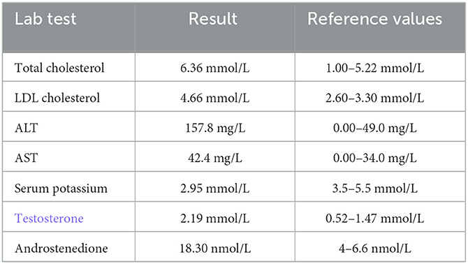
Adrenal CS was suspected on the basis of her Cushing’s signs, cortisol 25 μg/dL, and adrenocorticotropic hormone <1.5 pg/mL. She had hypokalemia, hypogammaglobulinemia, and liver dysfunction, and her condition was rapidly worsening. Given her pregnant state, she was admitted for intensive testing for the case of CS from 19 weeks of pregnancy. MRI revealed a well-defined 38-mm left adrenal tumor, which was the cause of the adrenal CS (Fig. 1). She was started on metyrapone with 250 mg per day, which increased androgens (0.53–0.69 ng/mL in 1 week). We considered that the management of CS by medication alone would be challenging and performed adrenalectomy during her pregnancy. The dose of metyrapone was increased to 1000 mg per day eventually.

Details are in the caption following the image

Magnetic resonance imaging on admission shows a left adrenal tumor with a long axis of 38 mm (arrowhead). Signal reduction was partially observed on opposed-phase images, leading to diagnosis of cortical adenoma.

She was admitted to the hospital at 23 weeks and 2 days of gestation, and laparoscopic left adrenalectomy was performed via a retroperitoneal approach in the right lateral and jackknife position on the following day (Fig. S1). During the surgery, blood pressure was carefully controlled by an anesthesiologist and the patient’s position and fetal heart rate were monitored by an obstetrician. The operation time, insufflation time, and general anesthesia time were 68, 59, and 123 min, respectively, and the blood loss volume was 75 mL, without any complications. Pathological findings revealed an adrenocortical adenoma. The specimen was positive for one of the nine Weiss criteria (Fig. 2).

Details are in the caption following the image

(a) Intraoperative findings of the retroperitoneal approach. Arrowheads indicate the tumor. (b) Gross appearance of the resected adrenal tumor; a brownish-toned, substantial mass, 60 × 34 × 15 mm in size. (c, d) Hematoxylin–eosin staining showed that nodular lesion with a fibrous capsule, with foci of homogeneous cells with eosinophilic or pale, foamy sporangia and small round nuclei.

Postoperatively, metyrapone was discontinued and both lower leg edema, facial acne, fatigue, and muscle weakness improved. Metyrapone was discontinued after surgery. Hydrocortisone, which had been administered at 150 mg/day during the perioperative period, was reduced every few weeks and was taken at 30 mg/day at delivery. She delivered by cesarean section at 38 weeks and 2 days of gestation, with good outcomes for the mother and her infant. Hydrocortisone was discontinued 15 weeks after delivery.

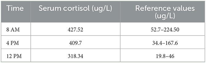
We showed the changes in cortisol and ACTH from the first visit to postpartum (Fig. 3).

Details are in the caption following the image

The transition of Cortisol and ACTH. Cortisol decreases rapidly after surgery and rises again before delivery. As cortisol improved, ACTH also increased.

Discussion

CS seldom occurs during pregnancy. Symptoms such as weight gain, skin striae, fatigue, and a round face can also occur in normal pregnancies. The dexamethasone suppression test can result in false positives because of ACTH produced by placenta in normal pregnancy. During pregnancy, there is a physiological state of high cortisol levels. The disappearance of diurnal rhythm is a useful indicator for diagnosis of CS in pregnancy because circadian rhythm is maintained in normal pregnancy. Useful diagnostic criteria include urine cortisol levels greater than three times the upper limit of normal, loss of diurnal cortisol rhythm, and presence of adrenal tumors on MRI.

The pharmacologic treatment of endogenous cortisol is complex, and hormonal management is challenging. While the management of the cortisol levels is important, metyrapone is a risk factor for gestational hypertension and may inhibit fetal cortisol production by crossing the placenta.16812

In this case, because androgens were also elevated and drug management was expected to be challenging, the surgery was aggressively considered. Despite the reports of successful adrenalectomy is after 28 weeks of gestation,61314 The surgery should be performed by an experienced team between 14 and 30 weeks of pregnancy, that is, after organogenesis phase and before the fetus grows too large.11315

A few pregnant women with adrenal CS undergo adrenalectomy. However, the laparoscopic approach is safe, and maternal and fetal complications were higher in women who did not undergo surgery.16 Less postoperative pain, faster wound healing, and faster postoperative recovery are the main advantages of laparoscopic surgery.17

In pregnant women, pneumoperitoneal pressure should be kept <12 mmHg because increased intraabdominal pressure decreases placental blood flow and can cause fetal acidosis due to the absorption of carbon dioxide used for insufflation.

Laparoscopic adrenalectomy can be safely performed through both transperitoneal and retroperitoneal approaches.18 However, in pregnant women, performing the surgery by the retroperitoneal approach in the lateral position is preferable to prevent putting pressure on the fetus during the surgery. The retroperitoneal approach is advantageous, as less pressure is placed on the uterus and adhesions are prevented. After taking the lateral position, the obstetrician is advised to check the position and confirm that the abdomen is not compressed and that the fetal heart rate is normal.

Conclusions

We present a case of a pregnant woman diagnosed with adrenal CS who underwent a unilateral laparoscopic adrenalectomy by a retroperitoneal approach without any problems. Adrenalectomy is a useful treatment when CS is difficult to control despite metyrapone and other medical support.

Author contributions

Nobuyoshi Takeuchi: Conceptualization; methodology; project administration; writing – original draft. Yusuke Imamura: Conceptualization; methodology; supervision; writing – review and editing. Kazuki Ishiwata: Data curation; supervision. Manato Kanesaka: Data curation; supervision. Yusuke Goto: Data curation; supervision. Tomokazu Sazuka: Data curation; supervision. Sawako Suzuki: Data curation; supervision. Hisashi Koide: Data curation; supervision. Shinichi Sakamoto: Data curation; supervision. Tomohiko Ichikawa: Data curation; supervision.

Conflict of interest

The authors declare no conflicts of interest.

Approval of the research protocol by an Institutional Reviewer Board

Not applicable.

Informed consent

Informed consent for the release of the case report and accompanying images has been obtained from the patient.

Registry and the Registration No. of the study/trial

Not applicable.

From https://onlinelibrary.wiley.com/doi/10.1002/iju5.12637

Rare Challenges in Diagnosing Cushing’s Syndrome and Primary Aldosteronism: A Case Report of a Female With a Negative Workup

Abstract

Cushing’s syndrome with concurrent primary aldosteronism (PA) is a rare presentation, and establishing an early diagnosis is imperative to preventing morbidity and long-term sequelae. The diagnosis is established by sequential lab work, showing an elevated cortisol and aldosterone level.

Taking the above into consideration, it is evident that repeatedly negative results on all three tests can present an extremely challenging case. In this report, we discuss a female who presented with an adrenal incidentaloma and features suggestive of primary hyperaldosteronism as well as Cushing’s syndrome but no elevations in serum, urine, or salivary cortisol.

In this study, we present a 37-year-old female with resistant hypertension and tachycardia. She had several features suggestive of Cushing’s syndrome including resistant hypertension, proximal muscle weakness, weight gain, easy bruising, hair loss, and a history of tachycardia and chest pain. Examination revealed an obese female with thin silvery abdominal striae. The patient’s labs revealed normal serum cortisol, urine-free cortisol (UFC), late-night salivary cortisol, and a normal dexamethasone suppression test. An abdominal computed tomography (CT) scan revealed a right adrenal mass measuring 2.1 x 1.5 x 2.5 cm. Due to a high index of suspicion, adrenal venous sampling was performed, which revealed high levels of cortisol and aldosterone in the right vein, confirming the diagnosis. The patient subsequently underwent a right adrenalectomy. She developed hypotension post-op, leading to the diagnosis of glucocorticoid-remediable aldosteronism.

Introduction

Primary aldosteronism (PA) is the excess production of aldosterone by the adrenal glands, despite a low serum renin level. The presentation of hyperaldosteronism can be vague and include symptoms such as muscle weakness, fatigue, headaches, numbness, and cramps. More specific findings include resistant hypertension, low serum potassium, and metabolic alkalosis. The etiologies are variable and can include an adrenal adenoma (Conn syndrome) or bilateral adrenal hyperplasia [1].

Cushing’s syndrome is also caused by excess hormone secretion by the adrenal glands. The etiologies include a primary adrenal adenoma, hyperplasia, carcinoma, or exogenous corticosteroid use. It can also be caused by an adrenocorticotropic hormone (ACTH)-secreting pituitary adenoma or as a result of paraneoplastic ACTH secretion. The clinical presentation is highly variable and leads to difficulties in establishing a diagnosis.

The concurrent existence of primary hyperaldosteronism and Cushing’s syndrome creates additional hindrances in diagnosis, yet further obscured in a patient with a repeatedly negative workup for both conditions.

Case Presentation

A 37-year-old female presented to her primary care physician with complaints of proximal muscle weakness, tachycardia, and chest pain. Repeated blood pressure readings revealed that she was hypertensive, and she was started on amlodipine and benazepril, which elevated her blood pressure further. A computed tomography (CT) scan (Figure 1) of the abdomen was performed due to resistant hypertension, which revealed an adrenal incidentaloma (right adrenal gland measuring 2.1 x 1.5 x 2.5 cm). Precontract density was 5 Hounsfield units, and a 15-minute delayed washout showed 11 Hounsfield units for a 72% washout. She was thus referred to endocrinology.

Abdominal-CT-scan-showing-a-nodule-in-the-right-adrenal-gland-measuring-2.1-x-1.5-x-2.5-cm
Figure 1: Abdominal CT scan showing a nodule in the right adrenal gland measuring 2.1 x 1.5 x 2.5 cm

She presented to the endocrinology clinic on March 12, 2021. A thorough physical examination was performed, which revealed a well-appearing obese female (BMI of 38.86 kg/m2) with no acute distress. Her blood pressure was 144/108 mmHg, her pulse was 95, and she was afebrile. Thin silvery striations were present on the abdomen, and alopecia was present on the crown. A review of all other systems was unremarkable. A detailed family history revealed early-onset hypertension in her brother (age: 35 years) and her mother (age: 30 years). Personal history included elevated anxiety, weight gain, headaches (frontal band distribution), increased thirst, easy bruising as well as delayed clearance of bruises, and proximal muscle weakness presenting as difficulty in climbing stairs and inability to lift heavy objects. She reported no change in menstrual cycles. There was no history of exogenous corticosteroid use.

Serum biochemistries were sent (Table 1), which showed normal levels of thyroid stimulating hormone (TSH), creatinine, liver function tests, and serum electrolytes. However, mildly elevated aldosterone (23 ng/dl), mild hypokalemia (3.3 mEq/L), and suppressed ACTH and dehydroepiandrosterone (DHEA) sulfate were discovered. The aldosterone to renin ratio was also elevated at 59.9 on spironolactone and was 71.4 three months later when spironolactone was discontinued. These findings lead to a preliminary diagnosis of primary hyperaldosteronism.

Test Result
Calcium 9.1 mmol/L
Sodium 137 mmol/L
Potassium 4.1 mmol/L
Chloride 106 mmol/L
CO2 27
BUN 15 mmol/L
Glucose 95 mmol/L
Creatinine 1.1 μmol/L
AST 24 U/L
ALT 20 U/L
Albumin 4.4 g/L
Total protein 7.0 g/L
Total bilirubin 0.4 μmol/L
Alkaline phosphatase 40 U/L
Renin 0.44
Table 1: Patient serum biochemistries

BUN: Blood urea nitrogen; AST: Aspartate transaminase; ALT: Alanine transaminase.

A workup for elevated cortisol was also performed as the patient was phenotypically Cushingoid, and the following biochemistries were sent sequentially: serum cortisol, 24-hour urine-free cortisol (UFC), salivary cortisol, and a low-dose dexamethasone suppression test (Table 2). The bloodwork was hence nonconfirmatory.

Endocrine workup
Serum cortisol 4.5 mcg/dL
Urine-free cortisol 1.57 g/24 h
Salivary cortisol <0.03 μg/dL
Dexamethasone suppression test 1.5 mcg/dL
Aldosterone <4.0
Table 2: Patient follow-up bloodwork

Despite a repeatedly negative workup for Cushing’s syndrome, adrenal venous sampling was performed due to a high index of suspicion. The results revealed an inferior vena cava (IVC) cortisol of 20, left adrenal venous (LAV) cortisol of 81, and right adrenal vein (RAV) cortisol of 1280. The results of the IVC aldosterone were 24, LAV aldosterone was 660 and RAV aldosterone was 1500. The elevated levels of cortisol in the RAV were in complete contradiction to the aforementioned workup. A diagnosis of Cushing’s syndrome and concurrent PA was determined.

Adrenal veinous sampling was instrumental in establishing the diagnosis but was equivocal and did not lateralize aldosterone and cortisol excess. However, the amount of aldosterone and cortisol were both significantly higher on the right side. After a panel discussion with doctors from several disciplines, a laparoscopic adrenalectomy was planned. The procedure was successful, and the patient was initially showing clinical improvement. The specimen was sent for pathological evaluation and revealed an adrenal cortical adenoma.

After initial improvement, the patient developed hypotension, which was likely due to adrenal insufficiency. The patient was supplemented with 1-mg dexamethasone tablets, which stabilized her condition, and a diagnosis of glucocorticoid-remediable-aldosteronism was made.

Based on a strong family history of early onset-resistant hypertension, a genetic component was suspected. Several genes associated with PA with autosomal dominant inheritance have been identified [2], such as CYP11B2, CLCN2, KCNJ5, CACNA1D, and CACNA1H. The patient was offered genetic testing but was unable to follow through due to financial reasons.

Discussion

This patient presented as an extremely rare example of PA and Cushing’s syndrome, with negative serum cortisol, 24-hour UFC, late-night salivary cortisol, and a dexamethasone suppression test. Despite repeatedly negative lab results, the patient presented with a markedly elevated cortisol on adrenal venous sampling. In our literature search, we found an instance of a patient with several negative UFCs [3]; however, to the best of our knowledge, there have been no reported instances of a completely negative workup in a patient who is positive for Cushing’s syndrome. In fact, in the practice guidelines published by the Journal of Clinical Endocrinology & Metabolism [4], it is recommended that patients with a suspected diagnosis of Cushing’s syndrome or an adrenal incidentaloma and two concordant negative test results need not undergo further investigations.

One proposed mechanism for the misleading workup could be assay interference. Interference occurs when a substance or process falsely alters an assay result [5]. This can lead to incorrect diagnosis and subsequent treatment and poses a threat to the patient. Another suggested mechanism causing false negative test results could be the hook effect [6]. The hook effect is described as a phenomenon that leads to falsely low results due to the presence of excessive analyte.

In a study by Friedman et al. [7], it was noted that patients with “episodic Cushing’s syndrome” or those with mild symptoms had a negative workup. The study recommended serial monitoring for the disease. The interesting fact is that our patient had several features suggestive of active Cushing’s syndrome, and the hypotension seen postoperatively was a testament to the fact that there was in fact a cortisol excess, which led to adrenal insufficiency. In light of the above, a consistently negative workup is perplexing.

Zhang et al. suggested performing a low-dose dexamethasone suppression test in individuals presenting with PA, prior to adrenal vein sampling (AVS) and surgery due to the high prevalence of Cushing’s syndrome in patients with PA [8]. A positive test result can lead to a straightforward diagnosis; however, in this rare case where the patient had severe negative tests, it can present as a challenge in diagnosis and treatment.

Conclusions

The presence of PA and concurrent Cushing’s syndrome can present as a diagnostic challenge. It is recommended to follow up on the signs of Cushing’s syndrome with preliminary tests and to presume its absence if two concordant tests are negative. Our patient, however, was an exceptional case.

This case highlighted the importance of maintaining a high index of suspicion for patients presenting with several signs and symptoms of the disease and a negative workup. More attention should be paid to the patient’s history, and a thorough physical examination should be conducted. In those with an uncertain diagnosis, adrenal venous sampling can provide a clearer picture and lead to a more accurate understanding of the case.

References

  1. Reincke M, Bancos I, Mulatero P, Scholl UI, Stowasser M, Williams TA: Diagnosis and treatment of primary aldosteronism. Lancet Diabetes Endocrinol. 2021, 9:876-92. 10.1016/S2213-8587(21)00210-2
  2. Dutta RK, Söderkvist P, Gimm O: Genetics of primary hyperaldosteronism. Endocr Relat Cancer. 2016, 23:R437-54. 10.1530/ERC-16-0055
  3. Moloney KJ, Mercado JU, Ludlam WH, Broyles FE: Diagnosis of Cushing’s disease in a patient with consistently normal urinary free cortisol levels: a case report. Clin Case Rep. 2016, 4:1181-3. 10.1002/ccr3.647
  4. Nieman LK, Biller BM, Findling JW, Newell-Price J, Savage MO, Stewart PM, Montori VM: The diagnosis of Cushing’s syndrome: an Endocrine Society Clinical Practice Guideline. J Clin Endocrinol Metab. 2008, 93:1526-40. 10.1210/jc.2008-0125
  5. Dimeski G: Interference testing. Clin Biochem Rev. 2008, 29:S43-8.
  6. The hook effect. (2014). Accessed: June 19, 2023: https://www.aacc.org/science-and-research/clinical-chemistry-trainee-council/trainee-council-in-english/pearls-of-lab….
  7. Friedman TC, Ghods DE, Shahinian HK, et al.: High prevalence of normal tests assessing hypercortisolism in subjects with mild and episodic Cushing’s syndrome suggests that the paradigm for diagnosis and exclusion of Cushing’s syndrome requires multiple testing. Horm Metab Res. 2010, 42:874-81. 10.1055/s-0030-1263128
  8. Zhang Y, Tan J, Yang Q, et al.: Primary aldosteronism concurrent with subclinical Cushing’s syndrome: a case report and review of the literature. J Med Case Rep. 2020, 14:32. 10.1186/s13256-020-2353-8

Adults with Cushing’s Syndrome Report High Burden Of Illness, Despite Ongoing Treatment

Key takeaways:

  • Cushing’s syndrome symptoms moderately impact quality of life for adults with the condition.
  • Weight gain, muscle fatigue and menstrual changes decline in severity from diagnosis to follow-up.

Adults with endogenous Cushing’s syndrome reported that the condition moderately affects their quality of life and causes them to have symptoms about 16 days in a given month, according to findings published in Pituitary.

“Our study aimed to evaluate the ongoing burden of Cushing’s syndrome in order to identify areas of unmet need,” Eliza B. Geer, MD, medical director of the Multidisciplinary Pituitary and Skull Base Tumor Center and associate attending of endocrinology and neurosurgery at Memorial Sloan Kettering Cancer Center, told Healio. “We found that patients with treated Cushing’s continue to experience ongoing symptoms more than half of the days in a given month, miss about 25 workdays per year and need twice the average number of outpatient visits per year, indicating a significant impact on daily function and work productivity. Some of these symptoms, like fatigue and pain, have not been well studied in Cushing’s patients, and need more attention.”

Geer and colleagues administered a cross-sectional survey to 55 adults aged 21 years and older who had been diagnosed with Cushing’s syndrome at least 6 months before the survey and were receiving at least one pharmacologic therapy for their disease (85% women; mean age, 43.4 years). The survey was conducted online from June to August 2021. Five patient-reported outcome scales were included. The CushingQoL was used to analyze quality of life, a visual analog scale was included to assess pain, the Brief Fatigue Inventory was used to measure fatigue, the Sleep Disturbance v1.0 scale assessed perceptions of sleep and the PROMIS Short Form Anxiety v1.0-8a scale was used to measure fear, anxious misery, hyperarousal and somatic symptoms related to arousal. Participants self-reported the impact of Cushing’s syndrome on daily life and their physician’s level of awareness of Cushing’s syndrome.

Some symptoms decline in severity over time

Of the study group, 81% had pituitary or adrenal tumors, and 20% had ectopic adrenocorticotropic hormone-producing tumors; 80% of participants underwent surgery to treat their Cushing’s syndrome.

The frequency of reported symptoms did not change from Cushing’s syndrome diagnosis to the time of the survey. The most frequently reported symptoms were weight gain, muscle fatigue and weakness and anxiety.

Participants reported a decline in symptom severity for weight gain, muscle fatigue and weakness and menstrual changes from diagnosis to the survey. Though symptom severity declined, none of the three symptoms were entirely eliminated. Adults did not report declines in severity for other symptoms. Hirsutism and anxiety were reported by few participants, but were consistently scored high in severity among those who reported it. There were no changes in patient satisfaction with medications from their first appointment to the time of the survey.

“It was surprising that anxiety and pain did not improve with treatment,” Geer said. “A quarter of patients at baseline reported anxiety and this percentage was exactly the same after treatment. Same for pain — nearly a quarter of patients reported pain despite treatment. While the presence of anxiety has been well-documented in Cushing’s patients, pain has not, and needs further study.”

Nearly half of primary care providers unable to diagnose Cushing’s syndrome

All participants reported having at least one challenge with being diagnosed with Cushing’s syndrome. Of the respondents, 49% said their primary care provider was unable to diagnose their Cushing’s syndrome and 33% initially received the wrong diagnosis. Physicians referred 49% of participants to a specialist, and 39% of adults said their doctor lacked knowledge or understanding of their condition.

The study group had a moderate level of quality of life impairment as assessed through the CushingQoL scale. The mean pain score was 3.6 of a possible 10, indicating low levels of pain. Moderate to severe levels of fatigue were reported by 69% of participants. Self-reported sleep and anxiety scores were similar to what is observed in the general population.

Participants said sexual activity, self-confidence and life satisfaction were most impacted by a Cushing’s syndrome diagnosis. Adults experienced symptoms a mean 16 days in a typical month and saw their outpatient physician an average of six times per year. Those who were employed said they miss 2 days of work per month, or about 25 days per year, due to Cushing’s syndrome.

“Longitudinal assessment of clinically relevant patient-reported outcomes based on validated measures and coupled with biochemical and treatment data is needed in a large cohort of Cushing’s patients,” Geer said. “This will allow us to identify clinically meaningful changes in symptom burden within each patient, as well as predictors of outcomes — which patients improve on which symptoms, and which patients do not feel better despite biochemical normalization. We need to improve our ability to help our patients feel better, not just achieve normal cortisol levels.”

For more information:

Eliza B. Geer, MD, can be reached at geere@mskcc.org.

From https://www.healio.com/news/endocrinology/20230830/adults-with-cushings-syndrome-report-high-burden-of-illness-despite-ongoing-treatment

Treatment-Resistant Depression with Acute Psychosis in an Adolescent Girl with Cushing’s Syndrome

Cushing’s syndrome (CS) is a rare disease with multiple somatic signs and a high prevalence of co-occurring depression. However, the characteristics of depression secondary to CS and the differences from major depression have not been described in detail. In this case, we report a 17-year-old girl with treatment-resistant depression with a series of atypical features and acute psychotic episodes, which is a rare condition secondary to CS. This case showed a more detailed profile of depression secondary to CS and highlighted the differences with major depression in clinical features, and it will improve insight into the differential diagnosis especially when the symptoms are not typical.

Introduction

Depression is a chronic medical problem with typical features, including sadness, decreased interest and cognitive impairments. In clinical practice, depression can occur in other medical conditions, especially endocrinopathies, making it a more complex problem and exhibiting a challenge in diagnosis, especially in first-contact patients or when the clinical presentations are atypical. It is generally accepted that patients who failed to respond to two or more adequate trials of first-line antidepressants for treatment of major depressive episode are considered to have treatment-resistant depression (TRD) (1). For patients with TRD, a throughout evaluation should be performed to investigate the underlying organic causes.

Cushing’s syndrome is a rare but serious endocrine disease due to chronic exposure to excess circulating glucocorticoids with multisystem effects (2). The etiology of CS can be divided into adrenocorticotropic hormone (ACTH)-dependent and ACTH-independent. It is characterized by a series of clinical features suggesting hypercortisolism, for example, metabolic abnormalities, hypertension and bone damages (3). A variety of neuropsychiatric symptoms, such as mood disturbance, cognitive impairment and psychosis, also occur in more than 70% CS patients (4). CS is life-threating if not timely diagnosed and treated, however, correct diagnosis can be delayed due to the wide range of phenotypes, especially when they are not classical (5).

Previous studies suggested that major depression was the most common co-morbid complication in CS patients, with a prevalence of 50–81% (6). Haskett’s study confirmed that 80% of subjects meet the criteria for major depression with melancholic features (7). As reported in most recent investigations, depression in CS was not qualitatively different from non-endocrine major depression and the similarity was even striking (38). However, some studies showed different conclusions and suggested a high prevalence of atypical depressive features other than melancholic features in CS (9). TRD and anxious depression has also been reported in CS patients (1011). All of the above conclusions suggest the complexity of depression with CS, and no distinct features have been found pertaining to hypercortisolism (1213). Although the intensity of depression secondary to CS is severe, suicidal depression is still an unusual condition (14).

Psychosis is a rare manifestation of CS, and the literature is limited. Only a few cases have been reported so far, especially combined with depression episode. In this case report, we presented a girl with CS, who experienced suicidal depression with a series of atypical features and acute psychotic symptoms, which was rarely reported in previous studies.

Case description

A 17-year-old girl with major depression for 3 years was involuntarily admitted for severe depressed mood with suicide attempts (neck cutting; tranquilizer overdose) and paranoid state in the last 2 weeks without any precipitating factors.

She experienced depressed and irritable mood in the last 3 years, and her condition had not improved although several adequate trials of antidepressants were used with satisfactory compliance (sertraline 200 mg/d; escitalopram oxalate 20 mg/d). Over the 2 weeks prior to admission, her depression continued to worsen with increasing irritability, she committed several suicide attempts, and once stated that she was unsafe at home. On admission, her heart rate was 116 bpm with blood pressure 139/81 mmHg and normal temperature; physical examination showed a cushingoid and virilising appearance (central obesity, swollen and hirsute face with acne, purple striae on the abdomen and bruises on the arms). No other abnormal signs were noted. She seemed drowsy but arousable, and she walked slowly, with bent shoulders and an inclined head. Mental state examination was hard to continue because she was passive and reluctant to answer our questions. Venlafaxine 150 mg/d has been used for more than 3 months with poor effects at that time.

Besides, weight gain (25 kg), irregular menstrual cycles and numbness of the hands and feet in the last half year were reported by her parents. Otherwise, No episodes of elevated mood and hyperactivity were found during the history taking. She does not have remarkable family history of serious physical or psychiatric illness; she was healthy, had an extroverted personality and had never used substances. Her premorbid social function and academic performance were good.

Several clinical characteristics found during the following mental state examinations were listed as follows:

• Prominent cognitive impairment without clouding of consciousness: Forgetfulness was frequently noted; she easily forgot important personal information such as her school and grade; she could not recall the suicide attempt committed recently and perfunctorily ascribed it to a casual event; and it was hard for her to recall her medical history (as it is for other depressive patients). The serial seven subtraction task could not be finished, and the interpretation of the proverb was superficial. Difficulty was found in attention maintenance; an effective conversation was hard to perform because she was mind-wandering (we needed to call her name to get her immediate attention) and often interrupted our conversations by introducing irrelevant topics or leaving without apparent reasons.

• Decreased language function that did not match her educational background: The patient could not find the proper words to articulate her feelings; instead, many simple, obscure and contradictory words were used, which made her response seem perfunctory. For example, she responded with “I do not know,” “I forgot,” or kept silent in response to our questions, which made the conversations hard to perform.

• Psychotic outbursts: Once she left the psychological therapy group, ranted about being persecuted and shook in fearfulness, stated “call the police” repeatedly, negative of explanations and comforts from others, but she cannot give any explanation about her behavior when calmed down. Sometimes she worried about being killed by the doctors but the worries were transient and fleeting.

• Depressed mood and negative thoughts (self-blame, worthlessness, and hopelessness) that were not persistent and profound: During most of her hospitalization, the patient seemed confused and apathetic, with intermittent anxiety, but she could not clearly express what made her anxious. Her crying and sadness happened suddenly, without obvious reasons, and she even denied low mood sometimes and said she had come to the hospital for cardiac disease treatment (she did not have any cardiac disease). Her description of her depressed mood was uncertain when specifically questioned, and she rarely reported her depressed feeling spontaneously as other depressed patients would. She did not even have the desire to get rid of her “depression”. Her suicidal ideation was transient and impulsive, and she could not provide a comprehensive explanation for her suicide attempts, such as emptiness, worthlessness or guilt. She was impatient and restless when interacting with others or when a more in-depth conversation was performed. She seemed apathetic, gave little response to emotional support from others and did not care about relevant important issues, such as hospital discharge or future plans. Elevated mood and motor activity were not found during the admission period.

• Social withdrawal and inappropriate behaviors: The patient often walked or stayed alone for long periods of time before speaking to other patients suddenly, which seemed improper or even odd in normal social interactions. During most hospitalization periods, lethargy and withdrawal were obvious.

Diagnostic assessment and therapeutic interventions

Basic laboratory tests reported abnormal results (Table 1), and the circulating cortisol level was far beyond the upper limit of normal, with a loss of circadian rhythm (Table 2); 24-h urinary free cortisol : >2897 nmol/24 h↑(69–345 nmol/24 h); serum ACTH (8 AM, 4 PM, 12 PM): 1.2 pg/ml, 1.3 pg/ml, <1 pg/ml (normal range: 1–46 pg/ml); low-dose dexamethasone suppression test (1 mg) (cortisol value): 1010.1 nmol/l (not suppressed; normal range: <50 nmol/L); high dose dexamethasone inhibition test (cortisol value): 879.0 nmol/l (not suppressed); OGTT and glycosylated hemoglobin; both normal. Other results used to rule out hyperaldosteronism and pheochromocytoma, such as the aldosterone/renin rate (ARR) and the vanillylmandelic acid, dopamine, norepinephrine and epinephrine levels, were reported to be within normal limits; ECG suggested sinus tachycardia; dual-energy X-ray bone density screening values were lower than the normal range; B-mode ultrasound showed a right adrenal tumor and fatty liver. The abdominal CT scan showed a tumor in her right adrenal gland. Brain MRI showed no abnormalities. Psychometric tests including HAMD (Hamilton depression scale), MADRS (Montgomery-Asberg Depression Rating Scale), WAIS (Wechsler Intelligence Scale) and MMSE (Mini-mental State Examination) were hard to perform due to her poor attention and non-cooperation presentation.

Table 1
www.frontiersin.orgTable 1. Abnormal lab results for the patient.

Table 2
www.frontiersin.orgTable 2. Circulating cortisol level.

The patient had little response to adequate antidepressants in our hospital, including fluoxetine 20–60 mg/d and aripiprazole 5–30 mg/d combined with 3 sessions of MECT (modified electroconvulsive therapy), which was stopped because of her poor cognitive function and poor response.

Her last diagnosis was right adrenal adenoma and non-ACTH-dependent Cushing’s syndrome. The adrenal adenoma was excised through laparoscopic resection in a general hospital. Hydrocortisone, amlodipine besylate, potassium chloride, metoprolol and escitalopram were used for treatment. Escitalopram 10 mg/d has been used until 2 weeks after her discharge. At the follow-up visit about 1 month after the surgery, her depressive mood had significantly improved, with no self-injury behaviors or psychiatric symptoms found. The patient was calm but still reacted slowly, and cognitive impairment was still found at the last visit.

Discussion

Previous studies have reported a close association between CS and depression (15). However, suicidal depression with atypical features and acute psychosis have rarely been reported, and the characteristics of depression secondary to CS and the differences from major depression have not been described in detail.

This case did not show a full-blown presentation of major depression according to the DSM-5. She presented with a series of features that were not typical as major depression, however, it should be emphasized that the atypical features were not identical to those noted in DSM5, especially regarding increased appetite and hypersomnia. The features suggesting difference from major depression were listed as follows: (a) depressed mood is not constant, it does not exist in most of the day; it is episodic without regular cyclicity, can happen or exacerbate suddenly; (b) the ability to describe anhedonia is poor, she can’t report her feeling voluntarily like other patients with major depression, which might be partially related with the decreased language function; (c) depressive thoughts such as self-accusation and feelings of guilt, the classical symptoms of major depression, were rarely found; (d) more exaggerated cognitive impairment and decrease language function; € partial or little useful effect of SSRIs (selective serotonin reuptake inhibitors). The above characteristics were similar to those reported in Starkman’s research (131617), in which increasing irritability was also regarded as one of the important features for depression in CS.

The literature about depression combined with psychosis episode in CS is rare. This patient showed acute episodes of persecutory delusion with disturbed behaviors; her psychotic symptoms occurred suddenly and were fragmentary, with poor sensitivity to antipsychotics; the content was not constant (she never referred to and even denied the unsafe feeling at home before admission), it changed with the environment and was not consistent with the mood state. However, we cannot reach an effective conclusion because the evidence was small; thus, these findings should be evaluated in combination with other clinical presentations.

Conclusion

Most reviews have concluded that mood disturbances in CS indicate “major depression”, but the detailed description of clinical features are lack, making clinicians uncertain about the presentation and confused about the diagnosis, especially when the somatic signs are indiscriminate. The clinical presentation in this case highlighted the fact that there is a wide range of phenotypes of depression in CS, for some CS patients, the depressive features are not highly consistent with the criteria of major depression regardless of the melancholic or atypical features in the DSM-5. Thus, a thorough and periodic evaluation is necessary to detect the underlying organic and psychosocial causes if the clinical symptoms are not typical (10).

Data availability statement

The original contributions presented in the study are included in the article/supplementary material, further inquiries can be directed to the corresponding author.

Ethics statement

Written informed consent was obtained from the individual(s), and minor(s)’ legal guardian/next of kin, for the publication of any potentially identifiable images or data included in this article.

Author contributions

XY, SC, XJ, and XH were responsible for clinical care. XY did literature search and drafted the manuscript. XH revised the manuscript. All authors contributed to the article and have approved the final manuscript.

Acknowledgments

We want to thank Juping Fu, Ying Zhang, and all other medical staff who gave careful nursing to the patient.

Conflict of interest

The authors declare that the research was conducted in the absence of any commercial or financial relationships that could be construed as a potential conflict of interest.

Publisher’s note

All claims expressed in this article are solely those of the authors and do not necessarily represent those of their affiliated organizations, or those of the publisher, the editors and the reviewers. Any product that may be evaluated in this article, or claim that may be made by its manufacturer, is not guaranteed or endorsed by the publisher.

References

1. Kverno KS, Mangano E. Treatment-resistant depression: approaches to treatment. J Psychosoc Nurs Ment Health Serv. (2021) 59:7–11. doi: 10.3928/02793695-20210816-01

PubMed Abstract | CrossRef Full Text | Google Scholar

2. Mokta J, Sharma R, Mokta K, Ranjan A, Panda P, Joshi I. Cushing’s disease presenting as suicidal depression. J Assoc Physicians India. (2016) 64:82–3.

PubMed Abstract | Google Scholar

3. Sonino N, Fava G. A Psychiatric disorders associated with Cushing’s syndrome epidemiology, pathophysiology and treatment. CNS Drugs. (2001) 15:361–73. doi: 10.2165/00023210-200115050-00003

PubMed Abstract | CrossRef Full Text | Google Scholar

4. Pivonello R, Simeoli C, De Martino MC, Cozzolino A, De Leo M, Iacuaniello D, et al. Neuropsychiatric disorders in Cushing’s syndrome. Front Neurosci. (2015) 9: 129. doi: 10.3389/fnins.2015.00129

PubMed Abstract | CrossRef Full Text | Google Scholar

5. Barbot M, Zilio M, Scaroni C. Cushing’s syndrome: Overview of clinical presentation, diagnostic tools and complications. Best Pract Res Clin Endocrinol Metab. (2020) 34:101380. doi: 10.1016/j.beem.2020.101380

PubMed Abstract | CrossRef Full Text | Google Scholar

6. Sonino N, Fava GA, Raffi AR, Boscaro, Fallo F. Clinical correlates of major depression in Cushing’s disease. Psychopathology. (1998) 31:302–6. doi: 10.1159/000029054

PubMed Abstract | CrossRef Full Text | Google Scholar

7. Haskett RF. Diagnostic categorization of psychiatric disturbance in Cushing’s syndrome. Am J Psychiatry. (1985) 142:911–6. doi: 10.1176/ajp.142.8.911

PubMed Abstract | CrossRef Full Text | Google Scholar

8. Fava GA. Affective disorders and endocrine disease. New insights from psychosomatic studies. Psychosomatics. (1994) 35:341–53. doi: 10.1016/S0033-3182(94)71755-2

PubMed Abstract | CrossRef Full Text | Google Scholar

9. Dorn LD, Burgess ES, Dubbert B, Simpson SE, Friedman T, Kling M, et al. Psychopathology in patients with endogenous Cushing’s syndrome: ‘atypical’ or melancholic features. Clin Endocrinol. (1995) 43:433–42. doi: 10.1111/j.1365-2265.1995.tb02614.x

PubMed Abstract | CrossRef Full Text | Google Scholar

10. Anil Kumar BN, Grover S. Cushing’s syndrome masquerading as treatment resistant depression indian. J Psychol Med. (2016) 38:246–8. doi: 10.4103/0253-7176.183095

PubMed Abstract | CrossRef Full Text | Google Scholar

11. Loosen PT, Chambliss B, DeBold CR, Shelton R, Orth D. Psychiatric phenomenology in Cushing’s disease. Pharmacopsychiatry. (1992) 25:192–8. doi: 10.1055/s-2007-1014405

PubMed Abstract | CrossRef Full Text | Google Scholar

12. Sonino N, Fava GA, Belluardo P, Girelli ME, Boscaro, M. Course of depression in Cushing’s syndrome: response to treatment and comparison with Graves’ disease. Horm Res. (1993) 39:202–6. doi: 10.1159/000182736

PubMed Abstract | CrossRef Full Text | Google Scholar

13. Starkman MN, Schteingart DE, Schork M. A depressed mood and other psychiatric manifestations of Cushing’s syndrome: relationship to hormone levels. Psychosom Med. (1981) 43:3–18. doi: 10.1097/00006842-198102000-00002

PubMed Abstract | CrossRef Full Text | Google Scholar

14. Al-Harbi SD, Mashi AH, Al Johani N. A case of Cushing’s disease presenting with isolated suicidal attempt clin med insights. Case Rep. (2021) 14:11795476211027668. doi: 10.1177/11795476211027668

PubMed Abstract | CrossRef Full Text | Google Scholar

15. Fujii Y, Mizoguchi Y, Masuoka J, Matsuda Y, Abe T, Anzai K, et al. Cushing’s syndrome and psychosis: a case report and literature review. prim care companion. CNS Disord. (2018) 20:279. doi: 10.4088/PCC.18br02279

PubMed Abstract | CrossRef Full Text | Google Scholar

16. Starkman MN, Giordani B, Berent S, Schork MA, Schteingart D. Elevated cortisol levels in Cushing’s disease are associated with cognitive decrements. Psychosom Med. (2001) 63:985–93. doi: 10.1097/00006842-200111000-00018

PubMed Abstract | CrossRef Full Text | Google Scholar

17. Starkman MN. Neuropsychiatric findings in Cushing syndrome and exogenous glucocorticoid administration. Endocrinol Metab Clin North Am. (2013) 42:477–88. doi: 10.1016/j.ecl.2013.05.010

PubMed Abstract | CrossRef Full Text | Google Scholar

Keywords: Cushing’s syndrome (CS), treatment-resistant depression, acute psychosis, adrenal adenoma (AA), adolescent girl

Citation: Yin X, Chen S, Ju X and Hu X (2023) Case report: Treatment-resistant depression with acute psychosis in an adolescent girl with Cushing’s syndrome. Front. Psychiatry 14:1170890. doi: 10.3389/fpsyt.2023.1170890

From https://www.frontiersin.org/articles/10.3389/fpsyt.2023.1170890/full

BMD may Underestimate Bone Deterioration for Women with Endogenous Cushing’s Syndrome

Nearly one-third of women with endogenous Cushing’s syndrome and normal bone mineral density have a low trabecular bone score, according to study data.

“A large proportion of patients had degraded microarchitecture despite normal BMD,” Hiya Boro, DM, MD, MBBS, consultant in endocrinology, diabetes and metabolism at Aadhar Health Institute in India, and colleagues wrote. “The risk of fracture may be underestimated if BMD alone is measured. Hence, trabecular bone score should be added as a routine complementary tool in the assessment of bone health in patients with Cushing’s syndrome.”

About one-third of women with endogenous Cushing's syndrome have normal BMD and low trabecular bone score. Data were derived from Boro H, et al. Clin Endocrinol. 2023;doi:10.1111/cen.14944.

Researchers conducted a cross-sectional study at a single center in India from March 2018 to August 2019. The study included 40 women with overt endogenous Cushing’s syndrome and 40 healthy sex-matched controls. Seum and salivary cortisol and plasma adrenocorticotropic hormone (ACTH) were measured. Participants were considered ACTH independent if they had a level of less than 2.2 pmol/L. Areal BMD was measured at the lumbar spine, femoral neck, total hip and distal one-third of the nondominant distal radius. Low BMD for age was defined as a z score of less than –2. Trabecular bone score was measured at the lumbar spine. Fully degraded microarchitecture was defined as a trabecular bone score of 1.2 or lower and partial degradation was a trabecular bone score of 1.21 to 1.34.

Of the 40 women with Cushing’s syndrome, 32 were ACTH-dependent and the other eight were ACTH independent. Of the independent group, seven had adrenal adenoma and one had adrenocortical carcinoma.

Women with Cushing’s syndrome had lower BMD at the lumbar spine (0.812 g/cm2 vs. 0.97 g/cm2< .001), femoral neck (0.651 g/cm2 vs. 0.773 g/cm2< .001) and total hip (0.799 g/cm2 vs. 0.9 g/cm2< .001) than the control group.

“No significant difference was noted in the distal radius BMD,” the researchers wrote. “This may be explained by the fact that cortisol excess predominantly affects trabecular rather than cortical bone.”

Absolute trabecular bone score was lower in the Cushing’s syndrome group compared with controls (1.2 vs. 1.361; P < .001). Based on trabecular bone score, 42.5% of women with Cushing’s syndrome had fully degraded bone microarchitecture, 45% had partially degraded microarchitecture and 12.5% had normal microarchitecture.

“In our study, 32.5% of patients had normal BMD with low trabecular bone score, thus highlighting the fact that patients may have normal BMD despite degraded microarchitecture,” the researchers wrote. “As such, assessment of BMD alone may underestimate the risk of fractures in patients with Cushing’s syndrome.”