Complete Surgical Resection of Cortisol Secreting Neuroendocrine Thymic Tumour

Abstract

Introduction

Neuroendocrine tumours of the thymus are extremely rare, with an estimated incidence of about 1 in 5 million people. Although data is limited, complete surgical resection remains the most significant prognostic factor for improved survival and disease-free outcomes, with adjuvant radiotherapy playing a role in cases where resection margins are close. This case report details the management of a cortisol secreting pT1bN0 atypical carcinoid of the thymus in a 43-year-old male.

Case report

43-year-old male presented with Cushing’s syndrome and was diagnosed with a cortical secreting atypical carcinoid of the thymus. He underwent a robotic thymectomy. Recurrent disease on a DOTATATE-PET CT scan resulted in a second surgery involving complete resection of the mediastinal tumour which had invaded the pericardium, as well as wedge resection of the lung and lymph node sampling. This was followed by adjuvant radiotherapy due to close proximity of the lesion to the margin (< 3 mm).

Discussion

Although paraneoplastic syndromes such as Cushing’s syndrome are rare manifestations of thymic neuroendocrine tumours and can result in challenging diagnoses, it is vital to have a high index of suspicion towards ectopic ACTH secretion in order to facilitate timely initiation of multimodal disease management for these patients including surgery and radiotherapy.

Conclusion

Surgical management has been shown to offer the greatest prognosis in terms of overall survival and disease-free survival. Adjuvant radiotherapy plays a role where resection margins are close.

Peer Review reports

Introduction

Neuroendocrine tumours of the thymus (NETT) are extremely rare, with an estimated incidence of about 1 in 5 million people [1], accounting for about 2–5% of thymic tumours and 0.4–3.4% of all carcinoid tumours [2]. Around 50% of neuroendocrine tumours of the thymus are hormonally active, with patients presenting with paraneoplastic symptoms such as Cushing’s syndrome due to ectopic production of ACTH. A review of 157 cases showed that males have a 3:1 increased risk of developing NETTs compared to women [3], with patients typically being heavy smokers and diagnosed between 40 and 60 years old. Atypical carcinoid tumours are differentiated from typical carcinoid tumours by their increased mitotic rate (2–10 per 2mm2/10 HPF) or areas of focal necrosis and account for about 40–50% of all thymic neuroendocrine tumours [1]. We present a case of a male patient who underwent complete excision and adjuvant radiotherapy for a neuroendocrine carcinoma with elevated mitotic count.

Case report

A previously fit and well 43-year-old male initially presented with features of Cushing’s syndrome, namely weight gain, hypertension and skin changes. He had a past medical history of polycystic kidney disease and asthma. The Cushing’s syndrome was found to be related to a 56 mm cortisol secreting pT1bN0 neuroendocrine carcinoma of the thymus. He underwent a right sided robotic thymectomy in July 2021.

A DOTATATE PET CT scan showed recurrence of his thymic carcinoid at the level of the groove between the right main pulmonary artery and right atrium, growing very close to the phrenic nerve. It was advised for him to undergo complete excision of the lesion requiring a joint approach from cardiac and thoracic surgeons. The patient subsequently underwent a median sternotomy, removal of recurrent mediastinal tumour invading the pericardium and wedge resection of the lung, lymph node sampling.

The patient was positioned supine. Femoral vessels were prepared in case there was bleeding which necessitated emergency bypass. Median sternotomy was performed with an oscillating saw due to previous right robotic thymectomy. The pericardium was opened. The tumour was identified at the level of the pulmonary vein and the cavo-atrial junction. En bloc resection of the tumour and the pericardium was performed, dissecting it away from the superior vena cava and the right atrium and wedge resection of the right lower lobe. The right phrenic was identified and spared. Due to previous surgery the phrenic nerve was surrounded by adhesions. A diaphragmatic plication was performed with 4 Ethibond no.5 sutures considering the risk of nerve palsy during dissection. The pericardium was reconstructed with a Prolene mesh fixed with Prolene 3/0.

Histology of the mediastinal tissue and right lung tissue sent showed a mitotic rate of more than 10 which according to the WHO classification of thoracic tumours, would make this a large cell neuroendocrine carcinoma. However, the morphological features were not of large cell type and therefore the tumour was best described as a NETT with elevated mitotic count. Histology confirmed the diagnosis as being a Neuroendocrine Carcinoma. The tumour had been excised completely with a 1.3 mm margin around the lesion. The patient required adjuvant radiotherapy due to the close proximity of the lesion to the margin (< 3 mm). A dose of 60 Gy over 30 daily fractions was selected in this postoperative adjuvant setting.

Discussion

This patient presented initially with Cushing’s syndrome associated with a cortisol secreting atypical carcinoid of the thymus. The excess glucocorticoid secretion presenting in the symptoms of Cushing’s syndrome can result from an ACTH secreting tumour of the pituitary which would be defined as Cushing’s disease, or less frequently from non-pituitary tumours secreting ACTH which would be defined as ectopic ACTH secretion [4]. Once the diagnosis of Cushing’s syndrome is made, it is essential to differentiate whether this is Cushing’s disease or an ectopic ACTH secretion. Current guidelines advise that inferior petrosal sinus sampling is the gold standard in distinguishing Cushing’s disease from ectopic ACTH secretion where a pituitary MRI is negative [5]. However, due to high cost, invasive nature and the risk of thromboembolic complications, investigations such as the CRH test and high dose dexamethasone suppression test are often preferred. A retrospective analysis looked at 719 patients with neuroendocrine tumours treated in EKPA-Laiko Hospital in Athens, Greece. They found that the prevalence of endocrine neoplastic syndromes in patients with neuroendocrine tumours was only 1.9% [6]. Kamp et al. studied the prevalence of specifically ectopic ACTH syndrome in 918 patients who had been diagnosed with either thoracic or gastroenteropancreatic neuroendocrine tumours. They found that 29 patients, or 3.2% had ectopic ACTH syndrome, with most of these cases being thoracic tumours and 4 of these patients having thymic tumours [7]. This study highlights that although the incidence of ectopic ACTH secretion in thoracic neuroendocrine tumours is relatively rare, resulting in challenging diagnosis, it is important to maintain a high index of suspicion in order to facilitate timely initiation of multimodal management such as surgery and radiotherapy.

A retrospective study at Fukuoka University Hospital looking at 9 cases of NETTs and 16 cases of thymic carcinomas, showed complete resection to be a statistically significant prognostic factor, with the 5-year survival rate and 5-year disease free survival rate being 87.5% and 75% in the thymic neuroendocrine tumour group, and 58.9% and 57.1% in the thymic carcinoma group respectively [8]. Chen et al. looked at a total of 104 patients diagnosed with NETTs, of which 97 underwent surgical resection, with 79 undergoing radical resection. The 1-year, 3-year and 5-year overall survival rates were found to be 91.8%, 70.2% and 54.6% respectively, with radical resection being found to be a significant factor in the overall survival of patients with NETTs [9]. Due to the rarity of NETTs, few cases are reported, and these studies are limited by their being retrospective in nature and spanning over several decades, possibly affecting the consistency and standardisation of patient treatment. However, it is important to note that complete radical resection of the tumour was consistently shown to be a strong prognostic factor in the overall survival and disease-free survival of patients and this should be attempted wherever possible.

The gentleman in our case required post operative radiotherapy due to the close proximity of the lesion to the margins. A large retrospective study looked at 205 patients treated for neuroendocrine thymic tumours, with 81 patients receiving radiotherapy and 70 out of the 81 receiving it as adjuvant therapy. In this particular study, radiotherapy was not shown to have any significant impact on survival outcomes [10]. An analysis of 12 cases of NETTs noted that 5 of the 6 patients who had presented with local recurrence during follow up had not received any post operative radiotherapy [1112], suggesting that adjuvant radiotherapy had resulted in better outcomes in terms of disease-free survival. A large retrospective analysis looking at 1489 patients diagnosed with NETTs or thymic carcinomas, found that the two factors which influenced positive survival outcomes were surgical resection and adjuvant radiotherapy. On sub-analysis, it was found that adjuvant radiotherapy had a good prognosis of survival in patients with margin positive tumours and was an independent predictor of survival for both thymic carcinomas and NETTs [12]. Wen et al. analysed 3947 patients in a retrospective study, including 293 neuroendocrine thymic tumours, 2788 thymomas and 866 thymic carcinomas. It was shown that post operative radiotherapy had a significant positive impact on overall survival and cancer specific survival in Masaoka-Koga stage III-IV thymic neuroendocrine tumour patients, as well as had a favourable impact on the overall survival of stage IIB patients [13]. Although these studies provide evidence of the benefits of adjuvant radiotherapy for favourable outcomes and prolonged survival, the last two studies are limited in that due to the rarity of neuroendocrine thymic tumours, they made up only 11 out of 329 (3.3%) of the thymic tumours analysed by Bakhos et al. and 7.4% of cases analysed by Wen et al.l, and the outcomes should therefore be interpreted with caution. The importance of multidisciplinary care involving maximal radical surgical excision as well as the involvement of oncologists and radiotherapists in the management of thoracic carcinoid tumours such as this, is emphasised in Busetto et al. [14].

Conclusion

Neuroendocrine tumours of the thymus (NETT) are exceedingly rare and often present diagnostic and therapeutic challenges due to their aggressive nature and associated paraneoplastic syndromes like Cushing’s syndrome. Complete surgical resection remains the most significant prognostic factor for improved survival and disease-free outcomes, with adjuvant radiotherapy playing a role in cases where resection margins are close. Although the data is limited by the rarity of the disease, existing studies suggest that a multidisciplinary, patient-specific approach, including surgery and radiotherapy, offers the best chance of long-term survival.

Fig. 1

figure 1

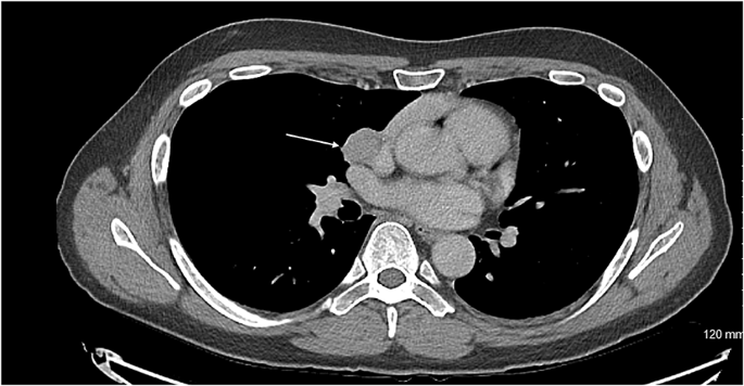
Axial CT showing the carcinoid tumour (demonstrated by arrow) in close proximity to innominate vein

Fig. 2

figure 2

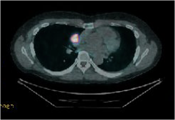
DOTATE PET demonstrating significant uptake in carcinoid tumour

Fig. 3

figure 3

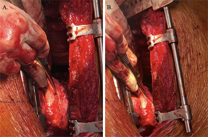
Intraoperative visualisation right phrenic nerve (demonstrated by forceps) overlying carcinoid tumour

Data availability

No datasets were generated or analysed during the current study.

References

  1. Bohnenberger H, Dinter H, König A, Ströbel P. Neuroendocrine Tumors Thymus Mediastinum J Thorac Dis [Internet]. 2017.

  2. Lau J, Ioan Cvasciuc T, Simpson D, C de Jong M, Parameswaran R. Continuing challenges of primary neuroendocrine tumours of the thymus: A concise review. Eur J Surg Oncol [Internet]. 2022.

  3. Chaer R, Massad MG, Evans A, Snow NJ, Geha AS. Primary neuroendocrine tumors of the thymus. Ann Thorac Surg. 2002. https://doi.org/10.1016/S0003-4975(02)03547-6.

    Article PubMed Google Scholar

  4. Isidori AM, Kaltsas GA, Pozza C, Frajese V, Newell-Price J, Reznek RH, et al. The ectopic adrenocorticotropin syndrome: clinical features, diagnosis, management, and long-term follow-up. J Clin Endocrinol Metab. 2006. https://doi.org/10.1210/jc.2005-1542.

    Article PubMed Google Scholar

  5. Ilias I, Torpy DJ, Pacak K, Mullen N, Wesley RA, Nieman LK. Cushing’s syndrome due to ectopic corticotropin secretion: twenty years’ experience at the National Institutes of Health. J Clin Endocrinol Metab. 2005. https://doi.org/10.1210/jc.2004-2527.

    Article PubMed Google Scholar

  6. Daskalakis K, Chatzelis E, Tsoli M, Papadopoulou-Marketou N, Dimitriadis GK, Tsolakis AV et al. Endocrine paraneoplastic syndromes in patients with neuroendocrine neoplasms. Endocrine [Internet]. 2018.

  7. Kamp K, Alwani RA, Korpershoek E, Franssen GJH, de Herder WW, Feelders RA. Prevalence and clinical features of the ectopic ACTH syndrome in patients with gastroenteropancreatic and thoracic neuroendocrine tumors. Eur J Endocrinol [Internet]. 2015.

  8. Midorikawa K, Miyahara S, Nishino N, Ueda Y, Waseda R, Shiraishi T, et al. Analysis of 25 surgical cases of thymic neuroendocrine tumors and thymic carcinoma. J Cardiothorac Surg. 2024. https://doi.org/10.1186/s13019-024-02723-w.

    Article PubMed PubMed Central Google Scholar

  9. Chen Y, Zhang J, Zhou M, Guo C, Li S. Real-world clinicopathological features and outcome of thymic neuroendocrine tumors: a retrospective single-institution analysis. Orphanet J Rare Dis. 2022. https://doi.org/10.1186/s13023-022-02366-x.

    Article PubMed PubMed Central Google Scholar

  10. Filosso PL, Yao X, Ahmad U, Zhan Y, Huang J, Ruffini E, et al. Outcome of primary neuroendocrine tumors of the thymus: a joint analysis of the international thymic malignancy interest group and the European society of thoracic surgeons databases. J Thorac Cardiovasc Surg. 2015. https://doi.org/10.1016/j.jtcvs.2014.08.061.

    Article PubMed Google Scholar

  11. Tiffet O, Nicholson AG, Ladas G, Sheppard MN, Goldstraw P. A clinicopathologic study of 12 neuroendocrine tumors arising in the thymus. Chest [Internet]. 2003.

  12. Bakhos CT, Salami AC, Kaiser LR, Petrov RV, Abbas AE. Thymic neuroendocrine tumors and thymic carcinoma: demographics, treatment, and survival. Innovations: Technology and Techniques in Cardiothoracic and Vascular Surgery. 2020. https://doi.org/10.1177/1556984520949287.

    Article PubMed Google Scholar

  13. Wen J, Chen J, Chen D, Liu D, Xu X, Huang L, et al. Evaluation of the prognostic value of surgery and postoperative radiotherapy for patients with thymic neuroendocrine tumors: a propensity-matched study based on the SEER database. Thorac Cancer. 2018. https://doi.org/10.1111/1759-7714.12868.

    Article PubMed PubMed Central Google Scholar

  14. Busetto A, Comacchio GM, Verzeletti V, Fares Shamshoum, Fortarezza F, Pezzuto F et al. Bilateral metachronous typical and atypical carcinoid tumors of the lung. Thorac Cancer. 2025;16(8).

Download references

Acknowledgements

No acknowledgements.

Funding

This report has not received funding.

Author information

Authors and Affiliations

  1. Department of Thoracic Surgery, Guy’s and St Thomas’ NHS Foundation Trust, London, England, UK

    Puiyee Sophia Chan, Akshay J. Patel, Ishaan Chauhan & Andrea Bille

  2. Institute of Immunology and Immunotherapy, University of Birmingham, Vincent Drive, Edgbaston, B15 2TT, England, UK

    Akshay J. Patel

Contributions

PSC and AJP wrote the main manuscript text and IC and AB prepared Figs. 12 and 3. All authors reviewed the manuscript.

Corresponding author

Correspondence to Akshay J. Patel.

Ethics declarations

Ethics approval and consent to participate

The authors are accountable for all aspects of the work, ensuring that questions related to the accuracy or integrity of any part of the work are appropriately investigated and resolved. All procedures performed in this study were in accordance with the ethical standards of the institutional and/or national research committee(s) and with the Helsinki Declaration (as revised in 2013).

Consent for publication

Written informed consent was obtained from the patients for the publication of this case report and accompanying images. A copy of the written consent is available for review by the editorial office of this journal.

Competing interests

The authors declare no competing interests.

Additional information

Publisher’s note

Springer Nature remains neutral with regard to jurisdictional claims in published maps and institutional affiliations.

Thymic Neuroendocrine Tumor With Metastasis to the Breast Causing Ectopic Cushing’s Syndrome

Ectopic adrenocorticotropic hormone secretion (EAS) is responsible for approximately 10%–18% of Cushing’s syndrome cases. Thymic neuroendocrine tumors (NETs) comprise 5%–16% of EAS; therefore, they are very rare and the data about this particular tumors is scarce.

We present a case of a 34-year-old woman with a rapid onset of severe hypercortisolism in April 2016. After initial treatment with a steroid inhibitor (ketoconazole) and diagnostics including 68Ga DOTA-TATE PET/CT, it was shown to be caused by a small thymic NET.

After a successful surgery and the resolution of all symptoms, there was a recurrence after 5 years of observation caused by a metastasis to the breast, shown in the 68Ga DOTA-TATE PET/CT result and confirmed with a breast biopsy.

Treatment with a steroid inhibitor (metyrapone) and tumor resection were again curative. The last disease relapse appeared 7 years after the initial treatment, with severe hypercortisolism treated with osilodrostat. There was a local recurrence in the mediastinum, and a thoracoscopic surgery was performed with good clinical and biochemical effect.

The patient remains under careful follow-up. Our case stays in accordance with recent literature data, showing that patients with thymic NETs are younger than previously considered and that the severity of hypercortisolism does not correlate with the tumor size. The symptoms of EAS associated with thymic NET may develop rapidly and may be severe as in our case. Nuclear medicine improves the effectiveness of the tumor search, which is crucial in successful EAS therapy. Our case also underlines the need for lifelong monitoring of patients with thymic NETs and EAS.

1 Introduction

Ectopic adrenocorticotropic hormone secretion (EAS) represents between 9% and 18% of adrenocorticotropic hormone (ACTH)-dependent Cushing’s syndrome (CS) cases (13). The tumors secreting ACTH may occur in many locations and present with different histopathological differentiation, resulting in various clinical outcomes. In the past, most of the EAS cases were associated with small cell lung cancer, characterized by rapid tumor progression and unfavorable prognosis. Recently, well-differentiated neuroendocrine tumors (NETs) from the foregut prevail in the clinical series of EAS, with most common locations in the lungs, thymus, and pancreas (1).

EAS is often associated with severe hypercortisolism. Typical Cushing’s appearance may not be present due to the rapid onset of the disease. Patients with this type of hypercortisolism need urgent treatment because they have the highest mortality of all forms of CS (4). A retrospective review of 43 patients with EAS reported deaths in 27 patients (62.8%) and a median overall survival of 32.2 months. The leading causes of mortality were the progression of primary malignancies and systemic infections; two patients died from pulmonary embolism (5).

Prompt surgical removal of the tumor secreting ACTH is the mainstay of the therapy. However, finding the tumor causing EAS can be challenging due to its small size and variety of locations. Most authors recommend a combination of computed tomography (CT) scanning of the chest, abdomen, and pelvis, with additional magnetic resonance imaging (MRI) of the pituitary, as the first-line examinations (167). However, the sensitivity of standard imaging modalities is suboptimal (8). In the analysis of 231 patients with EAS, cross-sectional imaging revealed the source of ACTH in 52.4% of them at initial evaluation, and another 29% was found during follow-up or due to nuclear medicine functional imaging, while 18.6% remained occult (9). Nuclear medicine improves the sensitivity of conventional radiology in the case of EAS, with the use of 18-fluorodeoxyglucose (18F-FDG) positron emission tomography (PET)/CT (18F-FDG PET/CT) expected to be useful in identifying EAS tumors with high proliferative activity and 68gallium-labeled somatostatin analogues (68Ga DOTA-TATE) PET/CT with the potential to detect NETs. In the head-to-head comparison, the detection rate of the source of EAS was 75% for 68Ga DOTA-TATE and 60% for 18F-FDG PET/CT, while the highest sensitivity (90%) was achieved when both methods were combined (10).

Thymic NETs comprise 2%–5% of all thymic neoplasms and may cause some paraneoplastic syndromes, with the most frequent being myasthenia gravis, syndrome of inappropriate antidiuretic hormone secretion, and hypercortisolism (11). EAS associated with thymic NETs are rare, representing between 5% and 16% of EAS in published case series (1). Because of the rarity and heterogeneity of the disease, no evidence-based guidelines are available.

We present a case of a patient with thymic NET causing EAS, with metastasis to the breast after 5 years of post-surgical remission and another local recurrence 7 years after the first operation.

Our case is unique because thymic NETs causing EAS are known as an aggressive disease with a median recurrence time of 24 months after thymectomy (12). There are only a few cases described of metastases to the breast from thymic NETs causing EAS (1316). Moreover, 68Ga-SSTR PET/CT was very helpful in detecting both primary and metastatic ectopic ACTH-secreting tumor, which underlines its role in the diagnostic workout of EAS.

2 Case description

A 32-year-old woman with no relevant medical history was admitted to the endocrinology department in April 2016 due to the rapid onset of symptoms: weight gain, hypertension, skin changes, and oligomenorrhoea.

The measurements at initial physical examination were as follows: body mass index (BMI)—29 kg/m2, blood pressure—180/90 mmHg, and heart rate—88/min. She had plethora, acne, moon face, buffalo hump, central obesity, many red striae in the abdominal area, and mild hirsutism. The baseline laboratory findings are presented in Table 1, with hypokalemia, diabetes, leukocytosis, high levels of serum cortisol, ACTH, and chromogranin A, and increased urine-free cortisol (UFC) secretion. There was no suppression of serum cortisol or UFC after a high-dose dexamethasone test. ACTH-dependent CS was diagnosed, and EAS was suspected. The patient’s family history was negative for endocrine diseases or genetic disorders.

Table 1

www.frontiersin.org

Table 1. Laboratory results at diagnosis (April 2016).

The first-line cross-sectional imaging studies (chest, abdomen, and pelvis CT and MRI of the pituitary gland) did not reveal the source of ACTH. Only a symmetrical enlargement of adrenals was observed. 68Ga DOTA-TATE PET/CT revealed an oval lesion in the anterior mediastinum (1.9 × 1.3 cm) with a subtle overexpression of somatostatin receptors (SUV max. 2.8, Figures 1A, B). The chest MRI confirmed a mass 1.5 × 2.0 × 2.5 cm, with high T2-weighted signal and high contrast enhancement, suggestive of NET. The patient was given ketoconazole (600 mg daily), spironolactone, potassium supplementation, antihypertensive drugs, and thromboembolic prophylaxis. In June 2016, thoracoscopic removal of the mediastinal tumor was performed. In the histopathological examination, the tumor was encapsulated, without evidence of invasion, and no lymph node metastases were described. The immunophenotype of the tumor was as follows: CgA (+), Syn (+), CKAE1+E3 (+) “dot-like”, S100 (-), calcitonin (-), EMA (+/-), Ki67 3% to 4% in hot spots, no necrosis, mitotic index 0/10HPF with conclusion: thymic NET—typical carcinoid (low-grade). The presence of paraganglioma was also taken into consideration, as such cases were described (17). However, the significant reaction with cytokeratin and lack of S100 protein expression made this diagnosis less probable.

Figure 1

www.frontiersin.org

Figure 168Ga-DOTATATE PET/CT scans. (A, B) Before the first surgery (April 2016). (C, D) Before the second surgery (May 2021). (E, F) Before the third surgery (January 2023).

The postoperative morning serum cortisol concentration was below 5 µg/dL, indicating biochemical remission. The patient received hydrocortisone substitution for a month. The clinical signs of CS disappeared, and there was a normalization of UFC.

During 5 years of follow-up, the patient got pregnant and delivered a healthy child. Genetic counseling was performed, and no germline mutation of MEN1 gene was identified. Other clinical manifestations of MEN1 (like primary hyperparathyroidism and pituitary secreting tumors) were excluded.

In May 2021, the patient experienced a sudden recurrence of CS symptoms. The laboratory findings confirmed severe hypercortisolism (Table 2); therefore, treatment with steroid inhibitor metyrapone was administered. The patient tolerated only 750 mg daily; there were side effects (skin rash and tachycardia) with higher doses. The chest MRI revealed no recurrence in the location of the primary tumor, only a lesion in the right breast (1.2 × 1.0 × 1.1 cm) with atypical contrast enhancement. The 68Ga-DOTA-TATE PET/CT result showed a subtle overexpression of the tracer (SUV max 1.9) in the right breast (Figures 1C, D). Breast ultrasonography confirmed a hypoechogenic, hypervascular mass in the right breast, BIRADS 3/4, diagnosed as NET in the breast biopsy. The tumor was removed in July 2021 without complications. The histopathological samples were compared with the primary lesion, confirming the metastasis from thymic NET to the breast—tumor size 0.7 × 1.5 cm, clear surgical margins (8 mm) with Ki67 3% (NET G2), and no lymph node metastases. After the breast surgery, the cortisol levels normalized in blood and urine and the CS symptoms disappeared. 18F-FDG PET/CT and 68Ga-DOTA-TATE PET/CT were performed, showing no pathological increase of radiotracer uptake in post-operative locations or mediastinal lymph nodes. The patient consulted with the oncology team, and no adjuvant therapy was recommended.

Table 2

www.frontiersin.org

Table 2. Laboratory results during 7 years of observation.

The next recurrence of the disease occurred in February 2023, with the symptoms developing suddenly during a very short period (1 to 2 weeks), additionally with significant mental deterioration (concentration disorders, anxiety, severe mood swing). The laboratory findings confirmed excessive hypercortisolism (Table 2). The patient was given osilodrostat (the initial dose was 20 mg daily but later reduced to 10 mg daily for 2 weeks until surgery) and symptomatic treatment with good clinical and biochemical effect. The 68Ga-DOTA-TATE PET/CT result showed a slightly increased uptake of the tracer in the left mediastinum, between cervical vessels, 0.9 × 1.2 cm (Figures 1E, F)—probably a local recurrence. Thoracotomy was performed in February 2023, with subsequent clinical and biochemical improvement (Table 2). In the histopathological examination, mediastinal NET G1 was diagnosed, without necrosis, mitotic activity 0/2 mm2, immunophenotype CgA (+), CD56 (+), Ki 67 1%, CK AE1/AE3 (+), CD117 (+), p40 (-), TdT (-), PAX8 (-), and the presence of tumor cell embolism in the vessels. One metastatic lesion was found in the pericardium (the maximal dimension of the tissue was 13 mm, resected radically). Two metastatic lesions in the fat tissue were found (one tissue fragment from the mediastinum, max. 16 mm diameter, and the second tissue fragment was surrounding the jugular vein, max. diameter up to 40 mm, both resected radically). Two of the 10 resected lymph nodes had metastatic lesions: one from the area of the jugular vein, diameter 11 mm, with capsular invasion, and the second lymph node N2R with capsular invasion, both resected radically. The symptoms of hypercortisolism disappeared, and the cortisol values were normalized after the operation. The patient is currently under careful monitoring, without signs of clinical or biochemical recurrence. 68Ga-DOTA-TATE PET/CT is performed every 6 months.

3 Discussion

Our case is representative for thymic NETs causing EAS presented in literature, but it also shows some distinct features, giving new insight into this rare condition.

In recent series, ACTH-secreting thymic NETs occurred often in young adults, like our patient. The typical age of presentation is 21–35 years in the largest case series, and 7.4% were children under 15 years (1213). In contrast, the former series of thymic NETs showed a peak incidence in the sixth decade of life (11).

ACTH-secreting thymic NETs show a slight male preponderance (58.6%); however, the patient’s gender does not seem to relate with the disease outcome (12). There was only an association between male sex and larger tumor size preoperatively as found in one case series (13).

Thymic NETs causing EAS are very rarely associated with MEN1; we have also excluded it in our patient. On the contrary, 30% of thymic NETs not associated with CS are found in patients with MEN1, mostly male smokers (18). It is not clear why thymic NETs with EAS are less likely caused by MEN1 gene mutation, but the possibility of this genetic predisposition should always be taken into consideration.

Thymic NETs associated with EAS are generally considered aggressive, presenting significant cellular atypia in the histopathological examination (19). However, the biology of the tumors is variable. In the histopathological examination of 92 thymic NETs secreting ACTH, the most common subtype was atypical NET (46.7%), while 30.4% of the cases were typical NETs and 21.7% were carcinomas, with the median Ki-67 10%, ranging from 1% to 40%. The median tumor size among 112 patients was 4.7 cm, ranging from 1 to 20 cm, and 55.7% of patients had metastases at presentation (12). It proves the significant heterogeneity of the disease.

Our patient had typical NET with small dimensions and localized disease at the time of diagnosis. Despite this, we observed aggressive Cushing’s syndrome with a short duration of symptoms and life-threatening hypokalemia. It has been observed that there is no correlation between tumor size and hormone levels (12). Thymic NETs associated with EAS are often large, which simplifies the diagnosis and localization. However, in the case of incidental sellar mass or small thymic tumor, the differential diagnosis might be difficult. The highest sensitivity in distinguishing thymic EAS from Cushing’s disease was documented in inferior petrosal sinus sampling and corticotropin-releasing hormone (CRH) stimulation test (1220).

In severe cases, when small ACTH-secreting NET needs to be found urgently, PET/CT is a very helpful diagnostic tool. In a prospective study comprising 20 patients with histologically proven EAS, the 68Ga-DOTATATE PET/CT result correctly identified the tumor in 75%, with SUV max. ranging from 1.4 to 20.7, while the 18F-FDG PET/CT findings had a slightly worse result (identified 60% tumors), with SUV max. ranging from 1.8 to 10.0. Those methods are believed to be complementary in case of localization and discrimination of EAS. The 68Ga-DOTATATE PET/CT result revealed tumor in six cases with a negative 18F-FDG PET/CT result, while the 18F-FDG PET/CT procedure was diagnostic in three cases with a negative 68Ga-DOTATATE uptake; the combined sensitivity of both methods was 90% (10). The typical first-line diagnostic modalities’ (CT and MRI) sensitivities range from 52% to 66% (9). Our case remains in accordance with those results, showing difficulties in localizing the ACTH source in first-line radiological methods and with 68Ga-DOTATATE PET/CT being the most useful diagnostic tool. It should also be noted that the 68Ga-DOTATATE uptake was only mildly elevated both in primary tumor and its recurrences despite excessive hormonal activity. We did not perform 18F-FDG PET/CT until second operation, as it was believed to be rather helpful in poorly differentiated tumors and 68Ga-DOTATATE PET/CT was diagnostic. Later, we performed it in search for other metastatic tumors, but the examination showed no tumor spread.

The recommended treatment of thymic NETs regarded radically resectable is thymectomy by median sternotomy or thoracotomy and lymph node dissection (112122). According to the last version of the ESMO Guidelines, available literature suggests no benefit from adjuvant therapy in ThCs. The majority of the authors of the Guidelines panel suggest individually discussing eventual postoperative therapies, including RT and/or systemic therapies, balancing the pros and cons only in selected patients with advanced stage R0 or R1-2 resection (22). Data on systemic therapies in thymic NETs are scarce; therefore, they should be discussed in a multidisciplinary expert team in case of morphologically progressive tumors, high tumor burden, or refractory hormonal syndromes. Somatostatin analogs are recommended as the first-line systemic therapy in typical carcinoids (22). We considered the adjuvant therapy with somatostatin analogs; however, due to the low uptake in PET examination and complete resolution of symptoms as well as the radical type of surgical removal, we did not decide to initiate such therapy. Other systemic treatment options include everolimus (second line in typical carcinoids or first line in atypical carcinoids), chemotherapy, peptide receptor radionuclide therapy (PRRT), and interferon-α (2223). There is also data on the benefits of combining long-acting lanreotide with temozolomide in progressive thymic NETs (24).

Due to the variable availability of steroid inhibitors during the course of the disease, our patient received three different preparations at each disease relapse. Both ketoconazole and osilodrostat were well tolerated and reduced the hypercortisolism within a few days, but metyrapone caused significant side effects (see below—”Patient’s perspective”), and it was not possible to normalize the cortisol values with this steroid inhibitor. It is worth noting that when using the most recent steroid inhibitor—osilodrostat—we initiated the therapy with a high dose without a previous dose titration. This strategy might be used in the case of severe hypercortisolism and proved effective and safe in our patient (25).

Most commonly, metastases from thymic NET producing ACTH are localized in lymph nodes, bone, lung, pleura, and, less commonly, liver and parotid gland (13). There are very few cases of EAS-related thymic NETs with breast metastases described in the literature, with some histopathological variability (one case related to atypical carcinoid, another to combined large-cell neuroendocrine carcinoma and atypical carcinoid, and third case of neuroendocrine carcinoma). All of them were female patients between 24 and 36 years of age, with mediastinal lymph nodes metastases at the time of presentation; one also had distant metastases to the bones (1315). Contrary to the reported cases, our patient had typical carcinoid (confirmed by three independent pathologists from different centers) but similarly presented with severe hypercortisolism. It suggests that there is no connection between tumor differentiation and the severity of hypercortisolism. Interestingly, in a review of 661 patients with metastatic NETs from Sweden, there were 20 patients with NETs and breast metastases, and among them only one case of thymic NET (Ki 67 12%), but without EAS. A total of 11 patients with breast metastases had a primary tumor in the small intestine and eight in the lung (16).

Our case underlines the necessity of long-term follow-up in EAS, as the recurrences occurred 5 and 7 years after the initial successful treatment. According to guidelines, follow-up after treatment of thymic NETs should be life-long (22).

The strength of our report is the presentation of a thymic NET with metastasis to the breast, diagnosed and treated with many currently available tools and with a long period of follow-up. The limitation is the low number of other similar cases to compare, which is a consequence of the rarity of this disease.

In conclusion, our case proves that thymic NETs with EAS might present in young patients with well-differentiated character in histopathological examination and severe, life-threatening hypercortisolism despite the small size of the primary lesion. 68Ga-DOTATATE PET/CT is a very helpful tool to localize the tumor. Finally, life-long follow-up should be performed despite complete remission after surgery.

4 Patient’s perspective

The first symptoms that I observed were face edema and mood changes. I rapidly lost muscle mass (approximately 6 kg in 2 weeks), and I was not able to climb stairs, especially with my child’s pram. The most difficult to accept were changes in my appearances—hirsutism, losing hair, changes of my facial features. My sense of pain (for example, during medical procedures) was diminished. Other disruptive symptoms were intensive sweating, increased appetite, thirst, brain fog, and digestive problems. At every relapse, the disease manifestations were fluctuating, all of them intensifying at the same time, which was very difficult for me. Also stress evoked disease symptoms. I experienced a strange feeling of warm during cortisol outbursts.

As for the treatment, I did not tolerate metyrapone well. I had skin rash, anxiety attacks with heart palpitations, and a metallic taste in my mouth. Other drugs (ketoconazole, osilodrostat) were better for me.

After operations of the relapses, the symptoms diminished very quickly, especially the most difficult ones. My blood pressure and glycemia normalized within a few days. Other manifestations, like loss of hair or skin changes, persisted up to 3 months.

Data availability statement

The datasets presented in this article are not readily available because the data are potentially identifiable. Requests to access the datasets should be directed to Aleksandra Zdrojowy-Wełna, aleksandra.zdrojowy-welna@umw.edu.pl.

Ethics statement

This study was exempt from ethical approval procedures being a case report of a single patient who has voluntarily provided oral and written consent to participate in the study and to have her case published for the sake of helping us better understand the clinical picture and the course of thymic neuroendocrine tumors with EAS and share it with the medical community for awareness about it. Written informed consent was obtained from the participant/patient(s) for the publication of this case report.

Author contributions

AZ-W: Conceptualization, Data curation, Investigation, Methodology, Software, Writing – original draft. MB: Conceptualization, Supervision, Writing – review & editing. JS: Data curation, Investigation, Methodology, Writing – review & editing. AJ-P: Data curation, Investigation, Writing – review & editing. JK-P: Conceptualization, Data curation, Investigation, Methodology, Supervision, Writing – original draft.

Funding

The author(s) declare that no financial support was received for the research, authorship, and/or publication of this article.

Acknowledgments

We would like to thank Prof. Barbara Górnicka and Prof. Michał Jeleń for their collaboration throughout the patient’s treatment.

Conflict of interest

The authors declare that the research was conducted in the absence of any commercial or financial relationships that could be construed as a potential conflict of interest.

The handling editor AJ declared a past co-authorship with the author MB.

The author(s) declared that they were an editorial board member of Frontiers, at the time of submission. This had no impact on the peer review process and the final decision.

Publisher’s note

All claims expressed in this article are solely those of the authors and do not necessarily represent those of their affiliated organizations, or those of the publisher, the editors and the reviewers. Any product that may be evaluated in this article, or claim that may be made by its manufacturer, is not guaranteed or endorsed by the publisher.


Supplementary material

The Supplementary Material for this article can be found online at: https://www.frontiersin.org/articles/10.3389/fonc.2025.1492187/full#supplementary-material

References

1. Young J, Haissaguerre M, Viera-Pinto O, Chabre O, Baudin E, Tabarin A. Management of endocrine disease: Cushing’s syndrome due to ectopic ACTH secretion: an expert operational opinion. Eur J Endocrinol. (2020) 182:29–58. doi: 10.1530/EJE-19-0877

PubMed Abstract | Crossref Full Text | Google Scholar

2. Feelders R, Sharma S, Nieman L. Cushing`s syndrome: epidemiology and developments in disease management. Clin Epidemiol. (2015) 7:281–93. doi: 10.2147/CLEP.S44336

PubMed Abstract | Crossref Full Text | Google Scholar

3. Lacroix A, Feelders RA, Stratakis CA, Nieman LK. Cushing’s syndrome. Lancet Lond Engl. (2015) 386:913–27. doi: 10.1016/S0140-6736(14)61375-1

PubMed Abstract | Crossref Full Text | Google Scholar

4. Javanmard P, Duan D, Geer EB. Mortality in patients with endogenous cushing’s syndrome. Endocrinol Metab Clin North Am. (2018) 47:313–33. doi: 10.1016/j.ecl.2018.02.005

PubMed Abstract | Crossref Full Text | Google Scholar

5. Ejaz S, Vassilopoulou-Sellin R, Busaidy NL, Hu MI, Waguespack SG, Jimenez C, et al. Cushing syndrome secondary to ectopic adrenocorticotropic hormone secretion: the University of Texas MD Anderson Cancer Center Experience. Cancer. (2011) 117:4381–9. doi: 10.1002/cncr.v117.19

PubMed Abstract | Crossref Full Text | Google Scholar

6. Sookur PA, Sahdev A, Rockall AG, Isidori AM, Monson JP, Grossman AB, et al. Imaging in covert ectopic ACTH secretion: a CT pictorial review. Eur Radiol. (2009) 19:1069–78. doi: 10.1007/s00330-008-1274-5

PubMed Abstract | Crossref Full Text | Google Scholar

7. Yogi-Morren D, Habra MA, Faiman C, Bena J, Hatipoglu B, Kennedy L, et al. Pituitary MRI findings in patients with pituitary and ectopic ACTH-dependent cushing syndrome: does A 6- mm pituitary tumor size cut-off value exclude ectopic acth syndrome? Endocr Pract. (2015) 21:1098–103. doi: 10.4158/EP15662.OR

PubMed Abstract | Crossref Full Text | Google Scholar

8. Ilias I, Torpy DJ, Pacak K, Mullen N, Wesley RA, Nieman LK. Cushing’s syndrome due to ectopic corticotropin secretion: twenty years’ Experience at the national institutes of health. J Clin Endocrinol Metab. (2005) 90:4955–62. doi: 10.1210/jc.2004-2527

PubMed Abstract | Crossref Full Text | Google Scholar

9. Isidori AM, Sbardella E, Zatelli MC, Boschetti M, Vitale G, Colao A, et al. Conventional and nuclear medicine imaging in ectopic cushing’s syndrome: A systematic review. J Clin Endocrinol Metab. (2015) 100:3231–44. doi: 10.1210/JC.2015-1589

PubMed Abstract | Crossref Full Text | Google Scholar

10. Liu Q, Zang J, Yang Y, Ling Q, Wu H, Wang P, et al. Head-to-head comparison of 68Ga- DOTATATE PET/CT and 18F-FDG PET/CT in localizing tumors with ectopic adrenocorticotropic hormone secretion: a prospective study. Eur J Nucl Med Mol Imaging. (2021) 48:4386–95. doi: 10.1007/s00259-021-05370-8

PubMed Abstract | Crossref Full Text | Google Scholar

11. Gaur P, Leary C, Yao JC. Thymic neuroendocrine tumors: a SEER database analysis of 160 patients. Ann Surg. (2010) 251:1117–21. doi: 10.1097/SLA.0b013e3181dd4ec4

PubMed Abstract | Crossref Full Text | Google Scholar

12. Guerrero-Pérez F, Peiró I, Marengo AP, Teulé A, Ruffinelli JC, Llatjos R, et al. Ectopic Cushing’s syndrome due to thymic neuroendocrine tumours: a systematic review. Rev Endocr Metab Disord. (2021) 22:1041–56. doi: 10.1007/s11154-021-09660-2

PubMed Abstract | Crossref Full Text | Google Scholar

13. Neary NM, Lopez-Chavez A, Abel BS, Boyce AM, Schaub N, Kwong K, et al. Neuroendocrine ACTH-producing tumor of the thymus—Experience with 12 patients over 25 years. J Clin Endocrinol Metab. (2012) 97:2223–30. doi: 10.1210/jc.2011-3355

PubMed Abstract | Crossref Full Text | Google Scholar

14. Dzialach L, Wojciechowska-Luzniak A, Maksymowicz M, Witek P. Case report: A challenging case of severe Cushing’s syndrome in the course of metastatic thymic neuroendocrine carcinoma with a synchronous adrenal tumor. Front Endocrinol. (2024). doi: 10.3389/fendo.2024.1399930/full

PubMed Abstract | Crossref Full Text | Google Scholar

15. Gaur S, Ayyappan AP, Nahleh Z. Breast metastases from an adrenocorticotropic hormone secreting thymic neuro-endocrine tumor. Breast Dis. (2013) 34:81–6. doi: 10.3233/BD-130354

PubMed Abstract | Crossref Full Text | Google Scholar

16. Crona J, Granberg D, Norlén O, Wärnberg F, Stålberg P, Hellman P, et al. Metastases from neuroendocrine tumors to the breast are more common than previously thought. A diagnostic pitfall? World J Surg. (2013) 37:1701–6. doi: 10.1007/s00268-013-2037-2

PubMed Abstract | Crossref Full Text | Google Scholar

17. Li B, Yan Z, Huang H. Case report: an unusual case of ectopic ACTH syndrome caused by mediastinal paraganglioma. Front Endocrinol. (2021) 12:790975. doi: 10.3389/fendo.2021.790975

PubMed Abstract | Crossref Full Text | Google Scholar

18. Ferolla P, Falchetti A, Filosso P, Tomassetti P, Tamburrano G, Avenia N, et al. Thymic neuroendocrine carcinoma (carcinoid) in multiple endocrine neoplasia type 1 syndrome: the Italian series. J Clin Endocrinol Metab. (2005) 90:2603–9. doi: 10.1210/jc.2004-1155

PubMed Abstract | Crossref Full Text | Google Scholar

19. Moran CA, Suster S. Neuroendocrine carcinomas (Carcinoid tumor) of the thymus. Am J Clin Pathol. (2000) 114:100–10. doi: 10.1309/3PDN-PMT5-EQTM-H0CD

PubMed Abstract | Crossref Full Text | Google Scholar

20. Kakade HR, Kasaliwal R, Jagtap VS, Bukan A, Budyal SR, Khare S, et al. Ectopic acth- secreting syndrome: A single-center experience. Endocr Pract. (2013) 19:1007–14. doi: 10.4158/EP13171.OR

PubMed Abstract | Crossref Full Text | Google Scholar

21. Girard N. Neuroendocrine tumors of the thymus: the oncologist point of view. J Thorac Dis. (2017) 9:1491–500. doi: 10.21037/jtd.2017.08.18

PubMed Abstract | Crossref Full Text | Google Scholar

22. Baudin E, Caplin M, Garcia-Carbonero R, Fazio N, Ferolla P, Filosso PL, et al. Lung and thymic carcinoids: ESMO Clinical Practice Guidelines for diagnosis, treatment and follow-up. Ann Oncol Off J Eur Soc Med Oncol. (2021) 32:439–51. doi: 10.1016/j.annonc.2021.01.003

PubMed Abstract | Crossref Full Text | Google Scholar

23. Ferolla P, Brizzi MP, Meyer T, Mansoor W, Mazieres J, Do Cao C, et al. Efficacy and safety of long-acting pasireotide or everolimus alone or in combination in patients with advanced carcinoids of the lung and thymus (LUNA): an open-label, multicentre, randomised, phase 2 trial. Lancet Oncol. (2017) 18:1652–64. doi: 10.1016/S1470-2045(17)30681-2

PubMed Abstract | Crossref Full Text | Google Scholar

24. Ferolla P, Berruti A, Spada F, Brizzi MP, Ibrahim T, Marconcini R, et al. Efficacy and safety of lanreotide autogel and temozolomide combination therapy in progressive thoracic neuroendocrine tumors (Carcinoid): results from the phase 2 ATLANT study. Neuroendocrinology. (2023) 113:332–42. doi: 10.1159/000526811

PubMed Abstract | Crossref Full Text | Google Scholar

25. Fleseriu M, Biller BMK. Treatment of Cushing’s syndrome with osilodrostat: practical applications of recent studies with case examples. Pituitary. (2022) 25:795–809. doi: 10.1007/s11102-022-01268-2

PubMed Abstract | Crossref Full Text | Google Scholar

Keywords: ectopic Cushing`s syndrome, thymic neuroendocrine tumor, thymic NET, ectopic ACTH secretion, case report

Citation: Zdrojowy-Wełna A, Bolanowski M, Syrycka J, Jawiarczyk-Przybyłowska A and Kuliczkowska-Płaksej J (2025) Case Report: Thymic neuroendocrine tumor with metastasis to the breast causing ectopic Cushing’s syndrome. Front. Oncol. 15:1492187. doi: 10.3389/fonc.2025.1492187

Received: 11 September 2024; Accepted: 31 January 2025;
Published: 25 February 2025.

Edited by:

Aleksandra Gilis-Januszewska, Jagiellonian University Medical College, Poland

Reviewed by:

Piero Ferolla, Umbria Regional Cancer Network, Italy
Lukasz Dzialach, Warsaw Medical University, Poland

Copyright © 2025 Zdrojowy-Wełna, Bolanowski, Syrycka, Jawiarczyk-Przybyłowska and Kuliczkowska-Płaksej. This is an open-access article distributed under the terms of the Creative Commons Attribution License (CC BY). The use, distribution or reproduction in other forums is permitted, provided the original author(s) and the copyright owner(s) are credited and that the original publication in this journal is cited, in accordance with accepted academic practice. No use, distribution or reproduction is permitted which does not comply with these terms.

*Correspondence: Aleksandra Zdrojowy-Wełna, aleksandra.zdrojowy-welna@umw.edu.pl

Disclaimer: All claims expressed in this article are solely those of the authors and do not necessarily represent those of their affiliated organizations, or those of the publisher, the editors and the reviewers. Any product that may be evaluated in this article or claim that may be made by its manufacturer is not guaranteed or endorsed by the publisher.

95% of researchers rate our articles as excellent or good

Learn more about the work of our research integrity team to safeguard the quality of each article we publish.

Find out more

People also looked at

Supplementary Material
  • Data Sheet 1.pdf

Day 8, Cushing’s Awareness Challenge

It’s Here!

Dr. Cushing was born in Cleveland Ohio. The fourth generation in his family to become a physician, he showed great promise at Harvard Medical School and in his residency at Johns Hopkins Hospital (1896 to 1900), where he learned cerebral surgery under William S. Halsted.

After studying a year in Europe, he introduced the blood pressure sphygmomanometer to the U.S.A. He began a surgical practice in Baltimore while teaching at Johns Hopkins Hospital (1901 to 1911), and gained a national reputation for operations such as the removal of brain tumors. From 1912 until 1932 he was a professor of surgery at Harvard Medical School and surgeon in chief at Peter Bent Brigham Hospital in Boston, with time off during World War I to perform surgery for the U.S. forces in France; out of this experience came his major paper on wartime brain injuries (1918). In addition to his pioneering work in performing and teaching brain surgery, he was the reigning expert on the pituitary gland since his 1912 publication on the subject; later he discovered the condition of the pituitary now known as “Cushing’s disease“.

Read more about Dr. Cushing

Today, April 8th, is Cushing’s Awareness Day. Please wear your Cushing’s ribbons, t-shirts, awareness bracelets or Cushing’s colors (blue and yellow) and hand out Robin’s wonderful Awareness Cards to get a discussion going with anyone who will listen.

And don’t just raise awareness on April 8.  Any day is a good day to raise awareness.


harvey-book

I found this biography fascinating!

I found Dr. Cushing’s life to be most interesting. I had previously known of him mainly because his name is associated with a disease I had – Cushing’s. This book doesn’t talk nearly enough about how he came to discover the causes of Cushing’s disease, but I found it to be a valuable resource, anyway.
I was so surprised to learn of all the “firsts” Dr. Cushing brought to medicine and the improvements that came about because of him. Dr. Cushing introduced the blood pressure sphygmomanometer to America, and was a pioneer in the use of X-rays.

He even won a Pulitzer Prize. Not for medicine, but for writing the biography of another Doctor (Sir William Osler).

Before his day, nearly all brain tumor patients died. He was able to get the number down to only 5%, unheard of in the early 1900s.

This is a very good book to read if you want to learn more about this most interesting, influential and innovative brain surgeon.


What Would Harvey Say?

harvey-book

(BPT) – More than 80 years ago renowned neurosurgeon, Dr. Harvey Cushing, discovered a tumor on the pituitary gland as the cause of a serious, hormone disorder that leads to dramatic physical changes in the body in addition to life-threatening health concerns. The discovery was so profound it came to be known as Cushing’s disease. While much has been learned about Cushing’s disease since the 1930s, awareness of this rare pituitary condition is still low and people often struggle for years before finding the right diagnosis.

Read on to meet the man behind the discovery and get his perspective on the present state of Cushing’s disease.

* What would Harvey Cushing say about the time it takes for people with Cushing’s disease to receive an accurate diagnosis?

Cushing’s disease still takes too long to diagnose!

Despite advances in modern technology, the time to diagnosis for a person with Cushing’s disease is on average six years. This is partly due to the fact that symptoms, which may include facial rounding, thin skin and easy bruising, excess body and facial hair and central obesity, can be easily mistaken for other conditions. Further awareness of the disease is needed as early diagnosis has the potential to lead to a more favorable outcome for people with the condition.

* What would Harvey Cushing say about the advances made in how the disease is diagnosed?

Significant progress has been made as several options are now available for physicians to use in diagnosing Cushing’s disease.

In addition to routine blood work and urine testing, health care professionals are now also able to test for biochemical markers – molecules that are found in certain parts of the body including blood and urine and can help to identify the presence of a disease or condition.

* What would Harvey Cushing say about disease management for those with Cushing’s disease today?

Patients now have choices but more research is still needed.

There are a variety of disease management options for those living with Cushing’s disease today. The first line and most common management approach for Cushing’s disease is the surgical removal of the tumor. However, there are other management options, such as medication and radiation that may be considered for patients when surgery is not appropriate or effective.

* What would Harvey Cushing say about the importance of ongoing monitoring in patients with Cushing’s disease?

Routine check-ups and ongoing monitoring are key to successfully managing Cushing’s disease.

The same tests used in diagnosing Cushing’s disease, along with imaging tests and clinical suspicion, are used to assess patients’ hormone levels and monitor for signs and symptoms of a relapse. Unfortunately, more than a third of patients experience a relapse in the condition so even patients who have been surgically treated require careful long-term follow up.

* What would Harvey Cushing say about Cushing’s disease patient care?

Cushing’s disease is complex and the best approach for patients is a multidisciplinary team of health care professionals working together guiding patient care.

Whereas years ago patients may have only worked with a neurosurgeon, today patients are typically treated by a variety of health care professionals including endocrinologists, neurologists, radiologists, mental health professionals and nurses. We are much more aware of the psychosocial impact of Cushing’s disease and patients now have access to mental health professionals, literature, patient advocacy groups and support groups to help them manage the emotional aspects of the disease.

Learn More

Novartis is committed to helping transform the care of rare pituitary conditions and bringing meaningful solutions to people living with Cushing’s disease. Recognizing the need for increased awareness, Novartis developed the “What Would Harvey Cushing Say?” educational initiative that provides hypothetical responses from Dr. Cushing about various aspects of Cushing’s disease management based on the Endocrine Society’s Clinical Guidelines.

For more information about Cushing’s disease, visit www.CushingsDisease.com or watch educational Cushing’s disease videos on the Novartis YouTube channel at www.youtube.com/Novartis.

From http://www.jsonline.com/sponsoredarticles/health-wellness/what-would-harvey-cushing-say-about-cushings-disease-today8087390508-253383751.html

Adrenocorticotropic Hormone-Dependent Cushing’s Syndrome Complicated With Gastric Ulcer Perforation in a 30-Year-Old Saudi Female

Abstract

Gastrointestinal perforation is a well-addressed complication of exogenous hypercortisolism; however, patients with endogenous Cushing’s syndrome (CS) do not usually experience this condition in clinical practice. The literature on this subject is limited and consists solely of clinical case reports/series with only 23 instances of gastrointestinal perforation occurring in individuals with endogenous Cushing’s syndrome. This is mainly attributed to the rarity of Cushing’s syndrome itself and the low chance of occurrence of such complications.

We report a case of a recently diagnosed adrenocorticotropic hormone (ACTH)-dependent Cushing’s syndrome in a 30-years-old female who presented initially with a three-month history of progressive weight gain, generalized weakness, acne, menstrual irregularity, and severe hypokalemia, and then developed a gastric ulcer perforation only one month after her ACTH-dependent Cushing’s syndrome diagnosis and was managed through emergent surgery.

Introduction

A disorder of the endocrine system characterized by excessive cortisol production, known as Cushing’s syndrome, rarely occurs. The main causes are pituitary tumors, ectopic adrenocorticotropic hormone (ACTH)-secreting tumors, or adrenal tumors that secrete cortisol independently [1]. Patients initially present with a wide range of symptoms, including weight gain, proximal myopathy, skin thinning, and abdominal striae [1]. Additionally, several metabolic disorders, such as diabetes mellitus, hypertension, and dyslipidemia, can occur, especially when the diagnosis is not established at an early stage [2]. There is a possibility of gastrointestinal complications among patients receiving exogenous glucocorticoids. However, there is limited information on gastrointestinal complications associated with endogenous hypercortisolemia [3,4]. Thus far, only 23 instances have been published addressing the co-occurrence of gastrointestinal perforation with endogenous Cushing’s syndrome [5-17]. To the best of our knowledge, this is the first case reporting gastric perforation in an ACTH-dependent Cushing’s syndrome, while the vast majority reported diverticular, sigmoid, or duodenal perforation with Cushing’s syndrome [5-17]. Herein, we describe the medical history, physical examination, and investigatory findings of a 30-year-old female with a recent diagnosis of ACTH-dependent Cushing’s syndrome that was complicated by gastric ulcer perforation, necessitating an urgent exploratory laparotomy. The primary motivator of this case report was the rarity of the described condition, the atypical location of the perforation in such patient group, and the relatively young age of the patient.

Case Presentation

History and examination

A 30-year-old female with a history of mental retardation was admitted to our emergency department (ER) with progressive weakness and fatigue. Upon taking the history, she had been having menstrual irregularities, progressive weight gain, and generalized weakness, which was significant enough to limit her physical activity and hinder her movement for the past three months. Initial vital signs showed that the patient had a body temperature of 37°C, a pulse rate of 90 beats per minute, and a blood pressure of 130/80 mmHg. On physical examination, the patient had a moon face with supraclavicular fullness, dorsocervical fat pad, purple abdominal striae, facial signs of hirsutism, and acne all over the face, shoulders, chest, and back.

Investigations

In the initial laboratory examination, hypokalemia of 2.1 mEq/L, hyperglycemia of 12.1 mmol/L, and metabolic alkalosis were detected (Table 1). The cortisol level after 1 mg dexamethasone suppression test was 2204 nmol/L (normal range 140-690), ACTH 123 pg/mL (normal range 7.2-63.3), DHEA-S 27.85 umol/L (normal range 2.6-13.9), And 24-hour urine cortisol level was 1560 mg/day (normal range 30-350) (Table 1). No suppression was observed in cortisol level with 8 mg dexamethasone suppression test.

Parameter Initial presentation Perforation presentation Refrence range
Na+ 143 mEq/L 139 mmol/L 135-147 mEq/L
Cl- 85 mEq/L 105 mmol/L 98-108 mEq/L
K+ 2.1 mEq/L 2.8 mmol/L 3.5-5.0 mEq/L
Mg2+ 0.79 mmol/L 0.77 mmol/L 0.85-1.110 mmol/L
PO3- 0.88 mmol/L 1.23 mmol/L 0.97-1.46 mmol/L
PH 7.54 7.36 7.35-7.45
PCO2 67.5 mmHg 42.7 mmHg 35-45 mmHg
PO2 27.7 mmHg 62.2 mmHg 75-100 mmHg
HCO3 49.8 mEq/L 23.6 mEq/L 22-26 mEq/L
Random blood glucose 12.1 mmol/L 24.1 mmol/L <5.5 mmol/L
Hemoglobin 13.5 g/dL 14.9 g/dL 13.7-16.8 g/dL
White blood cells 9,720 /uL 11,100 /uL 3,300-8,600 /uL
Lymphocyte 0.48% 0.33%
Neutrophil 8.55% 9.66%
Eosinophil 0.0% 0.0%
TSH 0.55 mIU/L Was not ordered 0.4-4.0 mIU/L
Cortisol 2204 nmol/L 4842 nmol/L 140-690 nmol/L
ACTH 123 pg/mL Was not ordered 7.2-63.3 pg/mL
Table 1: Laboratory findings on initial presentation and on perforation day

TSH – thyroid stimulating hormone; ACTH – adrenocorticotropic hormone

A series of CT scans for the neck, chest, abdomen, and pelvis was performed and failed to localize any tumors acting as an ectopic source. A pituitary MRI was performed, and no adenoma was found. To complete the diagnostic workup, we decided to do an inferior petrosal sinus sampling (IPSS) and PET scan with Gallium 68; however, the patient’s family refused and requested discharge and outpatient follow-ups. These results, together with the biochemical and clinical findings, supported the diagnostic hypothesis of ACTH-dependent Cushing’s syndrome.

Treatment/management

When addressing the issue of hypokalemia that the patient presented with initially, it was found to be resistant and difficult to correct. The patient was put on spironolactone 50 mg BID, and potassium chloride 20 mEq q8h, and her potassium level barely reached 3.5 mmol/L after several days. In addition, her magnesium level was corrected with magnesium oxide 800 mg every six hours. Her blood glucose level was controlled with insulin glargine 6 units daily and Novorapid as per the sliding scale. The patient was discharged on spironolactone tablets 50 mg BID (oral), potassium chloride 20 mEq q8h, cholecalciferol, calcium carbonate, insulin glargine 6 units daily, and Novorapid 4 units TID before meals.

Follow-up and outcomes

Seven days after discharge, she presented to the ER complaining of a new onset of abdominal pain, constipation, and reduced urine output. Her Glasgow Coma Scale (GCS) was 15, her blood pressure measurement was 146/90 mmHg, her pulse rate was 66 beats per minute, her respiratory rate was 21 breaths per minute, and her temperature was 36.7°C. Upon physical examination, the patient had distended non-tender abdomen without any other significant findings. Blood work was done, including renal functions, and all parameters, including potassium, were within normal limits. A chest X-ray was also performed and revealed no evidence of pneumoperitoneum. The patient was clinically stable after managing her abdominal pain with acetaminophen injection and administering fleet enema for constipation. After instructions on when to come again to the ER were given, the patient was discharged home on lactulose and paracetamol, and a close outpatient follow-up appointment was scheduled.

Five days after the ER visit, the patient presented again to the ER. She was still complaining of severe non-resolving abdominal pain, constipation, and reduced urine output. Upon physical examination in the ER, the patient was found to have developed a new onset of lower limb edema, abdominal rebound tenderness, and abdominal rigidity and guarding. She was hypotensive with a blood pressure of 91/46 mmHg, pulse rate of 80 beats per minute, respiratory rate of 16 breaths per minute, temperature of 38.2 °C, and SpO2 of 96%. The only significant laboratory finding was her potassium level dropping low to 2.8 mEq/L (Table 1). An X-ray of the chest was requested and showed a large pneumoperitoneum (Figure 1).

Posteroanterior-chest-X-ray-at-the-time-of-gastric-perforation-displaying-severe-air-under-the-diaphragm-with-bilateral-obstruction-indicating-massive-pneumoperitoneum-(red-arrow)
Figure 1: Posteroanterior chest X-ray at the time of gastric perforation displaying severe air under the diaphragm with bilateral obstruction indicating massive pneumoperitoneum (red arrow)

Abdominal CT was also urgently performed and confirmed the presence of gastric perforation likely related to an underlying perforated peptic ulcer with 0.8 cm defect at the distal greater curvature (Figures 23).

Coronal-section-CT-image-of-abdomen-and-pelvis-at-the-time-of-gastric-perforation-showing-features-of-gastric-perforation-likely-related-to-the-underlying-perforated-peptic-ulcer-with-0.8-cm-defect-at-the-distal-greater-curvature-
Figure 2: Coronal-section CT image of abdomen and pelvis at the time of gastric perforation showing features of gastric perforation likely related to the underlying perforated peptic ulcer with 0.8 cm defect at the distal greater curvature
Horizontal-section-CT-image-showing-features-of-gastric-perforation-likely-related-to-the-underlying-perforated-peptic-ulcer-with-0.8-cm-defect-at-the-distal-greater-curvature
Figure 3: Horizontal-section CT image showing features of gastric perforation likely related to the underlying perforated peptic ulcer with 0.8 cm defect at the distal greater curvature

The patient underwent an emergent gastric wedge resection for gastric perforation, and the pathology reported evidence of gastric ulcer with no evidence of malignancy. Furthermore, Helicobacter pylori test was performed on the sample, and it came back positive. The patient tolerated the surgery very well, and postoperative recovery was without any complications.

Later, the patient was prescribed metyrapone 250 mg Q4h, which was then increased to 500 mg Q4h four days after surgery, and her cortisol level significantly dropped to 634nmol/L. During that time, a gastrin level test was also performed to exclude the presence of gastrinomas, and the level was 45 pg/ml (normal range 13-115).

Discussion

A small percentage of the population suffers from Cushing’s syndrome, which is an endocrine disorder characterized by an endogenous overproduction of glucocorticoids, resulting in hypercortisolemia [1]. It is estimated to affect 0.7 to 2.4 people per million annually [1]. Hypercortisolemia alters psychologic, metabolic, and cardiovascular functions, resulting in increased mortality and morbidity rates, particularly if the diagnosis is delayed and long-term exposure to high cortisol levels occurs [2]. Women are more likely to suffer from this condition than men, and people in their 40s to 60s are most vulnerable to it [1]. Patients initially present with a wide range of symptoms, including weight gain, proximal myopathy, skin thinning, and abdominal striae [1]. Additionally, several metabolic disorders, such as diabetes mellitus, hypertension, and dyslipidemia, can occur [1]. Due to the rarity of this condition, there is often a significant delay in diagnosis and treatment, which could eventually lead to complications from prolonged hypercortisolism.

From another standpoint, in a systematic review, the incidence of peptic ulcer perforation ranges from 3.8 to 14 per 100,000 individuals in the general population [18]. In under-developed countries, patients are typically young, tobacco-using males [19]. However, patients in industrialized countries are typically older with multiple co-morbidities and are on long-term non-steroidal anti-inflammatory drugs (NSAIDs) or steroid use [19]. Patients may present with an abrupt onset of abdominal discomfort, abdominal rigidity, and tachycardia in the early stages of a perforated peptic ulcer [19]. Later, abdominal distention, pyrexia, hypotension, fever, and vomiting can occur [19]. Furthermore, when the diagnosis is made early, a perforated ulcer often has a good prognosis. However, the risk of adverse events increases if there is a delay in the diagnosis [20]. Therefore, making an early detection through different imaging modalities is crucial [20]. A history of peptic ulcer disease, NSAIDs, physiological stress, smoking, corticosteroids, and Helicobacter pylori are some of the well-established risk factors for a perforated peptic ulcer [20].

The prevalence of Helicobacter pylori among Saudi patients is high; in one study, the overall prevalence was 46.5% in patients with dyspepsia using gastric biopsy [21]. Several studies have explored the relationship between Helicobacter pylori and gastrointestinal perforation, but the results have been mixed. Some studies have suggested a higher prevalence of Helicobacter pylori infection among individuals with gastrointestinal perforation compared to those without, indicating a potential association. However, other studies have found no significant difference in the prevalence of Helicobacter pylori infection between perforated and non-perforated gastrointestinal ulcer cases [22]. Furthermore, they suggested that the presence of other risk factors like the use of NSAIDs, smoking, and alcohol may interact with Helicobacter pylori infection and contribute to the development of complications such as gastrointestinal perforation [22]. However, in our case, the patient did not have any established risk factors for gastric perforation, such as NSAIDs, smoking, or alcohol. Therefore, considering the low incidence of gastrointestinal perforation and high prevalence of Helicobacter pylori, the conflicting data regarding the association between Helicobacter pylori and gastrointestinal perforation, and the lack of established risk factors for gastrointestinal perforation in our patient, we suggest that prolonged excess glucocorticoids from Cushing’s syndrome may have contributed to the gastric perforation either independently or synergistically with Helicobacter pylori since hypercortisolism can lead to a weakened gastrointestinal wall integrity due to decreased collagen turnover and disruption of mucosal protection by prostacyclin [15]. In addition, because of hypercortisolism, perforation may not be contained or healed initially due to the immunosuppressive effects of hypercortisolism, whether endogenous or exogenous [15]. Additionally, high levels of cortisol may delay the diagnosis and treatment since it may mask the symptoms of the perforation [14]. Moreover, our patient was treated for severe hypokalemia with potassium supplementation for an extended period of time. Previous studies have linked potassium chloride supplementation to gastrointestinal ulceration and perforation, making this a possible additive cause of our patient’s condition [23,24].

A limited number of studies have addressed gastrointestinal perforations associated with endogenous hypercortisolemia [5-17]. The correlation between Cushing’s syndrome and gastrointestinal perforation is highlighted in our study and in the case reports that have been previously published (Table 2). Similar to our case, a female predominance was seen in gastrointestinal perforation among the reported cases of Cushing’s syndrome [6,7,12,13,15,16]. Additionally, the average age at which gastrointestinal perforation occurred in patients with endogenous hypercortisolism ranged from 45 to 80, which is a noticeably higher age range than the case we are presenting here (aged 30) [6-10,12]. Furthermore, unlike our case, in which gastrointestinal perforation occurred four months after the onset of Cushing’s symptoms, Intestinal perforation occurs approximately 9.8 months after Cushing’s symptoms first appear [15]. Furthermore, in our patient, gastric perforation occurred while she was hypercortisolemic and not in a remission state. Hence, in association with Helicobacter pylori infection, severe hypercortisolemia could have been a secondary contributing factor to gastric perforation. The complications of gastric ulceration, specifically with endogenous Cushing’s syndrome, have been addressed in two case reports [25,26]. It must be noted, however, that neither case is similar to ours. A case of gastric perforation was reported by Kubicka et al. in a patient who had a confirmed diagnosis of gastrinoma, and the patient was diagnosed with ectopic Cushing’s syndrome seven months after gastric perforation [25]. Therefore, since ectopic Cushing’s syndrome was diagnosed seven months after the perforation, it is more likely that the gastrinoma contributed to this complication. In contrast, our patient’s serum gastrin level was within the normal range, ruling out gastrinoma. Further, Hoshino et al. reported a case of gastrointestinal bleeding in a 39-year-old man with a confirmed diagnosis of Cushing’s disease secondary to pituitary adenoma [26]. He was found to have gastric ulceration and bleeding along with Helicobacter pylori infection and elevated cortisol levels [26]. In spite of the patient not developing a gastric perforation, it was suggested by the author that hypercortisolism might be a contributing factor for gastric ulcer complications by slowing down the ulcer healing process [26]

Reference Year of publication Age, gender Highest cortisol level plasma cortisol (PC, nmol/L) / UFC (nmol/L) Cause of Cushing’s syndrome Time from onset of Cushing’s symptoms to perforation (months) Reported site of gastrointestinal perforation
Current 2023 30, Female PC 4842 ACTH-dependant 4 Gastric perforation
Ishinoda et al. [17] 2023 24, Male PC 1647 Cushing’s disease 12 Sigmoid colon perforation
Wijewickrama et al. [16] 2021 32, Female PC 1147 Pituitary microadenoma 1 Diverticular perforation
Shahidi et al. [15] 2019 72, Female UFC 5296 Pancreatic neuroendocrine tumor 12 Diverticular perforation
Shahidi et al. [15] 2019 61, Female PC 1925 Metastatic medullary carcinoma of thyroid 12 Sigmoid colon and diverticular perforation
Shahidi et al. [15] 2019 68, Female UFC 410 Cushing’s disease 12 Sigmoid colon perforation
Shahidi et al. [15] 2019 71, Female UFC 1533 Cushing’s disease 4 Diverticular perforation
Shahidi et al. [15] 2019 54, Male UFC 374 Cushing’s disease 3 Sigmoid colon perforation
Shahidi et al. [15] 2019 52, Female UFC 885 Cushing’s disease 16 Diverticular perforation
Sater et al. [14] 2018 80, Female UFC 5601 Lung carcinoid 36 Diverticular perforation
Sater et al. [14] 2018 60, Female UFC 72726 Metastatic islet cell carcinoma 36 Diverticular perforation
Sater et al. [14] 2018 31, Male UFC 1297 Cushing’s disease 20 Diverticular perforation
Sater et al. [14] 2018 52, Female UFC 2371 Lung carcinoid 4 Diverticular perforation
Sater et al. [14] 2018 67, Male UFC 3836 Ectopic ACTH 10 Diverticular perforation
Sater et al. [14] 2018 51, Male UFC 13552 Metastatic thymic carcinoma 4 Diverticular perforation
Kaya et al. [9] 2016 70, Male PC 1432 Small cell lung cancer 1 Diverticular perforation
Dacruz et al. [12] 2016 60, Female UFC 4481 Metastatic parotid tumor 5 Sigmoid colon and diverticular perforation
Matheny et al. [10] 2016 67, Male UFC 11119 Metastatic medullary carcinoma of thyroid 4 Diverticular perforation
Flynn et al. [13]   2016 63, Female UFC 12465 Pheochromocytoma 1 Perforation at the splenic flexure
Balestrieri et al. [11] 2016 75, Male PC 2272 Neuroendocrine tumor 1 Intestinal perforation
Hara et al, [8] 2013 79, Male PC 1230 Cushing’s disease 6 Diverticular perforation
De Havenon et al. [7] 2011 71, Female PC 2593 Cushing’s disease 9 Diverticular perforation
Lutgers et al. [6] 2010 55, Female UFC 10152 Right pheochromocytoma 1 Sigmoid colon and diverticular perforation
Drake et al. [5] 1998 35, Male PC 1442 Islet cell tumor 4 Duodenal perforation and rupture of pancreatic pseudocyst
Table 2: Current case and previous reported 23 cases of patients with Cushing’s syndrome and gastrointestinal perforation

UFC – urinary free cortisol; PC – plasma cortisol; ACTH – adrenocorticotropic hormone

Conclusions

A high blood cortisol level can be associated with various clinical manifestations and diverse sets of complications. This case report sheds light on one of the less common complications of hypercortisolism in patients with Cushing’s syndrome, which is gastrointestinal perforation. Our report further supports the published evidence that gastrointestinal perforation is a rare but potentially fatal complication among patients with Cushing’s syndrome. Moreover, it highlights the possibility of developing gastric perforations in this patient group, even at younger ages than expected. This should elicit a high clinical suspicion and demand prompt investigation of Cushing’s syndrome patients in a hypercortisolism state presenting with modest gastrointestinal symptoms.

References

  1. Pivonello R, De Martino MC, De Leo M, Lombardi G, Colao A: Cushing’s syndrome. Endocrinol Metab Clin North Am. 2008, 37:135-49. 10.1016/j.ecl.2007.10.010
  2. Newell-Price J, Bertagna X, Grossman AB, Nieman LK: Cushing’s syndrome. Lancet. 2006, 367:1605-17. 10.1016/S0140-6736(06)68699-6
  3. Goethals L, Nieboer K, De Smet K, De Geeter E, Tabrizi NH, Van Eetvelde E, de Mey J: Cortisone associated diverticular perforation. JBR-BTR. 2011, 94:348-9. 10.5334/jbr-btr.705
  4. Piekarek K, Israelsson LA: Perforated colonic diverticular disease: the importance of NSAIDs, opioids, corticosteroids, and calcium channel blockers. Int J Colorectal Dis. 2008, 23:1193-7. 10.1007/s00384-008-0555-4
  5. Drake WM, Perry LA, Hinds CJ, Lowe DG, Reznek RH, Besser GM: Emergency and prolonged use of intravenous etomidate to control hypercortisolemia in a patient with Cushing’s syndrome and peritonitis. J Clin Endocrinol Metab. 1998, 83:3542-4. 10.1210/jcem.83.10.5156
  6. Lutgers HL, Vergragt J, Dong PV, de Vries J, Dullaart RP, van den Berg G, Ligtenberg JJ: Severe hypercortisolism: a medical emergency requiring urgent intervention. Crit Care Med. 2010, 38:1598-601. 10.1097/CCM.0b013e3181e47b7a
  7. de Havenon A, Ehrenkranz J: A perforated diverticulum in Cushing’s disease. Int J Surg Case Rep. 2011, 2:215-7. 10.1016/j.ijscr.2011.06.009
  8. Hara T, Akutsu H, Yamamoto T, Ishikawa E, Matsuda M, Matsumura A: Cushing’s disease presenting with gastrointestinal perforation: a case report. Endocrinol Diabetes Metab Case Rep. 2013, 2013:130064. 10.1530/EDM-13-0064
  9. Kaya T, Karacaer C, Açikgöz SB, Aydemir Y, Tamer A: Severe hypokalaemia, hypertension, and intestinal perforation in ectopic adrenocorticotropic hormone syndrome. J Clin Diagn Res. 2016, 10:OD09-11. 10.7860/JCDR/2016/17198.7127
  10. Matheny LN, Wilson JR, Baum HB: Ectopic ACTH production leading to diagnosis of underlying medullary thyroid carcinoma. J Investig Med High Impact Case Rep. 2016, 4:2324709616643989. 10.1177/2324709616643989
  11. Balestrieri A, Magnani E, Nuzzo F: Unusual Cushing’s syndrome and hypercalcitoninaemia due to a small cell prostate carcinoma. Case Rep Endocrinol. 2016, 2016:6308058. 10.1155/2016/6308058
  12. Dacruz T, Kalhan A, Rashid M, Obuobie K: An ectopic ACTH secreting metastatic parotid tumour. Case Rep Endocrinol. 2016, 2016:4852907. 10.1155/2016/4852907
  13. Flynn E, Baqar S, Liu D, et al.: Bowel perforation complicating an ACTH-secreting phaeochromocytoma. Endocrinol Diabetes Metab Case Rep. 2016, 2016:10.1530/EDM-16-0061
  14. Sater ZA, Jha S, McGlotten R, Hartley I, El Lakis M, Araque KA, Nieman LK: Diverticular perforation: A fatal complication to forestall in Cushing syndrome. J Clin Endocrinol Metab. 2018, 103:2811-4. 10.1210/jc.2018-00829
  15. Shahidi M, Phillips RA, Chik CL: Intestinal perforation in ACTH-dependent Cushing’s syndrome. Biomed Res Int. 2019, 2019:9721781. 10.1155/2019/9721781
  16. Wijewickrama PS, Ratnasamy V, Somasundaram NP, Sumanatilleke M, Ambawatte SB: A challenging case of Cushing’s disease complicated with multiple thrombotic phenomena following trans-sphenoidal surgery; a case report. BMC Endocr Disord. 2021, 21:29. 10.1186/s12902-021-00701-0
  17. Ishinoda Y, Uto A, Meshino H, et al.: Bowel perforation associated with Cushing’s disease: a case report with literature review. Endocr J. 2023, 70:933-9. 10.1507/endocrj.EJ23-0110
  18. Lau JY, Sung J, Hill C, Henderson C, Howden CW, Metz DC: Systematic review of the epidemiology of complicated peptic ulcer disease: incidence, recurrence, risk factors and mortality. Digestion. 2011, 84:102-13. 10.1159/000323958
  19. Chung KT, Shelat VG: Perforated peptic ulcer – an update. World J Gastrointest Surg. 2017, 9:1-12. 10.4240/wjgs.v9.i1.1
  20. Weledji EP: An overview of gastroduodenal perforation. Front Surg. 2020, 7:573901. 10.3389/fsurg.2020.573901
  21. Akeel M, Elmakki E, Shehata A, Elhafey A, Aboshouk T, Ageely H, Mahfouz MS: Prevalence and factors associated with H. pylori infection in Saudi patients with dyspepsia. Electron Physician. 2018, 10:7279-86. 10.19082/7279
  22. Thirupathaiah K, Jayapal L, Amaranathan A, Vijayakumar C, Goneppanavar M, Nelamangala Ramakrishnaiah VP: The association between Helicobacter pylori and perforated gastroduodenal ulcer. Cureus. 2020, 12:e7406. 10.7759/cureus.7406
  23. Farquharson-Roberts MA, Giddings AE, Nunn AJ: Perforation of small bowel due to slow release potassium chloride (slow-K). Br Med J. 1975, 3:206. 10.1136/bmj.3.5977.206
  24. Payan H, Blaustein A: Potassium chloride and small bowel perforation. Gastroenterology. 1965, 48:877-8. 10.1016/S0016-5085(65)80073-7
  25. Kubicka E, Zawadzka K, Syrycka J, Kałużny M, Pawluś A, Bolanowski M: A case of gastrinoma associated with ectopic Cushing syndrome. Pol Arch Intern Med. 2020, 130:328-9. 10.20452/pamw.15201
  26. Hoshino C, Satoh N, Narita M, Kikuchi A, Inoue M: Another ‘Cushing ulcer’. BMJ Case Rep. 2011, 2011:10.1136/bcr.02.2011.3888

From https://www.cureus.com/articles/196132-adrenocorticotropic-hormone-dependent-cushings-syndrome-complicated-with-gastric-ulcer-perforation-in-a-30-year-old-saudi-female-a-case-report-and-a-review-of-the-literature#!/

Challenging Case of Ectopic ACTH Secretion from Prostate Adenocarcinoma

Abstract

Cushing’s syndrome (CS) secondary to ectopic adrenocorticotrophic hormone (ACTH)-producing prostate cancer is rare with less than 50 cases reported. The diagnosis can be challenging due to atypical and variable clinical presentations of this uncommon source of ectopic ACTH secretion. We report a case of Cushing’s syndrome secondary to prostate adenocarcinoma who presented with symptoms of severe hypercortisolism with recurrent hypokalaemia, limb oedema, limb weakness, and sepsis. He presented with severe hypokalaemia and metabolic alkalosis (potassium 2.5 mmol/L and bicarbonate 36 mmol/L), with elevated 8 am cortisol 1229 nmol/L. ACTH-dependent Cushing’s syndrome was diagnosed with inappropriately normal ACTH 57.4 ng/L, significantly elevated 24-hour urine free cortisol and unsuppressed cortisol after 1 mg low-dose, 2-day low-dose, and 8 mg high-dose dexamethasone suppression tests. 68Ga-DOTANOC PET/CT showed an increase in DOTANOC avidity in the prostate gland, and his prostate biopsy specimen was stained positive for ACTH and markers for neuroendocrine differentiation. He was started on ketoconazole, which was switched to IV octreotide in view of liver dysfunction from hepatic metastases. He eventually succumbed to the disease after 3 months of his diagnosis. It is imperative to recognize prostate carcinoma as a source of ectopic ACTH secretion as it is associated with poor clinical outcomes, and the diagnosis can be missed due to atypical clinical presentations.

1. Introduction

Ectopic secretion of adrenocorticotropic hormone (ACTH) is responsible for approximately 10–20% of all causes of Cushing syndrome [1]. The classic sources of ectopic ACTH secretion include bronchial carcinoid tumours, small cell lung carcinoma, thymoma, medullary thyroid carcinoma (MTC), gastroenteropancreatic neuroendocrine tumours (NET), and phaeochromocytomas [2]. Ectopic adrenocorticotropic syndrome (EAS) is diagnostically challenging due to its variable clinical manifestations; however, prompt recognition and treatment is critical. Ectopic ACTH production from prostate carcinoma is rare, and there are less than 50 cases published to date. Here, we report a case of ectopic Cushing’s syndrome secondary to prostate adenocarcinoma who did not present with the typical physical features of Cushing’s syndrome, but instead with features of severe hypercortisolism such as hypokalaemia, oedema, and sepsis.

2. Case Presentation

A 61-year-old male presented to our institution with recurrent hypokalaemia, lower limb weakness, and oedema. He had a history of recently diagnosed metastatic prostate adenocarcinoma, for which he was started on leuprolide and finasteride. Other medical history includes poorly controlled diabetes mellitus and hypertension of 1-year duration. He presented with hypokalaemia of 2.7 mmol/L associated with bilateral lower limb oedema and weakness, initially attributed to the intake of complementary medicine, which resolved with potassium supplementation and cessation of the complementary medicine. One month later, he was readmitted for refractory hypokalaemia of 2.5 mmol/L and progression of the lower limb weakness and oedema. On examination, his blood pressure (BP) was 121/78 mmHg, and body mass index (BMI) was 24 kg/m2. He had no Cushingoid features of rounded and plethoric facies, supraclavicular or dorsocervical fat pad, ecchymoses, and no purple striae on the abdominal examination. He had mild bilateral lower limb proximal weakness and oedema.

His initial laboratory findings of severe hypokalaemia with metabolic alkalosis (potassium 2.5 mmol/L and bicarbonate 36 mmol/L), raised 24-hour urine potassium (86 mmol/L), suppressed plasma renin activity and aldosterone, central hypothyroidism, and elevated morning serum cortisol (1229 nmol/L) (Table 1) raised the suspicion for endogenous hypercortisolism. Furthermore, hormonal evaluations confirmed ACTH-dependent Cushing’s syndrome with inappropriately normal ACTH (56 ng/L) and failure of cortisol suppression after 1 mg low-dose, 2-day low-dose, and 8 mg high-dose dexamethasone suppression tests (Table 2). His 24-hour urine free cortisol (UFC) was significantly elevated at 20475 (59–413) nmol/day.

Table 1 
Investigations done during his 2nd admission.
Table 2 
Diagnostic workup for hypercortisolism.

To identify the source of excessive cortisol secretion, magnetic resonance imaging (MRI) of the pituitary fossa and computed tomography (CT) of the thorax, abdomen, and pelvis were performed. Pituitary MRI was unremarkable, and CT scan showed the known prostate lesion with extensive liver, lymph nodes, and bone metastases (Figure 1). To confirm that the prostate cancer was the source of ectopic ACTH production, gallium-68 labelled somatostatin receptor positron emission tomography (PET)/CT (68Ga-DOTANOC) was done, which showed an increased DOTANOC avidity in the inferior aspect of the prostate gland (Figure 2). Immunohistochemical staining of his prostate biopsy specimen was requested, and it stained positive for ACTH and markers of neuroendocrine differentiation (synaptophysin and CD 56) (Figures 3 and 4), establishing the diagnosis of EAS secondary to prostate cancer.

Figure 1 
CT thorax abdomen and pelvis showing prostate cancer (blue arrow) with liver metastases (red arrow).
Figure 2 
Ga68-DOTANOC PET/CT demonstrating increased DOTANOC avidity seen in the inferior aspect of the right side of the prostate gland (red arrow).
Figure 3 
Hematoxylin and eosin staining showing acinar adenocarcinoma of the prostate featuring enlarged, pleomorphic cells infiltrating as solid nests and cords with poorly differentiated glands (Gleason score 5 + 4 = 9).
Figure 4 
Positive ACTH immunohistochemical staining of prostate tumour (within the circle).

The patient was started on potassium chloride 3.6 g 3 times daily and spironolactone 25 mg once daily with normalisation of serum potassium. His BP was controlled with the addition of lisinopril and terazosin to spironolactone and ketoconazole, and his blood glucose was well controlled with metformin and sitagliptin. To manage the hypercortisolism, he was treated with ketoconazole 400 mg twice daily with an initial improvement of serum cortisol from 2048 nmol/L to 849 nmol/L (Figure 5). Systemic platinum and etoposide-based chemotherapy was recommended for the treatment of his prostate cancer after a multidisciplinary discussion, but it was delayed due to severe bacterial and viral infection. With the development of liver dysfunction, ketoconazole was switched to intravenous octreotide 100 mcg three times daily as metyrapone was not readily available in our country. However, the efficacy was suboptimal with marginal reduction of serum cortisol from 3580 nmol/L to 3329 nmol/L (Figure 5). The patient continued to deteriorate and was deemed to be medically unfit for chemotherapy or bilateral adrenalectomy. He was referred to palliative care services, and he eventually demised due to cancer progression within 3 months of his diagnosis.

Figure 5 
The trend in cortisol levels on pharmacological therapy.

3. Discussion

Ectopic ACTH secretion is an uncommon cause of Cushing’s syndrome accounting for approximately 9–18% of the patients with Cushing’s syndrome [3]. Clinical presentation is highly variable depending on the aggressiveness of the underlying malignancy, but patients typically present with symptoms of severe hypercortisolism such as hypokalaemiaa, oedema, and proximal weakness which were the presenting complaints of our patient [4]. The classical symptoms of Cushing’s syndrome are frequently absent due to the rapid clinic onset resulting in diagnostic delay [5].

Prompt diagnosis and localisation of the source of ectopic ACTH secretion are crucial due to the urgent need for treatment initiation. The usual sources include small cell lung carcinoma, bronchial carcinoid, medullary thyroid carcinoma, thymic carcinoid, and pheochromocytoma. CT of the thorax, abdomen, and pelvis should be the first-line imaging modality, and its sensitivity varies with the type of tumour ranging from 77% to 85% [6]. Functional imaging such as 18-fluorodeoxyglucose-PET and gallium-68 labelled somatostatin receptor PET/CT can be useful in localising the source of occult EAS, determining the neuroendocrine nature of the tumour or staging the underlying malignancy [36]. As prostate cancer is an unusual cause of EAS, we proceeded with 68Ga-DOTANOC PET/CT in our patient to localise the source of ectopic ACTH production.

The goals of management in EAS include treating the hormonal excess and the underlying neoplasm as well as managing the complications secondary to hypercortisolism [3]. Prompt management of the cortisol excess is paramount as complications such as hyperglycaemia, hypertension, hypokalaemia, pulmonary embolism, sepsis, and psychosis can develop especially when UFC is more than 5 times the upper limit of normal [3]. Ideally, surgical resection is the first-line management, but this may not be feasible in metastatic, advanced, or occult diseases.

Pharmacological agents are frequently required with steroidogenesis inhibitors such as ketoconazole and metyrapone, which reduce cortisol production effectively and rapidly [36], the main drawback of ketoconazole being its hepatic toxicity. The efficacy of ketoconazole is reported to be 44%, metyrapone 50–75%, and ketoconazole-metyrapone combination therapy 73% [37]. Mitotane, typically used in adrenocortical carcinoma, is effective in controlling cortisol excess but has a slow onset of action [38]. Etomidate infusion can be used for short-term rapid control of severe symptomatic hypercortisolism and can serve as a bridge to definitive therapy [9]. Mifepristone, a glucocorticoid receptor antagonist, is indicated mainly in difficult to control hyperglycaemia secondary to hypercortisolism [8]. Somatostatin analogue has been proposed as a possible pharmacological therapy due to the expression of somatostatin receptors by ACTH secreting tumours [810]. Bilateral adrenalectomy should be considered in patients with severe symptomatic hypercortisolism and life-threatening complications who cannot be optimally managed with medical therapies, especially in patients with occult EAS or metastatic disease [38]. Bilateral adrenalectomy results in immediate improvement in cortisol levels and symptoms secondary to hypercortisolism [11]. However, surgical complications, morbidity, and mortality are high in patients with uncontrolled hypercortisolism [8], and our patient was deemed by his oncologist and surgeon to have too high a risk for bilateral adrenalectomy. For the treatment of prostate carcinoma, platinum and etoposide-based chemotherapies have been used, but their efficacy is limited with a median survival of 7.5 months [412]. The side effects of chemotherapy can be severe with an enhanced risk of infection due to both cortisol and chemotherapy-mediated immunosuppression. Prompt control of hypercortisolism prior to chemotherapy and surgical procedure is strongly suggested to attenuate life-threatening complications such as infection, thrombosis, and bleeding with chemotherapy or surgery as well as to improve prognosis [313].

There are rare reports of ectopic ACTH secretion from prostate carcinoma. These tumours were predominantly of small cell or mixed cell type, and pure adenocarcinoma with neuroendocrine differentiation are less common [45]. There is a strong correlation between the prognosis and the types of malignancy in patients with EAS, and patients with prostate carcinoma have a poor prognosis [4]. These patients had metastatic disease at presentation, and the median survival was weeks to months despite medical treatment, chemotherapy, and even bilateral adrenalectomy [4], as seen with our patient who passed away within 3 months of his diagnosis.

In conclusion, adenocarcinoma of the prostate is a rare cause of EAS. The diagnosis and management are complex and challenging requiring specialised expertise with multidisciplinary involvement. The presentation can be atypical, and it is imperative to suspect and recognise prostate carcinoma as a source of ectopic ACTH secretion. Prompt initiation of treatment is important, as it is a rapidly progressive and aggressive disease associated with intense hypercortisolism resulting in high rates of mortality and morbidity.

Data Availability

The data used to support the findings of this study are included within the article.

Conflicts of Interest

The authors declare that there are no conflicts of interest.

Acknowledgments

The authors would like to thank the Pathology Department of Changi General Hospital for their contribution to this case.

References

  1. I. Ilias, D. J. Torpy, K. Pacak, N. Mullen, R. A. Wesley, and L. K. Nieman, “Cushing’s syndrome due to ectopic corticotropin secretion: twenty years’ experience at the national institutes of health,” Journal of Clinical Endocrinology & Metabolism, vol. 90, no. 8, pp. 4955–4962, 2005.View at: Publisher Site | Google Scholar
  2. J. Newell-Price, P. Trainer, M. Besser, and A. Grossman, “The diagnosis and differential diagnosis of cushing’s syndrome and pseudo-cushing’s states,” Endocrine Reviews, vol. 19, no. 5, pp. 647–672, 1998.View at: Publisher Site | Google Scholar
  3. J. Young, M. Haissaguerre, O. Viera-Pinto, O. Chabre, E. Baudin, and A. Tabarin, “Management of endocrine disease: cushing’s syndrome due to ectopic ACTH secretion: an expert operational opinion,” European Journal of Endocrinology, vol. 182, no. 4, pp. R29–R58, 2020.View at: Publisher Site | Google Scholar
  4. M. S. Elston, V. B. Crawford, M. Swarbrick, M. S. Dray, M. Head, and J. V. Conaglen, “Severe Cushing’s syndrome due to small cell prostate carcinoma: a case and review of literature,” Endocrine Connections, vol. 6, no. 5, pp. R80–R86, 2017.View at: Publisher Site | Google Scholar
  5. O. M. Alshaikh, A. A. Al-Mahfouz, H. Al-Hindi, A. B. Mahfouz, and A. S. Alzahrani, “Unusual cause of ectopic secretion of adrenocorticotropic hormone: cushing syndrome attributable to small cell prostate cancer,” Endocrine Practice, vol. 16, no. 2, pp. 249–254, 2010.View at: Publisher Site | Google Scholar
  6. A. Sundin, R. Arnold, E. Baudin et al., “ENETS consensus guidelines for the standards of care in neuroendocrine tumors: radiological, nuclear medicine and hybrid imaging,” Neuroendocrinology, vol. 105, no. 3, pp. 212–244, 2017.View at: Publisher Site | Google Scholar
  7. J.-B. Corcuff, J. Young, P. Masquefa-Giraud, P. Chanson, E. Baudin, and A. Tabarin, “Rapid control of severe neoplastic hypercortisolism with metyrapone and ketoconazole,” European Journal of Endocrinology, vol. 172, no. 4, pp. 473–481, 2015.View at: Publisher Site | Google Scholar
  8. L. K. Nieman, B. M. K. Biller, J. W. Findling et al., “Treatment of cushing’s syndrome: an endocrine society clinical practice guideline,” Journal of Clinical Endocrinology & Metabolism, vol. 100, no. 8, pp. 2807–2831, 2015.View at: Publisher Site | Google Scholar
  9. T. B. Carroll, W. J. Peppard, D. J. Herrmann et al., “Continuous etomidate infusion for the management of severe cushing syndrome: validation of a standard protocol,” Journal of the Endocrine Society, vol. 3, no. 1, pp. 1–12, 2019.View at: Publisher Site | Google Scholar
  10. K. Von Werder, O. A. Muller, and G. K. Stalla, “Somatostatin analogs in ectopic corticotropin production,” Metabolism, vol. 45, pp. 129–131, 1996.View at: Publisher Site | Google Scholar
  11. N. Klomjit, D. J. Rowan, A. G. Kattah, I. Bancos, and S. J. Taler, “New-onset resistant hypertension in a newly diagnosed prostate cancer patient,” American Journal of Hypertension, vol. 32, no. 12, pp. 1214–1217, 2019.View at: Publisher Site | Google Scholar
  12. R. Nadal, M. Schweizer, O. N. Kryvenko, J. I. Epstein, and M. A. Eisenberger, “Small cell carcinoma of the prostate,” Nature Reviews Urology, vol. 11, no. 4, pp. 213–219, 2014.View at: Publisher Site | Google Scholar
  13. F. A. Collichio, P. D. Woolf, and M. Brower, “Management of patients with small cell carcinoma and the syndrome of ectopic corticotropin secretion,” Cancer, vol. 73, no. 5, pp. 1361–1367, 1994.View at: Google Scholar

Copyright © 2022 Wanling Zeng and Joan Khoo. This is an open access article distributed under the Creative Commons Attribution License, which permits unrestricted use, distribution, and reproduction in any medium, provided the original work is properly cited.

From https://www.hindawi.com/journals/crie/2022/3739957/