Experts recommend tumor removal as first-line treatment for Cushing’s syndrome

The Endocrine Society today issued a Clinical Practice Guideline (CPG) on strategies for treating Cushing’s syndrome, a condition caused by overexposure to the hormone cortisol.

The CPG, entitled “Treatment of Cushing’s Syndrome: An Endocrine Society Clinical Practice Guideline,” was published online and will appear in the August 2015 print issue of the Journal of Clinical Endocrinology and Metabolism (JCEM), a publication of the Endocrine Society.

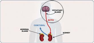
Cushing’s syndrome occurs when a person has excess cortisol in the blood for an extended period, according to the Hormone Health Network. When it is present in normal amounts, cortisol is involved in the body’s response to stress, maintains blood pressure and cardiovascular function, keeps the immune system in check, and converts fat, carbohydrates and proteins into energy. Chronic overexposure to the hormone can contribute to the development of cardiovascular disease, infections and blood clots in veins.

People who take cortisol-like medications such as prednisone to treat inflammatory conditions, including asthma and rheumatoid arthritis, can develop Cushing’s syndrome. The high cortisol levels return to normal when they stop taking the medication. This is called exogenous Cushing’s syndrome.

In other cases, tumors found on the adrenal or pituitary glands or elsewhere in the body cause the overproduction of cortisol and lead to the development of Cushing’s syndrome. The Clinical Practice Guidelines focus on this form of the condition, known as endogenous Cushing’s syndrome.

“People who have active Cushing’s syndrome face a greater risk of death – anywhere from nearly twice as high to nearly five times higher – than the general population,” said Lynnette K. Nieman, MD, of the National Institutes of Health’s Eunice Kennedy Shriver National Institute of Child Health and Human Development in Bethesda, MD, and chair of the task force that authored the guideline. “To reduce the risk of fatal cardiovascular disease, infections or blood clots, it is critical to identify the cause of the Cushing’s syndrome and restore cortisol levels to the normal range.”

In the CPG, the Endocrine Society recommends that the first-line treatment for endogenous Cushing’s syndrome be the removal of the tumor unless surgery is not possible or unlikely to address the excess cortisol. Surgical removal of the tumor is optimal because it leaves intact the hypothalamic-pituitary-adrenal axis, which is integral to the body’s central stress response.

Other recommendations from the CPG include:

  • Tumors should be removed by experienced surgeons in the following situations:— A tumor has formed on one or both of the two adrenal glands.— A tumor that secretes adrenocorticotropic hormone (ACTH) – the hormone that signals the adrenal glands to produce cortisol – has formed somewhere in the body other than the adrenal or pituitary gland.

    — A tumor has formed on the pituitary gland itself.

  • Patients who continue to have high levels of cortisol in the blood after surgery should undergo additional treatment.
  • People who had an ACTH-producing tumor should be screened regularly for the rest of their lives for high cortisol levels to spot recurrences.
  • If patients’ cortisol levels are too low following surgery, they should receive glucocorticoid replacement medications and be educated about adrenal insufficiency, a condition where the adrenal glands produce too little cortisol. This condition often resolves in 1-2 years.
  • Morning cortisol and/or ACTH stimulation tests, or insulin-induced hypoglycemia, can be used to test for the recovery of the hypothalamic-pituitary-adrenal axis in people who have low cortisol levels after surgery. Once the tests results return to normal, glucocorticoid replacement can be stopped.
  • People who have undergone pituitary surgery should be re-evaluated for other pituitary hormone deficiencies during the post-operative period.
  • Patients who have a pituitary tumor and have undergone surgery to remove both adrenal glands should be regularly evaluated for tumor progression using pituitary MRIs and tests for ACTH levels.
  • Radiation therapy may be used to treat a pituitary tumor, especially if it is growing. While awaiting the effect of radiation, which may take months to years, treatment with medication is advised.
  • To assess the effect of radiation therapy, the patient’s cortisol levels should be measured at 6- to 12-month intervals.
  • Medications may be used to control cortisol levels as a second-line treatment after surgery for a pituitary gland tumor, as a primary treatment for ACTH-secreting tumors that have spread to other parts of the body or suspected ACTH-secreting tumors that cannot be detected on scans. Medications also can be used as adjunctive treatment to reduce cortisol levels in people with adrenal cortical carcinoma, a rare condition where a cancerous growth develops in the adrenal gland.
  • People with Cushing’s syndrome should be treated for conditions associated with the disease, such as cardiovascular disease risk factors, osteoporosis and psychiatric symptoms.
  • Patients should be tested for recurrence throughout their lives except in cases where the person had a benign adrenal tumor removed.
  • Patients should undergo urgent treatment within 24 to 72 hours of detecting excess cortisol if life-threatening complications such as serious infection, pulmonary thromboembolism, cardiovascular complications and acute psychosis are present.

More information: The Hormone Health Network offers resources on Cushing’s syndrome at www.hormone.org/questions-and-answers/2012/cushing-syndrome

Bilateral Adrenal Incidentalomas May Have Different Etiology Than Unilateral

MedicalResearch.com Interview with:
Quan-Yang Duh MD
Chief, Section of Endocrine Surgery
UCSF Medical Center

Medical Research: What is the background for this study? What are the main findings?

Dr. Quan-Yang Duh: At UCSF we have a monthly Adrenal Conference (involving surgeons, endocrinologists and radiologists) to discuss patients we are consulted for adrenal tumors. About 30% of these are for incidentally discovered adrenal tumors (versus those found because of specific indications such as clinical suspicion or genetic screening). Of these 15-20% has bilateral adrenal tumors.

The evaluation of unilateral incidentaloma has been very well studied and many national guidelines have been published with specific management recommendations. So during our monthly adrenal conference, we have a routine “script” for evaluation and recommendations (rule out metastasis by looking for primary cancer elsewhere, rule out pheochromocytoma and Cushing, resect secreting tumors or large tumors, and if no operation recommended repeat scan in 6 months, etc.). This “script” has worked very well for patients with unilateral incidentaloma.

However, we were less certain when we made recommendations about bilateral incidentalomas because there was very little literature or guidelines written about it. We had some gut feelings, but we were not sure that we were recommending the right things. We needed more data. That was the main reason for the study.

What we found in our study was that although the possible subclinical diseases were the same – hypercortisolism and pheochromocytoma, the probabilities were different. The patients with bilateral incidentalomas were more likely to have subclinical Cushing’s and less likely to have pheochromocytomas than those with unilateral incidentalomas.

Medical Research: What should clinicians and patients take away from your report?

Dr. Quan-Yang Duh: The work up for bilateral adrenal incidentalomas is similar to that for unilateral incidentalomas. However, patients with bilateral incidentalomas are more likely to have subclinical Cushing’s and less likely to have pheochromocytoma. This difference should be kept in mind when clinicians evaluating these patients.

Medical Research: What recommendations do you have for future research as a result of this study?

Dr. Quan-Yang Duh: Because our study used data that were from patients who were referred to us to be discussed at our Adrenal Conference, there is likely to be a selection bias (probably higher proportion with clinically significant diseases). It would be interesting to prospectively study ALL patients with adrenal  from a radiology department and see whether our results are confirmed.

Citation:

What Genes are Related to Cushing’s Disease?

genetic


The genetic cause of Cushing disease is often unknown. In only a few instances, mutations in certain genes have been found to lead to Cushing disease. These genetic changes are called somatic mutations. They are acquired during a person’s lifetime and are present only in certain cells. The genes involved often play a role in regulating the activity of hormones.

Cushing disease is caused by an increase in the hormone cortisol, which helps maintain blood sugar levels, protects the body from stress, and stops (suppresses) inflammation. Cortisol is produced by the adrenal glands, which are small glands located at the top of each kidney. The production of cortisol is triggered by the release of a hormone called adrenocorticotropic hormone (ACTH) from the pituitary gland, located at the base of the brain. The adrenal and pituitary glands are part of the hormone-producing (endocrine) system in the body that regulates development, metabolism, mood, and many other processes.

Cushing disease occurs when a noncancerous (benign) tumor called an adenoma forms in the pituitary gland, causing excessive release of ACTH and, subsequently, elevated production of cortisol. Prolonged exposure to increased cortisol levels results in the signs and symptoms of Cushing disease: changes to the amount and distribution of body fat, decreased muscle mass leading to weakness and reduced stamina, thinning skin causing stretch marks and easy bruising, thinning of the bones resulting in osteoporosis, increased blood pressure, impaired regulation of blood sugar leading to diabetes, a weakened immune system, neurological problems, irregular menstruation in women, and slow growth in children. The overactive adrenal glands that produce cortisol may also produce increased amounts of male sex hormones (androgens), leading to hirsutism in females. The effect of the excess androgens on males is unclear.

Most often, Cushing disease occurs alone, but rarely, it appears as a symptom of genetic syndromes that have pituitary adenomas as a feature, such as multiple endocrine neoplasia type 1 (MEN1) or familial isolated pituitary adenoma (FIPA).

Cushing disease is a subset of a larger condition called Cushing syndrome, which results when cortisol levels are increased by one of a number of possible causes. Sometimes adenomas that occur in organs or tissues other than the pituitary gland, such as adrenal gland adenomas, can also increase cortisol production, causing Cushing syndrome. Certain prescription drugs can result in an increase in cortisol production and lead to Cushing syndrome. Sometimes prolonged periods of stress or depression can cause an increase in cortisol levels; when this occurs, the condition is known as pseudo-Cushing syndrome. Not accounting for increases in cortisol due to prescription drugs, pituitary adenomas cause the vast majority of Cushing syndrome in adults and children.

Read more about familial isolated pituitary adenoma.


How do people inherit Cushing disease?

Most cases of Cushing disease are sporadic, which means they occur in people with no history of the disorder in their family. Rarely, the condition has been reported to run in families; however, it does not have a clear pattern of inheritance.

The various syndromes that have Cushing disease as a feature can have different inheritance patterns. Most of these disorders are inherited in an autosomal dominant pattern, which means one copy of the altered gene in each cell is sufficient to cause the disorder.

From http://ghr.nlm.nih.gov/condition/cushing-disease

Given Adrenal Symptoms, Blood Test Recommended

adrenal-glands


Q: My husband’s recent CT scan of his stomach and digestive system revealed that he has nodules on both adrenal glands. It was suggested that he undergo a blood test to determine whether the nodules are producing hormones.

For 21 months, he has experienced high blood pressure, nausea, diarrhea, anxiety and abdominal pain. Could this be the source of his problems? If so, what course of action would you recommend?

A: The adrenal gland is responsible for the production of several essential hormones.

Tumors, or nodules, of the adrenal glands are common. They can be categorized into those that make hormones and those that don’t, and also by whether the tumors are benign or malignant.

The most common, by far, are benign, nonfunctioning tumors. These are usually discovered on an ultrasound or a CT scan obtained for some other reason.

More than 4 percent of people have an adrenal mass, and 85 percent are nonfunctional.

The symptoms that your husband has, however, raise a concern that he might have a hormone-producing tumor.

Four types of hormones are commonly produced by adrenal tumors: cortisone, aldosterone, sex hormones (estrogen or androgens) and catecholamines (epinephrine and norepinephrine).

A cortisone-producing adrenal tumor causes Cushing’s syndrome. It usually causes weight gain, especially in the abdomen; skin changes, including striae, or “stretch marks”; high blood pressure; and a predisposition to diabetes. Anxiety and abdominal pain are uncommon.

Aldosterone raises blood pressure, so a person with a functioning adrenal tumor making aldosterone usually has high blood pressure, but the other symptoms you mention for your husband aren’t common for this type of tumor.

Adrenal tumors that make epinephrine and the related norepinephrine are called pheochromocytomas. Hypertension is almost universal with this condition, and anxiety is frequently reported.

Tumors that produce sex hormones are rare, and they are present in men with androgen excess or feminization, in the case of estrogen-secreting tumors.

Although your husband’s symptoms aren’t specific for any one condition, the combination of his symptoms and adrenal nodules concerns me.

I agree with the recommendation to look for excess amounts of hormones in the blood. This can often be achieved with a simple blood test; however, a catheter is occasionally placed in the adrenal vein to sample blood coming from the gland (and its nodule) directly.

By comparing one side against the other, doctors can determine which side might be producing excess hormones.

An endocrinologist is the expert most likely to be familiar with these conditions.

Dr. Roach answers letters only in his North America Syndicate column but provides an order form of available health newsletters at http://www.rbmamail.com. Write him at 628 Virginia Dr., Orlando, FL 32853-6475; or ToYour GoodHealth@med. cornell.edu.

From http://www.dispatch.com/content/stories/life_and_entertainment/2015/07/27/given-adrenal-symptoms-blood-test-recommended.html

Narrowing in on Pituitary Tumors

0276f-pituitary-gland


As many as 20 percent of people may have a benign cyst or tumor in their pituitary gland. The vast majority of pituitary tumors are noncancerous, but can cause headaches and profound fatigue, and can also disrupt hormone function.

Currently, surgeons rely on radiologic images and MRIs to gather information about the size and shape of the tumor, but the resolution of such imaging technologies is limited, and additional surgeries to remove more of the tumor may be needed if a patient’s symptoms persist. In a new study published in the Proceedings of the National Academy of Sciences on July 27, investigators from Brigham and Women’s Hospital (BWH) present a new technique that could help surgeons more precisely define the locations of tumors in near real-time.

The new strategy uses a visualization technique (matrix-assisted laser desorption/ionization mass spectrometry imaging – MALDI MSI) that can analyze specific hormones, including growth hormone and prolactin, in tissue. In the newly published study, the researchers find that it’s possible to use MALDI MSI to determine the composition of such hormones in a pituitary sample in less than 30 minutes. This could give surgeons critical information to help distinguish tumor from normal gland.

“Our work is driven by a clinical need: we’ve developed a test specifically tailored for the needs of our neurosurgeon colleagues,” said corresponding author Nathalie Agar, PhD, director of the Surgical Molecular Imaging Laboratory in the Department of Neurosurgery at BWH. “A surgeon may sacrifice half of the pituitary gland in an effort to get the tumor out. Without a tool to distinguish healthy tissue from tumor, it’s hard to know in real-time if the surgery was a success. With this technology, in under 30 minutes a surgeon will be able to know if a sample contains normal pituitary tissue or a pituitary tumor.”

“Patients show up with the clinical symptoms of a pituitary tumor, but the tumor itself may not be visible on an MRI,” said co-author Edward Laws, MD, director of the Pituitary and Neuroendocrine Center at BWH. “This technique, which maps out where excess concentrations of hormone levels are located, has the potential to allow us to confirm that we’ve removed the abnormal tissue.”

“Evaluating whether a piece of pituitary tissue is abnormal can be challenging on frozen section,” said co-author Sandro Santagata, MD, PhD, of BWH’s Department of Pathology. “This approach has wonderful potential for enhancing our diagnostic capabilities. It is clearly an important step toward providing intra-operative molecular characterization of pituitary tissues.”

To test the technique, the research team analyzed hormone levels in 45 pituitary tumors and six normal pituitary gland samples, finding a distinct protein signature unique to the normal or tumor sample.

Mass spectrometry, a technique for measuring chemicals present in a sample, is currently used in the operating room to help inform clinical decisions, but up until now, the focus has been on small molecules – metabolites, fatty acids and lipids – using a different type of approach. By analyzing proteins, MALDI MSI offers a way to visualize hormone levels.

Current methods used to detect hormone levels take too long to fit the time constraints of surgical intervention. Surgeons must either remove a larger amount of potentially healthy pituitary gland or perform follow up surgery if the tumor has not been fully removed.

“We’re hoping that techniques like this one will help move the field toward more precise surgery: surgery that not only removes all of the tumor but also preserves the healthy tissue as much as possible,” said Agar.

In the next phase of their work, Agar and her colleagues plan to test out the technique in BWH’s AMIGO suite and analyze the impact of the technique on clinical decision making.

Other researchers who contributed to this study include David Calligaris, Daniel R. Feldman, Isaiah Norton, Olutayo Olubiyi, Armen N. Changelian, Revaz Machaidze, Matthew L. Vestal and Ian F. Dunn.

This work was funded in part by US National Institute of Health (NIH) Director’s New Innovator Award (1DP2OD007383-01 to N.Y.R.A.), U.S. Army Medical Research/CIMIT (2010A052245), the National Center for Image Guided Therapy grant P41RR019703, NIH K08NS064168, the Pediatric Low Grade Astrocytoma Program at Dana-Farber Cancer Institute, the Brain Science Foundation and the Daniel E. Ponton fund for the Neurosciences at BWH.

Brigham and Women’s Hospital 2015 | 75 Francis Street, Boston MA 02115 | 617-732-5500

From http://www.healthcanal.com/cancers/65676-narrowing-in-on-pituitary-tumors.html