Rare Case of Cushing’s Disease Diagnosed in 7-year-old Boy

A recent case report describes a 7-year-old boy with Cushing’s disease who had an unusual clinical presentation, which significantly delayed his diagnosis.

The study, “A variable course of Cushing’s disease in a 7 year old: diagnostic dilemma,” was published in the Journal of Pediatric Endocrinology and Metabolism.

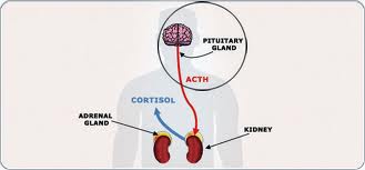
Rare in children and adolescents, Cushing’s disease refers to overproduction of cortisol caused by excessive adrenocorticotropic hormone (ACTH) secretion from a pituitary tumor. In pediatrics, early symptoms of excess cortisol include weight gain and delayed growth.

Despite being extremely unlikely in children younger than 7, some cases of Cushing’s disease in infancy have been reported.

“If undiagnosed or untreated it can lead to considerable morbidity and mortality, and the inability to detect a microadenoma [tumors smaller than 10 mm in diameter] on magnetic resonance imaging (MRI) can lead to a diagnostic dilemma leading to unnecessary delays in treatment initiation,” the researchers wrote.

Researchers from the Indraprastha Apollo Hospital in New Delhi, India, described a 7-year-old boy who complained of excessive appetite and weight gain in the previous five months. The child weighed 46.8 kg, was 127 cm tall, and had a body mass index (BMI) of 29, indicating he was overweight.

The child’s excess fat was mainly in his abdomen plus he had a round, red, puffy face, which are both common features of Cushing’s disease. He had no history of acute or chronic steroid intake, mood swings, sleep disorders, or issues with eyesight.

Given his clinical presentation, the investigators suspected the boy had Cushing’s disease or pseudo-Cushing’s disease, which refers to situations where the overproduction of cortisol is caused by something unrelated to the disease, such as stress or uncontrolled diabetes mellitus.

Biochemical testing showed the patient had high levels of cortisol, which remained unchanged after a dexamethasone suppression test. In addition, his levels of “bad” cholesterol, referring to low-density lipoprotein, were extremely elevated at 194 mg/dL, where a normal range is defined as less than 110 mg/dL.

Imaging revealed no lesions in the pituitary gland.

The boy was sent home with dietary recommendations. Eight weeks later, he had lost 4 kg, while his height remained the same; he also complained of headaches and various episodes of double vision.

This confused the clinical team as hallmarks of Cushing’s disease include short stature and weight loss triggered by pharmacological therapy. Despite having lost weight, he did not take any medications to help him with it, plus the boy’s height was normal for his age.

Nonetheless, the patient was complaining of neurological symptoms, suggesting progression of Cushing’s disease.

An ophthalmologist did not observe anything abnormal with the child’s eyes that could explain his double vision episodes.

A new series of tests revealed slightly elevated 24-hour urinary cortisol levels, decreased concentration of ACTH, and mildly increased cortisol levels after a two-day dexamethasone suppression test.

Magnetic resonance imaging (MRI) showed a small microadenoma in the right lobe of the pituitary gland.

Using Gamma Knife radiation therapy, a kind of high-precision radiation therapy, and surgery, doctors successfully removed the boy’s microadenoma. Six weeks post-procedure, his cortisol and ACTH concentrations returned to normal.

“MRI findings of the pituitary may be inconclusive in the beginning of the disease process and should be borne in mind during further follow-up. In cases where a clear-cut diagnosis may be difficult, a diligent follow-up is required to ascertain the course of the disease and to make timely diagnosis,” the investigators concluded.

From https://cushingsdiseasenews.com/2018/10/25/rare-case-cushings-disease-diagnosed-7-year-old-boy-case-study/

Science Teacher Receives Support After Cushing’s Disease Diagnosis

I find it amazing that it’s newsworthy in this day and age for anyone receiving support after a diagnosis.  Of course, a diagnosed person should be getting support as a matter of course.  If she had cancer, everyone would be all over this.

For Kara Murrow, the most rewarding moments as a teacher come when students learn about animals in the classroom. So it’s difficult for the Bonham Elementary fifth-grade science and social studies teacher to be away from school while she prepares for surgery.

“I enjoy it, and I know my kids enjoy the class and enjoy science because of it,” Murrow said. “With the science club I do after school once a week, the kids get upset when it gets canceled because of meetings. Not having it now is upsetting, too.”

Murrow was diagnosed this month with Cushing’s disease, a condition that develops when a tumor on the pituitary gland causes it to secrete too much adrenocorticotropic hormone. Murrow, who moved to West Texas from Arizona three years ago, said she has received support from Midland ISD employees and others in the local community.

Murrow’s mother, Louise Gonzalez, also appreciates Midlanders’ concerns for her daughter.

“People in Midland have been wonderful, considering how new we are to the area,” Gonzalez said. “The school district sent out the GoFundMe page and there’s been an outpouring of support for that. People at my church always ask me.”

Murrow’s family is collecting donations from the website GoFundMe to cover the costs of medical and travel expenses. Murrow and her husband, Kai, recently spent money on hospital stays connected to their 4-year-old son’s food sensitivities.

“They’ve been paying off those bills and doing OK until this came,” Gonzalez said. “Plus, she’s been going to the doctor about this. Because Cushing’s is so rare, doctors don’t recognize it.”

Murrow was diagnosed with the disease after medical professionals discovered a tumor on her pituitary gland. For six years, she experienced symptoms — including weight gain, dizziness and headaches — but said doctors couldn’t determine the cause. Murrow was thankful when she received an answer.

“It was a huge relief to finally have a diagnosis and know that I wasn’t crazy or making things up,” Murrow said. “It’s weird to be excited about a brain tumor. It’s a relief to know what was happening and that I have a solution.”

Murrow traveled this week to Barrow Neurological Institute in Phoenix, where she’s scheduled to undergo surgery to remove the tumor. Though Murrow said recovery lasts several months, she hopes to return to the classroom next school year.

Jaime White, fourth-grade language arts and social studies teacher at Bonham, said both staff and students miss her presence. She said Murrow expresses concern for her students during her time away.

“She’s worried about how kids will do on the STAAR [State of Texas Assessments of Academic Readiness],” White said. “She doesn’t want them to think she abandoned them. The disease has to take center stage.”

At school, White said she noticed her colleague’s dedication toward helping her students understand science.

“She’s hands-on,” White said. “When it comes to science, she’s always making sure the kids are doing some sort of experiment. She wants to make sure the kids grasp it.”

Murrow teaches students about animals through dissections and presentations. Before she became a teacher nine years ago, she coordinated outreach programs at an Arizona zoo.

When she came to MISD, Murrow saw an opportunity to generate enthusiasm about science. She launched an invite-only science club for fifth-graders who show interest in the subject.

“I started it because there wasn’t really anything,” Murrow said. “They have tutorials for reading and math. There’s not a lot kids can do with science after school. They get science in the younger grades, but the focus is on reading and math. Science is something kids really enjoy.”

Though Murrow is disappointed about not being able to facilitate the club, she recognizes the importance of her upcoming surgery. She’s happy her mother, husband and two children will be in Phoenix for support.

“I hope that it will bring about a sense of relief to all the symptoms I’ve been dealing with and provide a chance for myself and my family to continue along with a full life,” Murrow said.

From http://www.mrt.com/news/local/article/Science-teacher-receives-support-after-11026581.php

Neurosurgeon and Otolaryngologist Team Up to Remove Tumor in Pituitary Gland

After experiencing bad headaches and double vision, Kris Johnson was diagnosed with a pituitary adenoma, a tumor of the pituitary gland at the base of the brain. Loyola ENT surgeon Chirag Patel, MD, teamed up with neurosurgeon Anand Germanwala, MD, to remove the tumor, and Ms. Johnson now is “100 percent back to normal.”

Article ID: 668877

Released: 3-Feb-2017 2:05 PM EST

Source Newsroom: Loyola University Health System

Narrowing in on Pituitary Tumors

0276f-pituitary-gland


As many as 20 percent of people may have a benign cyst or tumor in their pituitary gland. The vast majority of pituitary tumors are noncancerous, but can cause headaches and profound fatigue, and can also disrupt hormone function.

Currently, surgeons rely on radiologic images and MRIs to gather information about the size and shape of the tumor, but the resolution of such imaging technologies is limited, and additional surgeries to remove more of the tumor may be needed if a patient’s symptoms persist. In a new study published in the Proceedings of the National Academy of Sciences on July 27, investigators from Brigham and Women’s Hospital (BWH) present a new technique that could help surgeons more precisely define the locations of tumors in near real-time.

The new strategy uses a visualization technique (matrix-assisted laser desorption/ionization mass spectrometry imaging – MALDI MSI) that can analyze specific hormones, including growth hormone and prolactin, in tissue. In the newly published study, the researchers find that it’s possible to use MALDI MSI to determine the composition of such hormones in a pituitary sample in less than 30 minutes. This could give surgeons critical information to help distinguish tumor from normal gland.

“Our work is driven by a clinical need: we’ve developed a test specifically tailored for the needs of our neurosurgeon colleagues,” said corresponding author Nathalie Agar, PhD, director of the Surgical Molecular Imaging Laboratory in the Department of Neurosurgery at BWH. “A surgeon may sacrifice half of the pituitary gland in an effort to get the tumor out. Without a tool to distinguish healthy tissue from tumor, it’s hard to know in real-time if the surgery was a success. With this technology, in under 30 minutes a surgeon will be able to know if a sample contains normal pituitary tissue or a pituitary tumor.”

“Patients show up with the clinical symptoms of a pituitary tumor, but the tumor itself may not be visible on an MRI,” said co-author Edward Laws, MD, director of the Pituitary and Neuroendocrine Center at BWH. “This technique, which maps out where excess concentrations of hormone levels are located, has the potential to allow us to confirm that we’ve removed the abnormal tissue.”

“Evaluating whether a piece of pituitary tissue is abnormal can be challenging on frozen section,” said co-author Sandro Santagata, MD, PhD, of BWH’s Department of Pathology. “This approach has wonderful potential for enhancing our diagnostic capabilities. It is clearly an important step toward providing intra-operative molecular characterization of pituitary tissues.”

To test the technique, the research team analyzed hormone levels in 45 pituitary tumors and six normal pituitary gland samples, finding a distinct protein signature unique to the normal or tumor sample.

Mass spectrometry, a technique for measuring chemicals present in a sample, is currently used in the operating room to help inform clinical decisions, but up until now, the focus has been on small molecules – metabolites, fatty acids and lipids – using a different type of approach. By analyzing proteins, MALDI MSI offers a way to visualize hormone levels.

Current methods used to detect hormone levels take too long to fit the time constraints of surgical intervention. Surgeons must either remove a larger amount of potentially healthy pituitary gland or perform follow up surgery if the tumor has not been fully removed.

“We’re hoping that techniques like this one will help move the field toward more precise surgery: surgery that not only removes all of the tumor but also preserves the healthy tissue as much as possible,” said Agar.

In the next phase of their work, Agar and her colleagues plan to test out the technique in BWH’s AMIGO suite and analyze the impact of the technique on clinical decision making.

Other researchers who contributed to this study include David Calligaris, Daniel R. Feldman, Isaiah Norton, Olutayo Olubiyi, Armen N. Changelian, Revaz Machaidze, Matthew L. Vestal and Ian F. Dunn.

This work was funded in part by US National Institute of Health (NIH) Director’s New Innovator Award (1DP2OD007383-01 to N.Y.R.A.), U.S. Army Medical Research/CIMIT (2010A052245), the National Center for Image Guided Therapy grant P41RR019703, NIH K08NS064168, the Pediatric Low Grade Astrocytoma Program at Dana-Farber Cancer Institute, the Brain Science Foundation and the Daniel E. Ponton fund for the Neurosciences at BWH.

Brigham and Women’s Hospital 2015 | 75 Francis Street, Boston MA 02115 | 617-732-5500

From http://www.healthcanal.com/cancers/65676-narrowing-in-on-pituitary-tumors.html

Recurrent sellar mass after resection of pituitary macroadenoma

A Puerto Rican woman aged 50 years presented to an ophthalmologist with complaints of vision changes, including difficulty seeing images in her peripheral vision in both eyes and difficulty in color perception. Her medical history was significant for menopause at age 43 years, type 2 diabetes and hypertension. She had no prior history of thyroid disease, changes in her weight, dizziness or lightheadedness, headaches, galactorrhea or growth of her hands or feet.

Formal visual fields showed bitemporal superior quadrantopsia, and she was sent to the ED for further evaluation.

Imaging and laboratory tests

A pituitary protocol MRI was performed that showed a large 3 cm x 2 cm x 2.2 cm mass in the pituitary with mild osseous remodeling of the sella turcica and mass effect on the optic chiasm (Figure 1). The mass was isointense with the brain parenchyma on T1-weighted and T2-weighted images and homogeneously enhanced after IV gadolinium contrast administration.

Baseline laboratory samples drawn at 11 p.m. in the ED showed a cortisol of 16.9 µg/dL (nighttime reference range: 3-16 µg/dL), adrenocorticotropic hormone 65 pg/mL (reference range: 6-50 pg/mL), prolactin 19.4 ng/mL (reference range: 5.2-26.5 ng/mL), thyroid-stimulating hormone 1.36 µIU/mL (reference range: 0.35-4.9 µIU/mL), free thyroxine 0.9 ng/dL (reference range: 0.6-1.8 ng/dL), triiodothyronine 85 ng/dL (reference range: 83-160 ng/dL), follicle-stimulating hormone (FSH) 11.1 mIU/mL (postmenopausal reference range: 26.7- 133.4 mIU/mL) and luteinizing hormone (LH) 1.2 mIU/mL (postmenopausal reference range: 5.2-62 mIU/mL).


Figure 1. T1-weighted MRI images with and without contrast of the pituitary. Coronal (A) and sagittal (C) images showed a large isodense (with brain parenchyma) 3 cm x 2 cm x 2.2 cm mass (red arrow) in the sella with superior extension to the optic chiasm. After gadolinium contrast, coronal (B) and sagittal (D) images show the mass homogenously enhances consistent with a pituitary adenoma.

Images courtesy of Pavani Srimatkandada, MD.

Given the patient’s high nighttime cortisol and adrenocorticotropic hormone (ACTH) levels, she underwent an overnight dexamethasone suppression test with 1 mg dexamethasone. Her morning cortisol was appropriately suppressed to less than 1 µg/dL, excluding Cushing’s disease.

Pituitary adenoma resection

The patient was diagnosed with a nonsecreting pituitary adenoma with suprasellar extension and optic chiasm compression with visual field deficits. The macroadenoma caused an inappropriately normal LH and FSH in a postmenopausal woman consistent with hypogonadotrophic hypogonadism.

She underwent transnasal transsphenoidal resection of the nonsecreting pituitary adenoma. The dural defect caused by the surgery was patched with an abdominal fat graft with a DuraSeal dura patch. A postoperative MRI showed complete resection of the adenoma with no evaluable tumor in the sella (Figure 2). Her postoperative course was complicated by transient diabetes insipidus requiring intermittent desmopressin; however, this resolved before her discharge from the hospital.

Figure 2. T1-weighted MRI images with contrast. Coronal views before (A) and after (B) transphenoidal tumor resection show complete resolution of the enhancing pituitary mass (A; red arrow) that is replaced with a new hypodense mass in the sella (B; yellow arrow). This mass is filled with cerebrospinal fluid with a residual rim of enhancing tissue. This is consistent with the development of a pseudomeningocele in the sella.


Postoperative testing confirmed secondary deficiency of the adrenal, thyroid and ovarian axes requiring hormone therapy. The patient had stable temporal hemianopia in the left eye with improved vision in the right eye.

Recurrent mass detected

One year after surgery, during a routine follow-up appointment, the patient reported no dizziness, lightheadedness, worsening vision changes, rhinorrhea or headache. She had a follow-up MRI of the brain with and without contrast, which showed the interval appearance of a mass in the sella that extended from the sphenoid sinus into the sella and came in contact with the optic nerve (Figure 3).

Figure 3. Axial MRI images of the sella after resection of pituitary adenoma. On T1-weighted images the mass (red arrow) in the sella is hypodense (black) compared with the brain parenchyma. On T2-weighted images, the mass (red arrow) is hyperdense (bright) compared with the brain, consistent with fluid. Cerebrospinal fluid in the sulci on the brain surface and the vitreous fluid within the eye are also hyperintense on T2-weighted images (yellow arrows).


On MRI, the mass was isodense with the cerebral spinal fluid (CSF) with a residual rim of enhancing normal pituitary tissue. This appearance is consistent with the postoperative development of a pseudomeningocele and not a solid mass in the sella (Table).

Pseudomeningoceles are abnormal collections of CSF that communicate with the CSF space around the brain; these occur after brain surgery involving duraplasty (incision and repair of the dura). Unlike meningoceles, pseudomeningoceles are not completely encased by a surrounding membrane, and they communicate with the circulating CSF. Similar to CSF, a pseudomeningocele is hypodense (dark) compared with brain on T1-weighted MRI images and hyperdense (bright) on T2-weight images.


Pseudomeningocele treatment

Treatment may be conservative or may involve neurosurgical repair if symptomatic. Little published data addresses the development of pseudomeningoceles after transsphenoidal pituitary surgery, but this complication occasionally occurs, especially if the dural incision is large. One study noted that pseudomeningoceles are one of the most common complications after suboccipital decompression for Chiari’s malformation, but the effect of this complication is unclear.

Endocrinologists must recognize that recurrent development of pituitary masses after transsphenoidal pituitary adenoma surgery may not represent regrowth of pituitary tissue, but instead development of a meningocele/pseudomeningocele. Pseudomeningocele can be easily confirmed because this fluid collection has very different MRI characteristics than pituitary adenoma (Table). Given that patients may remain asymptomatic after the development of a pseudomeningocele, periodic MRI imaging, hormonal evaluation and ophthalmologic monitoring of visual fields are required after transsphenoidal pituitary surgery.

References:
  • Hernandez Guilabert PM. Poster No C-1330. Presented at: European Society of Radiology; March 7-11, 2013; Vienna.
  • Parker SL, et al. J Neurosurg. 2013;doi:10.3171/2013.8.JNS122106.
For more information:
  • Stephanie L. Lee, MD, PhD, ECNU, is an associate professor of medicine and associate chief, in the Section of Endocrinology, Diabetes and Nutrition at Boston Medical Center. Lee can be reached at Boston Medical Center, 88 E. Newton St., Endocrinology Evans 201, Boston, MA 02118; email: stephanie.lee@bmc.org. Lee reports no relevant financial disclosures.
  • Pavani Srimatkandada, MD, is an endocrinology fellow in the Section of Endocrinology, Diabetes and Nutrition at Boston Medical Center. Srimatkandada can be reached at Boston Medical Center, 88 E. Newton St., Endocrinology Evans 201, Boston, MA 02118. She reports no relevant financial disclosures.

From http://www.healio.com/endocrinology/thyroid/news/print/endocrine-today/%7B82430fb6-bbe4-4908-a389-447eee8cd005%7D/recurrent-sellar-mass-after-resection-of-pituitary-macroadenoma

%d bloggers like this: