Osteoporosis as the First Sign of Cushing’s Disease in a Thin 16-Year-Old Boy

Abstract

Cushing’s disease (CD) is an extremely rare diagnosis in children. In this report, we present the case of an almost 16-year-old, short and thin boy with CD, the first symptoms of which were spinal pain and vertebral fractures as a result of osteoporosis. In light of his growth retardation and short stature, the boy underwent diagnostics, which excluded growth hormone (GH) deficiency, hypothyroidism and celiac disease. Finally, based on cortisol profile results, dexamethasone suppression tests and bilateral sampling during catheterization of the inferior petrosal sinuses, CD was diagnosed.

1. Introduction

Cushing’s disease (CD) is an extremely rare diagnosis in children; however, if it occurs, it is more likely to present in older children [1,2]. It is a type of ACTH-dependent Cushing’s syndrome (CS), in which the pituitary gland is the source of ACTH secretion. The highest incidence of CD occurs in children aged 12.3–14.1 years [3]. The incidence of CD during this developmental age is approximately 5% of that seen in adults (with an annual incidence of 0.89–1 per million pediatric patients) [1,2,4]. The rarest form of ACTH-dependent CS in children is ectopic Cushing’s syndrome (ECS), associated with ectopic production of ACTH or CRH, most commonly by neuroendocrine tumors such as bronchial carcinoids, gastrointestinal tumors, medullary thyroid carcinoma, or pheochromocytomas [2,4,5]. Children with ECS constitute 1% of patients with CS in the developmental age [2]. An even rarer disease is ACTH-independent Cushing’s syndrome—associated with adrenal lesions (adenoma, carcinoma, bilateral macronodular adrenal hyperplasia (BMAH), or primary pigmented nodular adrenocortical disease (PPNAD)) [2].
Regarding CD, ACTH is secreted in an overwhelming majority of cases by pituitary corticotropic microadenomas and—less commonly—by macroadenomas, the latter occurring in only 10% of adult CD cases and even more rarely in children (2%) [1,3]. Long-term hypercortisolemia can also lead to bone-mineralization disorders, including osteoporosis, especially in the bones of the central skeleton [4,6,7].
In children, the most common features of CD are rapid weight gain (93–98%), growth retardation (63–100%) and/or facial changes (63–100%) [4]. Mood disturbances, muscle weakness, osteopenia, and headaches are less frequent symptoms. Limited data are available about bone mineral density (BMD) in children with CD. Lonser et al. [8] observed fractures in 7% of patients with CD that were studied. Chronic glucocorticoid excess associated with CD has negative effects on bone turnover, leading to bone-mineralization disorders in both adults and children. Multiple factors contribute to decreased bone mineral density in CD, including the direct effect of glucocorticoids on osteoclasts and osteoblasts, both impairing bone formation and enhancing bone resorption. Glucocorticoids also act to decrease gastrointestinal calcium absorption and renal calcium reabsorption. Bone loss occurs more frequently in Cushing’s syndrome caused by adrenal tumors in CD [6,7].
An additional factor involved in bone-mineralization disorders, particularly in adult patients with CD, may be hypogonadotropic hypogonadism. Reproductive and sexual dysfunctions are highly prevalent in CS, with higher frequency observed in patients with pituitary-related CS, compared to those with adrenal-related CS. Hypogonadism is identified in as much as 50–75% of men with CS and menstrual irregularities are present in 43–80% of women diagnosed with this condition. During active disease, there is a significant reduction in plasma testosterone and gonadotropin levels in men [7,9]. These testosterone levels typically normalize during remission of the disease. Pivonello et al. [7] suggest that the lack of testosterone normalization three months after CS treatment indicates the need for administration of testosterone to protect the patient’s bone mass. In children, cortisol excess can also suppress gonadotropin, TSH and growth hormone secretion, contributing to the absence of pubertal characteristics or inhibiting its progression in patients who have already entered puberty [1].
So far, to our knowledge, there have been no reports on children where bone-mineralization disorders (without weight gain and hirsutism) are the first sign of CD.

2. Case Presentation

We present the case of an almost 16-year-old boy with short stature who, in May 2021, was referred to the Osteoporosis Outpatient Clinic of the Polish Mother’s Memorial Hospital—Research Institute (PMMH-RI) in Lodz, Poland, due to severe back pain. Low bone mass was diagnosed via dual-energy X-ray absorptiometry (DXA).
Initially, it seemed that the occurrence of those symptoms might be related to steroid therapy, because in November 2020 (just after SARS-CoV-2 infection) the child had developed severe abdominal pain, accompanied by an increase in the activity of liver enzymes, and after excluding an infectious cause, autoimmune hepatitis was diagnosed. Deflazacort (Calcort) therapy was prescribed in gradually reduced doses, with the initial dose being 24 mg in the morning and 18 mg in the afternoon. This therapy was discontinued on 1 October 2021. As early as on the fifth day of glucocorticosteroid treatment, pain presented in the lumbar spine region, increasing with movement. Initially, the pain was intermittent, then it became constant. No painkillers were needed. On 7 May 2021, on the basis of DXA, low bone mass was diagnosed (Z-score Spine: −4.2, Z-score TBLH: −1.9). In June 2021 (while still undergoing treatment with steroids) the boy was admitted to the Department of Endocrinology and Metabolic Diseases PMMH-RI for further diagnostics (Table 1).
Table 1. The medical history and the course of diagnostics and treatment.
The patient was a second child, born at 40 weeks of gestational age, weighing 4150 g, measuring 56 cm, and achieving a 10-point Apgar score. During infancy, he received vitamin D supplementation in accordance with Polish recommendations at that time. However, after his first year of life, the supplementation was not taken regularly. The boy received vaccinations according to the standard immunization schedule. There was no significant family medical history.
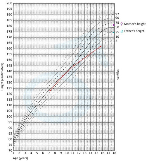
During the physical examination, apart from the presence of short stature, no other notable abnormalities were detected. The skin was clear, without pathological lesions; no features of hyperandrogenism were observed. The boy’s body weight was 47.4 kg (3rd–10th centile); his height, 162 cm (<3rd centile); and height SDS, −2.36; while his BMI was 18.06 kg/m2 (10th–25th centile). Pubarche was assessed as stage 4 according to the Tanner scale; the volume of the testes was 10–12 mL each. After available anthropometric measurements from the patient’s medical history were plotted on the growth chart for sex and chronological age, it became evident that the boy experienced growth retardation from the age of 11 (Figure 1).
Jcm 12 05967 g001
Figure 1. Growth chart for boys. The red line represents growth retardation from the age of 11.
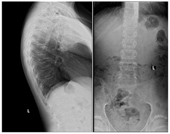
Apart from slightly increased calcium excretion in the 24 h urine collection (Calcium: 9.52 mmol/24 h), there were no significant abnormalities in the laboratory tests assessing calcium–phosphate metabolism (Calcium: 2.41 mmol/L, Phosphorus: 1.3 mmol/L). Serum parathormone (PTH) and vitamin D concentrations remained normal (PTH: 22.9 pg/mL, 25(OH)D: 46.7 ng/mL). Due to the described pain complaints, a thoracolumbar spine X-ray was performed. A decrease in the height of the Th5-Th9 vertebrae and central lowering of the upper border plate of the L4 and L5 were observed (Figure 2).
Jcm 12 05967 g002
Figure 2. A thoracolumbar spine X-ray with multilevel vertebral fractures. Decrease in the height of the Th5–Th9 vertebrae and central lowering of the upper border plate of the L4 and L5 were found.
Magnetic resonance imaging (MRI) of the spine confirmed multilevel vertebral fractures, which, together with the presence of low bone mass on DXA examination, allowed a diagnosis (according to ISCD guidelines) of osteoporosis to be made. Treatment included calcium supplements and cholecalciferol. The parents did not consent to treatment with bisphosphonates (sodium pamidronate), which is an off-label treatment.
In light of the patient’s short stature and growth retardation, an endocrinological assessment was conducted. The possibility of growth hormone (GH) deficiency and hypothyroidism as underlying causes for the growth retardation was ruled out. Gonadotropin and androgen levels were adequate for the pubertal stage (FSH—8.3 IU/L, LH—4.7 IU/L, testosterone—4.750 ng/mL, DHEA-S—230.30 µg/dL (normal range: 70.2–492), 17-OH-progesterone—0.78 ng/mL). The bone age was assessed to be 15 years.
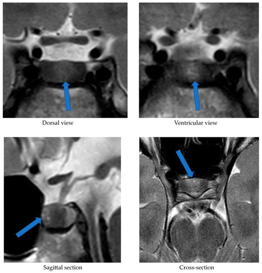
Alongside continued steroid therapy for autoimmune hepatitis, profiles of cortisol and ACTH secretion were performed. Due to the patient’s elevated cortisol levels during night hours (cortisol 24:00—10.7 µg/dL), an overnight dexamethasone suppression test (DST) and low-dose dexamethasone suppression test (LDDST) were performed. After administering 1 mg dexamethasone (23:00), his morning cortisol level (8:00) still remained elevated (cortisol—3.4 µg/dL). However, after administering 0.5 mg dexamethasone every 6 h for the next 2 days, cortisol levels (8:00) normalized (cortisol—1.0 µg/dL). An MRI of the pituitary gland showed only a poorly demarcated area in the anterior part of the glandular lobe, measuring approximately 2.0 × 3.5 × 5.0 mm on T2W images (Figure 3). A follow-up MRI examination was recommended, which was performed during the child’s next hospitalization in January 2022. The previously described area was still very faint.
Jcm 12 05967 g003
Figure 3. MRI examination image depicting the poorly demarcated area in the anterior part of the glandular lobe. The arrows point to a structure suspected of being an adenoma.
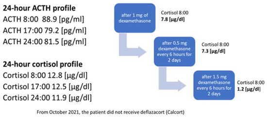
In October 2021, the administration of deflazocort as a treatment was discontinued. During hospitalization in January 2022, the diurnal pattern of ACTH and cortisol secretion was re-evaluated, yet no consistent diurnal rhythm was observed; cortisol levels remained elevated at night. For this reason, overnight DST and then LDDST were carried out again (Figure 4), in which no suppression of cortisol concentrations was obtained. Only after a high-dose DST (HDDST), in which a high 1.5 mg of dexamethasone was administered every 6 h (125 µg/kg/24 h), was cortisol secretion suppressed.
Jcm 12 05967 g004
Figure 4. Laboratory findings indicating the diagnosis of ACTH-dependent Cushing’s syndrome.
Based on the above results, CD was suspected as the cause of osteoporosis and growth retardation. In February 2022, a CRH test was performed upon the patient, which revealed a four-fold increase in ACTH levels and a two-fold increase in serum cortisol levels (Table 2).
Table 2. The results of human CRH (hCRH) stimulation test and bilateral inferior petrosal sinus sampling (BIPSS).
The CRH stimulation test was administered in the morning using human synthetic CRH (Ferring) at a dose of 1 μg/kg of body weight. During the test, cortisol and ACTH levels were measured in serum at the following time points: −15, 0, 15, 30, 60, and 90 min (see Table 2). As part of the diagnostic process, urinary free cortisol excretion was also measured over two consecutive days. Only on the first day was there a slight elevation in urinary free cortisol concentration, measuring 183.60 μg/24 h (normal range: 4.3–176). The measurement performed on the second day showed a normal urinary free cortisol concentration of 145.60 μg/24 h (normal range: 4.3–176). On 2 March 2022, the patient underwent a bilateral inferior petrosal sinus sampling (BIPSS). Human CRH stimulation was also used during the procedure. The presence of ACTH-dependent hypercortisolemia of pituitary origin was confirmed. The outcome of the CRH stimulation during the BIPSS is presented in Table 2. The boy qualified for transsphenoidal surgery (TSS) of the pituitary adenoma and was successfully operated on (8 March 2022). Postoperative histopathological examination revealed features of a corticotroph-rich pituitary adenoma.

3. Discussion

Osteoporosis, like CD, is extremely rare in the developmental age population. Bone-mineralization disorders among children may be primary (e.g., osteogenesis imperfecta), or secondary to other diseases or their treatment (e.g., with glucocorticosteroids). This case report presents a boy with osteoporosis, the cause of which was originally attributed to the treatment of autoimmune hepatitis with glucocorticosteroids. Steroid therapy is the most common cause of bone-mineralization disorders in children. However, osteoporosis is a late complication of steroid treatment. Briot et al. [10] demonstrated that the risk of fractures increases as early as 3 months after initiating steroid therapy. An additional factor increasing the risk of fractures is the dose of glucocorticosteroids used, corresponding to 2.5–5 mg of prednisolone per day [10]. In the case of the present patient, the appearance of spinal pain and thus vertebral fractures could not have been related to the deflazacort treatment started 5 days earlier. The bone-mineralization disorder must therefore have occurred much earlier. For this reason, the authors considered it necessary to search for other endocrine causes of osteoporosis development, including hypogonadism, growth hormone deficiency or Cushing’s syndrome/disease.
The serum vitamin D concentration can also influence bone mineral density. Every patient with mineralization disorders, especially with osteoporosis, requires a thorough assessment of calcium–phosphate metabolism [11]. Until the initiation of steroid therapy in March 2021, the patient did not undergo regular vitamin D supplementation. At the start of deflazacort treatment, his serum 25(OH)D concentration was 12.4 ng/mL. Consequently, additional cholecalciferol supplementation at a dose of 3000 IU/day was introduced. In a subsequent measurement conducted in June 2021, the concentration was within the reference range [25(OH)D: 46.7 ng/mL].
Considering the lack of regular supplementation before March 2021, it can be assumed that in October 2020, when the boy experienced SARS-CoV-2 infection, his serum vitamin D concentration was likely decreased as well, which could have had a further negative impact on the patient’s bone mineralization. Scientific reports indicate that adequate vitamin D levels reduce the risk of viral infections, including SARS-CoV-2 [12]. Di Filippo et al. [13] demonstrated that vitamin D deficiency observed in 68.2% of SARS-CoV-2-infected individuals correlated with a more severe course of the infection. In our patient, the course of COVID-19 was asymptomatic, and the diagnosis was established based on positive IgM antibody titers against SARS-CoV-2. The vitamin deficiency was most likely associated with irregular supplementation and lack of exposure to UV radiation (due to lockdown measures in Poland at that time). A reduced serum 25(OH)D concentration could have contributed to worsened bone mineral density and increased susceptibility to SARS-CoV-2 infection; however, it is the chronic hypercortisolism characteristic of CD that most likely led to the development of osteoporosis with accompanying fractures.
Another factor necessitating further diagnostic investigation into CD was the patient’s growth retardation observed since the age of 11. Both the pubertal state of the boy, and his gonadotropin and testosterone serum levels, allowed us to exclude hypogonadism. Maximum spontaneous nocturnal secretion of the growth hormone was 31.84 ng/mL. The diagnosis of CD was established on the basis of elevated cortisol levels at night and the lack of cortisol suppression in the test after administering dexamethasone. Final confirmation of the diagnosis was obtained in a post-CRH stimulation test. In pediatric cases, the absence of typical diurnal variation in serum corticosolemia, especially the nocturnal decline, and the inability to suppress cortisol secretion at midnight, are highly sensitive indicators of hypercortisolemia [6,8]. Consequently, in our patient, osteoporosis was a complication of diagnosed CD.
The patient in question was not obese, which is the predominant symptom of CD. This symptom, according to Ferrigno et al. [1], is present in 92–98% of examined children diagnosed with CD. Storr et al. [14] showed that facial changes and facial swelling were observed in 100% of subjects with CD, whereas Lonser et al. [8] observed this in only 63% of children with CD. In our patient, no changes in facial appearance were observed. Other symptoms typical of CS, such as hirsutism, acne, or bruises, were not noticed either. These symptoms were observed in all children with CD studied by Wędrychowicz et al. [3]. Non-specific symptoms of this condition may include mood changes, depression and emotional vacillation [1,8]. However, our patient’s parents did not observe any changes in the boy’s behavior. The indication for initiating the whole diagnostic process was (in addition to osteoporosis) growth retardation. Ferrigno et al. [1] point out that chronic hypercortisolemia most often leads to growth disorders accompanied by excessive weight gain. This is an early, highly sensitive and characteristic sign of CD. Short stature is not always observed and occurs in one in two children diagnosed with CD. The patient we present was short (height—162 cm (<3rd centile hSDS: −2.36)); growth retardation was observed from the age of 11 years.
The occurrence of vertebral fractures and the accompanying pain as the initial symptoms of hypercortisolism, the absence of obesity, and the confirmation of CD, an exceedingly rare condition in the pediatric population, collectively underscore the uniqueness of our patient’s disease presentation. A case involving a child with such an atypical course of ACTH-dependent CS has not been described before. Han et al. [15] reported a case of a 28-year-old lean woman (BMI: 19 kg/m²) with ACTH-independent CS due to a left adrenal adenoma, where, similarly to our patient, the initial manifestation of hypercortisolism was compression fractures of the thoracic vertebrae. The authors emphasize that vertebral fractures may affect 30–50% of patients with Cushing’s syndrome, with a higher frequency observed in patients with ACTH-independent CS compared to those in whom hypercortisolism results from the presence of pituitary adenoma [15].
The lack of obesity in a patient with hypercortisolism could be attributed to malnutrition, which accompanies the growth process in ECS. Hence, a crucial aspect was the differential diagnosis between CD and ECS. To this end, we performed a stimulation test using hCRH. We considered cut-off points for diagnosing CD to be a 35% increase in ACTH concentration at 15 and/or 30 min, and at least a 20% increase in cortisol concentration at 30 and 45 min [16,17]. In the case of ECS, a significant rise in CRH and cortisol concentrations is not observed. Recently published reports emphasize the need to explore new cut-off points to enhance the sensitivity and specificity of this test. Detomas et al. [5] indicate that an increase in ACTH ≥ 31% and cortisol ≥ 12% in the 30th minute of CRH tests allows for a highly sensitive and specific differentiation between CD and ECS. The authors highlight that measuring these hormones at the 60 min stage of the test does not provide diagnostic benefits. Notably, the study employed ovine CRH, which exhibits stronger and more prolonged stimulatory effects compared to the hCRH available in Europe that was used to diagnose our patient [5]. Conversely, Elenius et al. [16] suggest that optimal values for distinguishing between CD and ECS in the CRH stimulation test involve an increase in ACTH and/or cortisol levels of more than 40% during the test. In our patient, an over four-fold increase in ACTH levels and a more than two-fold increase in cortisol levels were observed at the 30 min mark of the test, thus independently and definitively excluding ECS regardless of the adopted cut-off points.
Our patient’s case also demonstrates that MRI is not a perfect method of visualizing an ACTH-secreting pituitary adenoma. In the first MRI examination performed upon our patient, a poorly demarcated area (2.0 × 3.5 × 5.0 mm) was described in the anterior part of the glandular lobe; in the examination performed 6 months later, this area maintained poor visibility, while laboratory results at the time clearly indicated an ACTH-dependent form of CS. It was only the bilateral inferior petrosal sinus sampling (BIPSS) that allowed a clear diagnosis. Data from the literature indicate that microadenomas smaller than 3–4 mm are visible on MRI in only half of cases. In two large studies including children, pituitary adenomas were found on MRI in 63% and 55% of cases [18]. Among the patients with CD studied by Wędrychowicz et al. [3], pituitary adenomas were described on MRI in all of them, but in two patients (50%) this was only achieved upon follow-up. In the standard procedure, in the absence of a pituitary lesion in the MRI examination, it is recommended that a BIPSS be performed. In the case of our patient, this examination was necessary to make a definitive diagnosis.
When analyzing the results of the BIPSS with hCRH stimulation, we employed the classical cut-offs for the ACTH IPS:P (Inferior Petrosal Sinus: Peripheral) ratio (i.e., ≥2 at baseline and ≥3 after hCRH stimulation) [1]. This allowed the confirmation of CD and determination of the pituitary adenoma’s localization, followed by the procedure for its surgical removal. The optimal cut-off values for the IPS:P ratio remain controversial. There are ongoing efforts to establish new, more precise cut-off points. Detomas et al. [19] demonstrated that an IPS:P ratio ≥ 2.1 during desmopressin stimulation in the BIPSS most accurately differentiates CD from ECS. Conversely, Chen et al. [20] showed that the optimal pre-desmopressin stimulation IPS:P ratio cut-off is 1.4, and post-stimulation it is 2.8. Both studies suggest the utilization of lower cut-off values for the IPS:P ratio than those traditionally adopted. Chen et al. [20] also advocate for avoiding stimulation during BIPSS. In most cases, the IPS:P ratio before stimulation is sufficient for diagnosing CD. According to the authors, desmopressin stimulation should be reserved for patients with ambiguous MRI findings or with a pituitary adenoma with diameter less than 6 mm. However, considering that the concentration of ACTH in the right inferior petrosal sinus in our patient was over 4 times higher than in the peripheral vessel and nearly 14 times higher after hCRH stimulation, regardless of the applied criteria, CD could be unequivocally diagnosed in our patient, and the lateralization of the microadenoma could be determined with certainty.
The rarity of CD, and the diagnostic difficulties stemming from its oligosymptomatic or atypical course, encourage description in the form of case reports. Eviz et al. [21] delineate the occurrence of cerebral cortical atrophy in two children with ECS. Additionally, other researchers have underscored the potential for thyroid disorders to manifest alongside hypercorticosolemia [22]. Although obesity typically stands out as a primary symptom of CD, Pomahacova et al. [23] reported a case involving two children with CD who maintained normal body weight, mirroring our patient’s situation. The symptoms that prompted diagnostic investigation in these instances included weakness, sleep disturbances and growth retardation. Interestingly, growth retardation, along with facial changes, was observed in all examined children with CD [23]. Nonetheless, to the best of our knowledge, we have yet to encounter a case report resembling ours. Therefore, it remains crucial to share our experiences.

4. Conclusions

Cushing’s disease is an extremely rare diagnosis in children. In Poland, there is no statistical record of occurrences of this disease among children. Wędrychowicz et al. reported that in their single Polish center, between 2012 and 2018, they identified four cases of children aged 7–15 who were diagnosed with CD [3]. The case we present shows that obesity, commonly considered as a predominant symptom of CD, is not necessarily observed in patients with this diagnosis in the developmental age population. Among children, it is growth disturbance that may be the first manifestation. On the other hand, a late complication of CD may be osteoporosis, so whenever a child is diagnosed with a bone-mineralization disorder, the cause of its development should be sought.
Diagnosis should be pursued until all potential causes of the described symptoms, including the rarest ones, are definitively ruled out—even if the clinical presentation, as in the case of our patient, initially does not point towards the final diagnosis. Thus far, no case of a child with CD exhibiting such subtle symptomatology has been described in the literature. The challenges in diagnosis we encountered primarily resulted from the atypical clinical outcome of CD in our patient—normal body weight, absence of hyperandrogenism, mood disturbances not apparent to caregivers and the patient’s immediate environment, as well as normal progression of puberty, did not immediately lead to the consideration of endocrinological causes of osteoporosis. The steroid therapy employed due to autoimmune hepatitis also complicated the diagnostic process. Only after discontinuing deflazacort treatment was it possible to definitively diagnose CD.
Our patient required hydrocortisone replacement in gradually decreasing doses for a year following TSS. Considering that pituitary adenomas in children can be genetically predisposed (e.g., MEN 1 mutation, AIP mutation, USP8 mutation, and other rarer ones), genetic consultation was sought [1]. However, the conducted tests have thus far excluded the most common mutations in our patient. Due to the diagnosed osteoporosis, chronic supplementation with calcium and cholecalciferol was recommended, along with annual follow-up DXA scans. Studies indicate that patients in remission from CD experience a gradual improvement in bone mineral density [3]. While we can currently observe remission in our patient’s case, the advanced bone age of the child (indicating the completion of the growth process) left limited potential for significant improvement in final growth. The patient still requires regular endocrinological and neurosurgical follow-ups, hormonal assessments, and pituitary MRI examinations.

Author Contributions

Conceptualization—R.S. and A.Ł.; software—S.A.; formal analysis—R.S.; investigation—A.Ł. and G.Z.; data curation—A.Ł.; writing—original draft preparation—A.Ł., R.S. and S.A.; writing—review and editing—A.L.; visualization—S.A.; supervision—A.L. All authors have read and agreed to the published version of the manuscript.

Funding

This study was funded by statutory funds from the Medical University of Lodz, Lodz, Poland (503/1-107-03/503-11-001).

Institutional Review Board Statement

Not applicable.

Informed Consent Statement

Written informed consent has been obtained from the patient to publish this paper.

Data Availability Statement

Not applicable.

Conflicts of Interest

The authors declare no conflict of interest.

References

  1. Ferrigno, R.; Hasenmajer, V.; Caiulo, S.; Minnetti, M.; Mazzotta, P.; Storr, H.L.; Isidori, A.M.; Grossman, A.B.; De Martino, M.C.; Savage, M.O. Paediatric Cushing’s disease: Epidemiology, pathogenesis, clinical management and outcome. Rev. Endocr. Metab. Disord. 2021, 22, 817–835. [Google Scholar] [CrossRef]
  2. Stratakis, A. Cushing syndrome in pediatrics. Endocrinol. Metab. Clin. N. Am. 2012, 41, 793–803. [Google Scholar] [CrossRef]
  3. Wędrychowicz, A.; Hull, B.; Tyrawa, K.; Kalicka-Kasperczyk, A.; Zieliński, G.; Starzyk, J. Cushing disease in children and adolescents—Assessment of the clinical course, diagnostic process, and effects of the treatment—Experience from a single paediatric centre. Pediatr. Endocrinol. Diabet. Metab. 2019, 25, 127–143. [Google Scholar] [CrossRef]
  4. Concepción-Zavaleta, M.J.; Armas, C.D.; Quiroz-Aldave, J.E.; García-Villasante, E.J.; Gariza-Solano, A.C.; Del Carmen Durand-Vásquez, M.; Concepción-Urteaga, L.A.; Zavaleta-Gutiérre, F.E. Cushing disease in pediatrics: An update. Ann. Pediatr. Endocrinol. Metab. 2023, 28, 87–97. [Google Scholar] [CrossRef]
  5. Detomas, M.; Ritzel, K.; Nasi-Kordhishti, I.; Wolfsberger, S.; Quinkler, M.; Losa, M.; Tröger, V.; Kroiss, M.; Fassnacht, M.; Vila, G.; et al. Outcome of CRH stimulation test and overnight 8 mg dexamethasone suppression test in 469 patients with ACTH-dependent Cushing’s syndrome. Front. Endocrinol. 2022, 13, 955945. [Google Scholar] [CrossRef] [PubMed]
  6. Lodish, M.B.; Hsiao, H.P.; Serbis, A.; Sinaii, N.; Rothenbuhler, A.; Keil, M.F.; Boikos, S.A.; Reynolds, J.C.; Stratakis, C.A. Effects of Cushing disease on bone mineral density in a pediatric population. J. Pediatr. 2010, 56, 1001–1005. [Google Scholar] [CrossRef] [PubMed]
  7. Pivonello, R.; Isidori, A.M.; De Martino, M.C.; Newell-Price, J.; Biller, B.M.; Colao, A. Complications of Cushing’s syndrome: State of the art. Lancet Diabetes Endocrinol. 2016, 4 (Suppl. S7), 611–629. [Google Scholar] [CrossRef] [PubMed]
  8. Lonser, R.R.; Wind, J.J.; Nieman, L.K.; Weil, R.J.; DeVroom, H.L.; Oldfield, E.H. Outcome of surgical treatment of 200 children with Cushing’s disease. J. Clin. Endocrinol. Metab. 2013, 98, 892–901. [Google Scholar] [CrossRef]
  9. Detomas, M.; Deutschbein, T.; Tamburello, M.; Chifu, I.; Kimpel, O.; Sbiera, S.; Kroiss, M.; Fassnacht, M.; Altieri, B. Erythropoiesis in Cushing syndrome: Sex-related and subtype-specific differences. Results from a monocentric study. J. Endocrinol. Investig. 2023; epub ahead of print. [Google Scholar] [CrossRef]
  10. Briot, K.; Roux, C. Glucocorticoid-induced osteoporosis. RMD Open 2015, 1, 14. [Google Scholar] [CrossRef]
  11. Laird, E.; Ward, M.; McSorley, E.; Strain, J.J.; Wallace, J. Vitamin D and bone health; potential mechanisms. Nutrients 2010, 2, 693–724. [Google Scholar] [CrossRef]
  12. di Filippo, L.; Frara, S.; Nannipieri, F.; Cotellessa, A.; Locatelli, M.; Querini, P.R.; Giustina, A. Low vitamin D levels are associated with long COVID syndrome in COVID-19 survivors. J. Clin. Endocrinol. Metab. 2023, 1–11. [Google Scholar] [CrossRef] [PubMed]
  13. di Filippo, L.; Allora, A.; Doga, M.; Formenti, A.M.; Locatelli, M.; Rovere Querini, P.; Frara, S.; Giustina, A. Vitamin D levels are associated with blood glucose and BMI in COVID-19 patients, predicting disease severity. J. Clin. Endocrinol. Metab. 2022, 107, 348–360. [Google Scholar] [CrossRef]
  14. Storr, H.L.; Chan, L.F.; Grossman, A.B.; Savage, M.O. Pediatric Cushing’s syndrome: Epidemiology, investigation and therapeutic advances. Trends Endocrinol. Metab. 2007, 18, 167–174. [Google Scholar] [CrossRef] [PubMed]
  15. Han, J.Y.; Lee, J.; Kim, G.E.; Yeo, J.Y.; Kim, S.H.; Nam, M.; Kim, Y.S.; Hong, S. Case of Cushing syndrome diagnosed by recurrent pathologic fractures in a young woman. J. Bone Metab. 2012, 19 (Suppl. S2), 153–158. [Google Scholar] [CrossRef] [PubMed]
  16. Elenius, H.; McGlotten, R.; Nieman, L.K. Ovine CRH stimulation and 8 mg dexamethasone suppression tests in 323 patients with ACTH-dependent Cushing’s syndrome. J. Clin. Endocrinol. Metab, 2023; Epub ahead of print. [Google Scholar] [CrossRef]
  17. Nieman, L.K.; Biller, B.M.K.; Findling, J.W.; Newell-Price, J.; Savage, M.O.; Stewart, P.M.; Montori, V.M. The diagnosis of Cushing’s syndrome: An Endocrine Society Clinical Practice Guideline. J. Clin. Endocrinol. Metab. 2008, 93 (Suppl. S5), 1526–1540. [Google Scholar] [CrossRef] [PubMed]
  18. Savage, M.O.; Storr, H.L. Pediatric Cushing’s disease: Management issues. Indian J. Endocrinol. Metab. 2012, 16 (Suppl. S2), 171–175. [Google Scholar] [CrossRef]
  19. Detomas, M.; Ritzel, K.; Nasi-Kordhishti, I.; Schernthaner-Reiter, M.H.; Losa, M.; Tröger, V.; Altieri, B.; Kroiss, M.; Kickuth, R.; Fassnacht, M.; et al. Bilateral inferior petrosal sinus sampling with human CRH stimulation in ACTH-dependent Cushing’s syndrome: Results from a retrospective multicenter study. Eur. J. Endocrinol. 2023, 188 (Suppl. S5), 448–456. [Google Scholar] [CrossRef]
  20. Chen, S.; Chen, K.; Wang, S.; Zhu, H.; Lu, L.; Zhang, X.; Tong, A.; Pan, H.; Wang, R.; Lu, Z. The optimal cut-off of BIPSS in differential diagnosis of ACTH-dependent Cushing’s syndrome: Is stimulation necessary? J. Clin. Endocrinol. Metab. 2020, 105 (Suppl. S4), 1673–1685. [Google Scholar] [CrossRef]
  21. Eviz, E.; Yesiltepe, M.G.; Arduc, A.A.; Erbey, F.; Guran, T.; Hatun, S. An overlooked manifestation of hypercortisolism—Cerebral cortical atrophy and challenges in identifying the etiology of hypercortisolism: A report of 2 pediatric cases. Horm. Res. Paediatr. 2023, 27. [Google Scholar] [CrossRef]
  22. Paragliola, R.M.; Corsello, A.; Papi, G.; Pontecorvi, A.; Corsello, S.M. Cushing’s syndrome effects on the thyroid. Int. J. Mol. Sci. 2021, 22, 3131. [Google Scholar] [CrossRef]
  23. Pomahacova, R.; Paterova, P.; Nykodymova, E.; Sykora, J.; Krsek, M. Pediatric Cushing’s disease: Case reports and retrospective review. Biomed. Pap. Med. Fac. Univ. Palacky. Olomouc. Czech Repub. 2022, 166. [Google Scholar] [CrossRef] [PubMed]
Disclaimer/Publisher’s Note: The statements, opinions and data contained in all publications are solely those of the individual author(s) and contributor(s) and not of MDPI and/or the editor(s). MDPI and/or the editor(s) disclaim responsibility for any injury to people or property resulting from any ideas, methods, instructions or products referred to in the content.

Share and Cite

MDPI and ACS Style

Łupińska, A.; Aszkiełowicz, S.; Zieliński, G.; Stawerska, R.; Lewiński, A. Osteoporosis as the First Sign of Cushing’s Disease in a Thin 16-Year-Old Boy—A Case Report. J. Clin. Med. 202312, 5967. https://doi.org/10.3390/jcm12185967

AMA StyleŁupińska A, Aszkiełowicz S, Zieliński G, Stawerska R, Lewiński A. Osteoporosis as the First Sign of Cushing’s Disease in a Thin 16-Year-Old Boy—A Case Report. Journal of Clinical Medicine. 2023; 12(18):5967. https://doi.org/10.3390/jcm12185967

Chicago/Turabian StyleŁupińska, Anna, Sara Aszkiełowicz, Grzegorz Zieliński, Renata Stawerska, and Andrzej Lewiński. 2023. “Osteoporosis as the First Sign of Cushing’s Disease in a Thin 16-Year-Old Boy—A Case Report” Journal of Clinical Medicine 12, no. 18: 5967. https://doi.org/10.3390/jcm12185967

Find Other Styles

Note that from the first issue of 2016, this journal uses article numbers instead of page numbers. See further details here.

Article Metrics

Citations

No citations were found for this article, but you may check on Google Scholar

Article Access Statistics

Article access statisticsArticle Views14. Sep15. Sep16. Sep17. Sep18. Sep19. Sep20. Sep21. Sep22. Sep23. Sep24. Sep25. Sep26. Sep27. Sep28. Sep29. Sep30. Sep1. Oct2. Oct3. Oct4. Oct5. Oct6. Oct7. Oct8. Oct9. Oct10. Oct11. Oct12. Oct13. Oct14. Oct0100200300400

For more information on the journal statistics, click here.

Multiple requests from the same IP address are counted as one view.

Rare Challenges in Diagnosing Cushing’s Syndrome and Primary Aldosteronism: A Case Report of a Female With a Negative Workup

Abstract

Cushing’s syndrome with concurrent primary aldosteronism (PA) is a rare presentation, and establishing an early diagnosis is imperative to preventing morbidity and long-term sequelae. The diagnosis is established by sequential lab work, showing an elevated cortisol and aldosterone level.

Taking the above into consideration, it is evident that repeatedly negative results on all three tests can present an extremely challenging case. In this report, we discuss a female who presented with an adrenal incidentaloma and features suggestive of primary hyperaldosteronism as well as Cushing’s syndrome but no elevations in serum, urine, or salivary cortisol.

In this study, we present a 37-year-old female with resistant hypertension and tachycardia. She had several features suggestive of Cushing’s syndrome including resistant hypertension, proximal muscle weakness, weight gain, easy bruising, hair loss, and a history of tachycardia and chest pain. Examination revealed an obese female with thin silvery abdominal striae. The patient’s labs revealed normal serum cortisol, urine-free cortisol (UFC), late-night salivary cortisol, and a normal dexamethasone suppression test. An abdominal computed tomography (CT) scan revealed a right adrenal mass measuring 2.1 x 1.5 x 2.5 cm. Due to a high index of suspicion, adrenal venous sampling was performed, which revealed high levels of cortisol and aldosterone in the right vein, confirming the diagnosis. The patient subsequently underwent a right adrenalectomy. She developed hypotension post-op, leading to the diagnosis of glucocorticoid-remediable aldosteronism.

Introduction

Primary aldosteronism (PA) is the excess production of aldosterone by the adrenal glands, despite a low serum renin level. The presentation of hyperaldosteronism can be vague and include symptoms such as muscle weakness, fatigue, headaches, numbness, and cramps. More specific findings include resistant hypertension, low serum potassium, and metabolic alkalosis. The etiologies are variable and can include an adrenal adenoma (Conn syndrome) or bilateral adrenal hyperplasia [1].

Cushing’s syndrome is also caused by excess hormone secretion by the adrenal glands. The etiologies include a primary adrenal adenoma, hyperplasia, carcinoma, or exogenous corticosteroid use. It can also be caused by an adrenocorticotropic hormone (ACTH)-secreting pituitary adenoma or as a result of paraneoplastic ACTH secretion. The clinical presentation is highly variable and leads to difficulties in establishing a diagnosis.

The concurrent existence of primary hyperaldosteronism and Cushing’s syndrome creates additional hindrances in diagnosis, yet further obscured in a patient with a repeatedly negative workup for both conditions.

Case Presentation

A 37-year-old female presented to her primary care physician with complaints of proximal muscle weakness, tachycardia, and chest pain. Repeated blood pressure readings revealed that she was hypertensive, and she was started on amlodipine and benazepril, which elevated her blood pressure further. A computed tomography (CT) scan (Figure 1) of the abdomen was performed due to resistant hypertension, which revealed an adrenal incidentaloma (right adrenal gland measuring 2.1 x 1.5 x 2.5 cm). Precontract density was 5 Hounsfield units, and a 15-minute delayed washout showed 11 Hounsfield units for a 72% washout. She was thus referred to endocrinology.

Abdominal-CT-scan-showing-a-nodule-in-the-right-adrenal-gland-measuring-2.1-x-1.5-x-2.5-cm
Figure 1: Abdominal CT scan showing a nodule in the right adrenal gland measuring 2.1 x 1.5 x 2.5 cm

She presented to the endocrinology clinic on March 12, 2021. A thorough physical examination was performed, which revealed a well-appearing obese female (BMI of 38.86 kg/m2) with no acute distress. Her blood pressure was 144/108 mmHg, her pulse was 95, and she was afebrile. Thin silvery striations were present on the abdomen, and alopecia was present on the crown. A review of all other systems was unremarkable. A detailed family history revealed early-onset hypertension in her brother (age: 35 years) and her mother (age: 30 years). Personal history included elevated anxiety, weight gain, headaches (frontal band distribution), increased thirst, easy bruising as well as delayed clearance of bruises, and proximal muscle weakness presenting as difficulty in climbing stairs and inability to lift heavy objects. She reported no change in menstrual cycles. There was no history of exogenous corticosteroid use.

Serum biochemistries were sent (Table 1), which showed normal levels of thyroid stimulating hormone (TSH), creatinine, liver function tests, and serum electrolytes. However, mildly elevated aldosterone (23 ng/dl), mild hypokalemia (3.3 mEq/L), and suppressed ACTH and dehydroepiandrosterone (DHEA) sulfate were discovered. The aldosterone to renin ratio was also elevated at 59.9 on spironolactone and was 71.4 three months later when spironolactone was discontinued. These findings lead to a preliminary diagnosis of primary hyperaldosteronism.

Test Result
Calcium 9.1 mmol/L
Sodium 137 mmol/L
Potassium 4.1 mmol/L
Chloride 106 mmol/L
CO2 27
BUN 15 mmol/L
Glucose 95 mmol/L
Creatinine 1.1 μmol/L
AST 24 U/L
ALT 20 U/L
Albumin 4.4 g/L
Total protein 7.0 g/L
Total bilirubin 0.4 μmol/L
Alkaline phosphatase 40 U/L
Renin 0.44
Table 1: Patient serum biochemistries

BUN: Blood urea nitrogen; AST: Aspartate transaminase; ALT: Alanine transaminase.

A workup for elevated cortisol was also performed as the patient was phenotypically Cushingoid, and the following biochemistries were sent sequentially: serum cortisol, 24-hour urine-free cortisol (UFC), salivary cortisol, and a low-dose dexamethasone suppression test (Table 2). The bloodwork was hence nonconfirmatory.

Endocrine workup
Serum cortisol 4.5 mcg/dL
Urine-free cortisol 1.57 g/24 h
Salivary cortisol <0.03 μg/dL
Dexamethasone suppression test 1.5 mcg/dL
Aldosterone <4.0
Table 2: Patient follow-up bloodwork

Despite a repeatedly negative workup for Cushing’s syndrome, adrenal venous sampling was performed due to a high index of suspicion. The results revealed an inferior vena cava (IVC) cortisol of 20, left adrenal venous (LAV) cortisol of 81, and right adrenal vein (RAV) cortisol of 1280. The results of the IVC aldosterone were 24, LAV aldosterone was 660 and RAV aldosterone was 1500. The elevated levels of cortisol in the RAV were in complete contradiction to the aforementioned workup. A diagnosis of Cushing’s syndrome and concurrent PA was determined.

Adrenal veinous sampling was instrumental in establishing the diagnosis but was equivocal and did not lateralize aldosterone and cortisol excess. However, the amount of aldosterone and cortisol were both significantly higher on the right side. After a panel discussion with doctors from several disciplines, a laparoscopic adrenalectomy was planned. The procedure was successful, and the patient was initially showing clinical improvement. The specimen was sent for pathological evaluation and revealed an adrenal cortical adenoma.

After initial improvement, the patient developed hypotension, which was likely due to adrenal insufficiency. The patient was supplemented with 1-mg dexamethasone tablets, which stabilized her condition, and a diagnosis of glucocorticoid-remediable-aldosteronism was made.

Based on a strong family history of early onset-resistant hypertension, a genetic component was suspected. Several genes associated with PA with autosomal dominant inheritance have been identified [2], such as CYP11B2, CLCN2, KCNJ5, CACNA1D, and CACNA1H. The patient was offered genetic testing but was unable to follow through due to financial reasons.

Discussion

This patient presented as an extremely rare example of PA and Cushing’s syndrome, with negative serum cortisol, 24-hour UFC, late-night salivary cortisol, and a dexamethasone suppression test. Despite repeatedly negative lab results, the patient presented with a markedly elevated cortisol on adrenal venous sampling. In our literature search, we found an instance of a patient with several negative UFCs [3]; however, to the best of our knowledge, there have been no reported instances of a completely negative workup in a patient who is positive for Cushing’s syndrome. In fact, in the practice guidelines published by the Journal of Clinical Endocrinology & Metabolism [4], it is recommended that patients with a suspected diagnosis of Cushing’s syndrome or an adrenal incidentaloma and two concordant negative test results need not undergo further investigations.

One proposed mechanism for the misleading workup could be assay interference. Interference occurs when a substance or process falsely alters an assay result [5]. This can lead to incorrect diagnosis and subsequent treatment and poses a threat to the patient. Another suggested mechanism causing false negative test results could be the hook effect [6]. The hook effect is described as a phenomenon that leads to falsely low results due to the presence of excessive analyte.

In a study by Friedman et al. [7], it was noted that patients with “episodic Cushing’s syndrome” or those with mild symptoms had a negative workup. The study recommended serial monitoring for the disease. The interesting fact is that our patient had several features suggestive of active Cushing’s syndrome, and the hypotension seen postoperatively was a testament to the fact that there was in fact a cortisol excess, which led to adrenal insufficiency. In light of the above, a consistently negative workup is perplexing.

Zhang et al. suggested performing a low-dose dexamethasone suppression test in individuals presenting with PA, prior to adrenal vein sampling (AVS) and surgery due to the high prevalence of Cushing’s syndrome in patients with PA [8]. A positive test result can lead to a straightforward diagnosis; however, in this rare case where the patient had severe negative tests, it can present as a challenge in diagnosis and treatment.

Conclusions

The presence of PA and concurrent Cushing’s syndrome can present as a diagnostic challenge. It is recommended to follow up on the signs of Cushing’s syndrome with preliminary tests and to presume its absence if two concordant tests are negative. Our patient, however, was an exceptional case.

This case highlighted the importance of maintaining a high index of suspicion for patients presenting with several signs and symptoms of the disease and a negative workup. More attention should be paid to the patient’s history, and a thorough physical examination should be conducted. In those with an uncertain diagnosis, adrenal venous sampling can provide a clearer picture and lead to a more accurate understanding of the case.

References

  1. Reincke M, Bancos I, Mulatero P, Scholl UI, Stowasser M, Williams TA: Diagnosis and treatment of primary aldosteronism. Lancet Diabetes Endocrinol. 2021, 9:876-92. 10.1016/S2213-8587(21)00210-2
  2. Dutta RK, Söderkvist P, Gimm O: Genetics of primary hyperaldosteronism. Endocr Relat Cancer. 2016, 23:R437-54. 10.1530/ERC-16-0055
  3. Moloney KJ, Mercado JU, Ludlam WH, Broyles FE: Diagnosis of Cushing’s disease in a patient with consistently normal urinary free cortisol levels: a case report. Clin Case Rep. 2016, 4:1181-3. 10.1002/ccr3.647
  4. Nieman LK, Biller BM, Findling JW, Newell-Price J, Savage MO, Stewart PM, Montori VM: The diagnosis of Cushing’s syndrome: an Endocrine Society Clinical Practice Guideline. J Clin Endocrinol Metab. 2008, 93:1526-40. 10.1210/jc.2008-0125
  5. Dimeski G: Interference testing. Clin Biochem Rev. 2008, 29:S43-8.
  6. The hook effect. (2014). Accessed: June 19, 2023: https://www.aacc.org/science-and-research/clinical-chemistry-trainee-council/trainee-council-in-english/pearls-of-lab….
  7. Friedman TC, Ghods DE, Shahinian HK, et al.: High prevalence of normal tests assessing hypercortisolism in subjects with mild and episodic Cushing’s syndrome suggests that the paradigm for diagnosis and exclusion of Cushing’s syndrome requires multiple testing. Horm Metab Res. 2010, 42:874-81. 10.1055/s-0030-1263128
  8. Zhang Y, Tan J, Yang Q, et al.: Primary aldosteronism concurrent with subclinical Cushing’s syndrome: a case report and review of the literature. J Med Case Rep. 2020, 14:32. 10.1186/s13256-020-2353-8

Unique Gene Expression Signature in Periadrenal Adipose Tissue Identifies a High Blood Pressure Group in Patients With Cushing Syndrome

Abstract

Background:

Cushing syndrome (CS) is a rare disease caused by excess cortisol levels with high cardiovascular morbidity and mortality. Hypertension in CS promotes hypercortisolism-associated cardiovascular events. Adipose tissue is a highly plastic tissue with most cell types strongly affected by the excess cortisol exposure. We hypothesized that the molecular and cellular changes of periadrenal adipose tissue in response to cortisol excess impact systemic blood pressure levels in patients with CS.

Methods:

We investigated gene expression signatures in periadrenal adipose tissue from patients with adrenal CS collected during adrenal surgery.

Results:

During active CS we observed a downregulation of gene programs associated with inflammation in periadrenal adipose tissue. In addition, we observed a clustering of the patients based on tissue gene expression profiles into 2 groups according to blood pressure levels (CS low blood pressure and CS high blood pressure). The 2 clusters showed significant differences in gene expression pattens of the renin-angiotensin-aldosterone-system. Renin was the strongest regulated gene compared with control patients and its expression correlated with increased blood pressure observed in our patients with CS. In the CS high blood pressure group, systemic renin plasma levels were suppressed indicative of an abnormal blood pressure associated with periadrenal adipose tissue renin-angiotensin-aldosterone-system activation.

Conclusions:

Here, we show for the first time a relevant association of the local renin-angiotensin-aldosterone-system and systemic blood pressure levels in patients with CS. Patients from the CS high blood pressure group still had increased blood pressure levels after 6 months in remission, highlighting the importance of local tissue effects on long-term systemic effects observed in CS.

Footnotes

*U. Stifel and F. Vogel contributed equally.

For Sources of Funding and Disclosures, see page xxx.

Supplemental Material is available at https://www.ahajournals.org/doi/suppl/10.1161/HYPERTENSIONAHA.123.21185.

Correspondence to: Martin Reincke, Department of Medicine IV, University Hospital, LMU Munich, GermanyEmail martin.reincke@med.uni-muenchen.de
Jan Tuckermann, Institute of Comparative Molecular Endocrinology (CME), Ulm University, GermanyEmail jan.tuckermann@uni-ulm.de

eLetters

eLetters should relate to an article recently published in the journal and are not a forum for providing unpublished data. Comments are reviewed for appropriate use of tone and language. Comments are not peer-reviewed. Acceptable comments are posted to the journal website only. Comments are not published in an issue and are not indexed in PubMed. Comments should be no longer than 500 words and will only be posted online. References are limited to 10. Authors of the article cited in the comment will be invited to reply, as appropriate.

Comments and feedback on AHA/ASA Scientific Statements and Guidelines should be directed to the AHA/ASA Manuscript Oversight Committee via its Correspondence page.

From https://www.ahajournals.org/doi/10.1161/HYPERTENSIONAHA.123.21185

Adults with Cushing’s Syndrome Report High Burden Of Illness, Despite Ongoing Treatment

Key takeaways:

  • Cushing’s syndrome symptoms moderately impact quality of life for adults with the condition.
  • Weight gain, muscle fatigue and menstrual changes decline in severity from diagnosis to follow-up.

Adults with endogenous Cushing’s syndrome reported that the condition moderately affects their quality of life and causes them to have symptoms about 16 days in a given month, according to findings published in Pituitary.

“Our study aimed to evaluate the ongoing burden of Cushing’s syndrome in order to identify areas of unmet need,” Eliza B. Geer, MD, medical director of the Multidisciplinary Pituitary and Skull Base Tumor Center and associate attending of endocrinology and neurosurgery at Memorial Sloan Kettering Cancer Center, told Healio. “We found that patients with treated Cushing’s continue to experience ongoing symptoms more than half of the days in a given month, miss about 25 workdays per year and need twice the average number of outpatient visits per year, indicating a significant impact on daily function and work productivity. Some of these symptoms, like fatigue and pain, have not been well studied in Cushing’s patients, and need more attention.”

Geer and colleagues administered a cross-sectional survey to 55 adults aged 21 years and older who had been diagnosed with Cushing’s syndrome at least 6 months before the survey and were receiving at least one pharmacologic therapy for their disease (85% women; mean age, 43.4 years). The survey was conducted online from June to August 2021. Five patient-reported outcome scales were included. The CushingQoL was used to analyze quality of life, a visual analog scale was included to assess pain, the Brief Fatigue Inventory was used to measure fatigue, the Sleep Disturbance v1.0 scale assessed perceptions of sleep and the PROMIS Short Form Anxiety v1.0-8a scale was used to measure fear, anxious misery, hyperarousal and somatic symptoms related to arousal. Participants self-reported the impact of Cushing’s syndrome on daily life and their physician’s level of awareness of Cushing’s syndrome.

Some symptoms decline in severity over time

Of the study group, 81% had pituitary or adrenal tumors, and 20% had ectopic adrenocorticotropic hormone-producing tumors; 80% of participants underwent surgery to treat their Cushing’s syndrome.

The frequency of reported symptoms did not change from Cushing’s syndrome diagnosis to the time of the survey. The most frequently reported symptoms were weight gain, muscle fatigue and weakness and anxiety.

Participants reported a decline in symptom severity for weight gain, muscle fatigue and weakness and menstrual changes from diagnosis to the survey. Though symptom severity declined, none of the three symptoms were entirely eliminated. Adults did not report declines in severity for other symptoms. Hirsutism and anxiety were reported by few participants, but were consistently scored high in severity among those who reported it. There were no changes in patient satisfaction with medications from their first appointment to the time of the survey.

“It was surprising that anxiety and pain did not improve with treatment,” Geer said. “A quarter of patients at baseline reported anxiety and this percentage was exactly the same after treatment. Same for pain — nearly a quarter of patients reported pain despite treatment. While the presence of anxiety has been well-documented in Cushing’s patients, pain has not, and needs further study.”

Nearly half of primary care providers unable to diagnose Cushing’s syndrome

All participants reported having at least one challenge with being diagnosed with Cushing’s syndrome. Of the respondents, 49% said their primary care provider was unable to diagnose their Cushing’s syndrome and 33% initially received the wrong diagnosis. Physicians referred 49% of participants to a specialist, and 39% of adults said their doctor lacked knowledge or understanding of their condition.

The study group had a moderate level of quality of life impairment as assessed through the CushingQoL scale. The mean pain score was 3.6 of a possible 10, indicating low levels of pain. Moderate to severe levels of fatigue were reported by 69% of participants. Self-reported sleep and anxiety scores were similar to what is observed in the general population.

Participants said sexual activity, self-confidence and life satisfaction were most impacted by a Cushing’s syndrome diagnosis. Adults experienced symptoms a mean 16 days in a typical month and saw their outpatient physician an average of six times per year. Those who were employed said they miss 2 days of work per month, or about 25 days per year, due to Cushing’s syndrome.

“Longitudinal assessment of clinically relevant patient-reported outcomes based on validated measures and coupled with biochemical and treatment data is needed in a large cohort of Cushing’s patients,” Geer said. “This will allow us to identify clinically meaningful changes in symptom burden within each patient, as well as predictors of outcomes — which patients improve on which symptoms, and which patients do not feel better despite biochemical normalization. We need to improve our ability to help our patients feel better, not just achieve normal cortisol levels.”

For more information:

Eliza B. Geer, MD, can be reached at geere@mskcc.org.

From https://www.healio.com/news/endocrinology/20230830/adults-with-cushings-syndrome-report-high-burden-of-illness-despite-ongoing-treatment

Asymptomatic Pheochromocytoma Associated with MEN Syndrome and Subclinical Cushing’s Syndrome

Abstract

Introduction and importance

Pheochromocytoma and Cushing’s syndrome are rare endocrine conditions caused by tumors in the adrenal gland. These conditions are classified under Multiple Endocrine Neoplasia (MEN) syndrome, characterized by the development of multiple tumors in the endocrine system. However, diagnosing these conditions can be challenging as they often lack clear symptoms, requiring careful evaluation, monitoring, and treatment to prevent complications.

Case presentation

A 23-year-old male recently presented with right-sided abdominal fullness and lipoma-like masses on the torso. Over a span of six months, the abdominal mass nearly doubled in size, accompanied by elevated levels of catecholaminescortisolparathyroid hormone (PTH), and calcitonin. Surprisingly, the patient remained asymptomatic despite these abnormal lab values. CT imaging revealed a substantial increase in the size of the mass in the right adrenal gland, from 6 × 7 cm to approximately 11.2 × 10.2 × 9 cm.

Clinical discussion

Pheochromocytoma secretes catecholamines and often leads to hypertension and related symptoms. Interestingly, most individuals with pheochromocytoma do not exhibit obvious symptoms, necessitating blood and urine tests, along with imaging studies, for accurate diagnosis. The size of the tumor does not necessarily indicate the severity of symptoms. MEN-2, a genetic syndrome, is characterized by pheochromocytoma, medullary thyroid carcinoma, and hyperparathyroidism. Additionally, methods for diagnosing Cushing’s syndrome, caused by excess cortisol production, are discussed.

Conclusion

Early diagnosis and genetic counseling are crucial in preventing complications associated with these conditions. By identifying them, appropriate treatment can be ensured for positive outcomes of patients and their families.

Keywords

Pheochromocytoma
Multiple Endocrine Neoplasia (MEN) syndrome
Cushing’s syndrome
Rare Case Report

Abbreviations

CT

computed tomography

MRI

Magnetic resonance imaging

USG

Ultrasonography

131I-MIBG

iodine 131 labeled meta-iodobenzylganidine

RAAS

Renin-angiotensin-aldosterone system

    1. Introduction

    Pheochromocytoma are catecholamine secreting tumors of chromaffin cells of adrenal medulla. It can be found anywhere in the body, with the majority being intra-abdominal and those other than adrenal medulla are referred to as paragangliomas [1,2]. Pheochromocytoma typically secretes norepinephrine and epinephrine, with norepinephrine being the primary catecholamine. However, some tumors may only secrete one of the two, and rarely, some may secrete dopamine or dopa [3].

    Vast majority >90 % of adrenal neoplasms are benign non-functional adenomas [4].About 10 % of pheochromocytomas are malignant and 10 % of cases are found on both sides. Additionally, approximately 40 % of pheochromocytomas are caused by genetic factors and can be associated with inherited syndromes [5].

    Pheochromocytoma is found to be associated with MEN-2. MEN-2 is a hereditary genetic condition that is caused by a de novo mutation in the RET gene. It is inherited in an autosomal dominant fashion and is mainly characterized by medullary thyroid carcinoma, pheochromocytoma and parathyroid adenoma or hyperplasia [6].

    MEN syndrome can be MEN-1, MEN-2A and MEN-2B. MEN-1 is characterized by pituitary tumors (prolactin or growth hormone), pancreatic endocrine tumors and parathyroid adenomas. Additionally, other tumors such as foregut carcinoidsadrenocortical adenomas, meningioma, lipomas, angiofibromas and collagenomas may also occur in MEN-1. MEN-2A is characterized by medullary thyroid carcinoma, pheochromocytoma, and parathyroid adenoma/hyperplasia; it can also be associated with cutaneous lichen amyloidosis and Hirschsprung disease. On the other hand, MEN-2B is characterized by familial medullary thyroid cancer, pheochromocytoma, mucosal neuromasgastrointestinal tract issues, musculoskeletal and spinal problems. [7].

    Cushing syndrome results from hypercortisolism and is characterized by hypertension, weight gain, easy bruising, and central obesity [4]. Cushing’s disease refers to ACTH-dependent cortisol excess caused by a pituitary adenoma, while ACTH-independent cortisol excess due to non-pituitary causes such as excess use of glucocorticoids, adrenal adenoma, hyperplasia, or carcinoma is referred to as Cushing syndrome [8].

    This case report has been written according to the SCARE checklist [9].

    2. Case presentation

    A 23-year-old male presented to our surgery department with the chief complaint of right sided abdominal fullness for six months. According to the patient a mass was incidentally reported six months back while he was under-evaluation for mild trauma due to road traffic accident. Six months back, the mass was approximately 6 × 7 cm, while at the time of presentation to our department the mass was approximately 11.2 × 10.2 × 9 cm (CT abdomen) which was globular in shape, had regular margin, and moved with respiration. He had no history of hypertension, headache, palpitation, sweating, pallor, recent weight loss, abdominal pain, psychological disturbance, dizzinessloss of consciousness, dark color urine, burning micturition, had normal bowel and bladder habit.

    Past history and family history were insignificant. He was not under any long-term medication and no known drug allergies. He occasionally smokes and consumes alcohol.

    On physical examination at the time of presentation, multiple soft, mobile, painless, subcutaneous nodules like lipoma were present over the torso. His height was 176.8 cm, weight 68 kg, BMI 21.8 kg/m2 (body mass index). He had blood pressure of 110/70 mm of Hg taken in left arm at sitting position, heart rate of 62 beats/min, respiratory rate of 24/min, temperature of 96.6 °F, SPO2 of 98 % at right hand. A mass was palpable on the right side of abdomen, otherwise abdomen was soft, non-tender, normal bowel sound was present. Chest, cardiac and neurologic examinations were all normal.

    Initial laboratory evaluation revealed 24 h. urine metanephrine of 5415 μg/24 h (normal: 25–312 μg/24 h.); 24 h. urine VMA of 32.2 mg/24 h. (normal: <13.60 mg/24 h.); serum cortisol of 535.16 nmol/l after overnight low dose dexamethasone(1 mg) suppression test (normal: <50 nmol/l);24 h. Urine free cortisol of 526.61 nmol/24 h. (normal: 30–145 nmol/24 h) PTH(intact) of 89.2 pg./ml (normal: 15–65 pg./ml); serum calcitonin of 15.2 pg./ml (normal: ≤8.4 pg./ml); serum CEA of 4.72 ng/ml (normal: 0.0–4.4 ng/ml); serum DHEA of 1.19 ng/ml (normal: 1.7–6.1 ng/ml). Baseline investigation: Hematology, urine routine/microscopic, electrolytes were within the normal range.

    Additional laboratory findings were as in the Table 1.

    Table 1.

    Lab evaluation Result Reference Unit
    Metanephrine, urine 24 h 5415 25–312 μg/24 h
    VMA, urine 24 h 32.2 <13.60 mg/24 h
    VMA, urine 12.88 ng/l
    Cortisol, serum, overnight DST 535.16 <50 nmol/l
    Cortisol, urine 24 h 526.61 30–145 nmol/24 h
    ACTH, complete 28.3 7.2–63.3 pg/ml
    DHEA, serum 1.19 1.7–6.1 ng/ml
    CEA, serum 4.72 0.0–4.4 ng/ml
    Phosphorus, serum 3.0 2.5–4.5 mg/dl
    Albumin, serum 5.2 3.5–5.2 g/dl
    Calcitonin, serum 15.2 ≤8.4 pg/ml
    Calcium, serum 8.94 8.6–10.0 mg/dl
    PTH (intact) 89.2 15–65 pg/ml
    aldosterone 8.7 7.0–30 g/dl
    Plasma rennin activity 1.42 0.10–6.56 ng/ml/h
    Aldosterone-rennin ratio 6.13 ≤20
    Creatinine, urine 36 mg/dl

    DST – dexamethasone suppression test; VMA – vanilmandelic acid; ACTH – adrenocorticotropic hormone; DHEA – dehydroepiandrosterone; CEA – carcino-embryonic-antigen; PTH – parathyroid hormone.

    2.1. USG abdomen

    USG abdomen (Fig. 1Fig. 2) showed well defined mixed echoic area in Right adrenal region measuring 12.7 × 10.7 cm in size. There was presence of internal vascularity with multiple foci of cystic compound. The lesion displaced the right kidney inferiorly.

    Fig. 1

    1. Download : Download high-res image (102KB)
    2. Download : Download full-size image

    Fig. 1. USG abdomen.

    Fig. 2

    1. Download : Download high-res image (106KB)
    2. Download : Download full-size image

    Fig. 2. USG abdomen.

    2.2. Plane and contrast CT scan of abdomen

    Plane and contrast CT scan of Abdomen (Fig. 3) showed approximately 11.2 × 10.2 × 9 cm sized, relatively well defined heterogeneous soft tissue density lesion with well-defined enhancing wall in right adrenal region. Non-enhancing areas were noted within the mass suggestive of necrosis. Few calcific foci were noted within the mass with no obvious hemorrhagic component. The lesion showed heterogeneous enhancement post contrast image.

    Fig. 3

    1. Download : Download high-res image (192KB)
    2. Download : Download full-size image

    Fig. 3. CT abdomen.

    After all the workup patient was given diagnosis of right sided Pheochromocytoma associated with MEN syndrome, with ACTH-independent Cushing’s syndrome and right adrenalectomy was performed.

    2.3. Pathology report

    2.3.1. Gross descriptions

    The specimen was globular mass measuring 14.5 × 10 cm, with smooth outer surface. On sectioning, the mass was well circumscribed, soft and yellow-brown, predominantly solid with cyst formation. The size of cyst ranges from 0.3 to 3.5 cm in diameter. Areas of hemorrhages were noted.

    2.3.2. Microscopic description

    Section showed tumor cells arranged in well-defined nests (Zellballen), alveolar and diffuse pattern with intervening fibrovascular stroma. The cells were intermediate to large sized, polygonal with finely granular amphophilic cytoplasm. The nuclei showed mild to moderate pleomorphism and were round to ovoid, with prominent nuclei noted. No capsular invasion, vascular invasion and necrosis. Areas of hemorrhage were seen. Mitosis 0–1/10 high power field was noted (Figs. 4 and 5).

    Unlabelled Image

    1. Download : Download high-res image (1MB)
    2. Download : Download full-size image

    Fig.a Diffuse Zellbalen pattern with intervening fibrous stroma.

    Fig.b Mild to moderate pleomorphic nuclei with abundant hemorrhage.

    Fig.c Low power field with intact capsule.

    Figs. 4 and 5

    1. Download : Download high-res image (352KB)
    2. Download : Download full-size image

    Figs. 4 and 5. Fig. 4 Intra-operative resection of tumor; Fig. 5 tumor after resection.

    3. Discussion

    In Pheochromocytoma activation of the alpha-one adrenergic receptor by catecholamine in the vascular bed causes vasoconstriction and leads to a rise in blood pressure. Similarly, activation of the beta-one receptor in the heart enhances the chronotropic and inotropic effect of the myocardium, leading to an increase in heart rate and cardiac output. In addition, activation of the beta-one receptor in the juxtaglomerular cells of the kidney activates the RAAS system. These receptor activation result in cardiovascular and sympathetic changes, such as hypertension, palpitation, headache, sweating, trembling, and anxiety [10].

    In Pheochromocytoma, the patient may have a 10-fold increase in plasma catecholamines, but the hemodynamic response can still fall within the normal range due to desensitization of the cardiovascular system. When catecholamine levels are elevated for a prolonged period, the alpha-one receptors in blood vessels may be down-regulated, making norepinephrine unresponsive in raising peripheral vascular resistance, which can lead to normal blood pressure. Similarly, a marked decrease in beta-one receptors in the heart could explain the normal heart rate, which was observed in our asymptomatic patient with Pheochromocytoma [11].

    Sometimes in asymptomatic patients, the size of the tumor tends to be larger than in those with hyperfunctioning tumors [12]. However, medical interventions such as surgery, anesthesia inductionintravenous urography contrast, or manipulation of the tumor can trigger adrenergic and hypertensive crises, so biopsy is usually contraindicated in pheochromocytoma [13].

    The diagnosis of pheochromocytoma is typically based on measuring plasma and urinary levels of catecholamines and their derivatives such as metanephrine and vanillylmandelic acid. The most reliable test is the measurement of urinary metanephrine as its excretion levels are relatively higher [13,14]. The combination of 131I-MIBG scintigraphy along with diagnostic urinary and blood tests can further enhance the sensitivity of the test. Specifically, the urinary normetanephrine test is considered the most sensitive single test for detecting Pheochromocytoma [15,16].

    In addition to a 24-h urine test and blood test, if the lab results are positive for Pheochromocytoma or paragangliomas, further diagnostic tests may be recommended, such as a CT scanMRI, m-iodobenzylganidine (MIBG) imaging, or positron emission tomography (PET) [16,17]. In our patient 24 h. urine metanephrine of 5415 μg/24 h (normal: 25–312 μg/24 h.); 24 h. urine VMA of 32.2 mg/24 h. (normal: <13.60 mg/24 h.) and imaging confirmation of right adrenal mass lead to the diagnosis of right sided pheochromocytoma.

    Our patient with pheochromocytoma was tested for parathyroid hormone and calcitonin due to the association of pheochromocytoma with MEN-2 [18]. MEN-2 can be diagnosed biochemically by measuring the baseline levels of calcitonin, parathyroid hormone and serum calcium along with blood tests for catecholamines and their metabolites to detect pheochromocytoma [19]. In our patient, multiple soft, mobile, painless, subcutaneous nodules like lipoma were present over the torso(MEN-1) and high levels of parathyroid hormone and calcitonin were detected(MEN-2). These findings can be correlated with MEN syndrome.

    USG of the neck revealed no abnormalities of thyroid and parathyroid gland in our patient so prophylactic thyroidectomy was not done, instead he was counseled for follow up if any symptoms or thyroid swelling appears.

    The diagnosis of Cushing’s syndrome typically involves measuring the levels of 24-h urine free cortisol and assessing the suppression of cortisol in response to a 1 mg overnight dexamethasone test. If cortisol levels remain elevated despite the test, the next step is to measure serum ACTH levels. If ACTH levels are suppressed, it suggests an ACTH-independent cause of Cushing’s syndrome, while elevated ACTH levels suggest an ACTH-dependent cause. Further evaluation may include a CT scan of the chest, abdomen, and pelvis to identify potential ectopic sources, as well as an MRI of the pituitary gland [8]. Our patient had a high level of 24 h. urine free cortisol of 526.61 nmol/24 h (reference range: 30–145 nmol/24 h) and serum cortisol of 535.16 nmol/L(reference range: <50 nmol/L) after overnight 1 mg dexamethasone suppression test, but normal level of ACTH of 28.3 pg./ml (reference range: 7.2–63.1 ng/ml), this suggests the diagnosis of ACTH independent Cushing’s syndrome.

    4. Conclusion

    Large Pheochromocytoma patients can be asymptomatic and can present in association with other endocrine disorders. So proper evaluation is necessary to find out associated conditions and manage accordingly to prevent the possible outcomes.

    Patient consent

    Written, informed consent was obtained from the patient for the publication of the report.

    Ethical approval

    It is exempted at my institution. We don’t need to take approval from ethical committee for case report.

    Funding

    N/A.

    Author contribution

    Conceptualization: Sanjit Kumar Shah.

    Clinical diagnosis and patient management: Mahipendra Tiwari.

    Microscopic slide preparation: Sneh Acharya.

    Writing original draft: Sanjit Kumar Shah and Avish Shah.

    All authors were involved in reviewing, editing, supervision and in preparing the final

    manuscript.

    Guarantor

    Guarantor: Sanjit Kumar Shah

    Email: sanjitshah023@gmail.com

    Conflict of interest statement

    N/A.

    References