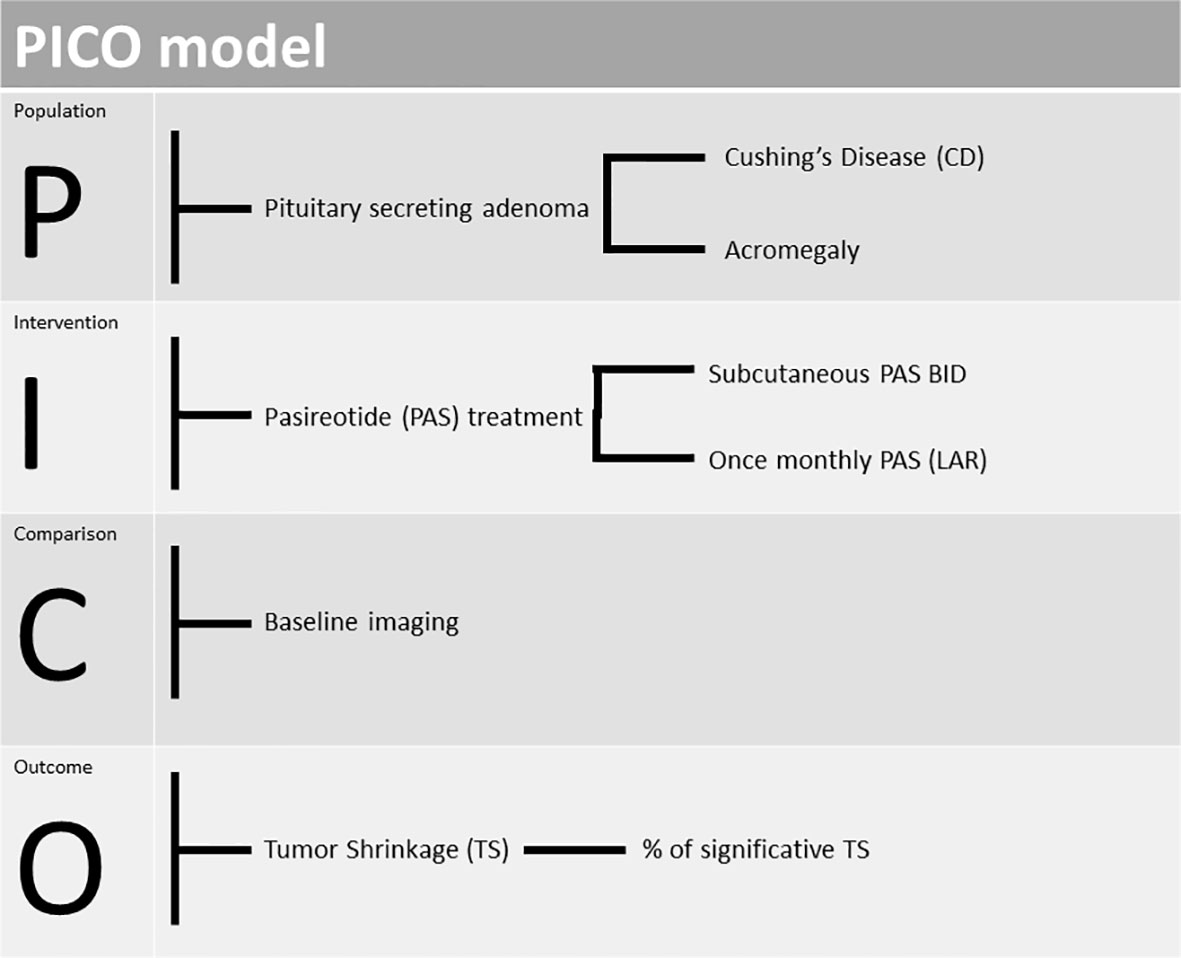
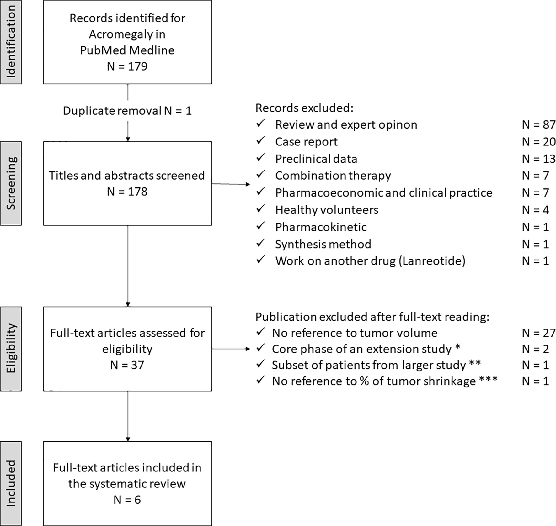
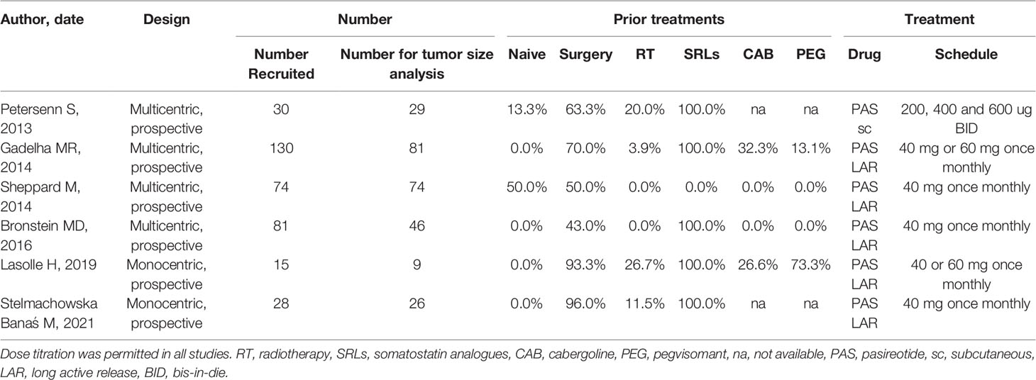
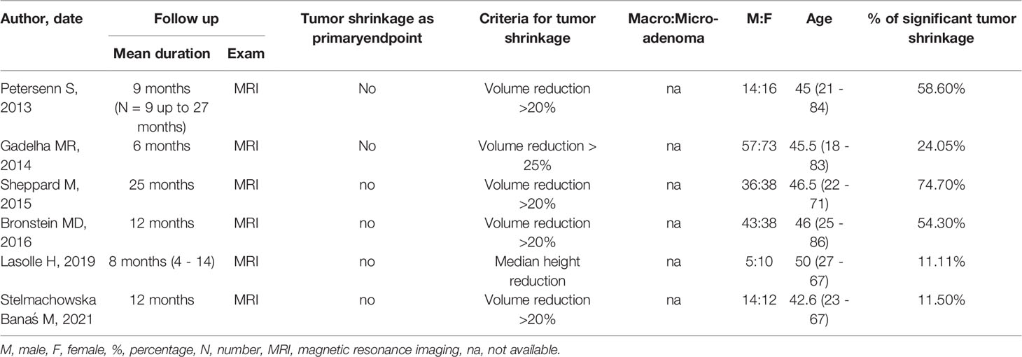
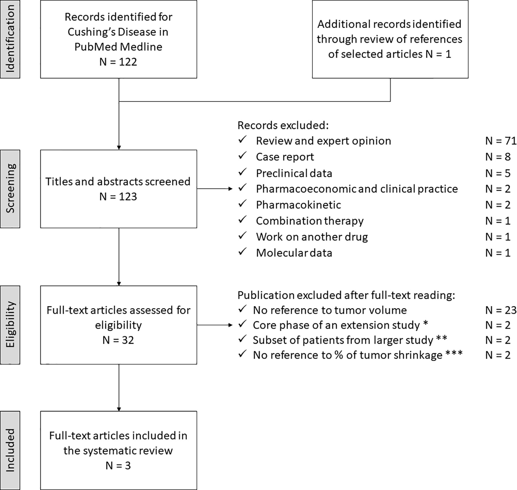
TP53 Mutations in Functional Corticotroph Tumors Are Linked to Invasion and Worse Clinical Outcome

Abstract

Corticotroph macroadenomas are rare but difficult to manage intracranial neoplasms. Mutations in the two Cushing’s disease mutational hotspots USP8 and USP48 are less frequent in corticotroph macroadenomas and invasive tumors. There is evidence that TP53 mutations are not as rare as previously thought in these tumors. The aim of this study was to determine the prevalence of TP53 mutations in corticotroph tumors, with emphasis on macroadenomas, and their possible association with clinical and tumor characteristics. To this end, the entire TP53 coding region was sequenced in 86 functional corticotroph tumors (61 USP8 wild type; 66 macroadenomas) and the clinical characteristics of patients with TP53 mutant tumors were compared with TP53/USP8 wild type and USP8 mutant tumors. We found pathogenic TP53 variants in 9 corticotroph tumors (all macroadenomas and USP8 wild type). TP53 mutant tumors represented 14% of all functional corticotroph macroadenomas and 24% of all invasive tumors, were significantly larger and invasive, and had higher Ki67 indices and Knosp grades compared to wild type tumors. Patients with TP53 mutant tumors had undergone more therapeutic interventions, including radiation and bilateral adrenalectomy. In conclusion, pathogenic TP53 variants are more frequent than expected, representing a relevant amount of functional corticotroph macroadenomas and invasive tumors. TP53 mutations associated with more aggressive tumor features and difficult to manage disease.

Introduction

Pituitary neuroendocrine tumors are the second most common intracranial neoplasm [1]. They are usually benign, but when aggressive they may be particularly difficult to manage, accompanied by high comorbidity and increased mortality [2]. Corticotroph tumors constitute 6–10% of all pituitary tumors, but they represent up to 45% of aggressive pituitary tumors and pituitary carcinomas [2]. Functional corticotroph tumors cause Cushing’s disease (CD), a debilitating condition accompanied by increased morbidity and mortality due to glucocorticoid excess [3]. Pituitary surgery is the first line treatment, but recurrence is observed in 15–20% of cases of whom most are macroadenomas (with a size of ≥ 10 mm) [4]. Treatment options include repeated pituitary surgery, radiation therapy, medical treatment and bilateral adrenalectomy (BADX) [3]. With respect to the latter, corticotroph tumor progression after bilateral adrenalectomy/Nelson’s syndrome (CTP-BADX/NS) is a frequent severe complication and may present with aggressive tumor behavior [5,6,7].

Corticotroph tumors (including CTP-BADX/NS) carry recurrent somatic mutations in the USP8 gene in ~ 40–60% of cases [8,9,10,11,12,13]. These USP8 mutant tumors are usually found in female patients and are generally less invasive [8,9,10,11]. Additional genetic studies identified a second mutational hotspot in the USP48 gene, but no other driver mutations [14,15,16,17,18]. Focusing on USP8 wild type corticotroph tumors, we recently discovered TP53 mutations in 6 out of 18 cases (33%) [17]. Subsequent reports documented TP53 mutations in small series of mainly aggressive corticotroph tumors and carcinomas [1920].

TP53 is the most commonly mutated gene in malignant neoplasms [2122], including brain and neuroendocrine tumors [2324]. Until our previous report [17], TP53 mutations were only described in isolated cases of aggressive pituitary tumors and carcinomas, and were therefore considered very rare events [81625,26,27,28]. A link between TP53 mutations and an aggressive corticotroph tumor phenotype has been hypothesized, but the heterogeneity and small size of the studies reported did not support significant clinical associations [1719].

To address this, we determined the prevalence of TP53 variants in a cohort of 86 patients with functional corticotroph tumors, including 61 with USP8 wild type tumors, and studied the associations between TP53 mutational status and clinical features.

Methods

Patients and samples

We analyzed tumor samples of 86 adult patients: 61 USP8 wild type and 25 USP8 mutant. Sixty-six patients (46 females, 20 males) were diagnosed with CD between 1994 and 2020 in Germany (Hamburg, Munich, Erlangen, and Tübingen) and Luxembourg. Twenty additional patients (16 females, 4 males) were diagnosed with CTP-BADX/NS, operated and followed up in 7 different international centers (Nijmegen, Munich, Erlangen, Hamburg, Paris, Rio de Janeiro, and Würzburg). Twenty-three out of 86 samples were collected prospectively between 2018 and 2021, and 63 were retrospective cases (of which 42 were investigated in the context of USP8 and USP48 screenings and published elsewhere) [9121317]. Seventy-one tumors were fresh frozen and 15 were formalin fixed paraffin embedded. Paired blood was available for 12 cases. The median follow-up time after initial diagnosis was 44 months (range 2–384 months).

Endogenous Cushing’s syndrome was diagnosed according to typical clinical signs and symptoms and established biochemical procedures suggesting glucocorticoid excess. Clinical features included central obesity, moon face, buffalo hump, muscle weakness, easy bruising, striae, acne, low-impact bone fractures, mood changes, irregular menstruation, infertility and impotency. Biochemical diagnosis was based on increased 24 h urinary free cortisol (UFC) and late-night salivary cortisol levels, and lack of serum cortisol suppression after low-dose dexamethasone test. A pituitary ACTH source was confirmed by > 2.2 pmol/l (10 pg/ml) basal plasma ACTH, > 50% suppression of serum cortisol during an 8 mg dexamethasone test, and ACTH and cortisol response to corticotrophin releasing hormone stimulation.

The clinical and pathological features of our study cohort are summarized in Additional file 1: Supplementary Table 1. All patients underwent pituitary surgery. The presence of an ACTH-producing pituitary tumor was confirmed histologically after surgical resection. Biochemical remission after surgery was defined as postoperative 24 h-UFC levels below or within the normal range, or serum cortisol levels < 5 µg/dl after low-dose (1 or 2 mg) dexamethasone suppression test. Tumor control was achieved when there was no evidence of regrowth or disease recurrence. Tumor invasion was defined as radiological or intraoperative evidence of tumor within the sphenoid and/or cavernous sinuses [29]. CTP-BADX/NS was defined as an expanding pituitary tumor after bilateral adrenalectomy (BADX) following expert consensus recommendations [5].

DNA extraction, TP53 amplification and sequencing

Genomic DNA was extracted using the Maxwell Tissue DNA Kit (Promega), Maxwell Blood DNA kit (Promega) or the FFPE DNA mini kit (Qiagen), depending on the type of sample, as described previously [912]. The entire coding sequence of TP53 (including exons 9β and 9γ) as well as noncoding regions adjacent to each exon were amplified using the GoTaq DNA polymerase (Promega) and specific primers (Additional file 1: Supplementary Table 2). Amplification of USP8 hotspot region and Sanger sequencing were performed as described previously [912]. Chromatograms were analyzed using the Mutation Surveyor v4.0.9 (Soft Genetics). Samples were examined for TP53 coding and splicing variants. Variant position and pathogenicity was investigated in ENSEMBL (www.ensembl.org), the UCSC Genome Browser (http://genome-euro.ucsc.edu), the IARC TP53 database (https://p53.iarc.fr/TP53GeneVariations.aspx), the Catalogue Of Somatic Mutations in Cancer (COSMIC; https://cancer.sanger.ac.uk/cosmic), ClinVar (https://www.ncbi.nlm.nih.gov/clinvar/), PHANTM (http://mutantp53.broadinstitute.org/), the Human Splicing Finder (HSF; http://www.umd.be/HSF3/) and VarSEAK splicing predictor (https://varseak.bio/). Variant frequencies on the general population were obtained from the Allele Frequency Aggregator (ALFA) project [30], the Genome Aggregation Database (gnomAD) [31] and the International Genome Sample Resource 1000Genome project [32]. Throughout the text, variants refer to NC_000017.11 (genomic DNA), ENST00000269305.9 (coding DNA) and ENSP00000269305.4 (protein), following the Human Genome Variation Society (HGVS) standard nomenclature system.

Statistical analysis

Statistical analysis was performed with the software package SPSS v24 (IBM). We used t-test or one-way ANOVA to analyze the association of TP53 variants with age, body mass index; Mann–Whitney U and Kruskal–Wallis to test non-parametric variables, such as tumor size, hormone levels, Ki67 index and p53 score. We corrected the analysis for multiple comparisons with the Bonferroni test. Categorical variables were analyzed using a chi-square test or Fisher exact test when needed. Survival analysis was performed using Kaplan–Meier curves with log-rank tests, and multivariate Cox regression. An exact, two-tailed significance level of P < 0.05 was considered to be statistically significant.

Results

Analysis of TP53 nucleotide variants

We analyzed all TP53 coding exons (including exons 9β and 9γ) and adjacent intronic noncoding sequences in 61 USP8 wild type tumors (49 CD and 12 CTP-BADX/NS). Of these, 13 were microadenomas (< 10 mm) and 48 macroadenomas (≥ 10 mm) at the time of the current operation. A separate group of 25 USP8 mutant tumors (17 CD and 8 CTP-BADX/NS) that were mainly macroadenomas (n = 19) was used for multiple comparison.

We found 59 variants in our cohort: 30 exclusively in USP8 wild type, 21 in USP8 mutant, and 8 in wild type and mutant tumors regardless of USP8 mutational status. No indels in the coding region of TP53 were detected. In addition, we did not find any genetic variant affecting TP53 splicing.

Nine out of 30 variants found in USP8 wild type tumors were either reported in the COSMIC database as pathogenic or absent from the common variant databases (1000Genomes, gnomAD, ALPHA) or had allele frequency < 0.0001. They were all described in cancer series: 5 as pathogenic or likely pathogenic in ClinVar, 2 as variants of uncertain significance (VUS) and 2 were not described in ClinVar (Table 1). All variants are reported to alter protein function and show clear loss of transactivation activity in a yeast based assay (Table 1) [33].

Table 1 Functionally relevant TP53 variants found in 9/86 corticotroph tumors

Seven variants target amino acids within the DNA-binding domain, essential for p53 activity, disrupting S2’ and S7 β-sheets or the L3 loop spatial conformation. The other two [c.1009C > G (p.Arg337Gly) and c.1031 T > C (p.Leu344Pro)] locate in the tetramerization domain and keep p53 protein as monomer impairing its transactivation activity [34]. From the 9 variants, 8 affect highly conserved p53 residues, while in c.1031 T > C (p.Met133Lys) the methionine alternates with leucine or valine among species. This variant alters protein folding, probably reducing DNA affinity [35], while the substitution of a methionine that acts as an alternative start codon abolishes the transcription of isoforms ∆133p53α, ∆133p53β and ∆133p53γ. The 9 variants were detected in nine cases (henceforth referred to as TP53 mutant; Table 1). Two tumors from unrelated patients (#6 and #7) carried the same variant c.818G > A (p.Arg273His), while one tumor (#4) carried two variants (c.718A > G and c.773A > C). Seven variants were found in heterozygosis, while the other two (from patients #1 and #2) in homozygosis. From these two, we only had paired blood/tumor samples from patient #1 and detected the variant only on the tumor sample, indicative of loss of heterozygosity (Additional file 1: Supplementary Fig. 1A). Similarly, we could demonstrate the somatic origin of the TP53 variants in four other patients with paired tumor/blood samples (#3, #5, #6 and #9).

The remaining 21/30 variants found in USP8 wild type and all 21 variants found in the USP8 mutant tumors were described as benign, likely benign or VUS with no evidence of affecting protein function. All tumors with these variants were considered TP53 wild type. From the 21 variants found in the USP8 wild type tumors (henceforth referred to as TP53/USP8 wild type group), 7 were non-synonymous variants, 8 synonymous variants and 6 non-coding variants without splicing effect. From the 21 variants found in the 25 USP8 mutant tumors, nine were synonymous, four non-synonymous and eight non-coding without splicing effect. In addition, eight variants were found in tumors regardless of USP8 mutational status that were not categorized as TP53 mutations. The intronic variant c.782 + 62G > A was found in heterozygosis in 6/70 samples. It was not reported in any database and is not predicted to have any splicing effect. The remaining seven are common variants classified as benign or likely benign in ClinVar and their allele frequencies were similar to those reported for the general population (ALFA, gnomAD and 1000Genome project) (Additional file 1: Supplementary Table 3).

Summarizing, all TP53 mutations were found in the USP8 wild type tumors, leading to a prevalence of 15% in this subgroup.

Clinical presentation of patients with TP53 mutant tumors

Patients with TP53 mutant tumors (n = 9) tended to be diagnosed at older age compared to TP53/USP8 wild type tumors (n = 52) (t-test P = 0.069; Table 2). This was significant after including the USP8 mutant group (n = 25) in the multiple comparison analysis (ANOVA P = 0.024, Table 2) and when TP53/USP8 wild type and USP8 mutant tumors were combined to a single group (TP53 wild type, n = 77; Additional file 1: Supplementary Table 4. We did not observe any sex specific predominance of TP53 mutations in contrast to USP8 mutants that are predominantly found in female patients. Furthermore, we did not find any statistically significant differences in ACTH and cortisol levels (Table2; Additional file 1: Supplementary Table 4).

Table 2 Clinical features of TP53 mutant versus TP53/USP8 wild type and USP8 mutant groups

Patients with TP53 mutant tumors underwent more surgeries and tumor resection was more frequently incomplete compared to TP53/USP8 wild type (Table 2). These patients also underwent a higher number of additional therapeutic procedures (radiation, n = 7; BADX, n = 4; temozolomide, n = 3; pasireotide, n = 2). Only one patient (#4) with TP53 mutant tumor, a 77 year-old man, had a single surgery without any other treatment, but his follow-up was short (< 6 months).

We observed TP53 mutations more frequently in CTP-BADX/NS (4/12, 33%) compared to CD (5/49, 10%), trending towards statistically significant difference (Fischer exact test P = 0.065 for TP53 mutant vs. TP53/USP8 wild type, P = 0.060 for comparison among the 3 groups; Table 2).

The TP53 mutant group associated with higher disease-specific mortality and shorter survival than USP8 mutant or TP53/USP8 wild type groups (log rank test, P = 0.023, Fig. 1). Three patients with TP53 mutant tumors (all CTP-BADX/NS) died of disease-related deaths: two from severe cerebral hemorrhage after surgery and stereotactic radiation and one from uncontrolled disease after five failed operations, radiotherapy (gamma knife, fractionated radiation) and chemotherapy (temozolomide, bevacizumab) at the ages of 75, 80 and 37, respectively. Ten-year survival was 27% for patients with TP53 mutant tumors, 100% for TP53/USP8 wild type and 86% for USP8 mutant. In our cohort, survival did not differ after adjusting for age (HR 7.7, 95%CI 0.6–107.7, P = 0.127).

Fig. 1

figure 1

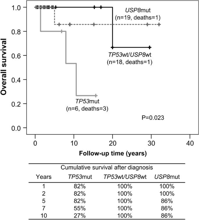
Kaplan–Meier curve showing overall survival in patients with TP53 mutant/USP8 wild type, USP8 mutant/TP53 wild type, and TP53 wild type/USP8 wild type corticotroph tumors. The table underneath the graph shows the 10-year cumulative survival after diagnosis

Tumor samples from prior surgeries were available from one TP53 mutant case (#8, Table 1). This male patient had his first pituitary surgery for CD when he was 30 years old and was treated with γ-knife one year later. He then underwent two more pituitary surgeries and BADX until the age of 35. He developed CTP-BADX/NS with para- and retrosellar tumor extension along with panhypopituitarism and underwent two more pituitary surgeries before dying at the age of 38 due to complications of the disease. We detected the TP53 variant c.1009C > G (p.Arg337Gly) in all available tumor specimens, including his first and latest surgeries (Additional file 1: Supplementary Fig. 1B).

No statistical association was found between clinical data and any of the 8 common variants.

Characteristics of TP53 mutant corticotroph tumors

All TP53 mutations were found in macroadenomas (9/66; Table 3). TP53 mutant tumors were larger that TP53/USP8 wild type (mm median [IQR] 20.0 [14.0] vs. 15.0 [14.3]), but this did not reach statistical significance (Table 3). Multiple comparison analysis showed that the difference in tumor size is significant only comparing TP53 mutant with USP8 mutant (median [IQR] 23.3 [14.0] vs. 14 [7.3] mm; Kruskal–Wallis P = 0.019; Bonferroni corrected P = 0.018).

Table 3 Tumor features of TP53 mutant versus TP53/USP8 wild type and USP8 mutant groups

Parasellar invasion was reported in 34 out of 64 cases, for which this information was available, and it was more common in TP53 mutant tumors (100% vs. 53% and 55% for TP53/USP8 wild type and USP8 mutant, respectively; Fischer exact test P = 0.006). TP53 mutant tumors had higher Knosp grade (Kruskal–Wallis P = 0.011) with the majority being Knosp 4 (Table 3, Additional file 1: Supplementary Table 4).

Ki67 proliferation index was available for 36 cases (6 TP53 mutant). Five out of six TP53 mutant tumors had Ki67 ≥ 3% and the overall Ki67 was higher than in the wild type tumors (Kruskal–Wallis P = 0.01; Bonferroni corrected P = 0.008 for TP53/USP8 wild type) (Table 3). Ki67 ≥ 10% was reported in 6 tumors, from which 5 were TP53 mutant (Fischer exact test P < 0.0001; the remaining case was TP53/USP8 wild type).

We had information on p53 immunostaining from 9 cases (all macroadenomas), four of which TP53 mutant: 3 tumors (from patients #5, 6 and 9) showed high p53 immunoreactivity, while the one (from patient #3) carrying a nonsense variant leading to a truncated protein was p53 negative. The five TP53 wild type cases showed isolated nuclear staining in < 1–3% of cells.

Summarizing, TP53 mutations were significantly associated with features related to a more aggressive tumor behavior, such as incomplete tumor resection, more frequent parasellar invasion, higher Knosp grade, and higher Ki67 proliferation index (Table 3; Additional file 1: Supplementary Table 4).

Discussion

Herein, we investigated the prevalence of TP53 mutations by screening a large cohort of 61 functional corticotroph tumors with USP8 wild type status, and found variants altering protein function in 15% of cases. We did not detect TP53 mutations in a separate group of 25 USP8 mutant tumors, which is in concordance with previously published small next-generation sequencing series [81819].

Since we focused on USP8 wild type tumors, macroadenomas were overrepresented in our cohort. Consequently, it should be noted that the prevalence of TP53 mutations is expected to be lower in the general CD population. In fact, ~ 50% of corticotroph tumors carry USP8 mutations, which others and we have shown to be mutually exclusive. Corticotroph tumors with USP8 mutations are associated with female predominance, younger age at presentation, and less invasiveness (despite shorter time to relapse) [911131836]. In contrast, TP53 mutant tumors were diagnosed mostly at older age, did not show sex predominance and were larger and more invasive, with lower complete resection rate. None of the 19 microadenomas included in our study carried TP53 mutations. Still, we need to acknowledge that since no sample was microdissected we may have lost microadenoma cases with TP53 mutations. Instead, we found TP53 mutations in 9/66 macroadenomas (14%) and 8/34 (24%) invasive tumors, supporting the findings from smaller series [1719].

Tumor size at presentation or invasiveness do not reliably predict aggressiveness. Instead, the European Society of Endocrinology Clinical Practice Guidelines for the management of aggressive pituitary tumors and carcinomas proposed a definition of pituitary tumor aggressiveness based on rapid or clinically relevant tumor growth despite optimal therapeutic options, along with bone invasion [37]. A recent study in a series of 9 aggressive pituitary tumors and carcinomas carrying ATRX mutations reported a high frequency of missense TP53 variants (5/9, 55.6%), further suggesting a link between TP53 mutational status and unfavorable outcome [20]. We do not have exact information on changes of tumor growth for the majority of our cases, but the higher number of surgical and radiation interventions, the higher Knosp grades, and the increased mortality rate indicate that patients with TP53 mutant tumors obviously follow a more aggressive disease course.

Ki67 proliferation index together with p53 immunostaining and mitotic count have been suggested as histological markers of pituitary tumor aggressiveness [2938]. In our series, Ki67 was significantly higher in TP53 mutant tumors, reinforcing our prior observation of a higher proportion of TP53 mutant tumors in the Ki67 ≥ 3 group [17]. We had limited information on p53 immunohistochemistry, since this measure is not routinely performed in our collaborative centers. Nevertheless, in the few tumors with known p53 immunopositivity, it was higher in the TP53 mutant group, which is in concordance with a previous study reporting high p53 immunoreactivity in all TP53 mutant tumors [19].

A mutagenic action of radiation on TP53 has been hypothesized by small series on radiation-induced tumors. For instance, TP53 mutations were reported in 58% of radiation-induced sarcomas [39], while a meta-analysis reported TP53 mutations in 14/30 radiation-induced gliomas [40]. A previous study reported a case with frameshift TP53 mutation in the CTP-BADX/NS tumor, but not in the initial CD surgeries, and the mutation was therefore suspected to be induced by radiotherapy [41]. In our series, however, 4 out of 7 TP53 mutant tumors were obtained before radiation.

In their case report, Pinto et al. suggested that TP53 mutations are acquired during tumorigenesis and condition tumor evolution [41]. In contrast, Casar-Borota et al. and Uzilov et al. reported high allele fraction of TP53 mutations, indicating that they are not a late event in corticotroph tumorigenesis [1920]. In addition, Uzilov et al. reported TP53 mutations in all tumor specimens from their two TP53 mutant cases with multiple surgeries [19]. Similarly, in our series we had tissue from multiple pituitary surgeries from one patient and found the TP53 variant in all samples (CD and CTP-BADX/NS), including specimens obtained before radiotherapy. Taken together, these observations suggest that in most cases, TP53 mutations may appear early during tumor development.

A limitation of our study is the short follow-up of patients who were prospectively included. Moreover, material from repeated surgeries was lacking from most patients with TP53 mutant tumors, hampering the examination of tumor evolution in these patients. Similarly, we had limited access to blood samples, so we could not demonstrate the somatic origin for all variants. Nevertheless, the older age at initial diagnosis of CD in patients with TP53 mutant tumors (53 ± 19.5 years old, with the youngest patient diagnosed at the age of 30) and the absence of additional neoplasias during follow-up also support a somatic instead of a germline origin. Furthermore, conditions related to germline TP53 mutations, such as Li-Fraumeni syndrome, very rarely present with pituitary tumor [42]. To our knowledge, the only published case so far was a pediatric patient with an aggressive lactotroph tumor [43].

In addition to the TP53 mutations, we detected several common variants. Variants rs59758982 and rs1042522 have been associated with increased cancer susceptibility [4445]. In some cancer types, the very frequent rs1042522 c.215G > C (p.Pro72Arg) alternative variant correlated to more efficient induction of apoptosis by DNA-damaging chemotherapeutic drugs, growth suppression and higher metastatic potential [46,47,48]. In nonfunctioning pituitary tumors, alternative allele C (leading to p.Arg72) was related to early age at presentation and reduced p21 expression [49]. Very recently, an overrepresentation of the rs1042522 alternative allele C (p.Arg72) was reported in 9 out of 10 corticotroph neoplasias including 5 functional tumors (allele frequency 0.900, vs 0.714 in Latino/admixed American in gnomAD [31]) without any association with clinical features [50]. In our cohort, we did not detect different allele frequencies in any of the investigated common variants (including rs1042522) compared with public databases, nor statistical association with any clinical variable, rendering their contribution to corticotroph pathophysiology unlikely.

Conclusion

Screening a large corticotroph tumor series revealed that TP53 mutations are more frequent than previously considered. Furthermore, we show that patients with TP53 mutant tumors had higher number of surgeries, more invasive tumors, and worse disease outcome. Our study provides evidence that patients with pathogenic or function altering variants may require more intense treatment and extended follow-up, and suggests screening for TP53 variants in macroadenomas with wild type USP8 status. Further work is needed to determine the potential use of TP53 status as a predictor of disease outcome.

Availability of data and materials

The authors declare that the relevant data supporting the conclusions of this article are included within the article and its supplementary information file. Additional clinical data are available from the corresponding authors MT and LGPR upon reasonable request.

Abbreviations

CD:
Cushing’s disease
BADX:
Bilateral adrenalectomy
CTP-BADX/NS:
Corticotroph tumor progression after bilateral adrenalectomy/Nelson’s syndrome
ACTH:
Adrenocorticotropic hormone
SD:
Standard deviation
IQR:
Interquartile range
HR:
Hazard ratio

References

  1. Ostrom QT, Gittleman H, Truitt G, Boscia A, Kruchko C, Barnholtz-Sloan JS (2018) CBTRUS statistical report: primary brain and other central nervous system tumors diagnosed in the United States in 2011–2015. Neuro Oncol 20:iv1-86

    PubMed PubMed Central Article Google Scholar

  2. McCormack A, Dekkers OM, Petersenn S, Popovic V, Trouillas J, Raverot G et al (2018) Treatment of aggressive pituitary tumours and carcinomas: results of a European society of endocrinology (ESE) survey 2016. Eur J Endocrinol 178:265–276

    CAS PubMed Article Google Scholar

  3. Fleseriu M, Auchus R, Bancos I, Ben-Shlomo A, Bertherat J, Biermasz NR et al (2021) Consensus on diagnosis and management of Cushing’s disease: a guideline update. Lancet Diabetes Endocrinol 9:847–875

    PubMed Article Google Scholar

  4. Dimopoulou C, Schopohl J, Rachinger W, Buchfelder M, Honegger J, Reincke M et al (2013) Long-term remission and recurrence rates after first and second transsphenoidal surgery for Cushing’s disease: care reality in the Munich metropolitan region. Eur J Endocrinol 170:283–292

    PubMed Article CAS Google Scholar

  5. Reincke M, Albani A, Assie G, Bancos I, Brue T, Buchfelder M et al (2021) Corticotroph tumor progression after bilateral adrenalectomy (Nelson’s syndrome): systematic review and expert consensus recommendations. Eur J Endocrinol 184:P1-16

    CAS PubMed PubMed Central Article Google Scholar

  6. Fountas A, Lim ES, Drake WM, Powlson AS, Gurnell M, Martin NM et al (2020) Outcomes of patients with Nelson’s syndrome after primary treatment: a multicenter study from 13 UK pituitary centers. J Clin Endocrinol Metab 105:1527–1537

    Article Google Scholar

  7. Kemink SA, Wesseling P, Pieters GF, Verhofstad AA, Hermus AR, Smals AG (1999) Progression of a Nelson’s adenoma to pituitary carcinoma; a case report and review of the literature. J Endocrinol Invest 22:70–75

    CAS PubMed Article Google Scholar

  8. Reincke M, Sbiera S, Hayakawa A, Theodoropoulou M, Osswald A, Beuschlein F et al (2015) Mutations in the deubiquitinase gene USP8 cause Cushing’s disease. Nat Genet 47:31–38

    CAS PubMed Article Google Scholar

  9. Pérez-Rivas LG, Theodoropoulou M, Ferraù F, Nusser C, Kawaguchi K, Stratakis CA et al (2015) The Gene of the ubiquitin-specific protease 8 is frequently mutated in adenomas causing Cushing’s disease. J Clin Endocrinol Metab 100:E997-1004

    PubMed PubMed Central Article Google Scholar

  10. Ma Z-Y, Song Z-J, Chen J-H, Wang Y-F, Li S-Q, Zhou L-F et al (2015) Recurrent gain-of-function USP8 mutations in Cushing’s disease. Cell Res 25:306–317

    CAS PubMed PubMed Central Article Google Scholar

  11. Hayashi K, Inoshita N, Kawaguchi K, Ardisasmita AI, Suzuki H, Fukuhara N et al (2016) The USP8 mutational status may predict drug susceptibility in corticotroph adenomas of Cushing’s disease. Eur J Endocrinol 174:213–226

    CAS PubMed Article Google Scholar

  12. Pérez-Rivas LG, Theodoropoulou M, Puar TH, Fazel J, Stieg MR, Ferraù F et al (2018) Somatic USP8 mutations are frequent events in corticotroph tumor progression causing Nelson’s tumor. Eur J Endocrinol 178:59–65

    Article Google Scholar

  13. Albani A, Pérez-Rivas LG, Dimopoulou C, Zopp S, Colón-Bolea P, Roeber S et al (2018) The USP8 mutational status may predict long-term remission in patients with Cushing’s disease. Clin Endocrinol (Oxf) 89:454–458

    CAS Article Google Scholar

  14. Bi WL, Horowitz P, Greenwald NF, Abedalthagafi M, Agarwalla PK, Gibson WJ et al (2017) Landscape of genomic alterations in pituitary adenomas. Clin Cancer Res 23:1841–1851

    CAS PubMed Article Google Scholar

  15. Song Z-J, Reitman ZJ, Ma Z-Y, Chen J-H, Zhang Q-L, Shou X-F et al (2016) The genome-wide mutational landscape of pituitary adenomas. Cell Res 26:1255–1259

    CAS PubMed PubMed Central Article Google Scholar

  16. Chen J, Jian X, Deng S, Ma Z, Shou X, Shen Y et al (2018) Identification of recurrent USP48 and BRAF mutations in Cushing’s disease. Nat Commun 9:3171

    PubMed PubMed Central Article CAS Google Scholar

  17. Sbiera S, Perez-Rivas LG, Taranets L, Weigand I, Flitsch J, Graf E et al (2019) Driver mutations in USP8 wild-type Cushing’s disease. Neuro Oncol 21:1273–1283

    CAS PubMed PubMed Central Article Google Scholar

  18. Neou M, Villa C, Armignacco R, Jouinot A, Raffin-Sanson ML, Septier A et al (2020) Pangenomic classification of pituitary neuroendocrine tumors. Cancer Cell 37:123-134.e5

    CAS PubMed Article Google Scholar

  19. Uzilov AV, Taik P, Cheesman KC, Javanmard P, Ying K, Roehnelt A et al (2021) USP8 and TP53 drivers are associated with CNV in a corticotroph adenoma cohort enriched for aggressive tumors. J Clin Endocrinol Metab 106:826–842

    PubMed Article Google Scholar

  20. Casar-Borota O, Boldt HB, Engström BE, Andersen MS, Baussart B, Bengtsson D et al (2021) Corticotroph aggressive pituitary tumors and carcinomas frequently harbor ATRX mutations. J Clin Endocrinol Metab 106:1183–1194

    PubMed Article Google Scholar

  21. Campbell PJ, Getz G, Korbel JO, Stuart JM, Jennings JL, Stein LD et al (2020) Pan-cancer analysis of whole genomes. Nature 578:82–93

    Article CAS Google Scholar

  22. Bouaoun L, Sonkin D, Ardin M, Hollstein M, Byrnes G, Zavadil J et al (2016) TP53 variations in human cancers: new lessons from the IARC TP53 database and genomics data. Hum Mutat 37:865–876

    CAS PubMed Article Google Scholar

  23. Horbinski C, Ligon KL, Brastianos P, Huse JT, Venere M, Chang S et al (2019) The medical necessity of advanced molecular testing in the diagnosis and treatment of brain tumor patients. Neuro Oncol 21:1498–1508

    CAS PubMed PubMed Central Article Google Scholar

  24. van Riet J, van de Werken HJG, Cuppen E, Eskens FALM, Tesselaar M, van Veenendaal LM et al (2021) The genomic landscape of 85 advanced neuroendocrine neoplasms reveals subtype-heterogeneity and potential therapeutic targets. Nat Commun 12:4612

    PubMed PubMed Central Article CAS Google Scholar

  25. Herman V, Drazin NZ, Gonsky R, Melmed S (1993) Molecular screening of pituitary adenomas for gene mutations and rearrangements. J Clin Endocrinol Metab 77:50–55

    CAS PubMed Google Scholar

  26. Levy A, Hall L, Yeudall WA, Lightman SL (1994) p53 gene mutations in pituitary adenomas: rare events. Clin Endocrinol (Oxf) 41:809–814

    CAS Article Google Scholar

  27. Tanizaki Y, Jin L, Scheithauer BW, Kovacs K, Roncaroli F, Lloyd RV (2007) P53 gene mutations in pituitary carcinomas. Endocr Pathol 18:217–222

    CAS PubMed Article Google Scholar

  28. Kawashima ST, Usui T, Sano T, Iogawa H, Hagiwara H, Tamanaha T et al (2009) P53 gene mutation in an atypical corticotroph adenoma with Cushing’s disease. Clin Endocrinol (Oxf) 2009:656–657

    Article Google Scholar

  29. Trouillas J, Roy P, Sturm N, Dantony E, Cortet-Rudelli C, Viennet G et al (2013) A new prognostic clinicopathological classification of pituitary adenomas: a multicentric case-control study of 410 patients with 8 years post-operative follow-up. Acta Neuropathol 126:123–135

    PubMed Article Google Scholar

  30. Phan J, Jin Y, Zhang H, Qiang W, Shekhtman E, Shao D et al (2020) ALFA: allele frequency aggregator: national center for biotechnology information, U.S. National Library of Medicine. Available from www.ncbi.nlm.nih.gov/snp/docs/gsr/alfa/

  31. Karczewski KJ, Francioli LC, Tiao G, Cummings BB, Alföldi J, Wang Q et al (2020) The mutational constraint spectrum quantified from variation in 141,456 humans. Nature 581:434–443

    CAS PubMed PubMed Central Article Google Scholar

  32. Fairley S, Lowy-Gallego E, Perry E, Flicek P (2020) The International genome sample resource (IGSR) collection of open human genomic variation resources. Nucleic Acids Res 48:D941–D947

    CAS PubMed Article Google Scholar

  33. Kato S, Han S-Y, Liu W, Otsuka K, Shibata H, Kanamaru R et al (2003) Understanding the function–structure and function–mutation relationships of p53 tumor suppressor protein by high-resolution missense mutation analysis. Proc Natl Acad Sci 100:8424–8429

    CAS PubMed PubMed Central Article Google Scholar

  34. Kawaguchi T, Kato S, Otsuka K, Watanabe G, Kumabe T, Tominaga T et al (2005) The relationship among p53 oligomer formation, structure and transcriptional activity using a comprehensive missense mutation library. Oncogene 24:6976–6981

    CAS PubMed Article Google Scholar

  35. Greenblatt MS, Chappuis PO, Bond JP, Hamel N, Foulkes WD (2001) TP53 mutations in breast cancer associated with BRCA1 or BRCA2 germ-line mutations: distinctive spectrum and structural distribution. Cancer Res 61:4092–4097

    CAS PubMed Google Scholar

  36. Sesta A, Cassarino MF, Terreni M, Ambrogio AG, Libera L, Bardelli D et al (2020) Ubiquitin-Specific Protease 8 mutant corticotrope adenomas present unique secretory and molecular features and shed light on the role of ubiquitylation on ACTH processing. Neuroendocrinology 110:119–129

    CAS PubMed Article Google Scholar

  37. Raverot G, Burman P, McCormack A, Heaney A, Petersenn S, Popovic V et al (2018) European society of endocrinology clinical practice guidelines for the management of aggressive pituitary tumours and carcinomas. Eur J Endocrinol 178:G1-24

    CAS PubMed Article Google Scholar

  38. Thapar K, Scheithauer BW, Kovacs K, Pernicone PJ, Laws ER (1996) p53 expression in pituitary adenomas and carcinomas: correlation with invasiveness and tumor growth fractions. Neurosurgery 38:765–70

    CAS PubMed Article Google Scholar

  39. Gonin-Laurent N, Gibaud A, Huygue M, Lefèvre SH, Le Bras M, Chauveinc L et al (2006) Specific TP53 mutation pattern in radiation-induced sarcomas. Carcinogenesis 27:1266–1272

    CAS PubMed Article Google Scholar

  40. Whitehouse JP, Howlett M, Federico A, Kool M, Endersby R, Gottardo NG (2021) Defining the molecular features of radiation-induced glioma: a systematic review and meta-analysis. Neuro-Oncol Adv 3:1–16

    Google Scholar

  41. Pinto EM, Siqueira SACC, Cukier P, Fragoso MCBVCBV, Lin CJ, De Mendonca BB et al (2011) Possible role of a radiation-induced p53 mutation in a Nelson’s syndrome patient with a fatal outcome. Pituitary 14:400–404

    PubMed Article Google Scholar

  42. Orr BA, Clay MR, Pinto EM, Kesserwan C (2020) An update on the central nervous system manifestations of Li–Fraumeni syndrome. Acta Neuropathol 139:669–87

    CAS PubMed Article Google Scholar

  43. Birk H, Kandregula S, Cuevas-Ocampo A, Wang CJ, Kosty J, Notarianni C (2022) Pediatric pituitary adenoma and medulloblastoma in the setting of p53 mutation: case report and review of the literature. Childs Nerv Syst. https://doi.org/10.1007/s00381-022-05478-8

    Article Google Scholar

  44. Granja F, Morari J, Morari EC, Correa LAC, Assumpção LVM, Ward LS (2004) Proline homozygosity in codon 72 of p53 is a factor of susceptibility for thyroid cancer. Cancer Lett 210:151–157

    CAS PubMed Article Google Scholar

  45. Sagne C, Marcel V, Amadou A, Hainaut P, Olivier M, Hall J (2013) A meta-analysis of cancer risk associated with the TP53 intron 3 duplication polymorphism (rs17878362): geographic and tumor-specific effects. Cell Death Dis 4:e492

    CAS PubMed PubMed Central Article Google Scholar

  46. Katkoori VR, Jia X, Shanmugam C, Wan W, Meleth S, Bumpers H et al (2009) Prognostic significance of p53 Codon 72 polymorphism differs with race in colorectal adenocarcinoma. Clin Cancer Res 15:2406–2416

    CAS PubMed PubMed Central Article Google Scholar

  47. Dumont P, Leu JIJ, Della Pietra AC, George DL, Murphy M (2003) The codon 72 polymorphic variants of p53 have markedly different apoptotic potential. Nat Genet 33:357–365

    CAS PubMed Article Google Scholar

  48. Basu S, Gnanapradeepan K, Barnoud T, Kung CP, Tavecchio M, Scott J et al (2018) Mutant p53 controls tumor metabolism and metastasis by regulating PGC-1α. Genes Dev 32:230–243

    CAS PubMed PubMed Central Article Google Scholar

  49. Yagnik G, Jahangiri A, Chen R, Wagner JR, Aghi MK (2017) Role of a p53 polymorphism in the development of nonfunctional pituitary adenomas. Mol Cell Endocrinol 446:81–90

    CAS PubMed PubMed Central Article Google Scholar

  50. Andonegui-Elguera S, Silva-Román G, Peña-Martínez E, Taniguchi-Ponciano K, Vela-Patiño S, Remba-Shapiro I et al (2022) The genomic landscape of corticotroph tumors: from silent adenomas to ACTH-secreting carcinomas. Int J Mol Sci. 23:4861

    CAS PubMed PubMed Central Article Google Scholar

Download references

Funding

Open Access funding enabled and organized by Projekt DEAL. The study was supported by the Deutsche Forschungsgemeinschaft (DFG) (Project number: 314061271-TRR 205 to MF, MR and MT; FA 466/5-1 to MF; DE 2657/1-1 to TD), Metiphys program of the LMU Medical Faculty (to AA), Else Kröner-Fresenius Stiftung (Project number: 2012_A103 and 2015_A228 to MR) and Fundação de Amparo à Pesquisa do Estado do Rio de Janeiro (FAPERJ; Project number: E-26/211.294/2021 to MRG).

Author information

Authors and Affiliations

  1. Medizinische Klinik und Poliklinik IV, Klinikum der Universität München, Ludwig-Maximilians-Universität München, Munich, GermanyLuis Gustavo Perez-Rivas, Julia Simon, Adriana Albani, Sicheng Tang, Günter K. Stalla, Martin Reincke & Marily Theodoropoulou
  2. Center for Neuropathology and Prion Research, Ludwig-Maximilians-Universität München, Munich, GermanySigrun Roeber & Jochen Herms
  3. Department of Endocrinology, Center for Rare Adrenal Diseases, Assistance Publique-Hôpitaux de Paris, Hôpital Cochin, Paris, FranceGuillaume Assié
  4. Université de Paris, Institut Cochin, Inserm U1016, CNRS UMR8104, F-75014, Paris, FranceGuillaume Assié
  5. Division of Endocrinology and Diabetes, Department of Internal Medicine I, University Hospital, University of Würzburg, Würzburg, GermanyTimo Deutschbein & Martin Fassnacht
  6. Medicover Oldenburg MVZ, Oldenburg, GermanyTimo Deutschbein
  7. Division of Endocrinology, Hospital Universitário Clementino Fraga Filho, Rio de Janeiro, BrazilMonica R. Gadelha
  8. Division of Endocrinology, Department of Internal Medicine, Radboud University Medical Centre, Nijmegen, The NetherlandsAd R. Hermus
  9. Medicover Neuroendocrinology, Munich, GermanyGünter K. Stalla
  10. Service d’Endocrinologie, Centre Hospitalier du Nord, Ettelbruck, LuxembourgMaria A. Tichomirowa
  11. Department of Neurosurgery, Universitätskrankenhaus Hamburg-Eppendorf, Hamburg, GermanyRoman Rotermund & Jörg Flitsch
  12. Department of Neurosurgery, University of Erlangen-Nürnberg, Erlangen, GermanyMichael Buchfelder
  13. Department of Neurosurgery, University of Tübingen, Tübingen, GermanyIsabella Nasi-Kordhishti & Jürgen Honegger
  14. Neurochirurgische Klinik und Poliklinik, Klinikum der Universität München, Ludwig-Maximilians-Universität München, Munich, GermanyJun Thorsteinsdottir
  15. Institute of Neuropathology, University Medical Center Hamburg-Eppendorf, Hamburg, GermanyWolfgang Saeger

Contributions

LPGR and MT designed the study. LPGR, JS, AA and ST implemented the study. LGPR did the data analysis. SR, GA, TD, MF, MRG, ARH, GKS, MAT, RR, JF, MB, INK, JH, JT, WS, JH and MR provided patient materials and data. LGPR and MT interpreted the data and composed the main draft of the manuscript. All authors have seen, corrected and approved the final draft.

Corresponding authors

Correspondence to Luis Gustavo Perez-Rivas or Marily Theodoropoulou.

Ethics declarations

Ethics approval and consent to participate

The study was performed in accordance with the Declaration of Helsinki and was approved by the ethics committee of the LMU Munich (Nr. 643-16). All patients provided written informed consent.

Competing interests

The authors declare that they have no competing interests.

Additional information

Publisher’s Note

Springer Nature remains neutral with regard to jurisdictional claims in published maps and institutional affiliations.

Supplementary Information

Additional file 1 of TP53 mutations in functional corticotroph tumors are linked to invasion and worse clinical outcome

Skip to file navigationSkip to generic navigation
1
Supplementary Table 1
. Description of study cohort.
Variable
mean/median
SD/IQR
Total n
Age at diagnosis (years), mean ±SD, [total n]
42
±15.2
86
Sex (female), n (%), [total n]
62
(72%)
86
BMI (kg/m2), mean ±SD, [total n]
28.9
±6.3
74
Disease presentation, n (%), [total n]
86
Cushing
66
(77%)
Nelson
20
(23%)
Number of prior pituitary surgeries, n (%), [total n]
80
0
50
(63%)
1
23
(29%)
≥2
7
(9%)
Total
number of pituitary surgeries, n (%), [total n]
82
1
46
(56%)
2
23
(28%)
≥3
13
(16%)
Complete tumor resection, n (%), [total n]
32
(60%)
53
Postoperative remission, n (%), [total n]
46
(59%)
78
Postoperative tumor control, n (%), [total
n]
34
(60%)
57
Radiation therapy, n (%), [total n]
24
(34%)
70
Radiation therapy before sample collection, n (%), [total n]
7
(13%)
53
Bilateral adrenalectomy, n (%), [total n]
23
(27%)
86
Pharmacological treatments
a
,
n (%), [total n]
18
(42%)
43
Preoperative hormone levels
Plasma ACTH (pg/mL), median (IQR)
98
(570.4)
75
Serum cortisol (
μ
g/dl), median (range)
29.1
(168.6)
50
24h
urinary free cortisol (
μ
g/24h), median (range)
432.5
(598.3)
30
Serum cortisol after low
dose DST (
μ
g/dl),
median (IQR)
20
(20.7)
46
Postoperative hormone levels
Plasma ACTH (pg/mL), median (IQR)
20
(107.6)
57
Serum cortisol nadir (
μ
g/dl), median (range)
8.8
(19.4)
58
Tumo
r size (mm), median (IQR), [total n]
15
(13.0)
85
Microadenoma
19
(22%)
Macroadenoma
66
(78%)
Granulation, n (%), [total n]
30
Sparsely
9
(30%)
Densely
21
(70%)
Ki67 index, median (IQR), [total n]
2.0
(3.8)
36
Ki67 index ≥3%, n (%)
14
(39%)
36
p53 positivity, median (IQR), [total n]
1
(26.5)
9
Invasion, n (%),
[total n]
34
(53%)
64
Hardy grade, n (%), [total n]
61
1
13
(21%)
2
22
(36%)
3
18
(30%)
4
8
(13%)
Knosp grade, n (%), [total n]
35
0
5
(14%)
1
12
(34%)
2
3
(9%)
3
7
(20%)
4
8
(7%)
Disease
specific death, n (%), [total
n]
5
(9%)
58
a
Pharmacological treatments: pasireotide (n=6), ketoconazole (n=5), mitotane (n=5), temozolamide
(n=4) metyrapone (n=5), cabergoline (n=3), bevazizumab (n=1). Five patients received >1
pharmacological agent.
2
Supplementary Table 2
. Primers used for
TP53
amplification and Sanger sequencing.
Primer
Sequence
DNA source
TP53
1
5′
TCTCATGCTGGATCCCCACT
3′
FF, FFPE
TP53
1rv
5′
GACCAGGTCCTCAGCC
3′
FFPE
TP53
2fw
5′
GGGGGCTGAGGACCTGGT
3′
FFPE
TP53
2rv
5′
ATACGGCCAGGCATTGAAGT
3′
FFPE
TP53
2
5′
AGAGGAATCCCAAAGTTCCA
3′
FF
TP53
3
5′
GTGCCCTGACTTTCAACTC
3′
FF, FFPE
TP53
3rv
5′
GGCAACCAGCCCTGTC
3′
FFPE
TP53
4fw
5′
GCCTCTGATTCCTCACTGAT
3′
FFPE
TP53
4
5′
CAGGAGAAAGCCCCCCTACT
3′
FF, FFPE
TP53
5
5′
CTTGCCACAGGTCTCCCCAA
3′
FF, FFPE
TP53
6
5′
AGGGGTCAGAGGCAAGCAGA
3′
FF, FFPE
TP53
7
5′
TAGGACCTGATTTCCTTA
3′
FF, FFPE
TP53
7rv
5′
AGTGAATCTGAGGCATAAC
3′
FFPE
TP53
7Bfw
5′
TGGAGGAGACCAAGGGTG
3′
FFPE
TP53
7Brv
5′
CGGCATTTTGAGTGTTAGAC
3′
FFPE
TP53
8
5′
TAAGCTATGATGTTCCTTAG
3′
FF, FFPE
TP53
8rv
5′
GACTGTTTTACCTGCAATTG
3′
FFPE
TP53
9
5′
CAATTGTAACTTGAACCATC
3′
FF, FFPE
TP53
10
5′
GGATGAGAATGGAATCCTAT
3′
FF, FFPE
TP53
11
5′
TCTCACTCATGTGATGTCATC
3′
FF, FFPE
TP53
12
5′
CACACCTATTGCAAGCAAGG
3′
FF, FFPE
FF, fresh frozen; FFPE, formalin
fixed
paraffin embedded.

Additional file 1

Supplementary Table 1: Description of study cohort. Supplementary Table 2: Primers used for TP53 amplification and Sanger sequencing. Supplementary Table 3: Common TP53 variants in the study cohort. Supplementary Table 4: Comparison of TP53 mutant versus TP53 wild type group. Supplementary Figure 1. Chromatograms showing the TP53 variants found in the corticotroph tumor of patient #1 and #8 (Table 1). A. The variant c.398T>A was present in homozygocity in the tumor and absent in the blood. B. The variant c.1009C>G is detected in all available surgical specimens in this patient. First and 2nd surgeries were Cushing’s disease tumors and 4th and 5th CTP-BADX/NS.


Persistent vs Recurrent Cushing’s Disease Diagnosed Four Weeks Postpartum

Abstract

Background. Cushing’s disease (CD) recurrence in pregnancy is thought to be associated with estradiol fluctuations during gestation. CD recurrence in the immediate postpartum period in a patient with a documented dormant disease during pregnancy has never been reported. Case Report. A 30-year-old woman with CD had improvement of her symptoms after transsphenoidal resection (TSA) of her pituitary lesion. She conceived unexpectedly 3 months postsurgery and had no symptoms or biochemical evidence of recurrence during pregnancy. After delivering a healthy boy, she developed CD 4 weeks postpartum and underwent a repeat TSA. Despite repeat TSA, she continued to have elevated cortisol levels that were not well controlled with medical management. She eventually had a bilateral adrenalectomy. Discussion. CD recurrence may be higher in the peripartum period, but the link between pregnancy and CD recurrence and/or persistence is not well studied. Potential mechanisms of CD recurrence in the postpartum period are discussed below. Conclusion. We describe the first report of recurrent CD that was quiescent during pregnancy and diagnosed in the immediate postpartum period. Understanding the risk and mechanisms of CD recurrence in pregnancy allows us to counsel these otherwise healthy, reproductive-age women in the context of additional family planning.

1. Introduction

Despite a relatively high prevalence of Cushing’s syndrome (CS) in women of reproductive age, it is rare for pregnancy to occur in patients with active disease [1]. Hypercortisolism leads to infertility through impairment of the hypothalamic gonadal axis. Additionally, while Cushing’s disease (CD) is the leading etiology of CS in nonpregnant adults, it is less common in pregnancy, accounting for only 30–40% of the CS cases in pregnant women [2]. It has been suggested that in CD there is hypersecretion of both cortisol and androgens, impairing fertility to a greater extent, while in CS of an adrenal origin, hypersecretion is almost exclusively of cortisol with minimal androgen production [3]. Regardless of the cause, active CS in pregnancy is associated with a higher maternal and fetal morbidity, hence, prompt diagnosis and treatment are essential.

Pregnancy is considered a physiological state of hypercortisolism, and the peripartum period is a common time for women to develop CD [34]. A recent study reported that 27% of reproductive-age women with CD had onset associated with pregnancy [4]. The high rate of pregnancy-associated CD suggests that the stress of pregnancy and peripartum pituitary corticotroph hyperstimulation may promote or accelerate pituitary tumorigenesis [46]. During pregnancy, the circulating levels of corticotropin-releasing hormone (CRH) in the plasma increase exponentially as a result of CRH production by the placenta, decidua, and fetal membranes rather than by the hypothalamus. Unbound circulating placental CRH stimulates pituitary ACTH secretion and causes maternal plasma ACTH levels to rise [4]. A review of the literature reveals many studies of CD onset during the peripartum period, but CD recurrence in the peripartum period has only been reported a handful of times [710]. Of these, most cases recurred during pregnancy. CD recurrence in the immediate postpartum period has only been reported once [7]. Below, we report for the first time a case of CD recurrence that occurred 4 weeks postpartum, with a documented dormant disease throughout pregnancy.

2. Case Presentation

A 30-year-old woman initially presented with prediabetes, weight gain, dorsal hump, abdominal striae, depression, lower extremity weakness, and oligomenorrhea with a recent miscarriage 10 months ago. Diagnostic tests were consistent with CD. Results included the following: three elevated midnight salivary cortisols: 0.33, 1.38, and 1.10 μg/dL (<0.010–0.090); 1 mg dexamethasone suppression test (DST) with cortisol 14 μg/dL (<1.8); elevated 24 hr urine cortisol (UFC) measuring 825 μg/24 hr (6–42); ACTH 35 pg/mL (7.2–63.3). MRI of the pituitary gland revealed a left 4 mm focal lesion (Figure 1(a)). After transsphenoidal resection (TSA), day 1, 2, and 3 morning cortisol values were 18, 5, and 2 μg/dL, respectively. Pathology did not show a definitive pituitary neoplasm. She was rapidly titrated off hydrocortisone (HC) by six weeks postresection. Her symptoms steadily improved, including improved energy levels, improved mood, and resolution of striae. She resumed normal menses and conceived unexpectedly around 3 months post-TSA. Hormonal evaluation completed a few weeks prior to her pregnancy indicated no recurrence: morning ACTH level, 27.8 pg/mL; UFC, 5 μg/24 hr; midnight salivary cortisol, 0.085 and 0.014 μg/dL. Her postop MRI at that time did not show a definitive adenoma (Figure 1(b)). During pregnancy, she had a normal oral glucose tolerance test at 20 weeks and no other sequela of CD. Every 8 weeks, she had 24-hour urine cortisol measurements. Of these, the highest was 93 μg/24 hr at 17 weeks and none were in the range of CD (Table 1). Towards the end of her 2nd trimester, she started to complain of severe fatigue. Given her low 24 hr urine cortisol level of 15 μg/24 hr at 36 weeks gestation, she was started on HC. She underwent a cesarean section at 40 weeks gestation for oligohydramnios and she subsequently delivered a healthy baby boy weighing 7.6 pounds with APGAR scores at 1 and 5 minutes being 9 and 9. HC was discontinued immediately after delivery. Around four weeks postpartum she developed symptoms suggestive for CD. Diagnostic tests showed an elevated midnight salivary cortisol of 0.206 and 0.723 μg/dL, and 24-hour urine cortisol of 400 μg/24 hr. MRI pituitary illustrated a 3 mm adenoma in the left posterior region of the gland, which was thought to represent a recurrent tumor (Figure 1(c)). A discrete lesion was found and resected during repeat TSA. Pathology confirmed corticotroph adenoma with MIB-1 < 3%. On postoperative days 1, 2, and 3, the cortisol levels were 26, 10, and 2.8 μg/dL, respectively. She was tapered off HC within one month. Her symptoms improved only slightly and she continued to report weight gain, muscle weakness, and fatigue. Three months after repeat TSA, biochemical data showed 1 out of 2 midnight salivary cortisols elevated at 0.124 μg/dL and elevated urine cortisol of 76 μg/24 hr. MRI pituitary demonstrated a 3 × 5 mm left enhancement, concerning for residual or enlarged persistent tumor. Subsequent lab work continued to show a biochemical excess of cortisol, and the patient was started on metyrapone but reported no significant improvement of her symptoms and only mild improvement of excess cortisol. After a multidisciplinary discussion, the patient made the decision to pursue bilateral adrenalectomy, as she refused further medical management and opted against radiation given the risk of hypogonadism.

(a)
(a)
(b)
(b)
(c)
(c)
(a)
(a)(b)
(b)(c)
(c)
Figure 1 
(a) Initial: MRI pituitary with and without contrast showing a coronal T1 postcontrast image immediately prior to our patient’s pituitary surgery. The red arrow points to a 3 × 3 × 5 mm hypoenhancing focus representing a pituitary microadenoma. (b) Postsurgical: MRI pituitary with and without contrast showing a coronal T1 postcontrast image obtained three months after transsphenoidal pituitary surgery. The red arrow shows that a hypoenhancing focus is no longer seen and has been resected. (c) Postpartum: MRI pituitary with and without contrast showing a coronal T1 postcontrast image obtained four weeks postpartum. The red arrow points to a 3 mm relatively hypoenhancing lesion representing a recurrent pituitary adenoma.
Table 1 
24-hour urine-free cortisol measurements collected approximately every 8 weeks throughout our patient’s pregnancy.

3. Discussion

The symptoms and signs of Cushing’s syndrome overlap with those seen in normal pregnancy, making diagnosis of Cushing’s disease during pregnancy challenging [1]. Potential mechanisms of gestational hypercortisolemia include increased systemic cortisol resistance during pregnancy, decreased sensitivity of plasma ACTH to negative feedback causing an altered pituitary ACTH setpoint, and noncircadian secretion of placental CRH during pregnancy causing stimulation of the maternal HPA axis [5]. Consequently, both urinary excretion of cortisol and late-night salivary cortisol undergo a gradual increase during normal pregnancy, beginning at the 11th week of gestation [2]. Cushing’s disease is suggested by 24-hour urinary-free cortisol levels greater than 3-fold of the upper limit of normal [2]. It has also been suggested that nocturnal salivary cortisol be used to diagnose Cushing’s disease by using the following specific trimester thresholds: first trimester, 0.25 μg/dL; second trimester, 0.26 μg/dL; third trimester 0.33, μg/dL [11]. By these criteria, our patient had no signs or biochemical evidence of CD during pregnancy but developed CD 4 weeks postpartum.

A recent study by Tang et al. proposed that there may be a higher risk of developing CD in the peripartum period, but did not test for CD during pregnancy, and therefore was not able to definitively say exactly when CD onset occurred in relation to pregnancy [4]. Previous literature suggests that there may be a higher risk of ACTH-secreting pituitary adenomas following pregnancy as there is a significant surge of ACTH and cortisol hormones at the time of labor. This increased stimulation of the pituitary corticotrophs in the immediate postpartum period may promote tumorigenesis [6]. It has also been suggested that the hormonal milieu during pregnancy may cause accelerated growth of otherwise dormant or small slow-growing pituitary corticotroph adenomas [45]. However, the underlying mechanisms of CD development in the postpartum period have yet to be clarified. We highlight the need for more research to investigate not only the development, but also the risk of CD recurrence in the postpartum period. Such research would be helpful for family planning.

4. Conclusion

Hypothalamic-pituitary-adrenal axis activation during pregnancy and the immediate postpartum period may result in higher rates of CD recurrence in the postpartum period, as seen in our patient. In general, more testing for CS in all reproductive-age females with symptoms suggesting CS, especially during and after childbirth, is necessary. Such testing can also help us determine when CD occurred in relation to pregnancy, so that we can further understand the link between pregnancy and CD occurrence, recurrence, and/or persistence. Learning about the potential mechanisms of CD development and recurrence in pregnancy will help us to counsel these reproductive-age women who desire pregnancy.

Abbreviations

CD: Cushing’s disease
TSA: Transsphenoidal resection
DST: Dexamethasone suppression test
ACTH: Adrenocorticotropic hormone
MRI: Magnetic-resonance imaging
HC: Hydrocortisone
CTH: Corticotroph-releasing hormone
HPA: Hypothalamic-pituitary-adrenal.

Data Availability

The data used to support the findings of this study are included within the article.

Additional Points

Note. Peripartum refers to the period immediately before, during, or after pregnancy and postpartum refers to any period after pregnancy up until 1 year postdelivery.

Disclosure

This case report is a follow up to an abstract that was presented in ENDO 2020 Abstracts. https://doi.org/10.1210/jendso/bvaa046.2128.

Conflicts of Interest

The authors declare that they have no conflicts of interest.

Acknowledgments

The authors thank Dr. Puneet Pawha for his help in reviewing MRI images and his suggestions.

References

  1. J. R. Lindsay and L. K. Nieman, “The hypothalamic-pituitary-adrenal axis in pregnancy: challenges in disease detection and treatment,” Endocrine Reviews, vol. 26, no. 6, pp. 775–799, 2005.View at: Publisher Site | Google Scholar
  2. W. Huang, M. E. Molitch, and M. E. Molitch, “Pituitary tumors in pregnancy,” Endocrinology and Metabolism Clinics of North America, vol. 48, no. 3, pp. 569–581, 2019.View at: Publisher Site | Google Scholar
  3. M. C. Machado, M. C. B. V. Fragoso, M. D. Bronstein, and M. Delano, “Pregnancy in patients with cushing’s syndrome,” Endocrinology and Metabolism Clinics of North America, vol. 47, no. 2, pp. 441–449, 2018.View at: Publisher Site | Google Scholar
  4. K. Tang, L. Lu, M. Feng et al., “The incidence of pregnancy-associated Cushing’s disease and its relation to pregnancy: a retrospective study,” Frontiers in Endocrinology, vol. 11, p. 305, 2020.View at: Publisher Site | Google Scholar
  5. S. K. Palejwala, A. R. Conger, A. A. Eisenberg et al., “Pregnancy-associated Cushing’s disease? an exploratory retrospective study,” Pituitary, vol. 21, no. 6, pp. 584–592, 2018.View at: Publisher Site | Google Scholar
  6. G. Mastorakos and I. Ilias, “Maternal and fetal hypothalamic-pituitary-adrenal axes during pregnancy and postpartum,” Annals of the New York Academy of Sciences, vol. 997, no. 1, pp. 136–149, 2003.View at: Publisher Site | Google Scholar
  7. G. F. Yaylali, F. Akin, E. Yerlikaya, S. Topsakal, and D. Herek, “Cushing’s disease recurrence after pregnancy,” Endocrine Abstracts, vol. 32, 2013.View at: Publisher Site | Google Scholar
  8. C. V. L. Fellipe, R. Muniz, L. Stefanello, N. M. Massucati, and L. Warszawski, “Cushing’s disease recurrence during peripartum period: a case report,” Endocrine Abstracts, vol. 70, 2020.View at: Publisher Site | Google Scholar
  9. P. Recinos, M. Abbassy, V. Kshettry et al., “Surgical management of recurrent Cushing’s disease in pregnancy: a case report,” Surgical Neurology International, vol. 6, no. 26, pp. S640–S645, 2015.View at: Publisher Site | Google Scholar
  10. A. Nakhleh, L. Saiegh, M. Reut, M. S. Ahmad, I. W. Pearl, and C. Shechner, “Cabergoline treatment for recurrent Cushing’s disease during pregnancy,” Hormones, vol. 15, no. 3, pp. 453–458, 2016.View at: Publisher Site | Google Scholar
  11. L. M. L. Lopes, R. P. V. Francisco, M. A. K. Galletta, and M. D. Bronstein, “Determination of nighttime salivary cortisol during pregnancy: comparison with values in non-pregnancy and cushing’s disease,” Pituitary, vol. 19, no. 1, pp. 30–38, 2015.View at: Publisher Site | Google Scholar

Copyright © 2022 Leena Shah et al. This is an open access article distributed under the Creative Commons Attribution License, which permits unrestricted use, distribution, and reproduction in any medium, provided the original work is properly cited.

From https://www.hindawi.com/journals/crie/2022/9236711/

Ectopic Adrenocorticotropic Hormone-Secreting Pituitary Adenoma in the Clivus Region: A Case Report

Yan Zhang, Danrong Wu, Ruoqiu Wang, Min Luo, Dong Wang, Kaiyue Wang, Yi Ai, Li Zheng, Qiao Zhang, Lixin Shi

Department of Endocrinology and Metabolism, Guiqian International General Hospital, Guiyang, People’s Republic of China

Correspondence: Qiao Zhang; Lixin Shi, Department of Endocrinology and Metabolism, Guiqian International General Hospital, Guiyang, People’s Republic of China, Tel/Fax +86 851-86277666, Email endocrine_zq@126.com; slx1962@medmail.com.cn

Abstract: Ectopic pituitary adenoma (EPA) is a pituitary adenoma unrelated to the intrasellar component and is an extremely rare disease. EPA resembles typical pituitary adenomas in morphology, immunohistochemistry, and hormonal activity, and it may present with specific or non-specific endocrine manifestations. Here, we report a rare case of ectopic adrenocorticotropic hormone (ACTH)-secreting pituitary adenoma in the clival region. Only three patients with ACTH-secreting pituitary adenomas occurring in the clivus have been previously reported, and the present case was diagnosed as a clivus-ectopic ACTH-secreting pituitary macroadenoma. Thus, in addition to the more common organs, such as the lung, thymus, and pancreas, in the diagnosis of ectopic ACTH syndrome, special attention should be paid to the extremely rare ectopic ACTH-secreting pituitary adenoma of the clivus region.

Keywords: ectopic pituitary adenoma, Cushing’s syndrome, clivus, adrenocorticotropic hormone, endocrine

Introduction

The diagnosis of Cushing’s syndrome (CS), particularly its localization diagnosis, has always been a challenge in clinical practice.1,2 Endogenous CS can be divided into adrenocorticotropic hormone (ACTH)-dependent and non-ACTH dependent with the former accounting for 70% of CS cases. Ectopic ACTH syndrome accounts for 5–10% of CS cases, and its lesions are mainly located in the lungs, thymus, pancreas, and the thyroid gland.3 Finding such lesions in non-pituitary intracranial regions is extremely rare, and ectopic ACTH in the clivus region is even rarer. To date, less than 60 cases of ectopic ACTH-secreting pituitary adenomas have been reported,4 and determining their localization is a formidable challenge in CS diagnosis. It is difficult to make an accurate and prompt diagnosis of ectopic ACTH-secreting pituitary adenoma caused by hypercortisolism based on its clinical manifestation, routine laboratory tests, and radiologic examinations.1,4 Ectopic pituitary adenomas (EPAs) are mainly concentrated in the sphenoid sinus, suprasellar region, and cavernous sinus, and rare regions include the clivus, ethmoid sinus, and nasal cavity.5 A literature review showed that only three cases of primary EPA in the clivus region have been reported worldwide.6–8 Recently, we diagnosed a patient with ectopic ACTH-secreting pituitary macroadenoma in the clivus region that was confirmed by surgery and immunohistochemistry.

Case Presentation

A 53-year-old female patient sought medical attention at our hospital for hypertension, headache, and dizziness with a blood pressure as high as 180/100 mmHg. Her medical history showed that she had developed similar symptoms 2 years ago. At that time, she had hypertension (180/100 mmHg), headache, and dizziness, and she was treated with amlodipine (5 mg per day), benazepril hydrochloride (10 mg per day), and metoprolol tartrate (50 mg per day). The patient was not hospitalized for treatment and did not undergo systemic examination. Three months before admission, the patient had a thoracic vertebrae fracture caused by moving heavy objects. One month before admission, she had a bilateral rib fracture due to falling on flat ground. Her physical examination results were as follows: blood pressure, 160/85 mmHg; height, 147 cm; weight, 55.2 kg; and body mass index (BMI), 25.54 kg/m2. In the physical examination, moon facies, buffalo hump, concentric obesity, facial plethora, and large patches of ecchymosis at the blood sampling site were observed. Purple striae were absent below the axilla, abdomen, and limbs. Her hematological examination results were as follows: cortisol (COR) rhythm with 33.52 µg/dL (reference range: 4.26–24.85) at 8:00 AM, 34.3 µg/dL at 4:00 PM, and 33.14 µg/dL at 12:00 AM; 1 mg dexamethasone overnight suppression test indicated 22.21 µg/dL COR at 8:00 AM; 24 h urine COR was 962.16 µg/24 h (reference range: 50–437 µg/24 h); 8:00 AM ACTH at two different times was 74 pg/mL and 90.8 pg/mL (reference range: <46); high-dose dexamethasone suppression test (HDDST) was 21.44 µg/dL COR (serum COR level was not suppressed by more than 50%); serum potassium was 3.38 mmol/L (reference range: 3.5–5.5); insulin-like growth factor-1 (IGF-1) was 106.6 ng/mL (reference range: 84–236); serum luteinizing hormone (LH) was <0.07 IU/L (reference range: 1.9–12.5); serum follicle stimulating hormone (FSH) was 0.37 IU/L (reference range: 2.5–10.2); prolactin (PRL), testosterone, progesterone, and estradiol test results were normal; FT4 was 8.25 pmol/L (reference range: 10.44–24.38); TSH was 1.116 mIU/L (reference range: 0.55–4.78); oral glucose tolerance test (OGTT) indicated that fasting blood glucose was 6.3 mmol/L and 2-h blood glucose was 18.72 mmol/L; and glycated hemoglobin (HbA1c) was 7.1%. A bone mineral density test suggested osteoporosis (dual energy X-rays: L1-L4 T values were −3.4).

Magnetic resonance (MR) scans were performed using a SIGNA Pioneer 3.0T (GE Healthcare, Waukesha, WI, USA), and computed tomography (CT) scans were performed using a 256 slice CT scanner (Revolution CT; GE Healthcare, Waukesha, WI, USA). The enhanced MR scan of the sellar lesion showed a soft tissue mass with abnormal signals in the occipital bone clivus. T1WI showed an isointense signal, and T2WI showed an isointense/slightly hyperintense signal in a large area of approximately 30 mm × 46 mm. The lesion extended anteriorly to completely fill the entire sphenoidal sinus, and it was in a close proximity to the right internal carotid arteries. Significant invasion, liquefaction, and necrosis were not observed in the bilateral cavernous sinuses. Pituitary gland morphology was normal with a superoinferior diameter of 3.14 mm, and the pituitary gland was located in the center. An occipital bone clival space-occupying lesion was considered with a tendency of low malignancy and a possibility of chordoma (Figure 1A–C). Non-enhanced high-resolution CT scans of the nasal sinuses showed osteolytic destruction, and a soft tissue mass was observed in the occipital bone clivus. The mass had a large area of 20 mm × 30 mm × 46 mm (Figure 1D). Enhanced CT of the adrenals showed bilateral adrenal gland hyperplasia.

Figure 1 (A) MR T1+T2 scan (transverse view). MR T1 scan (left) shows the soft tissue mass of the occipital clivus (white arrow), and MR T2 scan (right) shows that the right internal carotid artery, cavernous sinus, and tumor are within close proximity to each other (white arrow). (B) MR T1 enhanced scan (sagittal view) shows clear demarcation between normal pituitary gland and mass (white arrow). (C) MR T2 scan (sagittal view) shows that the pituitary fossa is normally present (white arrow). (D) CT (sagittal view) shows bony destruction of dorsum sellae, clivus, and sphenoid sinus by mass (white arrow).

Bilateral inferior petrosal sinus sampling (IPSS) combined with a desmopressin stimulation test had the following results: baseline ACTH at left inferior petrosal sinus/periphery (IPS/P), 5.4; post-stimulation IPS/P, 3.42; stimulation corrected (ACTHPRL) IPS/P, 2.8; right baseline IPS/P, 1.64; post-stimulation IPS/P, 9.34; and stimulation corrected IPS/P, 6.92. The left inferior petrosal sinus was the dominant side (Table 1).

Table 1 Bilateral Inferior Petrosal Sinus Sampling Combined with Desmopressin Stimulation Test

The patient underwent endoscopic transsphenoidal clival lesion resection surgery, and the postoperative pathology test results showed EPA (Figure 2). The immunohistochemistry staining results were as follows: CK (+), SYN (+), CgA (+), ACTH (+), growth hormone (GH) (−), LH (−), TSH (−), PRL (−), FSH (−), and Ki-67 (<1% +). The COR level at 10 days after surgery was 15.87 µg/dL, and the ACTH level was 31.37 pg/mL (Table 2).

Table 2 Changes in COR and ACTH Levels During Course of Treatment
Figure 2 Pathological diagnosis of (clivus) ectopic pituitary adenoma. (A) Pituitary adenoma revealing a trabecular and nested structure revealing vascular invasion (hematoxylin and eosin (HE) stain, 200x) composed of two distinct types of cells. (B) ACTH expression in the EPA (200x, ACTH-antibody, Dako).

After admission, her blood and urine COR levels were significantly elevated, and a qualitative diagnosis of CS was obtained. Etiological examination found that ACTH was also significantly elevated, suggesting that the CS was ACTH dependent. The HDDST results showed that the serum COR level was not suppressed by more than 50% and was accompanied by hypokalemia, suggesting that the ACTH-dependent CS may be ectopic ACTH syndrome. Ectopic ACTH syndrome is relatively rare, and the lesions are caused by non-pituitary tumors. No lesions were identified in the lung, thymus, pancreas, and thyroid of our patient. Regarding the IPSS examination, the IPS/P ratio was greater than 2, which suggested that the ectopic ACTH was located intracranially and not at the periphery. Radiologic testing suggested that the pituitary structure was normal and that a space-occupying lesion in the clivus region was present. Therefore, ectopic ACTH-secreting adenoma in the clivus region was considered, and postoperative pathological biopsy was used to confirm the diagnosis.

Discussion

EPA is an extremely rare disease that occurs outside of the sella turcica, and it is not linked to the intrasellar pituitary. The morphology, immunohistochemistry, and hormone activity of EPAs are similar to typical pituitary adenomas. EPAs can manifest as specific or non-specific endocrine disorders, and they account for 0.48% of all pituitary adenomas.9 The pathogenesis of EPA is still currently unknown. It is generally considered that during the development of the anterior pituitary lobe, the incompletely degraded Rathke cleft cyst remnants of the Rathke pouch lead to the formation of EPAs in the nasopharynx, sphenoid, and clivus.10,11 EPA is rare in China. Zhu et al5 recorded 14,357 pituitary gland patients in the last 20 years; of these patients, only 14 were diagnosed with EPA (0.098% of all cases), but none of the lesions originated from the clivus region. Previous literature reviews4,5 revealed that non-functioning EPAs in the clivus region are the most common (50%); the most common hormone-secreting functional adenomas are PRL adenomas and GH adenomas, which account for 25.0% and 21.4% of EPAs, respectively, whereas ACTH-secreting EPAs are extremely rare and only account for 3.6% of cases.

The postoperative pathological and immunohistochemical results of the tumor tissue in the patient demonstrated that it was an ectopic ACTH-secreting pituitary macroadenoma in the clivus region. Most EPAs are microadenomas (diameter <1 cm), except those in the clivus region, which are macroadenomas.5 Adenoma size generally does not affect the patient’s clinical and biochemical characteristics, and it may be related to tumor location or extension.12 Encasement of the internal carotid artery is a characteristic feature of EPA invasion into surrounding tissues.5 Encasement of the right internal carotid artery by the tumor was also observed in our patient. Therefore, surgery cannot completely remove the tumor and may ultimately affect surgical outcomes, and radiotherapy may even be required in the future. The serum COR and ACTH levels of our patient were evaluated 10 days after surgery. Although the levels were significantly lower than those before the surgery, the COR level was still significantly higher than the cutoff value of 1 µg/dL,13,14 suggesting that the patient may not have complete remission due to the incomplete tumor resection in the area adjacent to the carotid artery during surgery. Another feature that was observed in our patient was bone invasion. Because the clivus is composed of abundant cancellous bone that is connected to surrounding bone structures, EPAs or other tumors may cause bone destruction and affect the sphenoidal sinus and cavernous sinus, which is also consistent with literature reports.15,16

Due to the low incidence of EPAs, most EPA cases are reported as case reports in the literature. We performed an English literature search using the PubMed and Web of Science Core Collection databases with the following predetermined terms: “Cushing’s syndrome”, “pituitary adenomas”, “clivus”, “ectopic pituitary adenoma”, and “adrenocorticotropic”. The literature was included if it met the following criteria: (i) the confirmed diagnosis of CS or ectopic ACTH syndrome was described in the literature; (ii) the diagnosis of EPA was confirmed by postoperative inspection; and (iii) EPA occurred in the clivus. After excluding cases of clival invasion from other sites, we found only three reports of ectopic ACTH-secreting adenoma in the clivus region,6–8 and they were all female patients. Ortiz-Suarez and Erickson6 employed transfrontal craniotomy to demonstrate that the ectopic ACTH-secreting adenoma was an extension of extrasellar lesion to the clivus. In a case report by Pluta et al,7 the patient was found to have cavernous sinus and clival ACTH-positive tumors through transphenoidal surgery. In a case report by Aftab et al,8 the patient only presented a space-occupying lesion with unilateral vision loss; the patient was initially diagnosed with clival chordoma, but the postoperative results supported the diagnosis of EPA. Based on preoperative imaging, the possibility of chordoma was also considered to be high in our patient. We combined the clinical manifestation and laboratory test results of the patient and considered the etiology of CS to conclude that the patient had clival ectopic ACTH-secreting adenoma instead of chordoma.

Hormone tests in our patient suggested secondary pituitary-gonadal axis and decreased pituitary-thyroid axis function. These changes in endocrine function may be due to pituitary suppression by hypercortisolism. After surgery, the corresponding markers recovered, indicating that the suppression was transient. The patient has a history of fracture and a bone mineral density suggestive of osteoporosis, which may also be associated with CS hypercortisolemia.

Treatment modalities for EPA include adenoma resection surgery, radiotherapy, and drugs. The first-line recommended treatment is surgical resection. Craniotomy is considered the surgical procedure of choice for EPA, and endoscopic transsphenoidal surgery (TSS) is considered a feasible method for preserving pituitary function while simultaneously treating EPA. However, due to limitations with the surgical operation space, there are still concerns whether sufficient exploration and effective tumor resection can be achieved.17 Because there are few case reports of such patients, the long-term outcomes of these two surgical procedures require further validation. Due to differences in EPA sites and functions, the efficacy of surgery also differs. Zhu et al5 reported that compared to the radical resection rate of sphenoidal sinus and cavernous sinus EPA (72.3% and 73.3%, respectively), the radical resection rate of clival EPA is only 45.0%, and this difference is statistically significant.

The three clival EPA patients described in the three relevant publications6–8 all showed significant improvements in postoperative signs, symptoms, and hormone levels after complete surgical removal of the lesions or combined with radiation therapy. In our patient, however, radical resection of the tumor could not be achieved due to the close proximity of the tumor mass to the right internal carotid artery, and surgery could not be used to achieve complete remission, which is similar to the case reported by Zhu et al.5 For such patients, radiotherapy can be considered as a second-line treatment for EPA. To control hormone levels, drugs and bilateral adrenalectomy are also treatment options.5,18,19

Conclusion

EPA is a rare disease, and clival EPA is even rarer. From the entire diagnosis and treatment course, this unique and rare EPA case was preliminarily diagnosed through a comprehensive hormone panel and IPSS, and it was confirmed by pathology and immunohistochemistry after surgery. In the diagnosis of ectopic ACTH syndrome, attention should also be paid to extremely rare pituitary ectopic sites, such as the sphenoid sinuses, parasellar region, and the clivus, in addition to common sites, such as the lungs, thymus, pancreas, and thyroid.

Data Sharing Statement

The raw data supporting the conclusions of this article will be made available by the authors without undue reservation.

Informed Consent Statement

Prior written permission was obtained from the patient for treatment as well as for the preparation of this manuscript and for publication. Our institution approved the publication of the case details.

Acknowledgments

We would like to thank the patient and her family.

Author Contributions

All authors made a significant contribution to the work reported, whether that is in the conception, study design, execution, acquisition of data, analysis and interpretation, or in all these areas; took part in drafting, revising or critically reviewing the article; gave final approval of the version to be published; have agreed on the journal to which the article has been submitted; and agree to be accountable for all aspects of the work.

Funding

There is no funding to report.

Disclosure

The authors report no conflicts of interest in this work.

References

1. Senanayake R, Gillett D, MacFarlane J, et al. New types of localization methods for adrenocorticotropic hormone-dependent Cushing’s syndrome. Best Pract Res Clin Endocrinol Metab. 2021;35:101513. doi:10.1016/j.beem.2021.101513

2. Young J, Haissaguerre M, Viera-Pinto O, et al. Management of Endocrine Disease: cushing’s syndrome due to ectopic ACTH secretion: an expert operational opinion. Eur J Endocrinol. 2020;182:R29–r58. doi:10.1530/EJE-19-0877

3. Hayes AR, Grossman AB. The ectopic adrenocorticotropic hormone syndrome: rarely easy, always challenging. Endocrinol Metab Clin North Am. 2018;47:409–425. doi:10.1016/j.ecl.2018.01.005

4. Zhu J, Lu L, Yao Y, et al. Long-term follow-up for ectopic ACTH-secreting pituitary adenoma in a single tertiary medical center and a literature review. Pituitary. 2020;23:149–159. doi:10.1007/s11102-019-01017-y

5. Zhu J, Wang Z, Zhang Y, et al. Ectopic pituitary adenomas: clinical features, diagnostic challenges and management. Pituitary. 2020;23:648–664. doi:10.1007/s11102-020-01071-x

6. Ortiz-Suarez H, Erickson DL. Pituitary adenomas of adolescents. J Neurosurg. 1975;43:437–439. doi:10.3171/jns.1975.43.4.0437

7. Pluta RM, Nieman L, Doppman JL, et al. Extrapituitary parasellar microadenoma in Cushing’s disease. J Clin Endocrinol Metab. 1999;84:2912–2923. doi:10.1210/jcem.84.8.5890

8. Aftab HB, Gunay C, Dermesropian R, et al. “An Unexpected Pit” – ectopic pituitary adenoma. J Endocr Soc. 2021;5:A557–A558. doi:10.1210/jendso/bvab048.1137

9. Li X, Zhao B, Hou B, et al. Case report and literature review: ectopic thyrotropin-secreting pituitary adenoma in the suprasellar region. Front Endocrinol. 2021;12:619161. doi:10.3389/fendo.2021.619161

10. Agely A, Okromelidze L, Vilanilam GK, et al. Ectopic pituitary adenomas: common presentations of a rare entity. Pituitary. 2019;22:339–343. doi:10.1007/s11102-019-00954-y

11. Tajudeen BA, Kuan EC, Adappa ND, et al. Ectopic pituitary adenomas presenting as sphenoid or clival lesions: case series and management recommendations. J Neurol Surg B Skull Base. 2017;78:120–124. doi:10.1055/s-0036-1592081

12. Akirov A, Shimon I, Fleseriu M, et al. Clinical study and systematic review of pituitary microadenomas vs. macroadenomas in cushing’s disease: does size matter? J Clin Med. 2022;11:1558. doi:10.3390/jcm11061558

13. Badiu C. Williams textbook of endocrinology. Acta Endocrinologica. 2019;15:416. doi:10.4183/aeb.2019.416

14. Rollin GA, Ferreira NP, Junges M, et al. Dynamics of serum cortisol levels after transsphenoidal surgery in a cohort of patients with Cushing’s disease. J Clin Endocrinol Metab. 2004;89:1131–1139. doi:10.1210/jc.2003-031170

15. Hu S, Cheng S, Wu Y, et al. A large cavernous sinus giant cell tumor invading clivus and sphenoid sinus masquerading as meningioma: a case report and literature review. Front Surg. 2022;9:861739. doi:10.3389/fsurg.2022.861739

16. Wu X, Ding H, Yang L, et al. Invasive corridor of clivus extension in pituitary adenoma: bony anatomic consideration, surgical outcome and technical nuances. Front Oncol. 2021;11:689943. doi:10.3389/fonc.2021.689943

17. Sun X, Lu L, Feng M, et al. Cushing syndrome caused by ectopic adrenocorticotropic hormone-secreting pituitary adenomas: case report and literature review. World Neurosurg. 2020;142:75–86. doi:10.1016/j.wneu.2020.06.138

18. Szabo Yamashita T, Sada A, Bancos I, et al. Differences in outcomes of bilateral adrenalectomy in patients with ectopic ACTH producing tumor of known and unknown origin. Am J Surg. 2021;221:460–464. doi:10.1016/j.amjsurg.2020.08.047

19. Szabo Yamashita T, Sada A, Bancos I, et al. Bilateral adrenalectomy: differences between cushing disease and Ectopic ACTH-producing tumors. Ann Surg Oncol. 2020;27:3851–3857. doi:10.1245/s10434-020-08451-4

Creative Commons License This work is published and licensed by Dove Medical Press Limited. The full terms of this license are available at https://www.dovepress.com/terms.php and incorporate the Creative Commons Attribution – Non Commercial (unported, v3.0) License. By accessing the work you hereby accept the Terms. Non-commercial uses of the work are permitted without any further permission from Dove Medical Press Limited, provided the work is properly attributed. For permission for commercial use of this work, please see paragraphs 4.2 and 5 of our Terms.


Pasireotide-Induced Shrinkage in GH and ACTH Secreting Pituitary Adenoma

Introduction: Pasireotide (PAS) is a novel somatostatin receptor ligands (SRL), used in controlling hormonal hypersecretion in both acromegaly and Cushing’s Disease (CD). In previous studies and meta-analysis, first-generation SRLs were reported to be able to induce significant tumor shrinkage only in somatotroph adenomas. This systematic review and meta-analysis aim to summarize the effect of PAS on the shrinkage of the pituitary adenomas in patients with acromegaly or CD.

Materials and methods: We searched the Medline database for original studies in patients with acromegaly or CD receiving PAS as monotherapy, that assessed the proportion of significant tumor shrinkage in their series. After data extraction and analysis, a random-effect model was used to estimate pooled effects. Quality assessment was performed with a modified Joanna Briggs’s Institute tool and the risk of publication bias was addressed through Egger’s regression and the three-parameter selection model.

Results: The electronic search identified 179 and 122 articles respectively for acromegaly and CD. After study selection, six studies considering patients with acromegaly and three with CD fulfilled the eligibility criteria. Overall, 37.7% (95%CI: [18.7%; 61.5%]) of acromegalic patients and 41.2% (95%CI: [22.9%; 62.3%]) of CD patients achieved significant tumor shrinkage. We identified high heterogeneity, especially in acromegaly (I2 of 90% for acromegaly and 47% for CD), according to the low number of studies included.

Discussion: PAS treatment is effective in reducing tumor size, especially in acromegalic patients. This result strengthens the role of PAS treatment in pituitary adenomas, particularly in those with an invasive behavior, with progressive growth and/or extrasellar extension, with a low likelihood of surgical gross-total removal, or with large postoperative residual tissue.

Systematic Review Registration: https://www.crd.york.ac.uk/prospero/display_record.php?ID=CRD42022328152, identifier CRD42022328152

Introduction

Pasireotide (PAS) is a novel somatostatin receptor ligand (SRL) with a high affinity for the somatostatin receptor (SSR) type 5 (12). Somatotroph adenomas are usually responsive to first-generation SRLs (octreotide and lanreotide), as they are able to reduce growth hormone (GH) secretion through SSR type 2 (3). In the flow-chart of acromegaly treatment, PAS is suggested in case of resistance to first-generation SRLs, as SSR type 5 is also abundantly expressed in GH-secreting adenomas (3). A phase III study with PAS long-acting release (LAR) proved its efficacy in first-generation SRLs-resistant acromegalic patients after 6 months (4). In the extension study (Colao A et al.), 37% of patients achieved normalization of insulin-like growth factor 1 (IGF-1) and/or GH levels <1 µg/L, considering both those performing the extension treatment and those crossing over from the first-generation SRL-control group to the PAS LAR group. Nearly two-thirds of responses were achieved after at least 6 months of treatment. Up-titration of the dose from 40 to 60 mg monthly enriched the number of responders, suggesting that the PAS LAR effect may be both time- and dose-dependent (5). Concomitant improvement in signs and symptoms has also been confirmed in other series (69).

SSR type 5 is the predominant isoform in human corticotroph adenomas, since it is not down-regulated by high cortisol levels, as SSR type 2 does. Therefore, PAS is the only SRL available in patients with Cushing’s Disease (CD) (2). In a phase III study, subcutaneous (s.c.) PAS proved to be effective in normalizing urinary free cortisol (respectively in 13% and 25% of patients taking 600 µg or 900 µg bis-in-die for 12 months) (10), achieving significant clinical improvement (11). In the same clinical setting, PAS LAR showed similar efficacy and safety profiles (12). These benefits could be maintained for up to 5 years in an extension study (1314). In a recent meta-analysis, PAS treatment provided disease control in 44% of 522 patients with CD (15). Patients harbouring USP-8 mutations demonstrated an increased SSR type 5 expression in the corticotroph adenoma, increasing the likelihood of a positive response to PAS therapy (16). The safety profile of PAS is similar to that of first-generation SRLs, except for a significant worsening in glucose homeostasis (17).

Despite the normalization of hormonal excess, another goal of the medical treatment in GH-secreting pituitary adenomas is the reduction of the size of the adenoma (18). First-generation SRLs proved to be effective in achieving tumor shrinkage in acromegaly: Endocrine Society clinical practice guidelines suggested their role as primary therapy in poor surgical candidates and in those with extrasellar extension without chiasmal compression (18). Cozzi et al. reported in a large prospective cohort of acromegalic patients a significant Octreotide-induced tumor shrinkage in 82% of those receiving SRL as first-line treatment; most of them exhibited an early shrinkage with a progressive trend in reduction later on (19). A meta-analysis of 41 studies reported a significant tumor shrinkage in 50% of included patients (20). Data from the primary treatment with once-monthly lanreotide in surgical naïve patients demonstrated its efficacy in reducing tumor volume, achieving significant tumor shrinkage in 63% of them (21). Hypo-intensity on T2-weighted sequences at baseline magnetic resonance imaging (MRI) seems to predict tumor volume reduction during first-generation SRLs treatment (22). Regarding patients with CD, most patients presented a microadenoma, usually not aggressive or invasive: only in selected cases tumor shrinkage is an aim to achieve in patients with corticotropinoma.

As available data are scarce (or limited to selected studies), and the issue of pituitary adenoma shrinkage is of primary importance in the management of tumors that cannot be addressed through surgery, the aim of this systematic review and meta-analysis is to summarize available data regarding the effect of PAS on tumor size.

Materials and Methods

We used the Population-Intervention-Comparison-Outcome (PICO) model to formulate the research questions for the systematic review (23), as summarised in Figure 1. The systematic review and meta-analysis were conducted and are reported according to the Preferred Reporting Items for Systematic Reviews and Meta-Analysis of Diagnostic Test Accuracy Studies (PRISMA-DTA) statement (24). We registered the protocol on the International Prospective Register of Systematic Reviews database (PROSPERO, https://www.crd.york.ac.uk/PROSPERO, number CRD42022328152).

Figure 1
www.frontiersin.orgFIGURE 1 PICO (Population-Intervention-Comparison-Outcome) model design to our study.

Search Strategy

An extensive Medline search was performed for the research question by two of the authors (F.C. and A.M.) independently, discrepancies were resolved by discussion. The literature search was performed up to January 2022, no language restriction was applied. Research included the following keywords: 1) (“acromegalies” [All Fields] OR “acromegaly”[MeSH Terms] OR “acromegaly”[All Fields]) AND (“pasireotide”[Supplementary Concept] OR “pasireotide”[All Fields]); 2) (“pituitary ACTH hypersecretion”[MeSH Terms] OR (“pituitary”[All Fields] AND “ACTH”[All Fields] AND “hypersecretion”[All Fields]) OR “pituitary ACTH hypersecretion”[All Fields]) AND (“pasireotide”[Supplementary Concept] OR “pasireotide”[All Fields]).

Inclusion and exclusion criteria were specified in advance and protocol-defined, in order to avoid methodological bias for post-hoc analysis. The searches were designed to select all types of studies (retrospective, observational, controlled, randomized, and non-randomized) conducted in patients with acromegaly or CD treated with PAS as monotherapy; the assessment of the proportion of significant tumor shrinkage was an inclusion criterion. Search terms were linked to Medical Subject Headings when possible. Keywords and free words were used simultaneously. Additional articles were identified with manual searches and included a thorough review of other meta-analyses, review articles, and relevant references. Consolidation of studies was performed with Mendeley Desktop 1.19.8.

Study Selection

We included all original research studies conducted in adult patients that underwent PAS treatment used as monotherapy (s.c. bis-in-die and intramuscular once/monthly), that provided sufficient information about tumor size reduction during treatment. In case of overlapping cohorts of patients (as clinical trials with core and extension phases), we included only the extension study, in order to select those patients with measurable tumor shrinkage after long-term treatment. Local reports regarding patients involved in multicenter trials were excluded from the analysis, as they had been already considered in the larger series. Reviewers were not blinded to the authors or journals when screening articles.

Data Extraction and Quality Assessment

Two authors (F.C. and A.M.) read the included papers and extracted independently relevant data, any disagreements were resolved by discussion. If data were not clear from the original manuscript, the authors of the primary study were contacted to clarify the doubts.

Contents of data extraction in the selected paper included: name of the first author, year of publication, setting (referral centre, academic hospital, mono- or multi-centric collection), type of treatment, its dose schedule and duration, pituitary imaging method during follow- up, the endpoint type regarding adenoma size analysis (i.e. primary vs exploratory). When data were reported for each patient or for subgroups, a global percentage of significant tumor shrinkage was calculated considering all subjects involved in the study.

To assess the risk of bias in the included studies, the critical appraisal tool from Joanna Briggs’s Institute (JBI) was adapted to evaluate those considered in our metanalysis (25). Among the items proposed, we selected the most appropriate to our setting: 1. Were the inclusion criteria clearly identified? 2. Were diagnostic criteria for acromegaly or CD well defined? 3. Were valid methods applied to evaluate tumor shrinkage? 4. Was the inclusion of participants consecutive and complete? 5. Was the reporting of baseline participants’ features (demographic and clinical) complete? 6. Was the report of the outcomes clear? 7. Was the report of demographics of the involved sites complete? 8. Was statistical analysis appropriate? For each aspect we assigned as possible choices of answer: yes, no or unclear.

Data Synthesis and Analysis

A qualitative synthesis was performed summarizing the study design and population characteristics (age, male to female ratio, macro- to micro-adenoma ratio, prior treatments).

A random-effect model was used to estimate pooled effects. Forest plots for percentages of significant tumor size reduction were generated to visualize heterogeneity among the studies. In order to assess publication bias, despite the low number of articles considered, we performed funnel plot and asymmetry analysis adjusted for the low number of studies (an Egger’s regression test and a three-parameter selection model where two tailed p < 0.05 was considered statistically significant). The I2 test was conducted to analyze the heterogeneity between studies: an I2 >50% indicated a between-study heterogeneity.

Statistical analyses were performed with R: R-4.1.2 for Windows 10 (32/64 bit) released 2021-11-01 and R studio desktop RStudio Desktop 1.4.1717 for Windows 10 64 bit (R Foundation for Statistical Computing, Vienna, Austria, URL https://www.R-project.org/).

Results

Study Selection

The study selection process for acromegaly is depicted in Figure 2. The electronic search revealed 179 articles, with one duplicate (N = 178). After the first screening, 141 articles did not meet the eligibility criteria and were discarded. The full-text examination of the remaining studies excluded additional 31 articles: 27 did not provide adequate data about tumor size, two represented the core phase of an extension study, another one referred to a subset of patients from a larger study, and the last one did not provide sufficient data about the percentage of tumor size reduction. Thus, six studies fulfilling eligibility criteria (reported in Tables 12), were selected for data extraction and analysis.

Figure 2
www.frontiersin.orgFIGURE 2 Search strategy for acromegaly. * Petersenn S, 2010 (PAS sc, phase II) and Colao A, 2014 (PAS LAR). ** Shimon I, 2015 (PAS LAR). *** Tahara S, 2019 (PAS LAR, phase II). PAS, pasireotide, sc, subcutaneous, LAR, long-acting release.

Table 1
www.frontiersin.orgTABLE 1 Studies considered for the metanalysis in acromegaly.

Table 2
www.frontiersin.orgTABLE 2 Studies considered for the metanalysis in acromegaly.

The study selection process for CD is depicted in Figure 3. The electronic search revealed 122 articles; an additional one had been included post-hoc, through reference analysis of selected articles (N = 123). After the first screening, 91 articles did not meet the eligibility criteria and were discarded. The full-text examination of the remaining studies excluded 29 more articles: 23 did not provide sufficient data on tumor shrinkage, two of them represented the core phase of extension studies, two referred to subsets of patients included in a larger study and two did not provide sufficient data regarding the percentage of tumor size reduction. Thus, three studies fulfilling eligibility criteria (reported in Tables 34) were selected for data extraction and analysis.

Figure 3
www.frontiersin.orgFIGURE 3 Search strategy for Cushing’s Disease. * Lacroix A, 2018 (PAS LAR, phase III) and Lacroix A, 2020 (PAS sc, phase III post-hoc analysis). ** Simeoli C, 2014 (PAS sc) and Colao A 2012 (PAS sc, phase III). *** Daniel E, 2018 (PAS sc and LAR) and Trementino L, 2016 (PAS sc). PAS, pasireotide, sc, subcutaneous, LAR, long acting release.

Table 3
www.frontiersin.orgTABLE 3 Studies considered for the metanalysis in Cushing’s Disease.

Table 4
www.frontiersin.orgTABLE 4 Studies considered for the metanalysis in Cushing’s Disease.

Study Characteristics

Four multi- and two mono-centric studies in patients with acromegaly were considered and analyzed, all presenting a prospective design. Tumor size analysis was not one of the primary endpoints in any of the considered studies; from an initial overall recruitment of 358 patients, only 265 were included for tumor size reduction analysis. Most patients had previously undergone different treatments (Table 1). All studies, except one, used PAS LAR, dose titration was allowed in all trials. Median follow-up ranged from 6 to 25 months; MRI was performed to evaluate tumor size reduction and the criteria for considering it significant was mainly based on tumor volume analysis, except for Lasolle H et al. which considered median height reduction (26). Data from the PAOLA study provided separate percentages of significant tumor shrinkage for PAS at 40 mg or 60 mg once monthly; considering that respectively 12 and 7 patients showed a reduction >25%, a significant shrinkage was reported in 19 out of 79 considered cases (24%) (4). Stelmachowska-Banás et al. described one patient with McCune-Albright’s syndrome presenting with pituitary hyperplasia, without a visible adenoma at MRI; as its pituitary volume decreased during treatment, the patient was included in the group with significant tumor shrinkage (27). No study provided information about macro- to micro-adenoma ratio. Data regarding age and male to female ratio are also reported in Table 2.

Three studies including patients with CD met the eligibility criteria (Tables 34); all of them presented a multicentre prospective design, recruiting 139 patients, most of them assuming PAS as a second-line treatment, after a surgical failure. For tumor shrinkage analysis, a subgroup of 34 patients was considered, taking s.c. PAS bis-in-die in two studies and PAS LAR in the third; in all cases titration was admitted. Tumor size analysis was a secondary endpoint in all three studies. Follow-up ranged from 6 to 60 months; tumor size assessment was performed with pituitary MRI. Only Pivonello et al. evaluated maximum diameter, instead of tumor volume changes (28). The population analyzed for tumor shrinkage mainly presented with a microadenoma. Data regarding age and gender are reported in Table 4. In the trial reported by Petersenn S et al., we arbitrarily fixed the criterion to define a significant tumor volume reduction (at least 25% of the baseline size of the pituitary adenoma), and the proportion of responders was calculated from the supplementary materials accordingly (3/6 = 50%) (13). Pivonello et al. separated patients exhibiting mild-moderate from those with severe hypercortisolism; we considered them together for tumor size analysis obtaining an overall proportion of significant size reduction of 21.4% (3 out of 14 subjects) (28).

Risk of Bias

The evaluation of the risk of bias performed with the adapted JBI tool is reported in Table 5. All studies presented clear diagnostic and inclusion criteria, except that of Lasolle H et al. (26). Although all papers reported a valid tool for tumor shrinkage analysis (MRI), two of them did not analyse tumor volume and did not provide a clear definition of significant size reduction (2628). Regarding other items, the majority of the considered studies did not appear to present a clear source of bias.

Table 5
www.frontiersin.orgTABLE 5 Evaluation of the risk of bias performed with the adapted Joanna Briggs’s Institute (JBI) tool.

Meta-Analysis

In the six studies considered for acromegaly, 37.7% (95%CI: [18.7%; 61.5%]) of patients demonstrated a significant tumor size reduction (Figure 4). As expected, heterogeneity in tumor reduction between studies was high (I2 = 90%). We attempted to address publication bias despite the low-number of studies (Figure 6A): Egger’s regression test did not indicate the presence of funnel plot asymmetry (intercept = -3.15 with 95%CI: [-10.17; 3.85], t = -0.883, p = 0.427) and the three-parameter selection model performed for p < 0.05 (and p < 0.1 as a sensitivity analysis) suggested absence of publication bias (28).

Figure 4
www.frontiersin.orgFIGURE 4 Pooled effect for the proportion of responders (i.e. presenting significant tumor shrinkage) in acromegaly. CI, confidence interval.

In the three studies considered for CD, 41,2% (95%CI: [22.9%; 62.3%]) of patients overall demonstrated a significant tumor size reduction (Figure 5). The heterogeneity in tumor reduction between the studies represented by I2 amounted to 47%. Publication bias analysis (Figure 6B) was performed using Egger’s regression test (intercept = -1.828 with 95%CI: [-14.53; 10.88], t = -0.282, p = 0.825) without evidence of asymmetry. The three-parameter selection model on the contrary could not be performed due to the small number of studies.

Figure 5
www.frontiersin.orgFIGURE 5 Pooled effect for the proportion of responders (i.e. presenting significant tumor shrinkage) in Cushing’s Disease. CI, confidence interval.

Figure 6
www.frontiersin.orgFIGURE 6 (A) Funnel plot assessing publication bias for Acromegaly. (B) Funnel plot assessing publication bias for Cushing’s Disease.

Discussion

The biochemical efficacy of medical treatment with PAS in GH- or ACTH-secreting pituitary adenomas has been described in previous metanalyses for acromegaly (2930) and CD (15), the latter also exploring the clinical benefit. In addition to these reports, this meta-analysis shows that PAS treatment can induce an additional clinically significant tumor shrinkage in approximately 40% of patients.

Acromegaly

Overall, PAS treatment provided tumor shrinkage in 37.7% of the considered patients. A previous metanalysis on octreotide in acromegaly provided a higher percentage of tumor size reduction (over 50%) (20). Nevertheless, since PAS treatment is usually considered as a second- or third-line treatment in the therapeutic flow-chart of acromegaly, the population recruited is mainly composed of patients with first-generation SRL-resistant somatotroph adenomas. This bias in recruited populations of acromegalic patients may explain this difference in the outcome. In a direct comparison, although PAS LAR seemed more effective in achieving biochemical control, both the SRLs, the first- and the second-generation types, achieved similar percentages of tumor shrinkage (67). Moreover, in the crossover extension, the switch from octreotide to PAS was more effective than the reverse schedule, achieving a slightly higher percentage of further significant tumor shrinkage (8). Lasolle et al. reported that the expression of SSR type 5 and the granulation pattern are of limited value for the prediction of PAS responsiveness: 5 out of 9 somatotropinomas in their series were densely granulated (two did not respond to PAS), and the expression of SSR type 5 was modest in one controlled patient (26).

Other than SRLs, a further therapeutic option targeting the somatotroph adenoma is cabergoline, either as monotherapy in mild cases or as an add-on treatment for resistant adenomas (18). In a previous metanalysis, cabergoline in monotherapy resulted less effective than SRLs, achieving tumor shrinkage in about one third of the enrolled patients (31). It should also be mentioned that some studies reported an escape phenomenon from its treatment efficacy (32).

Data coming from the combination of PAS LAR and pegvisomant in acromegaly were not considered in our metanalysis, due to inclusion criteria and variable combination therapy of the two drugs (33). Since some cases of adenoma growth had been reported during pegvisomant use (3435), this combination therapy represents a rational approach, but tumor volume analysis is less reliable, given the purpose of our study. Despite concerns regarding tumor growth, pegvisomant effectiveness in acromegaly is well documented (1829), although the cost of this combination treatment can limit its applicability in real-life practice. Moreover, it is worth mentioning Coopmans and collaborators’ follow-up analysis, suggesting a PAS mediated anti-tumoral effect in acromegaly. During treatment, patients exhibited a significant increase in T2-weighted sequences signal at MRI; moreover, patients exhibiting this MRI characteristic in their adenomas showed a more evident decrease in IGF-1 levels, but not a similar pattern in reduction of pituitary adenoma size (36). This finding may be related to cell degeneration or tumor cell necrosis, without necessarily determining significant tumor size reduction. Further studies, probably with more data coming from histological reports, may be necessary to better understand these findings.

Cushing’s Disease

Overall, PAS treatment provided significant tumor shrinkage in 41.2% of CD patients. Regarding pituitary-directed drugs, at this moment available for CD treatment, the efficacy of cabergoline has been proven in vitro studies, but its efficacy in clinical trials is still debated (1537). In a previous prospective study, cabergoline induced significant tumor shrinkage (defined as tumor volume reduction >20%) in 4 out of 20 (20%) of the patients recruited after 24 months (38). PAS is the only pituitary-directed treatment for this condition approved by Drug Agencies. Although few studies have been considered in this metanalysis, due to the strict inclusion criteria, PAS appears more effective in tumor size reduction versus cabergoline, resulting in a better choice in CD therapy when aiming to control the pituitary adenoma.

In contrast to acromegaly, the majority of CD patients present a microadenoma, suggesting that tumor size might be a less relevant issue during medical treatment, even if the “cure” of the disease may forecast the resolution of the adenoma. Besides, up to 30% of CD patients, depending also on MRI accuracy and neuro-radiologist’s expertise, may present with negative imaging that prevents any evaluation of tumor shrinkage (39). In spite of that, endocrinologists, not so infrequently, deal with aggressive corticotroph adenomas, characterized by invasive local growth and/or resistance to conventional therapies. This challenging entity often requires multidisciplinary expertise to suggest different approaches, including PAS treatment (40). It should be mentioned that some non-pituitary targeting drugs, as inhibitors of cortisol synthesis, have been associated with tumor growth, due to cortisol-ACTH negative feedback. In particular, during osilodrostat treatment in a phase III study, four recruited patients discontinued osilodrostat after a significant increase in tumor volume (two with micro- and two with macro-adenomas 41), and this growth had also been described during ketoconazole and mitotane treatments (42). Thus, it may be speculated that PAS could provide a rational approach as an combination treatment with steroidogenesis inhibitors. Moreover, after bilateral adrenalectomy, pituitary adenoma tumor size is of the utmost importance, as patients may be at risk of developing a progression of the adenoma, the so-called Nelson’s syndrome. In a prospective study from Daniel E et al., PAS proved to be also effective in this setting, reducing ACTH levels and stabilizing the residual tumor over a treatment period of 7 months (43). Further studies, with longer treatment observation, may reveal whether PAS may achieve significant tumor shrinkage in these patients, as suggested by previous case reports in literature (4445).

Conclusion

The main limitation of our study resides in the scarce literature provided up to now (260 patients with acromegaly and 34 with CD), in the different therapy schedules and different criteria for tumor shrinkage in the selected study (largest tumor diameter vs a selected percentage of reduction). Moreover, in none of the study tumor reduction was one of the primary endpoints, and surgery was performed before PAS in most patients (78-88% of CD and 43-96% of acromegaly).

PAS is a novel compound, with a rising role in the treatment of secreting pituitary adenomas. Thus, this topic might be amplified with more data coming from further clinical studies, as real-life studies, possibly also addressing markers predictive of response to this treatment (e.g., expression of SSR type 2 and type 5 or somatic mutations in USP8 at tissue level of ACTH-secreting adenomas). Nevertheless, we can already state that PAS treatment is effective in reducing tumor size, especially in acromegaly. Our results strengthen the role of PAS treatment in somatotroph and corticotroph adenomas, especially when tumor volume is a relevant issue (i.e. tumor progression, extrasellar invasion) (1839), as a neoadjuvant treatment before surgery or as tailored treatment, alone or in combination, in persistent disease or when surgery is not feasible. Future research aiming to characterize markers predictive of response could help to identify optimal candidates for this treatment.

Data Availability Statement

The original contributions presented in the study are included in the article. Further inquiries can be directed to the corresponding author.

Ethics Statement

Informed consent was obtained from all subjects participating in the studies analyzed.

Author Contributions

Authors involved contributed to research as reported: literature search (FC, AM), preparation of original draft (FC, AM, MB, LD), literature review (CS, FC, AM, MF), manuscript editing (CS, FC, AM, MB, LD, MF) and supervision (RM, CS). All authors approved the final version of the paper.

Conflict of Interest

The authors declare that the research was conducted in the absence of any commercial or financial relationships that could be construed as a potential conflict of interest.

Publisher’s Note

All claims expressed in this article are solely those of the authors and do not necessarily represent those of their affiliated organizations, or those of the publisher, the editors and the reviewers. Any product that may be evaluated in this article, or claim that may be made by its manufacturer, is not guaranteed or endorsed by the publisher.

References

1. Chinezu L, Vasiljevic A, Jouanneau E, François P, Borda A, Trouillas J, et al. Expression of Somatostatin Receptors, SSTR2A and SSTR5, in 108 Endocrine Pituitary Tumors Using Immunohistochemical Detection With New Specific Monoclonal Antibodies. Hum Pathol (2014) 45(1):71–7. doi: 10.1016/j.humpath.2013.08.007

PubMed Abstract | CrossRef Full Text | Google Scholar

2. Ceccato F, Scaroni C, Boscaro M. Clinical Use of Pasireotide for Cushing’s Disease in Adults. Ther Clin Risk Manage (2015) 11:425–34. doi: 10.2147/TCRM.S37314

CrossRef Full Text | Google Scholar

3. Melmed S, Colao A, Barkan A, Molitch M, Grossman AB, Kleinberg D, et al. Guidelines for Acromegaly Management: An Update. J Clin Endocrinol Metab (2009) 94(5):1509–17. doi: 10.1210/jc.2008-2421

PubMed Abstract | CrossRef Full Text | Google Scholar

4. Gadelha MR, Bronstein MD, Brue T, Coculescu M, Fleseriu M, Guitelman M, et al. Pasireotide Versus Continued Treatment With Octreotide or Lanreotide in Patients With Inadequately Controlled Acromegaly (PAOLA): A Randomised, Phase 3 Trial. Lancet Diabetes Endocrinol (2014) 2(11):875–84. doi: 10.1016/S2213-8587(14)70169-X

PubMed Abstract | CrossRef Full Text | Google Scholar

5. Colao A, Bronstein MD, Brue T, De Marinis L, Fleseriu M, Guitelman M, et al. Pasireotide for Acromegaly: Long-Term Outcomes From an Extension to the Phase III PAOLA Study. Eur J Endocrinol (2020) 182(6):583. doi: 10.1530/EJE-19-0762

PubMed Abstract | CrossRef Full Text | Google Scholar

6. Colao A, Bronstein MD, Freda P, Gu F, Shen CC, Gadelha M, et al. Pasireotide Versus Octreotide in Acromegaly: A Head-to-Head Superiority Study. J Clin Endocrinol Metab (2014) 99(3):791–9. doi: 10.1210/jc.2013-2480

PubMed Abstract | CrossRef Full Text | Google Scholar

7. Sheppard M, Bronstein MD, Freda P, Serri O, De Marinis L, Naves L, et al. Pasireotide LAR Maintains Inhibition of GH and IGF-1 in Patients With Acromegaly for Up to 25 Months: Results From the Blinded Extension Phase of a Randomized, Double-Blind, Multicenter, Phase III Study. [Published Correction Appears in Pituitary]. (2015) 18(3):385–94. doi: 10.1007/s11102-014-0585-6

CrossRef Full Text | Google Scholar

8. Bronstein MD, Fleseriu M, Neggers S, Colao A, Sheppard M, Gu F, et al. Switching Patients With Acromegaly From Octreotide to Pasireotide Improves Biochemical Control: Crossover Extension to a Randomized, Double-Blind, Phase III Study. BMC Endocr Disord (2016) 16:16. doi: 10.1186/s12902-016-0096-8

PubMed Abstract | CrossRef Full Text | Google Scholar

9. Gadelha M, Bex M, Colao A, Pedroza García EM, Poiana C, Jimenez-Sanchez M, et al. Evaluation of the Efficacy and Safety of Switching to Pasireotide in Patients With Acromegaly Inadequately Controlled With First-Generation Somatostatin Analogs. Front Endocrinol (Lausanne). (2020) 10:931. doi: 10.3389/fendo.2019.00931

PubMed Abstract | CrossRef Full Text | Google Scholar

10. Colao A, Petersenn S, Newell-Price J, Findling JW, Gu F, Maldonado M, et al. A 12-Month Phase 3 Study of Pasireotide in Cushing’s Disease. N Engl J Med (2012) 366(10):914–24. doi: 10.1056/NEJMoa1105743

PubMed Abstract | CrossRef Full Text | Google Scholar

11. Pivonello R, Petersenn S, Newell-Price J, Findling JW, Gu F, Maldonado M, et al. Pasireotide Treatment Significantly Improves Clinical Signs and Symptoms in Patients With Cushing’s Disease: Results From a Phase III Study. Clin Endocrinol (Oxf). (2014) 81(3):408–17. doi: 10.1111/cen.12431

PubMed Abstract | CrossRef Full Text | Google Scholar

12. Lacroix A, Gu F, Gallardo W, Pivonello R, Yu Y, Witek P, et al. Efficacy and Safety of Once-Monthly Pasireotide in Cushing’s Disease: A 12 Month Clinical Trial. Lancet Diabetes Endocrinol (2018) 6(1):17–26. doi: 10.1016/S2213-8587(17)30326-1

PubMed Abstract | CrossRef Full Text | Google Scholar

13. Petersenn S, Salgado LR, Schopohl J, Portocarrero-Ortiz L, Arnaldi G, Lacroix A, et al. Long-Term Treatment of Cushing’s Disease With Pasireotide: 5-Year Results From an Open-Label Extension Study of a Phase III Trial. Endocrine. (2017) 57(1):156–65. doi: 10.1007/s12020-017-1316-3

PubMed Abstract | CrossRef Full Text | Google Scholar

14. Fleseriu M, Petersenn S, Biller BMK, Kadioglu P, De Block C, T’Sjoen G, et al. Long-Term Efficacy and Safety of Once-Monthly Pasireotide in Cushing’s Disease: A Phase III Extension Study. Clin Endocrinol (Oxf). (2019) 91(6):776–85. doi: 10.1111/cen.14081

PubMed Abstract | CrossRef Full Text | Google Scholar

15. Simões Corrêa Galendi J, Correa Neto ANS, Demetres M, Boguszewski CL, Nogueira VDSN. Effectiveness of Medical Treatment of Cushing’s Disease: A Systematic Review and Meta-Analysis. Front Endocrinol (Lausanne). (2021) 12:732240. doi: 10.3389/fendo.2021.732240

PubMed Abstract | CrossRef Full Text | Google Scholar

16. Hayashi K, Inoshita N, Kawaguchi K, Ibrahim Ardisasmita A, Suzuki H, Fukuhara N, et al. The USP8 Mutational Status may Predict Drug Susceptibility in Corticotroph Adenomas of Cushing’s Disease. Eur J Endocrinol (2016) 174(2):213–26. doi: 10.1530/EJE-15-0689

PubMed Abstract | CrossRef Full Text | Google Scholar

17. Samson SL, Gu F, Feldt-Rasmussen U, Zhang S, Yu Y, Witek P, et al. Managing Pasireotide-Associated Hyperglycemia: A Randomized, Open-Label, Phase IV Study. Pituitary. (2021) 24(6):887–903. doi: 10.1007/s11102-021-01161-4

PubMed Abstract | CrossRef Full Text | Google Scholar

18. Katznelson L, Laws ER Jr, Melmed S, Molitch ME, Murad MH, Utz A, et al. Acromegaly: An Endocrine Society Clinical Practice Guideline. J Clin Endocrinol Metab (2014) 99(11):3933–51. doi: 10.1210/jc.2014-2700

PubMed Abstract | CrossRef Full Text | Google Scholar

19. Cozzi R, Montini M, Attanasio R, Albizzi M, Lasio G, Lodrini S, et al. Primary Treatment of Acromegaly With Octreotide LAR: A Long-Term (Up to Nine Years) Prospective Study of its Efficacy in the Control of Disease Activity and Tumor Shrinkage. J Clin Endocrinol Metab (2006) 91(4):1397–403. doi: 10.1210/jc.2005-2347

PubMed Abstract | CrossRef Full Text | Google Scholar

20. Giustina A, Mazziotti G, Torri V, Spinello M, Floriani I, Melmed S. Meta-Analysis on the Effects of Octreotide on Tumor Mass in Acromegaly. PloS One (2012) 7(5):e36411. doi: 10.1371/journal.pone.0036411

PubMed Abstract | CrossRef Full Text | Google Scholar

21. Caron PJ, Bevan JS, Petersenn S, Flanagan D, Tabarin A, Prévost G, et al. Tumor Shrinkage With Lanreotide Autogel 120 Mg as Primary Therapy in Acromegaly: Results of a Prospective Multicenter Clinical Trial. J Clin Endocrinol Metab (2014) 99(4):1282–90. doi: 10.1210/jc.2013-3318

PubMed Abstract | CrossRef Full Text | Google Scholar

22. Potorac I, Petrossians P, Daly AF, Alexopoulou O, Borot S, Sahnoun-Fathallah M, et al. T2-Weighted MRI Signal Predicts Hormone and Tumor Responses to Somatostatin Analogs in Acromegaly. Endocr Relat Cancer. (2016) 23(11):871–81. doi: 10.1530/ERC-16-0356

PubMed Abstract | CrossRef Full Text | Google Scholar

23. Schardt C, Adams MB, Owens T, Keitz S, Fontelo P. Utilization of the PICO Framework to Improve Searching PubMed for Clinical Questions. BMC Med Inform Decis Mak. (2007) 7:16. doi: 10.1186/1472-6947-7-16

PubMed Abstract | CrossRef Full Text | Google Scholar

24. McInnes MDF, Moher D, Thombs BD, McGrath TA, Bossuyt PM. Preferred Reporting Items for a Systematic Review and Meta-Analysis of Diagnostic Test Accuracy Studies: The PRISMA-DTA Statement [Published Correction Appears in JAMA. 2019 Nov 26;322(20):2026]. JAMA. (2018) 319(4):388–96. doi: 10.1001/jama.2017.19163

CrossRef Full Text | Google Scholar

25. Munn Z, Barker TH, Moola S, Tufanaru C, Stern C, McArthur A, et al. Methodological Quality of Case Series Studies: An Introduction to the JBI Critical Appraisal Tool. JBI Evid Synth. (2020) 18(10):2127–33. doi: 10.11124/JBISRIR-D-19-00099

PubMed Abstract | CrossRef Full Text | Google Scholar

26. Lasolle H, Ferriere A, Vasiljevic A, Eimer S, Nunes ML, Tabarin A. Pasireotide-LAR in Acromegaly Patients Treated With a Combination Therapy: A Real-Life Study. Endocr Connect. (2019) 8(10):1383–94. doi: 10.1530/EC-19-0332

PubMed Abstract | CrossRef Full Text | Google Scholar

27. Stelmachowska-Banaś M, Czajka-Oraniec I, Tomasik A, Zgliczyński W. Real-World Experience With Pasireotide-LAR in Resistant Acromegaly: A Single Center 1-Year Observation. Pituitary. (2022) 25(1):180–90. doi: 10.1007/s11102-021-01185-w

PubMed Abstract | CrossRef Full Text | Google Scholar

28. Pivonello R, Arnaldi G, Scaroni C, Giordano C, Cannavò S, Iacuaniello D, et al. The Medical Treatment With Pasireotide in Cushing’s Disease: An Italian Multicentre Experience Based on “Real-World Evidence”. Endocrine. (2019) 64(3):657–72. doi: 10.1007/s12020-018-1818-7

PubMed Abstract | CrossRef Full Text | Google Scholar

29. Leonart LP, Ferreira VL, Tonin FS, Fernandez-Llimos F, Pontarolo R. Medical Treatments for Acromegaly: A Systematic Review and Network Meta-Analysis. Value Health (2018) 21(7):874–80. doi: 10.1016/j.jval.2017.12.014”

PubMed Abstract | CrossRef Full Text | Google Scholar

30. Qiao N, He M, Shen M, Zhang Q, Zhang Z, Shou X, et al. Comparative Efficacy Of Medical Treatment For Acromegaly: A Systematic Review And Network Meta-Analysis Of Integrated Randomized Trials And Observational Studies. Endocr Pract (2020) 26(4):454–62. doi: 10.4158/EP-2019-0528

PubMed Abstract | CrossRef Full Text | Google Scholar

31. Sandret L, Maison P, Chanson P. Place of Cabergoline in Acromegaly: A Meta-Analysis. J Clin Endocrinol Metab (2011) 96(5):1327–35. doi: 10.1210/jc.2010-2443

PubMed Abstract | CrossRef Full Text | Google Scholar

32. Giraldi EA, Ioachimescu AG. The Role of Dopamine Agonists in Pituitary Adenomas. Endocrinol Metab Clin North Am (2020) 49(3):453–74. doi: 10.1016/j.ecl.2020.05.006

PubMed Abstract | CrossRef Full Text | Google Scholar

33. Muhammad A, van der Lely AJ, Delhanty PJD, Dallenga AHG, Haitsma IK, Janssen JAMJL, et al. Efficacy and Safety of Switching to Pasireotide in Patients With Acromegaly Controlled With Pegvisomant and First-Generation Somatostatin Analogues (PAPE Study). J Clin Endocrinol Metab (2018) 103(2):586–95. doi: 10.1210/jc.2017-02017

PubMed Abstract | CrossRef Full Text | Google Scholar

34. uhk JH, Jung S, Psychogios MN, Göricke S, Hartz S, Schulz-Heise S, et al. Tumor Volume of Growth Hormone-Secreting Pituitary Adenomas During Treatment With Pegvisomant: A Prospective Multicenter Study. J Clin Endocrinol Metab (2010) 95(2):552–8. doi: 10.1210/jc.2009-1239

PubMed Abstract | CrossRef Full Text | Google Scholar

35. Marazuela M, Paniagua AE, Gahete MD, Lucas T, Alvarez-Escolá C, Manzanares R, et al. Somatotroph Tumor Progression During Pegvisomant Therapy: A Clinical and Molecular Study. J Clin Endocrinol Metab (2011) 96(2):E251–9. doi: 10.1210/jc.2010-1742

PubMed Abstract | CrossRef Full Text | Google Scholar

36. Coopmans EC, van der Lely AJ, Schneiders JJ, Neggers SJCMM. Potential Antitumour Activity of Pasireotide on Pituitary Tumours in Acromegaly. Lancet Diabetes Endocrinol (2019) 7(6):425–6. doi: 10.1016/S2213-8587(19)30113-5

PubMed Abstract | CrossRef Full Text | Google Scholar

37. Ferriere A, Cortet C, Chanson P, Delemer B, Caron P, Chabre O, et al. Cabergoline for Cushing’s Disease: A Large Retrospective Multicenter Study. Eur J Endocrinol (2017) 176(3):305–14. doi: 10.1530/EJE-16-0662

PubMed Abstract | CrossRef Full Text | Google Scholar

38. Pivonello R, De Martino MC, Cappabianca P, De Leo M, Faggiano A, Lombardi G, et al. The Medical Treatment of Cushing’s Disease: Effectiveness of Chronic Treatment With the Dopamine Agonist Cabergoline in Patients Unsuccessfully Treated by Surgery. J Clin Endocrinol Metab (2009) 94(1):223–30. doi: 10.1210/jc.2008-1533

PubMed Abstract | CrossRef Full Text | Google Scholar

39. Fleseriu M, Auchus R, Bancos I, Ben-Shlomo A, Bertherat J, Biermasz NR, et al. Consensus on Diagnosis and Management of Cushing’s Disease: A Guideline Update. Lancet Diabetes Endocrinol (2021) 9(12):847–75. doi: 10.1016/S2213-8587(21)00235-7

PubMed Abstract | CrossRef Full Text | Google Scholar

40. Ceccato F, Lombardi G, Manara R, Emanuelli E, Denaro L, Milanese L, et al. Temozolomide and Pasireotide Treatment for Aggressive Pituitary Adenoma: Expertise at a Tertiary Care Center. J Neurooncol. (2015) 122(1):189–96. doi: 10.1007/s11060-014-1702-0

PubMed Abstract | CrossRef Full Text | Google Scholar

41. Pivonello R, Fleseriu M, Newell-Price J, Bertagna X, Findling J, Shimatsu A, et al. Efficacy and Safety of Osilodrostat in Patients With Cushing’s Disease (LINC 3): A Multicentre Phase III Study With a Double-Blind, Randomised Withdrawal Phase. Lancet Diabetes Endocrinol (2020) 8(9):748–61. doi: 10.1016/S2213-8587(20)30240-0

PubMed Abstract | CrossRef Full Text | Google Scholar

42. Varlamov EV, Han AJ, Fleseriu M. Updates in Adrenal Steroidogenesis Inhibitors for Cushing’s Syndrome – A Practical Guide. Best Pract Res Clin Endocrinol Metab (2021) 35(1):101490. doi: 10.1016/j.beem.2021.101490

PubMed Abstract | CrossRef Full Text | Google Scholar

43. Daniel E, Debono M, Caunt S, Girio-Fragkoulakis C, Walters SJ, Akker SA, et al. A Prospective Longitudinal Study of Pasireotide in Nelson’s Syndrome. Pituitary. (2018) 21(3):247–55. doi: 10.1007/s11102-017-0853-3

PubMed Abstract | CrossRef Full Text | Google Scholar

44. Katznelson L. Sustained Improvements in Plasma ACTH and Clinical Status in a Patient With Nelson’s Syndrome Treated With Pasireotide LAR, a Multireceptor Somatostatin Analog. J Clin Endocrinol Metab (2013) 98(5):1803–7. doi: 10.1210/jc.2013-1497

PubMed Abstract | CrossRef Full Text | Google Scholar

45. He X, Spencer-Segal JL. Rapid Response of Nelson’s Syndrome to Pasireotide in Radiotherapy-Naive Patient. Clin Diabetes Endocrinol (2020) 6(1):22. doi: 10.1186/s40842-020-00110-7

PubMed Abstract | CrossRef Full Text | Google Scholar

Keywords: pasireotide, cushing, acromegaly, tumor volume, tumor size

Citation: Mondin A, Manara R, Voltan G, Tizianel I, Denaro L, Ferrari M, Barbot M, Scaroni C and Ceccato F (2022) Pasireotide-Induced Shrinkage in GH and ACTH Secreting Pituitary Adenoma: A Systematic Review and Meta-Analysis. Front. Endocrinol. 13:935759. doi: 10.3389/fendo.2022.935759

Received: 04 May 2022; Accepted: 06 June 2022;
Published: 01 July 2022.

Edited by:

Mohammad E. Khamseh, Iran University of Medical Sciences, Iran

Reviewed by:

Rosa Paragliola, Catholic University of the Sacred Heart, Rome, Italy
Marek Bolanowski, Wroclaw Medical University, Poland
Adriana G Ioachimescu, Emory University, United States

Copyright © 2022 Mondin, Manara, Voltan, Tizianel, Denaro, Ferrari, Barbot, Scaroni and Ceccato. This is an open-access article distributed under the terms of the Creative Commons Attribution License (CC BY). The use, distribution or reproduction in other forums is permitted, provided the original author(s) and the copyright owner(s) are credited and that the original publication in this journal is cited, in accordance with accepted academic practice. No use, distribution or reproduction is permitted which does not comply with these terms.

*Correspondence: Filippo Ceccato, filippo.ceccato@unipd.it

ORCID: Alessandro Mondin, orcid.org/0000-0002-6046-5198
Renzo Manara, orcid.org/0000-0002-5130-3971
Giacomo Voltan, orcid.org/0000-0002-3628-0492
Irene Tizianel, orcid.org/0000-0003-4092-5107
Luca Denaro, orcid.org/0000-0002-2529-6149
Marco Ferrari, orcid.org/0000-0002-4023-0121
Mattia Barbot, orcid.org/0000-0002-1081-5727
Carla Scaroni, orcid.org/0000-0001-9396-3815
Filippo Ceccato, orcid.org/0000-0003-1456-8716

Disclaimer: All claims expressed in this article are solely those of the authors and do not necessarily represent those of their affiliated organizations, or those of the publisher, the editors and the reviewers. Any product that may be evaluated in this article or claim that may be made by its manufacturer is not guaranteed or endorsed by the publisher.

From https://www.frontiersin.org/articles/10.3389/fendo.2022.935759/full

Hypercortisolemic Cushing’s Patients Possess a Distinct Class of Hematopoietic Progenitor Cells Leading to Erythrocytosis

Abstract

Although human cultures stimulated with dexamethasone suggest that the glucocorticoid receptor (GR) activates stress erythropoiesis, the effects of GR activation on erythropoiesis in vivo remains poorly understood.

We characterized the phenotype of a large cohort of patients with Cushing’s Disease, a rare condition associated with elevated cortisol levels. Results from hypercortisolemic patients with active Cushing’s were compared with those obtained from eucortisolemic patients after remission and from non-diseased volunteers. Active Cushing’s patients exhibit erythrocytosis associated with normal hemoglobin F levels. In addition, their blood contained elevated numbers of the GR-induced CD163+ monocytes and a unique class of CD34+ cells expressing CD110, CD36, CD133 and the GR-target gene CXCR4.

When cultured, these CD34+ cells generated similarly large numbers of immature erythroid cells in the presence and absence of dexamethasone, with raised expression of the GR-target gene GILZ. Of interest, blood from Cushing’s patients in remission maintained high numbers of CD163+ monocytes and, although their CD34+ cells had a normal phenotype, these cells were unresponsive to added dexamethasone.

Collectively, these results indicate that chronic exposure to excess glucocorticoids in vivo leads to erythrocytosis by generating erythroid progenitor cells with a constitutively active GR.

Although remission rescues the erythrocytosis and the phenotype of the circulating CD34+ cells, a memory of other prior changes is maintained in remission.

From https://haematologica.org/article/view/haematol.2021.280542