What Genes are Related to Cushing’s Disease?

genetic


The genetic cause of Cushing disease is often unknown. In only a few instances, mutations in certain genes have been found to lead to Cushing disease. These genetic changes are called somatic mutations. They are acquired during a person’s lifetime and are present only in certain cells. The genes involved often play a role in regulating the activity of hormones.

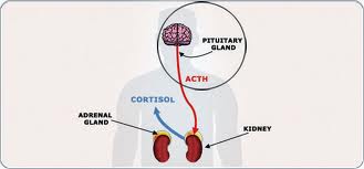
Cushing disease is caused by an increase in the hormone cortisol, which helps maintain blood sugar levels, protects the body from stress, and stops (suppresses) inflammation. Cortisol is produced by the adrenal glands, which are small glands located at the top of each kidney. The production of cortisol is triggered by the release of a hormone called adrenocorticotropic hormone (ACTH) from the pituitary gland, located at the base of the brain. The adrenal and pituitary glands are part of the hormone-producing (endocrine) system in the body that regulates development, metabolism, mood, and many other processes.

Cushing disease occurs when a noncancerous (benign) tumor called an adenoma forms in the pituitary gland, causing excessive release of ACTH and, subsequently, elevated production of cortisol. Prolonged exposure to increased cortisol levels results in the signs and symptoms of Cushing disease: changes to the amount and distribution of body fat, decreased muscle mass leading to weakness and reduced stamina, thinning skin causing stretch marks and easy bruising, thinning of the bones resulting in osteoporosis, increased blood pressure, impaired regulation of blood sugar leading to diabetes, a weakened immune system, neurological problems, irregular menstruation in women, and slow growth in children. The overactive adrenal glands that produce cortisol may also produce increased amounts of male sex hormones (androgens), leading to hirsutism in females. The effect of the excess androgens on males is unclear.

Most often, Cushing disease occurs alone, but rarely, it appears as a symptom of genetic syndromes that have pituitary adenomas as a feature, such as multiple endocrine neoplasia type 1 (MEN1) or familial isolated pituitary adenoma (FIPA).

Cushing disease is a subset of a larger condition called Cushing syndrome, which results when cortisol levels are increased by one of a number of possible causes. Sometimes adenomas that occur in organs or tissues other than the pituitary gland, such as adrenal gland adenomas, can also increase cortisol production, causing Cushing syndrome. Certain prescription drugs can result in an increase in cortisol production and lead to Cushing syndrome. Sometimes prolonged periods of stress or depression can cause an increase in cortisol levels; when this occurs, the condition is known as pseudo-Cushing syndrome. Not accounting for increases in cortisol due to prescription drugs, pituitary adenomas cause the vast majority of Cushing syndrome in adults and children.

Read more about familial isolated pituitary adenoma.


How do people inherit Cushing disease?

Most cases of Cushing disease are sporadic, which means they occur in people with no history of the disorder in their family. Rarely, the condition has been reported to run in families; however, it does not have a clear pattern of inheritance.

The various syndromes that have Cushing disease as a feature can have different inheritance patterns. Most of these disorders are inherited in an autosomal dominant pattern, which means one copy of the altered gene in each cell is sufficient to cause the disorder.

From http://ghr.nlm.nih.gov/condition/cushing-disease

Narrowing in on Pituitary Tumors

0276f-pituitary-gland


As many as 20 percent of people may have a benign cyst or tumor in their pituitary gland. The vast majority of pituitary tumors are noncancerous, but can cause headaches and profound fatigue, and can also disrupt hormone function.

Currently, surgeons rely on radiologic images and MRIs to gather information about the size and shape of the tumor, but the resolution of such imaging technologies is limited, and additional surgeries to remove more of the tumor may be needed if a patient’s symptoms persist. In a new study published in the Proceedings of the National Academy of Sciences on July 27, investigators from Brigham and Women’s Hospital (BWH) present a new technique that could help surgeons more precisely define the locations of tumors in near real-time.

The new strategy uses a visualization technique (matrix-assisted laser desorption/ionization mass spectrometry imaging – MALDI MSI) that can analyze specific hormones, including growth hormone and prolactin, in tissue. In the newly published study, the researchers find that it’s possible to use MALDI MSI to determine the composition of such hormones in a pituitary sample in less than 30 minutes. This could give surgeons critical information to help distinguish tumor from normal gland.

“Our work is driven by a clinical need: we’ve developed a test specifically tailored for the needs of our neurosurgeon colleagues,” said corresponding author Nathalie Agar, PhD, director of the Surgical Molecular Imaging Laboratory in the Department of Neurosurgery at BWH. “A surgeon may sacrifice half of the pituitary gland in an effort to get the tumor out. Without a tool to distinguish healthy tissue from tumor, it’s hard to know in real-time if the surgery was a success. With this technology, in under 30 minutes a surgeon will be able to know if a sample contains normal pituitary tissue or a pituitary tumor.”

“Patients show up with the clinical symptoms of a pituitary tumor, but the tumor itself may not be visible on an MRI,” said co-author Edward Laws, MD, director of the Pituitary and Neuroendocrine Center at BWH. “This technique, which maps out where excess concentrations of hormone levels are located, has the potential to allow us to confirm that we’ve removed the abnormal tissue.”

“Evaluating whether a piece of pituitary tissue is abnormal can be challenging on frozen section,” said co-author Sandro Santagata, MD, PhD, of BWH’s Department of Pathology. “This approach has wonderful potential for enhancing our diagnostic capabilities. It is clearly an important step toward providing intra-operative molecular characterization of pituitary tissues.”

To test the technique, the research team analyzed hormone levels in 45 pituitary tumors and six normal pituitary gland samples, finding a distinct protein signature unique to the normal or tumor sample.

Mass spectrometry, a technique for measuring chemicals present in a sample, is currently used in the operating room to help inform clinical decisions, but up until now, the focus has been on small molecules – metabolites, fatty acids and lipids – using a different type of approach. By analyzing proteins, MALDI MSI offers a way to visualize hormone levels.

Current methods used to detect hormone levels take too long to fit the time constraints of surgical intervention. Surgeons must either remove a larger amount of potentially healthy pituitary gland or perform follow up surgery if the tumor has not been fully removed.

“We’re hoping that techniques like this one will help move the field toward more precise surgery: surgery that not only removes all of the tumor but also preserves the healthy tissue as much as possible,” said Agar.

In the next phase of their work, Agar and her colleagues plan to test out the technique in BWH’s AMIGO suite and analyze the impact of the technique on clinical decision making.

Other researchers who contributed to this study include David Calligaris, Daniel R. Feldman, Isaiah Norton, Olutayo Olubiyi, Armen N. Changelian, Revaz Machaidze, Matthew L. Vestal and Ian F. Dunn.

This work was funded in part by US National Institute of Health (NIH) Director’s New Innovator Award (1DP2OD007383-01 to N.Y.R.A.), U.S. Army Medical Research/CIMIT (2010A052245), the National Center for Image Guided Therapy grant P41RR019703, NIH K08NS064168, the Pediatric Low Grade Astrocytoma Program at Dana-Farber Cancer Institute, the Brain Science Foundation and the Daniel E. Ponton fund for the Neurosciences at BWH.

Brigham and Women’s Hospital 2015 | 75 Francis Street, Boston MA 02115 | 617-732-5500

From http://www.healthcanal.com/cancers/65676-narrowing-in-on-pituitary-tumors.html

Hair Analysis Provides a Historical Record of Cortisol Levels in Cushing’s Syndrome

Exp Clin Endocrinol Diabetes. Author manuscript; available in PMC 2010 Sep 24.
Published in final edited form as:
PMCID: PMC2945912
NIHMSID: NIHMS235640
Hair Analysis Provides a Historical Record of Cortisol Levels in Cushing’s Syndrome

Abstract

The severity of Cushing’s Syndrome (CS) depends on the duration and extent of the exposure to excess glucocorticoids. Current measurements of cortisol in serum, saliva and urine reflect systemic cortisol levels at the time of sample collection, but cannot assess past cortisol levels. Hair cortisol levels may be increased in patients with CS, and, as hair grows about 1 cm/month, measurement of hair cortisol may provide historical information on the development of hypercortisolism.

We attempted to measure cortisol in hair in relation to clinical course in six female patients with CS and in 32 healthy volunteers in 1 cm hair sections. Hair cortisol content was measured using a commercially available salivary cortisol immune assay with a protocol modified for use with hair.

Hair cortisol levels were higher in patients with CS than in controls, the medians (ranges) were 679 (279–2500) and 116 (26–204) ng/g respectively (P <0.001). Segmental hair analysis provided information for up to 18 months before time of sampling. Hair cortisol concentrations appeared to vary in accordance with the clinical course.

Based on these data, we suggest that hair cortisol measurement is a novel method for assessing dynamic systemic cortisol exposure and provides unique historical information on variation in cortisol, and that more research is required to fully understand the utility and limits of this technique.

Keywords: glucocorticoids, pituitary adenoma, cancer, adrenal gland, hormones, cushing hair

Webinar: Diagnosis and Management of Acromegaly: A Clinical Update

Presented by
Lisa Nachtigall, MD
Co-director Neuroendocrine Clinical Center
Massachusetts General Hospital

Register Here

After registering you will receive a confirmation email with details about joining the webinar.

Contact us at webinar@pituitary.org with any questions or suggestions.

Date: Monday, July 27, 2015
Time: 2:00 PM – 3:00 PM Pacific Daylight Time

Presenter Bio
Lisa B. Nachtigall, MD, is an Associate Professor of Medicine at Harvard Medical School, the clinical co-director of the Neuroendocrine Clinical Center at Massachusetts General Hospital and course director in Clinical Neuroendocrine at Harvard Medical School.

Dr. Nachtigall earned her medical degree from New York University (NYU) School of Medicine in New York City. She completed her internship and residency in internal medicine at Bellevue Hospital Center/NYU school of Medicine, and a clinical fellowship in endocrinology and metabolism, as well as a research fellowship in reproductive endocrinology at Massachusetts General Hospital/Harvard Medical School.

Dr. Nachtigall’s work has been published in the New England Journal of Medicine, the Journal of Clinical Endocrinology and Metabolism, Neurosurgery, Pituitary, and the Clinical Endocrinology among others. She serves on the editorial board of Pituitary and as an ad hoc reviewer for many endocrine journals. Dr Nachtigall has been a presenter at national and international medical conferences, and she is currently an investigator on several clinical studies of acromegaly and pituitary tumors.

7 health conditions that are responsible for making you fat

Cushing’s syndrome: Cushing’s syndrome or hypercortisolism is a condition caused when the adrenal glands produce too much cortisol. This leads to a buildup of fat in the face, upper back and abdomen. Cushing’s syndrome can also be a side-effect of certain medications.

Read the other 6 at 7 health conditions that are responsible for making you fat | Read Health Articles & Blogs at TheHealthSite.com.