Cushing’s Disease Associated With Partially Empty Sella Turcica Syndrome

Abstract

The association between empty sella turcica (EST) syndrome and Cushing’s disease has been rarely reported. It is plausible to hypothesize that EST syndrome in association with Cushing’s disease can be attributed to intracranial hypertension. In this case report, we present a 47-year-old male patient who presented with weight loss, fatigue, easy bruising, acanthosis nigricans, and skin creases hyperpigmentation. Investigations revealed hypokalemia and confirmed the diagnosis of Cushing’s disease. Magnetic resonance imaging (MRI) brain showed a partial EST syndrome and a new pituitary nodule as compared with previous brain imaging. Transsphenoidal surgery was pursued and was complicated by cerebrospinal fluid leakage. This case reflects the rare association of EST syndrome and Cushing’s disease, suggesting the increased risk of postoperative complications in this setting and the diagnostic challenge that EST syndrome imposes. We review the literature for a possible mechanism of this association.

Introduction

Cushing’s disease is commonly caused by an adrenocorticotropic hormone (ACTH)-producing pituitary adenoma, which can be very challenging to be seen on brain magnetic resonance imaging (MRI) [1]. Empty sella turcica (EST) syndrome is a radiological diagnosis of apparently empty turcica secondary to outpouching of the arachnoid mater into the turcica, which can be attributed to intracranial hypertension (ICHTN). This can make the visual diagnosis of pituitary adenoma even more challenging in clinical practice. ICHTN has been also associated with Cushing’s disease and might explain this infrequent association between EST and Cushing’s disease [1]. EST syndrome can be either partial or complete, primary or secondary and has been seen infrequently with Cushing’s disease. In this setting, not only that it is likely to obscure an underlying pituitary lesion, but also it does contribute to the risk of postoperative complications [2].

Case Presentation

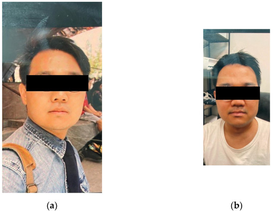
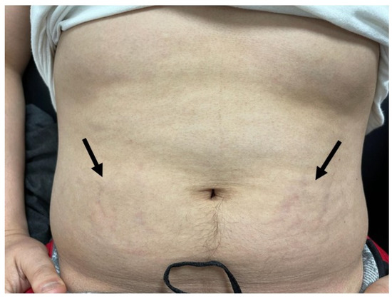
A 47-year-old male presented to the emergency department (ED) with slowly progressive generalized limb muscle weakness affecting both distal and proximal muscles over a few weeks and gait instability for three days prior to presentation. He also reported unintentional 40 pounds weight loss over the previous four months. Past medical history was significant for type II diabetes mellitus, hypothyroidism, hypertension, and dyslipidemia. In the ED, vital signs included a blood pressure of 140/90 mmHg, a heart rate of 66 beats per minute, a respiratory rate of 16 cycles per minute, and SpO2 of 97% on room air. Body mass index has decreased to 22 kg/m2 from a baseline of 26 kg/m2 one month prior. On the physical exam, he exhibited cachexia, easy bruising, acanthosis nigricans, and hyperpigmentation of skin creases. All other systems were negative. Complete metabolic panel and complete blood count were obtained showing hyperglycemia of 311 mg/dl, see Table 1. Further lab evaluation showed elevated salivary cortisol at 2.96 microgram/dl (reference range 0.094-1.551 mcg/dl), elevated 24-hour urinary free cortisol at 156 mcg/24 hour (reference 10-100 mcg/24h), positive overnight dexamethasone suppression test with serum cortisol at 2.8 mcg/dl (reference more than 2 mcg/dl), negative anti-adrenal antibodies, normal aldosterone, and elevated dehydroepiandrostenedione at 401 mcg/dl (reference 32-240 mcg/dl), with lack of suppression of the ACTH level at 35.1 pg/ml (reference 10-60 pg/ml). This confirmed the diagnosis of Cushing’s disease.

Variable Finding Reference
Random glucose 311 Less than 200 mg/dl
Sodium 141 137-145 mmol/L
Potassium 2.5 3.5-5.1 mmol/L
Chloride 96 98-107 mmol/L
Bicarbonate 32 22-30 mmol/L
Blood urea nitrogen 32 9-20 mg/dl
Creatinine 0.52 0.66-1.25 mg/dl
Calcium 8.7 8.6-10.3 mg/dl
Total protein 5.5 6.5-8.5 g/dl
Albumin 3.3 3.5-5 g/dl
Total bilirubin 0.6 0.2-1.3 mg/dl
Alkaline phosphatase 115 38-126 U/L
Aspartate transaminase 17 17-59 U/L
Alanine transaminase 39 Less than 49 U/L
White blood cell count 10×10^3 cells/mcl 4-10×1063 cells/mcl
Hemoglobin 15.3 13.7-17.5 g/dl
Platelet 281 150-400×10^3 cells/mcl
Table 1: Lab Findings

Computed tomography (CT) scan of the head was unremarkable. CT scan of the chest was also unremarkable. CT scan of abdomen and pelvis showed no adrenal mass. Ultrasound of the kidneys was unremarkable. Pituitary MRI brain protocol for adenoma showed a partial EST, shortening within neurohypophysis and a new 10 x 8 x 4 mm nodule along the floor of pituitary sella as compared to MRI four years ago (Figure 1).

Magnetic-Resonance-Imaging-(MRI)-Brain
Figure 1: Magnetic Resonance Imaging (MRI) Brain

MRI brain showing partially empty sella turcica syndrome ( black arrow) with a small nodule at the floor of the turcica (white arrow).

The diagnosis of Cushing’s disease was confirmed, and the patient underwent trans-sphenoidal resection of pituitary adenoma. Histological examination showed positive CAM 5.2, positive chromogranin, and ACTH immunostains. The patient presented to the ED five days after discharge home. He stated that he noticed drainage from the nose that transitioned from bloody to clear fluid and has been increasing in quantity for two days with associated intermittent headaches since the surgery. He was afebrile with stable vital signs. No signs of infection were noted on basic labs. These were significant only for mild asymptomatic hyponatremia of 131 mmol/L and hypokalemia of 3.3 mmol/L. The patient was diagnosed with cerebrospinal fluid (CSF) leakage and had a lumbar drain trial. The trial was unsuccessful after several days, and the patient underwent a transnasal endoscopic repair of CSF rhinorrhea using nasoseptal flaps. At an outpatient follow-up one month and three months after the surgery, prior lab abnormalities including hypokalemia, hyponatremia, and hyperglycemia resolved. No further evidence of CSF leakage was appreciated, and he remained asymptomatic.

Discussion

EST syndrome is characterized by herniation of the subarachnoid space into the intrasellar space with compression of the pituitary gland into the posteroinferior wall [3]. This is likely to obscure the presence of underlying pituitary mass. The incidence of EST syndrome in the general population is estimated at 20%. The association between EST syndrome and Cushing’s disease has been reported infrequently. A retrospective study of 68 patients with Cushing’s disease found that 16% of these have EST syndrome [3].

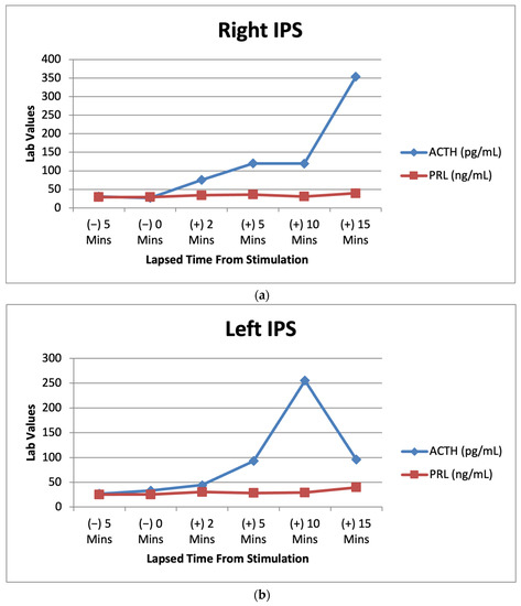
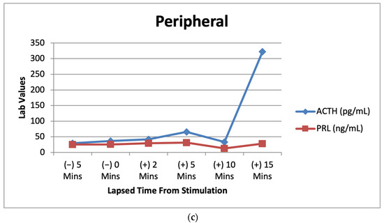
Cushing’s disease usually results from pituitary adenomas secreting ACTH, and even the smallest microadenomas can produce a systemic disease. These microadenomas can be very difficult to recognize on brain MRI [4]. This is complicated in EST syndrome and even further with the possibility of ectopic ACTH production. A retrospective study of 197 patients diagnosed with Cushing’s disease concluded that EST syndrome is associated with higher prevalence of MRI-negative Cushing’s disease. This was attributed to ICHTN and pituitary gland compression [1]. Although surgery is curative in 70-90% of cases, EST syndrome was found to have higher risk of postoperative complications among those with Cushing’s disease including diabetes insipidus, hypopituitarism, and CSF leakage [3]. This is usually because in the case of MRI-negative Cushing’s disease with total EST syndrome, empiric surgical exploration is sought after inferior petrosal sampling confirms the pituitary origin of excess ACTH, and postoperative remission indicates adequate tumor resection [2]. This entails a higher chance of uncertainty and injury to healthy pituitary tissue.

EST syndrome can be either primarily due to defects in the sellar diaphragm or anatomical variant or secondary to ICHTN. EST syndrome has been reported in association with many conditions associated with elevated intracranial pressure including tumors, thrombosis, meningitis, hydrocephalus, and Arnold-Chiari malformation [5]. Reversal of EST syndrome has been reported in those with idiopathic ICHTN with therapy by acetazolamide, ventriculoperitoneal shunt, and lumbar puncture [6,7]. A study has shown correlation between CSF circulation impairment or blockage and EST syndrome [8]. The incidence of EST syndrome in association with symptomatic intracranial hypertension is variable and ranges from 2.5% for total EST syndrome to 94% for partial EST syndrome [9]. Impaired CSF circulation and dynamics have been reported in 77% of patients with EST syndrome [10]. In addition to intracranial hypertension, EST syndrome has also been described in association with obesity, meningioma, pediatric nevoid basal cell carcinoma, therapy for growth hormone deficiency and even in healthy individuals [9]. Lack of symptoms of intracranial hypertension in this patient does not rule it out as intracranial hypertension in EST syndrome represents a spectrum that ranges from asymptomatic, milder intracranial hypertension to symptomatic intracranial hypertension with headache, visual disturbance, and papilledema [10]. This explains the fact that only 8-14% of EST syndrome progress to symptomatic ICHTN, while symptomatic ICHTN has been associated with EST syndrome in 94% of cases.

ICHTN has been seen in association with disturbance of the hypothalamic-pituitary-adrenal axis. This has been reported after surgical and medical treatment of Cushing’s disease, withdrawal of long-term steroid therapy, initial presentation of Addison’s disease, or relative glucocorticoids deficiency [11]. Cortisol excess increases CSF production and reduces its absorption, hence increasing intracranial pressure [12]. Another possible mechanism is the expression of both mineralocorticoid responsive epithelial sodium channel receptors on the basolateral membrane of the CSF producing epithelial cells of the choroid plexus as well as the expression of 11-beta hydroxysteroid dehydrogenase type 1 enzyme, which is a bidirectional enzyme that mainly functions to convert the inactive cortisone to active cortisol. These mechanisms play a role in maintaining the balance between CSF production and absorption [13,14].

In this case, the patient presented some clinical findings that are rarely associated with Cushing’s disease, combined with a radiological feature that masked the true diagnosis. Our patient presented with significant weight loss, rather than central obesity, which is normally associated with Cushing’s disease. Although possible, the increase in ACTH due to Cushing’s disease is not sufficient to cause hyperpigmentation, which is a classical finding of Addison’s disease, where the entire adrenal cortex is usually affected due to an autoimmune destruction; however, the zona glomerulosa of the adrenal cortex produces aldosterone and its deficiency would lead to hyperkalemia [15]. Our patient presented with both hyperpigmentation and hypokalemia.

Conclusions

EST syndrome is an uncommon radiological finding of apparently EST that has been reported in association with ICHTN. The latter has also been seen in association with Cushing’s disease/syndrome. This is likely to result from glucocorticoid excess-induced change in CSF flow dynamics. EST has been infrequently described in association with Cushing’s disease. This association has a clinical implication as it is likely to obscure the visualization of pituitary lesions responsible for Cushing’s disease, contribute to diagnostic uncertainty, and increase the risk of healthy pituitary tissue injury and the risk of postoperative complications including CSF leakage.

References

  1. Himes BT, Bhargav AG, Brown DA, Kaufmann TJ, Bancos I, Van Gompel JJ: Does pituitary compression/empty sella syndrome contribute to MRI-negative Cushing’s disease? A single-institution experience. Neurosurg Focus. 2020, 48:E3. 10.3171/2020.3.FOCUS2084
  2. Sun Y, Sun Q, Fan C, et al.: Diagnosis and therapy for Cushing’s disease with negative dynamic MRI finding: a single-centre experience. Clin Endocrinol (Oxf). 2012, 76:868-76. 10.1111/j.1365-2265.2011.04279.x
  3. Manavela MP, Goodall CM, Katz SB, Moncet D, Bruno OD: The association of Cushing’s disease and primary empty sella turcica. Pituitary. 2001, 4:145-51. 10.1023/a:1015310806063
  4. Chatain GP, Patronas N, Smirniotopoulos JG, et al.: Potential utility of FLAIR in MRI-negative Cushing’s disease. J Neurosurg. 2018, 129:620-8. 10.3171/2017.4.JNS17234
  5. Friedman DI, Jacobson DM: Diagnostic criteria for idiopathic intracranial hypertension. Neurology. 2002, 59:1492-5. 10.1212/01.wnl.0000029570.69134.1b
  6. Triggiani V, Giagulli VA, Moschetta M, Guastamacchia E: An unusual case of reversible empty sella. Endocr Metab Immune Disord Drug Targets. 2016, 16:154-6. 10.2174/1871530315666151001141507
  7. Wind JJ, Lonser RR, Nieman LK, DeVroom HL, Chang R, Oldfield EH: The lateralization accuracy of inferior petrosal sinus sampling in 501 patients with Cushing’s disease. J Clin Endocrinol Metab. 2013, 98:2285-93. 10.1210/jc.2012-3943
  8. Brismar K, Bergstrand G: CSF circulation in subjects with the empty sella syndrome. Neuroradiology. 1981, 21:167-75. 10.1007/BF00367338
  9. Ranganathan S, Lee SH, Checkver A, Sklar E, Lam BL, Danton GH, Alperin N: Magnetic resonance imaging finding of empty sella in obesity related idiopathic intracranial hypertension is associated with enlarged sella turcica. Neuroradiology. 2013, 55:955-61. 10.1007/s00234-013-1207-0
  10. Maira G, Anile C, Mangiola A: Primary empty sella syndrome in a series of 142 patients. J Neurosurg. 2005, 103:831-6. 10.3171/jns.2005.103.5.0831
  11. Zada G, Tirosh A, Kaiser UB, Laws ER, Woodmansee WW: Cushing’s disease and idiopathic intracranial hypertension: case report and review of underlying pathophysiological mechanisms. J Clin Endocrinol Metab. 2010, 95:4850-4. 10.1210/jc.2010-0896
  12. Sinclair AJ, Ball AK, Burdon MA, Clarke CE, Stewart PM, Curnow SJ, Rauz S: Exploring the pathogenesis of IIH: an inflammatory perspective. J Neuroimmunol. 2008, 201:212-20. 10.1016/j.jneuroim.2008.06.029
  13. Sinclair AJ, Onyimba CU, Khosla P, et al.: Corticosteroids, 11beta-hydroxysteroid dehydrogenase isozymes and the rabbit choroid plexus. J Neuroendocrinol. 2007, 19:614-20. 10.1111/j.1365-2826.2007.01569.x
  14. Amin MS, Wang HW, Reza E, Whitman SC, Tuana BS, Leenen FH: Distribution of epithelial sodium channels and mineralocorticoid receptors in cardiovascular regulatory centers in rat brain. Am J Physiol Regul Integr Comp Physiol. 2005, 289:R1787-97. 10.1152/ajpregu.00063.2005
  15. Stratakis CA: Skin manifestations of Cushing’s syndrome. Rev Endocr Metab Disord. 2016, 17:283-6. 10.1007/s11154-016-9399-3

From https://www.cureus.com/articles/161111-cushings-disease-associated-with-partially-empty-sella-turcica-syndrome-a-case-report#!/

Catastrophic ACTH-secreting Pheochromocytoma

Abstract

Summary

Cushing’s syndrome due to ectopic adrenocorticotropic hormone (ACTH) secretion (EAS) by a pheochromocytoma is a challenging condition. A woman with hypertension and an anamnestic report of a ‘non-secreting’ left adrenal mass developed uncontrolled blood pressure (BP), hyperglycaemia and severe hypokalaemia. ACTH-dependent severe hypercortisolism was ascertained in the absence of Cushingoid features, and a psycho-organic syndrome developed. Brain imaging revealed a splenial lesion of the corpus callosum and a pituitary microadenoma. The adrenal mass displayed high uptake on both 18F-FDG PET/CT and 68Ga-DOTATOC PET/CT; urinary metanephrine levels were greatly increased. The combination of antihypertensive drugs, high-dose potassium infusion, insulin and steroidogenesis inhibitor normalized BP, metabolic parameters and cortisol levels; laparoscopic left adrenalectomy under intravenous hydrocortisone infusion was performed. On combined histology and immunohistochemistry, an ACTH-secreting pheochromocytoma was diagnosed. The patient’s clinical condition improved and remission of both hypercortisolism and catecholamine hypersecretion ensued. Brain magnetic resonance imaging showed a reduction of the splenial lesion. Off-therapy BP and metabolic parameters remained normal. The patient was discharged on cortisone replacement therapy for post-surgical hypocortisolism. EAS due to pheochromocytoma displays multifaceted clinical features and requires prompt diagnosis and multidisciplinary management in order to overcome the related severe clinical derangements.

Learning points

  • A small but significant number of cases of adrenocorticotropic hormone (ACTH)-dependent Cushing’s syndrome are caused by ectopic ACTH secretion by neuroendocrine tumours, which is usually associated with severe hypercortisolism causing severe clinical and metabolic derangements.
  • Ectopic ACTH secretion by a pheochromocytoma is exceedingly rare but can be life-threatening, owing to the simultaneous excess of both cortisol and catecholamines.
  • The combination of biochemical and hormonal testing and imaging procedures is mandatory for the diagnosis of ectopic ACTH secretion, and in the presence of an adrenal mass, the possibility of an ACTH-secreting pheochromocytoma should be taken into account.
  • Immediate-acting steroidogenesis inhibitors are required for the treatment of hypercortisolism, and catecholamine excess should also be appropriately managed before surgical removal of the tumour.
  • A multidisciplinary approach is required for the treatment of this challenging entity.

Background

Cushing’s syndrome (CS) is a rare endocrine disease characterized by high levels of glucocorticoids; it increases morbidity and mortality due to cardiovascular and infectious diseases (123).

To diagnose CS, adrenocorticotropic hormone (ACTH)-dependent disease must be distinguished from ACTH-independent disease, and pituitary ACTH production from ectopic production. About 20% of ACTH-dependent cases arise from ectopic ACTH secretion (EAS) (234). EAS is most often due to aberrant ACTH production by small-cell lung carcinoma or neuroendocrine tumours originating in the lungs or gastrointestinal tract; this, in turn, strongly increases cortisol production by the adrenal glands (345).

Since the first-line treatment of EAS is the surgical removal of the ectopic ACTH-secreting tumour, its prompt and accurate localization is crucial.

Rapid cortisol reduction by means of immediate-acting steroidogenesis inhibitors (4) is mandatory in order to treat the related endocrine, metabolic and electrolytic derangements. EAS by a pheochromocytoma is exceedingly rare and can be life-threatening.

We describe the case of a woman with hypertension and a known ‘non-secreting’ left adrenal mass, who manifested uncontrolled blood pressure (BP), hyperglycaemia, hypokalaemia and psycho-organic syndrome associated with damage of the splenium of the corpus callosum. These findings were eventually seen to be related to an ACTH-secreting left pheochromocytoma, which was ascertained by hormonal evaluation and morphological and functional imaging assessment and confirmed by histopathology/immunostaining. Hormonal hypersecretion resolved after adrenalectomy and metabolic derangements normalized.

Case presentation

A 72-year-old woman with hypertension was taken to the emergency department because of increased BP (200/100 mm Hg). High BP (190/100 mmHg) was confirmed, whereas oxygen saturation (98%), heart rate (84 bpm) and lung and abdomen examination were normal. Electrocardiogram and chest x-ray were unremarkable. Captopril 50 mg orally, followed by intramuscular clonidine, normalized BP.

The patient looked thin and reported significant weight loss (10 kg) over the previous 6 months; she was on antihypertensive therapy with bisoprolol 5 mg/day and irbesartan 150 mg/day, and ezetimibe 10 mg/day for dyslipidaemia. The patient’s records included a previous diagnosis in another hospital of normofunctioning multinodular goitre and a 2.5 cm-left solid inhomogeneous adrenal mass with well-defined margins, which was found on CT performed 6 years earlier during the work-up for hypertension. On the basis of hormonal data and absent uptake on 123I metaiodobenzylguanidine scintigraphy, the adrenal lesion had been deemed to be non-functioning and follow-up had been advised. Unfortunately, only initial cortisol (15.7 μg/dL) and 24-h urine-free cortisol (UFC) levels (32.5 μg/24 h) were retrievable; both proved normal.

Investigations

Blood chemistry showed neutrophilic leucocytosis, hyperglycaemia with increased glycated haemoglobin, severe hypokalaemia and metabolic alkalosis (Table 1). Potassium infusion (50 mEq in 500 mL saline/24 h) was rapidly started, together with a subcutaneous rapid-acting insulin analogue and prophylactic enoxaparin. The patient experienced mental confusion, hallucinations and restlessness; non-enhanced computed tomography (CT) of the brain revealed a hypodense area of the splenium of the corpus callosum, possibly due to metabolic damage (Fig. 1A).

Figure 1View Full Size
Figure 1

Non-enhanced CT showing a hypodense area of the splenium of the corpus callosum (arrows), without mass effect (A, axial view). Contrast-enhanced MR image showing a hypointense pituitary lesion (arrow) which enhances more slowly than normal pituitary parenchyma, deemed suspicious for microadenoma (B, coronal view). FLAIR MR image showing hyperintense signal of the splenium of the corpus callosum (asterisk), which partially extended to the crux of the left fornix (arrow) (C, axial view). As the lesion showed no restricted diffusion on DWI (D, axial view), an ischaemic lesion was excluded. A progressive reduction in the extension of the hyperintense signal in the splenium of the corpus callosum (arrowheads) and in the crux of the left fornix (arrows) was observed on FLAIR MR images (2 months (E); 3 months (F); axial view). CT, computed tomography; DWI, diffusion-weighted imaging; FLAIR, fluid-attenuated inversion recovery; MR, magnetic resonance.

Citation: Endocrinology, Diabetes & Metabolism Case Reports 2023, 2; 10.1530/EDM-22-0308

Table 1Hormonal and biochemical evaluation of patient throughout hospitalization and follow-up.

Normal range On hospital admission After surgery
10 days 2 months 3 months 6 months 9 months 12 months 16 months
ACTH (pg/mL) 9–52 551 7 37 50 29.5 26 40.9 52
Morning cortisol† (µg/dL) 7–19.2 63.4 14 5.1 3.5 3.8 4.2 7.2 12.8
After 1 mg overnight dexamethasone
 ACTH 583
 Cortisol 60
DHEAS (µg/dL) 9.4–246 201
24-h urinalysis (µg/24 h)
 Adrenaline 0–14.9 95.5
 Noradrenaline 0–66 1133
 Metanephrine 74–297 1927
 Normetanephrine 105–354 1133
Chromogranin A 0–108 290
Renin (supine) (µU/mL) 2.4–29 3.9 14.6
Aldosterone (supine) (ng/dL) 3–15 3.4 12.5
LH (mIU/mL)* > 10 0.3 65.8
FSH (mIU/mL)* > 25 1.9 116
PRL (ng/mL) 3–24 13.7
FT4 (ng/dL) 0.9–1.7 1.1 1.2
FT3 (pg/mL) 1.8–4.6 1.1 2.7
TSH (µU/mL) 0.27–4.2 0.23 1.3
PTH (pg/mL) 15–65 166
Calcium (mg/dL) 8.2–10.2 8.2
Calcitonin (pg/mL) 0–10 1
Glycaemia (mg/dL) 60–110 212 69 73 83
Potassium (mEq/L) 3.5–5 2.4 3.3 3.9 4.2 3.7 5 4.4 3.9
Leucocytes (K/µL) 4.0–9.3 15.13
HbA1c (mmol/mol) 20–42 55 30
HCO3 (mEq/L) 22–26 41.8

*For menopausal age; †07:00–10:00 h.


The patient was transferred to the internal medicine ward. Although potassium infusion was increased to 120 mEq/day, serum levels did not normalize; a mineralocorticoid receptor antagonist (potassium canreonate) was therefore introduced, but the effect was partial. In order to control BP, the irbersartan dose was increased (300 mg/day) and amlodipine (10 mg/day) was added.

The combination of severe hypertension, newly occurring diabetes and resistant hypokalaemia prompted us to hypothesize a common endocrine aetiology.

A thorough hormonal array showed very high ACTH and cortisol levels, whereas supine renin and aldosterone levels were in the low-normal range (Table 1). Since our patient proved repeatedly non-compliant with 24-h urine collection, UFC could not be measured.

After an overnight 1 mg dexamethasone suppression test, cortisol levels remained unchanged, whereas ACTH levels slightly increased (Table 1). Notably, the patient showed no Cushingoid features. Gonadotropin levels were inappropriately low for the patient’s age; FT4 levels were normal, whereas FT3 and thyroid-stimulating hormone (TSH) levels were reduced and calcitonin levels were normal (Table 1). HbA1c levels were increased (Table 1).

Finally, secondary hyperparathyroidism, associated with low-normal calcium levels and reduced vitamin D levels, was found (Table 1).

Brain contrast-enhanced magnetic resonance (MR) imaging revealed a 5-mm median posterior pituitary microadenoma (Fig. 1B) and a hyperintense lesion of the splenium of the corpus callosum (Fig. 1C). Diffusion-weighted MR images of the lesion showed no restricted diffusion (Fig. 1D), thus excluding an ischaemic origin. Petrosal venous sampling for ACTH determination at baseline and after CRH stimulation was excluded, as it was deemed a high-risk procedure, given the patient’s poor condition.

Since the ACTH and cortisol levels were greatly increased and were associated with severe hypokalaemia, EAS was hypothesized; total-body contrast-enhanced CT revealed the left adrenal mass (3 cm), which showed regular margins and heterogeneous enhancement (Fig. 2A and B) and measured 25 Hounsfield units. There was no evidence of adrenal hyperplasia in the contralateral adrenal gland. The adrenal mass showed intense tracer uptake on both 18F-FDG PET/CT (Fig. 2C and D), suggestive of adrenal malignancy or functioning tumour, and 68Ga-DOTATOC PET/CT (Fig. 3), which is characteristic of a neuroendocrine lesion. No other sites of suspicious tracer uptake were detected.

Figure 2View Full Size
Figure 2

Contrast-enhanced abdominal computed tomography showing a 3-cm left adrenal mass (arrow) with well-defined margins and inhomogeneus enhancement, deemed compatible with an adenoma (A, coronal view; B, axial view). The adrenal mass showed high uptake (SUV max: 7.3) on 18F-FDG PET/CT (C, coronal view; D, axial view).

Citation: Endocrinology, Diabetes & Metabolism Case Reports 2023, 2; 10.1530/EDM-22-0308

Figure 3View Full Size
Figure 3

The left adrenal mass displaying very high uptake (SUV max: 40) on 68Ga-DOTATOC PET/CT (arrow, axial view).

Citation: Endocrinology, Diabetes & Metabolism Case Reports 2023, 2; 10.1530/EDM-22-0308


Bisoprolol was withdrawn, and 24-h urinary catecholamine, metanephrine and normetanephrine levels proved significantly increased, as were chromogranin A levels (Table 1). In sum, an ACTH-secreting pheochromocytoma was suspected and the pituitary microadenoma was deemed a likely incidental finding.

The patient’s mental state worsened, fluctuating from sopor to restlessness, which required parenteral neuroleptics and restraint. An electroencephalogram revealed a specific slowdown of cerebral electrical activity. Following rachicentesis, the cerebrospinal fluid showed pleocytosis (lympho-monocytosis), whereas a culture test and polymerase chain reaction for common neurotropic agents were negative. The neurologist hypothesized a psycho-organic syndrome secondary to severe metabolic derangement. Intravenous ampicillin, acyclovir and B vitamins were empirically started. The patient was transferred to the subintensive unit, where a nasogastric tube and central venous catheter were inserted, and enteral nutrition was started.

Treatment

Ketoconazole was started at a dosage of 200 mg twice daily; both cortisol and ACTH levels significantly decreased over a few days (Fig. 4), with a progressive decrease in glucose levels and normalization of potassium levels and BP on therapy. Subsequently, ketoconazole was titrated to 600 mg/day owing to a new increase in cortisol levels, which eventually normalized (Fig. 4). Of note, ACTH levels partially decreased on ketoconazole treatment (Fig. 4).

Figure 4View Full Size
Figure 4

ACTH and cortisol levels throughout the patient’s hospitalization and follow-up.

Citation: Endocrinology, Diabetes & Metabolism Case Reports 2023, 2; 10.1530/EDM-22-0308


Doxazosin 2 mg/day was added and the patient’s systolic BP blood settled at around 100 mm Hg; after a few days, bisoprolol was restarted. Contrast-enhanced MR showed a partial reduction of the hyperintense splenial lesion (Fig. 1E). Despite the severe clinical condition and the high risks of adrenal surgery, the patient’s relatives strongly requested the procedure and laparoscopic left adrenalectomy was planned. Alpha-blocker and fluid infusion were continued, ketoconazole was withdrawn the day before surgery, and a 100 mg IV bolus of hydrocortisone was administered just before the operation, followed by 200 mg/day, at first in continuous infusion, then as a 100 mg bolus every 8 h. After the removal of the left adrenal mass, noradrenaline infusion was required, owing to the occurrence of severe hypotension.

Outcome and follow-up

Pathology revealed a 2.5 cm reddish-brown encapsulated tumour, which was compatible with pheochromocytoma (Fig. 5A and B); ACTH immunostaining was positive in about 30% of tumour cells (Fig. 5C). This confirmed the diagnostic hypothesis of an ACTH-secreting pheochromocytoma. The tumour was stained for Chromogranin A (Fig. 5D). There were no signs of adrenal cortex hyperplasia in the resected gland. Thorough germinal genetic testing, comprising the commonest pheochromocytoma/paraganglioma genes: CDKN1B, KIF1B, MEN1, RET, SDHA, SDHB, SDHC, SDHD, SDHAF2 and TMEM127, was negative.

Figure 5View Full Size
Figure 5

Histological images of adrenal pheochromocytoma: the tumour is composed of well-defined nests of cells (‘zellballen’) (A; haematoxylin-eosin stain (HE), ×20) with pleomorphic nuclei with prominent nucleoli, basophilic or granular amphophilic cytoplasm (B; HE, ×40). The mitotic index was low: 1 mitosis per 30 high-power fields, and Ki-67 was 1%. On immunohistochemistry, cytoplasmatic ACTH staining was found in about 30% of tumour cells (C; ×20), whereas most tumour cells were stained for chromogranin A (D; ×20).

Citation: Endocrinology, Diabetes & Metabolism Case Reports 2023, 2; 10.1530/EDM-22-0308


One week after surgery ACTH levels had dropped to a low-normal value: 7 pg/mL, and cortisol levels (before morning hydrocortisone bolus administration) were normal: 14 µg/dL (Fig. 4). The patient’s clinical status slowly improved and the nasogastric tube was removed; intravenous hydrocortisone was carefully tapered until withdrawal and high-dose oral cortisone acetate (62.5 mg/day) was started. This dose was initially required since BP remained low (systolic: 90 mm Hg); thereafter, cortisone was reduced to 37.5 mg/day. Plasma cortisol levels before morning cortisone administration were reduced (Fig. 4). A new MR of the brain showed a further partial reduction of the splenial lesion (Fig. 1F). The patient was discharged with normal off-therapy BP and metabolic parameters.

During follow-up, she fully recovered, and BP and metabolic parameters remained normal. Gonadotropin levels became adequate for the patient’s age, and TSH and renin/aldosterone levels normalized (Table 1). Hypoadrenalism, however, persisted for more than 1 year; as the last hormonal evaluation, 16 months after surgery, showed normal baseline cortisol levels, the cortisone dose was tapered (12.5 mg/day) and further hormonal examination was scheduled (Table 1). ACTH and cortisol levels throughout the patient’s hospitalization and follow-up are shown in Fig. 4.

Discussion

The diagnosis of EAS is challenging and requires two steps: confirmation of increased ACTH and cortisol levels and anatomic distinction from pituitary sources of ACTH overproduction. Besides metabolic derangements (hyperglycaemia, hypertension), EAS-related severe hypercortisolism may cause profound hypokalaemia (345).

In our patient, the combination of worsening hypertension, newly occurring diabetes and resistant hypokalaemia raised the suspicion of a common endocrine cause.

ACTH-dependent severe hypercortisolism was ascertained, and subsequent brain MR revealed a pituitary microadenoma.

The diagnosis of CS requires the combination of two abnormal test results: 24-h UFC, midnight salivary cortisol and/or abnormal 1 mg dexamethasone suppression testing (26). ACTH evaluation (low/normal-high) is fundamental to tailoring the imaging technique.

The very high cortisol levels found in our patient were unchanged after overnight dexamethasone testing, whereas UFC could not be assessed owing to the lack of compliance with urine collection. The accuracy of the UFC assays, however, may be impaired by cortisol precursors and metabolites. Salivary cortisol assessment was not performed since the specific assay is not available in our hospital.

The combination of ACTH-dependent severe hypercortisolism and hypokalaemia prompted us to suspect EAS. The differential diagnosis between pituitary and ectopic ACTH-dependent CS involves high-dose (8 mg) dexamethasone suppression testing, which has relatively low diagnostic accuracy (6). Owing to the patient’s very high cortisol levels and severe hypokalaemia, this testing was not performed, on account of the risks of administering corticosteroids in a patient already exposed to excessive levels (6). Furthermore, owing to the increase in ACTH levels observed after overnight dexamethasone testing, we postulated the possible occurrence of glucocorticoid-driven positive feedback on ACTH secretion, which has been described in EAS, including cases of pheochromocytoma (7).

Finally, in the case of EAS suspected of being caused by pheochromocytoma, we do not recommend performing high-dose dexamethasone suppression testing, owing to the risk of triggering a catecholaminergic crisis (8).

The dynamic tests commonly used to distinguish patients with EAS from those with Cushing’s disease are the CRH stimulation test and the desmopressin stimulation test, either alone or in combination with CRH testing (6). Owing to the rapid worsening of our patient’s condition, dynamic testing was not done; however, the clinical picture and hormonal/biochemical data were suggestive of EAS.

EAS is mainly (45–50%) due to neuroendocrine tumours, mostly of the lung (small-cell lung cancer and bronchial tumours), thymus or gastrointestinal tract; however, up to 20% of ACTH-secreting tumours remain occult (345).

ACTH-secreting pheochromocytomas are responsible for about 5% of cases of EAS (34910). Indeed, this rate ranges widely, from 2.5% (11) to 15% (12), according to the different case series. Patients with EAS due to pheochromocytoma present with severe CS, overt diabetes mellitus, hypertension and hypokalaemia (3); symptoms of catecholamine excess may be unapparent (3), making the diagnosis more challenging.

A recent review of 99 patients with ACTH- and/or CRH-secreting pheochromocytomas found that the vast majority displayed a Cushingoid phenotype (10); by contrast, another review of 24 patients reported that typical Cushingoid features were observed in only 30% of patients, whereas weight loss was a prevalent clinical finding (13). We hypothesized that the significant weight loss reported by our patient was largely due to the hypermetabolic state induced by catecholamines, which directly reduce visceral and subcutaneous fat, as recently reported (14).

Our patient showed no classic stigmata of CS, owing to the rapid onset of severe hypercortisolism (1013), whereas she had worsening hypertension and newly occurring diabetes mellitus, which were related to both cortisol and catecholamine hypersecretion; hypokalaemia was deemed to be secondary to severe hypercortisolism. Indeed, greatly increased cortisol levels act on the mineralocorticoid receptors of the distal tubule after saturating 11β-hydroxysteroid dehydrogenase type 2, leading to hypokalaemia (4). Consequently, hypokalaemia is much more common (74–95% of patients) in EAS than in classic Cushing’s disease (10%) (3410). This apparent mineralocorticoid excess suppresses renin and aldosterone secretion, as was ascertained in our patient.

In this setting, the most effective way to manage hypokalaemia is to treat the hypercortisolism itself by administering immediate-acting steroidogenesis inhibitors, combined with potassium infusion and a mineralocorticoid receptor-antagonist (e.g. spironolactone) at an appropriate dosage (100–300 mg/day) (4).

In ACTH-secreting pheochromocytoma, cortisol hypersecretion potentiates catecholamine-induced hypertension by stimulating the phenol-etholamine-N-methyl–transferase enzyme, which transforms noradrenaline to adrenaline (4). Indeed, in our patient, the significant ketoconazole-induced reduction in cortisol secretion led to satisfactory BP control on antihypertensive drugs. After the biochemical diagnosis of pheochromocytoma, a selective alpha-blocker was added, and after a few days, a beta-blocker was restarted in order to control reflex tachycardia (15).

Our patient had greatly increased ACTH levels (>500 pg/mL) associated with very high cortisol levels (>60 µg/dL), which, together with the finding of hypokalaemia, prompted us to hypothesize EAS. With regard to these findings, ACTH levels are usually higher (>200 pg/mL) in patients with EAS than in those with CS due to a pituitary adenoma; however, considerable overlapping occurs (31116). Most patients with ACTH-secreting pheochromocytomas in those series had ACTH levels >300 pg/mL, and a few had normal ACTH levels (9), thus complicating the diagnosis. In addition, patients with EAS usually have higher cortisol levels than those with ACTH-secreting adenomas (311).

In our patient, the left adrenal mass was deemed the culprit of EAS, and owing to very high urinary metanephrine levels, a pheochromocytoma was suspected.

It can be assumed that the adrenal tumour, which was anamnestically reported as ‘non-secreting’, but on which only part of the initial hormonal data were available, was actually a pheochromocytoma at the time of the first diagnosis but displayed a silent clinical and hormonal behaviour. The mass subsequently showed significant uptake on both 18F-FDG PET/CT and 68Ga-DOTATOC PET/CT (45). It is claimed that 68Ga-DOTATOC PET/CT provides a high grade (90%) of sensitivity and specificity in the diagnosis of tumours that cause EAS (45); nevertheless, a recent systematic review reported much lower sensitivity (64%), which increased to 76% in histologically confirmed cases (17).

In patients with EAS, immediate-acting steroidogenesis inhibitors are required in order to achieve prompt control of severe hypercortisolism (4). Ketoconazole is one of the drugs of choice since it inhibits adrenal steroidogenesis at several steps. In our patient, ketoconazole rapidly reduced cortisol levels to normal values, without causing hepatic toxicity (4). Moreover, ketoconazole proved effective at a moderate dosage (600 mg/day), which falls within the mean literature range (1819). However, dosages up to 1200–1600 mg/day are sometimes required in severe cases (usually EAS) (1819). Speculatively, our results might reflect an enhanced inhibitory action of ketoconazole at the adrenal level, which was able to override the strong ectopic ACTH stimulation.

In addition, the finding that, following cortisol reduction, ACTH levels paradoxically decreased suggests an additive and direct effect of the drug. This effect has been observed in a few patients with EAS (20) and is supported by in vitro studies showing a direct anti-proliferative and pro-apoptotic effect of ketoconazole on ectopic ACTH secretion by tumours (21). Finally, the reduction in ACTH levels during treatment with steroidogenesis inhibitors prompts us to postulate the presence of glucocorticoid-driven positive feedback on ACTH secretion, as already described in neuroendocrine tumours (72021). The coexistence of EAS and ACTH-producing pituitary adenoma is very rare but must be taken into account. In our case, we deemed the pituitary mass found on MR to be a non-secreting microadenoma. This hypothesis was strengthened by the finding that, following exeresis of the ACTH-secreting pheochromocytoma, ACTH normalized, hypercortisolism vanished and pituitary function recovered. These findings suggest that: (i) altered pituitary function at the baseline was secondary to the inhibitory effect of hypercortisolism; (ii) the excessive production of cortisol was driven by ACTH overproduction outside the pituitary gland, specifically within the adrenal gland tumour.

In our patient, a few days after surgery, morning cortisol levels before hydrocortisone bolus administration were ‘normal’. Owing to both the half-life of hydrocortisone (8–12 h) and the supraphysiological dosage used, it is likely that a residual part of the drug, which cross-reacts in the cortisol assay, was still circulating at the time of blood collection, thus resulting in ‘normal’ cortisol values. Following the switch to oral cortisone, cortisol levels before therapy were low, thus confirming post-surgical hypocortisolism. Hypocortisolism remained throughout the first year after surgery, and glucocorticoid therapy was continued. Sixteen months after surgery, baseline cortisol levels returned to the normal range; cortisone therapy was therefore tapered and a further hormonal check was scheduled. Assessment of the cortisol response to ACTH stimulation testing would be helpful in order to check the resumption of the residual adrenal function.

A peculiar aspect of our case was the occurrence of a psycho-organic syndrome together with the finding of a splenial lesion on brain imaging, which was deemed secondary to metabolic injury. Indeed, the increased cortisol levels present in patients with Cushing’s disease are detrimental to the white matter of the brain, including the corpus collosum, causing subsequent clinical derangements (22).

Besides the direct effects of hypercortisolism, the splenial damage was also probably due to long-standing hypertension, worsened by newly occurring catecholamine hypersecretion and diabetes. Together with the normalization of cortisol and glycaemic levels, and of BP, a partial reduction in the splenial damage was observed on two subsequent MR examinations, and the patient’s neurological condition slowly improved until she fully recovered.

In our patient, thorough germinal genetic testing for the commonest pheochromocytoma/paraganglioma (PPGL) genes proved negative. Since approximately 40% of these tumours have germline mutations, genetic testing is recommended regardless of the patient’s age and family history. In the absence of syndromic, familial or metastatic presentation, the selection of genes for testing may be guided by the tumour location and biochemical phenotype.

Alterations of the PPGL genes can be divided into two groups: 10 genes (RET, VHL, NF1, SDHD, SDHAF2, SDHC, SDHB, SDHA, TMEM127 and MAX) that have well-defined genotype–phenotype correlations, thus allowing to tailor imaging procedures and medical management, and a group of other emerging genes, which lack established genotype–phenotype associations; for patients in whom mutations of genes belonging to this second group are detected, and hence hereditary predisposition is established, only general medical surveillance and family screening can be planned (2324).

In conclusion, our case highlights the importance of investigating patients with hypertension and metabolic derangements such as diabetes and hypokalaemia, since these findings may be a sign of newly occurring EAS, which, in rare cases, may be due to an ACTH-secreting pheochromocytoma. Since the additive effect of cortisol and catecholamine can cause dramatic clinical consequences, the possibility of an ACTH-secreting pheochromocytoma should be taken into account in the presence of an adrenal mass. EAS must be considered an endocrine emergency requiring urgent multi-specialist treatment. Surgery, whenever possible, is usually curative, and anatomic brain damage, as ascertained in our patient, may be at least partially reversible.

Declaration of interest

The authors declare that there is no conflict of interest that could be perceived as prejudicing the impartiality of the research reported.

Funding

This study did not receive any specific grant from any funding agency in the public, commercial or not-for-profit sector. The study was approved by the Local Ethics Committee (no: 732/2022).

Patient consent

The patient provided written informed consent.

Author contribution statement

All authors contributed equally to the conception, writing and editing of the manuscript. L Foppiani took care of the patient during hospitalization and in the outpatient department, performed the metabolic and endocrine work-up, conceived the study, analysed the data and wrote the manuscript. MG Poeta evaluated the patient during hospitalization with regard to neurological problems and planned the related work-up (brain imaging procedures and rachicentesis). M Rutigliani analysed the histological specimens and performed immunohistochemical studies. S Parodi performed CT and MR scans and analysed the related images. U Catrambone performed the left adrenalectomy. L Cavalleri performed general anaesthesia and assisted the patient during the surgical and post-surgical periods. G Antonucci revised the manuscript. P Del Monte helped in the endocrine work-up, in the evaluation of hormonal data and in the revision of the manuscript. A Piccardo performed 18F-FDG PET/CT and analysed the related images.

Acknowledgement

The work of Prof Silvia Morbelli in performing and analysing 68Ga-DOTATOC PET/CT is gratefully acknowledged.

References

From https://edm.bioscientifica.com/view/journals/edm/2023/2/EDM22-0308.xml


Complete and Sustained Remission of Hypercortisolism With Pasireotide Treatment of an Adrenocorticotropic Hormone (Acth)-Secreting Thoracic Neuroendocrine Tumour: an N-Of-1 Trial

Abstract

N-of-1 trials can serve as useful tools in managing rare disease. We describe a patient presenting with a typical clinical picture of Cushing’s Syndrome (CS).

Further testing was diagnostic of ectopic Adrenocorticotropic Hormone (ACTH) secretion, but its origin remained occult. The patient was offered treatment with daily pasireotide at very low doses (300 mg bid), which resulted in clinical and biochemical control for a period of 5 years, when a pulmonary typical carcinoid was diagnosed and dissected. During the pharmacological treatment period, pasireotide was tentatively discontinued twice, with immediate flare of symptoms and biochemical markers, followed by remission after drug reinitiation.

This is the first report of clinical and biochemical remission of an ectopic CS (ECS) with pasireotide used as first line treatment, in a low-grade lung carcinoid, for a prolonged period of 5 years. In conclusion, the burden of high morbidity caused by hypercortisolism can be effectively mitigated with appropriate pharmacological treatment, in patients with occult tumors. Pasireotide may lead to complete and sustained remission of hypercortisolism, until surgical therapy is feasible. The expression of SSTR2 from typical carcinoids may be critical in allowing the use of very low drug doses for achieving disease control, while minimizing the risk of adverse events.


Ketogenic Diet Initially Masks Symptoms of Hypercortisolism in Cushing’s Disease

Abstract

Cushing’s syndrome (CS) is a diagnosis used to describe multiple causes of serum hypercortisolism. Cushing’s disease (CD), the most common endogenous subtype of CS, is characterized by hypercortisolism due to a pituitary tumor secreting adrenocorticotropic hormone (ACTH). A variety of tests are used to diagnose and differentiate between CD and CS. Hypercortisolism has been found to cause many metabolic abnormalities including hypertension, hyperlipidemia, impaired glucose tolerance, and central adiposity. Literature shows that many of the symptoms of hypercortisolism can improve with a low carb (LC) diet, which consists of consuming <30 g of total carbohydrates per day. Here, we describe the case of a patient with CD who presented with obesity, hypertension, striae and bruising, who initially improved some of his symptoms by implementing a LC diet. Ultimately, as his symptoms persisted, a diagnosis of CD was made. It is imperative that practitioners realize that diseases typically associated with poor lifestyle choices, like obesity and hypertension, can often have alternative causes. The goal of this case report is to provide insight on the efficacy of nutrition, specifically a LC diet, on reducing metabolic derangements associated with CD. Additionally, we will discuss the importance of maintaining a high index of suspicion for CD, especially in those with resistant hypertension, obesity and pre-diabetes/diabetes.

1. Introduction

Cushing’s syndrome (CS) is a rare disorder of hypercortisolism related to exposure to high levels of cortisol (>20 mcg/dL between 0600–0800 or >10 mcg/dL after 1600) for an extended period [1,2]. CS affects 10 to 15 people per million and is more common among those with diabetes, hypertension, and obesity [3]. The metabolic derangements associated with CS include visceral obesity, elevated blood pressure, dyslipidemia, type II diabetes mellitus (T2DM) and insulin resistance [4]. CS physical exam findings include round face, dorsal fat pad, central obesity, abdominal striae, acne, and ecchymosis [3]. Other symptoms associated with CS include low libido, headache, change in menses, depression and lethargy [2,3,5]. The most common features of CS are weight gain, which is found in 82% of cases, and hypertension, which is found in 50–85% of cases [6]. CS can be caused by exogenous glucocorticoids, known as iatrogenic CS, ectopic ACTH secretion (EAS) from sources like a small cell lung cancer or adrenal adenoma, known as EAS CS, or excess production of ACTH from a pituitary tumor, known as CD [3]. In CD, ACTH subsequently causes increased production of cortisol from the adrenal glands. CD accounts for 80–85% of endogenous cases of CS [3]. Other conditions including alcoholism, depression, severe obesity, bulimia and anorexia nervosa can lead to a Cushing-like state, although are not considered true CS [3]. Many studies have demonstrated that LC diets can ameliorate some of the most common metabolic derangements seen in CD, namely hyperglycemia, weight gain, hypertension and insulin resistance.
A LC diet is a general term for diets which lower the total carbohydrates consumed per day [4]. A ketogenic diet is a subtype of LC that is described as having even fewer carbohydrates, typically less than 30 g/day. By reducing carbohydrate intake and thus limiting insulin production, the body achieves ketosis by producing an elevated number of ketones including β-hydroxybutyric acid, acetoacetic acid, and acetone, in the blood [7]. A carnivore diet, a specific type of a ketogenic diet, is defined as mainly eating animal food such as meat, poultry, eggs and fish. Contrarily, a standard American diet (SAD) is defined as a diet high in processed foods, carbs, added sugars, refined fats, and highly processed dairy products [8]. There are several therapeutic applications for LC diets that are currently supported by strong evidence. These include weight loss, cardiovascular disease, T2DM, and epilepsy. LC diets have clinical utility for acne, cancer, polycystic ovary syndrome (PCOS), and neurologic deficits [9].
In this case report, the patient endorsed initially starting a LC diet to address weight gain and high blood sugars that he noted on a glucometer. The patient noted a 35 pounds (lbs.) weight loss over the first 1.5 years on his LC diet, as well as improved blood pressure and in his overall health. He then adopted a carnivore diet but found that weight loss was difficult to maintain, although his body composition continued to improveand his clothes fit better. Later, he noted that his blood pressure would at times be poorly controlled despite multiple medications and strict dietary adherence. The patient reported “being in despair” and “not trusting his doctors” because they did not understand how much his diet had helped him. Despite strict adherence, his symptoms of insulin resistance and hypertension persisted. In this report, we will describe how his symptoms of CD were ameliorated by the ketogenic diet. This case report also highlights that when patients are unable to overcome hormonal pathology, clinicians should not blame patients for lack of adherence to a diet, but instead understand the need to evaluate for complex pathology.

2. Detailed Case Description

A male patient in his thirties, of Asian descent, had a past medical history of easy bruising, central obesity, headaches, hematuria, and hypertension and past family medical history of hypertension in his father and brother. In 2015, he was at his heaviest weight of 179 lbs. with a body mass index (BMI) of 28 kg/m2, placing him in the overweight category (25.0–29.9 kg/m2). At that time the patient reported he was following a SAD diet and was active throughout the day. The patient stated he ate a diet of vegetables, fruits and carbohydrates, but he was not able to lose weight. The patient stated that he switched to a LC diet, to address weight gain and hyperglycemia, and he reported that he lost approximately 35 lbs. in 1.5 years. The patient described his LC diet as eating green leafy vegetables, low carb fruits, fish, poultry, beef and dairy products. The patient then later switched to a carnivore diet. He noted despite aggressively adhering to his diet, that his weight-loss had plateaued, although his waist circumference continued to decrease. The patient noted his carnivore diet consisted of eating a variety of different meats, poultry, fish and eggs.
The metabolic markers seen in Table 1 were obtained after the patient had started a carnivore diet. The patient’s blood glucose levels decreased overtime despite impaired glucose metabolism being a known side effect of hypercortisolism [4]. The patient’s high-density lipoprotein (HDL) remained in a healthy range (40–59 mg/dL) and his triglycerides stayed in an optimal range (<100 mg/dL), despite dyslipidemia being a complication of CD [4]. When the patient was consuming a SAD diet, he was not under the care of a physician and was unable to provide us with previous biomarkers.
Table 1. Patient’s metabolic markers on a carnivore diet. Glucose (70 to 99 mg/dL), total cholesterol (desirable <200 mg/dL, borderline high 200–239 mg/dL, high >239 mg/dL), triglycerides (optimal: <100 mg/dL), HDL (low male: <40 mg/dL), low density lipoprotein (LDL) (Optimal: <100 mg/dL).
Table
Despite strict adherence to his diet and initial improvement in his weight, his blood pressure and his blood sugar levels, in October of 2021 the patient was admitted to the hospital for hypertensive urgency, with a blood pressure of 216/155. His complaints at the time were unexplained ecchymosis, hematuria and significant headaches that were resistant to Excedrin (acetaminophen-aspirin-caffeine) use. At the hospital, the patient underwent a computed tomography (CT) scan of the head and radiograph of the chest, and both images were negative for acute pathology. During his hospital admission, the patient denied any changes in vision, chest pain or edema of the legs. Ultimately, the patient was told to eat a low-salt diet and to follow-up with a cardiologist. At discharge, the patient was placed on hydrochlorothiazide, labetalol, amlodipine and lisinopril. The patient was then seen by his primary care physician in November of 2021 and his urinalysis at that time showed 30 mg/mL (Negative/Trace) of protein in his urine, without hematuria. The patient’s primary care physician discontinued his hydrochlorothiazide and started the patient on furosemide. Additionally, the primary care physician reinforced cutting out salt and limiting his calories to prevent any further weight gain, which his physician explained would contribute further to his hypertension. He was referred to hematology and oncology in November of 2021 for his symptoms of hematuria and abnormal ecchymosis to his abdomen, thighs and arms. The patient’s coagulation and platelet counts were normal, and his symptoms were noted to be improving. His hematuria and ecchymosis were attributed to his significant Excedrin use from the past 1–2 months, secondary to his headaches, and their anti-platelet effect. It was noted that the patient had significant hemolysis during his hospital admission. However, in his follow up examination, there were no signs of hemolysis, and it was attributed to his hypertensive urgency. Again, a low-salt, calorie-limited diet was recommended. The patient was referred to cardiology where he was evaluated for secondary hypertension, because despite his weight loss and his strict adherence to his diet, his blood pressure was still uncontrolled on multiple medications. He had a normal echocardiogram and renal ultrasound which showed no signs of renal artery stenosis bilaterally. At that time the patient’s serum renin, aldosterone and urine metanephrine levels were all normal. His cardiologist increased his lisinopril, and continued him on amlodipine, furosemide and labetalol and reinforced the recommendations of lowering his salt and preventing weight gain.
The patient first contacted our office in January of 2022. At that time his blood pressure was noted to be 160/120 despite being compliant with current blood pressure medications. The patient reported strict adherence to his carnivore diet by sharing his well-documented meals on his social media accounts. Given the persistent symptoms, despite his significant change in diet and weight loss, we were concerned that a hormonal etiology may be driving his symptoms. The patient was seen in-person, in our office, in March of 2022. At the request of the patient, we again reviewed his social media profile to assess his meal choices and diet. While the patient was eager to show us his carnivore meals, what we incidentally noted in his photos was despite weight loss and strict diet adherence, he had developed moon facies (Figure 1a,b). On the physical exam, we noted his prominent abdominal striae (Figure 2). Several screening tests for Cushing’s syndrome were ordered. A midnight salivary cortisol was ordered, with values of 0.884 ug/dL (<0.122 ug/dL) and 0.986 ug/dL (<0.122 ug/dL) and a urinary free cortisol excretion (UFC) was ordered, with values of 8.8 ug/L (5–64 ug/L). At this point our suspicion was confirmed that the patient had inappropriately elevated cortisol.
Metabolites 12 01033 g001 550
Figure 1. The patient’s progression of moon facies, (a) photo from 2019 after initial weight loss (b) photo from office visit in 2022.
Metabolites 12 01033 g002 550
Figure 2. The arrows demonstrate early striae visualized on the lower abdomen bilaterally, unclear in image due to poor office lighting.
Based on screening tests and significant physical exam findings, we referred the patient to endocrinology for a low dose dexamethasone suppression test (DST). They performed a low dose DST revealing a dehydroepiandrosterone (DHEA) of 678 ug/dL (89–427 ug/dL) and ACTH of 23.9 pg/mL (7.2–63.3 pg/mL). The low dose DST and midnight salivary cortisol were both positive indicating hypercortisolism. To begin determining the source of hypercortisolism, the plasma ACTH was evaluated and was 27.2 pg/mL (7.2–63.3 pg/mL). While ACTH was within normal range, a plasma ACTH > 20 pg/mL is suggestive of ACTH-dependent CS, so a magnetic resonance imaging (MRI) of the brain was ordered [2]. The MRI revealed a 4 mm heterogeneous lesion in the central pituitary gland which is suspicious of a cystic microadenoma. To confirm that a pituitary tumor was the cause of the patient’s increased cortisol, the patient was sent for inferior petrosal sinus sampling (IPSS). The results of the IPSS indicated an increase in ACTH in both inferior petrosal sinuses and peripheral after corticotropin-releasing hormone (CRH) stimulation (Figure 3a–c), which was consistent with hypercortisolism.
Metabolites 12 01033 g003a 550Metabolites 12 01033 g003b 550
Figure 3. (a) Right IPS venous sampling values for ACTH and prolactin after CRH stimulation over multiple time intervals. (b) Left IPS venous sampling values for ACTH and prolactin after CRH stimulation over multiple time intervals. (c) Peripheral sampling values for ACTH and prolactin after CRH stimulation over multiple time intervals.
Lab results from the patient’s IPSS venous sampling can be seen above. The graphs depict the lab values of ACTH (7.2–63.3 pg/mL) and prolactin (PRL) (2.1–17.7 ng/mL) before and after CRH stimulation during IPSS. PRL acts as a baseline to indicate successful catheterization in the procedure [10].
Using the ACTH levels from our patient’s IPSS we calculated a ratio of inferior petrosal sinus to peripheral (IPS:P). These results can be seen below (Table 2). The right IPS:P was calculated as 3.60 at 10 min and the left IPS:P as 7.65 at 10 min. These ratios confirmed that the hypercortisolism was due to the pituitary tumor, as it is higher than the 3:1 ratio necessary for diagnosis of CD [11]. The patient is currently scheduled to undergo surgical resection of the pituitary microadenoma.
Table 2. Right and left petrosal sinus to peripheral serum ACTH ratios.
Table

3. Clinical Evaluation for CS

In this case, the patient presented with uncontrolled hypertension, weight gain despite a strict diet, hyperglycemia, abdominal striae and moon facies. Despite evaluation, both inpatient and outpatient, a diagnosis of CS was not yet explored. When CS is suspected based on clinical findings, the use of exogenous steroids must first be excluded as it is the most common cause of hypercortisolism [3]. If there is still concern for CS, there are three screening tests that can be done which are sensitive but not specific for hypercortisolism. The screening tests include: a 24-h UFC, 2 late night salivary cortisol tests, low dose (1 g) DST [3]. To establish the preliminary diagnosis of hypercortisolism two screening tests must be abnormal [2].
The first step to determine the cause of hypercortisolism is to measure the plasma level of ACTH. Low values of ACTH < 5 pg/mL indicate the cause is likely ACTH-independent CS and imaging of the adrenal glands is warranted as there is a high suspicion of an adrenal adenoma [2,3]. When the serum ACTH is elevated >/20 pg/mL it is likely an ACTH-dependent form of CS [2]. To further evaluate an ACTH-dependent hypercortisolism, an MRI should be obtained as there is high suspicion that the elevated cortisol is coming from a pituitary adenoma. If there is a pituitary mass >6 mm there is a strong indication for the diagnosis of CD [2]. However, pituitary tumors can be quite small and can be missed on MRIs in 20–58% of patients with CD [2]. If there is still a high suspicion of CD with an inconclusive MRI, a high dose DST (8 g) is done. Patients with CD should not respond and their ACTH and DHEA, a steroid precursor, should remain high. Similarly, CRH stimulation test is done and patients with CD should have an increase in ACTH and/or cortisol within 45 min of CRH being given. If the patient has a positive high-dose DST, CRH-stimulation test and an MRI with a pituitary tumor >6 mm no further testing is needed as it is likely the patient has CD [2]. If either of those tests are abnormal, the MRI shows a pituitary tumor < 6 mm, or there is diagnostic ambiguity, the patient should undergo IPSS with ACTH measurements before and after the administration of CRH [4]. IPSS is the gold standard for determining the source of ACTH secretion and confirming CD. In this invasive procedure, ACTH, prolactin, and cortisol levels are sampled prior to CRH stimulation and after CRH stimulation. PRL acts as a baseline to indicate successful catheterization in the procedure [12]. To confirm CD, a ratio of IPS:P is calculated for values prior to and after CRH stimulation. A peak ratio greater than 2.0 before CRH stimulation or a peak ratio greater than 3.0 after CRH stimulation is indicative of CD. In comparing the right and left petrosal sinus sample, an IPS:P ratio greater than 1.4 suggests adenoma lateralization. However, due to high variability, IPSS should not be used for diagnosing lateralization [13].

4. Discussion

Surgical intervention remains the primary treatment for CD [4]. However, remission is not guaranteed as symptoms and metabolic diseases have been shown to persist afterwards. In the literature it has been shown that nutrition can have a powerful impact on suppressing, or even reversing metabolic disorders and comorbidities associated with CD. A LC diet has been shown to promote significant weight loss, reduce hypertension, improve dyslipidemia, reverse T2DM and improve cortisol levels (2, 14–15, 18–21).
There are reports of weight loss on a LC diet in the literature. A LC significantly reduced weight and BMI of 30 male subjects [14]. In a group of 120 participants over 24 weeks who followed a LC versus low fat (LF) diet, showed a greater weight loss in the LC group vs. the LF group [15]. Patients diagnosed and treated for CD found that their weight remained largely unchanged even after treatment [6]. In many cases, surgical treatment does not always resolve the associated comorbidity of central adiposity in CD. In such cases, a LC diet can be used before, during and after treatment, as an adjunct, to decrease associated weight gain and comorbidities.
Nutritional intervention can be a powerful adjunct to reduce comorbidities associated with CD. As seen in this case report, the patient’s symptoms of CD, especially hypertension and weight gain, improved with dietary changes despite him having a pituitary microadenoma. Multiple studies showed that a LC diet was able to decrease blood pressure parameters. In a group of 120 participants over 24 weeks who followed a LC versus a LF diet showed a greater decrease in both systolic and diastolic blood pressure in the LC group vs. the LF group [15]. Other literature which studied the effect of a LC diet on hypertension demonstrated the reduction of blood pressure and is thought to be due to ketogenesis. It is thought the production of ketones have a natriuretic effect on the body therefore lowering systemic blood pressure [16].
A LC diet improves lipid profiles and inflammatory markers associated with metabolic syndrome [14]. Literature shows that a LC diet has a greater impact on decreasing triglyceride levels and increasing HDL levels, when compared to a LF diet [15]. Triglyceride levels in patients in CD remission remained high [17]. Therefore, it can be hypothesized that a LC diet would be beneficial, in addition to standard CD treatment, to lower the associated comorbidity of hypertriglyceridemia and metabolic syndrome.
Insulin resistance, a precursor to T2DM, is a common comorbidity of hypercortisolism which can be treated with a LC diet. One study showed that in subjects with T2DM, a decrease in A1c and a reduction in antidiabetic therapy were seen with consumption of a LC diet [18]. Additionally, a cohort of 9 participants following a LC diet were able to collectively lower their A1c on average by 1% while concurrently discontinuing various antidiabetic therapies including insulin [19].
Literature shows that a LC diet can minimize systemic cortisol levels through various mechanisms. Current treatment of CD includes medications which block cortisol production and/or cortisol secretion [2]. LC can imitate similar results seen through medication intervention for CD. Carbohydrate restriction can lower cortisol levels, as carbohydrates stimulate adrenal cortisol secretion and extra-adrenal cortisol regeneration [4]. A ketogenic diet can lower the level of ghrelin, a peptide produced in the stomach that has orexigenic properties [20,21]. Literature shows that ghrelin increases levels of serum cortisol [22]. Therefore, implementing a ketogenic diet would decrease ghrelin, and subsequently minimize the effects of increased ghrelin on serum cortisol. A LC diet decreases visceral fat which itself is an endocrine organ and can increase the synthesis of cortisol [14]. Therefore, decreasing visceral fat also decreases the production of cortisol. A LC was shown to significantly reduced weight, BMI and cortisol levels of 30 obese male subjects [14]. Further, a LC diet excludes foods with a high glycemic index which cause increased stress on the body which subsequently leads to the activation of the hypothalamic-pituitary-axis which causes increased levels of cortisol [14].
This case report illustrated how a LC diet was initially successful at ameliorating the patient’s associated symptoms of hypertension and obesity, making his diagnosis of CD go undetected. Literature shows that while the prevalence of CS on average is a fraction of a percent, it is much higher among patients with poorly controlled diabetes, hypertension and early onset osteoporosis [3]. Two hundred patients with diabetes mellitus were studied and 5.5% were found to have CS [23]. Another study discovered that in subjects with CD, 36.4% were found to have hyperlipidemia, 73.1% with hypertension, and 70.2% with impaired glucose metabolism [17]. It can be concluded that a higher index of suspicion and lower threshold for screening for CS may be necessary in obese and diabetic patient populations. A lower threshold for screening can allow for earlier diagnosis for many patients, and therefore provide better outcomes for those diagnosed with CS.
It is important for clinicians to consider alternative pathology for patients combating metabolic derangements. As depicted in this case, the patient lost 35 lbs. while on a LC diet, despite having hypercortisolism, presumably for months to years prior to the diagnosis of his condition. The patient noted a tendency to gain weight, have elevated blood sugar and blood pressure which prompted him to begin self-treatment with increasingly strict carbohydrate restriction. The patient was able to keep his symptoms of hypercortisolism managed, potentially making the diagnosis difficult for his team of clinicians. From a diagnostic perspective, it’s important to understand that strict dietary adherence can have profound impacts on even the most severe hormonal pathology. Ultimately, this case serves as a reminder of the power of nutrition to address metabolic derangements and simultaneously as a reminder to diagnosticians to never rely on lack of dietary adherence as a reason for persistent metabolic symptoms. The reflexive advice to “not gain weight” and “lower salt intake” in retrospect appears both dogmatic and careless. In this case, the patient had seen several doctors and was even hospitalized and yet his disease state remained unclear and the dietary messaging cursory.

5. Conclusions

Many chronic diseases, including diabetes, hypertension and obesity, are generally thought to be caused by dietary and lifestyle choices. However, as exemplified in this report underlying medical problems, such as endocrine disorders, can be the cause of such metabolic derangements. It is critical that practitioners consider other causes of metabolic derangements, as assuming that they are due to poor dietary adherence, can allow them to go undiagnosed. While there is extensive literature on LC diets and their effect on the metabolic derangements associated with hypercortisolism, there needs to be further research on LC as an adjunctive therapy to conventional CD treatment. Ultimately, nutrition can have a powerful impact on suppressing, or even reversing metabolic disorders. As depicted in this case study, a LC diet is powerful enough to temporarily suppress symptoms of CD.

Author Contributions

M.K.D., E.-C.P.-M. and T.K. equally contributed to this case report. All authors have read and agreed to the published version of the manuscript.

Funding

This research received no external funding.

Institutional Review Board Statement

Not applicable.

Informed Consent Statement

Written informed consent has been obtained from the patient to publish this paper.

Data Availability Statement

The data presented in this study are available in article.

Acknowledgments

We would like to thank our patients and the Society of Metabolic Health Practitioners.

Conflicts of Interest

T.K. is an unpaid member of the Board of Directors of the Society of Metabolic Health Practitioners and a producer of podcasts on health and nutrition, with all proceeds donated to humanitarian charities; his spouse has ownership interest in a food company. The other author reports no conflicts of interest.

References

  1. Nieman, L.K. UpToDate. Available online: https://www.uptodate.com/contents/measurement-of-cortisol-in-serum-and-saliva?search=cortisol%20level&source=search_result&selectedTitle=1~150&usage_type=default&display_rank=1 (accessed on 27 September 2022).
  2. Feelders, R.; Sharma, S.; Nieman, L. Cushing’s Syndrome: Epidemiology and Developments in Disease Management. Clin. Epidemiol. 2015, 7, 281. [Google Scholar] [CrossRef] [PubMed]
  3. Guaraldi, F.; Salvatori, R. Cushing Syndrome: Maybe Not so Uncommon of an Endocrine Disease. J. Am. Board Fam. Med. 2012, 25, 199–208. [Google Scholar] [CrossRef] [PubMed]
  4. Guarnotta, V.; Emanuele, F.; Amodei, R.; Giordano, C. Very Low-Calorie Ketogenic Diet: A Potential Application in the Treatment of Hypercortisolism Comorbidities. Nutrients 2022, 14, 2388. [Google Scholar] [CrossRef] [PubMed]
  5. Nieman, L.K. UpToDate. Available online: https://www.uptodate.com/contents/epidemiology-and-clinical-manifestations-of-cushings-syndrome?search=cushings%20diagnosis%20symptoms&source=search_result&selectedTitle=2~150&usage_type=default&display_rank=2 (accessed on 27 September 2022).
  6. Schernthaner-Reiter, M.H.; Siess, C.; Gessl, A.; Scheuba, C.; Wolfsberger, S.; Riss, P.; Knosp, E.; Luger, A.; Vila, G. Factors Predicting Long-Term Comorbidities in Patients with Cushing’s Syndrome in Remission. Endocrine 2018, 64, 157–168. [Google Scholar] [CrossRef] [PubMed]
  7. Giordano, C.; MarchiÃ2, M.; Timofeeva, E.; Biagini, G. Neuroactive Peptides as Putative Mediators of Antiepileptic Ketogenic Diets. Front. Neurol. 2014, 5, 63. [Google Scholar] [CrossRef]
  8. Standard American Diet (SAD). Available online: https://piviohealth.com/knowledge-bank/glossary/standard-american-diet-sad/ (accessed on 2 October 2022).
  9. Paoli, A.; Rubini, A.; Volek, J.S.; Grimaldi, K.A. Beyond Weight Loss: A Review of the Therapeutic Uses of Very-Low-Carbohydrate (Ketogenic) Diets. Eur. J. Clin. Nutr. 2013, 67, 789–796. [Google Scholar] [CrossRef] [PubMed]
  10. Sharma, S.T.; Nieman, L.K. Is Prolactin Measurement of Value during Inferior Petrosal Sinus Sampling in Patients with ACTH-Dependent Cushing’s Syndrome? J. Endocrinol. Investig. 2013, 36, 1112–1116. [Google Scholar] [CrossRef]
  11. Kline, G.; Chin, A.C. Chapter 5—Adrenal disorders. In Endocrine Biomarkers: Clinical Aspects and Laboratory Determination; Elsevier: Amsterdam, The Netherlands, 2017; Available online: https://www.sciencedirect.com/science/article/pii/B9780128034125000057 (accessed on 18 October 2022).
  12. Ghorbani, M.; Akbari, H.; Griessenauer, C.J.; Wipplinger, C.; Dastmalchi, A.; Malek, M.; Heydari, I.; Mollahoseini, R.; Khamseh, M.E. Lateralization of Inferior Petrosal Sinus Sampling in Cushing’s Disease Correlates with Cavernous Sinus Venous Drainage Patterns, but Not Tumor Lateralization. Heliyon 2020, 6, e05299. [Google Scholar] [CrossRef]
  13. Knecht, L. Inferior Petrosal Sinus Sampling in the Diagnosis of Cushing’s Disease. Available online: https://csrf.net/doctors-articles/inferior-petrosal-sinus-sampling-diagnosis-cushings-disease/ (accessed on 18 October 2022).
  14. Polito, R.; Messina, G.; Valenzano, A.; Scarinci, A.; Villano, I.; Monda, M.; Cibelli, G.; Porro, C.; Pisanelli, D.; Monda, V.; et al. The Role of Very Low Calorie Ketogenic Diet in Sympathetic Activation through Cortisol Secretion in Male Obese Population. J. Clin. Med. 2021, 10, 4230. [Google Scholar] [CrossRef] [PubMed]
  15. Yancy, W.S.; Olsen, M.K.; Guyton, J.R.; Bakst, R.P.; Westman, E.C. A Low-Carbohydrate, Ketogenic Diet versus a Low-Fat Diet to Treat Obesity and Hyperlipidemia. Ann. Intern. Med. 2004, 140, 769. [Google Scholar] [CrossRef] [PubMed]
  16. Khan, S.S.; Ning, H.; Wilkins, J.T.; Allen, N.; Carnethon, M.; Berry, J.D.; Sweis, R.N.; Lloyd-Jones, D.M. Association of Body Mass Index with Lifetime Risk of Cardiovascular Disease and Compression of Morbidity. JAMA Cardiol. 2018, 3, 280–287. [Google Scholar] [CrossRef]
  17. Sun, X.; Feng, M.; Lu, L.; Zhao, Z.; Bao, X.; Deng, K.; Yao, Y.; Zhu, H.; Wang, R. Lipid Abnormalities in Patients with Cushing’s Disease and Its Relationship with Impaired Glucose Metabolism. Front. Endocrinol. 2021, 11, 600323. [Google Scholar] [CrossRef] [PubMed]
  18. Bolla, A.; Caretto, A.; Laurenzi, A.; Scavini, M.; Piemonti, L. Low-Carb and Ketogenic Diets in Type 1 and Type 2 Diabetes. Nutrients 2019, 11, 962. [Google Scholar] [CrossRef] [PubMed]
  19. Norwitz, N.G.; Soto-Mota, A.; Kalayjian, T. A Company Is Only as Healthy as Its Workers: A 6-Month Metabolic Health Management Pilot Program Improves Employee Health and Contributes to Cost Savings. Metabolites 2022, 12, 848. [Google Scholar] [CrossRef] [PubMed]
  20. Ebbeling, C.B.; Feldman, H.A.; Klein, G.L.; Wong, J.M.W.; Bielak, L.; Steltz, S.K.; Luoto, P.K.; Wolfe, R.R.; Wong, W.W.; Ludwig, D.S. Effects of a Low Carbohydrate Diet on Energy Expenditure during Weight Loss Maintenance: Randomized Trial. BMJ 2018, 363, k4583. [Google Scholar] [CrossRef] [PubMed]
  21. Marchiò, M.; Roli, L.; Lucchi, C.; Costa, A.M.; Borghi, M.; Iughetti, L.; Trenti, T.; Guerra, A.; Biagini, G. Ghrelin Plasma Levels after 1 Year of Ketogenic Diet in Children with Refractory Epilepsy. Front. Nutr. 2019, 6, 112. [Google Scholar] [CrossRef] [PubMed]
  22. Kärkkäinen, O.; Farokhnia, M.; Klåvus, A.; Auriola, S.; Lehtonen, M.; Deschaine, S.L.; Piacentino, D.; Abshire, K.M.; Jackson, S.N.; Leggio, L. Effect of Intravenous Ghrelin Administration, Combined with Alcohol, on Circulating Metabolome in Heavy Drinking Individuals with Alcohol Use Disorder. Alcohol. Clin. Exp. Res. 2021, 45, 2207–2216. [Google Scholar] [CrossRef] [PubMed]
  23. Catargi, B.; Rigalleau, V.; Poussin, A.; Ronci-Chaix, N.; Bex, V.; Vergnot, V.; Gin, H.; Roger, P.; Tabarin, A. Occult Cushing’s Syndrome in Type-2 Diabetes. Available online: https://academic.oup.com/jcem/article/88/12/5808/2661485 (accessed on 27 September 2022).
Publisher’s Note: MDPI stays neutral with regard to jurisdictional claims in published maps and institutional affiliations.

Recurrent Neuroendocrine Tumor of the Cervix Presenting With Ectopic Cushing’s Syndrome

Abstract

Neuroendocrine carcinomas (NEC) of the cervix are a rare disease entity and account for only 1-2% of cervical carcinomas. The small-cell variant is the most common, with a worse prognosis and a higher rate of lymphatic and hematogenous metastases when compared with other subtypes of NEC. The diagnosis is usually made when the extra-pelvic disease is already apparent. Cushing’s syndrome due to adrenocorticotropic hormone (ACTH)-secreting tumors of the cervix is exceedingly rare. To date, there have been no reported cases in the literature of Cushing’s syndrome induced by the recurrence of metastases years after the initial diagnosis. This is a case of recurrent small-cell neuroendocrine carcinoma of the cervix presenting with Cushing’s syndrome five years after her original diagnosis. We present here the workup, management, and follow-up of this patient, including multisystemic, coordinated medical care.

Introduction

Neuroendocrine carcinomas (NECs) are heterogenous groups of tumors derived from neuroendocrine cells. NECs of the cervix are rare and account for 1-2% of all cervical carcinomas, with the small-cell variant being the most common [1,2]. Small-cell NECs have a high rate of lymphatic and hematogenous metastasis even when the carcinoma is limited to the cervix. Patients usually present at a late stage, with the extra-pelvic disease being apparent at the time of diagnosis [2]. Among the different histologic variants of NEC of the cervix, the small-cell variant has the highest rate of recurrence [3]. Adrenocorticotropic hormone (ACTH)-secreting tumors of the cervix are rare [4]. We present a case of recurrent metastatic NEC of the cervix five years after the original diagnosis of NEC of the cervix, now presenting with Cushing’s syndrome [1,2].

Case Presentation

A 39-year-old female with a history of recurrent small-cell cervical cancer presented to the emergency department (ED) of our hospital with complaints of weight gain, generalized facial edema, lightheadedness, tingling sensation of her entire face, bilateral leg edema, and abdominal distention.

Her problems started a month prior to her ED visit, when she started to complain of abdominal distention. She had a computed tomography (CT) abdomen with contrast, which revealed evidence of metastatic disease, including multiple large liver lesions (Figure 1). Subsequently, she had a positron emission tomography (PET) scan, which confirmed the presence of hypermetabolic lesions in the right peritonsillar tissue, liver, right lower quadrant of the abdomen, and bilateral pulmonary nodules with lymphadenopathy in the left hilum (Figure 2). A liver biopsy was done, with the final pathology consistent with recurrent NEC of the cervix. She was started on cisplatin, etoposide, and atezolizumab by gynecologic oncology but started to develop facial swelling and progressive abdominal distention, prompting this ED consult and subsequent admission.

Abdomial-CT-with-contrast-done-one-month-prior-showed-evidence-of-metastatic-disease-including-multiple-large-liver-lesions.
Figure 1: Abdomial CT with contrast done one month prior showed evidence of metastatic disease including multiple large liver lesions.
PET/CT-demonstrated-the-presence-of-hypermetabolic-lesions-in-the-liver-and-right-lower-quadrant-of-the-abdomen.
Figure 2: PET/CT demonstrated the presence of hypermetabolic lesions in the liver and right lower quadrant of the abdomen.

She had a significant medical history of being diagnosed with cervical cancer (FIGO stage 1B2 NEC) five years prior by gynecologic oncology, at which time she underwent concurrent chemo-radiation followed by surgical assessment of her pelvic lymph nodes with robotic pelvic lymph node dissection and bilateral ovarian transposition to avoid premature menopause. She was subsequently treated with cisplatin and pelvic radiation. She had a follow-up cervical biopsy several months after chemotherapy, which showed persistent NEC, but her PET scan showed no evidence of metastatic disease. After undergoing a robotic total laparoscopic hysterectomy, the final pathology showed a persistent microscopic focus of NEC of the cervix with negative margins. She received adjuvant chemotherapy with cisplatin and etoposide for six cycles with regular follow-up pap smears and annual PET scans, with no evidence of recurrence for five years.

On admission, her vital signs were: blood pressure = 129/79 mm Hg, pulse rate = 85/min, respiratory rate = 18/min, and temperature = 98.5 °F (36.9 °C). Her physical examination was notable for moon facies (a noticeable change from her pictures as recent as two months prior), supraclavicular and dorsocervical fat pads, multiple bruises on her arms, edema of her face and legs, acne of her face and neck, and hair growth of her chin area. No purple striae were seen on the abdomen.

Laboratory tests revealed leukopenia and thrombocytopenia (which were attributed to her chemotherapy), recently diagnosed diabetes (occasional hyperglycemia and HbA1c 7.7%), and electrolyte imbalances (hypokalemia and hypophosphatemia) (Table 1).

Sodium 142 mEq/L (135–145 mEq/L)
Potassium 2.0 mEq/L (3.5–5.0 mEq/L)
Chloride 98 mEq/L (98–108 mEq/L)
CO2 35 mEq/L (21–32 mEq/L)
Anion gap 9 mEq/L (8–16 mEq/L)
BUN 14 mg/dL (7–13 mEq/L)
Creatinine 1.13 mg/dL (0.6–1.1 mg/dL)
Glucose 460 mg/dL (74–100 mg/dL)
Calcium 7.8 mg/dL (8.5–10.1 mg/dL)
Phosphorous 1.0 mg/dL (2.5–4.5 mg/dL)
Albumin 2.5 mg/dL (3.1–4.5 mg/dL)
AST 43 U/L (15–27 U/L)
ALT 76 U/L (12–78 U/L)
White blood cell count 0.6 k/cmm (4.5–10.0 k/cmm)
Red blood cell count 3.55 million cells/μL (3.7–5 × 2)
Hemoglobin 11.9 g/dL (12.0–16.0)
Hematocrit 34.3% (35.0–47.0)
Platelet 45 k/cmm (150–440 k/cmm)
Table 1: Initial laboratory work showed leukopenia, thrombocytopenia, hyperglycemia, hypokalemia, and hypophosphatemia.

AST: aspartate aminotransferase, CO2: carbon dioxide, BUN: blood urea nitrogen, ALT: alanine aminotransferase.

Her chest X-ray showed bilateral pleural effusions. Magnetic resonance imaging (MRI) of the brain showed no evidence of pituitary masses, abnormalities, or metastatic disease in the brain. A CT of the chest showed new bilateral non-calcified lung nodules when compared to the previous PET scan, pathologic-sized left hilar adenopathy, and multiple peripherally enhancing hepatic nodules and masses (Figure 3). The adrenal glands were unremarkable. Workup for facial swelling and bilateral leg edema showed no evidence of superior vena cava (SVC) syndrome on both her chest CT and transthoracic echocardiogram.

Contrast-enhanced-chest-CT-showing-bilateral-noncalcified-lung-nodules.
Figure 3: Contrast-enhanced chest CT showing bilateral noncalcified lung nodules.

She was admitted to the intensive care unit (ICU) and started on empiric antibiotics and filgrastim for neutropenia. Replacement therapy for both hypokalemia and hypophosphatemia was given. After both electrolytes were normalized, the patient was started on basal-bolus insulin therapy.

Based on her clinic presentation of excessive weight gain, new-onset hyperglycemia, hypertension with hypokalemia, and a history of NEC, suspicion of Cushing’s syndrome was high. Further workup showed elevated serum cortisol after 1 mg overnight dexamethasone suppression, elevated 24-hour urine cortisol, and elevated midnight salivary cortisol, which confirmed Cushing’s syndrome (Table 2). ACTH was also elevated, but dehydroepiandrosterone sulfate (DHEAS) was normal. Thyroid function tests showed a slightly low free thyroxine, but this was attributed to an acute illness.

HgbA1C 7.7% (4.0-6.0%)
ACTH 1207 pg/mL (7.2–63.3 pg/mL)
24-hour urine cortisol 7070 μg/24 hr (6–42 μg/24 hr)
Salivary cortisol >1.000 μg /dL (0.025–0.600 μg/dL)
Serum cortisol after 1 mg overnight dexamethasone suppression 143.0 μg/dL (3.1–16.7 μg/dL)
Total testosterone 77 ng/dL (14–76 ng/dL)
DHEAS 250.0 μg/dL (57.3–279.2 μg/dL)
Chromogranin A 970.9 ng/mL (0.0–101.8 ng/mL)
TSH 0.572 mIU/L (0.358–3.74mIU/L)
Free T4 0.70 ng/dl (0.76–1.46) ng/dl
Table 2: Work up showed elevated ACTH, elevated 24-hour urine cortisol, elevated salivary cortisol, and elevated serum cortisol after 1 mg overnight dexamethasone suppression test.

HgbA1C: hemoglobin A1C; ACTH: adrenocorticotropic hormone; DHEAS: dehydroepiandrosterone sulfate; TSH: thyroid stimulating hormone; free T4: free thyroxine.

A diagnosis of Cushing’s syndrome due to metastatic small-cell neuroendocrine carcinoma of the cervix was assumed. A bilateral adrenalectomy, which is the definitive treatment of hypercortisolism when surgical removal of the source of excess ACTH is done, was not done because gynecologic oncology wanted to treat her with chemotherapy urgently due to her metastases and the nature of the disease and felt that surgery and recovery would delay the start of chemotherapy. Ketoconazole was felt to be a poor choice in the setting of liver metastases with worsening liver function tests. The patient was thus started on mifepristone 300 mg daily, as it is indicated for hypercortisolism secondary to endogenous Cushing’s syndrome with diabetes. Nephrology was consulted, and potassium supplementation was transitioned to oral potassium chloride 40 meq tablets four times a day; spironolactone 50 mg twice daily was added for the hypokalemia and hypertension, which occurred after the patient started bevacizumab. Hypokalemia is a common side effect of mifepristone therapy due to the glucocorticoid receptor blockade, which leads to cortisol’s spillover effect on unopposed mineralocorticoid receptors. She was discharged home with a basal-bolus insulin regimen.

Her posthospitalization course was complicated by compression fractures of her lumbar spine one week after discharge with no history of falls. An MRI of the spine showed chronic compression fractures of the T11-L3 vertebral bodies with no evidence of osseous metastatic disease. Dual-energy X-ray absorptiometry (DXA) scan interpretation demonstrated osteoporosis. Vertebral fracture assessment showed morphometric fractures in the lower thoracic and upper lumbar vertebrae. She was subsequently treated with IV administration of 5 mg of zoledronic acid. She was also readmitted multiple times after her initial admission due to the patient’s developing neutropenic fever, which was treated with filgrastim and antibiotics.

After starting mifepristone, her glycemic control improved to the point that insulin therapy could be subsequently discontinued. Her liver enzymes normalized, and ketoconazole was subsequently added for adjunct therapy to treat hypercortisolism, but the dose could not be optimized due to persistently elevated liver function tests. Hypokalemia management and resistant hypertension were additional challenges encountered by this patient.

At her follow-up visits, she had notably lost weight with the improvement of her leg edema. She continued to follow up with a nephrologist on an outpatient basis, and her normal potassium levels were normal on 40 meq of oral potassium chloride tablets four times a day and spironolactone 150 mg twice a day. She was followed up closely by her gynecologic oncologist and was on bevacizumab, topotecan, and paclitaxel before her unfortunate demise a few months later.

Discussion

Cushing’s syndrome due to ectopic ACTH secretion only represents 9-18% of cases. Most primary endocrine tumors responsible for ectopic ACTH secretion are located in the chest [5]. Abdominal and retroperitoneal neuroendocrine tumors are the second- and third-most reported sites [5]. Neuroendocrine tumors of the cervix are incredibly rare [6-9].

A unique feature of this case is that the patient presented with Cushing’s syndrome due to neuroendocrine tumor metastases found five years after the primary site of the tumor was resected. For this patient, a biopsy of the liver confirmed a metastatic neuroendocrine tumor, but it is unknown if the other sites of metastases are implicated in the production of excess ACTH.

The management of this disease focuses on controlling hypercortisolism, consequent hyperglycemia, and hypokalemia. Surgical excision of ACTH-secreting neuroendocrine tumors is the most effective, but in cases where that is not possible, bilateral adrenalectomy and medical treatment are the next best treatments for this disease entity [10]. For this patient, bilateral adrenalectomy was not done as gynecologic oncology wanted to treat her with chemotherapy urgently due to the metastases and nature of the disease and felt that surgery and recovery would delay the start of chemotherapy.

We provided medical management for the patient’s hypercortisolism. Pharmacological therapy for hypercortisolism can be categorized into immediate-acting steroidogenesis inhibitors (metyrapone, ketoconazole, and etomidate), slow-acting cortisol-lowering drugs (mitotane), and glucocorticoid receptor antagonists (mifepristone) [5]. We initially chose mifepristone because it is indicated in patients with type 2 diabetes mellitus and could be given safely despite the patient’s worsening liver function levels [11].

As demonstrated, the management of recurrent hypokalemia proved challenging in this patient. The phenomenon is well known to be induced by ectopic ACTH. Several mechanisms contribute to this. Activation of renal tubular type 1 (mineralocorticoid) receptors by cortisol is thought to be the mechanism that applies mainly to patients with severe hypercortisolism due to ectopic ACTH secretion. Additionally, there may also be an increase in the production of renin substrate from the liver. The high serum cortisol concentrations may not be completely inactivated by 11β-hydroxysteroid dehydrogenase type 2 in the kidney and overwhelm its ability to convert cortisol to cortisone, resulting in activation of mineralocorticoid receptors resulting in potassium loss in the distal tubules [12]. Hypokalemia may also result from adrenal hypersecretion of mineralocorticoids, such as deoxycorticosterone and corticosterone. This can also be amplified by mifepristone, as it is a glucocorticoid receptor antagonist that increases circulating cortisol levels [12].

Complications such as hypokalemia, hyperglycemia, acute respiratory distress syndrome, infections, muscle wasting, hypertension, and bone fractures can occur and can arise at any time throughout the course of the disease when urine-free cortisol is fivefold or more above the upper limit of normal [5]. Ketoconazole was initially considered for medical treatment, but due to mildly elevated liver enzymes during the initial presentation, we decided to use mifepristone instead. A small cohort study showed that severe hypercortisolism and increased baseline transaminase levels could be due to cortisol-induced hepatic steatosis [13]. Later in her course, ketoconazole was added to her mifepristone therapy to decrease adrenal cortisol production. Unfortunately, her dose could not be increased due to the patient’s persistently elevated liver enzymes.

Recurrent pancytopenia due to chemotherapy contributed to the protracted nature of this patient’s clinical course. Due to cortisol’s immunosuppressive and anti-inflammatory effects, opportunistic infections can arise [14]. Since her initial hospitalization, she has been readmitted several times due to neutropenic fever, which was treated with filgrastim and antibiotics.

Conclusions

Ectopic Cushing’s syndrome due to metastatic neuroendocrine small-cell carcinoma is a rare condition with a poor prognosis. The options for treatment are few and not necessarily curative. There needs to be increased awareness of this serious and rare complication. Managing the condition can be a challenge and requires a multidisciplinary team approach to improve outcomes.


References

  1. Cohen JG, Kapp DS, Shin JY, et al.: Small cell carcinoma of the cervix: treatment and survival outcomes of 188 patients. Am J Obstet Gynecol. 2010, 203:347.e1-6. 10.1016/j.ajog.2010.04.019
  2. Salvo G, Gonzalez Martin A, Gonzales NR, Frumovitz M: Updates and management algorithm for neuroendocrine tumors of the uterine cervix. Int J Gynecol Cancer. 2019, 29:986-95. 10.1136/ijgc-2019-000504
  3. Stecklein SR, Jhingran A, Burzawa J, Ramalingam P, Klopp AH, Eifel PJ, Frumovitz M: Patterns of recurrence and survival in neuroendocrine cervical cancer. Gynecol Oncol. 2016, 143:552-7. 10.1016/j.ygyno.2016.09.011
  4. Chen J, Macdonald OK, Gaffney DK: Incidence, mortality, and prognostic factors of small cell carcinoma of the cervix. Obstet Gynecol. 2008, 111:1394-402. 10.1097/AOG.0b013e318173570b
  5. Young J, Haissaguerre M, Viera-Pinto O, Chabre O, Baudin E, Tabarin A: Management of Endocrine Disease: Cushing’s syndrome due to ectopic ACTH secretion: an expert operational opinion. Eur J Endocrinol. 2020, 182:R29-58. 10.1530/EJE-19-0877
  6. Hashi A, Yasumizu T, Yoda I, et al.: A case of small cell carcinoma of the uterine cervix presenting Cushing’s syndrome. Gynecol Oncol. 1996, 61:427-31. 10.1006/gyno.1996.0168
  7. Iemura K, Sonoda T, Hayakawa A, et al.: Small cell carcinoma of the uterine cervix showing Cushing’s syndrome caused by ectopic adrenocorticotropin hormone production. Jpn J Clin Oncol. 1991, 21:293-8.
  8. Barghouthi N, Perini J, Cheng J: Ectopic adrenocorticotropic hormone production: a case of neuroendocrine cervical small cell carcinoma presenting as Cushing syndrome. AACE Clin Case Rep. 2018, 4:e367-e369. 10.4158/ACCR-2018-0080
  9. Di Filippo L, Vitali G, Taccagni G, Pedica F, Guaschino G, Bosi E, Martinenghi S: Cervix neuroendocrine carcinoma presenting with severe hypokalemia and Cushing’s syndrome. Endocrine. 2020, 67:318-20. 10.1007/s12020-020-02202-x
  10. Ilias I, Torpy DJ, Pacak K, Mullen N, Wesley RA, Nieman LK: Cushing’s syndrome due to ectopic corticotropin secretion: twenty years’ experience at the National Institutes of Health. J Clin Endocrinol Metab. 2005, 90:4955-62. 10.1210/jc.2004-2527
  11. Biller BM, Grossman AB, Stewart PM, et al.: Treatment of adrenocorticotropin-dependent Cushing’s syndrome: a consensus statement. J Clin Endocrinol Metab. 2008, 93:2454-62. 10.1210/jc.2007-2734
  12. Fleseriu M, Biller BM, Findling JW, Molitch ME, Schteingart DE, Gross 😄 Mifepristone, a glucocorticoid receptor antagonist, produces clinical and metabolic benefits in patients with Cushing’s syndrome. J Clin Endocrinol Metab. 2012, 97:2039-49. 10.1210/jc.2011-3350
  13. Young J, Bertherat J, Vantyghem MC, Chabre O, Senoussi S, Chadarevian R, Castinetti F: Hepatic safety of ketoconazole in Cushing’s syndrome: results of a Compassionate Use Programme in France. Eur J Endocrinol. 2018, 178:447-58. 10.1530/EJE-17-0886
  14. Sarlis NJ, Chanock SJ, Nieman LK: Cortisolemic indices predict severe infections in Cushing syndrome due to ectopic production of adrenocorticotropin. J Clin Endocrinol Metab. 2000, 85:42-47. 10.1210/jcem.85.1.6294


From https://www.cureus.com/articles/111698-recurrent-neuroendocrine-tumor-of-the-cervix-presenting-with-ectopic-cushings-syndrome