Adrenal Cushing’s Syndrome in Pregnancy Complicated by Fetal Growth Restriction Following Retroperitoneoscopic Adrenalectomy

Abstract

A 29-year-old Japanese pregnant woman, G5P3A1, conceived spontaneously and was referred to our hospital because of uncontrolled hypertension at 24 weeks of gestation. On admission, she presented with physical findings characteristic of Cushing’s syndrome (CS), such as moon face, buffalo hump, and reddish-purple striae. Laboratory examination revealed hyperglycemia and hypercortisolism with suppressed adrenocorticotropic hormone levels. Imaging studies revealed a right adrenocortical adenoma, and the patient was clinically diagnosed with adrenal CS. At 28 weeks, she underwent retroperitoneoscopic adrenalectomy, which normalized maternal cortisol levels and improved metabolic abnormalities. Despite these improvements, she was diagnosed with fetal growth restriction accompanied by superimposed preeclampsia at approximately 33 weeks. The maternal serum soluble fms-like kinase 1 (sFlt-1)/placental growth factor (PlGF) ratio was markedly elevated. At 36 weeks, an emergency cesarean section was performed for fetal compromise, resulting in the delivery of a small-for-gestational-age infant. Histopathological examination of the placenta revealed ischemic changes consistent with placental insufficiency. Both the mother and infant were discharged in stable conditions. The present case shows that although adrenalectomy during pregnancy can correct endocrine abnormalities, it does not necessarily prevent subsequent fetal growth restriction.

Introduction

Cushing’s syndrome (CS) is an endocrine disorder caused by chronic hypercortisolism. Because cortisol can disrupt ovulation, leading to menstrual irregularities and infertility [1,2], pregnancy in women with CS is exceedingly rare. Moreover, diagnosis during pregnancy is particularly challenging as many hallmark features of hypercortisolism – fatigue, weight gain, acne, and mood instability – are common in normal pregnancies.

Untreated CS during gestation is associated with substantially increased maternal and perinatal morbidity and mortality. Aggressive management during gestation, including cortisol synthesis inhibitors or surgical resection of pituitary adenomas or adrenal tumors, has been shown to improve maternal and fetal outcomes [3-5]. However, intensive treatment may not fully reduce the risks of fetal growth restriction and preterm delivery [5,6], and the underlying reason for this remains unclear.

Herein, we report a case of adrenal CS in a pregnant woman who underwent retroperitoneoscopic adrenalectomy at 28 weeks of gestation. Despite achieving biochemical remission of hypercortisolism after surgery, she developed fetal growth restriction and required preterm cesarean delivery due to fetal compromise.

This article was previously presented as a meeting abstract at (1) the 97th Annual Congress of the JES on June 7, 2024; (2) the 60th Annual Congress of JSPNM on July 15, 2024; and (3) the 47th Annual Meeting of JSGOS on November 24, 2024.

Case Presentation

A 29-year-old Japanese woman with a G5P3A1 conceived spontaneously. She had no medical history other than asthma and no particular familial history. She began receiving antenatal care at a nearby facility during the first trimester. She did not undergo screening tests for predicting the development of preeclampsia (PE), such as the first-trimester ultrasound at 11-14 weeks or pregnancy-associated plasma protein A assessment. Her casual blood glucose level was 87 mg/dL at 10+6 weeks of gestation. Initially, she was normotensive, but her blood pressure gradually increased to 144/100 mmHg at 18 weeks of gestation, and diagnosed as having chronic hypertension. Thereafter, her hypertension worsened, reaching 177/100 mmHg at 21 weeks of gestation, and she was diagnosed with superimposed PE. Around the same time, her body weight increased by 11.5 kg from the pre-pregnancy weight (from 58.5 kg to 70 kg), and generalized edema developed. As a result, she was admitted to the referring hospital and started taking antihypertensive treatment with oral methyldopa 750 mg/day, which lowered her blood pressure to a range of 130-150/80-100 mmHg, decreased her body weight to 66.5 kg, and improved the generalized edema. Although she was discharged from the hospital, her blood pressure increased again; thus, she was transferred to our institution, a tertiary referral perinatal medical center, at 24+6 weeks of gestation for subsequent perinatal management.

At her initial visit, her height and body weight were 153 cm and 66.2 kg, respectively. Her vital signs were as follows: body temperature 36.0℃, blood pressure 159/115 mmHg with the use of antihypertensive medication, and heart rate 80/min. She had an obvious full-moon face, acne vulgaris (Figure 1A), a buffalo hump, and reddish-purple striae on her abdomen and thighs (Figures 1B1C). She also had bilateral pitting edema in her lower legs and thin skin on the backs of her hands. No anemic palpebral conjunctiva, cervical lymphadenopathy, or thyroid enlargement was observed.

Macroscopic-findings-characteristic-of-Cushing’s-syndrome
Figure 1: Macroscopic findings characteristic of Cushing’s syndrome

(A) Moon face, (B) reddish-purple striae

on abdomen, and (C) reddish-purple striae on thighs.

An increased neutrophil count and decreased eosinophil count were observed, although the white blood cell count was within the normal range (Table 1). Biochemical analysis showed that the serum potassium level was decreased (2.3 mEq/L). The serum total protein, albumin, blood urea nitrogen, and cholinesterase levels were mildly decreased. Renal function, hepatic function, and lipid profiles were within normal limits, except for elevated triglyceride levels. A spot urine test indicated an elevated urine protein-to-creatinine ratio (0.436 g/gCr) (Table 2). Regarding diabetes-related tests, fasting plasma glucose (91 mg/dL), glycated hemoglobin (HbA1c) (5.4%), and glycated albumin (GA) (12.9%) were all within their normal ranges. The serum C-peptide level was elevated. A 75 g oral glucose tolerance test (OGTT) conducted at 25+4 weeks of gestation showed serum glucose levels of 191 mg/dL at one hour and 212 mg/dL at two hours (Table 2), indicating postprandial hyperglycemia. Endocrinological evaluation revealed elevated morning serum cortisol levels with loss of diurnal variation. This hypercortisolism is accompanied by suppressed plasma adrenocorticotropic hormone (ACTH) levels (Table 3). The 24-hour urinary free cortisol (UFC) level was markedly elevated (1,380 μg/day). In contrast, dehydroepiandrosterone sulfate (DHEA-S) levels decreased. Serum thyroid-stimulating hormone (TSH) was markedly decreased (0.091 IU/mL), accompanied by mild reductions in free T3 (1.65 pg/mL) and free T4 (0.65 ng/dL), which indicated central hypothyroidism. Abdominal ultrasonography revealed a nodule in the right adrenal gland with a maximum diameter of approximately 30 mm (28 × 27 × 25 mm) (Figure 2A). Abdominal magnetic resonance imaging (MRI) detected a 27-mm well-defined nodular lesion at the same location, which demonstrated a signal drop on opposed-phase images (Figure 2B). Obstetric ultrasonography revealed an estimated fetal body weight of 742 g (adequate for gestational age) (Figures 3A3C), an amniotic fluid index of 16.4 cm (Figure 3D), and no major structural anomalies of the fetus. From the day of referral, oral nifedipine (40 mg/day) was initiated as antihypertensive therapy. Potassium chloride (KCl) was administered orally.

Parameter Test value Reference range
CBC
WBC 8.1×10⁹/L 3.3-8.6 ×10⁹/L
Neut 83.5% 38.5-80.5%
Lymph 10.5% 16.5-49.5%
Mono 5.8% 2.0-10%
Eosino 0.1% 0.0-8.5%
RBC 3.17×10¹²/L 3.86-4.92 ×10¹²/L
Hb 11.5 g/dL 11.4-16.8 g/dL
Plt 190×109/L 158-348 ×109/L
Serum Biochemistry
TP 5.7 g/dL 6.6-8.1 g/dL
Alb 3.3 g/dL 4.1-5.1 g/dL
T-Bil 1 mg/dL 0.4-1.5 mg/dL
AST 15 U/L 13-30 U/L
ALT 27 U/L 7-23 U/L
LDH 326 U/L 124-222 U/L
ALP 55 U/L 38-113 U/L
γ-GTP 29 U/L 9-32 U/L
Na 146 mEq/L 138-145 mEq/L
K 2.3 mEq/L 3.6-4.8 mEq/L
Cl 107 mEq/L 101-108 mEq/L
Ca 8.5 mg/dL 8.8-10.1 mg/dL
P 2.1 mg/dL 2.7-4.6 mg/dL
BUN 6 mg/dL 8-20 mg/dL
UA 3.4 mg/dL 2.6-5.5 mg/dL
Cr 0.45 mg/dL 0.46-0.79 mg/dL
CRP 0.1 mg/dL 0-0.14 mg/dL
HDL-C 66 mg/dL 48-103 mg/dL
LDL-C 134 mg/dL 65-163 mg/dL
TG 211 mg/dL 30-117 mg/dL
FPG 91 mg/dL 73-109 mg/dL
HbA1c 5.4% 4.9-6.0%
GA 12.9% 12.3-16.5%
C-peptide 3.7 ng/mL 0.6-1.8 ng/mL
Endocrinology
Adrenaline <0.01 ng/mL <0.17 ng/mL
Noradrenaline 0.09 ng/mL 0.15-0.57 ng/mL
Dopamine <0.02 ng/mL <0.03 ng/mL
Cortisol 24.7 μg/dL 3.7-19.4 μg/dL
Aldosterone <4.0 pg/mL 4.0-82.1 pg/mL
Renin activity 0.7 ng/mL/hr 0.2-3.9 ng/mL/hr
DHEA-S 43 μg/dL 92-399 μg/dL
TSH 0.091 IU/mL 0.350-4.940 IU/mL
FT3 1.65 pg/mL 1.68-3.67 pg/mL
FT4 0.65 ng/dL 0.70-1.48 ng/dL
Table 1: Laboratory data of CBC, serum biochemistry, and endocrinology

CBC: complete blood count, WBC: white blood cell count, Neut: neutrophil, Lymph: lymphocyte, Mono: monocyte, Eosino: eosinophil, RBC: red blood cell count, Hb: hemoglobin, Plt: platelet count, TP: total protein, Alb: albumin, T-Bil: total bilirubin, AST: aspartate aminotransferase, ALT: alanine aminotransferase, LDH: lactate dehydrogenase, ALP: alkaline phosphatase, γ-GTP: gamma-glutamyl transpeptidase, Na: sodium, K: potassium, Cl: chloride, Ca: calcium, P: phosphorus, BUN: blood urea nitrogen, UA: uric acid, Cr: creatinine, CRP: C-reactive protein, HDL-C: high-density lipoprotein cholesterol, LDL-C: low-density lipoprotein cholesterol, TG: triglyceride, FPG: fasting plasma glucose, HbA1c: hemoglobin A1c, GA: glycated albumin, C-peptide: connecting peptide, DHEA-S: dehydroepiandrosterone sulfate, TSH: thyroid-stimulating hormone, FT3: free triiodothyronine, FT4: free thyroxine

Parameter Test value
75-g OGTT
PG
0 min 91 mg/dL
30 min 153 mg/dL
60 min 191 mg/dL
90 min 204 mg/dL
120 min 225 mg/dL
IRI
0 min 10.8 μU/mL
30 min 29.3 μU/mL
60 min 48.7 μU/mL
90 min 64.6 μU/mL
120 min 91.2 μU/mL
Urinalysis
U-Cr 39 mg/dL
U-TP 17 mg/dL
U-TP/Cr 0.436 g/gCr
Table 2: Laboratory data of 75-g OGTT and urinalysis

OGTT: oral glucose tolerance test, PG: plasma glucose, IRI: immunoreactive insulin, U-Cr: urinary creatinine, U-TP: urinary total protein

Parameter Test value Reference range
ACTH/F diurnal rhythm
ACTH
6:00 AM 2.1 pg/mL 7.2-63.3 pg/mL
4:00 PM 2.0 pg/mL 7.2-63.3 pg/mL
11:00 PM 2.3 pg/mL 7.2-63.3 pg/mL
F
6:00 AM 24.7 μg/dL 3.7-19.4 μg/dL
4:00 PM 25 μg/dL 3.7-19.4 μg/dL
11:00 PM 25.8 μg/dL 3.7-19.4 μg/dL
Table 3: Laboratory data of ACTH/F diurnal rhythm

ACTH: adrenocorticotropic hormone, F: cortisol

Radiological-findings-of-the-right-adrenal-tumor-(white-arrow)
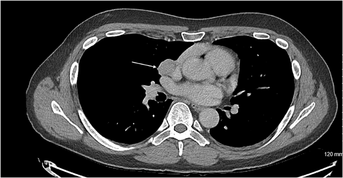
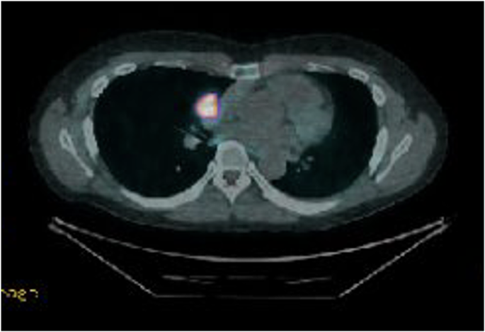
Figure 2: Radiological findings of the right adrenal tumor (white arrow)

(A) Trans-abdominal ultrasonography image and (B) coronal section of the trunk on MRI.

Obstetric-ultrasonography
Figure 3: Obstetric ultrasonography

(A) The plane used for biparietal diameter measurement, (B) the plane used for abdominal circumference measurement, (C) the plane used for femoral length measurement, and (D) the plane used for amniotic fluid index measurement.

Physical examination revealed typical signs of CS, such as a moon face, buffalo hump, and reddish-purple striae. In addition, laboratory findings showed elevated UFC, increased nocturnal serum cortisol levels (>5.0 μg/dL), and suppressed ACTH levels (<5.0 pg/mL). On the basis of these findings, the patient was diagnosed with ACTH-independent CS. Furthermore, imaging studies identified a right adrenal mass, leading to a final diagnosis of CS caused by a right adrenal tumor. Both central hypothyroidism and impaired glucose tolerance were considered secondary complications, primarily caused by hypercortisolemia due to CS. The serum potassium level was maintained at approximately 3.0 mEq/L after the administration of oral KCl. An increase in the nifedipine dose from 20 mg/day to 40 mg/day stabilized the blood pressure at approximately 140/90 mmHg (Figure 4A). Intensive insulin therapy with insulin lispro was initiated on hospital day 4 (Figure 4B), and the insulin dosage was gradually increased for postprandial hyperglycemia. The maximum insulin dose was 41 units/day on day 23 of hospitalization. Throughout this period, the UFC levels remained persistently elevated (Figure 4C).

Clinical-course-between-hospitalization-and-cesarean-delivery
Figure 4: Clinical course between hospitalization and cesarean delivery

(A) Blood pressure trend, (B) total dose of insulin, and (C) urinary free cortisol trend.

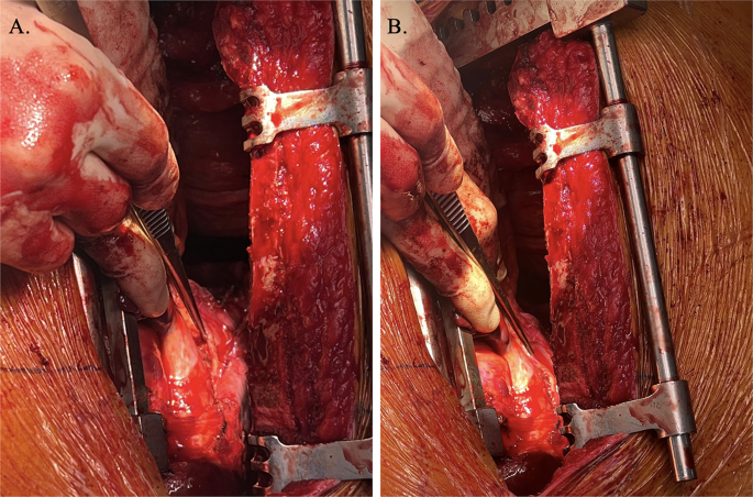
A clinical team of obstetricians, urologists, and endocrinologists discussed the treatment plans for CS and perinatal management. Pharmacological treatment had two problems: radicality and risk of fetal adrenal insufficiency due to placental passage of medication; therefore, we decided to perform adrenalectomy during pregnancy. At 28+3 weeks of gestation, a retroperitoneoscopic adrenalectomy was performed by urologists. After the induction of general anesthesia, the patient lay on the bed in a complete left lateral position (Figures 5A5B). Consequently, the endoscope and instrument ports were placed in the same configuration as those used in the conventional retroperitoneal approach for nonpregnant patients. Port placements were planned guided by abdominal ultrasonography to identify the uterine position, and the assistant port was positioned at a location that minimized potential interference with the uterus. The surgery was completed without complications. The operative time was 83 minutes, and bleeding was minimal. Histopathological examination indicated that the tumor was an adrenocortical adenoma (Figures 6A6C).

Photograph-showing-the-patient-in-the-left-lateral-decubitus-position-after-general-anethesia
Figure 5: Photograph showing the patient in the left lateral decubitus position after general anethesia

(A) Abdominal area and (B) dorsal area.

Histopathological-findings-of-the-right-adrenal-gland-(A,-B,-C)-and-placenta-(D)
Figure 6: Histopathological findings of the right adrenal gland (A, B, C) and placenta (D)

(A) Macroscopic view of the right adrenal gland showing the normal adrenal tissue (black asterisk) and the adrenal tumor (white asterisk). (B, C) Microscopic findings of the right adrenal gland and tumor (H&E staining).

(B) Normal adrenal gland (black asterisk) and adrenal tumor (white asterisk) separated by a thin fibrous capsule (black arrow).

(C) Tumor cells with abundant eosinophilic to clear cytoplasm arranged in a trabecular to microacinar growth pattern.

(D) Microscopic findings of the placenta (H&E staining) showing fibrin deposition within villous vessels (black arrow) and chorionic villi with loss of nuclear detail and crowding (black asterisk).

After surgery, the maternal glucose tolerance rapidly improved, and intensive insulin therapy with insulin lispro became unnecessary (Figure 4B). To avoid postoperative adrenal insufficiency, replacement therapy with hydrocortisone was initiated at 200 mg/day immediately after surgery, and the dosage was gradually tapered to 25 mg/day before delivery (Figure 4C). Maternal thyroid function normalized two weeks after surgery. At 29 weeks of gestation, oral nifedipine (40 mg/day) was stopped and blood pressure was monitored; however, high blood pressure was sustained. Therefore, oral nifedipine was resumed at 20 mg/day at 31 weeks of gestation. At approximately 33 weeks of gestation, the fetus exhibited slow growth, leading to a diagnosis of fetal growth restriction. The levels of serum soluble fms-like kinase 1 (sFlt-1)/placental growth factor (PlGF) were 173 (7990/46.1) at 33+0, 299 (11600/38.9) at 34+1, and 316 (15200/48.1) at 35+5 weeks. Trends in the estimated fetal body weight and standard deviation are shown in Figure 7. At 36+1 weeks of gestation, cardiotocography revealed severely prolonged deceleration regardless of the absence of uterine contraction, and an emergency cesarean section was performed. A male infant weighing 1,726 g and 41 cm in height, diagnosed as small for gestational age, was born with Apgar scores of 8 at one minute and 9 at five minutes. The umbilical arterial pH was 7.36. The size and weight of the placenta were 14.7 × 12.8 × 3.0 cm and 315 g, respectively, and histopathological examination revealed findings consistent with ischemic infarction (Figure 6D). Antihypertensive drugs administered to the mother were discontinued on day 8. The mother and neonate were discharged on POD 20. The child achieved normal development at the age of two years.

Trends-in-estimated-fetal-body-weight-(EFBW)-and-standard-deviation-(SD)
Figure 7: Trends in estimated fetal body weight (EFBW) and standard deviation (SD)

Discussion

This case illustrates adrenal CS in pregnancy, complicated by the subsequent development of fetal growth restriction, despite retroperitoneoscopic adrenalectomy at 28 weeks of gestation. Notably, a markedly increased maternal serum sFlt-1/PlGF ratio was detected at the time of diagnosis of fetal growth restriction. To the best of our knowledge, this is the first case in which angiogenic markers were evaluated in a pregnant woman with adrenal CS.

The coexistence of CS and pregnancy is extremely rare [4]. The primary reason for this rarity is infertility, often caused by the hypercortisolism characteristic of CS. Specifically, hypercortisolism suppresses the hypothalamic-pituitary-gonadal axis, leading to impaired follicular development and anovulation by disrupting the secretion of gonadotropin-releasing hormone (GnRH) [1,7]. Pregnancy poses significant challenges in patients with ACTH-dependent CS, in whom excessive ACTH production is accompanied by androgen overproduction. As a result, adrenal etiologies of CS are more common than pituitary-dependent etiologies during pregnancy [3]. Several factors make it difficult to diagnose CS during pregnancy. First, the characteristic physical findings of CS closely mimic physiological changes in normal pregnancy. For example, weight gain, abdominal striae, and edema are common symptoms of both conditions. Therefore, this overlap can cause delayed diagnosis or misdiagnosis of CS during pregnancy [3]. It has been reported that 21.5% of pregnant women with CS are diagnosed only after delivery [3]. Second, physiological hormonal changes during pregnancy complicate the diagnostic process. During gestation, the placenta produces corticotropin-releasing hormone (CRH) and ACTH [8]. Additionally, elevated estrogen levels increase the synthesis of corticosteroid-binding globulin, resulting in a state of physiological hypercortisolism in pregnant women [9,10]. Consequently, the dexamethasone suppression test, which is key to the diagnosis of CS, is often unreliable in pregnant women because of the high incidence of false-positive results [4].

Despite these diagnostic hurdles, certain findings are highly valuable in identifying CS during pregnancy. First, careful examination of physical signs specific to CS, such as skin thinning and the presence of wide, reddish-purple striae, is crucial. Second, the evaluation of diurnal cortisol rhythms was informative. While this rhythm is preserved in normal pregnancy, it is characteristically absent in CS. Therefore, measuring late-night serum cortisol levels is useful for differentiating between these two states [11]. Third, a 24-hour UFC level exceeding three times the upper limit of normal for non-pregnant individuals is strongly suggestive of CS [4,7,9]. In the present case, these key features were decisive for the diagnosis. We found wide, reddish-purple striae, a loss of diurnal cortisol rhythm, and a markedly elevated 24-hour UFC level. Based on these findings, we definitively diagnosed the patient with CS complicating pregnancy.

According to a systematic review of 263 pregnancies complicated by CS, untreated pregnant women were significantly more likely to develop PE than those treated beforehand (26.5% vs. 2.3%) [3]. PE is characterized by defective placentation and impaired spiral artery remodeling, leading to placental ischemia during early pregnancy. Placental ischemia produces sFlt-1, a splice variant of Flt-1 that binds to vascular endothelial growth factor and PlGF and serves as a biochemical marker of endothelial dysfunction that inhibits angiogenesis [12]. Systemic endothelial dysfunction leads to maternal hypertension, proteinuria, and damage to other organs, including the placenta. In this case, placental histopathology indicated ischemic changes without retroplacental hematoma. In addition, a marked elevation of the sFlt-1/PlGF ratio – resulting from both increased sFlt-1 and decreased PlGF – was detected, supporting the presence of placental ischemia due to impaired placentation in early pregnancy.

In this case, several factors may have contributed to the placental ischemia. First, poor control of maternal hyperglycemia or hypertension may have played a role. As hyperglycemia is known to induce oxidative stress [13], it is possible that hyperglycemia in early pregnancy causes placental ischemia indirectly via oxidative stress. Recent studies suggest that hypertension in early pregnancy may contribute to impaired placentation, thereby increasing the risk of subsequent superimposed PE [14,15]. Therefore, chronic hypertension associated with CS may also be related to placental ischemia, although the maternal outpatient blood pressure was within the normal range during early pregnancy in the present case. Second, chronic hypercortisolemia can directly contribute to abnormal placentation. Previous animal experiments have shown that elevated maternal serum cortisol levels enhance uterine arterial contractions [16], which may induce placental ischemia. Furthermore, chronic hypercortisolism may exceed the protective capacity of 11β-hydroxysteroid dehydrogenase type 2 (11β-HSD2), which shields the fetus from excessive cortisol, thereby directly affecting the fetus [17]. Based on these findings, it is presumed that irreversible placental damage had already occurred at the time of the surgical resection in this case. Preconceptional or at least early diagnosis and treatment of CS are crucial for preventing fetal growth restriction associated with superimposed PE after surgery.

The second trimester is generally considered the optimal period for adrenalectomy in pregnant patients with adrenal CS [18]; however, successful procedures have been reported even during the third trimester [6,19]. Endoscopic adrenalectomy is favored over open approaches owing to its reduced morbidity, although direct comparisons between the transperitoneal and retroperitoneal approaches in pregnancy are lacking. In non-pregnant patients, both approaches yield similar operative times, blood loss, and hospital stays [20]. In this case, the retroperitoneal approach was used. This technique offers several advantages during pregnancy as follows: it allows surgery in the lateral position, minimizes inferior vena cava compression by the gravid uterus, avoids entry into the peritoneal cavity, thereby preventing interference from the enlarged uterus, and reduces the risk of intra-abdominal inflammatory spread to the uterus and adjacent organs. Based on our experience and considering the potential advantages of the retroperitoneoscopic approach, we propose that retroperitoneoscopic adrenalectomy should be considered even in the early third trimester, as it may safely prolong gestation and reduce the need for preterm delivery.

Conclusions

This case highlights the challenges of managing adrenal CS during pregnancy. Uncontrolled CS may impair placental development during early pregnancy; therefore, preconceptional or at least early recognition and appropriate management are crucial to minimize the risk of subsequent fetal growth restriction. Further research is needed to clarify the pathophysiological relationship between hypercortisolism and impaired placentation in early pregnancy and to refine strategies for managing this rare but high-risk condition.

References

  1. Castinetti F, Brue T: Impact of Cushing’s syndrome on fertility and pregnancy. Ann Endocrinol (Paris). 2022, 83:188-90. 10.1016/j.ando.2022.04.001
  2. Eschler DC, Kogekar N, Pessah-Pollack R: Management of adrenal tumors in pregnancy. Endocrinol Metab Clin North Am. 2015, 44:381-97. 10.1016/j.ecl.2015.02.006
  3. Caimari F, Valassi E, Garbayo P, Steffensen C, Santos A, Corcoy R, Webb SM: Cushing’s syndrome and pregnancy outcomes: a systematic review of published cases. Endocrine. 2017, 55:555-63. 10.1007/s12020-016-1117-0
  4. Hamblin R, Coulden A, Fountas A, Karavitaki N: The diagnosis and management of Cushing’s syndrome in pregnancy. J Neuroendocrinol. 2022, 34:e13118. 10.1111/jne.13118
  5. Sammour RN, Saiegh L, Matter I, et al.: Adrenalectomy for adrenocortical adenoma causing Cushing’s syndrome in pregnancy: a case report and review of literature. Eur J Obstet Gynecol Reprod Biol. 2012, 165:1-7. 10.1016/j.ejogrb.2012.05.030
  6. Martínez García R, Martínez Pérez A, Domingo del Pozo C, Sospedra Ferrer R: Cushing’s syndrome in pregnancy. Laparoscopic adrenalectomy during pregnancy: the mainstay treatment. J Endocrinol Invest. 2016, 39:273-6. 10.1007/s40618-015-0345-0
  7. Younes N, St-Jean M, Bourdeau I, Lacroix A: Endogenous Cushing’s syndrome during pregnancy. Rev Endocr Metab Disord. 2023, 24:23-38. 10.1007/s11154-022-09731-y
  8. Sasaki A, Shinkawa O, Margioris AN, et al.: Immunoreactive corticotropin-releasing hormone in human plasma during pregnancy, labor, and delivery. J Clin Endocrinol Metab. 1987, 64:224-9. 10.1210/jcem-64-2-224
  9. Jung C, Ho JT, Torpy DJ, et al.: A longitudinal study of plasma and urinary cortisol in pregnancy and postpartum. J Clin Endocrinol Metab. 2011, 96:1533-40. 10.1210/jc.2010-2395
  10. Petraglia F, Sawchenko PE, Rivier J, Vale W: Evidence for local stimulation of ACTH secretion by corticotropin-releasing factor in human placenta. Nature. 1987, 328:717-19. 10.1038/328717a0
  11. Savas M, Mehta S, Agrawal N, van Rossum EF, Feelders RA: Approach to the patient: diagnosis of Cushing syndrome. J Clin Endocrinol Metab. 2022, 107:3162-74. 10.1210/clinem/dgac492
  12. Jung E, Romero R, Yeo L, et al.: The etiology of preeclampsia. Am J Obstet Gynecol. 2022, 226:S844-66. 10.1016/j.ajog.2021.11.1356
  13. González P, Lozano P, Ros G, Solano F: Hyperglycemia and oxidative stress: an integral, updated and critical overview of their metabolic interconnections. Int J Mol Sci. 2023, 24:9352. 10.3390/ijms24119352
  14. Ueda A, Hasegawa M, Matsumura N, et al.: Lower systolic blood pressure levels in early pregnancy are associated with a decreased risk of early-onset superimposed preeclampsia in women with chronic hypertension: a multicenter retrospective study. Hypertens Res. 2022, 45:135-45. 10.1038/s41440-021-00763-6
  15. Burton GJ, Jauniaux E: Pathophysiology of placental-derived fetal growth restriction. Am J Obstet Gynecol. 2018, 218:S745-61. 10.1016/j.ajog.2017.11.577
  16. Xiao D, Huang X, Bae S, Ducsay CA, Zhang L: Cortisol-mediated potentiation of uterine artery contractility: effect of pregnancy. Am J Physiol Heart Circ Physiol. 2002, 283:H238-46. 10.1152/ajpheart.00842.2001
  17. Albiston AL, Obeyesekere VR, Smith RE, Krozowski ZS: Cloning and tissue distribution of the human 11 beta-hydroxysteroid dehydrogenase type 2 enzyme. Mol Cell Endocrinol. 1994, 105:11-17. 10.1016/0303-7207(94)90176-7
  18. Wang Y, An Y, Hou X, et al.: Cushing’s syndrome in pregnancy secondary to adrenocortical adenoma: a case series and review. Endocrinol Diabetes Metab. 2024, 7:e00474. 10.1002/edm2.474
  19. Shaw JA, Pearson DW, Krukowski ZH, Fisher PM, Bevan JS: Cushing’s syndrome during pregnancy: curative adrenalectomy at 31 weeks gestation. Eur J Obstet Gynecol Reprod Biol. 2002, 105:189-91. 10.1016/s0301-2115(02)00148-3
  20. Nigri G, Rosman AS, Petrucciani N, et al.: Meta-analysis of trials comparing laparoscopic transperitoneal and retroperitoneal adrenalectomy. Surgery. 2013, 153:111-19. 10.1016/j.surg.2012.05.042

From https://www.cureus.com/articles/425273-adrenal-cushings-syndrome-in-pregnancy-complicated-by-fetal-growth-restriction-following-retroperitoneoscopic-adrenalectomy#!/

Cushing’s, Cancer and Other Serious Diseases

I was drawn to this blog post because the author mentioned that she had both Cushing’s and cancer, a kind of unusual combination.

1974 to Today: Seal it up
By Experience
I still haven’t heard what the consensus is on my aftercare: Cushing’s and Cancer. I don’t know what I will be expecting to feel like after surgery. My endocrinologist said that I should get sick after the surgery and need some kind of
1974 to Today – http://1974totoday.blogspot.com/

I don’t usually comment on blog posts but I did on this one because we seem to share so much, disease-wise.

I said

Hi, I was drawn to your blog post because I have a blog with the same name, Cushings & Cancer.

I had my Cushing’s long ago and my cancer (kidney aka renal cell carcinoma) was 3 years ago but I sure know where you’re coming for.

My surgeon contacted my endo for the amounts of steroids during surgery (they came through the IV) then post-op, they kept cutting my dose in half until I was back down to normal.
Generally, you stress-dose after surgery if you feel like you have a flu coming on. Has your endo given you Cortef or another steroid to take for emergencies like this? Sometimes, they will give you an injectible to be faster acting.

Best of luck with the cancer surgery AND your Cushing’s.
MaryO

I sure hope that this isn’t a trend, Cushies getting cancer although I know of a couple others on the boards getting cancer.

I suppose Cushing’s doesn’t make us any more immune to other diseases but it seems like it should.

Haven’t we already “done our time”?

OTOH, I have a friend with a serious cancer (aren’t they all?)  who recently learned that she has a second, unrelated, cancer.  Makes you wonder sometimes.

What other diseases have you had in addition to your Cushing’s?

Helping others learn more about Cushing’s/Acromegaly

I found this article especially interesting.  This question was asked of a group of endos at an NIH conference a few years ago – if you saw someone on the street who looked like they had symptoms of fill-in-the disease, would you suggest that they see a doctor.  The general answer was no.  No surprise there.

Patients, if you see someone who looks like s/he has Cushing’s, give them a discrete card.

Spread The Word! Cushing’s Pocket Reference

Robin Writes:

This has been a concern of mine for some time. Your post spurred me on to do something I’ve been meaning to do. I’ve designed something you can print that will fit on the business cards you can buy just about anywhere (Wal-mart included). You can also print on stiff paper and cut with a paper cutter or scissors. I’ve done a front and a back.

Cushing's Pocket Reference

Here are the links:

Front: This card is being presented by a person who cares.
Back (The same for everyone)

This Topic on the Message Boards

~~~~~~~~~~~~~~~~~~

And now, the article from http://www.guardian.co.uk/lifeandstyle/2009/nov/03/doctor-diagnosis-stranger:

Are doctors ever really off duty?

Which potentially serious symptoms would prompt them to stop and advise a stranger on a bus?

By Lucy Atkins

Bus

Passengers on a London bus. Photograph: David Levene

A Spanish woman of 55, Montse Ventura, recently met the woman she refers to as her “guardian angel” on a bus in Barcelona. The stranger – an endocrinologist – urged Ventura to have tests for acromegaly, a rare disorder involving an excesss of growth hormone, caused by a pituitary gland tumour. How had the doctor made this unsolicited diagnosis on public transport? Apparently the unusual, spade-like shape of Ventura’s hands was a dead giveaway.

But how many off-duty doctors would feel compelled to alert strangers to symptoms they spot? “If I was sitting next to someone on a bus with a melanoma, I’d say something or I wouldn’t sleep at night,” says GP Mary McCullins. “We all have a different threshold for interfering and you don’t want to terrify people, but this is the one thing I’d urge a total stranger to see a doctor about.” So what other symptoms might prompt a doctor to approach someone on the street?

Moon face

Cushing’s syndrome is another rare hormone disorder which can be caused by a non-cancerous tumour in the pituitary gland. “A puffy, rounded ‘moon face’ is one of the classic signs of Cushing’s,” says Dr Steve Field, chair of the Royal College of GPs. “In a social situation, I wouldn’t just say, ‘You’re dangerously ill’ but I’d try to elicit information and encourage them to see a doctor.”

Different-sized pupils

When one pupil is smaller than the other, perhaps with a drooping eyelid, it could be Horner’s syndrome, a condition caused when a lung tumour begins eating into the nerves in the neck. This can be the first obvious sign of the cancer. “I’d encourage someone to get this checked out,” says Dr Simon Smith, consultant in emergency medicine at the Oxford Radcliffe Hospitals Trust. “People often have an inkling that something’s wrong, and you might spur them to get help sooner.”

Clubbing fingers

Some people are born with club-shaped fingers, but if, over time, they become “drumstick-like”, this could signify serious problems such as lung tumours, chronic lung infections or congenital heart disease. “Because it happens gradually, some people disregard clubbing,” says Smith. “But I’d say something because it can be an important symptom in many serious illnesses.”

Lumpy eyelids

Whitish yellowy lumps around the eyelids can be a sign of high cholesterol, a major factor in heart disease. Sometimes you also get a yellow circle around the iris. “I would suggest they got a cholesterol test with these symptoms,” says Smith. “They can do something about it that could save their life.”

Suntan in unlikely places

A person with Addison’s disease, a rare but chronic condition brought about by the failure of the adrenal glands, may develop what looks like a deep tan, even in non sun-exposed areas such as the palms. Other symptoms (tiredness, dizziness) can be non-specific so the condition is often advanced by the time it is diagnosed. Addison’s is treatable with lifelong steroid replacement therapy. “If someone was saying they hadn’t been in the sun but had developed a tan, alarm bells would ring and I’d probably ask how they were feeling,” says McCullins.

Trench mouth

Putrid smelling breath – even if the teeth look perfect – can be a sign of acute necrotising periodontitis. “I’d be able to tell when someone walks through the door,” says dentist Laurie Powell. “But people become accustomed to it and don’t notice.” Untreated, the condition damages the bones and connective tissue in the jaw. It can also be a sign of other diseases such as diabetes or Aids.

Complete Surgical Resection of Cortisol Secreting Neuroendocrine Thymic Tumour

Abstract

Introduction

Neuroendocrine tumours of the thymus are extremely rare, with an estimated incidence of about 1 in 5 million people. Although data is limited, complete surgical resection remains the most significant prognostic factor for improved survival and disease-free outcomes, with adjuvant radiotherapy playing a role in cases where resection margins are close. This case report details the management of a cortisol secreting pT1bN0 atypical carcinoid of the thymus in a 43-year-old male.

Case report

43-year-old male presented with Cushing’s syndrome and was diagnosed with a cortical secreting atypical carcinoid of the thymus. He underwent a robotic thymectomy. Recurrent disease on a DOTATATE-PET CT scan resulted in a second surgery involving complete resection of the mediastinal tumour which had invaded the pericardium, as well as wedge resection of the lung and lymph node sampling. This was followed by adjuvant radiotherapy due to close proximity of the lesion to the margin (< 3 mm).

Discussion

Although paraneoplastic syndromes such as Cushing’s syndrome are rare manifestations of thymic neuroendocrine tumours and can result in challenging diagnoses, it is vital to have a high index of suspicion towards ectopic ACTH secretion in order to facilitate timely initiation of multimodal disease management for these patients including surgery and radiotherapy.

Conclusion

Surgical management has been shown to offer the greatest prognosis in terms of overall survival and disease-free survival. Adjuvant radiotherapy plays a role where resection margins are close.

Peer Review reports

Introduction

Neuroendocrine tumours of the thymus (NETT) are extremely rare, with an estimated incidence of about 1 in 5 million people [1], accounting for about 2–5% of thymic tumours and 0.4–3.4% of all carcinoid tumours [2]. Around 50% of neuroendocrine tumours of the thymus are hormonally active, with patients presenting with paraneoplastic symptoms such as Cushing’s syndrome due to ectopic production of ACTH. A review of 157 cases showed that males have a 3:1 increased risk of developing NETTs compared to women [3], with patients typically being heavy smokers and diagnosed between 40 and 60 years old. Atypical carcinoid tumours are differentiated from typical carcinoid tumours by their increased mitotic rate (2–10 per 2mm2/10 HPF) or areas of focal necrosis and account for about 40–50% of all thymic neuroendocrine tumours [1]. We present a case of a male patient who underwent complete excision and adjuvant radiotherapy for a neuroendocrine carcinoma with elevated mitotic count.

Case report

A previously fit and well 43-year-old male initially presented with features of Cushing’s syndrome, namely weight gain, hypertension and skin changes. He had a past medical history of polycystic kidney disease and asthma. The Cushing’s syndrome was found to be related to a 56 mm cortisol secreting pT1bN0 neuroendocrine carcinoma of the thymus. He underwent a right sided robotic thymectomy in July 2021.

A DOTATATE PET CT scan showed recurrence of his thymic carcinoid at the level of the groove between the right main pulmonary artery and right atrium, growing very close to the phrenic nerve. It was advised for him to undergo complete excision of the lesion requiring a joint approach from cardiac and thoracic surgeons. The patient subsequently underwent a median sternotomy, removal of recurrent mediastinal tumour invading the pericardium and wedge resection of the lung, lymph node sampling.

The patient was positioned supine. Femoral vessels were prepared in case there was bleeding which necessitated emergency bypass. Median sternotomy was performed with an oscillating saw due to previous right robotic thymectomy. The pericardium was opened. The tumour was identified at the level of the pulmonary vein and the cavo-atrial junction. En bloc resection of the tumour and the pericardium was performed, dissecting it away from the superior vena cava and the right atrium and wedge resection of the right lower lobe. The right phrenic was identified and spared. Due to previous surgery the phrenic nerve was surrounded by adhesions. A diaphragmatic plication was performed with 4 Ethibond no.5 sutures considering the risk of nerve palsy during dissection. The pericardium was reconstructed with a Prolene mesh fixed with Prolene 3/0.

Histology of the mediastinal tissue and right lung tissue sent showed a mitotic rate of more than 10 which according to the WHO classification of thoracic tumours, would make this a large cell neuroendocrine carcinoma. However, the morphological features were not of large cell type and therefore the tumour was best described as a NETT with elevated mitotic count. Histology confirmed the diagnosis as being a Neuroendocrine Carcinoma. The tumour had been excised completely with a 1.3 mm margin around the lesion. The patient required adjuvant radiotherapy due to the close proximity of the lesion to the margin (< 3 mm). A dose of 60 Gy over 30 daily fractions was selected in this postoperative adjuvant setting.

Discussion

This patient presented initially with Cushing’s syndrome associated with a cortisol secreting atypical carcinoid of the thymus. The excess glucocorticoid secretion presenting in the symptoms of Cushing’s syndrome can result from an ACTH secreting tumour of the pituitary which would be defined as Cushing’s disease, or less frequently from non-pituitary tumours secreting ACTH which would be defined as ectopic ACTH secretion [4]. Once the diagnosis of Cushing’s syndrome is made, it is essential to differentiate whether this is Cushing’s disease or an ectopic ACTH secretion. Current guidelines advise that inferior petrosal sinus sampling is the gold standard in distinguishing Cushing’s disease from ectopic ACTH secretion where a pituitary MRI is negative [5]. However, due to high cost, invasive nature and the risk of thromboembolic complications, investigations such as the CRH test and high dose dexamethasone suppression test are often preferred. A retrospective analysis looked at 719 patients with neuroendocrine tumours treated in EKPA-Laiko Hospital in Athens, Greece. They found that the prevalence of endocrine neoplastic syndromes in patients with neuroendocrine tumours was only 1.9% [6]. Kamp et al. studied the prevalence of specifically ectopic ACTH syndrome in 918 patients who had been diagnosed with either thoracic or gastroenteropancreatic neuroendocrine tumours. They found that 29 patients, or 3.2% had ectopic ACTH syndrome, with most of these cases being thoracic tumours and 4 of these patients having thymic tumours [7]. This study highlights that although the incidence of ectopic ACTH secretion in thoracic neuroendocrine tumours is relatively rare, resulting in challenging diagnosis, it is important to maintain a high index of suspicion in order to facilitate timely initiation of multimodal management such as surgery and radiotherapy.

A retrospective study at Fukuoka University Hospital looking at 9 cases of NETTs and 16 cases of thymic carcinomas, showed complete resection to be a statistically significant prognostic factor, with the 5-year survival rate and 5-year disease free survival rate being 87.5% and 75% in the thymic neuroendocrine tumour group, and 58.9% and 57.1% in the thymic carcinoma group respectively [8]. Chen et al. looked at a total of 104 patients diagnosed with NETTs, of which 97 underwent surgical resection, with 79 undergoing radical resection. The 1-year, 3-year and 5-year overall survival rates were found to be 91.8%, 70.2% and 54.6% respectively, with radical resection being found to be a significant factor in the overall survival of patients with NETTs [9]. Due to the rarity of NETTs, few cases are reported, and these studies are limited by their being retrospective in nature and spanning over several decades, possibly affecting the consistency and standardisation of patient treatment. However, it is important to note that complete radical resection of the tumour was consistently shown to be a strong prognostic factor in the overall survival and disease-free survival of patients and this should be attempted wherever possible.

The gentleman in our case required post operative radiotherapy due to the close proximity of the lesion to the margins. A large retrospective study looked at 205 patients treated for neuroendocrine thymic tumours, with 81 patients receiving radiotherapy and 70 out of the 81 receiving it as adjuvant therapy. In this particular study, radiotherapy was not shown to have any significant impact on survival outcomes [10]. An analysis of 12 cases of NETTs noted that 5 of the 6 patients who had presented with local recurrence during follow up had not received any post operative radiotherapy [1112], suggesting that adjuvant radiotherapy had resulted in better outcomes in terms of disease-free survival. A large retrospective analysis looking at 1489 patients diagnosed with NETTs or thymic carcinomas, found that the two factors which influenced positive survival outcomes were surgical resection and adjuvant radiotherapy. On sub-analysis, it was found that adjuvant radiotherapy had a good prognosis of survival in patients with margin positive tumours and was an independent predictor of survival for both thymic carcinomas and NETTs [12]. Wen et al. analysed 3947 patients in a retrospective study, including 293 neuroendocrine thymic tumours, 2788 thymomas and 866 thymic carcinomas. It was shown that post operative radiotherapy had a significant positive impact on overall survival and cancer specific survival in Masaoka-Koga stage III-IV thymic neuroendocrine tumour patients, as well as had a favourable impact on the overall survival of stage IIB patients [13]. Although these studies provide evidence of the benefits of adjuvant radiotherapy for favourable outcomes and prolonged survival, the last two studies are limited in that due to the rarity of neuroendocrine thymic tumours, they made up only 11 out of 329 (3.3%) of the thymic tumours analysed by Bakhos et al. and 7.4% of cases analysed by Wen et al.l, and the outcomes should therefore be interpreted with caution. The importance of multidisciplinary care involving maximal radical surgical excision as well as the involvement of oncologists and radiotherapists in the management of thoracic carcinoid tumours such as this, is emphasised in Busetto et al. [14].

Conclusion

Neuroendocrine tumours of the thymus (NETT) are exceedingly rare and often present diagnostic and therapeutic challenges due to their aggressive nature and associated paraneoplastic syndromes like Cushing’s syndrome. Complete surgical resection remains the most significant prognostic factor for improved survival and disease-free outcomes, with adjuvant radiotherapy playing a role in cases where resection margins are close. Although the data is limited by the rarity of the disease, existing studies suggest that a multidisciplinary, patient-specific approach, including surgery and radiotherapy, offers the best chance of long-term survival.

Fig. 1

figure 1

Axial CT showing the carcinoid tumour (demonstrated by arrow) in close proximity to innominate vein

Fig. 2

figure 2

DOTATE PET demonstrating significant uptake in carcinoid tumour

Fig. 3

figure 3

Intraoperative visualisation right phrenic nerve (demonstrated by forceps) overlying carcinoid tumour

Data availability

No datasets were generated or analysed during the current study.

References

  1. Bohnenberger H, Dinter H, König A, Ströbel P. Neuroendocrine Tumors Thymus Mediastinum J Thorac Dis [Internet]. 2017.

  2. Lau J, Ioan Cvasciuc T, Simpson D, C de Jong M, Parameswaran R. Continuing challenges of primary neuroendocrine tumours of the thymus: A concise review. Eur J Surg Oncol [Internet]. 2022.

  3. Chaer R, Massad MG, Evans A, Snow NJ, Geha AS. Primary neuroendocrine tumors of the thymus. Ann Thorac Surg. 2002. https://doi.org/10.1016/S0003-4975(02)03547-6.

    Article PubMed Google Scholar

  4. Isidori AM, Kaltsas GA, Pozza C, Frajese V, Newell-Price J, Reznek RH, et al. The ectopic adrenocorticotropin syndrome: clinical features, diagnosis, management, and long-term follow-up. J Clin Endocrinol Metab. 2006. https://doi.org/10.1210/jc.2005-1542.

    Article PubMed Google Scholar

  5. Ilias I, Torpy DJ, Pacak K, Mullen N, Wesley RA, Nieman LK. Cushing’s syndrome due to ectopic corticotropin secretion: twenty years’ experience at the National Institutes of Health. J Clin Endocrinol Metab. 2005. https://doi.org/10.1210/jc.2004-2527.

    Article PubMed Google Scholar

  6. Daskalakis K, Chatzelis E, Tsoli M, Papadopoulou-Marketou N, Dimitriadis GK, Tsolakis AV et al. Endocrine paraneoplastic syndromes in patients with neuroendocrine neoplasms. Endocrine [Internet]. 2018.

  7. Kamp K, Alwani RA, Korpershoek E, Franssen GJH, de Herder WW, Feelders RA. Prevalence and clinical features of the ectopic ACTH syndrome in patients with gastroenteropancreatic and thoracic neuroendocrine tumors. Eur J Endocrinol [Internet]. 2015.

  8. Midorikawa K, Miyahara S, Nishino N, Ueda Y, Waseda R, Shiraishi T, et al. Analysis of 25 surgical cases of thymic neuroendocrine tumors and thymic carcinoma. J Cardiothorac Surg. 2024. https://doi.org/10.1186/s13019-024-02723-w.

    Article PubMed PubMed Central Google Scholar

  9. Chen Y, Zhang J, Zhou M, Guo C, Li S. Real-world clinicopathological features and outcome of thymic neuroendocrine tumors: a retrospective single-institution analysis. Orphanet J Rare Dis. 2022. https://doi.org/10.1186/s13023-022-02366-x.

    Article PubMed PubMed Central Google Scholar

  10. Filosso PL, Yao X, Ahmad U, Zhan Y, Huang J, Ruffini E, et al. Outcome of primary neuroendocrine tumors of the thymus: a joint analysis of the international thymic malignancy interest group and the European society of thoracic surgeons databases. J Thorac Cardiovasc Surg. 2015. https://doi.org/10.1016/j.jtcvs.2014.08.061.

    Article PubMed Google Scholar

  11. Tiffet O, Nicholson AG, Ladas G, Sheppard MN, Goldstraw P. A clinicopathologic study of 12 neuroendocrine tumors arising in the thymus. Chest [Internet]. 2003.

  12. Bakhos CT, Salami AC, Kaiser LR, Petrov RV, Abbas AE. Thymic neuroendocrine tumors and thymic carcinoma: demographics, treatment, and survival. Innovations: Technology and Techniques in Cardiothoracic and Vascular Surgery. 2020. https://doi.org/10.1177/1556984520949287.

    Article PubMed Google Scholar

  13. Wen J, Chen J, Chen D, Liu D, Xu X, Huang L, et al. Evaluation of the prognostic value of surgery and postoperative radiotherapy for patients with thymic neuroendocrine tumors: a propensity-matched study based on the SEER database. Thorac Cancer. 2018. https://doi.org/10.1111/1759-7714.12868.

    Article PubMed PubMed Central Google Scholar

  14. Busetto A, Comacchio GM, Verzeletti V, Fares Shamshoum, Fortarezza F, Pezzuto F et al. Bilateral metachronous typical and atypical carcinoid tumors of the lung. Thorac Cancer. 2025;16(8).

Download references

Acknowledgements

No acknowledgements.

Funding

This report has not received funding.

Author information

Authors and Affiliations

  1. Department of Thoracic Surgery, Guy’s and St Thomas’ NHS Foundation Trust, London, England, UK

    Puiyee Sophia Chan, Akshay J. Patel, Ishaan Chauhan & Andrea Bille

  2. Institute of Immunology and Immunotherapy, University of Birmingham, Vincent Drive, Edgbaston, B15 2TT, England, UK

    Akshay J. Patel

Contributions

PSC and AJP wrote the main manuscript text and IC and AB prepared Figs. 12 and 3. All authors reviewed the manuscript.

Corresponding author

Correspondence to Akshay J. Patel.

Ethics declarations

Ethics approval and consent to participate

The authors are accountable for all aspects of the work, ensuring that questions related to the accuracy or integrity of any part of the work are appropriately investigated and resolved. All procedures performed in this study were in accordance with the ethical standards of the institutional and/or national research committee(s) and with the Helsinki Declaration (as revised in 2013).

Consent for publication

Written informed consent was obtained from the patients for the publication of this case report and accompanying images. A copy of the written consent is available for review by the editorial office of this journal.

Competing interests

The authors declare no competing interests.

Additional information

Publisher’s note

Springer Nature remains neutral with regard to jurisdictional claims in published maps and institutional affiliations.

Secondary Adrenal Insufficiency and Iatrogenic Cushing’s Syndrome in a 13-Year-Old Male With Vogt-Koyanagi-Harada Disease

ABSTRACT

Vogt-Koyanagi-Harada disease (VKH) is a rare autoimmune disorder, especially in children, requiring long-term corticosteroids. We report a 13-year-old male with VKH who developed iatrogenic Cushing’s syndrome and secondary adrenal insufficiency after prolonged prednisone treatment. Despite adding mycophenolate mofetil, tapering failed due to relapses. He showed weight gain, growth delay, striae, and suppressed cortisol and adrenocorticotropic hormone, confirming hypothalamic-pituitary-adrenal axis suppression. Hydrocortisone was given for stress coverage. A relapse followed steroid discontinuation. This case highlights the risk of endocrine complications in pediatric VKH and emphasizes the importance of early hormonal evaluation and individualized tapering during chronic steroid therapy.

KEYWORDS

Vogt-Koyanagi-syndrome
Cushing syndrome
Adrenal insufficiency
Pediatrics

INTRODUCTION

Vogt-Koyanagi-Harada disease (VKH) is a rare autoimmune disorder that can significantly affect the eyes, skin, and central nervous system (Stern & Nataneli, 2025). Among the various forms of autoimmune uveitis, VKH is particularly notable for its broad clinical spectrum, encompassing ocular, neurologic, and cutaneous manifestations (Herbort & Mochizuki, 2007). In pediatric patients, age-specific considerations become paramount, as prolonged corticosteroid therapy is frequently required to control inflammation but can result in serious endocrine complications. One such complication is iatrogenic Cushing’s syndrome (ICS), which may predispose to secondary adrenal insufficiency (SAI), especially when steroid withdrawal is abrupt or inadequately tapered (Improda et al., 2024Prete & Bancos, 2021). Despite increasing recognition of pediatric VKH, endocrine consequences of its treatment remain underreported.
We present the case of a 13-year-old male with VKH who displayed overt signs of hypercortisolism and biochemical evidence of adrenal suppression after discontinuing corticosteroids, underscoring the importance of vigilant monitoring and a carefully structured tapering protocol in pediatric patients requiring long-term steroid therapy. Given that many pediatric patients with VKH and steroid-related complications are managed not only by pediatric endocrinologists but also by pediatric providers, including nurse practitioners, this case highlights aspects relevant to a broad clinical audience.

CASE PRESENTATION

A 13-year-old male with a known history of VKH was referred to our clinic for growth and pubertal assessment following significant weight gain and clinical features suggestive of ICS. His perinatal period was uneventful; he was born at term via cesarean section for maternal indications (bicornuate uterus), with a birth weight of 2980 g and a length of 49 cm. Family history was notable for celiac disease in the mother, mixed hypercholesterolemia in the father, vitiligo in the maternal grandfather, and autoimmune diseases (Sjögren’s syndrome and multiple sclerosis) in second-degree maternal relatives.
The patient first presented, at age 11 years and 11 months, with redness, pain, and photophobia of the right eye [Figure 1]. Initial ophthalmological examination revealed panuveitis, with signs of posterior synechiae and optic disc edema. Fluorescein and indocyanine green angiography confirmed bilateral granulomatous involvement. Systemic workup excluded other infectious and autoimmune causes of uveitis. Neurological imaging revealed a non-specific thalamic lesion, classified as a radiological isolated syndrome, with no clinical neurological deficits.
FIGURE 1

  1. Download: Download high-res image (311KB)
  2. Download: Download full-size image

FIGURE 1. Timeline of notable events. Timeline summarizing key events including clinical course, treatments, and relapses.

Abbreviations: ACTH, adrenocorticotropic hormone; VKH, Vogt-Koyanagi-Harada disease.
Oral prednisone (25 mg/day) was initiated, along with topical ocular corticosteroids, leading to clinical improvement. The first tapering and discontinuation of prednisone occurred after seven months of therapy. Three months later, a clinical relapse occurred, requiring re-initiation of prednisone and subsequent addition of mycophenolate mofetil as a steroid-sparing agent. Prednisone was then tapered and discontinued again after another seven months of treatment. Over the course of therapy, the patient gained approximately 15 kg and developed progressive cushingoid features [Table 1].

TABLE 1. Clinical and biochemical features of ICS and SAI in the patient

Empty Cell Clinical Findings Interpretation
Growth and development Height: 143.5 cm (3rd percentile); mid-parental height: 171 ± 8 cm Growth deceleration likely related to chronic glucocorticoid exposure and ICS
Weight and body composition Weight: 53.3 kg (75th–90th percentile); BMI: 25.8 kg/m²; central obesity Suggestive of glucocorticoid-induced lipogenesis and altered fat distribution
Skin and soft tissue Striae rubrae on flanks; mild dorsal fat pad (“buffalo hump”) Classic phenotypic features of ICS
Pubertal status Tanner stage I; testicular volume 5–6 mL; pubic hair stage I Early puberty with preserved testicular volume; no signs of delayed or precocious puberty
HPA axis function Cortisol: 0.5 → 9.9 → 3.1 µg/dL; ACTH: 7–23 pg/mL Suppressed HPA axis consistent with SAI
Glucose metabolism HbA1c: 5.9%; fasting glucose: 72 mg/dL; insulin: 16.9 mcU/mL Normal glucose metabolism; mild hyperinsulinemia possibly due to steroid exposure
Thyroid function TSH: 2.32 µU/mL; free T4: 1.59 ng/dL Euthyroid; no evidence of central or primary thyroid dysfunction
Neurologic imaging Right thalamic signal abnormality; stable; no neurological deficits No CNS involvement of VKH; imaging excluded alternative diagnoses
Family history Autoimmune conditions in maternal relatives; vitiligo in grandfather Suggests genetic predisposition to autoimmune diseases; relevant to VKH etiology
Therapeutic course Initial improvement with prednisone; relapses on tapering; mycophenolate added; steroids reintroduced Demonstrates difficulty in achieving steroid-free remission and the need for steroid-sparing agents
Abbreviations: ACTH, adrenocorticotropic hormone; BMI, body mass index; CNS, central nervous system; HPA, hypothalamic-pituitary-adrenal; ICS, iatrogenic Cushing’s syndrome; SAI, secondary adrenal insufficiency; TSH, thyroid-stimulating hormone; VKH, Vogt-Koyanagi-Harada disease.
Summary of patient’s clinical signs and biochemical parameters during corticosteroid therapy, including features of ICS and evidence of SAI.
Laboratory testing during steroid tapering attempts revealed HbA1c of 5.9% (41 mmol/mol), fasting glucose of 72 mg/dL, and insulin of 16.9 mcU/mL; morning serum cortisol was markedly reduced (0.5 mcg/dL; ref. 2.4–22.9), raising concerns for SAI. Gonadotropins (follicle-stimulating hormone 4.3 mcU/mL, luteinizing hormone 1.1 mcU/mL) and testosterone (0.03 ng/mL) were consistent with early puberty. Thyroid function (thyroid-stimulating hormone 2.32 mcU/mL, free thyroxine 1.59 ng/dL) and celiac serology were normal. Brain magnetic resonance imaging confirmed a stable right thalamic signal abnormality and minor asymmetry of cerebral arteries, in line with prior findings; cardiac and abdominal ultrasounds were unremarkable.
When first evaluated in our endocrinology clinic (at age 13 years and 6 months), the patient’s height was 143.5 cm (3rd percentile; mid-parental height target: 171 ± 8 cm), and his weight was 53.3 kg (75th–90th percentile), corresponding to a body mass index of 25.8 kg/m². He exhibited central obesity, striae rubrae on the flanks, and a mild dorsal hump. Genital examination showed bilateral testicular volumes of 5–6 mL and pubic hair at Tanner stage I, compatible with early puberty. The remainder of the physical exam was unremarkable.
In light of clinical and biochemical evidence of hypothalamic-pituitary-adrenal (HPA) axis suppression, further hormonal testing was performed. Serum cortisol had partially recovered (9.9 mcg/dL; ref. 2.7–18.4) with adrenocorticotropic hormone (ACTH) at 23.1 pg/mL (ref. 7.3–63.3). Hydrocortisone was prescribed for use during stressful events. However, two months after prednisone discontinuation, at age 13 years and 8 months, a clinical relapse of VKH occurred, requiring escalation of mycophenolate mofetil and re-initiation of prednisone therapy.
The patient currently remains under combined rheumatologic, ophthalmologic, and endocrinologic management. Steroids have been successfully tapered and discontinued, but signs of chronic adrenal suppression and cushingoid features persist. Mycophenolate mofetil is ongoing as maintenance immunosuppression, and adrenal function is being closely monitored.

DISCUSSION

VKH is a rare granulomatous autoimmune condition targeting melanocyte-containing tissues, including the uveal tract, meninges, inner ear, and skin. While more frequently diagnosed in adults, pediatric-onset VKH is increasingly recognized and often presents with bilateral panuveitis, optic disc edema, serous retinal detachments, and systemic manifestations such as meningismus, tinnitus, hearing loss, vitiligo, and poliosis (Abu El-Asrar et al., 2021Reiff, 2020). Early and aggressive immunosuppression is essential to prevent chronic recurrent uveitis and progressive vision loss (Abu El-Asrar et al., 2008).
Systemic corticosteroid therapy—using high-dose oral prednisone or intravenous pulse methylprednisolone—is the first-line treatment for pediatric VKH, and is effective in rapidly controlling intraocular inflammation and achieving favorable visual outcomes when initiated early (Leal et al., 2024Reiff, 2020). Gradual tapering of corticosteroids over at least six months is critical to minimize recurrence and prevent chronic disease evolution (Ei Ei Lin Oo et al., 2020Wang et al., 2023). Rapid tapering is associated with higher rates of relapse and chronicity. Nonetheless, corticosteroid monotherapy is often insufficient to prevent long-term recurrence and chronic complications in pediatric VKH (Abu El-Asrar et al., 2021Park et al., 2022Sakata et al., 2015). Early addition of immunosuppressive agents—such as mycophenolate mofetil or methotrexate—within three months of disease onset improves long-term control, reduces the risk of chronic recurrent uveitis, and enhances visual outcomes (Ei Ei Lin Oo et al., 2020Park et al., 2022). Long-term remission rates are higher when immunosuppressive therapy is maintained for several years with sustained inflammation control (Wang et al., 2023).
Children are especially vulnerable to the adverse effects of prolonged corticosteroid exposure, including growth failure, pubertal delay, obesity, insulin resistance, ICS, and suppression of the HPA axis with subsequent SAI (Bornstein et al., 2016Messazos & Zacharin, 2016Santos-Oliveira et al., 2025). ICS results from chronic exposure to supraphysiologic doses of glucocorticoids and may present with weight gain, central obesity, facial rounding, and violaceous striae—many of which were observed in our patient. In children, these manifestations may overlap with common features of puberty or lifestyle-related obesity, making early diagnosis more challenging (Savage & Storr, 2012). SAI is a potentially life-threatening complication that occurs when exogenous glucocorticoids suppress the endogenous production of corticotropin-releasing hormone and ACTH. The risk is highest with longer treatment durations (typically > 12 weeks) and higher cumulative doses, particularly with long-acting steroids such as betamethasone or dexamethasone (Beuschlein et al., 2024).
Our patient presented with markedly reduced morning cortisol levels and low-normal ACTH, consistent with central adrenal suppression. Partial biochemical recovery occurred months after discontinuation, yet persistently suboptimal cortisol levels indicated incomplete restoration of HPA function. These findings align with a meta-analysis by Broersen et al., which showed that although adrenal recovery improves over time, a significant proportion of patients remain functionally insufficient even six months after stopping corticosteroids (Broersen et al., 2015).
To our knowledge, this is among the first reported pediatric cases of VKH complicated by both ICS and SAI. Although the literature contains extensive documentation of glucocorticoid side effects in autoimmune and inflammatory conditions (Arroyo et al., 2023), there remains a notable gap in addressing endocrine sequelae within VKH, particularly in children. Most published pediatric VKH case reports focus on ophthalmologic or immunologic outcomes, with limited attention to longitudinal hormonal monitoring and risk mitigation. VKH is rare in childhood, representing an uncommon cause of uveitis, with pediatric-onset forms accounting for fewer than 10% of all VKH cases (Martin et al., 2010Yang et al., 2023). Several works have documented its course and treatment (Abu El-Asrar et al., 2008Albaroudi et al., 2020Sadhu et al., 2024); none of the reports explicitly addressed endocrine complications, highlighting a major gap in longitudinal follow-up and inter-specialty collaboration in such cases.
The recent 2024 Joint Clinical Guideline from the European Society of Endocrinology and the Endocrine Society offers important insight into the diagnosis and management of glucocorticoid-induced adrenal insufficiency (Beuschlein et al., 2024). Although not providing pediatric-specific recommendations, it emphasizes that children are included among at-risk populations, and that the same diagnostic and tapering principles apply across age groups. It highlights that the risk of SAI depends not only on dose and duration, but also on the glucocorticoid formulation, route of administration, and individual susceptibility. The guideline recommends transitioning from long-acting to short-acting glucocorticoids (e.g., prednisone or hydrocortisone) to facilitate tapering and adrenal recovery. Tapering should begin only after adequate disease control and must proceed gradually—especially once physiologic dose equivalents are reached (4–6 mg/day of prednisone). Morning serum cortisol serves as the initial screening tool for HPA recovery, with levels > 10 µg/dL (> 300 nmol/L) indicating recovery and < 5 µg/dL (< 150 nmol/L) indicating suppression. Importantly, symptoms of glucocorticoid withdrawal (e.g., fatigue, myalgias, mood changes) may mimic adrenal insufficiency and require temporary increases in glucocorticoid dose and a slower taper.
In our case, hydrocortisone was prescribed for use during stress, such as illness or surgery, in accordance with these recommendations. Given his partial biochemical recovery, the patient was also advised to carry steroid warning documentation and to continue close endocrine follow-up. This approach reflects best practice in managing patients transitioning off chronic corticosteroid therapy, particularly in pediatric populations where risks are amplified (Beuschlein et al., 2024).
We strongly advocate for multidisciplinary collaboration in managing complex VKH cases [Figure 2]. Ophthalmologists and rheumatologists should remain alert to endocrine warning signs such as growth deceleration, cushingoid appearance, and fatigue (Santos-Oliveira et al., 2025), while endocrinologists should consider autoimmune or inflammatory etiologies in children with ICS or SAI. Importantly, the early use of steroid-sparing immunosuppressants—as was done with mycophenolate mofetil in our case—can reduce glucocorticoid burden and mitigate downstream complications. Agents such as azathioprine, methotrexate, or mycophenolate have demonstrated efficacy in reducing steroid dependence in pediatric uveitis (Simonini et al., 2013Sood & Angeles-Han, 2017).
FIGURE 2

  1. Download: Download high-res image (519KB)
  2. Download: Download full-size image

FIGURE 2. Multidisciplinary management plan for pediatric VKH with chronic corticosteroid therapy. Schematic representation of the recommended multidisciplinary team for pediatric patients with VKH requiring prolonged corticosteroid therapy. The model emphasizes collaboration among health professionals for early recognition and management of VKH manifestations.

(abbreviations: CNS, central nervous system; HPA, hypothalamic-pituitary-adrenal; VKH, Vogt-Koyanagi-Harada disease).

CONCLUSION

This case highlights the dual endocrine risks associated with prolonged corticosteroid therapy in pediatric patients with VKH: ICS and SAI. It underscores the importance of routinely monitoring growth, pubertal development, and HPA axis function both during and after steroid treatment.
Given the widespread use of systemic corticosteroids in pediatric inflammatory disorders, proactive endocrine screening, multidisciplinary collaboration, and adherence to guideline-based tapering protocols are essential to ensure effective disease management while minimizing preventable hormonal complications. Further research and the development of pediatric-specific guidelines are warranted to optimize endocrine care in children receiving long-term glucocorticoid therapy.

REPORTING CHECKLIST DISCLOSURE

We are submitting this case report using the CARE checklist.

DATA AVAILABILITY STATEMENT

Data sharing is not applicable to this article as no new data were created or analyzed in this study.

FUNDING

The authors did not receive support from any organization for the submitted work.

PATIENT CONSENT

Written informed consent and permission to share this case were obtained from the legal guardians/parents.

ETHICAL STATEMENTS

Please find attached the AIFA regulation regarding observational studies, provided in Italian. For your convenience, we have translated the relevant section (highlighted in light blue, pages 7-8) into English:
“The registration of studies covered by this provision in the Register of Observational Studies (RSO) is mandatory for review by the Ethics Committee, except for the exemptions listed below. This guideline does not apply to the following categories: […] Case reports and case series (typically involving 3-5 patients at most) that do not have a methodological approach qualifying them as clinical studies.”
Our study falls precisely into the category of a case report, rather than a clinical study.

CRediT authorship contribution statement

Roberto Paparella: Writing – original draft, Conceptualization. Irene Bernabei: Writing – original draft. Arianna Bei: Writing – original draft. Cinzia Fiorentini: Resources. Norma Iafrate: Resources. Roberta Lucibello: Resources. Francesca Pastore: Resources. Ida Pucarelli: Writing – review & editing, Supervision, Conceptualization. Luigi Tarani: Writing – review & editing, Supervision.

CONFLICTS OF INTEREST

None to report.

REFERENCES