


April is always Cushing’s Awareness Challenge month because Dr. Harvey Cushing was born on April 8th, 1869.
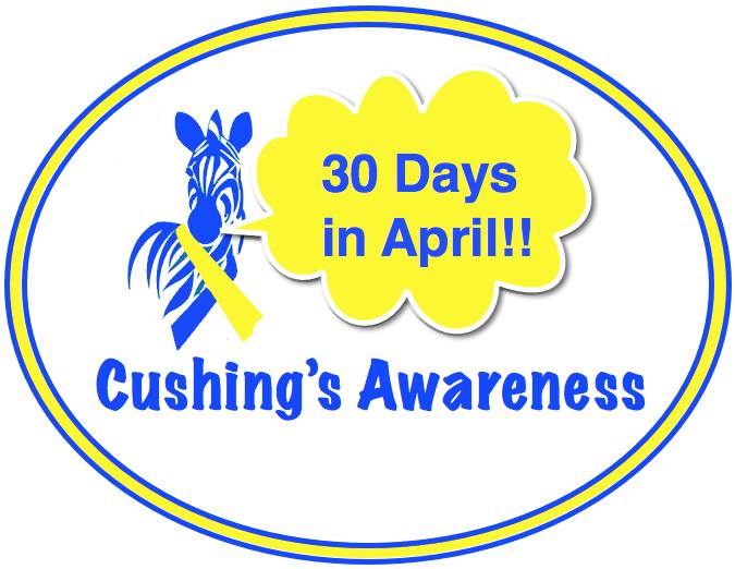
Thanks to Robin for this wonderful past logo! I’ve participated in these 30 days for Cushing’s Awareness several times so I’m not quite sure what is left to say this year but I always want to get the word out when I can.
As I see it, there have been some strides the diagnosis or treatment of Cushing’s since last year. More drug companies are getting involved, more doctors seem to be willing to test, a bit more awareness, maybe.

How fitting that this challenge should begin on April Fool’s Day. So much of Cushing’s Syndrome/Disease makes us Cushies seem like we’re the April Fool. Maybe, just maybe, it’s the doctors who are the April Fools…
Doctors tell us Cushing’s is too rare – you couldn’t possibly have it. April Fools!
All you have to do is exercise and diet. You’ll feel better. April Fools!
Those bruises on your legs? You’re just clumsy. April Fools!
Sorry you’re growing all that hair on your chin. That happens as you age, you know. April Fools!
Did you say you sleep all day? You’re just lazy. If you exercised more, you’d have more energy. April Fools!
You don’t have stretch marks. April Fools!
You have stretch marks but they are the wrong [color/length/direction] April Fools!
The hump on the back of your neck is from your poor posture. April Fools!
Your MRI didn’t show a tumor. You couldn’t have Cushing’s. April Fools!
This is all in your mind. Take this prescription for antidepressants and go home. April Fools!
If you have this one surgery, your life will get back to normal within a few months. April Fools!
What? You had transsphenoidal surgery for Cushing’s? You wasted your time and money. April Fools!
I am the doctor. I know everything. Do not try to find out any information online. You could not have Cushing’s. It’s too rare… April FOOL!
All this reminds me of a wonderful video a message board member posted a while ago:
So now – who is the April Fool? It wasn’t me. Don’t let it be you, either!

Filed under: Cushing's, Diagnostic Testing, Rare Diseases, symptoms, Treatments | Tagged: April Fool, awareness, bruise, Cushing's Awareness Challenge 2016, diagnosis, doctors, Dr. Harvey Cushing, drug companies, endocrinologist, exercise, fatigue, hirsuitism, MRI, pituitary, sleep, stretch marks, surgery, symptoms, transsphenoidal, treatment, tumor, video, weight | 2 Comments »


