First-of-its Kind Hormone Replacement Treatment Shows Promise in Patient Trials

Cortisol
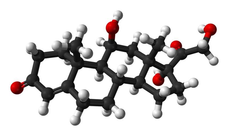
Ball-and-stick model of the cortisol (hydrocortisone) molecule. Credit: Public Domain

A first-of-its kind hormone replacement therapy that more closely replicates the natural circadian and ultradian rhythms of our hormones has shown to improve symptoms in patients with adrenal conditions. Results from the University of Bristol-led clinical trial are published today in the Journal of Internal Medicine.

Low levels of a key hormone called cortisol is typically a result of conditions such as Addison’s and congenital adrenal hyperplasia. The hormone regulates a range of vital processes, from cognitive processes such as memory formation, metabolism and immune responses, through to  and blood sugar levels. When low, it can trigger symptoms of debilitating fatigue, nausea, , dangerously  and depression. Although rare, these adrenal conditions require lifelong daily hydrocortisone replacement therapy.

Although existing oral hormone replacement treatment can restore , it is still associated with an impaired quality of life for patients. Scientists believe this is because the current treatment does not mimic the body’s normal physiological timing, missing cortisol’s anticipatory rise and lacking its underlying ultradian and .

The new “pulsatility” therapy, the culmination of ten years’ research by the Bristol team, is designed to deliver standard hydrocortisone replacement to patients via a pump which replicates more closely cortisol’s natural rhythmic secretion pattern. The pulsatile subcutaneous pump has now revealed promising results in its first clinical trial.

Twenty participants aged 18 to 64 years with adrenal insufficiency conditions were assessed during the double-blinded PULSES six-week trial and treated with usual dose hydrocortisone replacement therapy administered either via the pump or the standard three times daily oral treatment.

While only psychological and metabolic symptoms were assessed during the trial, results revealed the pump therapy decreased fatigue by approximately 10%, improved mood and increased patient energy levels by 30% first thing in the morning—a key time frame when many patients struggle. Patient MRI scans also revealed alteration in the way that the brain processes emotional information.

Dr. Georgina Russell, Honorary Lecturer at the University’s Bristol Medical School, and the lead author, explained, “Patients on cortisol replacement therapy often have side effects which makes it difficult for them to lead normal lives. We hope this  will offer greater hope for the thousands of people living with hormone insufficiency conditions.”

Stafford Lightman, a neuroendocrinology expert and Professor of Medicine at Bristol Medical School: Translational Health Sciences (THS), and the study’s joint lead author, added, “Besides reduction in dosage, cortisol replacement has remained unchanged for many decades. It is widely recognized that current replacement therapy is unphysiological due to its lack of pre-awakening surge, ultradian rhythmicity, and post dose supraphysiological peaks. The new therapy clearly shows that the timing of cortisol delivery- in line with the body’s own rhythmic pattern of cortisol secretion—is important for normal cognition and behavior.

“Our findings support the administration of hormone  that mimics natural physiology, and is one of the first major advances in adrenal insufficiency treatment to date.”

Joe Miles, a participant on the PULSES trial, explained, “The Crono P pump has been life-changing. I noticed a very quick improvement compared to tablets when I was on the PULSES study. I went from feeling tired all the time to having sudden energy.

“When the PULSES study ended and I had to return the pump, I simply couldn’t cope with going back to how I used to be, so I made it my mission to write to as many doctors to have it prescribed privately.

“I’ve now been on it for six years and have introduced a number of other people with Addison’s disease to the pump, and all of them have said it’s life changing. Some have gone from being seriously ill to feeling better than they have done for years.”

Dr. Russell said, “Approximately 1% of the UK population is taking steroids at any moment in time; these individuals can experience debilitating psychological side effects. This trial has shown that even at physiological levels, brain functioning is disrupted and that we need to explore not only the dose but the pattern of steroids delivery when considering any type of steroid treatment.”

More information: Ultradian hydrocortisone replacement alters neuronal processing, emotional ambiguity, affect and fatigue in adrenal insufficiency: The PULSES trial, Journal of Internal Medicine (2023). DOI: 10.1111/joim.13721

Journal information: Journal of Internal Medicine

How AI Could Help Make Brain Surgery Safer

I’m not sure I like this!

STORY: Could artificial intelligence be used to make brain surgery safer?

At this university in London, trainee surgeon Danyal Khan is taking part in a mock operation during which he’s assisted by a real-time video feed, as is typical in brain surgery.

But what’s new here is that the footage is being analyzed by AI to help Khan better understand what he’s seeing.

The AI system, which is under development at University College London (UCL), highlights sensitive or critical structures in the brain.

Neurosurgeon Hani Marcus believes it has the potential to make brain surgery safer and more effective:

“So I’m very bullish that in the medium to long term, the A.I. will be helping lots of surgeons do lots of operations better than they otherwise can.”

Marcus says the AI system analyzed video of more than 200 pituitary gland tumor operations, and gained around 10 years-worth of experience in a fraction of the time.

That knowledge means the AI can now not only help navigate to the correct area of the brain, but also know what should be happening at any stage of the procedure, making it a valuable training aid.

“So, what we’re really trying to do is apply AI or artificial intelligence to support surgeons doing brain tumor surgery at the base of the brain. And what this practically entails is us training the AI with hundreds of videos, telling it, if you like, what structures are what and then at some point over that period, the AI becomes really good itself at recognising things, and able to support other surgeons who’re perhaps less experienced in advising them what to do next.”

Assistant Professor of Robotics and A.I. Sophia Bano explains how that might look in a real operation:

“There can be scenarios where clinicians, unintentionally, are very close to a very critical structure such as the optic nerve. This can have, any damage or a slight more pressure on the optic nerve, can have long term complications on the patient side. So, this whole tool will alert the surgeon during the procedure if there is any risk of potential complication so they can recalibrate themselves during the procedure.”

Khan, who was also involved in developing the software that is now helping him learn, says the A.I. system has been a valuable tool in his training.

It could also provide him with step-by-step guidance during a procedure, similar to having a senior surgeon standing over your shoulder.

“I think as I progress as a surgeon, there might be stages where I wonder, you know, have I done enough of a particular part of the procedure and should I move on? And actually having that sort of assistant in the background as a reassurance to look at and say, ‘well, yeah, actually, at this stage, out of the hundreds of videos of experts that this algorithm has watched, the experts would probably start moving on to the next phase’. It’s a useful double check.”

According to UCL, the system could be ready to be used in operating theaters within two years.

From https://news.yahoo.com/ai-could-help-brain-surgery-080156167.html

​

Hormones and High Blood Pressure: Study Reveals Endocrine Culprits and Targeted Treatments

In a recent study published in Hypertension Research, scientists examine the endocrine causes of hypertension (HTN) and investigate the efficacy of treatments to alleviate HTN.


What is HTN?

About 30% of the global population is affected by HTN. HTN is a modifiable cardiovascular (CV) risk factor that is associated with a significant number of deaths worldwide.

There are two types of HTN known as primary and secondary HTN. As compared to primary HTN, secondary HTN causes greater morbidity and mortality.

The most common endocrine causes of HTN include primary aldosteronism (PA), paragangliomas and pheochromocytomas (PGL), Cushing’s syndrome (CS), and acromegaly. Other causes include congenital adrenal hyperplasia, mineralocorticoid excess, cortisol resistance, Liddle syndrome, Gordon syndrome, and thyroid and parathyroid dysfunction.

What is PA?

PA is the most common endocrine cause of hypertension, which is associated with excessive aldosterone secretion by the adrenal gland and low renin secretion. It is difficult to estimate the true prevalence of PA due to the complexity of its diagnosis.

Typically, the plasma aldosterone-to-renin ratio (ARR) is measured to diagnose PA. The diagnosis of PA can also be confirmed using other diagnostic tools like chemiluminescent enzyme immunoassays (CLEIAs) and radio immune assay (RIA).

Continuous aldosterone secretion is associated with organ damage due to chronic activation of the mineralocorticoid (MR) receptor in many organs, including fibroblasts and cardiomyocytes. An elevated level of aldosterone causes diastolic dysfunction, endothelial dysfunction, left ventricular hypertrophy, and arterial stiffness.

Increased aldosterone secretion also leads to obstructive sleep apnea and increases the risk of osteoporosis. This is why individuals with PA are at a higher risk of cardiovascular events (CVDs), including heart failure, myocardial infarction, coronary artery disease, and atrial fibrillation.

PA is treated by focusing on normalizing potassium and optimizing HTN and aldosterone secretion. Unilateral adrenalectomy is a surgical procedure proposed to treat PA.

Young patients who are willing to stop medication are recommended surgical treatment. The most common pharmaceutical treatment for PA includes mineralocorticoid receptor antagonists such as spironolactone and eplerenone.

Pheochromocytomas and paragangliomas

PGL are tumors that develop at the thoracic-abdominal-pelvic sympathetic ganglia, which are present along the spine, as well as in the parasympathetic ganglia located at the base of the skull. The incidence rate of PGL is about 0.6 for every 100,000 individuals each year. PGL tumors synthesize excessive catecholamines (CTN), which induce HTN.

Some of the common symptoms linked to HTN associated with PGL are palpitations, sweating, and headache. PGL can be diagnosed by determining metanephrines (MN) levels, which are degraded products of CTN. Bio-imaging tools also play an important role in confirming the diagnosis of PGL.

Excessive secretion of CTN increases the risk of CVDs, including Takotsubo adrenergic heart disease, ventricular or supraventricular rhythm disorders, hypertrophic obstructive or ischaemic cardiomyopathy, myocarditis, and hemorrhagic stroke. Excessive CTN secretion also causes left ventricular systolic and diastolic dysfunction.

Typically, PGL treatment is associated with surgical procedures. Two weeks before the surgery, patients are treated with alpha-blockers. For these patients, beta-blockers are not used as the first line of treatment without prior use of alpha-adrenergic receptors.

Patients with high CTN secretion are treated with metyrosine, as this can inhibit tyrosine hydroxylase. Hydroxylase converts tyrosine into dihydroxyphenylalanine, which is related to CTN synthesis.

What is CS?

CS, which arises due to persistent exposure to glucocorticoids, is a rare disease with an incidence rate of one in five million individuals each year. The most common symptoms of CS include weight gain, purple stretch marks, muscle weakness, acne, and hirsutism. A high cortisol level causes cardiovascular complications such as HTN, hypercholesterolemia, and diabetes.

CS is diagnosed based on the presence of two or more biomarkers that can be identified through pathological tests, such as salivary nocturnal cortisol, 24-hour urinary-free cortisol, and dexamethasone suppression tests.

CS is treated through surgical procedures based on the detected lesions. Patients with severe CS are treated with steroidogenic inhibitors, such as metyrapone, ketoconazole, osilodrostat, and mitotane. Pituitary radiotherapy and bilateral adrenalectomy are performed when other treatments are not effective.

Acromegaly

Acromegaly arises due to chronic exposure to growth hormone (GH), leading to excessive insulin-like growth factor 1 (IGF1) synthesis. This condition has a relatively higher incidence rate of 3.8 million person-years. Clinical symptoms of acromegaly include thickened lips, widened nose, a rectangular face, prominent cheekbones, soft tissue overgrowth, or skeletal deformities.

Prolonged exposure to GH leads to increased water and sodium retention, insulin resistance, reduced glucose uptake, and increased systemic vascular resistance. These conditions increase the risk of HTN and diabetes in patients with acromegaly. Acromegalic patients are also at a higher risk of cancer, particularly those affecting the thyroid and colon.

Acromegaly is diagnosed using the IGF1 assay, which determines IGF1 levels in serum. After confirming the presence of high IGF1 levels, a GH suppression test must be performed to confirm the diagnosis. Bioimaging is also conducted to locate adenoma.

Acromegaly is commonly treated through surgical procedures. Patients who refuse this line of treatment are treated with somatostatin receptor ligands, growth hormone receptor antagonists, dopaminergic agonists, or radiotherapy.

Journal reference:
  • De Freminville, J., Amar, L., & Azizi, M. (2023) Endocrine causes of hypertension: Literature review and practical approach. Hypertension Research; 1-14. doi:10.1038/s41440-023-01461-1

From https://www.news-medical.net/news/20231015/Hormones-and-high-blood-pressure-Study-reveals-endocrine-culprits-and-targeted-treatments.aspx

Crooke Cell Adenoma Confers Poorer Endocrinological Outcomes Compared with Corticotroph Adenoma

Abstract

Background and Objectives

Crooke cell adenomas (CCA) are a rare, aggressive subset of adrenocorticotrophin secreting pituitary corticotroph adenomas (sCTA) found in 5–10% of patients with Cushing’s disease. Multiple studies support worse outcomes in CCA but are limited by small sample size and single-institution databases. We compared outcomes in CCA and sCTA using a multicenter, international retrospective database of high-volume skull base centers.

Methods

Patients surgically treated for pituitary adenoma from January 2017 through December 2020 were included.

Results

2826 patients from 12 international centers were compared (n=20 CCA and n=480 sCTA). No difference in baseline demographics, tumor characteristics or postoperative complications was seen. Microsurgical approaches (60% CCA vs. 62.3% sCTA) were most common. Gross total resection (GTR) was higher in CCA patients (100% vs. 83%, p=0.05). Among patients that had GTR according to intraoperative findings, fewer CCA patients had postoperative hormone normalization of pituitary function (50% vs. 77.8%, p<0.01) and remission of hypersecretion by 3-6 months (75% vs. 84.3%, p<0.01). This was present despite CCA having better local control rates (100% vs. 96%, p<0.01) and fewer patients with remnant on MRI (0% vs. 7.2%, p<0.01). A systematic literature review of 35 studies reporting on various treatment strategies reiterated the high rate of residual tumor, persistent hypercortisolism, and tumor-related mortality in CCA patients.

Conclusion

This modern, multicenter series of patients with CCA reflects their poor prognosis and reduced post-surgical hormonal normalization. Further work is necessary to better understand the pathophysiology of CCA to devise more targeted treatment approaches.

References (0)

Cited by (0)

Previous presentations: none

Previous publications: none

Disclosures

No relevant disclosures to report

CREDIT statement

Matthew Finlay: conceptualization, writing – review and editing

Richard Drexler: conceptualization, writing – review and editing

All: data curation, writing – review and editing

Michael Karsy: conceptualization, data curation, methodology, writing – original draft, writing – review and editing, supervision

Funding and Disclosures: none

View full text

From https://www.sciencedirect.com/science/article/abs/pii/S187887502301344X

Osteoporosis as the First Sign of Cushing’s Disease in a Thin 16-Year-Old Boy

Abstract

Cushing’s disease (CD) is an extremely rare diagnosis in children. In this report, we present the case of an almost 16-year-old, short and thin boy with CD, the first symptoms of which were spinal pain and vertebral fractures as a result of osteoporosis. In light of his growth retardation and short stature, the boy underwent diagnostics, which excluded growth hormone (GH) deficiency, hypothyroidism and celiac disease. Finally, based on cortisol profile results, dexamethasone suppression tests and bilateral sampling during catheterization of the inferior petrosal sinuses, CD was diagnosed.

1. Introduction

Cushing’s disease (CD) is an extremely rare diagnosis in children; however, if it occurs, it is more likely to present in older children [1,2]. It is a type of ACTH-dependent Cushing’s syndrome (CS), in which the pituitary gland is the source of ACTH secretion. The highest incidence of CD occurs in children aged 12.3–14.1 years [3]. The incidence of CD during this developmental age is approximately 5% of that seen in adults (with an annual incidence of 0.89–1 per million pediatric patients) [1,2,4]. The rarest form of ACTH-dependent CS in children is ectopic Cushing’s syndrome (ECS), associated with ectopic production of ACTH or CRH, most commonly by neuroendocrine tumors such as bronchial carcinoids, gastrointestinal tumors, medullary thyroid carcinoma, or pheochromocytomas [2,4,5]. Children with ECS constitute 1% of patients with CS in the developmental age [2]. An even rarer disease is ACTH-independent Cushing’s syndrome—associated with adrenal lesions (adenoma, carcinoma, bilateral macronodular adrenal hyperplasia (BMAH), or primary pigmented nodular adrenocortical disease (PPNAD)) [2].
Regarding CD, ACTH is secreted in an overwhelming majority of cases by pituitary corticotropic microadenomas and—less commonly—by macroadenomas, the latter occurring in only 10% of adult CD cases and even more rarely in children (2%) [1,3]. Long-term hypercortisolemia can also lead to bone-mineralization disorders, including osteoporosis, especially in the bones of the central skeleton [4,6,7].
In children, the most common features of CD are rapid weight gain (93–98%), growth retardation (63–100%) and/or facial changes (63–100%) [4]. Mood disturbances, muscle weakness, osteopenia, and headaches are less frequent symptoms. Limited data are available about bone mineral density (BMD) in children with CD. Lonser et al. [8] observed fractures in 7% of patients with CD that were studied. Chronic glucocorticoid excess associated with CD has negative effects on bone turnover, leading to bone-mineralization disorders in both adults and children. Multiple factors contribute to decreased bone mineral density in CD, including the direct effect of glucocorticoids on osteoclasts and osteoblasts, both impairing bone formation and enhancing bone resorption. Glucocorticoids also act to decrease gastrointestinal calcium absorption and renal calcium reabsorption. Bone loss occurs more frequently in Cushing’s syndrome caused by adrenal tumors in CD [6,7].
An additional factor involved in bone-mineralization disorders, particularly in adult patients with CD, may be hypogonadotropic hypogonadism. Reproductive and sexual dysfunctions are highly prevalent in CS, with higher frequency observed in patients with pituitary-related CS, compared to those with adrenal-related CS. Hypogonadism is identified in as much as 50–75% of men with CS and menstrual irregularities are present in 43–80% of women diagnosed with this condition. During active disease, there is a significant reduction in plasma testosterone and gonadotropin levels in men [7,9]. These testosterone levels typically normalize during remission of the disease. Pivonello et al. [7] suggest that the lack of testosterone normalization three months after CS treatment indicates the need for administration of testosterone to protect the patient’s bone mass. In children, cortisol excess can also suppress gonadotropin, TSH and growth hormone secretion, contributing to the absence of pubertal characteristics or inhibiting its progression in patients who have already entered puberty [1].
So far, to our knowledge, there have been no reports on children where bone-mineralization disorders (without weight gain and hirsutism) are the first sign of CD.

2. Case Presentation

We present the case of an almost 16-year-old boy with short stature who, in May 2021, was referred to the Osteoporosis Outpatient Clinic of the Polish Mother’s Memorial Hospital—Research Institute (PMMH-RI) in Lodz, Poland, due to severe back pain. Low bone mass was diagnosed via dual-energy X-ray absorptiometry (DXA).
Initially, it seemed that the occurrence of those symptoms might be related to steroid therapy, because in November 2020 (just after SARS-CoV-2 infection) the child had developed severe abdominal pain, accompanied by an increase in the activity of liver enzymes, and after excluding an infectious cause, autoimmune hepatitis was diagnosed. Deflazacort (Calcort) therapy was prescribed in gradually reduced doses, with the initial dose being 24 mg in the morning and 18 mg in the afternoon. This therapy was discontinued on 1 October 2021. As early as on the fifth day of glucocorticosteroid treatment, pain presented in the lumbar spine region, increasing with movement. Initially, the pain was intermittent, then it became constant. No painkillers were needed. On 7 May 2021, on the basis of DXA, low bone mass was diagnosed (Z-score Spine: −4.2, Z-score TBLH: −1.9). In June 2021 (while still undergoing treatment with steroids) the boy was admitted to the Department of Endocrinology and Metabolic Diseases PMMH-RI for further diagnostics (Table 1).
Table 1. The medical history and the course of diagnostics and treatment.
The patient was a second child, born at 40 weeks of gestational age, weighing 4150 g, measuring 56 cm, and achieving a 10-point Apgar score. During infancy, he received vitamin D supplementation in accordance with Polish recommendations at that time. However, after his first year of life, the supplementation was not taken regularly. The boy received vaccinations according to the standard immunization schedule. There was no significant family medical history.
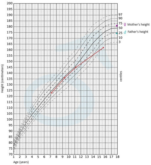
During the physical examination, apart from the presence of short stature, no other notable abnormalities were detected. The skin was clear, without pathological lesions; no features of hyperandrogenism were observed. The boy’s body weight was 47.4 kg (3rd–10th centile); his height, 162 cm (<3rd centile); and height SDS, −2.36; while his BMI was 18.06 kg/m2 (10th–25th centile). Pubarche was assessed as stage 4 according to the Tanner scale; the volume of the testes was 10–12 mL each. After available anthropometric measurements from the patient’s medical history were plotted on the growth chart for sex and chronological age, it became evident that the boy experienced growth retardation from the age of 11 (Figure 1).
Jcm 12 05967 g001
Figure 1. Growth chart for boys. The red line represents growth retardation from the age of 11.
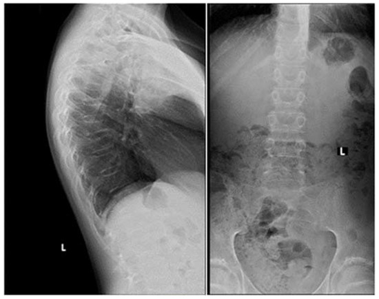
Apart from slightly increased calcium excretion in the 24 h urine collection (Calcium: 9.52 mmol/24 h), there were no significant abnormalities in the laboratory tests assessing calcium–phosphate metabolism (Calcium: 2.41 mmol/L, Phosphorus: 1.3 mmol/L). Serum parathormone (PTH) and vitamin D concentrations remained normal (PTH: 22.9 pg/mL, 25(OH)D: 46.7 ng/mL). Due to the described pain complaints, a thoracolumbar spine X-ray was performed. A decrease in the height of the Th5-Th9 vertebrae and central lowering of the upper border plate of the L4 and L5 were observed (Figure 2).
Jcm 12 05967 g002
Figure 2. A thoracolumbar spine X-ray with multilevel vertebral fractures. Decrease in the height of the Th5–Th9 vertebrae and central lowering of the upper border plate of the L4 and L5 were found.
Magnetic resonance imaging (MRI) of the spine confirmed multilevel vertebral fractures, which, together with the presence of low bone mass on DXA examination, allowed a diagnosis (according to ISCD guidelines) of osteoporosis to be made. Treatment included calcium supplements and cholecalciferol. The parents did not consent to treatment with bisphosphonates (sodium pamidronate), which is an off-label treatment.
In light of the patient’s short stature and growth retardation, an endocrinological assessment was conducted. The possibility of growth hormone (GH) deficiency and hypothyroidism as underlying causes for the growth retardation was ruled out. Gonadotropin and androgen levels were adequate for the pubertal stage (FSH—8.3 IU/L, LH—4.7 IU/L, testosterone—4.750 ng/mL, DHEA-S—230.30 µg/dL (normal range: 70.2–492), 17-OH-progesterone—0.78 ng/mL). The bone age was assessed to be 15 years.
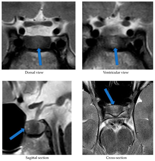
Alongside continued steroid therapy for autoimmune hepatitis, profiles of cortisol and ACTH secretion were performed. Due to the patient’s elevated cortisol levels during night hours (cortisol 24:00—10.7 µg/dL), an overnight dexamethasone suppression test (DST) and low-dose dexamethasone suppression test (LDDST) were performed. After administering 1 mg dexamethasone (23:00), his morning cortisol level (8:00) still remained elevated (cortisol—3.4 µg/dL). However, after administering 0.5 mg dexamethasone every 6 h for the next 2 days, cortisol levels (8:00) normalized (cortisol—1.0 µg/dL). An MRI of the pituitary gland showed only a poorly demarcated area in the anterior part of the glandular lobe, measuring approximately 2.0 × 3.5 × 5.0 mm on T2W images (Figure 3). A follow-up MRI examination was recommended, which was performed during the child’s next hospitalization in January 2022. The previously described area was still very faint.
Jcm 12 05967 g003
Figure 3. MRI examination image depicting the poorly demarcated area in the anterior part of the glandular lobe. The arrows point to a structure suspected of being an adenoma.
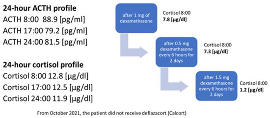
In October 2021, the administration of deflazocort as a treatment was discontinued. During hospitalization in January 2022, the diurnal pattern of ACTH and cortisol secretion was re-evaluated, yet no consistent diurnal rhythm was observed; cortisol levels remained elevated at night. For this reason, overnight DST and then LDDST were carried out again (Figure 4), in which no suppression of cortisol concentrations was obtained. Only after a high-dose DST (HDDST), in which a high 1.5 mg of dexamethasone was administered every 6 h (125 µg/kg/24 h), was cortisol secretion suppressed.
Jcm 12 05967 g004
Figure 4. Laboratory findings indicating the diagnosis of ACTH-dependent Cushing’s syndrome.
Based on the above results, CD was suspected as the cause of osteoporosis and growth retardation. In February 2022, a CRH test was performed upon the patient, which revealed a four-fold increase in ACTH levels and a two-fold increase in serum cortisol levels (Table 2).
Table 2. The results of human CRH (hCRH) stimulation test and bilateral inferior petrosal sinus sampling (BIPSS).
The CRH stimulation test was administered in the morning using human synthetic CRH (Ferring) at a dose of 1 μg/kg of body weight. During the test, cortisol and ACTH levels were measured in serum at the following time points: −15, 0, 15, 30, 60, and 90 min (see Table 2). As part of the diagnostic process, urinary free cortisol excretion was also measured over two consecutive days. Only on the first day was there a slight elevation in urinary free cortisol concentration, measuring 183.60 μg/24 h (normal range: 4.3–176). The measurement performed on the second day showed a normal urinary free cortisol concentration of 145.60 μg/24 h (normal range: 4.3–176). On 2 March 2022, the patient underwent a bilateral inferior petrosal sinus sampling (BIPSS). Human CRH stimulation was also used during the procedure. The presence of ACTH-dependent hypercortisolemia of pituitary origin was confirmed. The outcome of the CRH stimulation during the BIPSS is presented in Table 2. The boy qualified for transsphenoidal surgery (TSS) of the pituitary adenoma and was successfully operated on (8 March 2022). Postoperative histopathological examination revealed features of a corticotroph-rich pituitary adenoma.

3. Discussion

Osteoporosis, like CD, is extremely rare in the developmental age population. Bone-mineralization disorders among children may be primary (e.g., osteogenesis imperfecta), or secondary to other diseases or their treatment (e.g., with glucocorticosteroids). This case report presents a boy with osteoporosis, the cause of which was originally attributed to the treatment of autoimmune hepatitis with glucocorticosteroids. Steroid therapy is the most common cause of bone-mineralization disorders in children. However, osteoporosis is a late complication of steroid treatment. Briot et al. [10] demonstrated that the risk of fractures increases as early as 3 months after initiating steroid therapy. An additional factor increasing the risk of fractures is the dose of glucocorticosteroids used, corresponding to 2.5–5 mg of prednisolone per day [10]. In the case of the present patient, the appearance of spinal pain and thus vertebral fractures could not have been related to the deflazacort treatment started 5 days earlier. The bone-mineralization disorder must therefore have occurred much earlier. For this reason, the authors considered it necessary to search for other endocrine causes of osteoporosis development, including hypogonadism, growth hormone deficiency or Cushing’s syndrome/disease.
The serum vitamin D concentration can also influence bone mineral density. Every patient with mineralization disorders, especially with osteoporosis, requires a thorough assessment of calcium–phosphate metabolism [11]. Until the initiation of steroid therapy in March 2021, the patient did not undergo regular vitamin D supplementation. At the start of deflazacort treatment, his serum 25(OH)D concentration was 12.4 ng/mL. Consequently, additional cholecalciferol supplementation at a dose of 3000 IU/day was introduced. In a subsequent measurement conducted in June 2021, the concentration was within the reference range [25(OH)D: 46.7 ng/mL].
Considering the lack of regular supplementation before March 2021, it can be assumed that in October 2020, when the boy experienced SARS-CoV-2 infection, his serum vitamin D concentration was likely decreased as well, which could have had a further negative impact on the patient’s bone mineralization. Scientific reports indicate that adequate vitamin D levels reduce the risk of viral infections, including SARS-CoV-2 [12]. Di Filippo et al. [13] demonstrated that vitamin D deficiency observed in 68.2% of SARS-CoV-2-infected individuals correlated with a more severe course of the infection. In our patient, the course of COVID-19 was asymptomatic, and the diagnosis was established based on positive IgM antibody titers against SARS-CoV-2. The vitamin deficiency was most likely associated with irregular supplementation and lack of exposure to UV radiation (due to lockdown measures in Poland at that time). A reduced serum 25(OH)D concentration could have contributed to worsened bone mineral density and increased susceptibility to SARS-CoV-2 infection; however, it is the chronic hypercortisolism characteristic of CD that most likely led to the development of osteoporosis with accompanying fractures.
Another factor necessitating further diagnostic investigation into CD was the patient’s growth retardation observed since the age of 11. Both the pubertal state of the boy, and his gonadotropin and testosterone serum levels, allowed us to exclude hypogonadism. Maximum spontaneous nocturnal secretion of the growth hormone was 31.84 ng/mL. The diagnosis of CD was established on the basis of elevated cortisol levels at night and the lack of cortisol suppression in the test after administering dexamethasone. Final confirmation of the diagnosis was obtained in a post-CRH stimulation test. In pediatric cases, the absence of typical diurnal variation in serum corticosolemia, especially the nocturnal decline, and the inability to suppress cortisol secretion at midnight, are highly sensitive indicators of hypercortisolemia [6,8]. Consequently, in our patient, osteoporosis was a complication of diagnosed CD.
The patient in question was not obese, which is the predominant symptom of CD. This symptom, according to Ferrigno et al. [1], is present in 92–98% of examined children diagnosed with CD. Storr et al. [14] showed that facial changes and facial swelling were observed in 100% of subjects with CD, whereas Lonser et al. [8] observed this in only 63% of children with CD. In our patient, no changes in facial appearance were observed. Other symptoms typical of CS, such as hirsutism, acne, or bruises, were not noticed either. These symptoms were observed in all children with CD studied by Wędrychowicz et al. [3]. Non-specific symptoms of this condition may include mood changes, depression and emotional vacillation [1,8]. However, our patient’s parents did not observe any changes in the boy’s behavior. The indication for initiating the whole diagnostic process was (in addition to osteoporosis) growth retardation. Ferrigno et al. [1] point out that chronic hypercortisolemia most often leads to growth disorders accompanied by excessive weight gain. This is an early, highly sensitive and characteristic sign of CD. Short stature is not always observed and occurs in one in two children diagnosed with CD. The patient we present was short (height—162 cm (<3rd centile hSDS: −2.36)); growth retardation was observed from the age of 11 years.
The occurrence of vertebral fractures and the accompanying pain as the initial symptoms of hypercortisolism, the absence of obesity, and the confirmation of CD, an exceedingly rare condition in the pediatric population, collectively underscore the uniqueness of our patient’s disease presentation. A case involving a child with such an atypical course of ACTH-dependent CS has not been described before. Han et al. [15] reported a case of a 28-year-old lean woman (BMI: 19 kg/m²) with ACTH-independent CS due to a left adrenal adenoma, where, similarly to our patient, the initial manifestation of hypercortisolism was compression fractures of the thoracic vertebrae. The authors emphasize that vertebral fractures may affect 30–50% of patients with Cushing’s syndrome, with a higher frequency observed in patients with ACTH-independent CS compared to those in whom hypercortisolism results from the presence of pituitary adenoma [15].
The lack of obesity in a patient with hypercortisolism could be attributed to malnutrition, which accompanies the growth process in ECS. Hence, a crucial aspect was the differential diagnosis between CD and ECS. To this end, we performed a stimulation test using hCRH. We considered cut-off points for diagnosing CD to be a 35% increase in ACTH concentration at 15 and/or 30 min, and at least a 20% increase in cortisol concentration at 30 and 45 min [16,17]. In the case of ECS, a significant rise in CRH and cortisol concentrations is not observed. Recently published reports emphasize the need to explore new cut-off points to enhance the sensitivity and specificity of this test. Detomas et al. [5] indicate that an increase in ACTH ≥ 31% and cortisol ≥ 12% in the 30th minute of CRH tests allows for a highly sensitive and specific differentiation between CD and ECS. The authors highlight that measuring these hormones at the 60 min stage of the test does not provide diagnostic benefits. Notably, the study employed ovine CRH, which exhibits stronger and more prolonged stimulatory effects compared to the hCRH available in Europe that was used to diagnose our patient [5]. Conversely, Elenius et al. [16] suggest that optimal values for distinguishing between CD and ECS in the CRH stimulation test involve an increase in ACTH and/or cortisol levels of more than 40% during the test. In our patient, an over four-fold increase in ACTH levels and a more than two-fold increase in cortisol levels were observed at the 30 min mark of the test, thus independently and definitively excluding ECS regardless of the adopted cut-off points.
Our patient’s case also demonstrates that MRI is not a perfect method of visualizing an ACTH-secreting pituitary adenoma. In the first MRI examination performed upon our patient, a poorly demarcated area (2.0 × 3.5 × 5.0 mm) was described in the anterior part of the glandular lobe; in the examination performed 6 months later, this area maintained poor visibility, while laboratory results at the time clearly indicated an ACTH-dependent form of CS. It was only the bilateral inferior petrosal sinus sampling (BIPSS) that allowed a clear diagnosis. Data from the literature indicate that microadenomas smaller than 3–4 mm are visible on MRI in only half of cases. In two large studies including children, pituitary adenomas were found on MRI in 63% and 55% of cases [18]. Among the patients with CD studied by Wędrychowicz et al. [3], pituitary adenomas were described on MRI in all of them, but in two patients (50%) this was only achieved upon follow-up. In the standard procedure, in the absence of a pituitary lesion in the MRI examination, it is recommended that a BIPSS be performed. In the case of our patient, this examination was necessary to make a definitive diagnosis.
When analyzing the results of the BIPSS with hCRH stimulation, we employed the classical cut-offs for the ACTH IPS:P (Inferior Petrosal Sinus: Peripheral) ratio (i.e., ≥2 at baseline and ≥3 after hCRH stimulation) [1]. This allowed the confirmation of CD and determination of the pituitary adenoma’s localization, followed by the procedure for its surgical removal. The optimal cut-off values for the IPS:P ratio remain controversial. There are ongoing efforts to establish new, more precise cut-off points. Detomas et al. [19] demonstrated that an IPS:P ratio ≥ 2.1 during desmopressin stimulation in the BIPSS most accurately differentiates CD from ECS. Conversely, Chen et al. [20] showed that the optimal pre-desmopressin stimulation IPS:P ratio cut-off is 1.4, and post-stimulation it is 2.8. Both studies suggest the utilization of lower cut-off values for the IPS:P ratio than those traditionally adopted. Chen et al. [20] also advocate for avoiding stimulation during BIPSS. In most cases, the IPS:P ratio before stimulation is sufficient for diagnosing CD. According to the authors, desmopressin stimulation should be reserved for patients with ambiguous MRI findings or with a pituitary adenoma with diameter less than 6 mm. However, considering that the concentration of ACTH in the right inferior petrosal sinus in our patient was over 4 times higher than in the peripheral vessel and nearly 14 times higher after hCRH stimulation, regardless of the applied criteria, CD could be unequivocally diagnosed in our patient, and the lateralization of the microadenoma could be determined with certainty.
The rarity of CD, and the diagnostic difficulties stemming from its oligosymptomatic or atypical course, encourage description in the form of case reports. Eviz et al. [21] delineate the occurrence of cerebral cortical atrophy in two children with ECS. Additionally, other researchers have underscored the potential for thyroid disorders to manifest alongside hypercorticosolemia [22]. Although obesity typically stands out as a primary symptom of CD, Pomahacova et al. [23] reported a case involving two children with CD who maintained normal body weight, mirroring our patient’s situation. The symptoms that prompted diagnostic investigation in these instances included weakness, sleep disturbances and growth retardation. Interestingly, growth retardation, along with facial changes, was observed in all examined children with CD [23]. Nonetheless, to the best of our knowledge, we have yet to encounter a case report resembling ours. Therefore, it remains crucial to share our experiences.

4. Conclusions

Cushing’s disease is an extremely rare diagnosis in children. In Poland, there is no statistical record of occurrences of this disease among children. Wędrychowicz et al. reported that in their single Polish center, between 2012 and 2018, they identified four cases of children aged 7–15 who were diagnosed with CD [3]. The case we present shows that obesity, commonly considered as a predominant symptom of CD, is not necessarily observed in patients with this diagnosis in the developmental age population. Among children, it is growth disturbance that may be the first manifestation. On the other hand, a late complication of CD may be osteoporosis, so whenever a child is diagnosed with a bone-mineralization disorder, the cause of its development should be sought.
Diagnosis should be pursued until all potential causes of the described symptoms, including the rarest ones, are definitively ruled out—even if the clinical presentation, as in the case of our patient, initially does not point towards the final diagnosis. Thus far, no case of a child with CD exhibiting such subtle symptomatology has been described in the literature. The challenges in diagnosis we encountered primarily resulted from the atypical clinical outcome of CD in our patient—normal body weight, absence of hyperandrogenism, mood disturbances not apparent to caregivers and the patient’s immediate environment, as well as normal progression of puberty, did not immediately lead to the consideration of endocrinological causes of osteoporosis. The steroid therapy employed due to autoimmune hepatitis also complicated the diagnostic process. Only after discontinuing deflazacort treatment was it possible to definitively diagnose CD.
Our patient required hydrocortisone replacement in gradually decreasing doses for a year following TSS. Considering that pituitary adenomas in children can be genetically predisposed (e.g., MEN 1 mutation, AIP mutation, USP8 mutation, and other rarer ones), genetic consultation was sought [1]. However, the conducted tests have thus far excluded the most common mutations in our patient. Due to the diagnosed osteoporosis, chronic supplementation with calcium and cholecalciferol was recommended, along with annual follow-up DXA scans. Studies indicate that patients in remission from CD experience a gradual improvement in bone mineral density [3]. While we can currently observe remission in our patient’s case, the advanced bone age of the child (indicating the completion of the growth process) left limited potential for significant improvement in final growth. The patient still requires regular endocrinological and neurosurgical follow-ups, hormonal assessments, and pituitary MRI examinations.

Author Contributions

Conceptualization—R.S. and A.Ł.; software—S.A.; formal analysis—R.S.; investigation—A.Ł. and G.Z.; data curation—A.Ł.; writing—original draft preparation—A.Ł., R.S. and S.A.; writing—review and editing—A.L.; visualization—S.A.; supervision—A.L. All authors have read and agreed to the published version of the manuscript.

Funding

This study was funded by statutory funds from the Medical University of Lodz, Lodz, Poland (503/1-107-03/503-11-001).

Institutional Review Board Statement

Not applicable.

Informed Consent Statement

Written informed consent has been obtained from the patient to publish this paper.

Data Availability Statement

Not applicable.

Conflicts of Interest

The authors declare no conflict of interest.

References

  1. Ferrigno, R.; Hasenmajer, V.; Caiulo, S.; Minnetti, M.; Mazzotta, P.; Storr, H.L.; Isidori, A.M.; Grossman, A.B.; De Martino, M.C.; Savage, M.O. Paediatric Cushing’s disease: Epidemiology, pathogenesis, clinical management and outcome. Rev. Endocr. Metab. Disord. 2021, 22, 817–835. [Google Scholar] [CrossRef]
  2. Stratakis, A. Cushing syndrome in pediatrics. Endocrinol. Metab. Clin. N. Am. 2012, 41, 793–803. [Google Scholar] [CrossRef]
  3. Wędrychowicz, A.; Hull, B.; Tyrawa, K.; Kalicka-Kasperczyk, A.; Zieliński, G.; Starzyk, J. Cushing disease in children and adolescents—Assessment of the clinical course, diagnostic process, and effects of the treatment—Experience from a single paediatric centre. Pediatr. Endocrinol. Diabet. Metab. 2019, 25, 127–143. [Google Scholar] [CrossRef]
  4. Concepción-Zavaleta, M.J.; Armas, C.D.; Quiroz-Aldave, J.E.; García-Villasante, E.J.; Gariza-Solano, A.C.; Del Carmen Durand-Vásquez, M.; Concepción-Urteaga, L.A.; Zavaleta-Gutiérre, F.E. Cushing disease in pediatrics: An update. Ann. Pediatr. Endocrinol. Metab. 2023, 28, 87–97. [Google Scholar] [CrossRef]
  5. Detomas, M.; Ritzel, K.; Nasi-Kordhishti, I.; Wolfsberger, S.; Quinkler, M.; Losa, M.; Tröger, V.; Kroiss, M.; Fassnacht, M.; Vila, G.; et al. Outcome of CRH stimulation test and overnight 8 mg dexamethasone suppression test in 469 patients with ACTH-dependent Cushing’s syndrome. Front. Endocrinol. 2022, 13, 955945. [Google Scholar] [CrossRef] [PubMed]
  6. Lodish, M.B.; Hsiao, H.P.; Serbis, A.; Sinaii, N.; Rothenbuhler, A.; Keil, M.F.; Boikos, S.A.; Reynolds, J.C.; Stratakis, C.A. Effects of Cushing disease on bone mineral density in a pediatric population. J. Pediatr. 2010, 56, 1001–1005. [Google Scholar] [CrossRef] [PubMed]
  7. Pivonello, R.; Isidori, A.M.; De Martino, M.C.; Newell-Price, J.; Biller, B.M.; Colao, A. Complications of Cushing’s syndrome: State of the art. Lancet Diabetes Endocrinol. 2016, 4 (Suppl. S7), 611–629. [Google Scholar] [CrossRef] [PubMed]
  8. Lonser, R.R.; Wind, J.J.; Nieman, L.K.; Weil, R.J.; DeVroom, H.L.; Oldfield, E.H. Outcome of surgical treatment of 200 children with Cushing’s disease. J. Clin. Endocrinol. Metab. 2013, 98, 892–901. [Google Scholar] [CrossRef]
  9. Detomas, M.; Deutschbein, T.; Tamburello, M.; Chifu, I.; Kimpel, O.; Sbiera, S.; Kroiss, M.; Fassnacht, M.; Altieri, B. Erythropoiesis in Cushing syndrome: Sex-related and subtype-specific differences. Results from a monocentric study. J. Endocrinol. Investig. 2023; epub ahead of print. [Google Scholar] [CrossRef]
  10. Briot, K.; Roux, C. Glucocorticoid-induced osteoporosis. RMD Open 2015, 1, 14. [Google Scholar] [CrossRef]
  11. Laird, E.; Ward, M.; McSorley, E.; Strain, J.J.; Wallace, J. Vitamin D and bone health; potential mechanisms. Nutrients 2010, 2, 693–724. [Google Scholar] [CrossRef]
  12. di Filippo, L.; Frara, S.; Nannipieri, F.; Cotellessa, A.; Locatelli, M.; Querini, P.R.; Giustina, A. Low vitamin D levels are associated with long COVID syndrome in COVID-19 survivors. J. Clin. Endocrinol. Metab. 2023, 1–11. [Google Scholar] [CrossRef] [PubMed]
  13. di Filippo, L.; Allora, A.; Doga, M.; Formenti, A.M.; Locatelli, M.; Rovere Querini, P.; Frara, S.; Giustina, A. Vitamin D levels are associated with blood glucose and BMI in COVID-19 patients, predicting disease severity. J. Clin. Endocrinol. Metab. 2022, 107, 348–360. [Google Scholar] [CrossRef]
  14. Storr, H.L.; Chan, L.F.; Grossman, A.B.; Savage, M.O. Pediatric Cushing’s syndrome: Epidemiology, investigation and therapeutic advances. Trends Endocrinol. Metab. 2007, 18, 167–174. [Google Scholar] [CrossRef] [PubMed]
  15. Han, J.Y.; Lee, J.; Kim, G.E.; Yeo, J.Y.; Kim, S.H.; Nam, M.; Kim, Y.S.; Hong, S. Case of Cushing syndrome diagnosed by recurrent pathologic fractures in a young woman. J. Bone Metab. 2012, 19 (Suppl. S2), 153–158. [Google Scholar] [CrossRef] [PubMed]
  16. Elenius, H.; McGlotten, R.; Nieman, L.K. Ovine CRH stimulation and 8 mg dexamethasone suppression tests in 323 patients with ACTH-dependent Cushing’s syndrome. J. Clin. Endocrinol. Metab, 2023; Epub ahead of print. [Google Scholar] [CrossRef]
  17. Nieman, L.K.; Biller, B.M.K.; Findling, J.W.; Newell-Price, J.; Savage, M.O.; Stewart, P.M.; Montori, V.M. The diagnosis of Cushing’s syndrome: An Endocrine Society Clinical Practice Guideline. J. Clin. Endocrinol. Metab. 2008, 93 (Suppl. S5), 1526–1540. [Google Scholar] [CrossRef] [PubMed]
  18. Savage, M.O.; Storr, H.L. Pediatric Cushing’s disease: Management issues. Indian J. Endocrinol. Metab. 2012, 16 (Suppl. S2), 171–175. [Google Scholar] [CrossRef]
  19. Detomas, M.; Ritzel, K.; Nasi-Kordhishti, I.; Schernthaner-Reiter, M.H.; Losa, M.; Tröger, V.; Altieri, B.; Kroiss, M.; Kickuth, R.; Fassnacht, M.; et al. Bilateral inferior petrosal sinus sampling with human CRH stimulation in ACTH-dependent Cushing’s syndrome: Results from a retrospective multicenter study. Eur. J. Endocrinol. 2023, 188 (Suppl. S5), 448–456. [Google Scholar] [CrossRef]
  20. Chen, S.; Chen, K.; Wang, S.; Zhu, H.; Lu, L.; Zhang, X.; Tong, A.; Pan, H.; Wang, R.; Lu, Z. The optimal cut-off of BIPSS in differential diagnosis of ACTH-dependent Cushing’s syndrome: Is stimulation necessary? J. Clin. Endocrinol. Metab. 2020, 105 (Suppl. S4), 1673–1685. [Google Scholar] [CrossRef]
  21. Eviz, E.; Yesiltepe, M.G.; Arduc, A.A.; Erbey, F.; Guran, T.; Hatun, S. An overlooked manifestation of hypercortisolism—Cerebral cortical atrophy and challenges in identifying the etiology of hypercortisolism: A report of 2 pediatric cases. Horm. Res. Paediatr. 2023, 27. [Google Scholar] [CrossRef]
  22. Paragliola, R.M.; Corsello, A.; Papi, G.; Pontecorvi, A.; Corsello, S.M. Cushing’s syndrome effects on the thyroid. Int. J. Mol. Sci. 2021, 22, 3131. [Google Scholar] [CrossRef]
  23. Pomahacova, R.; Paterova, P.; Nykodymova, E.; Sykora, J.; Krsek, M. Pediatric Cushing’s disease: Case reports and retrospective review. Biomed. Pap. Med. Fac. Univ. Palacky. Olomouc. Czech Repub. 2022, 166. [Google Scholar] [CrossRef] [PubMed]
Disclaimer/Publisher’s Note: The statements, opinions and data contained in all publications are solely those of the individual author(s) and contributor(s) and not of MDPI and/or the editor(s). MDPI and/or the editor(s) disclaim responsibility for any injury to people or property resulting from any ideas, methods, instructions or products referred to in the content.

Share and Cite

MDPI and ACS Style

Łupińska, A.; Aszkiełowicz, S.; Zieliński, G.; Stawerska, R.; Lewiński, A. Osteoporosis as the First Sign of Cushing’s Disease in a Thin 16-Year-Old Boy—A Case Report. J. Clin. Med. 2023, 12, 5967. https://doi.org/10.3390/jcm12185967

AMA StyleŁupińska A, Aszkiełowicz S, Zieliński G, Stawerska R, Lewiński A. Osteoporosis as the First Sign of Cushing’s Disease in a Thin 16-Year-Old Boy—A Case Report. Journal of Clinical Medicine. 2023; 12(18):5967. https://doi.org/10.3390/jcm12185967

Chicago/Turabian StyleŁupińska, Anna, Sara Aszkiełowicz, Grzegorz Zieliński, Renata Stawerska, and Andrzej Lewiński. 2023. “Osteoporosis as the First Sign of Cushing’s Disease in a Thin 16-Year-Old Boy—A Case Report” Journal of Clinical Medicine 12, no. 18: 5967. https://doi.org/10.3390/jcm12185967

Find Other Styles

Note that from the first issue of 2016, this journal uses article numbers instead of page numbers. See further details here.

Article Metrics

Citations

No citations were found for this article, but you may check on Google Scholar

Article Access Statistics

Article access statisticsArticle Views14. Sep15. Sep16. Sep17. Sep18. Sep19. Sep20. Sep21. Sep22. Sep23. Sep24. Sep25. Sep26. Sep27. Sep28. Sep29. Sep30. Sep1. Oct2. Oct3. Oct4. Oct5. Oct6. Oct7. Oct8. Oct9. Oct10. Oct11. Oct12. Oct13. Oct14. Oct0100200300400

For more information on the journal statistics, click here.

Multiple requests from the same IP address are counted as one view.