Unilateral Adrenalectomy for Pediatric Cyclical Cushing Syndrome With Novel PRKAR1A Variant Associated Carney Complex

Abstract

Primary pigmented nodular adrenocortical disease is a rare cause of Cushing syndrome accounting for less than 1% of cases. We present a 9-year-old boy who presented at age 4 with cyclical Cushing syndrome and was eventually diagnosed with a novel, previously unreported, unpublished variant in PRKAR1A associated with Carney complex. He was treated with unilateral left adrenalectomy. At 1-year follow-up, he continues to be in remission of his symptoms of Cushing syndrome.

Introduction

Cushing syndrome is characterized by prolonged exposure to excess glucocorticoids and is broadly classified as either ACTH-dependent or ACTH-independent [12]. Primary pigmented nodular adrenocortical disease (PPNAD) is a rare cause of ACTH-independent Cushing syndrome, characterized by bilateral adrenal hyperplasia with autonomous, hyperfunctioning nodules [12]. Approximately 90% of PPNAD cases occur in the context of Carney complex, with isolated cases being exceedingly uncommon [12].

PPNAD was first described in 1984 by Carney et al, who coined the term in a case series of 4 patients and a review of 24 previously reported cases [1]. In that series, patients presented with ACTH-independent Cushing syndrome and no radiographic evidence of adrenal tumors. All underwent bilateral adrenalectomy, with histopathology revealing bilateral pigmented nodules in otherwise small or normal-sized adrenal glands [1]. Histologically, the classic features of PPNAD include multiple small black or brown cortical nodules surrounded by an atrophic adrenal cortex—reflecting chronic ACTH suppression [1].

Clinically, PPNAD most often presents with cyclical Cushing syndrome, characterized by alternating periods of hypercortisolism and normocortisolemia [2]. This intermittent pattern poses a substantial diagnostic challenge, as biochemical confirmation requires detection of cortisol excess during active phases of the cycle.

Carney complex is a multiple neoplasia syndrome involving endocrine, cardiac, cutaneous, and neural tumors. First described by Carney et al in 1985, it is typically inherited in an autosomal dominant fashion. Approximately 70% of cases occur in familial settings, while the remaining 30% arise from de novo pathogenic variants [34]. Over half of affected individuals harbor pathogenic variants in the PRKAR1A tumor suppressor gene on chromosome 17q24.2, while approximately 20% of cases are linked to alternate loci such as 2p16 [24].

Diagnostic criteria for Carney complex include either 2 clinical manifestations or 1 clinical manifestation in combination with a pathogenic PRKAR1A variant or an affected first-degree relative [2]. The most common endocrine manifestation is PPNAD, reported in approximately 25% of patients with Carney complex, though this likely underestimates the true prevalence, as autopsy studies reveal histologic evidence of PPNAD in nearly all affected individuals [2].

The Endocrine Society clinical practice guidelines recommend bilateral adrenalectomy as the definitive treatment for PPNAD, effectively curing hypercortisolism but necessitating lifelong glucocorticoid and mineralocorticoid replacement therapy due to resultant adrenal insufficiency [5]. Unilateral adrenalectomy has emerged as an alternative approach, particularly in pediatric patients, with the potential to preserve endogenous adrenal function.

Herein, we present the case of a 9-year-old boy with Carney complex and cyclical Cushing syndrome due to PPNAD, successfully managed with unilateral adrenalectomy.

Case Presentation

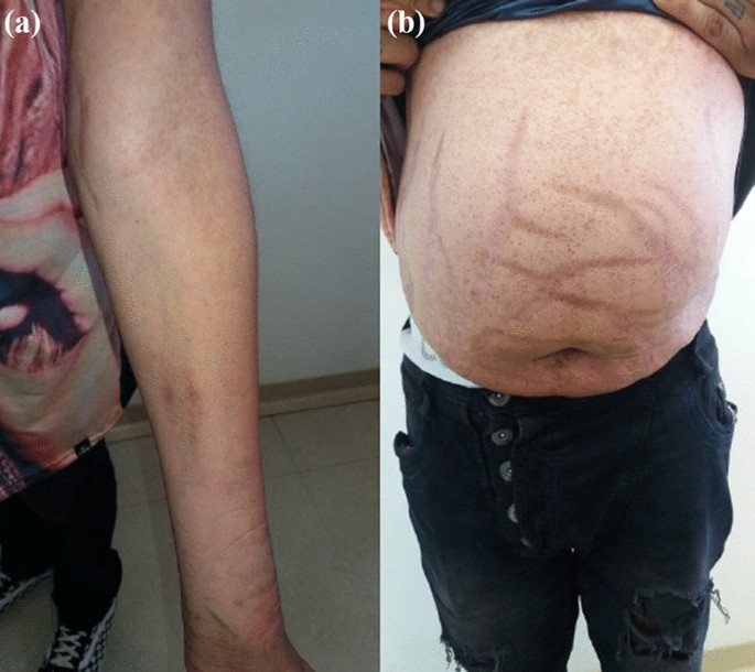
A 4-year-old boy presented with a week-long history of facial swelling, hyperphagia, weight gain, and scrotal swelling. At presentation, his weight was 22 kg (99th percentile) and body mass index (BMI) was 18 kg/m² (96th percentile). Initial workup revealed normal 24-hour urinary free cortisol <0.0913 µg/day (SI: 274 nmol/day) with low urinary creatinine 215 mg/day (SI: 1.9 mmol/day) (normal reference range 973-2195 mg/day; SI: 8.6-19.4 mmol/day) suggesting an incomplete sample. A repeat collection produced similar results. A 1 mg dexamethasone suppression test demonstrated nonsuppressed cortisol (27.9 µg/dL; SI: 772 nmol/L), suggestive of Cushing syndrome.

Over 5 years, the patient experienced 2 to 3 episodes per year of rapid weight gain (20-50 lbs), facial flushing, abdominal distention, and mood changes. Despite persistent obesity (>97th percentile), linear growth remained normal.

Diagnostic Assessment

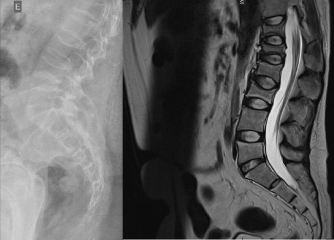
At age 7, midnight salivary cortisol was markedly elevated at 3.7 µg/dL (SI: 103 nmol/L) (normal reference range < 0.4 µg/dL; SI: < 11.3 nmol/L), raising suspicion for cyclical Cushing syndrome. Magnetic resonance imaging of the abdomen was negative for adrenal lesions. At age 8, during an active episode, 2 elevated salivary cortisol samples, 2.0 µg/dL (SI: 55.1 nmol/L) and 2.2 µg/dL (SI: 61.9 nmol/L) (normal reference range < 0.4 µg/dL; SI: < 11.3 nmol/L), were obtained. A high-dose dexamethasone suppression test yielded a low baseline cortisol 3.2 µg/dL (SI: 89 nmol/L) and nonsuppressed cortisol post-dexamethasone 3.0 µg/dL (SI: 83 nmol/L). Baseline ACTH was 7.7 pg/mL (SI: 1.7 pmol/L), suppressed to <3.2 pg/mL (SI: < 0.7 pmol/L) post-dexamethasone—consistent with ACTH-independent cortisol excess.

At age 9, the patient underwent the gold standard diagnostic testing for cyclical Cushing, the Liddle test. The test involves 6 days of urine collection: days 1 to 2 establish baseline urinary cortisol levels, days 3 to 4 assess response to low-dose dexamethasone, and days 5 to 6 evaluate response to high-dose dexamethasone. The patient’s cortisol increased paradoxically from 118.5 µg/day (SI: 327 nmol/day) to 402.0 µg/day (SI: 1109 nmol/day) over 6 days, consistent with PPNAD physiology. Genetic testing was performed with the following report: “A heterozygous variant, NM_002734.4(PRKAR1A):c.550-2_553delinsG, p.(Val184_Tyr185delinsAsp), was detected in exon 7 of this gene. This variant does not appear to have been reported in population (gnomAD, ESP, dbSNP) and clinical databases (ClinVar), or in the literature. The impact of this variant on RNA splicing as assessed by multiple algorithms (Alamut Suite) is: abolishment of canonical acceptor splice site. Based on the current evidence, this variant was classified as likely pathogenic, American College for Medical Genetics category 2”. Family testing revealed this to be a de novo pathogenic variant.

Further workup included echocardiogram and thyroid ultrasound, both of which were normal. During workup for scrotal swelling at initial presentation, the patient was found to have bilateral testicular masses with negative testicular cancer tumor markers: α-fetoprotein, human chorionic gonadotropin, and lactate dehydrogenase. The family declined invasive biopsy of these lesions. He was followed by pediatric urology with yearly serial ultrasound, and these were felt to be benign testicular tumors, presumed noncalcifying Sertoli cell tumors, associated with Carney complex (Fig. 1).


Ultrasound of bilateral testicular lesions. A) Left testicle. B) Right testicle.

Figure 1.

Ultrasound of bilateral testicular lesions. A) Left testicle. B) Right testicle.

Based on the presence of 2 major diagnostic criteria in combination with the molecular diagnosis of a likely pathogenic variant of PRKAR1A, the diagnosis of Carney complex was established.

Treatment

The patient was referred for surgical evaluation for consideration of adrenalectomy. A comprehensive discussion was conducted regarding the potential benefits and risks of unilateral vs bilateral adrenalectomy. The family was counseled that unilateral adrenalectomy might not fully resolve the hypercortisolemia and that a subsequent contralateral adrenalectomy could be necessary. In contrast, bilateral adrenalectomy would definitively address cortisol excess but result in permanent adrenal insufficiency requiring lifelong glucocorticoid and mineralocorticoid replacement. After multidisciplinary consultation with endocrinology and surgery, the decision was made to proceed with unilateral adrenalectomy.

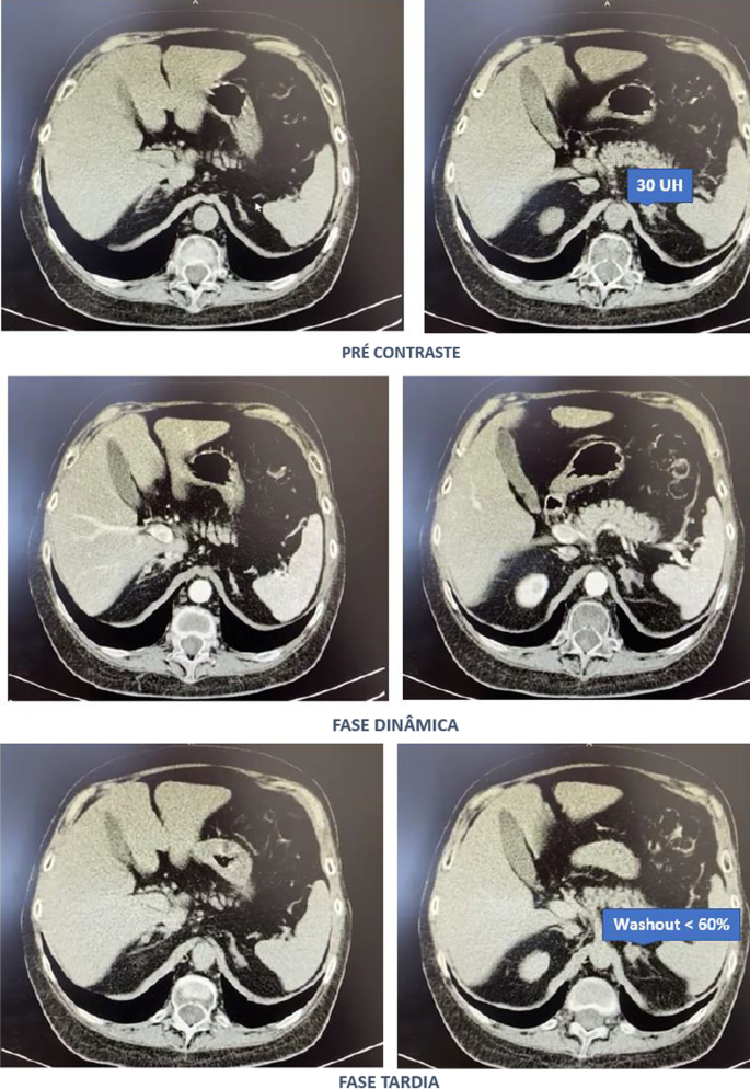
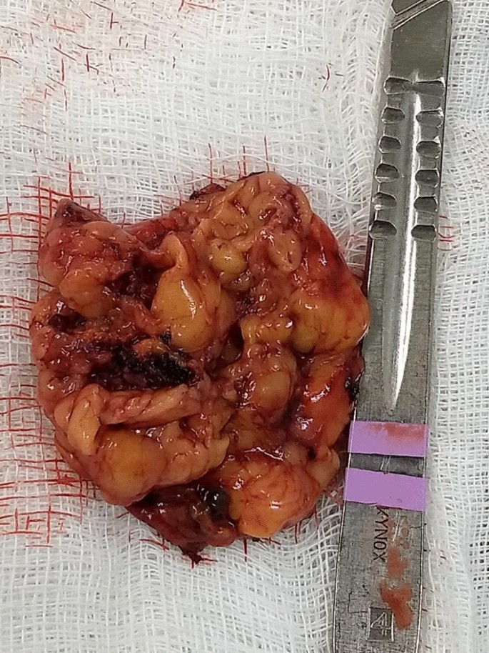
Preoperative IV contrast-enhanced computed tomography (CT), reviewed by a physician experienced in PPNAD, demonstrated greater nodularity in the left adrenal gland compared to the right. Therefore, a laparoscopic left adrenalectomy was performed electively without intraoperative complications. The patient was discharged on postoperative day 1. At the time of surgery (age 9), his weight was 70 kg (100th percentile), and BMI was 31.6 kg/m² (99th percentile). The resected left adrenal gland was submitted for histopathologic evaluation. Gross examination revealed no overt nodularity (Fig. 2); however, microscopic analysis identified multiple pigmented cortical nodules consistent with PPNAD (Fig. 3).


Left adrenal gland gross morphology. No macroscopic nodularity appreciable.

Figure 2.

Left adrenal gland gross morphology. No macroscopic nodularity appreciable.


Hematoxylin and Eosin staining on microscopy of left adrenal gland demonstrating hyperpigmented nodule.

Figure 3.

Hematoxylin and Eosin staining on microscopy of left adrenal gland demonstrating hyperpigmented nodule.

Outcome and Follow-up

The patient was followed closely in the postoperative period and was last evaluated 11 months after adrenalectomy. He remained clinically well, with complete resolution of Cushingoid features and no evidence of recurrence. Since surgery, he had experienced significant weight loss of 11.4 kg, with a current weight of 58.6 kg and a BMI of 25 kg/m² (97th percentile).

In summary, this case describes a 9-year-old boy with ACTH-independent, cyclical Cushing syndrome secondary to PPNAD, associated with a de novo likely pathogenic variant in the PRKAR1A gene, consistent with Carney complex. Histopathologic analysis of the resected adrenal gland confirmed the diagnosis of PPNAD. At nearly 1 year post-unilateral adrenalectomy, the patient remains asymptomatic with no biochemical or clinical signs of disease recurrence.

Discussion

Diagnosis of cyclical Cushing is challenging due to the cyclical nature of the disease and the challenges with current available testing modalities. Late-night salivary cortisol testing was a more reliable screening tool in this case as the 24-hour urinary cortisol were affected by inaccurate collection. The cyclical nature of the disease, coupled with the necessity for appropriately timed testing, contributed to a prolonged interval before definitive diagnosis and treatment. Additionally, initial imaging was interpreted as normal, and it was only upon review by a clinician with expertise in PPNAD that subtle adrenal nodularity was identified on CT. Ultimately, the Liddle test and genetic testing were the highest yield for confirmation of disease. This test measures the suppressibility of endogenous cortisol following exogenous dexamethasone administration. In patients with PPNAD, a paradoxical increase in cortisol excretion may occur, attributed to glucocorticoid receptor–mediated activation of protein kinase A catalytic subunits [6]. The likely pathogenic variant found in this case is a novel, previously unreported, variant in the PRKAR1A gene. This rare variant impact both the canonical acceptor splice site in intron 6 as well as results in an in-frame protein change in exon 7 (Val184_Tyr185delinsAsp).

The treatment of PPNAD in the context of Carney complex is typically with bilateral adrenalectomy, as per Endocrine Society guidelines [5]. The drawback of bilateral adrenalectomy is the resultant adrenal insufficiency resulting in lifelong adrenal replacement. Unilateral adrenalectomy is an attractive option for the treatment of PPNAD given the ability to avoid adrenal insufficiency brought upon by bilateral adrenalectomy. Case reports and case series in adult patients have demonstrated variable success in unilateral treatment. In a case series of 17 patients with classic cyclical Cushing, 3 patients had recurrence of Cushing syndrome after unilateral adrenalectomy and were cured with contralateral adrenalectomy [7]. One patient had subtotal (<90%) left adrenalectomy and did not have recurrence with 66 years of follow-up [7].

A case series by Xu et al 2013 described 12 out of 13 patients with PPNAD successfully cured with unilateral adrenalectomy at median 47 months follow-up [8]. The side of adrenalectomy was selected based on CT/magnetic resonance imaging in 3 patients and adrenal iodine131-norcholesterol scintigraphy in the remaining. At our center, the iodine131-norcholesterol scintigraphy was not available so CT was the chosen imaging modality.

Ultimately, the efficacy and morbidity of unilateral adrenalectomy remains unclear. Furthermore, due to the rarity of PPNAD, the criteria for selection of patients who are candidates for unliteral adrenalectomy is challenging to establish. This case reports adds to the existing literature the clinical characteristics of one patient treated successfully by unilateral adrenalectomy.

Learning Points

  • Diagnosis of cyclical Cushing can be very challenging. Late-night salivary cortisol is more reliable than 24-hour urinary cortisol.
  • The paradoxical rise in cortisol in the Liddle test is confirmatory for cyclical Cushing, hence the testing should be considered early in affected patients.
  • Genetic testing assessing for Carney complex, PRAKA1A pathogenic variant, should be considered early in a patient with concern for cyclical Cushing and another system involved like testicular lesions.
  • Although bilateral adrenalectomy is the recommendation for PPNAD; in selected patients, unilateral adrenalectomy might provide several years of remission.

Acknowledgements

Thank you to Dr. Hong Wang, MD, PhD, DABMGG, FACMG, FCCMG, for her support on this project and in all things. Thank you to Dr. Andre Lacroix MD, FCAHS, for reviewing the preoperative CT adrenals with the team.

Contributors

All authors made individual contributions to authorship. F.B. was involved in the diagnosis and management of the patient. N.S. was responsible for the patient’s surgery. C.J.Z. was involved in the patient’s surgery and postoperative care. R.S., M.S., and P.W. were all medical professionals involved in his management and care. All authors contributed, reviewed, and approved the final draft.

Funding

No public or commercial funding.

Disclosures

None declared.

Informed Patient Consent for Publication

Signed informed consent obtained directly from the patient’s relatives or guardians

Data Availability Statement

Data sharing is not applicable to this article as no datasets were generated or analyzed during the current study.

Author notes

Natashia Seemann and Funmbi Babalola co-senior author.

© The Author(s) 2025. Published by Oxford University Press on behalf of the Endocrine Society.
This is an Open Access article distributed under the terms of the Creative Commons Attribution-NonCommercial-NoDerivs licence (https://creativecommons.org/licenses/by-nc-nd/4.0/), which permits non-commercial reproduction and distribution of the work, in any medium, provided the original work is not altered or transformed in any way, and that the work is properly cited. For commercial re-use, please contact reprints@oup.com for reprints and translation rights for reprints. All other permissions can be obtained through our RightsLink service via the Permissions link on the article page on our site—for further information please contact journals.permissions@oup.com. See the journal About page for additional terms.

Adrenal Gland Volume Measurement Could Assist Surgery Option in Patients With Primary Pigmented Nodular Adrenocortical Disease

Abstract

Background

Primary pigmented nodular adrenocortical disease is a rare form of adrenocorticotropic hormone–independent Cushing syndrome originating from bilateral adrenal lesions. Current guidelines do not specify a recommended strategy for determining the optimal surgery. This study evaluates the concordance between bilateral adrenal gland volume and adrenal venous sampling results and the predictive value of adrenal gland volume for postoperative outcomes in patients with primary pigmented nodular adrenocortical disease.

Method

This is a retrospective study conducted at a single center. The study cohort included 10 hospitalized patients with primary pigmented nodular adrenocortical disease from 2011 to 2023. Patients underwent thin-slice adrenal computed tomography scan. An nnU-NET–based automatic segmentation model segmented the adrenal region of interest, and adrenal gland volume were computed. The ratio of left to right adrenal gland volume were also determined. All patients underwent either unilateral or bilateral adrenalectomy and received postoperative follow-up.

Results

Adrenal gland volume enlargement was asymmetrical between the 2 sides. Larger adrenal gland volumes typically corresponded to the side of dominant cortisol production as indicated by adrenal venous sampling. Clinical and biochemical remission was achieved with left adrenalectomy when left to right adrenal gland volume exceeded 1.2, and with right adrenalectomy when left to right adrenal gland volume was below 0.9. When the left to right adrenal gland volume was approximately 1, unilateral adrenalectomy proved less effective, often necessitating bilateral adrenalectomy, either simultaneously or sequentially.

Conclusion

Measuring adrenal gland volume can aid in formulating the optimal surgical approach for patients with primary pigmented nodular adrenocortical disease.

Introduction

Primary pigmented nodular adrenocortical disease (PPNAD) is an uncommon cause of adrenocorticotropic hormone (ACTH)-independent Cushing syndrome (ACS).1 Frequently, PPNAD is associated with the Carney complex (CNC), a rare multiple endocrine neoplasia syndrome characterized by distinctive pigmented lesions on skin and mucous membranes, cardiac and extracardiac myxomas, and multiple endocrine tumors.2 Approximately 45–68.6% of patients with CNC develop PPNAD. CNC is most commonly linked to mutations in the PRKAR1A gene, which follows an autosomal-dominant inheritance pattern, although approximately 25% of cases emerge sporadically from de novo mutations.1,2
The adrenal morphology in PPNAD typically includes multiple small nodules forming a “string of beads” appearance1; however, some patients exhibit atypical features such as a normal adrenal contour, unilateral large nodules, or adenomas.3, 4, 5 In cases lacking other CNC components, these atypical features increase the risk of diagnostic errors.
To date, no universally endorsed surgical strategies exist for PPNAD. Although bilateral adrenalectomy was once the standard treatment to eliminate autonomous cortisol secretion, it leads to lifelong adrenal insufficiency, necessitating continuous glucocorticoid and mineralocorticoid replacement, and poses an ongoing risk of adrenal crisis.1 Accumulating evidence suggests that unilateral adrenalectomy can diminish cortisol levels and ameliorate metabolic disturbances associated with glucocorticoid excess, with some patients experiencing temporary adrenal insufficiency.1,6 This suggests that cortisol production may not be synchronously increased in bilateral adrenals in patients with PPNAD. Selecting the dominant cortisol-producing adrenal for resection could control the metabolic effects of autonomous cortisol production while avoiding the need for lifelong hormone replacement and the risk of an adrenal crisis.
Bilateral adrenal venous sampling (AVS), typically used to identify the dominant aldosterone-secreting side in primary aldosteronism,7 also has been employed to determine the dominant cortisol-secreting side in PPNAD, thus guiding surgical decisions.8,9 However, AVS is technically demanding, involves radiation exposure, has a notable failure rate, and is costly. Moreover, there are no standardized criteria for successful AVS or for determining the dominant side in patients with PPNAD. Therefore, exploring simpler, cost-effective, and reliable criteria for surgical decision-making is crucial.
In this study, we included previously diagnosed patients with PPNAD to apply machine-learning algorithms for segmenting adrenal region of interest (ROI) and analyze the relationship between adrenal morphologic changes and clinical outcomes, thereby providing guidance for surgical planning.

Access through your organization

Check access to the full text by signing in through your organization.

Access through your organization

Section snippets

Patients and diagnoses

From December 2011 to August 2024, 321 patients with ACS were diagnosed and treated in the Department of Endocrinology and Metabolism at West China Hospital of Sichuan University. Among them, 12 patients with PPNAD were identified, and 10 of them with preoperative adrenal computed tomography (CT) imaging, comprising 2 male and 8 female patients, were included in this study. Among them, 8 patients were found to carry PRKAR1A gene mutations, as identified by next-generation sequencing of DNA

Patient clinical characteristics

The study analyzed data from 10 patients, comprising 8 women and 2 men, with a mean age of 30.5 years (range, 15–55 years). Eight patients were diagnosed with arterial hypertension, 4 exhibited impaired glucose regulation, and 2 had normal glucose levels and arterial blood pressure. Nine patients displayed typical features of Cushing syndrome, with the exception of 1 individual who presented solely with hypertension and central obesity. In addition, all female participants experienced menstrual

Discussion

This retrospective study examined the relationships among AGV, AVS, and surgical outcomes in 10 patients diagnosed with PPNAD. We observed that AGVs in patients with PPNAD were not uniformly enlarged. Variability in enlargement was noted, with some patients developing larger left adrenal lesions, others larger right adrenal lesions, and some exhibiting equivalently sized bilateral adrenal lesions. Generally, larger AGVs correlated with the dominant side of cortisol production as indicated by

Funding/Support

The study was supported by a grant from the Science &Technology Department of Sichuan Province (2023YFS0262) and a grant from the Ministry of Science and Technology of the People’s Republic of China (2022YFC2505303).

CRediT authorship contribution statement

Tao Chen: Writing – review & editing, Writing – original draft, Visualization, Validation, Software, Resources, Project administration, Methodology, Investigation, Funding acquisition, Formal analysis, Data curation, Conceptualization. Sikui Shen: Resources, Project administration, Investigation. Yeyi Tang: Resources. Wei Xie: Resources. Huaiqiang Sun: Software, Methodology, Data curation. Yuchun Zhu: Resources. Mingxi Zou: Resources. Ying Chen: Resources. Haoming Tian: Supervision. Xiaomu Li:

Conflict of Interest/Disclosure

The authors have no relevant financial disclosures.

References (18)

  • K.L. Hackman et al.

    Cushing syndrome in a young woman due to primary pigmented nodular adrenal disease

    Endocr Pract

    (2010)
  • G. Luo et al.

    An optimized two-stage cascaded deep neural network for adrenal segmentation on CT images

    Comput Biol Med

    (2021)
  • A.C. Powell et al.

    Operative management of Cushing syndrome secondary to micronodular adrenal hyperplasia

    Surgery

    (2008)
  • L. Bouys et al.

    Bilateral adrenocortical nodular disease and Cushing’s syndrome

    J Clin Endocrinol Metab

    (2024)
  • L. Bouys et al.

    Management of endocrine disease: carney complex: clinical and genetic update 20 years after the identification of the CNC1 (PRKAR1A) gene

    Eur J Endocrinol

    (2021)
  • L. Groussin et al.

    Mutations of the PRKAR1A gene in Cushing’s syndrome due to sporadic primary pigmented nodular adrenocortical disease

    J Clin Endocrinol Metab

    (2002)
  • K.M. Lowe et al.

    Cushing syndrome in carney complex: clinical, pathologic, and molecular genetic findings in the 17 affected mayo clinic patients

    Am J Surg Pathol

    (2017)
  • D. Vezzosi et al.

    Hormonal, radiological, NP-59 scintigraphy, and pathological correlations in patients with Cushing’s syndrome due to primary pigmented nodular adrenocortical disease (PPNAD)

    J Clin Endocrinol Metab

    (2015)
  • Y. Zhu et al.

    Primary pigmented nodular adrenocortical disease: report of 5 cases

    Chin Med J (Engl)

    (2006)
There are more references available in the full text version of this article.

A Rare Case of PRKACA Duplication–Associated Childhood-Onset Primary Pigmented Nodular Adrenocortical Disease

Abstract

Primary pigmented nodular adrenocortical disease (PPNAD) is a rare but important cause of adrenocorticotropic hormone (ACTH)-independent Cushing syndrome (CS). It usually presents as cyclical CS in young adults. Childhood onset of PPNAD is exceedingly rare. About 90% of cases of PPNAD are associated with Carney complex (CNC). Both PPNAD and CNC are linked to diverse pathogenic variants of the PRKAR1A gene, which encodes the regulatory subunit type 1 alpha of protein kinase A (PKA). Pathogenic variants of PRKACA gene, which encodes the catalytic subunit alpha of PKA, are extremely rare in PPNAD. We report a case of a female child, aged 8 years and 3 months, who presented with features suggestive of CS, including obesity, short stature, hypertension, moon facies, acne, and facial plethora but without classical striae or signs of CNC. Hormonal evaluation confirmed ACTH-independent CS. However, abdominal imaging revealed normal adrenal morphology. Genetic analysis identified a duplication of the PRKACA gene on chromosome 19p, which is linked to PPNAD. The patient underwent bilateral laparoscopic adrenalectomy, and histopathological study confirmed the PPNAD diagnosis. Postoperative follow-up showed resolution of cushingoid features and hypertension. To our knowledge, this is the first reported case of a female child with PRKACA duplication presenting as CS due to PPNAD.

Introduction

Endogenous Cushing syndrome (CS) is a multisystem disorder caused by excessive production of cortisol. It can result from either adrenocorticotropic hormone (ACTH)-dependent or ACTH-independent etiologies. The incidence of endogenous CS is estimated to be 0.7 to 2.4 cases per million annually, with 10% of cases occurring in children [1]. Adrenal causes account for 65% of endogenous CS in children and 2% of these are due to primary pigmented nodular adrenocortical disease (PPNAD) [2]. PPNAD is associated with Carney complex (CNC) in 90% of patients, while the remaining 10% occur as isolated cases [3]. CNC is an autosomal dominant disorder characterized by spotty skin pigmentation, mesenchymal tumors, peripheral nerve tumors, and various other neoplasms [2].

The PRKAR1A gene on chromosome 17 is most commonly implicated in CNC and PPNAD. It encodes the regulatory subunit type 1 alpha of protein kinase A (PKA) [4]. Pathogenic variants in the PDE11A gene, encoding phosphodiesterase 11A, are the second most common genetic abnormality in PPNAD [4]. PRKACA gene on chromosome 19 encodes the catalytic subunit alpha of PKA. Pathogenic variants in the PRKACA gene are rarely reported in PPNAD [5]. To date, only 3 cases of pathogenic variants in PRKACA have been reported as a cause of PPNAD, with 1 case occurring in childhood [6‐8]. We report a rare case of PPNAD in a female child, caused by a duplication of the PRKACA gene.

Case Presentation

A female child aged 8 years and 3 months presented with a 1-year history of acne, poor linear growth, and a weight gain of 9 kg over the past 6 months. She was the first-born child of non-consanguineous parents and had an uneventful perinatal and postnatal history until the age of 7 years. There were no episodes of vomiting, seizures, headache, visual disturbances, flushing, or abdominal pain. The family history was unremarkable with no similar symptoms reported in either siblings or parents. Auxological evaluation was carried out at the age of 8 years and 3 months, and it revealed a height of 114.5 cm, which was 2 SD below the mean for her age. The parental target height was 148.56 cm, which was 1.6 SD below the mean for adult height (Fig. 1). Her weight was 37 kg and body mass index (BMI) was 28.22 kg/m2, which was above the 95th percentile, categorizing her as obese. Tanner pubertal staging showed breast stage B1 bilaterally, pubic hair stage P1, and absent axillary hair. Physical examination revealed grade 3 acanthosis nigricans, moon facies, facial plethora, acne on the face, and a dorsocervical fat pad (Fig. 2). However, there were no characteristic wide purple striae, easy bruisability, or hyperpigmentation of the skin. Signs of hyperandrogenism, such as hirsutism or clitoromegaly were absent, except for facial acne. Cutaneous examination showed no features of CNC, such as spotty skin pigmentation, blue nevi, or cutaneous myxomas. Her blood pressure was 160/100 mm of Hg, exceeding the 99th percentile for her age and height, without a postural drop. Systemic examination was unremarkable, with no breast masses, nerve thickening, or other stigmata of CNC.

Growth chart by the Indian Academy of Pediatrics [9] illustrating the patient's progression. At baseline, the patient's height was 114.5 cm, placing her below the 3rd percentile for her age, while her weight was 37 kg, corresponding to the 75th to 90th percentile range. Five months after bilateral adrenalectomy, she exhibited a 9-cm increase in height and a 10-kg reduction in weight.

Figure 1.

Growth chart by the Indian Academy of Pediatrics [9] illustrating the patient’s progression. At baseline, the patient’s height was 114.5 cm, placing her below the 3rd percentile for her age, while her weight was 37 kg, corresponding to the 75th to 90th percentile range. Five months after bilateral adrenalectomy, she exhibited a 9-cm increase in height and a 10-kg reduction in weight.

A and B, clinical signs of Cushing syndrome observed during physical examination: moon facies, dorsocervical fat pad, generalized obesity, short stature, and facial acne. C, Follow-up photograph taken 5 months after bilateral adrenalectomy, showing a reduction in weight, resolution of facial acne and acanthosis, and an increase in height.

Figure 2.

A and B, clinical signs of Cushing syndrome observed during physical examination: moon facies, dorsocervical fat pad, generalized obesity, short stature, and facial acne. C, Follow-up photograph taken 5 months after bilateral adrenalectomy, showing a reduction in weight, resolution of facial acne and acanthosis, and an increase in height.

Diagnostic Assessment

Biochemical investigations revealed dyslipidemia, while fasting plasma glucose, 2-hour post-glucose plasma glucose, liver function tests, and renal function tests were within normal limits. Hematological evaluation showed neutrophilic leukocytosis. Fasting serum insulin levels and homeostatic model assessment of insulin resistance (HOMA-IR) were elevated, signifying marked insulin resistance (Table 1). Serum cortisol levels measured at 08:00 hours, 16:00 hours, and midnight were elevated, indicating a loss of the normal diurnal cortisol rhythm (Table 2). Serum cortisol levels following the overnight dexamethasone suppression test (ONDST), low-dose dexamethasone suppression test (LDDST), and high-dose dexamethasone suppression test (HDDST) were non-suppressible, confirming the presence of endogenous CS. There was no paradoxical rise in serum cortisol following HDDST. Serum ACTH levels were suppressed both at 08:00 hours and at midnight, indicating an ACTH-independent etiology of hypercortisolism (Table 2). The levels of androgens such as serum testosterone and dehydroepiandrosterone sulfate were within normal limits. Plasma aldosterone concentration (PAC), plasma renin activity (PRA) and PAC to PRA ratio were all within the normal range as shown in Table 2.

Table 1.

Results of biochemical and hematological testing

Parameter (reference range) Value (baseline) Value (5 months postsurgery)
Fasting plasma glucose
(70-100 mg/dL; 3.9-5.6 mmol/L)
81 mg/dL(4.4 mmol/L) 63 mg/dL (3.5 mmol/L)
2-hour post-glucose plasma glucose
(70-100 mg/dL (3.9-7.8 mmol/L)
110 mg/dL (6 mmol/L) 79 mg/dL (4.4 mmol/L)
Serum insulin (3-35 mU/L; 21.5-251 pmol/L) 44.6 mU/L (319.6 pmol/L) 14 mU/L (100.3 pmol/L)
HbA1c
(4-5.6%; 20-38 mmol/mol)
5.5% (37 mmol/mol) 5.5% (37 mmol/mol)
HOMA-IR
(0.5-1.4)
8.9 2.2
Serum total cholesterol
(<200 mg/dL; <5.2 mmol/L)
Age 0-19 years:
(<170 mg/dL; 4.3 mmol/L)
188 mg/dL (4.9 mmol/L) 130 mg/dL (3.4 mmol/L)
Serum LDL
(<100 mg/dL; <2.6 mmol/L)
123 mg/dL (3.2 mmol/L) 85 mg/dL (2.2 mmol/L)
Serum HDL
Males: (>40 mg/dL; >1 mmol/L)
Females: (>50 mg/dL; >1.3 mmol/L)
Age 0-19 years:
(>45 mg/dL; >1.2 mmol/L)
46 mg/dL (1.2 mmol/L) 23 mg/dL (0.6 mmol/L)
Serum triglyceride
(<150 mg/dL; <1.7 mmol/L)
Age 0-9 years:
(<75 mg/dL; <1.0 mmol/L)
93 mg/dL (1.0 mmol/L) 85 mg/dL (0.9 mmol/L)
Hemoglobin
(11-16 g/dL; 6.8-9.9 mmol/L)
13.6 g/dL (8.4 mmol/L) 12.7 g/dL (7.8 mmol/L)
Total leukocyte count
(4000-11 000 cells/µL)
16 170 cells/µL 6550 cells/µL
Total platelet count
(1.54×10⁵ cells/µL)
4.79×10⁵ cells/µL 2.00×10⁵ cells/µL
Differential count
Neutrophils
(40%-75%)
Lymphocytes
(20%-45%)
Eosinophils
(1%-6%)
Monocytes
(2%-10%)
Basophils
(0%-0.5%)
71.8%
24%
1.2%
3%
0%
41%
52%
5%
2%
0%

Abbreviations: HbA1c, glycated hemoglobin; HDL, high-density lipoprotein; HOMA-IR, homeostatic model assessment of insulin resistance; LDL, low-density lipoprotein.

Table 2.

Results of dynamic testing of serum cortisol, serum ACTH, and other hormonal assessment

Parameter (reference range) Value
Serum cortisol
0800 Am (5-25 µg/dL; 138-690 nmol/L) 28.5 µg/dL (786.6 nmol/L)
0400 Pm (3-10 µg/dL; 82.8-276 nmol/L) 24.9 µg/dL (686.1 nmol/L)
Midnight (awake) (<7.5 µg/dL; <207 nmol/L) 25.9 µg/dL (714.6 nmol/L)
Post ONDST (<1.8 µg/dL; <50 nmol/L) 31.9 µg/dL (879.8 nmol/L)
Post LDDST (<1.8 µg/dL; <50 nmol/L) 24.7 µg/dL (680.6 nmol/L)
Post HDDST (<1.8 µg/dL; <50 nmol/L) 25 µg/dL (690 nmol/L)
Serum ACTH
Midnight (5-22 pg/mL; 1.1-4.8 pmol/L) 1.5 pg/mL (0.34 pmol/L)
0800 Am (10-60 pg/mL; 2.3-13.6 pmol/L) 1.2 pg/mL (0.27 pmol/L)
Androgens
Serum DHEAS (10-193 µg/dL; 0.27-5.23 µmol/L) 13.6 µg/dL(0.37 µmol/L)
Serum testosterone (5-13 ng/dL; 0.17-0.45 nmol/L) 11.41 ng/dL(0.39 nmol/L)
Renin-aldosterone axis
PAC (<40 ng/dL; <1100 pmol/L) 8 ng/dL (220 nmol/L)
PRA (0.8-2.0 ng/mL/h; 10.24-25.6 pmol/L/min) 1.2 ng/mL/h (15.36 pmol/L/min)
PAC to PRA ratio (<30 ng/dL per ng/mL/h; <60 pmol/L per pmol/L/min) 6.67 ng/dL per ng/mL/h (14.3 pmol/L per pmol/L/min)

Abbreviations: ACTH, adrenocorticotropic hormone; DHEAS, dehydroepiandrosterone sulfate; HDDST, high-dose dexamethasone suppression test; LDDST, low-dose dexamethasone suppression test; ONDST, overnight dexamethasone suppression test; PAC, plasma aldosterone concentration; PRA, plasma renin activity.

Adrenal imaging with both computed tomography (CT) and magnetic resonance imaging (MRI) showed no abnormalities in either adrenal gland (Fig. 3). Based on these clinical findings, hormonal profile, and normal imaging results, PPNAD was suspected.

Adrenal computed tomography (CT) showing normal adrenals bilaterally (white arrows).

Figure 3.

Adrenal computed tomography (CT) showing normal adrenals bilaterally (white arrows).

Blood was collected in an EDTA vial, and DNA was extracted for targeted gene capture using a custom kit. Sequences were aligned to the human reference genome (GRCh38) using BWA aligner (Sentieon, PMID: 20080505). Variants were identified with Sentieon haplotype caller, and copy number variants were detected using ExomeDepth (PMID: 22942019) method. This identified a heterozygous exonic duplication ∼24.97 Kb at genomic location chr19:g.(? 14092580)(14117547_? )dup on chromosome 19p13, which comprises the PRKACA gene. This was a heterozygous autosomal dominant variant and confirmed the diagnosis of PPNAD.

Treatment

The child was started on antihypertensive therapy, requiring a combination of 3 medications; amlodipine, enalapril, and spironolactone to achieve adequate blood pressure control. She subsequently underwent bilateral laparoscopic adrenalectomy at our institute. During the procedure, she received steroid coverage with a continuous infusion of hydrocortisone at 4 mg per hour, which was maintained for 48 hours postoperatively. This was followed by oral hydrocortisone replacement therapy at a dose of 15 mg/m²/day in 3 divided doses along with oral fludrocortisone at 100 µg/day. The intraoperative and postoperative periods were uneventful.

On gross examination, the excised adrenal glands appeared unremarkable (Fig. 4A). However, histopathological examination using hematoxylin and eosin (H&E) staining revealed multiple round-to-oval nodules within the adrenal cortex of both glands (Fig. 4B and 4C). Nodules were well-defined but unencapsulated. These nodules were composed of large polygonal lipid-poor cells with abundant eosinophilic granular cytoplasm containing lipofuscin granules. The peri-nodular cortex showed compression atrophy. These findings were consistent with a diagnosis of PPNAD [10].

A, Gross image of the excised adrenal glands B, Histopathological findings of adrenal tissue stained with hematoxylin and eosin (H&E) stain, showing nonencapsulated micronodules (green arrows) with internodular cortical atrophy. C, Magnified image of a single cortical nodule showing an unencapsulated nodule composed of large polygonal lipid-poor cells with abundant eosinophilic granular cytoplasm with lipofuscin granules. Nuclei show prominent nucleoli. Peri-nodular cortex shows compression atrophy (H&E stain, 400X).

Figure 4.

A, Gross image of the excised adrenal glands B, Histopathological findings of adrenal tissue stained with hematoxylin and eosin (H&E) stain, showing nonencapsulated micronodules (green arrows) with internodular cortical atrophy. C, Magnified image of a single cortical nodule showing an unencapsulated nodule composed of large polygonal lipid-poor cells with abundant eosinophilic granular cytoplasm with lipofuscin granules. Nuclei show prominent nucleoli. Peri-nodular cortex shows compression atrophy (H&E stain, 400X).

Outcome and Follow-Up

By postoperative day 7, the patient’s blood pressure had normalized, allowing discontinuation of antihypertensive medications. She was initially started on hydrocortisone in 3 divided doses which was later converted to 2 divided doses. She was stable and reported no adrenal crises during the follow-up period of 5 months. Throughout this period, she demonstrated consistent clinical improvement, with resolution of acne, improvement in cushingoid facies, and sustained normotension without the need for antihypertensive medications. At 5 months after surgery, she showed significant clinical recovery, evidenced by a weight loss of 10 kg, a height gain of 9 cm, and a reduction in BMI from 28.22 to 16 kg/m², as shown in Figs. 1 and 2. Biochemical analysis at this stage revealed normalization of serum insulin levels, a reduction in HOMA-IR, and a normalized lipid profile.

Discussion

The diagnosis of PPNAD is often challenging in the absence of characteristic features of CNC. Approximately 90% of PPNAD cases occur as part of CNC. CNC is associated with typical manifestations such as spotty skin pigmentation, blue cutaneous nevi, cardiac myxomas, and tumors at various sites [23]. PPNAD typically presents in young adults, often as cyclical CS and less frequently as classical CS [11]. Childhood onset of PPNAD is exceedingly rare [12]. In the absence of CNC, certain diagnostic indicators, such as a paradoxical rise in serum cortisol following a HDDST, may serve as important clues for diagnosing PPNAD. However, no paradoxical rise was observed in our case. The utility of imaging in diagnosing PPNAD is limited, as adrenal CT scans are often unremarkable [13]. A case series of 88 patients with confirmed PPNAD reported normal-appearing adrenals in 45% of cases, while bilateral adrenal nodularity or enlargement was identified in only 12% and 27% of cases, respectively [14]. MRI adds minimal diagnostic value. Given these limitations, a high index of clinical suspicion and genetic analysis are crucial for establishing a definitive diagnosis of PPNAD. Genetic confirmation is particularly important, as bilateral adrenalectomy, which is curative, requires lifelong steroid replacement therapy. Pathogenic variants in the PRKAR1A gene are the most common genetic abnormality in PPNAD, found in 79.5% of cases. Pathogenic variants in the PDE11A gene are the second most common and are found in 26.5% cases [15].

PKA is a heterotetramer composed of 2 regulatory subunits and 2 catalytic subunits. Four regulatory subunits (RIα, RIβ, RIIα, and RIIβ) and 4 catalytic subunits (Cα, Cβ, Cγ and Prkx) have been identified [15]. In its inactive state, the regulatory subunits are bound to the catalytic subunits, maintaining the complex in an inhibited configuration. Under normal physiological conditions, ACTH binds to the melanocortin-2 receptor (MC2R) on zona fasciculata cells of the adrenal cortex, activating adenylate cyclase. Adenylate cyclase enhances the conversion of adenosine triphosphate (ATP) to cyclic adenosine monophosphate (cAMP) [15]. Increased intracellular cAMP induces a conformational change in PKA, resulting in the release of the catalytic subunits. The liberated catalytic subunits phosphorylate downstream targets, such as cAMP–response element-binding protein (CREB), which in turn drives the transcription of genes involved in cortisol synthesis and adrenocortical cell proliferation. Duplication of PRKACA gene results in constitutive activation of the catalytic subunit alpha of PKA [16]. This aberrant activation enhances downstream signaling pathways of PKA, leading to increased cortisol biosynthesis and adrenocortical cell proliferation, ultimately culminating in PPNAD.

Pathogenic variants of the PRKACA gene causing PPNAD are exceedingly rare, with only 3 cases reported in the literature to date (Table 3) [6‐8]. To the best of our knowledge, the present case is the first reported female patient with PPNAD caused by a pathogenic variant of PRKACA gene, presenting in the first decade of life. This case highlights that PPNAD caused by pathogenic PRKACA variants can manifest as an isolated condition in childhood without other features of CNC.

Table 3.

Previously reported cases of PPNAD with pathogenic variants of PRKACA

S. No. Age (years) Gender PRKACA defect Clinical features Authors (year of reporting)
1. 22 Female Copy number gain variation of size 431 kb spanning genomic region 19p13.13p13.12, which contains the PRKACA gene PPNAD with Cushing syndrome and features of CNC Wang-Rong Yang et al (2024) [6]
2. 8 Male Copy number duplication in PRKACA gene PPNAD with Cushing syndrome, without any features of CNC Xu Yuying et al (2023) [8]
3. 21 Female Point mutation in PRKACA gene at 95th nucleotide, substituting Adenine with Thymine (c.95 A > T) PPNAD with Cushing syndrome, without any features of CNC Wan Shuang et al (2022) [7]
4.
(current case)
8 Female Heterozygous duplication of size 24.9 kb, spanning genomic location chr19:g.(?_14092580)_(14117547_?)dup, comprising the PRKACA gene PPNAD with Cushing syndrome, without any features of CNC

Abbreviations: CNC, Carney complex; PPNAD, primary pigmented nodular adrenocortical disease; PRKACA, catalytic subunit alpha of protein kinase A.

Learning Points

  • PRKACA duplication is a rare but important cause of PPNAD and should be considered during genetic testing, especially in the absence of pathogenic variants of PRKAR1A gene and classical CNC features.

  • Normal adrenal imaging and absence of CNC manifestations do not exclude the diagnosis of PPNAD, emphasizing the importance of comprehensive clinical evaluation and genetic testing.

  • The potential genotypic correlation between pathogenic variants of the PRKACA gene and CNC remains uncertain and requires further research.

Acknowledgments

We acknowledge the contributions of the Departments of Urology, Paediatric Surgery, Anaesthesiology and Paediatrics at our institute for surgical management and postoperative care of the reported case. We extend our sincere gratitude to Dr. Manoj Kumar Patro for his significant contributions to the histopathological evaluation of the case.

Contributors

All authors made individual contributions to authorship. P.R.K., D.K.D., D.P., B.D., J.K.M., and B.S.D. were involved in the diagnosis, management, and manuscript submission. All authors reviewed and approved the final draft.

Funding

No public or commercial funding.

Disclosures

None declared

Informed Patient Consent for Publication

Signed informed consent obtained directly from the patient’s relatives or guardians.

Data Availability Statement

Some or all datasets generated during and/or analyzed during the current study are not publicly available but are available from the corresponding author on reasonable request.

Abbreviations


    • ACTH

      adrenocorticotropic hormone


    • BMI

      body mass index


    • cAMP

      cyclic adenosine monophosphate


    • CNC

      Carney complex


    • CS

      Cushing syndrome


    • CT

      computed tomography


    • HOMA-IR

      homeostatic model assessment of insulin resistance


    • HDDST

      high-dose dexamethasone suppression test


    • LDDST

      low-dose dexamethasone suppression test


    • MRI

      magnetic resonance imaging


    • ONDST

      overnight dexamethasone suppression test


    • PAC

      plasma aldosterone concentration


    • PKA

      protein kinase A


    • PPNAD

      primary pigmented nodular adrenocortical disease


  • PRA

    plasma renin activity

This is an Open Access article distributed under the terms of the Creative Commons Attribution-NonCommercial-NoDerivs licence (https://creativecommons.org/licenses/by-nc-nd/4.0/), which permits non-commercial reproduction and distribution of the work, in any medium, provided the original work is not altered or transformed in any way, and that the work is properly cited. For commercial re-use, please contact reprints@oup.com for reprints and translation rights for reprints. All other permissions can be obtained through our RightsLink service via the Permissions link on the article page on our site—for further information please contact journals.permissions@oup.com. See the journal About page for additional terms.

Severe Osteoporosis in a Young Man with Bilateral Cushing’s Syndrome

Abstract

Background

The diagnosis of Cushing’s syndrome is challenging; however, through the clinical picture and the search for secondary causes of osteoporosis, it was possible to reach the diagnosis of the case reported. There was an independent, symptomatic ACTH hypercortisolism manifested by typical phenotypic changes, severe secondary osteoporosis and arterial hypertension in a young patient.

Case presentation

A 20-year-old Brazilian man with low back pain for 8 months. Radiographs showed fragility fractures in the thoracolumbar spine, and bone densitometry showed osteoporosis, especially when evaluating the Z Score (− 5.6 in the lumbar spine). On physical examination, there were wide violaceous streaks on the upper limbs and abdomen, plethora and fat increase in the temporal facial region, hump, ecchymosis on limbs, hypotrophy of arms and thighs, central obesity and kyphoscoliosis. His blood pressure was 150 × 90 mmHg. Cortisol after 1 mg of dexamethasone (24.1 µg/dL) and after Liddle 1 (28 µg/dL) were not suppressed, despite normal cortisoluria. Tomography showed bilateral adrenal nodules with more severe characteristics. Unfortunately, through the catheterization of adrenal veins, it was not possible to differentiate the nodules due to the achievement of cortisol levels that exceeded the upper limit of the dilution method. Among the hypotheses for the differential diagnosis of bilateral adrenal hyperplasia are primary bilateral macronodular adrenal hyperplasia, McCune–Albright syndrome and isolated bilateral primary pigmented nodular hyperplasia or associated with Carney’s complex. In this case, primary pigmented nodular hyperplasia or carcinoma became important etiological hypotheses when comparing the epidemiology in a young man and the clinical-laboratory-imaging findings of the differential diagnoses. After 6 months of drug inhibition of steroidogenesis, blood pressure control and anti-osteoporotic therapy, the levels and deleterious metabolic effects of hypercortisolism, which could also impair adrenalectomy in the short and long term, were reduced. Left adrenalectomy was chosen, given the possibility of malignancy in a young patient and to avoid unnecessary definitive surgical adrenal insufficiency if the adrenalectomy was bilateral. Anatomopathology of the left gland revealed expansion of the zona fasciculate with multiple nonencapsulated nodules.

Conclusion

The early identification of Cushing’s syndrome, with measures based on the assessment of risks and benefits, remains the best way to prevent its progression and reduce the morbidity of the condition. Despite the unavailability of genetic analysis for a precise etiological definition, it is possible to take efficient measures to avoid future damage.

Peer Review reports

Background

Cushing’s syndrome may be exogenous or endogenous and, in this case, can be ACTH-dependent or independent. In the case reported, there was an independent, symptomatic ACTH hypercortisolism manifested by typical phenotypic changes, severe secondary osteoporosis and arterial hypertension in a young patient. Osteoporosis secondary to hypercortisolism occurs due to chronic reduction in bone formation, loss of osteocytes and increased reabsorption caused by intense binding of cortisol to glucocorticoid receptors present in bone cells [1]. In addition, excess cortisol impairs vitamin D metabolism and reduces endogenous parathyroid hormone secretion, intestinal calcium reabsorption, growth hormone release, and lean body mass [2]. Subclinical Cushing disease occurs in up to 11% of individuals diagnosed with early-onset osteoporosis and 0.5–1% of hypertension patients. [3] A cross-sectional study published in 2023 revealed a prevalence of 81.5% bone loss in 19 patients with Cushing’s syndrome [2]. The prevalence of osteopenia ranges from 60 to 80%, and the prevalence of osteoporosis ranges from 30 to 65% in patients with Cushing’s syndrome. Additionally, the incidence of fragility fractures ranges from 30 to 50% in these patients [4] and is considered the main cause of morbidity affecting the quality of life. The diagnosis is challenging, given the presence of confounding factors; however, through the clinical picture and the search for secondary causes of osteoporosis, it was possible to reach a syndromic diagnosis. Early identification of this syndrome, with measures based on the assessment of risks and benefits, remains the best way to prevent progression and reduce morbidity related to this disease [2].

Case presentation

A 20-year-old Brazilian male patient reported low back pain that had evolved for 8 months, with no related trauma. He sought emergency care and performed spinal radiographs on this occasion (03/2019). Due to the several alterations observed in the images, he was referred to the Orthopedics Service of the Hospital of Federal University of Juiz de Fora, which prescribed orthopedic braces, indicated physical therapy and was referred again to the Osteometabolic Diseases outpatient clinic of the Endocrinology and Rheumatology Services of the Hospital of Federal University of Juiz de Fora on 10/2019.

The radiographs showed a marked reduction in the density of bone structures, scoliotic deviation with convexity toward the left and reduction in the height of the lumbar vertebrae, with partial collapses of the vertebral bodies at the level of T12, L1, L2, L3 and L5, with recent collapses in T12 and L1, suggesting bone fragility fractures. The same can be seen in posterior magnetic resonance imaging (Fig. 1).

Fig. 1

figure 1

Radiography and Magnetic Resonance Imaging (MRI) of lumbosacral spine in profile

Bone scintigraphy on 08/2019 did not reveal hyper flow or anomalous hyperemia in the topography of the thoracolumbar spine, and in the later images of the exam, there was a greater relative uptake of the tracer in the lumbar spine (vertebrae T10–T12, L2–L4), of nonspecific aspect, questioning the presence of osteoarticular processes or ankylosing spondylitis.

It was also observed in the bone densitometry requested in October 2019, performed by dual-energy X-ray absorptiometry (DXA), low bone mineral density (BMD) in the lumbar spine, femoral neck and total femur, when comparing the results to evaluating the Z Score (Table 1).

Table 1 Dual-energy X-ray absorptiometry (DXA)

Thus, the diagnosis of osteoporosis was established, and treatment with vitamin D 7000 IU per week was started due to vitamin D3 insufficiency associated with the bisphosphonate alendronate 70 mg, also weekly. The patient had a past pathological history of fully treated syphilis (2018) and perianal condyloma with a surgical resection on 09/2017 and 02/2018. In the family history, it was reported that a maternal uncle died of systemic sclerosis. In the social context, the young person denied drinking alcohol and previous or current smoking.

On physical examination, there were no lentiginous skin areas or blue nevi; however, wide violet streaks were observed on the upper limbs and abdomen, with plethora and increased fat in the temporal facial region and hump (Fig. 2a, b), limb ecchymosis, hypotrophy of the arms and thighs, central obesity and kyphoscoliosis. Systemic blood pressure (sitting) was 150 × 90 mmHg, BMI was 26.09 kg/m2, and waist circumference was 99 cm, with no reported reduction in height, maintained at 1.55 m.

Fig. 2

figure 2

Changes in the physical examination. a Violet streaks on the upper limbs, b Violet streaks on abdomen

An investigation of secondary causes for osteoporosis was initiated, with the following laboratory test results (Table 2).

Table 2 Laboratory tests

Computed tomography of the abdomen with adrenal protocol performed on 08/13/2020 characterized isodense nodular formation in the body of the left adrenal and in the lateral portion of the right adrenal, measuring 1.5 cm and 0.6 cm, respectively. The lesions had attenuation of approximately 30 HU, showing enhancement by intravenous contrast, with an indeterminate washout pattern in the late phase after contrast (< 60%) (Fig. 3).

Fig. 3

figure 3

Computed tomography abdomen with adrenal protocol

After contact with the interventional radiology of the Hospital of Federal University of Juiz de Fora, catheterization of adrenal veins was performed on 10/2020; however, it was not possible to perform adequate lesion characterization due to obtaining serum cortisol levels that extrapolated the dilutional upper limit of the method (Table 3).

Table 3 Adrenal catheterization

The calculation of the selectivity index was 6.63 (Reference Value (RV) > 3), confirming the good positioning of the catheter within the vessels during the procedure. The calculated lateralization index was 1.1296 (VR < 3), denoting bilateral hormone production. However, as aldosterone was not collected from a peripheral vein, it was not possible to obtain the contralateral rate and define whether there was contralateral suppression of aldosterone production [5].

Due to pending diagnoses for a better therapeutic decision and Cushing’s syndrome in clear evolution and causing organic damage, it was decided, after catheterization, to make changes in the patient’s drug prescription. Ketoconazole 400 mg per day was started, the dose of vitamin D was increased to 14,000 IU per week, and ramipril 5 mg per day was prescribed due to secondary hypertension. In addition, given the severity of osteoporosis, it was decided to replace previously prescribed alendronate with zoledronic acid.

Magnetic resonance imaging of the upper abdomen was performed on 06/19/2021, which demonstrated lobulated nodular thickening in the left adrenal gland with areas of decreased signal intensity in the T1 out-phase sequence, denoting the presence of fat, and homogeneous enhancement using contrast, measuring approximately 1.7 × 1.5 × 1.3 cm, suggestive of an adenoma. There was also a small nodular thickening in the lateral arm of the right adrenal, measuring approximately 0.8 × 0.6 cm, which was difficult to characterize due to its small dimensions and nonspecific appearance.

PPNAD or carcinoma became an important etiological hypothesis for the case described when comparing the epidemiology in a young man and the clinical-laboratory-imaging findings of the differential diagnoses. According to a dialog with the patient and family, the group of experts opted for unilateral glandular surgical resection on the left side (11/11/2021), where more significant changes were visualized, as there was a possibility of malignancy in a young patient and to avoid a definitive adrenal insufficiency condition because of bilateral adrenalectomy. This would first allow the analysis of the material and follow-up of the evolution of the condition with the permanence of the contralateral gland.

In the macroscopic analysis of the adrenalectomy specimen, adrenal tissue weighing 20 g and measuring 9.3 × 5.5 × 2.0 cm was described, completely surrounded by adipose tissue. The gland has a multinodular surface and varies between 0.2 and 1.6 cm in thickness, showing a cortex of 0.1 cm in thickness and a medulla of 1.5 cm in thickness (Fig. 4).

Fig. 4

figure 4

Left adrenal

The microscopic analysis described the expansion of the zona fasciculate, with the formation of multiple nonencapsulated nodules composed of polygonal cells with ample and eosinophilic cytoplasm and frequent depletion of intracytoplasmic lipid content. No areas of necrosis or mitotic activity were observed. The histopathological picture is suggestive of cortical pigmented micronodular hyperplasia of the adrenal gland.

For the final etiological definition and an indication of contralateral adrenalectomy, which could be unnecessary and would avoid chronic corticosteroid therapy, or else, it would be necessary to protect the patient from future complications with the maintenance of the disease in the right adrenal gland, it would be essential to search for mutations in the PRKAR1A, PDE11A, PDE8B and PRKACA genes [15]; however, such genetic analysis is not yet widely available, and the impossibility of carrying it out at the local level did not allow a complete conclusion of the case.

Discussion

Through the clinical picture presented and the research of several secondary causes for osteoporosis, it was possible to arrive at the diagnosis of Cushing syndrome [6]. There was symptomatic independent ACTH hypercortisolism, manifested by typical phenotypic changes, severe secondary osteoporosis, and arterial hypertension in a young patient.

The diagnosis of Cushing’s syndrome is always challenging, given the presence of confounding factors such as the following:

  • Physiological states of hypercortisolism—pseudo Cushing (strenuous exercise, pregnancy, uncontrolled diabetes, sleep apnea, chronic pain, alcohol withdrawal, psychiatric disorders, stress, obesity, glucocorticoid resistance syndromes);
  • Cyclic or mild—subclinical Cushing’s pictures;
  • Frequent and, even unknown, short- and long-term use of corticosteroids under different presentations;
  • Increase in the general population incidence of diabetes and obesity;
  • Screening tests with singularities for collection and individualized for different patient profiles.

It is important to note that the basal morning cortisol measurement is not the ideal test to assess hypercortisolism and is better applied to the assessment of adrenal insufficiency. However, the hypercortisolism of the case was unequivocal, and this test was also shown to be altered several times. As no test is 100% accurate, the current guidelines suggest the use of at least two first-line functional tests that focus on different aspects of the pathophysiology of the hypothalamic‒pituitary‒adrenal axis to confirm the hypercortisolism state: 24-hours cortisol, nocturnal salivary cortisol, morning serum cortisol after suppression with 1 mg of dexamethasone or after Liddle 1. Given that night-time salivary cortisol would require hospitalization, the other suggested tests were chosen, which are easier to perform in this context [78].

Subsequently, tests were performed to determine the cause of hypercortisolism, such as serum ACTH levels and adrenal CT. The suppressed ACTH denoted the independence of its action. CT showed bilateral adrenal nodules with more severe features: solid lesion, attenuation > 10 UI on noncontrast images, and contrast washout speed < 60% in 10 minutes. In this case, it is essential to make a broad clinical decision and dialog with the patient to weigh and understand the risks and benefits of surgical treatment [9].

Among the main diagnostic hypotheses for the differential diagnosis of bilateral adrenal hyperplasia are primary bilateral macronodular adrenal hyperplasia, McCune–Albright syndrome (MAS) and bilateral primary pigmented nodular hyperplasia (PPNAD) isolated or associated with Carney’s complex. Another possibility would be bilateral adrenocorticotropic hormone (ACTH)-dependent macronodular hyperplasia secondary to long-term adrenal stimulation in patients with Cushing’s disease (ACTH-secreting pituitary tumor) or ectopic ACTH production, but the present case did not present with ACTH elevation.

Primary macronodular adrenal hyperplasia (nodules > 1 cm) predominates in women aged 50–60 years and may also be detected in early childhood (before 5 years) in the context of McCune–Albright syndrome. Most cases are considered sporadic; however, there are now several reports of familial cases whose presentation suggests autosomal dominant transmission. Several pathogenic molecular causes were identified in the table, indicating that it is a heterogeneous disease [10]. The pathophysiology occurs through the expression of anomalous ectopic hormone receptors or amplified eutopic receptors in the adrenals. It usually manifests in an insidious and subclinical way, with cortisol secretion mediated through receptors for gastric inhibitory peptide (GIP), vasopressin (ADH), catecholamines, interleukin 1 (IL-1), leptin, luteinizing hormone (LH), serotonin or others. Nodular development is not always synchronous or multiple; thus, hypercortisolism only manifests when there is a considerable increase in the number of adrenocortical cells, with severe steroidogenesis observed by cortisoluria greater than 3 times the upper limit of normal. Patients with mild Cushing’s syndrome should undergo screening protocols to identify aberrant receptors, as this may alter the therapeutic strategy. If there is evidence of abnormal receptors, treatment with beta-blockers is suggested for patients with beta-adrenergic receptors or with gonadotropin-releasing hormone (GnRH) agonists (and sex steroid replacement) for patients with LH/hCG receptors. In patients in whom aberrant hormone receptors are not present or for whom no specific pharmacological blockade is available or effective, the definitive treatment is bilateral adrenalectomy, which is known to make the patient dependent on chronic corticosteroid therapy [11]. Studies have shown the effectiveness of unilateral surgery in the medium and long term, opting for the resection of the adrenal gland of greater volume and nodularity by CT, regardless of the values obtained by catheterization of adrenal veins, but with the possibility of persistence or recurrence in the contralateral gland. Another possibility would be total unilateral adrenalectomy associated with subtotal contralateral adrenalectomy [12].

In McCune–Albright syndrome (MAS), there are activating mutations in the G-protein GNAS1 gene, generating autonomic hyperfunction of several tissues, endocrine or not, and there may be, for example, a constant stimulus similar to ACTH on the adrenal gland. In this case, pituitary levels of ACTH are suppressed, and adrenal adenomas with Cushing’s syndrome appear. Hypercortisolism may occur as an isolated manifestation of the syndrome or be associated with the triad composed of polyostotic fibrous dysplasia, café au lait spots with irregular borders and gonadal hyperfunction with peripheral precocious puberty. The natural history of Cushing’s syndrome in McCune-Albright syndrome (MAS) is heterogeneous, with some children evolving with spontaneous resolution of hypercortisolism, while others have a more severe condition, eventually requiring bilateral adrenalectomy [13].

PPNAD predominates in females, in people younger than 30 years, multiple and small (< 6 mm) bilateral pigmented nodules (surrounded by atrophied cortex), which can reach 1.5 cm in adulthood, with family genetic inheritance (66%) or sporadic inheritance (33%), and as part of the Carney complex reported in 40% of cases. In 70% of cases, inactivating mutations are identified in the PKA regulatory 1-alpha subunit (PRKAR1A), a tumor suppressor gene [14]. Osteoporosis is often associated with this condition [15]. One test that can distinguish patients with PPNAD from other primary adrenocortical lesions is cortisoluria after sequential suppression with low- and high-dose dexamethasone. In contrast to most patients with primary adrenocortical disease, who demonstrate no change in urinary cortisol, 70% of PPNAD patients have a paradoxical increase in urinary cortisol excretion [16]. The treatment of choice for PPNAD is bilateral adrenalectomy due to the high recurrence rate for primary adrenal disease [17].

Carney complex is a multiple neoplastic syndrome with autosomal dominant transmission, characterized by freckle-like cutaneous hyperpigmentation (lentiginosis), endocrine tumors [(PPNAD), testicular and/or thyroid tumors and acromegaly] and nonendocrine tumors, including cutaneous, cardiac, mammary, and osteochondral myxomas, among others. In the above case, the transthoracic echocardiogram of the patient on 03/18/2021 showed cavities of normal dimensions, preserved systolic and diastolic functions, no valve changes and no lentiginous skin areas and blue nevi, making the diagnosis of the syndrome less likely. The definitive diagnosis of Carney requires two or more main manifestations. Several related clinical components may suggest the diagnosis but not define it. The diagnosis can also be made if a key criterion is present and a first-degree relative has Carney or an inactivating mutation of the gene encoding PRKAR1A [18].

The adenoma is usually small in size (< 3 cm), similar to the nodules in this case; however, it is usually unilateral, with an insidious and mild evolution, especially in adult women over 35 years of age, producing only 1 steroid class. Carcinomas are usually large (> 6 cm), and only 10% are bilateral. They should be suspected mainly when the tumor presents with hypercortisolism associated with hyperandrogenism. They have a bimodal age distribution, with peaks in childhood and adolescence, as well as at the end of life [3].

Conclusion

Early identification of Cushing’s syndrome, with measures based on the assessment of risks and benefits, remains the best way to prevent progression and reduce morbidity [2]. After 6 months of drug inhibition of steroidogenesis, blood pressure control and anti-osteoporotic therapy, the objective was to minimize the levels and deleterious metabolic effects of hypercortisolism, which could also harm the surgical procedure in the short and long term through infections, dehiscence, nonimmediate bed mobilization and cardiovascular events. Unilateral adrenalectomy was chosen, given the possibility of malignancy in a young patient and to avoid definitive surgical adrenal insufficiency if the adrenalectomy was bilateral. Despite the unavailability of genetic analysis for a precise etiological definition, it is possible to take efficient measures to avoid unnecessary consequences or damage.

Availability of data and materials

All data generated or analysed during this study are included in this published article [and its Additional file 1]. The datasets generated and/or analysed during the current study are available in the link https://ufjfedubr-my.sharepoint.com/:v:/g/personal/barbara_reis_ufjf_edu_br/EVpIR005sPZGlQvMJhIwSaUB0Hig4KOjhkG4D4cMggUwHA?e=Dk8tng.

Abbreviations

ACTH:
Adrenocorticotropic hormone
PPNAD:
Bilateral primary pigmented nodular hyperplasia
DXA:
Dual energy X-ray absorptiometry
GIP:
Gastric inhibitory peptide
GnRH:
Gonadotropin-releasing hormone
IL-1:
Interleukin 1
BMD:
Low bone mineral density
LH:
Luteinizing hormone
MAS:
McCune–Albright syndrome
PRKAR1A:
PKA regulatory 1-alpha subunit
ADH:
Vasopressin

References

  1. Pedro AO, Plapler PG, Szejnfeld VL. Manual brasileiro de osteoporose: orientações práticas para os profissionais de saúde. 1st ed. São Paulo: Editora Clannad; 2021. ISBN 978-65-89832-00-3.

  2. Naguib R, Elkemary EZ, Elsharkawi KM. The severity of bone loss: a comparison between Cushing’s disease and Cushing’s syndrome. J Endocrinol Metab. 2023;13(1):33–8. https://doi.org/10.14740/jem857.

    Article Google Scholar

  3. Vilar L, et al. Endocrinologia Clínica. 6th ed. Rio de Janeiro: Guanabara Koogan; 2016.

    Google Scholar

  4. Wang D, Dang CX, Hao YX, Yu X, Liu PF, Li JS. Relationship between osteoporosis and Cushing syndrome based on bioinformatics. Medicine (Baltimore). 2022;101(43): e31283.

    Article CAS PubMed Google Scholar

  5. Williams TA, Reincke M. Management of Endocrine Disease: diagnosis and management of primary aldosteronism: the Endocrine Society guideline 2016 revisited. Eur J Endocrinol. 2018;179(1):R19–29. https://doi.org/10.1530/EJE-17-0990.

    Article CAS PubMed Google Scholar

  6. Compston J, Cooper A, Cooper C, Gittoes N, Gregson C, Harvey N, National Osteoporosis Guideline Group (NOGG), et al. UK clinical guideline for the prevention and treatment of osteoporosis. Arch Osteoporos. 2017;12(1):43. https://doi.org/10.1007/s11657-017-0324-5.

    Article CAS PubMed PubMed Central Google Scholar

  7. Nieman LK. Diagnosis of Cushing’s syndrome in the modern era. Endocrinol Metab Clin N Am. 2018;47(2):259–73. https://doi.org/10.1016/j.ecl.2018.02.001.

    Article Google Scholar

  8. Herr K, Muglia VF, Koff WJ, Westphalen AC. Imaging of the adrenal gland lesions. Radiol Bras. 2014;47(4):228–39. https://doi.org/10.1590/0100-3984.2013.1762.

    Article PubMed PubMed Central Google Scholar

  9. Hsiao HP, Kirschner LS, Bourdeau I, Keil MF, Boikos SA, Verma S, et al. Clinical and genetic heterogeneity, overlap with other tumor syndromes, and atypical glucocorticoid hormone secretion in adrenocorticotropin-independent macronodular adrenal hyperplasia compared with other adrenocortical tumors. J Clin Endocrinol Metab. 2009;94(8):2930–7. https://doi.org/10.1210/jc.2009-0516.

    Article CAS PubMed PubMed Central Google Scholar

  10. Mircescu H, Jilwan J, N’Diaye N, et al. Are ectopic or abnormal membrane hormone receptors frequently present in adrenal Cushing’s syndrome? J Clin Endocrinol Metab. 2000;85(10):3531–6. https://doi.org/10.1210/jcem.85.10.6865.

    Article CAS PubMed Google Scholar

  11. Miller BS, Auchus RJ. Evaluation and treatment of patients with hypercortisolism: a review. JAMA Surg. 2020;155(12):1152–9. https://doi.org/10.1001/jamasurg.2020.3280.

    Article PubMed Google Scholar

  12. Haddad NG, Eugster EA. Peripheral precocious puberty including congenital adrenal hyperplasia: causes, consequences, management and outcomes. Best Pract Res Clin Endocrinol Metab. 2019;33(3):101273. https://doi.org/10.1016/j.beem.2019.04.007.

    Article PubMed Google Scholar

  13. Bonnet-Serrano F, Bertherat J. Genetics of tumors of the adrenal cortex. Endocr Relat Cancer. 2018;25(3):R131–52. https://doi.org/10.1530/ERC-17-0361.

    Article CAS PubMed Google Scholar

  14. Carney JA, Young WF Jr. Primary pigmented nodular adrenocortical disease and its associated conditions. Endocrinologist. 1992;2:6.

    Article Google Scholar

  15. Stratakis CA, Sarlis N, Kirschner LS, Carney JA, Doppman JL, Nieman LK, et al. Paradoxical response to dexamethasone in the diagnosis of primary pigmented nodular adrenocortical disease. Ann Intern Med. 1999;131(8):585–91. https://doi.org/10.7326/0003-4819-131-8-199910190-00006.

    Article CAS PubMed Google Scholar

  16. Powell AC, Stratakis CA, Patronas NJ, Steinberg SM, Batista D, Alexander HR, et al. Operative management of Cushing syndrome secondary to micronodular adrenal hyperplasia. Surgery. 2008;143(6):750–8. https://doi.org/10.1016/j.surg.2008.03.022.

    Article PubMed Google Scholar

  17. Almeida MQ, Stratakis CA. Carney complex and other conditions associated with micronodular adrenal hyperplasias. Best Pract Res Clin Endocrinol Metab. 2010;24(6):907–14. https://doi.org/10.1016/j.beem.2010.10.006.

    Article CAS PubMed PubMed Central Google Scholar

  18. Hannah-Shmouni F, Stratakis CA. A gene-based classification of primary adrenocortical hyperplasias. Horm Metab Res. 2020;52(3):133–41. https://doi.org/10.1055/a-1107-2972.

    Article CAS PubMed Google Scholar

Download references

Acknowledgements

Not applicable.

Funding

Not applicable.

Author information

Authors and Affiliations

  1. Serviço de Endocrinologia, Hospital Universitário da Universidade Federal de Juiz de Fora, Juiz de Fora, Minas Gerais, Brazil

    Bárbara Oliveira Reis, Christianne Toledo Sousa Leal, Danielle Guedes Andrade Ezequiel, Ana Carmen dos Santos Ribeiro Simões Juliano, Flávia Lopes de Macedo Veloso, Leila Marcia da Silva, Lize Vargas Ferreira, Mariana Ferreira & Gabriel Zeferino De Oliveira Souza

Contributions

All the authors contributed to the conception and design of the work and have approved the submitted version. All authors read and approved the final manuscript.

Corresponding author

Correspondence to Bárbara Oliveira Reis.

Ethics declarations

Ethics approval and consent to participate

Not applicable.

Consent for publication

Written informed consent was obtained from the patient for publication of this case report and any accompanying images. A copy of the written consent is available for review by the Editor-in-Chief of this journal.

Competing interests

The authors declare that they have no competing interests.

Additional information

Publisher’s Note

Springer Nature remains neutral with regard to jurisdictional claims in published maps and institutional affiliations.

Supplementary Information

Additional file 1. Surgical removal of adrenal gland.

Cushing’s Syndrome Revealing Carney Complex: A Case Report


Carney complex (CNC) is a rare multisystem disorder, inherited in an autosomal dominant manner and characterized by distinctive spotty skin pigmentation, myxomas and endocrine abnormalities.

We report a case of a 35-year-old patient diagnosed with Cushing’s syndrome complicated with an impaired glucose tolerance (IGT) and a severe psychiatric disturbance. The diagnosis of CNC was made by having two major criteria, namely a primary pigmented nodular adrenal disease (PPNAD) and thyroid carcinoma.

Read the entire report here: Cushing’s Syndrome Revealing Carney Complex: A Case
Report