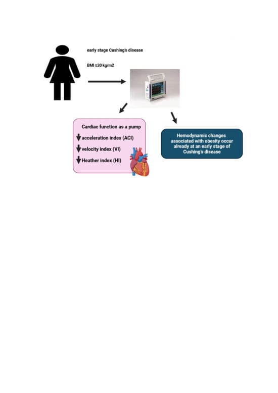
Different Patient Versus Provider Perspectives on Living with Cushing’s Disease

Abstract

Context

Patients with Cushing’s disease (CD) face challenges living with and receiving appropriate care for this rare, chronic condition. Even with successful treatment, many patients experience ongoing symptoms and impaired quality of life (QoL). Different perspectives and expectations between patients and healthcare providers (HCPs) may also impair well-being.

Objective

To examine differences in perspectives on living with CD between patients and HCPs, and to compare care goals and unmet needs.

Design

Memorial Sloan Kettering Pituitary Center established an annual pituitary symposium for pituitary patients and HCPs. Through anonymous pre-program surveys distributed at the 2020 and 2022 symposia, patients and HCPs answered questions related to their own sense, or perception of their patients’ sense, of hope, choice, and loneliness in the context of living with CD.

Participants

From 655 participants over two educational events, 46 patients with CD and 116 HCPs were included. Median age of both groups was 51 years. 78.3% of the patients were female vs. 53.0% of the HCPs.

Results

More patients than HCPs reported they had no choices in their treatment (21.7% vs. 0.9%, P < 0.001). More patients reported feeling alone living with CD than HCPs’ perception of such (60.9% vs. 45.5%, P = 0.08). The most common personal care goal concern for patients was ‘QoL/mental health,’ vs. ‘medical therapies/tumor control’ for HCPs. The most common CD unmet need reported by patients was ‘education/awareness’ vs. ‘medical therapies/tumor control’ for HCPs.

Conclusions

CD patients experience long term symptoms and impaired QoL which may in part be due to a perception of lack of effective treatment options and little hope for improvement. Communicating experiences and care goals may improve long term outcomes for CD patients.

Introduction

Patients with rare diseases face challenges receiving appropriate care. Cushing’s disease (CD), a condition associated with excess endogenous glucocorticoids due to an ACTH-secreting pituitary tumor, is a rare disease, occurring in 0.7 to 2.4 per million per year [1]. Patients with CD are at high risk for metabolic, cardiovascular, and psychiatric disease, in addition to long-term symptom burden and impaired quality of life (QoL), despite adequate treatment [1,2,3].

A critical aspect of effective patient care is communication and mutual understanding between healthcare provider (HCP) and patient. Patients with pituitary tumors experience significant anxiety associated with their diagnosis, in large part due to difficulties interacting with healthcare systems and limited communication of information [4]. Many pituitary patients express concern regarding the complexity of their care, and satisfaction improves with the delivery of more information by the HCP [4]. Patients with pituitary tumors, and CD specifically, require multidisciplinary care which necessitates effective communication in order to provide the best possible outcomes [5].

Similar to acromegaly patients [6], CD patients’ long-term well-being may be adversely affected by different perspectives and expectations between patients and HCPs, especially after treatment [7]. While HCPs primarily use biochemical data to define successful treatment, patients rely more on their symptoms and ability to regain normal functioning [7]. Despite achieving biochemical remission, CD patient perception of having persistent disease negatively impacts QoL [8]. In addition, 67.5% of Cushing’s syndrome patients report receiving insufficient information from their HCPs regarding the recovery experience after surgery despite the fact that all HCPs report providing this information [9]. Improved communication between HCPs and CD patients is vital to optimizing patients’ QoL and long term outcomes.

Recently there has been a growing emphasis on the use of internet-based platforms for healthcare delivery and education [10]. With the goals of offering HCP and patient education and assessing pituitary patients’ needs, since 2019 the pituitary center at Memorial Sloan Kettering (MSK) has offered annual virtual educational programs for pituitary patients, caregivers, HCPs, and members of the pharmaceutical industry. For the current study, we gathered deidentified information from 2020 to 2022 MSK program participants on CD patients’ and HCPs’ attitudes about CD, related to their sense of hope, choice, and loneliness, through anonymous pre-program surveys. Our specific aims were to: (1) Assess differences in perspectives between patients’ and HCPs’ responses in the pre-program survey; (2) Compare patients’ and HCPs’ perceived care goals and unmet needs.

Methods

Educational program enrollment

The MSK program was offered to patients with any type of pituitary tumor as well as HCPs, family members, caregivers, and members of industry. The role of the registrant as a patient, caregiver/family member, HCP, and/or member of industry was determined for all registrants of the virtual programs.

Any patient with a pituitary tumor treated at our center and outside institutions, inclusive of patients at all points along their treatment journey, were invited to register for the virtual education program. HCPs, including endocrinologists, neurosurgeons, otolaryngologists, radiation oncologists, neurologists, ophthalmologists, neuro-oncologists, family medicine and internal medicine physicians, physicians in training and other allied health professionals who treat and manage patients with pituitary diseases were also invited to register. Invitations were sent through email to neuroendocrine experts and endocrinologists, patient support groups on social media, direct messaging to patients with pituitary tumors by their treating physicians and via patient databases, advertisements through endocrine societies, brochure/postcard mailing, and Eventbrite, a virtual platform for live events.

Study participants

Registrants from MSK virtual programs held on December 5, 2020, (n = 328) and April 9, 2022, (n = 327) were included in the pool of subjects, among which the qualifying participants were determined.

Of the 655 total registrants from the 2020 and 2022 programs, 320 (48.9%) were patients or caregivers and 309 (47.2%) were HCPs (Fig. 1). Of the 147 providers (88 in 2020 and 59 in 2022) that attended and filled out a pre-program survey 31 were excluded from our analysis. Eight filled out surveys in both 2020 and 2022, 4 were members of industry, 3 did not fill out any responses, and 1 was not in the healthcare field. In addition, 12 providers had at least three fields missing in the survey and 3 had filled out two surveys for the same year, so they were also excluded. A total of 116 providers (72 from 2020 to 44 from 2022) were included in the analysis.

Fig. 1
figure 1

Enrollment flowchart

Among the 320 pituitary patients who attended the programs (157 from 2020 to 163 from 2022), 53 identified as ‘patients with Cushing’s’ and submitted surveys (34 participants from 2020 to 19 from 2022). Seven patients were excluded from the 2022 surveys as they had also filled out surveys in 2020, leaving a final group of 46 patients who were included in the analysis.

Virtual education programs

For each program, there was a single day of live interactive programming, meaning that all participants attended at the same time. The programs were recorded and made available for several weeks as enduring material for registrants on an online website.

After joint sessions in the morning, both programs consisted of two tracks in the afternoon: the ‘provider/clinical track’ and the ‘patient/caregiver track’. During the programs, an ongoing chat reeled through the virtual program which allowed patients to continually ask questions. Faculty experts answered these questions in written responses directly within the chat and/or in spoken responses during one of the live broadcasted Q&A sessions. Additionally, the programs both included panel discussions answering patient questions and moderated patient discussions with invited patient speakers.

Study procedures

Through anonymous pre-program surveys distributed at the 2020 and 2022 symposia, patients and HCPs answered questions related to their own sense, or perception of their patients’ sense, of hope, choice, and loneliness in the context of living with CD. This survey was developed by a multidisciplinary team and has been reported previously [11]. Demographic and clinical information was also assessed including year of diagnosis, prior treatments, and current medications (for patients) and specialty and practice type (for providers), as shown in Tables 1 and 2. Multiple-choice questions assessing patients’ attitudes toward their disease included possible answers of ‘I have no hope for improvement,’ ‘I have some hope for improvement,’ and ‘I have a lot of hope for improvement;’ and ‘I have no choice in my treatment,’ ‘I have some choices in my treatment,’ and ‘I have many choices in my treatment.’ Patients were also asked to respond ‘TRUE’ or ‘FALSE’ to the following statements: ‘I feel alone living with my Cushing’s,’ ‘Hearing the journeys of other patients helps me better understand my own,’ and ‘I feel anxious about my Cushing’s diagnosis.’

Table 1 Patient demographic data
Table 2 Provider demographic data

Multiple-choice questions assessing providers’ attitudes about their patients’ Cushing’s included possible answers of ‘I have no hope for their improvement,’ ‘I have some hope for their improvement,’ and ‘I have a lot of hope for their improvement;’ and ‘my patients have no choice in their treatment,’ ‘my patients have some choices in their treatment,’ and ‘my patients have many choices in their treatment.’ Providers were also asked to respond ‘TRUE’ or ‘FALSE’ to the following statements: ‘my patients feel alone living with their Cushing’s,’ ‘Hearing the journeys of other patients helps will help my patients better understand their own,’ and ‘my patients feel anxious about their Cushing’s diagnosis.’

Additionally, patients were surveyed on care goals and unmet needs related to their treatment. Specifically, patients were asked, ‘What are the healthcare outcomes/goals that matter to you the most?’ and ‘What do you think are unmet needs for the diagnosis or treatment of your condition?’ The first question was intended to refer to the patient specifically, while the second question was meant to examine how the condition is treated in general. Survey responses were submitted as free text and subsequently grouped by the authors (AH and EBG) into nine different categories: (a) Quality of life (QoL)/Mental Health; (b) Medical Therapies/Tumor Control; (c) Education/Awareness; (d) Communications/Multidisciplinary Care; (e) Insurance/Access; (f) Fertility; (g) Controlling Comorbidities; (h) Support System and (i) none. Responses could receive multiple designations if applicable. AH coded the free text themes independently, then EBG reviewed each answer and corresponding grouping to confirm accuracy. If there was disagreement or confusion, coding from our prior work [11] was reviewed.

HCPs were also surveyed on care goals and unmet needs related to their patient’s treatment. Providers were asked, ‘What are the healthcare outcomes/goals that matter to you the most?’ and ‘what do you think are unmet needs for the diagnosis or treatment of your patient’s condition?’ The first question was intended to refer to the provider and their goals related to Cushing’s, while the second question was meant to examine how the condition is treated in general. Survey responses were submitted as free text and subsequently grouped by the authors (AH and EBG) into nine different categories: (a) Quality of life (QoL)/Mental Health; (b) Medical Therapies/Tumor Control; (c) Education/Awareness; (d) Communications/Multidisciplinary Care; (e) Insurance/Access; (f) Fertility; (g) Controlling Comorbidities; (h) Support System and (i) none. Responses could receive multiple designations if applicable.

Statistical analysis

Descriptive statistics were presented as counts and percentages for categorical variables and as medians and interquartile range (IQR) for continuous variables. The Chi-square test or Fisher’s exact test was used to compare gender and survey responses between the CD patient group and the HCP group. All statistical tests were two-tailed, and a P-value of < 0.05 was considered statistically significant. SAS Software® (version 9.4; SAS Institute Inc., Cary, NC) was used for all analyses.

Results

Between the 2020 and 2022 events, there was combined representation from 25 different countries. A map and a full list of the countries is shown in Fig. 2.

Fig. 2

figure 2

Map of registrant locations. Locations (listed alphabetically): Argentina, Australia, Belgium, Brazil, Canada, Chile, China, Greece, Hong Kong, India, Israel, Jamaica, Latvia, Malaysia, Netherlands, New, Zealand, Oman, Peru, Philippines, Qatar, Romania, Saudi Arabia, Singapore, UK, US

From a total of 655 participants over two educational events, 46 patients with CD and 116 HCP caring for CD patients were included in the analysis. The demographic data of the patients and HCPs are outlined in Tables 1 and 2, respectively. Median age of the patients and HCPs was 51 years. 78.3% of the CD group was female vs. 53.0% of the HCP group (P = 0.003).

CD patients ranged from newly diagnosed to being diagnosed 33 years prior. The HCPs who filled out the pre-program surveys were in practice for a mean duration of 18.5 years, with a range from 1 to 54 years.

As shown in Table 1, CD patients had a mean duration of suspected active disease prior to diagnosis of 5.26 years, as defined by onset of CD symptoms until diagnosis, and a mean duration of disease since diagnosis of 5.9 years. 42 (91%) had undergone surgical treatment of their Cushing’s. For those who underwent surgery, the mean number of surgeries was 1.17 (range 0–4). 20% had received pituitary radiation. Overall, 31% of patients were on medical therapy for Cushing’s. Metyrapone was the most used CD therapy (in 11%), followed by ketoconazole (in 9%). Of those requiring pituitary hormone replacement, 34.8% had one pituitary hormone deficiency and 21.7% had multiple hormone deficiencies. Thyroid hormone replacement (37%) and adrenal replacement (30%) were the most common.

As shown in Table 2, the majority of the HCPs were endocrinologists (72%) followed by neurosurgeons (9%) and nurses (8%). There was a total of 9 different specialties represented by the provider group. 16% of the providers worked in private practice, 16% were hospital based, and 16% worked in ‘unspecified clinical care.’ 38% of the providers practice type was ‘unspecified.’

Based on the pre-program survey responses, we identified different attitudes between patients and HCPs in several domains. Table 3 depicts pre-program survey responses from CD patients and HCPs assessing their attitudes about CD. 21.7% of patients reported they had no choices in their treatment, compared to 0.9% of HCPs (P < 0.001). Almost all HCPs (99.1%) reported that CD patients had least some choice in their management. In addition, less than half (45.7%) of patients reported they had a lot of hope for improvement whereas 71.3% of HCPs had a lot of hope for their patients’ improvement. Surprisingly, fewer CD patients reported feeling anxious about their diagnosis compared to HCPs’ perceived patient anxiety (65.2% vs 94.6%, P < 0.001). However, more patients tended to feel more alone living with CD than HCPs’ perception of such (60.9% vs. 45.5%, P = 0.08). Both CD patients and HCPs agreed that hearing the journeys of other CD patients would help patients better understand their own disease (97.8% vs 100%).

Table 3 Patient and provider attitudes by pre-program survey

CD patients and HCPs were also surveyed on their personal care goals and unmet needs, results of which are shown in Figs. 3A, B and 4A, B. The most common personal care goal concern for patients was ‘QoL/mental health’ which was reported by 70%, followed by ‘controlling comorbidities’ (39%) and ‘medical therapies/tumor control’ (24%). HCPs prioritized the same three care goals as patients but ‘medical therapies/tumor control’ was the most common (44%). ‘Controlling comorbidities’ and ‘QoL/mental health’ were the second and third most often HCP reported care goals (31 and 22% respectively). ‘Education/awareness’ was the most common perceived CD unmet need by patients (59%). HCPs reported both ‘medical therapies/tumor control’ and ‘education/awareness’ to be the most common unmet needs (35 and 26%, respectively). Examples of patient and provider responses, and how they were coded, are shown in Supplemental Table 1.

Fig. 3
figure 3

A Care goals according to participants with Cushing’s who completed pre-program survey. This pie graph represents the free-text survey response from patients regarding their personal care goals as categorized by topic. B Care goals according to providers who completed pre-program survey. This pie graph represents the free-text survey response from providers regarding their personal care goals as categorized by topic

Fig. 4
figure 4

A Unmet needs for the field of Cushing’s disease according to participants with Cushing’s who completed pre-program survey. This pie graph represents the free-text survey response from patients regarding unmet needs in Cushing’s as categorized by topic. B Unmet needs for the field of Cushing’s disease according to providers who completed pre-program survey. This pie graph represents the free-text survey response from providers regarding unmet needs in Cushing’s as categorized by topic

Discussion

This study examined the differences between patients and HCP-reported perceptions of living with CD. We identified several differences in disease outlook between CD patients and HCPs. We found that more patients than HCPs reported they had no choices in their treatment. Furthermore, less than half of patients reported they had a lot of hope for improvement whereas most (71.3%) of HCPs had a lot of hope for their patients’ improvement. Interestingly, fewer CD patients reported feeling anxious about their diagnosis compared to HCPs’ perceived patient anxiety, although a higher percentage of patients reported feeling alone living with CD compared to the HCPs’ perception of patient loneliness. We also identified HCP and patient differences in reported personal care goals and perceived unmet needs in the field. The most common personal care goal concern for patients was ‘QoL/mental health,’ whereas it was ‘medical therapies/tumor control’ for HCPs. ‘Education/awareness’ was the most commonly perceived unmet need by patients, whereas it was ‘medical therapies/tumor control’ for HCPs.

Our findings support prior work demonstrating a discrepancy between patients and HCPs regarding the need for improved multidisciplinary care [12]. 43% of patients listed ‘communication/multidisciplinary care’ as an unmet need in the field, compared to 3% of providers. Pituitary centers of excellence provide expert multidisciplinary care in the neuroendocrine, neurosurgical, and radiation oncology domains, but often lack expertise in mental and physical health domains salient for CD patients, who suffer from depression, anxiety, myopathy and joint pain. In order to offer comprehensive care, psychiatrists, psychologists, social workers, pain medicine experts, physical therapists, and nutritionists with expertise in CD should be included in the pituitary center multidisciplinary team [13]. Our findings suggest that pituitary centers of excellence should take into account the most important personal care goal reported by CD patients, which is Qol/mental health, and provide expert treatment in this domain.

It is not surprising that Qol/mental health is the personal care goal most reported by CD patients. Prior assessment of acromegaly patients demonstrated the same finding: QoL/mental health was the most common personal care goal concern [11]. While surgical [14] and medical [15,16,17,18] treatment of Cushing’s improves QoL, QoL has been shown to remain impaired over time after treatment [19]. Several factors may contribute to long-term Qol impairments, including the presence of persistent disease, imperfect treatment modalities which themselves may be associated with burden and adverse side effects, and persistent comorbidities including depression, anxiety, fatigue, and overweight. Perception of disease status may also play a role in QoL. In surgically remitted CD patients, there may be discordance between biochemical remission and perceived disease status [8]. Specifically, this study found that of those with self-identified persistence of disease, 65% were in fact biochemically remitted. This group had lower QoL scores than the concordant group who self-identified as in remission with biochemical evidence of remission.

CD patients’ outlook on their condition, including their perception of choices and hope for change, has not been previously well described, despite the fact that these perceptions likely inform long term Qol. Patient outlook may be a modifiable target that if addressed, could improve long term patient well-being and outcomes. Aside from continuing progress in the development of new therapies for CD patients which can offer patients more objective choices in their treatment, other modalities should be considered. Prior work has shown that virtual educational programs improve acromegaly patients’ hope for improvement, perception of having choices in their treatment, and sense of loneliness [11]. Educational programs have also been shown to result in improved physical activity and sleep, and reduced pain levels in CS patients [20]. More work is needed to develop effective education programming tailored for CD patients to provide the appropriate support that these patients need.

Difference in HCP and patient disease perceptions may also play a role in Cushing’s patients’ quality of life and outcomes. Among a cohort of patients who underwent surgical resection for Cushing’s, 32.4% reported not receiving information from their doctors about the recovery experience, despite the fact that all physicians surveyed reported giving information about the recovery process [9]. Furthermore, 16.1% of patients in this cohort reported that not enough medical professionals were familiar with the symptoms of Cushing’s. Recovery time was also reported to be longer by patients than providers [9]. Similarly, discordance was found between acromegaly patients and HCPs regarding reported severity of symptoms, with patients more frequently reporting symptoms as severe compared to HCPs, and many patients reporting symptoms which were not reported by HCPs [6]. Improving communication between HCP and patients may positively affect CD patient outlook and QoL.

We identified a similar disparity between CD patients and HCP regarding care goals and unmet needs. 70% of patients surveyed considered QoL/mental health to be a top care goal, but only 22% of provider shared this goal. 59% of patients reported education/awareness as an unmet need, compared to 26% of HCPs. These findings support data shown by Acre et al. in which Cushing’s patients report a lack of symptom recognition by their providers [9]. HCPs should be aware that their patients may have different treatment priorities.

Our finding that more HCPs reported patient anxiety living with CD compared to patients themselves needs further exploration. This could reflect inadequate communication between HCP and patient, or skewed HCP perceptions of CD. This, and other findings in our study should be viewed in light of the small cohort, and as such, needs confirmation in larger cohorts and more in-depth symptom assessments. Additional limitations of our study include lack of paired patient-HCP responses as the HCPs included were not providing care for this specific CD cohort. Since this was a pituitary educational forum, likely most or all patients who identified as having Cushing’s had CD. However, our survey did not specify the type of surgery patients underwent or the etiology of their Cushing’s. Additionally, we used multidisciplinary team agreed upon measures and not validated assessments. Further work should consider validating a tool to assess patient-provider discordances. Our findings may also be confounded by selection bias, given that the patients participating in our virtual education programs are more likely to be under the care of experts in the field and may not represent the attitudes of all patients living with CD. Finally, the included HCPs were representatives from a range of specialties with different levels of experience taking care of patients with CD which may also affect their responses.

Our findings highlight the importance of understanding CD patients’ outlook and perspective in their condition, and that they may differ from their HCP. More than half of CD patients did not have a lot of hope for improvement and reported feeling alone, and many patients felt they had no choices in their treatment. QOL/mental health was the most commonly reported care goal for patients, which was not the case for HCPs. Comprehensive multidisciplinary care for CD patients should include mental health professionals with expertise in CD. Regular open communication between HCPs and CD patients will help bridge perception differences and facilitate personalized care, which will ultimately improve long-term outcomes for CD patients.

Data availability

The data that support the findings of this study are available from the authors upon request.

References

  1. Newell-Price J, Bertagna X, Grossman AB, Nieman LK (2006) Cushing’s syndrome. Lancet 367(9522):1605–1617. https://doi.org/10.1016/S0140-6736(06)68699-6

    Article CAS PubMed Google Scholar

  2. Kreitschmann-Andermahr I, Siegel S, Gammel C et al (2018) Support needs of patients with Cushing’s disease and Cushing’s syndrome: results of a survey conducted in Germany and the USA. Int J Endocrinol 2018:9014768. https://doi.org/10.1155/2018/9014768

    Article PubMed PubMed Central Google Scholar

  3. Feelders RA, Pulgar SJ, Kempel A, Pereira AM (2012) The burden of Cushing’s disease: clinical and health-related quality of life aspects. Eur J Endocrinol 167(3):311–326. https://doi.org/10.1530/EJE-11-1095

    Article CAS PubMed Google Scholar

  4. Donegan D, Gowan T, Gruber R et al (2021) The need for patient-centered education among patients newly diagnosed with a pituitary tumor. J Endocr Soc. https://doi.org/10.1210/jendso/bvab061

    Article PubMed PubMed Central Google Scholar

  5. Frara S, Rodriguez-Carnero G, Formenti AM, Martinez-Olmos MA, Giustina A, Casanueva FF (2020) Pituitary tumors centers of excellence. Endocrinol Metab Clin North Am 49(3):553–564. https://doi.org/10.1016/j.ecl.2020.05.010

    Article PubMed Google Scholar

  6. Geer EB, Sisco J, Adelman DT et al (2020) Observed discordance between outcomes reported by acromegaly patients and their treating endocrinology medical provider. Pituitary 23(2):140–148. https://doi.org/10.1007/s11102-019-01013-2

    Article PubMed Google Scholar

  7. Webb SM, Valassi E (2022) Quality of life impairment after a diagnosis of Cushing’s syndrome. Pituitary 25(5):768–771. https://doi.org/10.1007/s11102-022-01245-9

    Article PubMed PubMed Central Google Scholar

  8. Carluccio A, Sundaram NK, Chablani S et al (2015) Predictors of quality of life in 102 patients with treated Cushing’s disease. Clin Endocrinol (Oxf) 82(3):404–411. https://doi.org/10.1111/cen.12521

    Article PubMed Google Scholar

  9. Acree R, Miller CM, Abel BS, Neary NM, Campbell K, Nieman LK (2021) Patient and provider perspectives on postsurgical recovery of Cushing syndrome. J Endocr Soc. https://doi.org/10.1210/jendso/bvab109

    Article PubMed PubMed Central Google Scholar

  10. Ca BM, Badiu C, Bonomi M et al (2017) Developing and evaluating rare disease educational materials co-created by expert clinicians and patients: the paradigm of congenital hypogonadotropic hypogonadism. Orphanet J Rare Dis 12(1):57. https://doi.org/10.1186/s13023-017-0608-2

    Article Google Scholar

  11. Geer EB, Kilgallon JL, Liebert KJP, Kimball A, Nachtigall LB (2022) Virtual education programming for patients with acromegaly: a pilot study. Eur J Endocrinol 186(3):341–349. https://doi.org/10.1530/EJE-21-1071

    Article CAS PubMed Google Scholar

  12. Valassi E, Santos A, Yaneva M et al (2011) The European registry on Cushing’s syndrome: 2-year experience. Baseline demographic and clinical characteristics. Eur J Endocrinol 165(3):383–392. https://doi.org/10.1530/EJE-11-0272

    Article CAS PubMed Google Scholar

  13. Casanueva FF, Barkan AL, Buchfelder M et al (2017) Criteria for the definition of pituitary tumor centers of excellence (PTCOE): a pituitary society statement. Pituitary 20(5):489–498. https://doi.org/10.1007/s11102-017-0838-2

    Article PubMed PubMed Central Google Scholar

  14. Santos A, Resmini E, Martinez-Momblan MA et al (2012) Psychometric performance of the Cushing QoL questionnaire in conditions of real clinical practice. Eur J Endocrinol 167(3):337–342. https://doi.org/10.1530/EJE-12-0325

    Article CAS PubMed Google Scholar

  15. Colao A, Petersenn S, Newell-Price J et al (2012) A 12-month phase 3 study of pasireotide in Cushing’s disease. N Engl J Med 366(10):914–924. https://doi.org/10.1056/NEJMoa1105743

    Article CAS PubMed Google Scholar

  16. Fleseriu M, Biller BM, Findling JW et al (2012) Mifepristone, a glucocorticoid receptor antagonist, produces clinical and metabolic benefits in patients with Cushing’s syndrome. J Clin Endocrinol Metab 97(6):2039–2049. https://doi.org/10.1210/jc.2011-3350

    Article CAS PubMed Google Scholar

  17. Geer EB, Salvatori R, Elenkova A et al (2021) Levoketoconazole improves clinical signs and symptoms and patient-reported outcomes in patients with Cushing’s syndrome. Pituitary 24(1):104–115. https://doi.org/10.1007/s11102-020-01103-6

    Article CAS PubMed Google Scholar

  18. Pivonello R, Fleseriu M, Newell-Price J et al (2020) Efficacy and safety of osilodrostat in patients with Cushing’s disease (LINC 3): a multicentre phase III study with a double-blind, randomised withdrawal phase. Lancet Diabetes Endocrinol 8(9):748–761. https://doi.org/10.1016/S2213-8587(20)30240-0

    Article CAS PubMed Google Scholar

  19. Page-Wilson G, Oak B, Silber A et al (2023) Evaluating the burden of endogenous Cushing’s syndrome using a web-based questionnaire and validated patient-reported outcome measures. Pituitary. https://doi.org/10.1007/s11102-023-01314-7

    Article PubMed PubMed Central Google Scholar

  20. Martínez-Momblán MA, Gómez C, Santos A et al (2016) A specific nursing educational program in patients with Cushing’s syndrome. Endocrine 53:199–209. https://doi.org/10.1007/s12020-015-0737-0

    Article CAS PubMed Google Scholar

Download references

Acknowledgements

The authors would like to thank the HCP and patient participants who attended the events, the MSK faculty, invited speakers, Leslie Edwin of Cushing’s Support and Research Foundation, Amy Edouard and the MSK CME team, and Recordati Rare Diseases, Inc., Amryt Pharma (previously Chiasma, Inc.), Crinetics, Sparrow Pharmaceuticals, Corcept Therapeutics, and Xeris Biopharma (previously Strongbridge Biopharma) for providing educational grants for these educational activities.

Funding

This research was funded in part through the NIH/NCI Cancer Center Support Grant P30 CA008748.

Author information

Authors and Affiliations

  1. Division of Endocrinology, Department of Medicine, Weill Cornell Medicine, New York, NY, USA

    Amanda Halstrom

  2. Department of Epidemiology and Biostatistics, Memorial Sloan Kettering Cancer Center, New York, NY, USA

    I.-Hsin Lin

  3. Multidisciplinary Pituitary & Skull Base Tumor Center, Memorial Sloan Kettering Cancer Center, New York, NY, USA

    Andrew Lin, Marc Cohen, Viviane Tabar & Eliza B. Geer

  4. Department of Neurology, Memorial Sloan Kettering Cancer Center, New York, NY, USA

    Andrew Lin

  5. Department of Neurosurgery, Memorial Sloan Kettering Cancer Center, New York, NY, USA

    Andrew Lin, Marc Cohen & Eliza B. Geer

  6. Head and Neck Service, Department of Surgery, Memorial Sloan Kettering Cancer Center, New York, NY, USA

    Marc Cohen & Viviane Tabar

  7. Department of Medicine, Memorial Sloan Kettering Cancer Center, New York, NY, USA

    Eliza B. Geer

Contributions

A.H. and E.B.G. wrote the manuscript text and prepared the figures. All authors reviewed the manuscript.

Corresponding author

Correspondence to Eliza B. Geer.

Ethics declarations

Competing interests

The authors declare no competing interests.

Ethical approval

As an educational quality initiative project using de-identified data, it was determined that our project did not constitute human subjects research and thus did not require IRB oversight.

Additional information

Publisher’s Note

Springer Nature remains neutral with regard to jurisdictional claims in published maps and institutional affiliations.

Supplementary Information

Below is the link to the electronic supplementary material.

Radiation-induced Undifferentiated Malignant Pituitary Tumor After 5 Years of Treatment for Cushing Disease

Abstract

The occurrence of a second neoplasm possibly constitutes an adverse and uncommon complication after radiotherapy. The incidence of a second pituitary tumor in patients irradiated for adrenocorticotropic hormone secreting pituitary adenoma is rare. We report a case of a 40-year-old female with Cushing disease who underwent surgical management followed by radiotherapy. After 5 years of initial treatment, an increase in tumor size was evident at the same location, with a significant interval growth of the parasellar component of the lesion. Histology revealed an undifferentiated highly malignant sarcoma. In the span of next 2 years, the patient was followed with 2 repeat decompression surgeries and radiotherapy because of significant recurrent compressive symptoms by locally invasive malignant tumor. Despite the best efforts, the patient remained unresponsive to multiple treatment strategies (eg, surgical resections and radiotherapy) and succumbed to death.

Introduction

Radiation therapy is a commonly used modality for primary or adjuvant treatment of pituitary adenoma. It is also used as an adjuvant therapy for Cushing disease with persistent or aggressive tumor growth or recurrent disease after surgery. The immediate sequelae of radiotherapy for pituitary tumors include nausea, fatigue, diminished taste and olfaction, and hair loss [1]. One frequent long-term side effect is hypopituitarism. The incidence rate of new-onset hypopituitarism after conventional radiotherapy is approximately 30% to 100% after a follow-up of 10 years, whereas after stereotactic radiosurgery or fractionated radiotherapy, the incidence is approximately 10% to 40% at 5 years [2].

The occurrence of a second neoplasm after cranial radiotherapy constitutes possibly one of the most adverse complications. Tumors such as meningioma, glioma, and sarcoma are the most frequently reported secondary neoplasms after pituitary irradiation [3]. The cumulative probability of a second brain tumor in patients irradiated for pituitary adenoma and craniopharyngioma is approximately 4% [4].

We report 1 such case with detailed clinical, histopathological, and radiological characteristics because of its rarity and associated high mortality of radiation-induced sarcoma.

Case Presentation

The patient first presented at 40 years of age with complaints of weight gain, new-onset diabetes mellitus, hypertension, and cushingoid features in 2014. She was diagnosed with Cushing disease (24-hour urinary cortisol 1384 mcg/24 hours [3819 nmol/24 hours; reference >2 upper limit of normal], low-dose dexamethasone suppression test serum cortisol 16.6 mcg/dL [457.9 nmol/L], ACTH 85 pg/mL [18.7 pmol/L; reference range, <46 pg/mL, <10 pmol/L]) caused by invasive adrenocorticotropic hormone-secreting giant adenoma. The initial imaging revealed a homogenously enhanced pituitary macroadenoma with a size of 42 × 37 × 35 mm with suprasellar extension and encasing both the internal carotid arteries with mass effect on optic chiasma and sellar erosion. The patient underwent tumor excision by endoscopic transsphenoidal transnasal approach. Partial excision of the tumor was achieved because of cavernous sinus invasion. Histopathology and immunohistochemical stains demonstrated a corticotrophin-secreting (ACTH-staining positive) pituitary adenoma with MIB labeling index of 1% to 2%. Because biochemical remission was not achieved (urinary cortisol 794 mcg/24 hours [2191 nmol/24 hours]; ACTH 66 pg/mL [14.5 pmol/L; reference range, <46 pg/mL, <10 pmol/L]), the patient was started on ketoconazole and was received fractionated radiotherapy with a dose of 5040 cGy in 28 fractions.

Diagnostic Assessment

For the next 5 years, at yearly follow-up, 400 mg ketoconazole was continued in view of insufficient control of ACTH secretion. During follow-up, the size of the tumor was stable at approximately 23 × 16 × 33 mm after radiotherapy with no significant clinical and biochemical changes.

Five years after surgery and radiotherapy, the patient developed cerebrospinal fluid rhinorrhea; imaging revealed a cystic transformation of the suprasellar component and increase in the size of the tumor to 39 × 22 × 26 mm, which included visualization of a parasellar component of size 29 × 19 × 15 mm. The patient continued on ketoconazole. The patient was also advised to undergo hypofractionated radiotherapy but did not return for follow-up.

Treatment

In 2021, 1.5 years after the last visit, the patient developed severe headache, altered sensorium, ptosis, focal seizures, and left-sided hemiparesis. During this episode, the patient had an ACTH of 66 pg/mL (14.53 pmol/L; reference range, <46 pg/mL [<10 pmol/L]) and baseline cortisol of 25 mcg/dL (689 nmol/L; reference range, 4-18 mcg/dL [110-496 nmol/L]). Repeat imaging revealed a significant decrease in the suprasellar cystic component but an increase in the size of the parasellar component to 38 × 21 × 25 mm from 29 × 19 × 15 mm, which was isointense on T1 and T2 with heterogeneous enhancement. Significant brain stem compression and perilesional edema was also visible. The patient underwent urgent frontotemporal craniotomy and decompression of the tumor. On pathological examination, the tumor tissue was composed of small pleomorphic round cells arranged in sheets and cords separated by delicate fibrocollagenous stroma. Cells had a round to oval hyperchromatic nucleus with scanty cytoplasm. Areas of hemorrhage, necrosis, and a few apoptotic bodies were seen. The tumor tissue had very high mitotic activity of >10/10 hpf and MIB labeling index of 70%. Immunohistochemistry demonstrated positivity for vimentin, CD99, and TLE-1. Dot-like positivity was present for HMB 45, synaptophysin. INI-1 loss was present in some cells. Ten percent patchy positivity was present for p53. The tumor cells, however, consistently failed to express smooth muscle actin, CD34, Myf-4, epithelial membrane antigen, desmin, LCA, SADD4, CD138, and S-100 protein. ACTH and staining for other hormones was negative. Based on the immunological and histochemical patterns, a diagnosis of high-grade poorly differentiated malignant tumor with a probability of undifferentiated sarcoma was made.

Because of the invasion of surrounding structures and surgical inaccessibility, repeat fractionated radiotherapy was given with a dose of 4500 cGy over 25 fractions at 1.8 Gy daily to the planned target volume via image-guided fractionated radiotherapy. During the next 1.5 years, patient improved clinically with no significant increase in the size of tumor (Fig. 1). The patient was gradually tapered from ketoconazole and developed hypopituitarism requiring levothyroxine and glucocorticoid replacement. There was a significant improvement in the power of the left side and ptosis.


Figure 1.

Contrast-enhanced T1 magnetic resonance imaging dynamic pituitary scan (A, sagittal; B, axial; C, coronal sections) reveals postoperative changes with residual enhancing tumor in the right lateral sella cavity with extension into the right cavernous sinus and parasellar region encasing the cavernous and inferiorly extends through the foramen ovale below the skull base up to approximately 1.5 cm. Anteriorly, it extends up to the right orbital apex and posteriorly extends along the right dorsal surface of clivus.

Outcome and Follow-up

After 1.5 years of reradiation in 2022, the patient again developed palsies of the abducens, trigeminal, oculomotor, and trochlear cranial nerve on the right side and left-sided hemiparesis. A significant increase in tumor size to 50 × 54 × 45 mm with anterior, parasellar, and infratentorial extension was seen (Fig. 2). Again, repeat decompression surgery was done. Two months after surgery, there was no improvement in clinical features and repeat imaging suggested an increased size of the tumor by 30%, to approximately 86 × 68 × 75 mm. Nine years after initial presentation, the patient had an episode of aspiration pneumonia and died.


Figure 2.

Contrast-enhanced T1 magnetic resonance imaging dynamic pituitary images (A, sagittal; B, axial; C, coronal sections) after 1.5 years of a second session of radiotherapy reveal a significant interval increase in size of heterogeneously enhancing irregular soft tissue in sellar cavity with extension into the right cavernous sinus and parasellar region when compared with previous imaging. Superiorly, it extends in the suprasellar region, causing mass effect on the optic chiasma with encasement of the right prechiasmatic optic nerve and right-sided optic chiasma. Inferiorly, the lesion extends into the sphenoid sinus. Posteriorly, there is interval increase in the lesion involving the clivus and extending into the prepontine and interpeduncular cistern. Anteriorly, mass has reached up to the right orbital apex optic nerve canal, which shows mild interval increase.

Discussion

Radiation-induced tumors were initially described by Cahan et al in 1948. They also described the prerequisites for a tumor to be classified as a radiation-induced sarcoma [5]. The modified Cahan criteria state that (1) the presence of nonmalignancy or malignancy of a different histological type before irradiation, (2) development of sarcoma within or adjacent to the area of the radiation beam, (3) a latent period of at least 3 years between irradiation and diagnosis of secondary tumor, and (4) histological diagnosis of sarcoma, can be classified as radiation-induced sarcoma [5].

Our patient fulfilled the criteria for a radiation-induced sarcoma with a highly malignant tumor on histopathology. Radiation-induced sarcomas after functional pituitary tumors, especially Cushing disease, are rarely reported. One of the case reports revealed a high-grade osteoblastic osteosarcoma 30 years after treatment for Cushing disease with transsphenoidal resection and external beam radiotherapy [6]. In our case, there was a lag period of approximately 5 years before the appearance of a second highly undifferentiated, malignant, histologically distinct tumor. The cellular origin of this relatively undifferentiated tumor cannot be determined with certainty. However, the interlacing sarcomatous and adenomatous components resulting from distinct positive immunohistochemistry may indicate that the sarcomatous component may be derived from the preexisting pituitary adenoma.

A hormonally functional pituitary tumor is not itself expected to be associated with an increased risk of secondary malignancy, except in the case of GH-secreting tumors and those with a hereditary cancer syndrome. Although not proven, immunosuppression from hypercortisolism in Cushing disease has been proposed as a contributor to secondary tumor development [7]. Other mechanisms causing increased risk of secondary malignancy can be double-stranded DNA damage and genomic instability caused by ionizing radiation and germline mutations in tumor suppressor genes such as TP53 and Rb [7].

Radiation-induced intracranial tumors were studied in a multicenter, retrospective cohort of 4292 patients with pituitary adenoma or craniopharyngioma. Radiotherapy exposure was associated with an increased risk of a second brain tumor with a rate ratio of 2.18 (95% CI, 1.31-3.62, P < .0001). The cumulative probability of a second brain tumor was 4% for the irradiated patients and 2.1% for the controls at 20 years [7]. In another study including 426 patients irradiated for pituitary adenoma between 1962 and 1994, the cumulative risk of second brain tumors was 2.0% (CI, 0.9-4.4) at 10 years and 2.4% (95% CI, 1.2-5.0) at 20 years. The relative risk of a second brain tumor compared with the incidence in the normal population is 10.5 (95% CI, 4.3-16.7) [8].

The incidence of radiation-induced sarcomas has been estimated at 0.03% to 0.3% of patients who have undergone radiation therapy. The risk of radiation-induced sarcomas increases with field size and dose. In a systemic review and analysis of 180 cases of radiation-induced intracranial sarcomas, the average dose of radiation delivered was 51.4 ± 18.6 Gy and latent period of sarcoma onset was 12.4 ± 8.6 years. A total of 49 cases were developed after radiation treatment of pituitary adenomas (27.2%). The median overall survival time for all patients with sarcoma was 11 months, with a 5-year survival rate of 14.3% [9].

Our patient received approximately 50 Gy twice through fractionated radiotherapy, resulting in larger field size and significantly higher dose than one would expect with a modern stereotactic treatment. Such a high dose of radiation is indeed a risk factor for secondary malignancy. In our patient, in a period of 2 months, there was already >30% tumor growth after recent repeat decompression surgery.

The risk of secondary malignancy is thought to be much lower with stereotactic radiosurgery than conventional external beam radiation therapy, with an estimated cumulative incidence of 0.045% over 10 years (95% CI, 0.00-0.34) [10]. However, long-term follow-up data for patients receiving stereotactic radiation therapy are shorter and thus definitive conclusions cannot be made at this stage.

Our case highlights a rare but devastating long-term complication of pituitary tumor irradiation after Cushing disease. The limited response to various available treatment options defines the aggressive nature of radiation-induced malignancy.

Learning Points

  • The occurrence of a second neoplasm constitutes possibly one of the most adverse and rare complication after radiotherapy.
  • The incidence of radiation-induced sarcomas has been estimated at 0.03% to 0.3% of patients, but cases after Cushing disease are rarely reported.
  • Patients often present with advanced disease unresponsive to various treatment modalities because of aggressive clinical course.
  • New modalities with stereotactic radiosurgery and proton beam therapy are to be reviewed closely for risk assessment of secondary tumor.

Acknowledgments

The authors acknowledge Dr. Ishani Mohapatra for her support with histopathology and interpretation.

Contributors

All authors made individual contributions to authorship. G.B., S.K.M., and V.A.R. were involved in diagnosis and management of the patient. G.B. was involved in the writing of this manuscript and submission. V.P.S. was responsible for patient surgeries. All authors reviewed and approved the final draft.

Funding

The authors received no financial support for the research, authorship, and/or publication of this article.

Disclosures

The authors have nothing to disclose.

Informed Patient Consent for Publication

Signed informed consent could not be obtained from the patient or a proxy but was approved by the treating institute.

Data Availability Statement

Data sharing is not applicable to this article as no data sets were generated or analyzed during the current study.

© The Author(s) 2023. Published by Oxford University Press on behalf of the Endocrine Society.
This is an Open Access article distributed under the terms of the Creative Commons Attribution License (https://creativecommons.org/licenses/by/4.0/), which permits unrestricted reuse, distribution, and reproduction in any medium, provided the original work is properly cited.

Restoration of Intra-patient Variability and Diurnal Range of ACTH with Remission in Cushing’s Disease

The following is a summary of “Diurnal Range and Intra-patient Variability of ACTH Is Restored With Remission in Cushing’s Disease,” published in the November 2023 issue of Endocrinology by Alvarez, et al.


Distinguishing Cushing’s disease (CD) remission from other conditions using single adrenocorticotropic hormone (ACTH) measurements poses challenges. For a study, researchers sought to analyze changes in ACTH levels before and after transsphenoidal surgery (TSS) to identify trends confirming remission and establish ACTH cutoffs for targeted clinical trials.

A retrospective analysis involved 253 CD patients undergoing TSS at a referral center from 2005 to 2019. Remission outcomes were assessed based on postoperative ACTH levels.

Among 253 patients, 223 achieved remission post-TSS. The remission group exhibited higher ACTH variability at morning (AM) (P = .02) and evening (PM) (< .001) time points compared to the nonremission group. Nonremission cases had a significantly narrower diurnal ACTH range (P < .0001). A ≥50% decrease in plasma ACTH from mean preoperative levels, especially in PM values, predicted remission. Absolute plasma ACTH concentration and the ratio of preoperative to postoperative values were associated with nonremission (adj P < .001 and .001, respectively).

ACTH variability suppression was observed in CD, with remission linked to restored variability. A ≥50% decrease in plasma ACTH may predict CD remission post-TSS. The insights can guide clinicians in developing rational outcome measures for interventions targeting CD adenomas.

Source: academic.oup.com/jcem/article-abstract/108/11/2812/7187942?redirectedFrom=fulltext

Long-Term Efficacy and Safety of Subcutaneous Pasireotide Alone or In Combination With Cabergoline in Cushing’s Disease

Objective: This study evaluated short- and long-term efficacy and safety of the second-generation somatostatin receptor ligand pasireotide alone or in combination with dopamine agonist cabergoline in patients with Cushing’s disease (CD).

Study design: This is an open-label, multicenter, non-comparative, Phase II study comprising 35-week core phase and an optional extension phase. All patients started with pasireotide, and cabergoline was added if cortisol remained elevated. Eligible patients had active CD, with or without prior surgery, were pasireotide naïve at screening or had discontinued pasireotide for reasons other than safety. Primary endpoint was proportion of patients with a mean urinary free cortisol (mUFC) level not exceeding the upper limit of normal (ULN) at week 35 with missing data imputed using last available post-baseline assessments.

Results: Of 68 patients enrolled, 26 (38.2%) received pasireotide monotherapy and 42 (61.8%) received pasireotide plus cabergoline during the core phase. Thirty-four patients (50.0%; 95% CI 37.6–62.4) achieved the primary endpoint, of whom 17 (50.0%) received pasireotide monotherapy and 17 (50.0%) received combination therapy. Proportion of patients with mUFC control remained stable during the extension phase up to week 99. Treatment with either mono or combination therapy provided sustained improvements in clinical symptoms of hypercortisolism up to week 99. Hyperglycemia and nausea (51.5% each), diarrhea (44.1%) and cholelithiasis (33.8%) were the most frequent adverse events.

Conclusion: Addition of cabergoline in patients with persistently elevated mUFC on maximum tolerated doses of pasireotide is an effective and well-tolerated long-term strategy for enhancing control of hypercortisolism in some CD patients.

Clinical trial registration: https://clinicaltrials.gov/ct2/show/NCT01915303, identifier NCT01915303.

1 Introduction

Cushing’s disease (CD) is a rare condition arising from chronic overproduction of cortisol, secondary to an adrenocorticotropic hormone (ACTH)-secreting pituitary tumor (1). Untreated hypercortisolism results in substantial multisystem morbidity, impaired quality of life (QoL) and premature mortality (14).

Pasireotide is a second-generation, multireceptor-targeted somatostatin receptor ligand (SRLs), with affinity for 4 of the 5 known somatostatin receptor subtypes (SSTRs) (5) and is approved for the treatment of patients with CD for whom surgery has failed or is not an option (6). Phase III trials of pasireotide monotherapy have shown sustained biochemical and clinical benefits up to 5 years (69). These benefits are also reflected in real-world evidence (10).

Cabergoline, a potent dopamine agonist with high affinity for dopamine type 2 receptors (D2), is commonly used off-label for the treatment of CD (2). Small, retrospective, non-randomized studies have demonstrated long-term urinary free cortisol (UFC) control (24−;60 months) in 23−;40% of patients with CD, especially those with mild hypercortisolism (1113). A meta-analysis of individual patient data from six observational studies (n=124) reported normalization of mean UFC (mUFC) levels in 34% of patients (1415). However, a short prospective study on cabergoline monotherapy showed a limited value in controlling UFC, possibly linked to short duration (16). As most corticotropinomas co­express SSTR5 and D2, combining pasireotide and cabergoline in a stepwise approach could potentially improve efficacy with achieving more rapid biochemical control (17), a premise supported by results from an 80-day pilot study of 17 patients with CD treated with cabergoline- pasireotide combination, and low-dose ketoconazole (in case of lack of complete control with the two-drug combination) (18).

The current study aims to report the efficacy and safety of prolonged treatment with pasireotide alone or in combination with cabergoline from the largest prospective, multicentre study to date of a pituitary-targeting combination treatment regimen in patients with CD (NCT01915303).

2 Materials and methods

2.1 Patients

Adults (≥18 years) with a confirmed diagnosis of CD or de novo CD, if they were not candidates for surgery or refused surgery were enrolled. Cushing’s disease was defined by a mean 24-hour (24h) UFC level greater than the upper limit of normal (ULN, 137.95 nmol/24h), calculated from three 24h samples collected within 2 weeks; a morning plasma ACTH level within or above the normal range; and a confirmed pituitary source of Cushing’s syndrome, determined by MRI confirmation of pituitary adenoma >6mm or inferior petrosal sinus sampling (IPSS) gradient >3 after CRH stimulation (or >2 if IPSS without CRH stimulation) for those patients with a tumor ≤6mm. For patients who had prior pituitary surgery, histopathology confirming an ACTH staining adenoma was considered confirmatory of CD. Key exclusion criteria included optic chiasm compression requiring surgery, poorly controlled diabetes (glycated hemoglobin [HbA1c] >8%) and having risk factors for torsades de pointes (for further details, see the Supplementary Appendix).

2.2 Study design

This was a single-arm, open-label, multicenter, non-comparative, Phase II study. After 4 weeks of screening, patients were treated in a stepwise approach during the core phase. Patients received subcutaneous pasireotide 0.6 mg twice daily (bid) for 8 weeks. Patients with a mUFC level exceeding ULN after 8 weeks received pasireotide 0.9 mg bid for another 8 weeks. If mUFC level remained elevated with pasireotide 0.9 mg bid, oral cabergoline 0.5 mg once daily (qd) was added for 8 weeks and could be increased to 1.0 mg qd for another 8 weeks (Supplementary Figure S1). After 35 weeks of treatment in the core phase, patients could enter the extension phase of the trial. Addition or titration of cabergoline during the extension phase was at the discretion of investigators.

Collection of extension data commenced from week 43, and patients continued their current study treatment up to study end (4 September 2019; date of last patient visit), week 257. Data beyond week 99 are not reported here because of small patient numbers.

2.3 End points and assessments

The primary endpoint of the study was the proportion of patients with mUFC ≤ULN at week 35. Secondary endpoints (reported at 4-week intervals up to week 35 and 8-week intervals from week 43 to the date of the last patient visit) included changes from baseline in mUFC, plasma ACTH, serum cortisol, total cholesterol, and clinical signs (systolic/diastolic blood pressure, body mass index (BMI), weight, waist circumference, facial rubor, hirsutism, striae, supraclavicular and dorsal fat pads) and symptoms (CushingQoL). Treatment escape was defined as an increase in one UFC above the normal range during follow-up of complete responders (14). Cushing Quality of Life Questionnaire (CushingQoL) (19) scores were reported up to week 35 only. Details on the safety assessments are provided in the Supplementary Appendix.

2.4 Statistical analyses

No formal hypothesis testing was performed because of the exploratory design of the study. Efficacy analyses were conducted on full analysis set, i.e., all patients to whom study treatment was assigned. Safety analyses were conducted on all patients who received ≥1 dose of pasireotide per day during the study. For patients with missing mUFC value at week 35, including those who discontinued, the last available assessment was carried forward. Details on the post hoc analyses and sample size estimation is provided in the Supplementary Appendix. Enrolled patients, who were observed for failed inclusion or exclusion criteria during the monitoring visits, were classified under protocol deviation. However, patients with no safety concerns were allowed to continue in the study and included in the full analysis set as intention to treat – assessing the study outcome, while some patients were excluded from the per protocol analysis.

3 Results

3.1 Study population

A total of 68 patients were enrolled in the study. At baseline, 66 (97.1%) patients were pasireotide naïve, while 2 (2.9%) were treated with pasireotide previously with 4 weeks of washout period prior to screening (Table 1). Of 68 patients received treatment during the core phase, 26 (38.2%) received pasireotide monotherapy and 42 (61.8%) received combination therapy. Fifty-two (76.5%) patients completed the 35-week core phase while 16 (23.5%) discontinued (Figure 1). All 68 patients were included in the full analysis set based on the intention to treat (ITT) principle. One of the protocol deviations observed during the study, was inclusion of 3 patients with normal mUFC value at screening visit (baseline) and assigning a treatment. The deviation category for the 3 patients was ‘failed inclusion criteria’ with screening mUFC value ≤ULN (137.95 nmol/24h) or mUFC calculated using ❤ UFC values or 2 out of 3 UFC values ≤ULN. One of these patients (baseline mUFC 37.37 nmol/24h ≤ULN) was discontinued from the study at Week 2 and due to lack of post-baseline mUFC assessment, was classified ‘non-responder’ at Week 35 assessment. The 2nd patient’s baseline mUFC value of 135.20 nmol/24h was close to ULN (137.95 nmol/24h) and was rescreened. Based on the rescreened mUFC value 306.5 nmol/24h, this patient was included in study, and the mUFC at Week 35 was 192.30 nmol/24h (non-responder at Week 35 assessment). For all study assessments, the scheduled screening visit’s first mUFC value (≤ULN) was used as baseline value. The 3rd patient (baseline mUFC value 131.77 nmol/24h) was discontinued from the study at Week 26 and was also observed for non-compliant schedule visit and medication dosages. The mUFC value recorded at Week 26 (88.95 nmol/24h) was ≤ULN and this last observation was carried forward to Week 35. Hence, the patient was classified ‘responder’, leaving one patient included in the study as responder as a protocol deviation.

Table 1
www.frontiersin.orgTable 1 Patient demographics and baseline characteristics.

Figure 1
www.frontiersin.orgFigure 1 Patient disposition. *If the study drugs were locally available at the end of the core phase, patients could switch over to the commercial supply and exit the extension phase. Only in countries where the drug was not locally available were patients given the option to enter the extension phase. Percentage for patients not entering the extension phase was calculated from the total number of patients enrolled in the study.

Twenty-nine (42.6%) patients continued treatment in the extension phase; 10 (34.5%) received pasireotide monotherapy and 19 (65.5%) received combination therapy. Twelve (41.4%) patients completed the extension phase, while 17 (58.6%) discontinued treatment before study end, most commonly for unsatisfactory therapeutic effect (n=8). The most common reason for discontinuation was adverse events (AEs): 5 (17.2%) patients with pasireotide monotherapy and 2 (5.1%) patients with combination therapy.

3.2 Efficacy: biochemical response

Overall, 34/68 (50.0%; 95% CI 37.6–62.4) patients achieved the primary endpoint, of whom 17 (50.0%) were receiving pasireotide monotherapy and 17 (50.0%) were receiving combination therapy. Patients with mild hypercortisolism (mUFC 1.0–<2.0 x ULN) at baseline were more likely to respond to both pasireotide monotherapy and combination therapy (n=15; 22.1%, Figure 2). Seven of 17 patients in the pasireotide monotherapy group met the primary endpoint based on their last available assessment prior to week 35. Even if the 3 patients who had mUFC ≤ULN at baseline were excluded from the primary analysis, 33/65 (50.7%; 95% CI 38.1–63.4) patients would have achieved the primary endpoint. The results are similar to the original analysis (34/68 (50.0%; 95% CI 37.6–62.4) based on the full analysis set.

Figure 2
www.frontiersin.orgFigure 2 Patients achieving mUFC ≤ULN at week 35. At baseline there were 23 patients with mild, 30 with moderate and 12 with severe hypercortisolism. mUFC, mean urine free cortisol; ULN, upper limit of normal.

For the overall study population (n=68), mUFC rapidly decreased from 501.6 nmol/24h (3.6 x ULN; SD: 488.66 nmol/24h) to 242.1 nmol/24h (1.8 x ULN; SD: 203.47 nmol/24h) at week 4 and mUFC remained below baseline levels up to week 35 (184.8 nmol/24h; 1.3 x ULN; SD:140.13 nmol/24h). For patients who received pasireotide monotherapy (n=26), mUFC( ± SD) decreased from baseline (442.1± 557.13 nmol/24h [n=26]; 3.2 x ULN) to week 35 (136.6 ± 127.77 nmol/24h [n=14]; 1 x ULN) and at the end of the study (111.2 ± 40.39 nmol/24h [n=5]; 0.8 x ULN) using the last-observation-carried-forward (LOCF). For those who did not normalize on pasireotide monotherapy (n=42), mUFC ( ± SD) decreased from baseline, i.e., last observation before starting cabergoline (280.20 ± 129.03 nmol/24h [n=40]; 2.0 x ULN) to week 35 (206.6 ± 141.96 nmol/24h [n=31]; 1.5 x ULN) and at the end of the study (219.60 ± 83.78 nmol/24h [n=7]; 1.6 x ULN) using the LOCF. During the core phase, mean serum cortisol decreased from 738.6 nmol/L (1.3 x ULN) at baseline to 538.2 nmol/L (0.95 x ULN) and ACTH levels from 16.3 pmol/L (2.7 x ULN) to 11.0 pmol/L (1.8 x ULN) at week 35.

During the extension phase, 25 patients had a mUFC assessment; of whom 12 (48%) had a mUFC ≤ULN at the end of the extension phase. During the extension phase, mUFC levels decreased slightly and fluctuated above and below the ULN up to the week 139 (Figure 3A), while mean serum cortisol remained below ULN (404 nmol/L; Figure 3B) and ACTH levels fluctuated from 8.2 pmol/L to 11.5 pmol/L) and remained above the ULN value (Figure 3C).

Figure 3
www.frontiersin.orgFigure 3 Mean actual change over time in (A) mUFC (B) serum cortisol, and (C) ACTH. ACTH, adrenocorticotropic hormone; mUFC, mean urine free cortisol; ULN, upper limit of normal .

Twenty-one of 38 (55%) patients achieved control with combination therapy at some point during the core or extension study, of whom 13 (62%) experienced escape (at least one UFC >ULN after previous control). The time to achieve control after starting cabergoline ranged from 14−;343 days. Notably, one patient received pasireotide 0.6 mg bid initially, dose increased to 0.9 mg bid at Week 17, followed by addition of cabergoline 0.5 mg od at Week 31. The patient achieved biochemical control (mUFC value of 120.15 nmol/24h) on the same day of the start of combination therapy. Clinically it is highly unlikely that biochemical control was achieved with single dose of cabergoline administration. Therefore, it could be considered that normalization was achieved while receiving pasireotide monotherapy. Also, the physician might have prescribed combination therapy before receiving the mUFC value of the (urinary) sample delivered on the morning of combination therapy initiation (while the patient was still on monotherapy). The patient continued combination therapy and maintained biochemical control up to Week 35 and beyond. Furthermore, at Week 59 the cabergoline dose was increased to 1.0 mg/day due to mUFC >ULN at previous visit (Week 51). The patient remained on pasireotide 0.9 mg bid/cabergoline 1.0 mg od combination therapy until the study end.

The median time to escape after achieving control with the addition of cabergoline was 58 days (range 28−;344). 10/13patients regained biochemical control with combination therapy. No patients on pasireotide alone experienced escape, probably due to the short observation time.

3.3 Clinical signs and symptoms of CD

Relative to baseline, pasireotide monotherapy was accompanied by reductions in median blood pressure, weight, BMI, waist circumference, and total cholesterol. Overall improvement in clinical measures persisted over time (Supplementary Table S1). Clinical improvements were also seen following the addition of cabergoline, particularly for hirsutism (Supplementary Figures S2S3).

Mean( ± SD) standardized CushingQoL score was 41.6(± 20.2) at baseline and increased to 47.6(± 20.8) at week 35 (Supplementary Table S2), indicating improvements in patients’ QoL (19).

3.4 Safety and tolerability

Median duration of exposure to pasireotide was 35.0 weeks (range 0−;268), with a median dose of 1.53 mg/day (range 0.29−;1.80). Median duration of exposure to cabergoline was 16.9 weeks (range 1−;215), with a median dose of 0.50 mg/day (range 0.44−;0.97).

All patients (N=68) reported at least one AE and 28/68 (41.2%) patients had a grade 3/4 AE (Table 2). The most common AEs (≥30%) were hyperglycemia and nausea (51.5% each), diarrhea (44.1%) and cholelithiasis (33.8%). Treatment-related AEs (TRAEs) were reported in 66/68 (97.1%) patients; the most frequent TRAEs (≥30%) were hyperglycemia and nausea (47.1% each), diarrhea (39.7%), and cholelithiasis (32.4%). Fourteen (20.6%) patients had ≥1 AE leading to discontinuation.

Table 2
www.frontiersin.orgTable 2 Summary of adverse events (≥10%), overall and by treatment regimen.

The most common AEs leading to discontinuation were increased gamma-glutamyl transferase (GGT) and hyperglycemia (two patients each, 2.9%). Twenty-three (33.8%) patients had ≥1 AE leading to dose adjustment or interruption. Details on special safety assessments such as hyperglycemia-related AEs, blood glucose, HbA1c, IGF-1 as well as hematological and biochemical abnormalities are presented in the Supplementary Appendix.

Three (4.4%) patients died during the study, two (2.9%) during the core phase and one (1.5%) during the extension. All deaths were considered unrelated to study medication. The causes during the core phase were multi-organ dysfunction syndrome for one patient aged 79 years and unknown for the other aged 34 years. Uncontrolled hypertension was reported as the cause of death for the patient aged 47 during the extension phase.

4 Discussion

The severe morbidity and increased mortality with uncontrolled CD highlight the importance of identifying an effective medical strategy. This study explored the potential of a synergistic benefit of the addition of cabergoline to pasireotide treatment in patients with CD.

Complete normalization of cortisol production is required to reverse the risks of morbidity and mortality in patients with CD (1). Two small studies showed clinical improvement of normalized UFC when cabergoline and ketoconazole were combined (2021). Benefit has also been reported with triple therapy with pasireotide, cabergoline and ketoconazole (18) and triple therapy with ketoconazole, metyrapone and mitotane in severe CD (22). In the current study, 50% of patients achieved the primary endpoint of mUFC ≤ULN at week 35 and a similar proportion (48%) sustained biochemical control throughout the extension phase. Notably, combination treatment doubled the number of patients who attained mUFC ≤ULN from the core phase to the end of the extension phase. In particular, mUFC was rapidly reduced with treatment, i.e., in most patients within 2 months, while measures of patient-reported outcomes also improved including QoL. Twenty-three patients (33.8%) who completed the core phase did not enter the extension phase. This was because only patients from countries where a commercial supply was unavailable were given the option to enter the extension phase.

This study confirms previous reports that patients with mild hypercortisolism at baseline were more likely to achieve mUFC control with pasireotide monotherapy than patients with moderate or severe hypercortisolism (623). In addition, patients with moderate hypercortisolism at baseline were more likely to achieve mUFC control with the addition of cabergoline. This supports that a combination therapy can be effective for patients with a wider range of disease severity. Accordingly, in vitro data may indeed indicate synergism between SSTR and D2 that might increase therapeutic efficacy (2425).

Improvements in clinical signs and symptoms with pasireotide monotherapy were consistent with published data (610). In the core phase, an improvement of blood pressure and BMI was observed with pasireotide monotherapy and, to a lesser extent, with combination therapy which may related to the difference in duration of biochemical remission.

The overall safety profile was consistent with that expected for pasireotide, with most AEs being mild/moderate (2627). There were no new safety signals identified with the addition of cabergoline. Common AEs including nausea, headache, dizziness, and fatigue are suggestive of steroid withdrawal symptoms associated with the decrease in UFC, although direct drug effects cannot fully be excluded. Adrenal insufficiency was not reported as side effect. Rates of hyperglycemia-related AEs (68%) were consistent with those in previous reports of pasireotide monotherapy (610). FPG increased with pasireotide monotherapy during the first 8 weeks of treatment and stabilized for the remainder of the study, including following the addition of cabergoline. These data highlight the vital role of blood glucose monitoring in these patients.

Both pasireotide and cabergoline are pituitary-targeted agents that act directly on the source of the disease via inhibition of ACTH release by the corticotroph tumor, which may be an advantage over steroid synthesis inhibitors. This study further confirms previous data reporting the benefits of pasireotide in combination with cabergoline in patients with CD (18). While not entirely elucidated, down-regulation of dopamine D2 receptors (D2R) expression, and post-receptor desensitization and/or tumor regrowth of corticotroph tumor cell were suggested as possible mechanisms for treatment escape (15). Moreover, different dopamine receptor patterns and/or D2R isoforms also influence the response and eventually the treatment escape. Treatment escape has been observed in some studies after long-term (7−;12 months) treatment with cabergoline (13), however it is possible that use of concomitant SRLs could potentially reduce the rate of escape. In this study, a total of 13 patients experienced treatment escape. However, 10 of these patients regained biochemical control. For 7 of these 10 patients, there was up titration of doses to a maximum of 1.8 mg/day of pasireotide and 1 mg/day of cabergolineAlthough pasireotide and cabergoline have shown long-term reduction in IGF-1 levels in patients with acromegaly (2829), there is little evidence for this effect in patients with CD (430). One study (n=17) found significant decreases in IGF-1 after 28 days’ treatment with pasireotide that was independent of UFC reduction. One-third of patients had low IGF-1 (30). Our study showed that almost half of patients (47.6%) had IGF-1 levels either above ULN or below LLN prior to the addition of cabergoline, and IGF-1 levels decreased relative to the baseline, with majority of values within the normal range during the core and extension phases up to week 99. Baseline levels of IGF-1 may already be low because of the suppressive effect of excess cortisol on the somatotropic axis (31).

Although clinicians have several therapeutic options at their disposal to treat hypercortisolemia associated with CD, the optimal treatment approach should be based on the individual clinical situation and the benefit–risk considerations for each patient. In this study, 13 patients had history of pituitary radiation, with a duration of at least 2.6 years (median 3.3 years) between the last radiation treatment and the observed response date. However, only 7/13 patients achieved the therapeutic target. Although there was a gap of > 2 years, we cannot exclude the role of radiation in normalizing UFC. Contrastingly, 6/13 patients treated with radiation did not achieve mUFC ≤ULN (responders) at Week 35. The impact of the adjuvant radiation therapy remains unclear.

The strengths of this study are that this is the largest and longest prospective study with pituitary-directed pharmacotherapy, to date, evaluating the addition of cabergoline to pasireotide in patients with CD, and this stepwise approach reflects real-world clinical practice (18). The study is limited by the open-label design and the fact that it was not a head-to-head comparative study of pasireotide only versus pasireotide plus cabergoline. This may be of importance in interpreting patient-reported outcomes. Several patients continued treatment for almost 2 years; however, interpretation of long-term data should be made with caution because of the small patient numbers. Notably, the last available assessment was carried forward for patients with missing mUFC value at week 35 including those who discontinued and were considered for response analysis. It should also be noted that the definition of loss of response, also known as escape, used in this study (at least one UFC value >ULN after previously achieving UFC ≤ULN) may overestimate the rate of apparent escape as UFC values may have fluctuated about the ULN range or been marginally elevated. The definition of treatment escape differs across studies, and we have used a very stringent one in this study, requiring only a single high UFC to meet the classification as escape. Thus, it is likely that some loss of biochemical control interpreted as escape is actually fluctuation of cortisol around the upper limit of normal range.

Other limitations include protocol deviations in including 3 patients with normal UFC at baseline (one patient was uncontrolled at rescreen, and one was discontinued at 2 weeks – both classified as

non-responders), lack of data on impact of radiation therapy without study drug in patients who gained biochemical control with adjuvant radiation therapy, lack of pituitary magnetic resonance imaging to detect pituitary tumor changes, lack of data about effective cabergoline dose and absence of cardiac valve assessment for mild to moderate severity in the medium term. Both pasireotide and cabergoline can induce tumor shrinkage in CD (693235) and it would be interesting to examine the combined effect on tumor size. This study used the subcutaneous formulation of pasireotide, whereas the most common usage currently is the long-acting formulation. Efficacy of long-acting pasireotide (36) seems higher compared to the subcutaneous formulation (7) and the effect of combination of long-acting pasireotide with cabergoline should be evaluated in future studies. No formal assessments were made for impulsive control disorders, which have been associated with dopamine agonists, including cabergoline (32333738). The reason that several different terms were used for hyperglycemia-related AEs is that they were reported as per discretion of each investigator. No additional psychiatric AEs were reported, although they were not exhaustively searched.

5 Conclusions

This is the first study demonstrating that pituitary-targeted combination treatment with pasireotide and cabergoline doubled the number of patients who attained mUFC ≤ULN. Both short- and long-term safety profile are consistent with known data for pasireotide and cabergoline. The low rate of discontinuation due to AEs suggests that pasireotide alone or as combination treatment is generally well-tolerated if appropriately monitored, even with prolonged treatment. The addition of cabergoline to pasireotide treatment in patients with persistently elevated mUFC could be an effective long-term strategy for enhancing the control of CD in a subset of patients, with close monitoring for possible escape.

Data availability statement

The original contributions presented in the study are included in the article/Supplementary Material. Further inquiries can be directed to the corresponding author.

Ethics statement

The studies involving humans were approved by Hospital Britanico, Buenos Aires, Argentina; Ethische commissie University Hospitals Leuven, Leuven, Belgium; Universitair Ziekenhuis Gent, Gent, Belgium; Comite de Etica em Pesquisa Hospital Moinhos de Vento, Porto Alegre-RS, Brazil; Comitê de Ética em Pesquisa do Hospital de Clı́nicas, Universidade Federal do Paraná, Curitiba-PR, Brazil; Comissão de Ética para Análise de Projetos de Pesquisa, São Paulo – SP, Brazil; Ethics Committee for clinical trials, Sofia, Bulgaria; Comité Corporativo de Ética en Investigación, Bogotá DC, Colombia; Comite De Protection Des Personnes, Groupe Hospitalier Pellegrin – Bat, Bordeaux Cedex, France; Friedrich-Alexander Universitat Erlangen-Nurnberg, Medizinische Fakultat, Erlangen, Germany;National Ethics Committee, Cholargos, Athens, Greece; Ethics Committee for Clinical Pharmacology (ECCP), Budapest, Hungary; Institute Ethics Committee, New Delhi, India; Institutional Review Board (IRB) Ethics Committee Silver, Christian Medical College, Vellore, Tamil Nadu, India; Institute Ethics Committee, PGIMER, Chandigarh, India; Comitato Etico Dell’irccs Istituto Auxologico Italiano Di Milano, Milano, Italy; Comitato Etico Universita’ Federico Ii Di Napoli, Napoli, Italy; Jawatankuasa Etika & Penyelidikan Perubatan (Medical Research and Ethics Committee), d/a Institut Pengurusan Keshatan Jalan Rumah Sakit, Kuala Lumpur, Malaysia; Institutd Nacional De Neurologia Y Neurocirugia, Mexico City, Mexico; Clinica Bajio (CLINBA), Guanajuato, Mexico; Medische Ethische Toetsings Commissie, Rotterdam; Netherlands; CEIm Provincial de Málaga, Málaga, Spain; Istanbul University Cerrahpasa Medical Faculty, Istanbul, Turkey; WIRB, Puyallup, WA, USA; Research Integrity Office, Oregon Health & Science University Portland, OR USA. The studies were conducted in accordance with local legislations and institutional requirements. The participants provided their written informed consent to participate in this study.

Author contributions

All authors directly participated in the planning, execution, or analysis, and have had full control of complete primary data, and hold responsibility for data integrity and accuracy. All authors contributed to the article and approved the submitted version.

Acknowledgments

We thank Julie Brown, Mudskipper Business Ltd, and Manojkumar Patel and Sashi Kiran Goteti, Novartis Healthcare Private Limited, for medical editorial assistance with this manuscript. We would also like to thank all investigators, sub-investigators, study nurses and coordinators, and patients who have made this study possible.

Conflict of interest

HP and RM were Novartis employees and owned Novartis stocks. AMP was employed by Novartis and Recordati. AC is a Novartis employee and owns Novartis stocks. RF received research grants from Strongbridge and Corcept, consulting fee from Recordati, honoraria and financial support for meetings and/or travel from HRA Pharma and Recordati, and attended advisory boards for Recordati. MF has received research support to Oregon Health & Science University as a principal investigator from Recordati and Xeris Strongbridge and has performed occasional scientific consultancy for Recordati, HRA Pharma, Sparrow, and Xeris Strongbridge. PK attended advisory boards for Recordati. MB’s institution received consulting fee and attended advisory boards from Recordati. DG-D received research grants from Recordati Rare Disease and Bayer, consulting fee from Abbott-Lafrancol, Biotoscana, PTC lab, Glaxo/Helou, Recordati Rare Disease, and Bayer, honoraria from Valentech Pharma, Sanofi, and Bayer, travel grants from Recordati Rare Disease, advocacy groups and other leadership roles from Asociación Colombiana de Endocrinologia and Asociación Colombiana de Osteoporosis y Metabolismo, and other financial and non-financial interests include Asociacion Colombiana de Endocrinologia y Metabolismo, Hospital Universitario Fundación Santa Fé de Bogota, and Asociación Colombiana de Osteoporosis y Metabolismo. CB received research grants from Novartis and Recordati, and consulting and speaker fee from Novartis. BB served as the principal investigator for grants to Massachusetts General Hospital from Cortendo/Strongbridge Xeris, Millendo, and Novartis and has occasionally consulted for Cortendo/Strongbridge Xeris, HRA Pharma, Novartis Recordati, and Sparrow. RP and his institution received research grants and honoraria from Pfizer, Ipsen, Novartis, Merck Serono, IBSA Farmaceutici, Corcept, Shire, HRA Pharma, ICON, Covance, Neuroendocrine CAH, Camurus, Recordati, Janssen Cilag, and CMED Clinical Services, received consulting fee from Recordati Rare Disease, Organon Italia, Siunergos Pharma, Corcept, S&R Farmaceutici S.p.A., DAMOR Farmaceutici, and Pfizer, attended advisory boards from Crinetics Pharmaceuticals, Recordati Rare Disease, Pfizer, and HRA Pharma.

The remaining author declares that the research was conducted in the absence of any commercial or financial relationships that could be construed as a potential conflict of interest.

The authors declare that this study received funding from Novartis Pharma AG. Novartis was involved in the study design, analysis, interpretation of data, and providing financial support for medical editorial assistance of this article.

Publisher’s note

All claims expressed in this article are solely those of the authors and do not necessarily represent those of their affiliated organizations, or those of the publisher, the editors and the reviewers. Any product that may be evaluated in this article, or claim that may be made by its manufacturer, is not guaranteed or endorsed by the publisher.

Supplementary material

The Supplementary Material for this article can be found online at: https://www.frontiersin.org/articles/10.3389/fendo.2023.1165681/full#supplementary-material

References

1. Lacroix A, Feelders RA, Stratakis CA, Nieman LK. Cushing’s syndrome. Lancet (2015) 386(9996):913–27. doi: 10.1016/s0140-6736(14)61375-1

PubMed Abstract | CrossRef Full Text | Google Scholar

2. Feelders RA, Newell-Price J, Pivonello R, Nieman LK, Hofland LJ, Lacroix A. Advances in the medical treatment of Cushing’s syndrome. Lancet Diabetes Endocrinol (2019) 7(4):300–12. doi: 10.1016/s2213-8587(18)30155-4

PubMed Abstract | CrossRef Full Text | Google Scholar

3. Newell-Price J. Mortality in cushing disease. Nat Rev Endocrinol (2016) 12(9):502–3. doi: 10.1038/nrendo.2016.118

PubMed Abstract | CrossRef Full Text | Google Scholar

4. Fleseriu M, Auchus R, Bancos I, Ben-Shlomo A, Bertherat J, Biermasz NR, et al. Consensus on diagnosis and management of Cushing’s disease: a guideline update. Lancet Diabetes Endocrinol (2021) 9(12):847–75. doi: 10.1016/s2213-8587(21)00235-7

PubMed Abstract | CrossRef Full Text | Google Scholar

5. Schmid HA. Pasireotide (SOM230): Development, mechanism of action and potential applications. Mol Cell Endocrinol (2008) 286(1):69–74. doi: 10.1016/j.mce.2007.09.006

PubMed Abstract | CrossRef Full Text | Google Scholar

6. Colao A, Petersenn S, Newell-Price J, Findling JW, Gu F, Maldonado M, et al. A 12-month Phase 3 study of pasireotide in Cushing’s disease. N Engl J Med (2012) 366(10):914–24. doi: 10.1056/NEJMoa1105743

PubMed Abstract | CrossRef Full Text | Google Scholar

7. Lacroix A, Gu F, Gallardo W, Pivonello R, Yu Y, Witek P, et al. Efficacy and safety of once-monthly pasireotide in Cushing’s disease: a 12 month clinical trial. Lancet Diabetes Endocrinol (2018) 6(1):17–26. doi: 10.1016/s2213-8587(17)30326-1

PubMed Abstract | CrossRef Full Text | Google Scholar

8. Fleseriu M, Petersenn S, Biller BMK, Kadioglu P, De Block C, T’Sjoen G, et al. Long-term efficacy and safety of once-monthly pasireotide in Cushing’s disease: A Phase III extension study. Clin Endocrinol (2019) 91(6):776–85. doi: 10.1111/cen.14081

CrossRef Full Text | Google Scholar

9. Petersenn S, Salgado LR, Schopohl J, Portocarrero-Ortiz L, Arnaldi G, Lacroix A, et al. Long-term treatment of Cushing’s disease with pasireotide: 5-year results from an open-label extension study of a Phase III trial. Endocrine (2017) 57(1):156–65. doi: 10.1007/s12020-017-1316-3

PubMed Abstract | CrossRef Full Text | Google Scholar

10. Fleseriu M, Iweha C, Salgado L, Mazzuco TL, Campigotto F, Maamari R, et al. Safety and efficacy of subcutaneous pasireotide in patients with Cushing’s disease: results from an open-label, multicenter, single-arm, multinational, expanded-access study. Original Res Front Endocrinol (Lausanne) (2019) 10:436. doi: 10.3389/fendo.2019.00436

CrossRef Full Text | Google Scholar

11. Pivonello R, De Martino MC, Cappabianca P, De Leo M, Faggiano A, Lombardi G, et al. The medical treatment of Cushing’s disease: effectiveness of chronic treatment with the dopamine agonist cabergoline in patients unsuccessfully treated by surgery. J Clin Endocrinol Metab (2009) 94(1):223–30. doi: 10.1210/jc.2008-1533

PubMed Abstract | CrossRef Full Text | Google Scholar

12. Godbout A, Manavela M, Danilowicz K, Beauregard H, Bruno OD, Lacroix A. Cabergoline monotherapy in the long-term treatment of Cushing’s disease. Eur J Endocrinol (2010) 163(5):709–16. doi: 10.1530/eje-10-0382

PubMed Abstract | CrossRef Full Text | Google Scholar

13. Ferriere A, Cortet C, Chanson P, Delemer B, Caron P, Chabre O, et al. Cabergoline for Cushing’s disease: a large retrospective multicenter study. Eur J Endocrinol (2017) 176(3):305–14. doi: 10.1530/eje-16-0662

PubMed Abstract | CrossRef Full Text | Google Scholar

14. Palui R, Sahoo J, Kamalanathan S, Kar SS, Selvarajan S, Durgia H. Effect of cabergoline monotherapy in Cushing’s disease: an individual participant data meta-analysis. J Endocrinol Invest (2018) 41(12):1445–55. doi: 10.1007/s40618-018-0936-7

PubMed Abstract | CrossRef Full Text | Google Scholar

15. Palui R, Sahoo J, Kamalanathan S, Kar SS, Selvarajan S, Durgia H. Correction to: Palui R, Sahoo J, Kamalanathan S, Kar SS, Selvarajan S, Durgia H. Effect of cabergoline monotherapy in Cushing’ disease: an individual participant data meta-analysis. J Endocrinol Invest. 2018; 41(12):1445-1455. Doi: 10.1007/s40618-018-0936-7. J Endocrinol Invest (2022) 45(4):899–900. doi: 10.1007/s40618-021-01723-0

PubMed Abstract | CrossRef Full Text | Google Scholar

16. Burman P, Edén-Engström B, Ekman B, Karlsson FA, Schwarcz E, Wahlberg J. Limited value of cabergoline in Cushing’s disease: a prospective study of a 6-week treatment in 20 patients. Eur J Endocrinol (2016) 174(1):17–24. doi: 10.1530/eje-15-0807

PubMed Abstract | CrossRef Full Text | Google Scholar

17. Colao A, Filippella M, Pivonello R, Di Somma C, Faggiano A, Lombardi G. Combined therapy of somatostatin analogues and dopamine agonists in the treatment of pituitary tumours. Eur J Endocrinol (2007) 156:S57–63. doi: 10.1530/eje.1.02348

PubMed Abstract | CrossRef Full Text | Google Scholar

18. Feelders RA, de Bruin C, Pereira AM, Romijn JA, Netea-Maier RT, Hermus AR, et al. Pasireotide alone or with cabergoline and ketoconazole in Cushing’s disease. N Engl J Med (2010) 362(19):1846–8. doi: 10.1056/NEJMc1000094

PubMed Abstract | CrossRef Full Text | Google Scholar

19. Webb SM, Badia X, Barahona MJ, Colao A, Strasburger CJ, Tabarin A, et al. Evaluation of health-related quality of life in patients with Cushing’s syndrome with a new questionnaire. Eur J Endocrinol (2008) 158(5):623–30. doi: 10.1530/eje-07-0762

PubMed Abstract | CrossRef Full Text | Google Scholar

20. Barbot M, Albiger N, Ceccato F, Zilio M, Frigo AC, Denaro L, et al. Combination therapy for Cushing’s disease: effectiveness of two schedules of treatment: should we start with cabergoline or ketoconazole? . Pituitary (2014) 17(2):109–17. doi: 10.1007/s11102-013-0475-3

PubMed Abstract | CrossRef Full Text | Google Scholar

21. Vilar L, Naves LA, Azevedo MF, Arruda MJ, Arahata CM, Moura E, et al. Effectiveness of cabergoline in monotherapy and combined with ketoconazole in the management of Cushing’s disease. Pituitary (2010) 13(2):123–9. doi: 10.1007/s11102-009-0209-8

PubMed Abstract | CrossRef Full Text | Google Scholar

22. Kamenický P, Droumaguet C, Salenave S, Blanchard A, Jublanc C, Gautier JF, et al. Mitotane, metyrapone, and ketoconazole combination therapy as an alternative to rescue adrenalectomy for severe ACTH-dependent Cushing’s syndrome. J Clin Endocrinol Metab. (2011) 96(9):2796–804. doi: 10.1210/jc.2011-0536

PubMed Abstract | CrossRef Full Text | Google Scholar

23. Boscaro M, Ludlam WH, Atkinson B, Glusman JE, Petersenn S, Reincke M, et al. Treatment of pituitary-dependent Cushing’s disease with the multireceptor ligand somatostatin analog pasireotide (SOM230): a multicenter, Phase II trial. J Clin Endocrinol Metab (2009) 94(1):115–22. doi: 10.1210/jc.2008-1008

PubMed Abstract | CrossRef Full Text | Google Scholar

24. Rocheville M, Lange DC, Kumar U, Patel SC, Patel RC, Patel YC. Receptors for dopamine and somatostatin: formation of hetero-oligomers with enhanced functional activity. Science (2000) 288(5463):154–7. doi: 10.1126/science.288.5463.154

PubMed Abstract | CrossRef Full Text | Google Scholar

25. Baragli A, Alturaihi H, Watt HL, Abdallah A, Kumar U. Heterooligomerization of human dopamine receptor 2 and somatostatin receptor 2 Co-immunoprecipitation and fluorescence resonance energy transfer analysis. Cell Signal (2007) 19(11):2304–16. doi: 10.1016/j.cellsig.2007.07.007

PubMed Abstract | CrossRef Full Text | Google Scholar

26. Pivonello R, Ferrigno R, De Martino MC, Simeoli C, Di Paola N, Pivonello C, et al. Medical treatment of Cushing’s disease: an overview of the current and recent clinical trials. Front Endocrinol (Lausanne) (2020) 11:648. doi: 10.3389/fendo.2020.00648

CrossRef Full Text | Google Scholar

27. Capatina C, Hinojosa-Amaya JM, Poiana C, Fleseriu M. Management of patients with persistent or recurrent Cushing’s disease after initial pituitary surgery. Expert Rev Endocrinol Metab (2020) 15(5):321–39. doi: 10.1080/17446651.2020.1802243

PubMed Abstract | CrossRef Full Text | Google Scholar

28. Colao A, Bronstein MD, Brue T, De Marinis L, Fleseriu M, Guitelman M, et al. Pasireotide for acromegaly: long-term outcomes from an extension to the Phase III PAOLA study. Eur J Endocrinol (2020) 182(6):583. doi: 10.1530/EJE-19-0762

PubMed Abstract | CrossRef Full Text | Google Scholar

29. Marazuela M, Ramos-Leví A, Sampedro-Núñez M, Bernabeu I. Cabergoline treatment in acromegaly: pros. Endocrine (2014) 46(2):215–9. doi: 10.1007/s12020-014-0206-1

PubMed Abstract | CrossRef Full Text | Google Scholar

30. Varewijck AJ, Feelders RA, de Bruin C, Pereira AM, Netea-Maier RT, Zelissen PMJ, et al. P3-535, Pasireotide treatment in cushing disease: effects on the IGF-I system. Endocr Rev (2011) 32(Suppl 1):1–3095. doi: 10.1210/endo-meetings.2011.PART4.P5.P3-535

CrossRef Full Text | Google Scholar

31. Mazziotti G, Giustina A. Glucocorticoids and the regulation of growth hormone secretion. Nat Rev Endocrinol (2013) 9(5):265–76. doi: 10.1038/nrendo.2013.5

PubMed Abstract | CrossRef Full Text | Google Scholar

32. Batista RL, Musolino NRC, Cescato VAS, da Silva GO, Medeiros RSS, Herkenhoff CGB, et al. Cabergoline in the management of residual nonfunctioning pituitary adenoma: a single-center, open-label, 2-year randomized clinical trial. Am J Clin Oncol (2019) 42(2):221–7. doi: 10.1097/coc.0000000000000505

PubMed Abstract | CrossRef Full Text | Google Scholar

33. Lacroix A, Gu F, Schopohl J, Kandra A, Pedroncelli AM, Jin L, et al. Pasireotide treatment significantly reduces tumor volume in patients with Cushing’s disease: results from a Phase 3 study. Pituitary (2020) 23(3):203–11. doi: 10.1007/s11102-019-01021-2

PubMed Abstract | CrossRef Full Text | Google Scholar

34. Simeoli C, Auriemma RS, Tortora F, De Leo M, Iacuaniello D, Cozzolino A, et al. The treatment with pasireotide in Cushing’s disease: effects of long-term treatment on tumor mass in the experience of a single center. Endocrine (2015) 50(3):725–40. doi: 10.1007/s12020-015-0557-2

PubMed Abstract | CrossRef Full Text | Google Scholar

35. Manavela MP, Danilowicz K, Bruno OD. Macrocorticotropinoma shrinkage and control of hypercortisolism under long-term cabergoline therapy: case report. Pituitary (2012) 15:33–6. doi: 10.1007/s11102-011-0309-0

PubMed Abstract | CrossRef Full Text | Google Scholar

36. Lacroix A, Bronstein MD, Schopohl J, Delibasi T, Salvatori R, Li Y, et al. Long-acting pasireotide improves clinical signs and quality of life in Cushing’s disease: results from a phase III study. J Endocrinol Invest. (2020) 43(11):1613–22. doi: 10.1007/s40618-020-01246-0

PubMed Abstract | CrossRef Full Text | Google Scholar

37. Hinojosa-Amaya JM, Johnson N, González-Torres C, Varlamov EV, Yedinak CG, McCartney S, et al. Depression and impulsivity self-assessment tools to identify dopamine agonist side effects in patients with pituitary adenomas. Front Endocrinol (Lausanne) (2020) 11:579606. doi: 10.3389/fendo.2020.579606

PubMed Abstract | CrossRef Full Text | Google Scholar

38. Grall-Bronnec M, Victorri-Vigneau C, Donnio Y, Leboucher J, Rousselet M, Thiabaud E, et al. Dopamine agonists and impulse control disorders: a complex association. Drug Saf. (2018) 41(1):19–75. doi: 10.1007/s40264-017-0590-6

PubMed Abstract | CrossRef Full Text | Google Scholar

Keywords: somatostatin, pasireotide, cabergoline, Cushing’s disease, hypercortisolism

Citation: Feelders RA, Fleseriu M, Kadioglu P, Bex M, González-Devia D, Boguszewski CL, Yavuz DG, Patino H, Pedroncelli AM, Maamari R, Chattopadhyay A, Biller BMK and Pivonello R (2023) Long-term efficacy and safety of subcutaneous pasireotide alone or in combination with cabergoline in Cushing’s disease. Front. Endocrinol. 14:1165681. doi: 10.3389/fendo.2023.1165681

Received: 14 February 2023; Accepted: 11 August 2023;
Published: 09 October 2023.

Edited by:

Renato Cozzi, Endocrinology Unit Ospedale Niguarda, Italy

Reviewed by:

Przemyslaw Witek, Warsaw Medical University, Poland
Athanasios Fountas, General Hospital of Athens G. Genimatas, Greece

Copyright © 2023 Feelders, Fleseriu, Kadioglu, Bex, González-Devia, Boguszewski, Yavuz, Patino, Pedroncelli, Maamari, Chattopadhyay, Biller and Pivonello. This is an open-access article distributed under the terms of the Creative Commons Attribution License (CC BY). The use, distribution or reproduction in other forums is permitted, provided the original author(s) and the copyright owner(s) are credited and that the original publication in this journal is cited, in accordance with accepted academic practice. No use, distribution or reproduction is permitted which does not comply with these terms.

*Correspondence: Richard A. Feelders, r.feelders@erasmusmc.nl

Present addresses: Alberto M. Pedroncelli, Chief Medical Office, Camurus AB, Lund, SwedenRicardo Maamari, Global Medical Affairs, Mayne Pharma, Raleigh, NC, United States

These authors have contributed equally to this work

Disclaimer: All claims expressed in this article are solely those of the authors and do not necessarily represent those of their affiliated organizations, or those of the publisher, the editors and the reviewers. Any product that may be evaluated in this article or claim that may be made by its manufacturer is not guaranteed or endorsed by the publisher.

From https://www.frontiersin.org/articles/10.3389/fendo.2023.1165681/full

Multiple Clinical Indications of Mifepristone: A Systematic Review

​Abstract

Mifepristone and misoprostol are globally used medications that have become disparaged through the stigmatization of reproductive healthcare. Patients are hindered from receiving prompt treatment in clinical scenarios where misoprostol and mifepristone are the drugs of choice. It is no exaggeration to emphasize that in cases where reproductive healthcare is concerned. The aim of this paper is to discuss the different indications of mifepristone and to delineate where the discrepancy in accessibility arises. For this systematic review, we included publications citing clinical trials involving the use and efficacy of mifepristone published in English within the date range of 2000 to 2023. Five databases were searched to identify relevant sources. These databases are Google Scholar, MEDLINE with full text through EBSCO, and three National Center for Biotechnology Information (NCBI) databases (NCBI Bookshelf, PubMed, and PubMed Central). Twenty-three records were ultimately included in this review. Mifepristone has been shown to have therapeutic effects in the treatment of psychiatric disorders, such as major depressive disorder and psychotic depression. There was a significant decrease in depression and psychiatric rating symptoms for patients taking mifepristone versus placebo with no adverse events. Mifepristone has also been shown to improve treatment course in patients with Cushing’s disease (CD) who failed or are unable to undergo surgical treatment. In addition, mifepristone has been shown to be a successful treatment option for adenomyosis and leiomyomas. Patients had a statistically significant decrease in uterine volumes following mifepristone treatment, which aided in the alleviation of other symptoms, such as blood loss and pelvic discomfort. Mifepristone is a synthetic steroid that has immense potential to provide symptomatic relief in patients suffering from a wide array of complicated diseases. Historically, mifepristone has been proven to have an incredible safety profile. While further research is certainly needed, the politicization of its medical use for only one of its many indications has unfortunately led to the willful ignorance of its potential despite its evidence-based safety profile and efficacy.

Introduction & Background

Mifepristone is a synthetic steroid derived from norethindrone and therefore has antagonistic activity against progesterone and glucocorticoid receptors. Misoprostol is a synthetic prostaglandin E1 analog that works through the direct stimulation of prostaglandin E1 receptors. Recently, these medications have become disparaged due to their associations with the controversial medical procedure known as abortion. Abortions, however, have been so common that one out of four women will have had an abortion by the time they reach the age of 45 [1]. It is estimated that 3.7 million women have used mifepristone and misoprostol for medication abortions since they were first approved by the Food and Drug Administration (FDA) in 2000 [1]. Mifepristone followed by misoprostol is up to 14 times safer than carrying the patient’s pregnancy to term [1]. Aside from abortion, mifepristone is used for both gynecologic and obstetric conditions. Obstetric conditions include induction of labor, postpartum hemorrhage, intrauterine fetal demise, ectopic pregnancies, and miscarriages [2]. Gynecological conditions that can be treated with mifepristone include abnormal uterine bleeding, post-coital contraception, and treatment of gynecological cancers [3]. Due to the stigmatized nature of abortion, however, patients are hindered from receiving prompt treatment in clinical scenarios where mifepristone is the drug of choice. It is no exaggeration to emphasize that in cases where reproductive healthcare is concerned, every second counts [3]. Legislation that varies across states further impacts patients who risk their lives and health as they attempt to navigate their care plan across borders. Travel costs, time-off, childcare, transportation, and living accommodations are just a few more of the factors patients must take into consideration when they are forced to seek life-saving care outside of their homes [3].

Mifepristone is a medication that has multiple therapeutic applications, such as treating leiomyomas, psychotic depression, and post-traumatic stress disorder (PTSD). However, its use is restricted in many countries because of its abortifacient effect. This is a logical fallacy that deprives patients of a beneficial and safe treatment option. This systematic review aims to explore the evidence-based uses of mifepristone and how it can improve patients’ health outcomes. The clinical indications that will be discussed are adenomyosis, leiomyomas, psychotic depression, PTSD, and Cushing’s disease (CD).

Review

Methods

Eligibility Criteria

For this systematic review, we included publications of clinical trials and systematic reviews citing clinical trials relating to the clinical use of mifepristone and published in English within the date range of 2000 to 2023.

Info Sources

Five databases were searched to identify relevant sources. These databases include Google Scholar, MEDLINE with full text through EBSCO, and three National Center for Biotechnology Information (NCBI) databases (NCBI Bookshelf, PubMed, and PubMed Central).

Search Strategy

For each database, we inputted “clinical use of mifepristone” as our search term. The populated results were then narrowed down to those published in the English language and within the date range of 2000 to 2023 using automated search tools.

Selection Process

The titles and abstracts of the remaining records were then screened, and those deemed relevant to clinical uses of mifepristone and its efficacy were included for comprehensive review. This initial record search in three of the four databases (Google Scholar, MEDLINE, and PubMed) was completed by three separate reviewers. The initial record search in the remaining two databases (NCBI Bookshelf and PubMed Central) was completed by another individual reviewer.

Data Collection Process

After the initial record search, 60 records were deemed relevant to the study topic and compiled for a more comprehensive review. Two records were found to be duplicates and removed. Each of the four reviewers read the remaining 58 records and voted on the eligibility of the publication for inclusion in our review. Older publications that were expanded upon in more recent study trials were excluded to reduce redundancy. In addition, for records with similar study protocols, only the more recently published record was included. Ten records were excluded from the review due to ineligible study design. For those records that were not unanimously accepted (at least one reviewer voted for exclusion), the record was excluded. To ensure that the data utilized in this review were backed by sufficient evidence, the reviewers organized the remaining records into groups based on the disease mifepristone was being studied to treat. After further discussion, it was decided to exclude the records in the groups that lacked at least three separate clinical trials on the use of mifepristone in the treatment of the disease. Thirty articles were excluded. Seven of the 18 remaining records were systematic reviews, and citation searching of the records found four additional records that met the eligibility criteria. The remaining 23 records were included for further review.

Data Items

Of the remaining 23 records deemed acceptable for inclusion, only studies with statistically significant findings regarding the clinical use of mifepristone were included for detailed analysis. One record was excluded due to early termination of the trial. Our records include two open-label studies, four retrospective studies, seven reviews (systematic, meta-analysis), one wet lab (human specimen was used), five long-term safety extension articles, and seven randomized control experimental trials.

Study Risk-of-Bias Assessment

We assessed the risk of bias (RoB) in the studies included in the review using the revised Cochrane RoB tool for randomized trials (RoB 2). The five domains assessed were (1) RoB arising from the randomization process, (2) RoB due to deviations from the intended interventions (effect of assignment to intervention and effect of adhering to intervention), (3) missing outcome data, (4) RoB in the measurement of the outcome, and (5) RoB in the selection of the reported result. Each randomized control trial included in this review was assessed for RoB by two authors working independently using the RoB 2. For those studies in which the assessing authors came to different conclusions, the remaining two authors completed independent RoB 2 assessments of the study in question, and the majority of findings was accepted. Utilizing the methodology for assigning the overall RoB for each study as outlined by the RoB 2 tool, each study was designated as having “low risk of bias” or “high risk of bias.” After an initial assessment, both authors deemed the nine randomized control studies had a low RoB.

Effect Measures

Analysis of the studies included a focus on statistically significant findings that varied between control and intervention groups as defined by a p-value less than 0.5. As each study had its own parameters and primary and secondary endpoints, we focused our analysis on the safety and clinical efficacy of mifepristone as measured and reported by the authors of the studies included.

Synthesis Methods

As previously mentioned, as the studies included in this review vary widely in their study population and intervention design, our analysis focused on qualitative synthesis of study outcomes. These outcomes were categorized as the clinical efficacy and safety of mifepristone for CD, psychiatric disorders, and select gynecological diseases (adenomyosis and leiomyomas).

Certainty Assessment

To assess the certainty of the body of evidence regarding the studies included in our review, two reviewers applied the five Grading of Recommendations, Assessment, Development, and Evaluations (GRADE) considerations (study limitations, inconsistency of results, indirectness of evidence, imprecision, and publication bias) to each study. Accordingly, the included studies were categorized as having high, moderate, low, or very low certainty of evidence based on the GRADE criteria. After the assessment, both reviewers deemed that all records had high certainty of evidence.

PRISMA-2020-flow-diagram-for-new-systematic-reviews-that-included-searches-of-databases,-registers,-and-other-sources
Figure 1: PRISMA 2020 flow diagram for new systematic reviews that included searches of databases, registers, and other sources

*Consider, if feasible to do so, reporting the number of records identified from each database or register searched (rather than the total number across all databases/registers). **If automation tools were used, indicate how many records were excluded by a human and how many were excluded by automation tools.

PRISMA: Preferred Reporting Items for Systematic Reviews and Meta-Analyses

Results

Psychiatric Implications

Based on the analyses, numerous trials demonstrated the profound therapeutic effect that mifepristone can have on psychiatric disorders. In a double-blind study following 19 patients with bipolar disorder, researchers studied neurocognitive function and mood in patients treated with mifepristone vs. the placebo [4]. Significant improvements in verbal fluency and spatial working memory were seen in the group treated with mifepristone. The Hamilton Depression Rating Scale (HDRS) and Montgomery-Asberg Depression Rating Scale (MADRS) scores also improved from baseline (i.e., lower scores) measurements in these patients. It is worth noting that these improvements were seen in as little as two weeks, which is quicker than what is normally seen with typical therapeutic agents for bipolar disorder (lithium/valproic acid) [4].

The most extensive research demonstrated the benefits of using mifepristone with major or psychotic depression [5]. It is important to note that approximately 20% of patients living with major depression experience psychotic symptoms [6]. A randomized, double-blind study looked at 30 participants with psychotic major depression (PMD) and treated them with mifepristone 600 mg or a placebo for eight days. Using the HDRS and Brief Psychiatric Rating Scale (BPRS) to quantify baseline levels of symptoms, results from eight days later showed that mifepristone was significantly more effective in reducing psychotic symptoms compared to the placebo group [6]. By day 8, nearly half of the participants attained a 50% reduction in the BPRS compared to the placebo group (p<0.046) in addition to lower HDRS scores (although this was not found to be significant). Moreover, when researchers looked further into the use of mifepristone in psychotic depression disorders, they discovered a correlation between higher plasma levels of mifepristone and a reduction in psychotic symptoms [7]. More specifically, the strongest reduction in psychosis symptoms was found to be associated with doses of 1200 mg/day of mifepristone, which resulted in a statistically significant reduction in psychotic symptoms (p<0.0004) [7]. The drug was also well tolerated and demonstrated a large safety margin in contrast to the numerous common adverse effects that patients experience when placed on standard treatment options (i.e., antipsychotics). In another double-blind, placebo-controlled study that took place over four days, five participants diagnosed with psychotic major depression were administered 600 mg of mifepristone [5]. The HDRS and BPRS scores were used, and the results showed that all five participants’ depression ratings decreased – a nearly statistically significant finding (p<.07) [5]. Likewise, four out of the five BPRS scores declined, approximating to a 32.5% decline, which is comparable to the 40% decline seen with traditional antipsychotic treatments that span six to eight weeks. Once again, no adverse effects were reported.

The use of mifepristone has been explored in many cognitive disorders, including Alzheimer’s disease. One study found that patients with mild to moderate Alzheimer’s disease displayed improvement on the Alzheimer’s disease assessment cognitive subtest – by 2.67 as opposed to the 1.67 decline in patients treated with a placebo [5]. Although not statistically significant, this finding encourages further studies to continue exploring the psychiatric and neurologic use of mifepristone.

Cushing’s Disease

Multiple trials have been conducted regarding the use and efficacy of mifepristone in the treatment of CD. Although surgical intervention to remove the source of excess cortisol production is the current mainstay of treatment, clinical trials have focused on the treatment with mifepristone for medical therapy, especially in patients who have failed surgical intervention or for those who are not good candidates for surgery.

Accordingly, a retrospective study of 20 patients with hypercortisolism (12 with adrenocortical carcinoma, three with ectopic adrenocorticotropic hormone (ACTH) secretion, four with CD, and one with bilateral adrenal hyperplasia) found clinically significant improvement in excess cortisol-induced symptoms in 15 out of 20 patients [4]. Patient responses to mifepristone treatment were monitored by clinical signs of hypercortisolism (signs of hypercortisolism, blood pressure measurements, and signs of adrenal insufficiency) and serum potassium and glucose. The study found that 15 out of 20 patients showed significant clinical improvement in excess cortisol-induced symptoms. Psychiatric symptoms and blood glucose levels also improved in the patients [4]. Of note, 11 out of 20 trial participants exhibited moderate to severe hypokalemia as a side effect, although only one patient had to leave the study early due to severe adverse effects [4].

In another well-known study, 50 patients were assessed at baseline and during intervention (total of six times) for 24 weeks, referred to as the SEISMIC study [8]. Changes in oral glucose tolerance tests over time were used to assess the mifepristone effect in type 2 diabetes millets (T2DM)/impaired glucose tolerance patients. Changes in diastolic blood pressure (BP) over time were used to measure the effect of mifepristone in hypertensive cardiogenic shock (CS) patients [8]. Results found a statistically significant improvement in symptoms in both groups: diabetic patients had improvement in response to oral glucose test, decreased A1C, and decreased fasting glucose, and hypertensive patients had decreased diastolic BP or reduction in antihypertensive medications [8]. In addition, the waist circumference and hemoglobin A1C (HbA1C) also improved, and study findings concluded that mifepristone use has an acceptable risk-benefit ratio for six months of treatment [8].

Several extension studies were later performed utilizing the data found during the SEISMIC study [9]. One such study assessing weight loss in patients who participated in the SEISMIC study also found statistically significant improvement in patients with CD. After one-week mifepristone period (patients who chose to participate in this follow-up study had to be assessed to ensure it was safe for them to enroll in this study), 30 patients were enrolled and started on once daily mifepristone at the dose they were taking when the SEISMIC study concluded [9]. The patient’s weight was assessed at baseline and week 24 of the SEISMIC study, and for this study, the follow-up weight was taken at months 6, 12, 18, and 24 and a final visit. Data were assessed for 29 of the participants and statistically significant decreases in weight were found for all participants from baseline to end of the SEISMIC study, and the maintenance of weight loss was statistically significant in all participants at their final visit to this study as well [9].

Another SEISMIC extension study focused on monitoring the effects of mifepristone treatment in CD on ACTH levels and pituitary MRI findings [10]. Serum ACTH, urinary, and salivary cortisol levels were monitored during the SEISMIC study (baseline, day 14, and weeks 6, 10, 16, and 24) and once after a six-week mifepristone-free “washout” period. ACTH levels were then monitored one month later and then routinely every three months during the intervention period, which varied per participant [10]. Serum cortisol measures were assessed during the SEISMIC study at the intervals mentioned previously and then every six months during the extension study. Pituitary MRI studies were taken prior to mifepristone administration during the SEISMIC study and at weeks 10 and 24 [10]. Repeat imaging was then taken every six months during the extension study. On average, ACTH levels increased greater than twofold (2.76 ± 1.65-fold over baseline; p<0.0001 vs. baseline) in patients during the SEISMIC and extension study periods and decreased to near baseline levels after six weeks of mifepristone discontinuation [10]. Serum cortisol levels in both the initial intervention and extension period increased as well, although a higher mean cortisol level was seen during the extension study intervention (SEISMIC: 1.97 ± 1.02-fold increase; p<0.0001 vs. baseline; extension study: 2.85 ± 1.05-fold increase; p<0.0001 vs. baseline) [10]. In comparing the baseline and post-intervention MRI images, 30 out of 36 patients showed no progression in pituitary tumor size with mifepristone intervention, two patients showed regression of tumor size, and three patients showed evidence of tumor progression. One patient was found to have a tumor post-intervention despite a negative initial MRI at baseline [10].

A retrospective analysis of data collected during the SEISMIC study utilized oral glucose tolerance test data to assess the mifepristone treatment effect on the total body insulin sensitivity, beta cell function, weight, waist circumference, and additional parameters [11]. The analysis found improved total body insulin sensitivity in all participants, with the greatest improvement occurring from baseline to week 6. The weight and waist circumference both decreased by week 24 [11].

An additional important six-month study was done on 46 patients with refractory CS and either DM2, impaired glucose tolerance, or diagnosis of HTN in which mifepristone treatment was administered daily [12]. Patients were examined by three separate reviewers using global clinical response assessments (-1 = worsening, 0 = no change, 1 = improving) measured by eight clinical categories: glucose control, lipids, blood pressure, body composition, clinical appearance, strength, psychiatric/cognitive symptoms, and quality of life at weeks 6, 10, 16, and 24. A positive correlation with increasing GCR scores was found by week 24, with 88% of participants showing statistically significant improvement (p<0.001) [12].

Adenomyosis/Leiomyoma

Adenomyosis and leiomyomas are common gynecological conditions that affect large portions of the female population. Multiple trials have proven mifepristone’s success in treating endometriosis and various forms of cancer. Current data shows that mifepristone is well tolerated and has mild side effects in certain long-term clinical settings.

In one trial following mifepristone and its effects on adenomyosis, 20 patients were treated with 5 mg oral mifepristone/day for three months [13]. After the three-month trial, patients demonstrated a statistically significant (p<0.001) reduction in uterine volume as was measured through transvaginal ultrasound. These patients were also found to have significantly decreased CA-125 markers (a marker of adenomyosis and an increase in uterine size) and significantly increased hemoglobin concentration The patient’s endometrial tissue was then obtained from each patient during their hysterectomy [13]. The endometrial tissue samples were treated with varying concentrations of mifepristone for 48 hours. They found that mifepristone significantly decreased the viability of endometrial epithelial and stromal cells in adenomyosis and can induce their apoptosis as well [13]. This concentration-dependent inhibitory effect was most significantly seen with concentrations of mifepristone above 50 μmol/L at 48 hours. The same study showed that mifepristone demonstrated another dose-dependent relationship in the inhibition of the migration of ectopic endometrial and stromal cells. This finding is significant as the migratory nature of the patient’s endometrial and stromal cells is the pathogenesis behind adenomyosis [13].

Another study looked at the effect of mifepristone in combination with high-intensity focused ultrasound (HIFU) and levonorgestrel-releasing intrauterine system (LNG-IUS) in the treatment of adenomyosis [13]. Out of 123 patients, 34 patients were treated with HIFU alone, 29 patients were treated with HIFU combined with mifepristone, 10 patients with HIFU combined with LNG-IUS, and 50 patients with HIFU combined with mifepristone and LNG-IUS [13]. In the group treated with HIFU combined with mifepristone and LNG-IUS, the uterine volume was significantly reduced after treatment at 3, 6, 12, and 24 months compared to the previous treatment (p<0.05). Dysmenorrhea was measured using a visual analog score (VAS). In the combination group of mifepristone, HIFU, and LNG-IUS, VAS scores decreased from 80.82 ± 12.49 to 29.58 ± 9.29 at 24 months [13]. This was significantly lower than the three other treatment groups (p<0.05). The combination group of mifepristone, HIFU, and LNG-IUS also demonstrated statistically significant decreases in the menstrual volume and CA-125 serum markers [13]. Hemoglobin levels were not statistically different among the four treatment groups, but it is postulated that this could have been due to the fact that the patients who were anemic had been treated with different medications to improve their Hb aside from the trial medications [13].

Uterine leiomyomas are another gynecological condition that has been found to improve with the use of mifepristone as well. Insulin-like growth factor 1 (IGF-1) has been found to be overexpressed in leiomyomas [14]. This study showed that mifepristone inhibited the gene expression of IGF-1, and the reduction in symptoms correlated with a decrease in IGF-1 expression although the mechanism is not fully understood [14]. A meta-analysis studied the effects of mifepristone on uterine and leiomyoma volumes of 780 women from 11 randomized controlled trials. Mifepristone at doses from 2.5, 5, and 10 mg was found to effectively reduce uterine and leiomyoma volumes and alleviate leiomyoma symptoms at six months [6]. Pelvic pain, pelvic pressure, and dysmenorrhea were found to be alleviated after three months of treatment. Mifepristone also decreased the mean loss of blood during menstruation and a statistically significant increase in hemoglobin. No significant difference was found among varying dosages of 2.5, 5, and 10 mg other than increased frequency of hot flashes in patients of the 10 mg group. Another review investigated six clinical trials involving 166 women and the effects of 5-50 mg mifepristone for three to six months on leiomyomas [3]. The review demonstrated that daily treatment with all doses of mifepristone resulted in reductions in pelvic pain, pelvic pressure, dysmenorrhea, and uterine and leiomyoma volume size by 26-74%. Even doses of 2.5 mg of mifepristone resulted in significant improvement in the quality of life scores although there was little reduction in leiomyoma size at this dose [3]. This review also reported the rapid correction of uterine bleeding, amenorrhea, and increases in hemoglobin levels following treatment with 50 mg of mifepristone on alternating days. Even vaginal mifepristone has demonstrated efficacious results in the improvement of leiomyomas. In one such trial, the effects of daily 10 mg vaginal mifepristone were studied in 33 women from the ages of 30-53 [15]. Vaginal mifepristone significantly reduced leiomyoma volume and reduced the effects of symptoms on the patient’s quality of life as measured by the Uterine-Fibroid Symptoms Quality of Life questionnaire (UFS-QoL). It is important to note that the only significant side effect found in this review of trials was hot flashes at doses of mifepristone at 10 mg or more. Mifepristone was otherwise generally well tolerated with minimal if any adverse effects [15].

Discussion

Adenomyosis is a gynecologic condition that is characterized by the growth of endometrial cells into the myometrium, resulting in a globally enlarged uterus and an associated increase in CA-125 [16]. This marker is classically known to be an ovarian tumor marker; however, in this class, it reflects the increase in uterine glandular size. Although it is often labeled as a “benign” disease, it affects around 20% of reproductive-aged women. This condition can lead to dysmenorrhea, infertility, and menorrhagia in addition to detrimental effects on a patient’s mental health [16]. Despite 20% of affected patients being under the age of 40, the gold standard of treatment is a hysterectomy. Hysterectomies may often not be wanted by patients as it is an invasive surgery that comes with several potential complications of its own. It is important to note that due to the large percentage of patients with adenomyosis who are of reproductive age, hysterectomies may not be an appropriate standard method of treatment. To rob patients of their fertility without attempting medication therapy with mifepristone first is an act of injustice. Surgery alone comes with many complications and the possibility of recurrence. The ability of physicians to manage their patient’s pain and symptoms should be guided medically before surgical sterilization is considered. Many of these patients are forced to seek alternative non-invasive treatments instead of medication therapies to preserve their fertility.

HIFU and LNG-IUS are noninvasive therapies for adenomyosis that can be used in patients who refuse hysterectomies or for those who are not good candidates [16]. The pitfalls of these procedures include the fact that 20% of patients on HIFU alone end up relapsing, and LNG-IUS cannot be used in patients with a uterine size that is >12 weeks gestation or a uterine cavity depth that is >9 cm. Because adenomyosis is an estrogen-dependent disease, gonadotropin-releasing hormone agonists (GnRH-a) are also often used in combination with HIFU and LNG-US. Through the inhibition of the secretion of estrogen, GnRH-as facilitate reduced pelvic pain, reduced bleeding, and reduced uterine cavity size [16]. Reduction in cavity size is significant as this alone can lead to improved pain and reduced bleeding and allows patients to qualify for LNG-US where their previous uterine cavity size would have prevented their candidacy. Its current limitations include price (>$200/month), induction of premenopausal syndrome, and high rates of relapse following drug cessation [16]. Mifepristone offers a cheaper alternative (<$4/month) with significantly improved outcomes in reduced uterine cavity size, decreased dysmenorrhea pain scale score, and lower menstruation volume scores [16]. Mifepristone is also able to provide such results without the bone loss that is commonly associated with GnRH-analogs [3]. This is because mifepristone allows for serum estradiol to remain within the patient’s physiologic follicular phase range [3]. In addition, mifepristone is able to significantly reduce serum levels of CA-125 and improve hemoglobin levels in patients with menorrhagia. These reductions in CA-125 demonstrate marked reductions in the size of glands of the uterus of these patients. Through the reduction of cavity size, mifepristone can not only offer therapeutic relief but also allow patients to qualify for noninvasive LNG-US procedures, which can offer further therapeutic benefits. Patients should have the option to explore all potential medical therapies before opting for surgical correction.

Leiomyomas, or uterine fibroids, are another commonly encountered gynecologic condition and represent the most common benign tumors found in the female population. These benign smooth muscle tumors are estrogen-sensitive and can rarely develop into malignant leiomyosarcomas. Nearly 20-50% of patients with these fibroids experience symptoms, such as abnormal uterine bleeding (AUB), infertility, pelvic pain, and miscarriages [17]. Currently, the only treatment for this common condition is surgery. Two medications that are commonly used for preoperative reductions in leiomyoma size are mifepristone and enantone. Enantone is a gonadotropin-releasing hormone analog that has shown significant improvement in leiomyoma shrinkage, correction of anemia, and correction of AUB [17]. Through its MOA, however, enantone can lead to harmful adverse effects, such as menopausal symptoms and bone mineral loss. Using hormone supplementation to negate these side effects leads to reduced effectiveness of enantone in fibroid size reduction. Several studies have shown that progesterone plays a large role in the proliferation of leiomyoma growth [17]. Mifepristone, therefore, offers an effective alternate solution by producing the same results without enantone’s adverse effects. When comparing enantone to mifepristone, the two medications both resulted in statistically significant reductions in fibroid size, reduction in dysmenorrhea, reduction in non-menstrual abdominal pain, and increased Hgb/Hct/and RBC count despite differences in dosage [17]. However, mifepristone was able to maintain the patients’ premenopausal levels of estrogen, whereas patients on enantone were found to have estrogen levels of menopausal patients. Furthermore, patients who were treated with enantone also reported more adverse events compared to those in the mifepristone group [17]. Vaginal use of mifepristone has also been shown to significantly reduce leiomyoma size and improve symptoms of anemia while lowering systemic bioavailability of mifepristone [15]. Through its concentrated distribution to uterine tissue, vaginal mifepristone can lead to increased improvement in its clinical outcomes. Vaginal mifepristone showed statistically significant improvements in leiomyoma volume change, USF-QoL, and decreased bleeding intensity at the end of the three-month trial and three months after treatment [15]. For these reasons, mifepristone can be used effectively for conservative therapy in patients suffering from leiomyomas and should be considered a viable option for patients not wishing to undergo surgery.

CD refers to hypercortisolism that is caused by pituitary adenomas, adrenal neoplasias, or paraneoplastic ACTH secretion. Hypercortisolism in these patients leads to the development of skin changes, HTN, obesity, insulin resistance, dyslipidemia, anovulation, skeletal disorders, and neuropsychiatric disorders [18]. Patients suffering from these conditions endure a severely decreased quality of life and increased morbidity and mortality. The syndromic nature of this disease prompts delayed diagnosis and further increases the mortality and morbidity of this population [18]. CS therefore necessitates effective and rapid treatment options to diminish harm and clinical burden. The current first-line treatment for CD is pituitary surgery despite its nearly ⅓ relapse rate within 10 years postoperatively [18]. In these patients and patients with recurrent CD, further treatment options are necessitated. These options include adrenal surgery, pituitary radiotherapy, or medication therapy. Radiotherapy further delays symptomatic relief as it usually takes years before excess cortisol levels are managed. It also carries the risk of the patient developing hypopituitarism due to subsequent pituitary damage [18]. While surgery of the adrenal glands can quickly achieve control of excess cortisol, it also carries a risk of permanent adrenal insufficiency. Medication therapy can be used preoperatively, postoperatively, and as adjunctive therapy to radiotherapy. These drug classes include somatostatin analogs, dopamine agonists, and adrenal steroidogenesis inhibitors [18]. The most commonly used medication is the adrenal steroidogenesis inhibitor ketoconazole. While it has been proven to be effective and rapid in its success, doses may need to be frequently increased due to the cortisol blockade that occurs in CD patients [8]. In fact, due to the hormonal imbalances in CD patients, many medications often have to be dose adjusted to achieve therapeutic effect. It is also important to note that many of the medications that are used are not easily tolerated when doses are increased or adjusted frequently. The use of mifepristone has demonstrated statistically significant results in weight reduction, insulin resistance, depression, HTN, and quality of life in CD patients [10]. Furthermore, mifepristone can also be used effectively in patients experiencing cortisol-induced psychosis during acute exacerbations of hypercortisolism. While not included in the classes of more commonly used drugs for CD, mifepristone has been approved by the FDA for the treatment of CD when associated with disorders of glucose metabolism. This is undoubtedly due to the stigmatization of mifepristone and the subsequent reluctance of clinicians to incorporate it into their treatment plans.

Neuropsychiatric disorders have been investigated for their associations with dysregulations of the hypothalamic-pituitary-adrenal axis (HPA) and increases in cortisol levels. Studies have shown that patients suffering from depression, schizophrenia, and psychotic depression have elevated levels of cortisol and increased activity of their HPA [19]. The role of cortisol in psychiatric disorders is evidenced by the adverse psychiatric effects that patients can develop in response to exogenous glucocorticoid use through subsequent increases in cortisol. These include delirium, depression, mania, or psychosis. When functioning normally, HPA activity and cortisol secretion are maintained through sensitive negative feedback systems involving glucocorticoid receptors (GCRs) and mineralocorticoid receptors (MCR) [19]. At low doses, cortisol preferentially binds to MCR. As cortisol levels rise, it begins to bind to GCR and thereby initiates the negative feedback loop. Antipsychotics that are typically used work by reducing cortisol levels. Mifepristone, when dosed at >200 mg/day, selectively binds only to GCR and has no effect on MCR [19]. Through its sole inhibition of GCR, it ensures that normal cortisol homeostasis is maintained while ensuring that excess high levels of cortisol are blocked. This was evidenced by the statistically significant correlation between rising plasma concentrations of mifepristone and improvement of psychotic symptoms [20].

The hippocampus is a region of the temporal lobe that is most notably recognized for its role in learning and memory. Further studies have shown correlations between hippocampal atrophy and patients with severe depression, PTSD, and schizophrenia. It is postulated that this hippocampal atrophy leads to persistently high levels of cortisol, worsening these patient’s psychiatric symptoms. Administration of mifepristone to patients with combat-related PTSD demonstrated significant benefits in quality of life and psychiatric improvement. Psychotic major depression is another psychiatric condition that affects around 20% of patients with major depression [7]. When mifepristone was used to treat psychotic depression, patients were able to achieve rapid antipsychotic effects that lasted for weeks after the medication therapy ended. It should be noted that patients suffering from PMD generally have increased cortisol levels even with standard antidepressant therapy alone [7]. Some patients are even unresponsive to electroconvulsive therapy. The ability of patients suffering from psychotic depression to achieve rapid relief is imperative as these patients are more susceptible to suicidal ideation, especially during an episode of psychosis [7]. Bipolar disorder is another mood disorder that has been found to be associated with high levels of cortisol, dysfunction of the HPA axis, and GR dysfunction. Several neuroendocrine studies demonstrated that around 43% of bipolar patients with depression were also dexamethasone-suppression-test (DST) nonsuppressors [7]. Further studies found that bipolar patients suffering through relapse and recovery had abnormal dexamethasone/corticotropin-releasing hormone (dex/CRH) test results [21]. These abnormal (dex/CRH) findings were also seen in healthy patients who had certain genetic predispositions for mood disorders [21]. Regarding these HPA dysfunctions, GR has been implicated in being an important modulator of neurocognitive function and mood. This can be evidenced through research findings that report increased GR number and GR binding in brain tissue following the administration of antidepressants in depressed patients [21].

Mifepristone’s unique advantage is that its selective role as a GR antagonist was also found to increase both MR and GR binding in the frontal cortex. In fact, data from Young et al. [21] reveals significant improvement in frontal cortex functioning following clinical mifepristone trials. These results were seen through improvements in spatial working memory function and reductions in the HDRS17 and MADRS. They also demonstrated significant improvement in verbal fluency from baseline. These improvements in neurocognitive functioning were measured when the subjects’ mood was similar to their baseline or did not vary when compared to the placebo group [21]. This key finding suggests that improvements in neurocognitive functioning were not solely related to improvements in mood or depression. Mifepristone achieves these improvements in neurocognitive function through its selective activity towards GR within the frontal cortex. Furthermore, patients are also able to achieve symptomatic improvement two weeks after the initiation of treatment [21]. The rapid nature of mifepristone adds further clinical benefit as classic bipolar treatments take longer to achieve therapy and the fact that treatment plans for patients with bipolar disorder are tricky to individualize. Other commonly known psychiatric disorders are treated with antipsychotics. While these medications often come with a large array of adverse effects, weight gain, metabolic derangements, and glucose intolerance have been a few of the more frequently reported negative effects. While the exact cause of the weight gain is unknown, mifepristone was shown to significantly reduce weight gain in patients when taken alongside risperidone or olanzapine [21]. As discussed previously, mifepristone also has the ability to significantly improve insulin resistance, thereby further improving the AE patients may experience on antipsychotics. Therefore, through mifepristone’s selective activity as a GCR antagonist, it has immense potential as a psychiatric therapeutic agent.

Conclusions

Mifepristone is a synthetic steroid that has immense potential to provide symptomatic relief in patients suffering from a wide array of complicated diseases. Prednisone, dexamethasone, and anabolic steroids are also synthetic steroids that are commonly used. Despite being a part of the same class as mifepristone, none of these medications fall under as much legal, political, and social duress as mifepristone. This is in spite of the fact that mifepristone has been proven to have an incredible safety profile since its introduction to the public in the 1980s. In fact, its mortality rate is significantly lower than that of Tylenol, NSAIDs, penicillin, and phosphodiesterase inhibitors. While further research is certainly needed, its involvement in politics has unfortunately led to the willful ignorance of its medical potential despite its evidence-based safety profile and efficacy.

References

  1. Beaman J, Prifti C, Schwarz EB, Sobota M: Medication to manage abortion and miscarriage. J Gen Intern Med. 2020, 35:2398-405. 10.1007/s11606-020-05836-9
  2. Hagey JM, Givens M, Bryant AG: Clinical update on uses for Mifepristone in obstetrics and gynecology. Obstet Gynecol Surv. 2022, 77:611-23. 10.1097/OGX.0000000000001063
  3. Spitz IM: Mifepristone: where do we come from and where are we going? Clinical development over a quarter of a century. Contraception. 2010, 82:442-52. 10.1016/j.contraception.2009.12.012
  4. Castinetti F, Fassnacht M, Johanssen S, et al.: Merits and pitfalls of mifepristone in Cushing’s syndrome. Eur J Endocrinol. 2009, 160:1003-10. 10.1530/EJE-09-0098
  5. Belanoff JK, Flores BH, Kalezhan M, et al.: Rapid reversal of psychotic depression using mifepristone. J Clin Psychopharmacol. 2001, 21:516-21.
  6. Eisinger SH, Meldrum S, Fiscella K, et al.: Low-dose mifepristone for uterine leiomyomata. Obstet Gynecol. 2003, 101:243-50. 10.1016/S0029-7844(02)02511-5
  7. Flores BH, Kenna H, Keller J, Solvason HB, Schatzberg AF: Clinical and biological effects of mifepristone treatment for psychotic depression. Neuropsychopharmacology. 2006, 31:628-36. 10.1038/sj.npp.1300884
  8. Fleseriu M, Biller BM, Findling JW, Molitch ME, Schteingart DE, Gross 😄 Mifepristone, a glucocorticoid receptor antagonist, produces clinical and metabolic benefits in patients with Cushing’s syndrome. J Clin Endocrinol Metab. 2012, 97:2039-49. 10.1210/jc.2011-3350
  9. Fein HG, Vaughan TB 3rd, Kushner H, Cram D, Nguyen 😧 Sustained weight loss in patients treated with mifepristone for Cushing’s syndrome: a follow-up analysis of the SEISMIC study and long-term extension. BMC Endocr Disord. 2015, 15:63. 10.1186/s12902-015-0059-5
  10. Fleseriu M, Findling JW, Koch CA, Schlaffer SM, Buchfelder M, Gross 😄 Changes in plasma ACTH levels and corticotroph tumor size in patients with Cushing’s disease during long-term treatment with the glucocorticoid receptor antagonist mifepristone. J Clin Endocrinol Metab. 2014, 99:3718-27. 10.1210/jc.2014-1843
  11. Wallia A, Colleran K, Purnell JQ, Gross C, Molitch ME: Improvement in insulin sensitivity during mifepristone treatment of Cushing syndrome: early and late effects. Diabetes Care. 2013, 36:e147-8. 10.2337/dc13-0246
  12. Katznelson L, Loriaux DL, Feldman D, Braunstein GD, Schteingart DE, Gross 😄 Global clinical response in Cushing’s syndrome patients treated with mifepristone. Clin Endocrinol (Oxf). 2014, 80:562-9. 10.1111/cen.12332
  13. Che X, Wang J, He J, et al.: A new trick for an old dog: the application of mifepristone in the treatment of adenomyosis. J Cell Mol Med. 2020, 24:1724-37. 10.1111/jcmm.14866
  14. Shen Q, Zou S, Sheng B, et al.: Mifepristone inhibits IGF-1 signaling pathway in the treatment of uterine leiomyomas. Drug Des Devel Ther. 2019, 14:3161-70.
  15. Yerushalmi GM, Gilboa Y, Jakobson-Setton A, Tadir Y, Goldchmit C, Katz D, Seidman DS: Vaginal mifepristone for the treatment of symptomatic uterine leiomyomata: an open-label study. Fertil Steril. 2014, 101:496-500. 10.1016/j.fertnstert.2013.10.015
  16. Zhu H, Ma Q, Dong G, Yang L, Li Y, Song S, Mu Y: Clinical evaluation of high-intensity focused ultrasound ablation combined with mifepristone and levonorgestrel-releasing intrauterine system to treat symptomatic adenomyosis. Int J Hyperthermia. 2023, 40:10.1080/02656736.2022.2161641
  17. Liu C, Lu Q, Qu H, et al.: Different dosages of mifepristone versus enantone to treat uterine fibroids: a multicenter randomized controlled trial. Medicine (Baltimore). 2017, 96:e6124. 10.1097/MD.0000000000006124
  18. Pivonello R, De Leo M, Cozzolino A, Colao A: The treatment of Cushing’s disease. Endocr Rev. 2015, 36:385-486. 10.1210/er.2013-1048
  19. Hartmann J, Bajaj T, Klengel C, et al.: Mineralocorticoid receptors dampen glucocorticoid receptor sensitivity to stress via regulation of FKBP5. Cell Rep. 2021, 35:109185. 10.1016/j.celrep.2021.109185
  20. Block TS, Kushner H, Kalin N, Nelson C, Belanoff J, Schatzberg A: Combined analysis of mifepristone for psychotic depression: plasma levels associated with clinical response. Biol Psychiatry. 2018, 84:46-54. 10.1016/j.biopsych.2018.01.008
  21. Young AH, Gallagher P, Watson S, Del-Estal D, Owen BM, Ferrier IN: Improvements in neurocognitive function and mood following adjunctive treatment with mifepristone (RU-486) in bipolar disorder. Neuropsychopharmacology. 2004, 29:1538-45. 10.1038/sj.npp.1300471

From https://www.cureus.com/articles/191397-multiple-clinical-indications-of-mifepristone-a-systematic-review#!/