Cushing Syndrome in Paediatric Population

Introduction

Cushing’s syndrome (CS) may be defined as a clinical condition characterised by signs and symptoms resulting from excessive and prolonged exposure to glucocorticoids. CS can be differentiated into an exogenous form due to high-dose and prolonged glucocorticoid treatments and an endogenous form caused by excessive cortisol secretion.
In paediatric population, the exogenous CS represents the most frequent type of CS due to the widespread therapeutic use of glucocorticoids (given by systemic or local routes) for pulmonary, renal, haematological, or rheumatological diseases, more rarely due to an unappropriated administration of glucocorticoids by parents (medical child abuse or “Munchausen syndrome by proxy”). Endogenous CS is very rare, with an overall incidence of 1.2-5 per million per year [14], of which 10% of cases occurs in paediatric age [56].
According to the origin of the hypercortisolism, endogenous CS can be also differentiated into an ACTH-dependent form resulting from ACTH-secreting pituitary neuroendocrine tumours (Cushing’s disease, CD) or ACTH-and or corticotropin releasing hormone (CRH) secreting neuroendocrine tumours outside the hypothalamic-pituitary area (ectopic Cushing syndrome, ECS), and an ACTH-independent form of adrenal origin (adrenal Cushing’s syndrome, ACS) (adenoma, carcinoma or bilateral adrenal hyperplasia). Finally, there are some clinical conditions, such as psychiatric disorders, severe obesity, poorly controlled diabetes mellitus, anorexia or intense physical exercise, that are associated with non-physiological hypercortisolism (non-neoplastic hypercortisolism, NNH, formerly known as Pseudo-Cushing’s syndrome) caused by chronic stimuli on hypothalamic-pituitary-adrenal axis.
NNH, particularly when characterized by moderate hypercortisolism, have often several clinical characteristics similar to CS and the first-line tests for screening endogenous hypercortisolism may provide misleading results, making the differential diagnosis very challenging. Besides the clinical history, the duration of symptoms and the first-line tests, second-line dynamic tests can be performed to better discriminate NNH from CS [79]. A recent systematic review and metanalysis provide an overview about the usefulness of the second-line tests to differentiate NNH from CS [10].
Similar to adult population, CD represents the most common form of CS in paediatric age (about 75–80%), while about 15–20% of cases are ascribed to ACS and less than 2% to ectopic origin, although there is a different distribution by age [1112]. In fact, CD occurs often in adolescent and pre-adolescent age, while endogenous CS in children younger than 8 years is mainly caused by adrenal tumours [1314]. CD in younger children with a relevant family history may be caused by rare genetic causes, since the pituitary adenomas should be the first presentation of MEN1, AIP gene mutations or more rare genetic mutations (as CDKNIB or DICER1 gene) [15].
According to some large epidemiological series studies, adrenocortical tumours present a peak incidence during the first decade, with a median age at diagnosis of 3–4 years [16] and are relatively more frequent in paediatric age than in adulthood. Paediatric adrenocortical tumours are almost always functional, presenting with virilization due to excess androgen secretion alone or in combination with hypercortisolism in about 80% of cases [16]. Adrenal tumours can be isolated or in the context of predisposing genetic syndromes as Li-Fraumeni or Beckwith-Wiedemann syndrome. Primary pigmented nodular adrenocortical disease (PPNAD) is a rare congenital disorder, occurring in late adolescence, mostly (about 95% of cases) associated with the multiple endocrine neoplasia (MEN) syndrome known as Carney complex [13]. Macronodular adrenal hyperplasia is rarely reported in the paediatric population, while another form of bilateral adrenocortical hyperplasia includes the adrenal lesions in McCune-Albright syndrome, which represents the first cause of CS in infants [5131718]. ECS is extremely rare in childhood, and is associated to neuroendocrine tumours, mostly bronchial, thymic, renal and duodenal or pancreatic carcinoids [5131920].

Methods

An extensive MEDLINE search was performed in 2023 for the research question by two authors (LC, GP) independently, and discrepancies were resolved by discussion. A literature search was performed from 1970 to 2023. The following search words were included: “Cushing’s Syndrome, Cushing’s disease, children, childhood, diagnosis, endogenous hypercortisolism”. Search terms were linked to the Medical Subject Headings (MeSH) when possible. Keywords and free words were used simultaneously. Additional articles were identified with manual searches and included thorough review of other meta-analyses, review articles, and relevant references.

Clinical presentation of CS in children

The diagnosis of CS is often difficult due to the insidious onset of hypercortisolism, in absence of relevant early signs of the disease, as well as the rarity of the disease in childhood. For these reasons, the time to diagnosis has been reported as a mean of 33 months (95% CI 29–38) and not dissimilar to adult population [21].
In childhood, the most common and earliest sign of CS is weight gain, which becomes pathognomonic when combined with concomitant growth failure. Generally, the discrepancy between height SDS and BMI SDS is suggestive of CS, although short stature (defined as height inferior to -2 SDS) is not always reported [2223]. On the other hand, decreased height velocity or growth arrest always occurs in childhood CS, due to the inhibitory action of glucocorticoids on growth plate cartilage, except for subjects presenting a concomitant hyperandrogenism in which growth may be normal or even increased. Some authors have suggested to consider children with height inferior to 0 SDS and BMI over + 1.5 SDS for CS diagnosis, allowing to differentiate from subjects with simple obesity, which often present tall stature [624]. Ultimately, growth arrest could be considered the main red flag sign for paediatricians in suspected CS.
Other common signs reported in childhood and adolescence include swelling of the face (as plethora or moon face), headaches, striae rubrae, acanthosis nigricans, dorsal cervical or supraclavicular fat pads and osteopenia. The main clinical findings in paediatric CS are showed in Table 1.
Table 1

Clinical characteristics of paediatric cushing syndrome (CS)
Magiakou 1994 [23]
Devoe 1997 [25]
Storr 2011 [26]
Shah 2011 [22]
Lonser 2013 [27]
Guemes 2016 [28]
Number of patients (F/M)
59 (37/22)
42 (25/17)
41 (15/26)
48 (19/29)
200 (106/94)
30 (14/16)
Period of observation
1982–1992
1974–1993
1983–2010
1988–2008
1982–2010
1983–2013
Subtype of CS
Pituitary (50)
Adrenal (6)
Ectopic (3)
Pituitary
Pituitary
Pituitary
Pituitary
Pituitary (16), Adrenal (11), Ectopic (2), Unknown (1)
Mean age at onset (y) or duration of symptoms (m)
11±4 y
9±6 y
10±3 y
NA
NA
23.6 ± 14.2 m
10.6 ± 3.6 y
12 m (6–18)
Mean age at diagnosis (range)
14±4
10±5
11±4
13.1 y (6.5–18)a
12.3 ± 3.5 y (5.7–17.8)
14.85 ± 2.5 y (9–19)
13.7 ± 3.7 y
8.9 (0.2–15.5)a
SDS Height at diagnosis (range)
-1.3±1.5
-1.0±1.3
-0.1±0.9
-1.8 (-3.5 to + 0.3)
-1.8 ± 1.3 (-1.2 to -4.2)
NAb
NA
-0.3 (-3.2 to + 3.0)c
Signs and symptoms (%)
Weight gain
90
92
98
98
93
76.6
Growth retardation
83
84
100
83
63
36.6
Facial changes
46
100
98
63
Fatigue
44
67
61
48
40
Pubertal lack or delay
60
10
Hirsutism
78
46
59
56
56.6
Acne
47
46
44
47
50
Amenorrhea (primary or secondary
78
49
Virilization
38
76
26.6
Gynecomastia
16
Osteopenia
74
Dorsal cervical fat pad
28
69
Striae rubrae
61
36
49
58
55
26.6
Acanthosis nigricans
12
75
32
Headache
26
51
38
Hypertension
47
63
49
71
36
50
Psychiatric disorders
19
44
59
46
31
43.3
Sleep disturbances
20
Muscle weakness
48
Easy bruising
28
17
25
20
Glucose intolerance or diabetes
25
7
Abbreviation. F: female; M: male; SDS: Standard deviation score; y: years; m: months; NA: not available. a median age; b 56% of subjects presented short stature; c median SDS height
The excess of adrenal androgens is responsible for the appearance of acne, hirsutism and early secondary sexual development (i.e., precocious pubic hair growth) in prepubertal children, while the consequent inhibition of gonadotropins secretion may lead of a lack or delay of pubertal development. However, in adolescence, adrenal hyperandrogenism and hypogonadism may result in menstrual changes (as oligo- or amenorrhea), virilization or gynecomastia. Adrenocortical tumours are often characterised by severe concomitant hyperandrogenism, presenting with hirsutism, acne or virilization.
Additional clinical features reported in paediatric population include depression, behaviour disorders (as anxiety, mood swings, emotional lability) and asthenia, while other typical signs of CS in adulthood as myopathy-related fatigue, easy bruising or hypertension are less common during childhood and adolescence [622232529].
Considering the extreme rarity of CS and the increasing incidence of obesity in childhood, an extensive screening of the entire paediatric population with obesity is not recommended. It is however important to raise awareness amongst paediatricians to recognize few key features of CS, like facial changes, weight gain with simultaneous growth failure, prepubertal virilisation as menstrual changes or hypogonadism signs in adolescence.
Since the clinical features of NNH are often indistinguishable from neoplastic CS, a good history and examination (as individual growth charts), in addition to specific diagnostic tests, are needed to better rule out any physical or psychological causes of NNH [9].
In identify the different origin of CS based on symptoms, it should be considered that ECS is more commonly associated with catabolic signs (muscle weakness, osteoporotic fractures), little or no weight gain, hypertension and hypokalaemia due to the mineralocorticoid effect of cortisol excess. In fact, very high cortisol levels can cause the saturation of the type-2 11β-Hydroxysteroid Dehydrogenase (11βHSD-2) enzyme, expressed in renal cortex and responsive to convert cortisol into inactive cortisone, leading to spillover of cortisol to the mineralocorticoid receptor. Because of this biochemical mechanism, severe hypercortisolism may be considered as a functional mineralocorticoid excess state causing hypokalaemia, increased renal tubular sodium reabsorption, consequent intravascular volume expansion and hypertension [3032]. However, since the clinical spectrum of presentation of ECS may overlap with CD, the differential diagnosis is challenging and requires the combination of dynamic biochemical testing and multimodal imaging, each with its own pitfalls [172033].

Diagnostic workup for CS

Once a possible intake of exogenous corticosteroids has been ruled out through a careful medical history, the first step in the diagnostic workup is the identification of endogenous hypercortisolism.

Screening for endogenous hypercortisolism

Endogenous hypercortisolism in the paediatric population is essentially demonstrated with the following tests: 24-h urinary free cortisol (UFC), late-night salivary or serum cortisol and dexamethasone-suppression testing. Because none of these tests has 100% of diagnostic accuracy, as for adulthood, at least two tests are usually needed to confirm endogenous CS [7]. Table 2 shows the statistical features of the three diagnostic tests reported in the paediatric population.
Table 2

Diagnostic tests performed for endogenous hypercortisolism screening in the paediatric population
Author
Population Age (mean)
Subject characteristics (N)
Test
Cut-off
Sensibility
Specificity
Bickler 1994 [54]
15.7 y (pituitary)
8.1 y (adrenal)
Pituitary (10)
Adrenal (2)
UFC
> 60 mg/m2
100% (8/8)
LDDST
< 50% of basal serum cortisol
91% (10/11)
Devoe 1997 [25]
13.1 y (6.5–18)a
Pituitary (42)
UFC
> 70 µg/m2
86% (25/29)
Martinelli 1999 [49]
10.2 ± 5 y
Pituitary (5), Adrenal (6), Obese controls (21)
Late-night salivary cortisol
> 7.5 nmol/l
100% (11/11)
95.2% (20/21)
Gafni 2000 [39]
5–17 y
CS patients (14), Healthy controls (53)
UFC
> 72 µg/m2
93% (13/14)
100% (53/53)
Late-night salivary cortisol
> 7.5 nmol/l
93% (13/14)
100% (53/53)
Davies 2005 [47]
12.2 y
Pituitary (14)
Late-night serum cortisol
> 50 nmol/l [1.8 µg/dl]
100% (14/14)
Batista 2007 [38]
3–18 y
Pituitary (80), Adrenal (25), Controls (20)
UFC
> 70 µg/m2
88% (92/105) [PPV 98%]
90% (18/20) [NPV 58%]
Late-night serum cortisol
> 4.4 µg/dl
99% (104/105)
[PPV 100%]
100% (20/20) [NPV 95%]
Shah 2011 [22]
14.85 ± 2.5 y
Pituitary (48)
Late-night serum cortisol
> 3.2 µg/dl
100% (38/38)
LDDST (30 µg/kg/day [max 2 mg/day] divided every 6 h for 48 h
≥ 1.8 µg/dl
100% (48/48)
≥ 5 µg/dl
94% (45/48)
Storr 2011 [26]
12.3 ± 3.5 y
Pituitary (41)
LDDST (30 µg/kg/day [max 2 mg/day] divided every 6 h for 48 h)
< 50 nmol/l [1.8 µg/dl]
92% (35/38)
Lonser 2013 [27]
13.7 ± 3.7 y
Pituitary (200)
UFC
Age-appropriate reference
99% (177/179)
> 70 µg/m2
88% (155/177)
Late-night serum cortisol
> 7.5 µg/dl
97% (188/193)
Shapiro 2016 [40]
11.7 y (pituitary), 12.9 y (adrenal), 11.5 y (controls)
Pituitary (39), Adrenal (8), Control (19)
UFC (different assays)
Corrected for BSA
89% (34/38)
100%
Wędrychowicz 2019 [55]
11.7 y
Pituitary (4)
UFC
> 55 µg/24 h
100% (4/4)
Late-night serum cortisol
> 4.4 µg/dl
100% (4/4)
Overnight DST (1 mg at 11.00 p.m.)
< 1.8 µg/dl
75% (3/4)
Guemes 2016 [28]
8.9 y (0.2–15.5)a
Pituitary (16), Adrenal (11), Ectopic (2), Unknown (1)
UFC
> 275 nmol [100 µg]/24 h
94% (17/18)
Late-night serum cortisol
> 138 nmol/l [5 µg/dl]
100% (27/27)
LDDST (20 µg/kg/day [max 2 mg/day] divided every 6 h for 48 h)
< 50 nmol/l [1.8 µg/dl]
100% (20/20)
Abbreviation. N: number; y: years; UFC: Urinary free cortisol; DST: dexamethasone suppression test; LDDST: low-dose DST; PPV: Positive Predictive Value; NPV: Negative Predictive Value; BSA: body surface area. a median age
Recently, some authors have reported the value of hair cortisol measurements as a good marker of hypercortisolism also in paediatric population [34], although further studies are needed to validate this test in the diagnostic workup for CS.

24-h Urinary free cortisol (UFC)

24-h UFC is a long-time used screening test for CS, widely performed in childhood for its non-invasive characteristics and the possibility to collect the 24-h samples at home, although this collection may be difficult for younger subjects. Differently from adults, in paediatric population UFC should be corrected for body surface area, conventionally used to make the normal range homogeneous despite the different cortisol secretion during childhood and puberty [3537]. The cut-off of 70 µg/m2/day is associated with an acceptable sensitivity and specificity (over 88% and 90% respectively) [273839], even if the normal ranges varied among different paediatric studies, due to assay-specific reference range [252840]. In order to reduce intra-patient variability and to provide a better diagnostic accuracy, it is now recognised that at least two UFC measurements should be performed in subjects suspected of CS [27384041].
Mild forms of hypercortisolism may have a false-negative UFC assay, because free cortisol appears in the urine only when serum cortisol exceeds the plasma protein binding capacity. On the other hand, false-positive elevation of UFC measurements should be caused by NNH, as physical or emotional stress, severe obesity or depression. In fact, obese children and adolescents may present slightly elevated UFC, particularly when the obesity is associated with metabolic syndrome [4243].
Considering the extremely low prevalence of CS in the paediatric population, the positive predictive value of UFC measurements is considerably low. For this reason, UFC alone is not recognised as an ideal screening tool, while its use combined with another screening tests is desirable to better detect subjects with endogenous hypercortisolism.
In the last decades, liquid chromatography-tandem mass spectrometry (LC-MS/MS) assays had demonstrated superior sensitivity and specificity compared to traditional immunoassays [4446], reducing a considerable analytical bias thanks to its ability to differentiate various glucocorticoid metabolites.

Late night cortisol

Abnormal circadian rhythm of cortisol secretion is a hallmark of CS. The lack of the physiological evening nadir in cortisol secretion is detectable with late-night serum or salivary cortisol tests. As for UFC, at least two late-night cortisol measurements are desirable to improve the diagnostic accuracy, particularly in patients with mild CS.
For serum cortisol measurement, an indwelling intravenous cannula should be placed before sleeping and the blood sample should be taken without waking the child. The assessment of midnight serum cortisol gives the highest sensitivity and specificity for the diagnosis of CS in childhood (99 and 100% respectively using the cut-off of 4.4 µg/dl) [38], despite different normal ranges (between 1.8 and 5 µg/dl) have been considered for paediatric subjects [222847].
However, the late-night serum sample requires hospitalization and its use as a screening test for CS is limited.
On the other hand, late-night salivary cortisol measurement represents an easily executable, stress-free test also in outpatient setting. Conventionally, the salivary samples are collected at 11–12 pm, even if some authors suggest to performed it at usual bedtime in order to achieve unstressed levels, resulting from the request to the patient to stay awake beyond the usual bedtime [48]. This precaution, suggested for adult subjects, should be considered also for paediatric population to reduce a potential false-positive rate of the test.
Although the available data in paediatric population are limited, the sensitivity and specificity of late-night salivary cortisol assessment appear to be close to late-night serum cortisol (93–100% and 95–100% respectively) [3949].
For all these reasons, late-night salivary cortisol seems to be the best screening test for endogenous hypercortisolism in childhood.
Although the traditional immunoassay methods already have a very high sensitivity, LC-MS/MS assays had demonstrated an improvement of diagnostic specificity and appear to be the most accurate analytical tools also for modern salivary or serum steroid measurements [5052]. In fact, the use of LC-MS/MS assay allows the dosage of different cortisol metabolites (as cortisone) in order to better identify the endogenous cortisol production and consequently to reduce false-positive results [851].

Low-dose dexamethasone suppression tests (DST)

In healthy individuals, a supraphysiological exogenous dexamethasone dose inhibits ACTH and consequently cortisol secretion. Therefore, a decrease of serum cortisol concentration below the value of 1.8 µg/dl after 1 or 2 mg dexamethasone dose is considered to be a normal response. The low-dose DST should be performed through two different forms: the 1 mg “overnight” (or Nugent) and the two-day 2 mg (or low-dose Liddle) test.
The “overnight” DST is performed with the administration of 1 mg (or 25 µg/kg in children with body weight < 40 kg) of dexamethasone at 11 PM to 12 AM (midnight), measuring serum cortisol at 8 AM the next morning. In order to ensure a proper DST in adult population, Ceccato et al. propose to measure also dexamethasone after 1 mg-DST with LC-MS/MS assay [53]. At present, no similar data are available among paediatric population, although dexamethasone measurement should be suggested also in children and adolescents to reduce false-positive results due to inadequate bioavailability or incorrect administration of dexamethasone.
The “low-dose Liddle” DST (LDDST) consists of the administration of 2 mg/day of dexamethasone (or 20–30 µg/kg/day in children < 30 kg), divided in 0.5 mg doses every six hours for 48 h, and measurement of serum cortisol within six hours after the last dose.
For both DST, the lack of the physiological serum cortisol suppression (< 1.8 µg/dL) is suspicious for CS. LDDST has demonstrated a good sensitivity (over 90%) for CS in paediatric patients [2628], whereas less data regarding the overnight DST sensitivity and specificity are available in childhood [5455].
For its ease analysis in an out-patient setting, LDDST is therefore a useful screening test for paediatric patients suspected of CS.
Recently, some authors have investigated the utility of salivary cortisone measurement after DST, that is characterized by a more linear relationship with serum cortisol than salivary cortisol [56]. Moreover, a prospective use of salivary cortisol/cortisone after DST in childhood should be encouraged for its non-invasive and stress-free peculiarity, avoiding venipuncture.

Etiological diagnosis of endogenous CS

Basal electrolytes and ACTH

Levels of serum electrolytes are usually normal, but potassium may be decreased, especially in children with ECS [57]. In children with CD, morning plasma ACTH is commonly detectable (> 5 pg/ml) while those with ACS showed suppressed ACTH [29]. Batista et al. showed that a cut-off of morning ACTH of 29 pg/ml had a sensitivity of 70% and specificity of 100% to differentiate ACTH-dependent from ACTH-independent CS [38]. ACTH concentrations are usually very high in patients with ECS but may be normal in patients with pituitary adenomas [172957]. CD should be suspected in patients with biologically moderate signs, without hypokalaemia or marked plasma ACTH elevation and with progressive onset [172033].

CRH stimulation test

The CRH test has been suggested as the best non-invasive tool for diagnosing CD. Sensitivity and specificity are reported to be around 80 and 92% (according to study in adults) [175860]. This test consists in the intravenous injection of 1 µg/kg CRH (maximum dose 100 µg) [29]. The criterion for diagnosis of CD is a mean increase of 20% above baseline for cortisol value at 15 and 30 min and an increase in the mean ACTH concentration of at least 35% over basal value at 15 and 30 min after CRH administration [1729]. Some authors reported the use of ovine CRH (the only available form in the United States, until the mid-2020) in paediatric population [3861] as alternative to human CRH. Although it has been described as the ovine CRH can induce a stronger, more prolonged increase in ACTH and, particularly, cortisol compared with human CRH in adult subjects [62], no data are available comparing ovine and human CRH in paediatric population.
Despite children with CD seem to have a more evident cortisol response than adults, making this test more useful in the paediatric age than in adults [17262963], the recent synthetic human CRH shortage [64] will make CRH test less feasible in favour of other dynamic tests as Desmopressin test [65].

Desmopressin test

Desmopressin is a preferential vasopressin receptor V2 and V3 agonist. Because of the overexpression of the V3 in human ACTH-secreting adenomas, the administration of desmopressin causes a significant rise in ACTH and cortisol levels in most patients with CD [1758]. This makes desmopressin administration a suitable test enabling the distinction between neoplastic from NNH [91026]. Like CRH test, Desmopressin test results effective, well-tolerated, less expensive, and relatively non-invasive. While the sensitivity is comparable to CRH test, the specificity seems to be lower [17586066]. Like the other tests, it is probabilistic: the more significant the elevation of ACTH and cortisol, the more probable the diagnosis of corticotropic adenoma [1758]. Different cut-off criteria were used to define a positive response. Malerbi et al. showed that the administration of Desmopressin 5–10 µg intravenous determines a cortisol increase above baseline ranging from 61 to 379% in patients with pituitary disease [67]. Sakai et al. using a high percent ACTH rise threshold of 120% reported a positive ACTH response in all 10 patients with CD, whereas all 3 patients with ECS were unresponsive to desmopressin [68]. Tsagarakis et al. showed that desmopressin test (10 µg intravenous) can produce a significant overlap of responses between CD and patients with ECS and therefore it is of limited value in the differential diagnosis of ACTH-dependent CS. This is probably due to the expression of the V2 receptors in tumours with ECS [69]. Desmopressin (10 µg intravenous) in combination with CRH may provide an improvement over the standard CRH test in the differential diagnosis of ACTH-dependent CS [70]. However, the benefit of a desmopressin-CRH combined test results limited [66]. It should be considered that all the above studies included adults [6769].
Desmopressin test proved to be effective in increasing the sensibility of Bilateral Petrosal Sinus Sampling (BIPSS) [71]. In a retrospective study including 16 children with CD, Chen et al. showed an increase of the sensitivity of BIPSS from 64.7% at baseline to 83.3% after desmopressin stimulation [72]. Many CD patients respond aberrantly to the desmopressin test. Loss of the desmopressin response, performed in the early post-operative period, is a good predictor for a favourable long-term outcome. Moreover, during follow-up, the return of desmopressin response is predictive of recurrence [6671].

Standard high dose dexamethasone suppression test (HDDST)

HDDST or high-dose Liddle test is the oldest described and it is used to differentiate CD from ECS. This test consists in the administration of dexamethasone at a dosage of 80–120µgr/kg/day divided into four doses every 6 h (maximum 2 mg/dose) for 48 h or a single cumulative dose of 80–120µgr/kg (maximum 8 mg) at 11 pm. Plasma cortisol is measured at 8–9 am the morning after the last administration of dexamethasone; the suppression of serum cortisol up to 50% of baseline is suspicious for CD as for adult population [1726282938].
Liu et al. showed that HDDST in combination with pituitary dynamic enhanced MRI (dMRI) had a positive predictive value (98.6%), higher than that of Bilateral Petrosal Sinus Sampling (BIPSS) for the diagnosis of CD [73].
Despite HDDST had reported a good sensibility to identify CD in childhood, this test seems to have a low specificity to exclude ECS because of the high degrees of cortisol suppression after HDDST in children with ECS [192829]. In addition, the administration of high-dose dexamethasone in CS patients with high cortisol level can cause severe side effects, including exacerbation of their hypertension and fluctuation of blood glucose. Because of the low accuracy and the risk of severe side effects, this test is less frequently used [29].
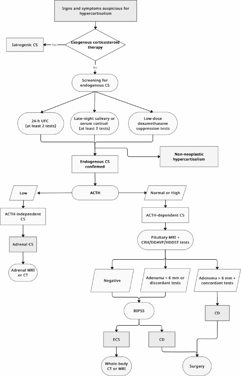
https://static-content.springer.com/image/art%3A10.1007%2Fs40618-024-02452-w/MediaObjects/40618_2024_2452_Fig1_HTML.png

Fig. 1

Diagnostic algorithm for screening and differential diagnosis of cushing syndrome in paediatric population

Imaging

Pituitary magnetic resonance imaging (MRI)

Since ACTH-secreting pituitary adenomas are very small (usually < 6 mm in diameter), it is difficult to localize these tumours. Diagnostic workup of CD includes pituitary MRI, but in many patients no tumour is identified. Conventional MRI, even with contrast enhancement, mostly failed to identify ACTH-secreting microadenomas in children with CD. Up to one-third of paediatric and adolescent patients with CD don’t have pituitary tumour detectable at brain MRI. The acquisition protocol should comprise coronal and sagittal spin-echo (SE) slices with gadolinium-enhanced T1 and T2 and millimetric 3D T1 slices [17295774]. In a retrospective study including 30 children with CD (mean age 12 ± 3 years), Batista et al. showed that pre- and post-contrast spoiled gradient-recalled acquisition in the steady state (SPGR) was superior to conventional pre- and post-contrast T1-weighted SE acquisition MRI in the identification of the microadenomas. In particular, the post-contrast SPGR-MRI identified the location of the tumour in 18 of 28 patients, whereas post-contrast SE-MRI identified the location and accurately estimated the size of the tumour in only 5 of 28 patients (p < 0.001) [74].

Bilateral petrosal sinus sampling (BIPSS)

BIPSS is another powerful diagnostic tool with high sensitivity and specificity, but its invasiveness and high cost limit its wide application, and the indication for BIPSS is still controversial [717297576]. It consists of the placement of femoral catheters that reach the inferior petrosal sinuses. Successively, blood samples are collected for measurement of ACTH from petrosal sinuses and from peripheral pathway before and after the administration of CRH. Inferior petrosal sinus (IPS) to peripheral (P) ACTH ratio and interpetrosal sinus gradient of one of the two sides to the contralateral side are calculated [7576]. In order to avoid incorrect results, it is recommended to verify hypercortisolism with serum cortisol sample immediately before performing BIPSS. Detomas et al. recently described the largest study on BIPSS.
According to the authors, the cut-offs for the ACTH IPS: P ≥ 1.9 at baseline (sensitivity 82.1%, specificity 85.7%) and ≥ 2.1 at 5 min post-CRH (sensitivity 91.3%, specificity 92.9%) allow for the best discrimination between CD and ECS [77]. In a multicentre study including 16 children aged between 4 and 16.5 years, Turan et al. showed that BIPSS is a superior diagnostic work-up than MRI to confirm the diagnosis of CD. Moreover, it showed a significantly higher sensitivity (92.8%) than MRI (53.3%) in detecting adenoma localization at pituitary level, which is crucial for surgical intervention [75]. The use of desmopressin has been reported in alternative to CRH [76]. In a review including case series of children with CS [76], the overall accuracy of BIPSS was 84.1% and became 92.3% after stimulation with desmopressin. The overall lateralizing accuracy of BIPSS was 50%. While BIPSS has a high diagnostic accuracy for the localization to the pituitary gland, it is not reliable for tumour lateralization to the right or left side of the gland. BIPSS is considered the gold standard to reliably exclude ECS and should performed in a specialized centre due to potential patient risk. However, BIPSS is not routinely available in many centres, it may have decreased specificity in children, especially when the pituitary tumour is not lateralized showing misleading results [7778]. For these reasons and for the risks related to the invasiveness of the procedure, BIPSS should be reserved only for exceptional cases in children [177576].

Radiological anatomic imaging

Subjects with ACS should perform an adrenal Computer Tomography (TC) or MRI to determine the adrenal cause. Despite abdominal TC with contrast-enhanced studies is the cornerstone of imaging of adrenal tumours in adults, MRI scan should be initially preferred in childhood to avoid radiation exposure [79]. Adrenocortical carcinomas are usually unilateral, larger than adenomas, with irregular margins, inhomogeneous contents (with areas of necrosis, haemorrhage and calcification) and avidly enhancement after contrast administration due to their high vascularity [80]. PPNAD is more difficult di identify with radiological studies, because it usually presents normal- or small-sized adrenal glands.
In subjects with suspected ECS, a thin-multislice neck-chest-abdomen-pelvic CT, alone or eventually followed by MRI, should be performed to identify neuroendocrine tumours that generally are very small and difficult to identify [11].

Functional imaging

Second-line functional imaging studies (as Positron Emission Tomography, PET, or scintigraphy) may be useful to provide an accurate etiological diagnosis of CS, particularly when the traditional radiological exams are inconclusive to differentiate CD from ECS. Because of the rarity of ECS, a univocal algorithm regarding the use of new molecular imaging techniques is not well established.
Whereas the ectopic ACTH-secreting tumours express the cell-surface receptors for somatostatin, 111In-pentetreotide (OCT) scintigraphy is often chosen as confirmatory exam [81].
The 68Gallium-DOTATATE PET/CT scan, using a modified octreotide molecule that also binds to somatostatin receptors, has shown a greater sensitivity for small tumours and may be useful for the tumoral identification in case of negative OCT scan [7]. Finally, 18FDG-PET/CT seems to be highly sensitive for the detection of aggressive pancreatic lesions [81].
In ACS cases, when adrenocortical carcinoma is suspected and traditional imaging studies (MRI or TC) are not diriment, 11C-metomidate-PET/CT scan allows a non-invasive characterization and staging of the adrenal lesion [8283].

Algorithm approach

Clinical history and the age at presentation of symptoms should guide throughout the different diagnosis of endogenous CS. A careful personal history, supported by patient growth charts, physical examination and screening tests should be able to rule out any physical or neuropsychiatric causes of NNH, even if second-line dynamic tests are sometimes needed to distinguish NNH from neoplastic CS.
Although CD is the main cause of CS in children older than 8 years, the clinical presentation of ECS may overlap with CD and the differential diagnosis of CS may be challenging, requiring the combination of dynamic biochemical tests and multimodal imaging.
Since none of the dynamic tests show a perfect sensitivity and specificity, using more than one dynamic test might improve accuracy. A non-invasive approach using a combination of three or four tests, specifically CRH and desmopressin stimulation tests plus MRI, followed by total-body CT, if biochemical and anatomical findings are discordant, correctly diagnose CD in approximately half of patients, potentially eliminating the need for BIPSS [1784]. If a pituitary tumour is detected on MRI and dynamic testing results are consistent with CD, BIPSS is not necessary for diagnosis. Since ECS in children is extremely rare, the algorithm approach in children may differ from the adult approach. Findings of ACTH-dependent CS, doubtful CRH test and normal pituitary MRI should be followed by extended imaging (whole-body CT/MRI or functional imaging). Considering the extremely rarity of ECS, the great majority of ACTH-dependent hypercortisolism, even with normal pituitary MRI, corresponds to CD due to a pituitary lesion not yet visible [17]. For this reason, BIPSS should be used only exceptionally in children. A diagnostic algorithm is proposed in Fig. 1.

Conclusions

We provide detailed revision on the diagnostic evaluation of children and adolescents presenting with signs and symptoms suspicious for CS and guidance on the workup from the confirmation of endogenous hypercortisolism to the etiological diagnosis of such a rare challenging condition.

Declarations

Ethical approval

This article does not include research on human participants and/or animals.
Informed consent is not required.

Conflict of interest

The authors have no relevant financial or non-financial interests to disclose.
Open Access This article is licensed under a Creative Commons Attribution 4.0 International License, which permits use, sharing, adaptation, distribution and reproduction in any medium or format, as long as you give appropriate credit to the original author(s) and the source, provide a link to the Creative Commons licence, and indicate if changes were made. The images or other third party material in this article are included in the article’s Creative Commons licence, unless indicated otherwise in a credit line to the material. If material is not included in the article’s Creative Commons licence and your intended use is not permitted by statutory regulation or exceeds the permitted use, you will need to obtain permission directly from the copyright holder. To view a copy of this licence, visit http://​creativecommons.​org/​licenses/​by/​4.​0/​.

Publisher’s note

Springer Nature remains neutral with regard to jurisdictional claims in published maps and institutional affiliations.

Clinical Features and Treatment Options for Pediatric Adrenal Incidentalomas

Abstract

Background

The aim of this study was to investigate the clinical features and treatment options for pediatric adrenal incidentalomas(AIs) to guide the diagnosis and treatment of these tumors.

Methods

The clinical data of AI patients admitted to our hospital between December 2016 and December 2022 were collected and retrospectively analyzed. All patients were divided into neonatal and nonneonatal groups according to their age at the time of the initial consultation.

Results

In the neonatal group, 13 patients were observed and followed up, and the masses completely disappeared in 8 patients and were significantly reduced in size in 5 patients compared with the previous findings. Four patients ultimately underwent surgery, and the postoperative pathological diagnosis was neuroblastoma in three patients and teratoma in one patient. In the nonneonatal group, there were 18 cases of benign tumors, including 9 cases of ganglioneuroma, 2 cases of adrenocortical adenoma, 2 cases of adrenal cyst, 2 cases of teratoma, 1 case of pheochromocytoma, 1 case of nerve sheath tumor, and 1 case of adrenal hemorrhage; and 20 cases of malignant tumors, including 10 cases of neuroblastoma, 9 cases of ganglioneuroblastoma, and 1 case of adrenocortical carcinoma.

Conclusions

Neuroblastoma is the most common type of nonneonatal AI, and detailed laboratory investigations and imaging studies are recommended for aggressive evaluation and treatment in this population. The rate of spontaneous regression of AI is high in neonates, and close observation is feasible if the tumor is small, confined to the adrenal gland and has no distant metastasis.

Peer Review reports

Background

The incidence of adrenal incidentaloma (AI) is increasing due to the increased frequency of imaging and improved imaging sensitivity [1]. AI is relatively common in adults, and several organizations, such as the American Association of Clinical Endocrinologists/American Association of Endocrine Surgeons and the European Society Endocrinology, have proposed specific protocols to guide the evaluation, treatment, and follow-up management of AI in adults [2]. Although AI, a nonfunctioning adrenocortical adenoma, is most common in adults, neuroblastoma is the most common incidental tumor of the adrenal gland in children. In addition, in the neonatal period, which is a more complex stage of childhood, the biology of adrenal masses found in this age group is also more specific, and the nature of these masses can range from spontaneous regression to rapid progression to aggressive disease with metastatic dissemination and even death. Given that AI is the most common malignant tumor, the management of AI in children cannot be simply based on the measurements used in adult AI. In this study, we retrospectively analyzed the clinical data of pediatric AI patients in a single center to investigate the clinical characteristics and management of AI in children.

Methods

A total of 66 children with adrenal tumors were diagnosed and treated at the Department of Urology of the Children’s Hospital of Nanjing Medical University from December 2016 to December 2022. A total of 55 cases were detected during physical examination, or the patients were diagnosed and received treatment for diseases other than adrenal disease after excluding adrenal tumors detected due to typical clinical manifestations or signs such as centripetal obesity and precocious puberty. Research protocols involving human materials were approved by the Medical Ethics Committee of the Children’s Hospital of Nanjing Medical University. All clinical information, radiological diagnosis, laboratory test results, intervention results, and follow-up data were collected from the department’s database.

All the children underwent ultrasonography and CT scanning, and 11 children underwent MRI. In addition to routine tests such as blood routine and biochemical indexes, the examination and evaluation of adrenal endocrine hormones and tumor markers included (1) plasma cortisol and ACTH levels, (2) plasma catecholamine and metabolite determination, (3) plasma renin and plasma aldosterone, (4) urinary vanillylmandelic acid/homovanillic acid(VMA/HVA), and (5) AFP, CEA, NSE, and CA19-9. Five patients underwent a low-dose dexamethasone suppression test. Seventeen of the 55 patients were treated with watch-waiting therapy, 4 of the 17 ultimately underwent surgery, 4 of the 38 patients underwent tumor biopsy, and 34 underwent adrenalectomy.

The data were analyzed using Graph Pad Prism 8. The measurement data are expressed as ‾x ± sd. The maximum diameter of the tumors, age of the patients with benign and malignant tumors, and maximum diameter of the tumors between the laparoscopic surgery group and the open surgery group were compared using paired t tests, and the percentages of the count data were compared using Fisher’s exact test.

Results

In this study, all patients were divided into two groups according to their age at the time of consultation: the neonate group and the nonneonate group.

Neonate group:

There were 7 male and 10 female patients, 7 of whom were diagnosed via prenatal examination and 10 of whom were diagnosed after birth. Five patients were diagnosed with lesions on the left side, 12 patients were diagnosed with lesions on the right side, and the maximal diameters of the masses ranged from 16 to 48 mm. The characteristics of the AIs in the neonate group are presented in Table 1.

Table 1 Characteristics of AI in the neonates group

Among the 17 patients, 8 had cystic masses with a maximum diameter of 1648 mm, 5 had cystic-solid masses with a maximum diameter of 3339 mm, and 4 had solid masses with a maximum diameter of 1845 mm. Two patients with solid adrenal gland masses suggested by CT scan had obvious elevations in serum NSE and maximum diameters of 44 and 45 mm, respectively. These patients underwent adrenal tumor resection, and the pathology diagnosed that they had neuroblastomas(NB). In one patient, the right adrenal gland was 26 × 24 × 27 mm in size with slightly elevated echogenicity at 38 weeks after delivery, and the mass increased to a size of 40 × 39 × 29 mm according to the 1-month postnatal review. MRI suggested that the adrenal gland tumor was associated with liver metastasis, and the pathology of the tumor suggested that it was NB associated with liver metastasis after surgical resection (stage 4 S, FH). One child was found to have 25 × 24 × 14 mm cystic echoes in the left adrenal region during an obstetric examination, and ultrasound revealed 18 × 11 mm cystic solid echoes 5 days after birth. Ultrasound revealed 24 × 15 mm cystic solid echoes at 2 months. Serum NSE and urinary VMA were normal, and the tumor was excised due to the request of the parents. Pathology suggested a teratoma in the postoperative period. A total of 13 children did not receive surgical treatment or regular review via ultrasound, serum NSE or urine VMA. The follow-up time ranged from 1 to 31 months, with a mean of 9.04 ± 7.61 months. Eight patients had complete swelling, and 5 patients were significantly younger than the previous patients. Nonneonate group:

There were 24 male and 14 female patients in the nonneonate group; 24 patients had lesions on the left side, 14 patients had lesions on the right side, and the maximal diameters of the masses ranged from 17 to 131 mm. Most of these tumors were found during routine physical examinations or incidentally during examinations performed for various complaints, such as gastrointestinal symptoms, respiratory symptoms, or other related conditions. As shown in Table 2, abdominal pain was the most common risk factor (44.7%) for clinical onset, followed by routine physical examination and examination for respiratory symptoms.

Table 2 Clinical presentations leading to discovery of AI in non-neonate group

Among the 38 patients, 10 had NBs with maximum diameters ranging from 20 to 131 mm, 9 had ganglion cell neuroblastomas with maximum diameters ranging from 33.6 to 92 mm, 9 had ganglion cell neuromas with maximum diameters ranging from 33 to 62 mm, 2 had adrenal adenomas with maximum diameters ranging from 17 to 70 mm, 1 had a cortical carcinoma with a maximum diameter of 72 mm, 2 had adrenal cysts with maximum diameters ranging from 26 to 29 mm, 2 had mature teratomas with maximum diameters of 34 and 40 mm, 1 had a pheochromocytoma with a diameter of 29 mm, 1 had a nerve sheath tumor with a diameter of 29 mm, and 1 patient with postoperative pathological confirmation of partial hemorrhagic necrosis of the adrenal gland had focal calcification with a maximum diameter of 25 mm (Table 3).

Table 3 Distribution of different pathologies among AI with various sizes in non-neonate group

The mean age of children with malignant tumors was significantly lower than that of children with benign tumors (57.95 ± 37.20 months vs. 105.0 ± 23.85 months; t = 4.582, P < 0.0001). The maximum diameter of malignant tumors ranged from 20 to 131 mm, while that of benign tumors ranged from 17 to 72 mm, and the maximum diameter of malignant tumors was significantly greater than that of benign tumors (65.15 ± 27.61 mm v 37.59 ± 12.98 mm; t = 3.863, P = 0.0004). Four biopsies, 5 laparoscopic adrenal tumor resections and 11 open adrenal tumor resections were performed for malignant tumors, and 16 laparoscopic adrenal tumor resections and 2 open procedures were performed for benign tumors. The maximum diameter of the tumors ranged from 17 to 62 mm in 21 children who underwent laparoscopic surgery and from 34 to 99 mm in 13 children who underwent open resection; there was a statistically significant difference in the maximum diameter of the tumors between the laparoscopic surgery group and the open surgery group (35.63 ± 10.36 mm v 66.42 ± 20.60 mm; t = 5.798, P < 0.0001).

Of the 42 children with definitive pathologic diagnoses at surgery, 23 had malignant tumors, and 19 had benign tumors. There were 15 malignant tumors with calcification on imaging and 5 benign tumors. The percentage of malignant tumors with calcifications in was significantly greater than that of benign tumors (65.22% v 26.32%; P = 0.0157). In the present study, all the children underwent CT, and 31 patients had postoperative pathological confirmation of NB. A total of 26 patients were considered to have neurogenic tumors according to preoperative CT, for a diagnostic compliance rate of 83.97%. Three children were considered to have cortical adenomas by preoperative CT, and 1 was ultimately diagnosed by postoperative pathology, for a diagnostic compliance rate of 33.33%. For 4 patients with teratomas and adrenal cysts, the CT findings were consistent with the postoperative pathology. According to our research, NB 9-110HU, GNB 15-39HU, GB 19-38HU, ACA 8HU, adrenal cyst 8HU, and cellular achwannoma 17HU.

Discussion

According to the clinical practice guidelines developed by the European Society of Endocrinology and European Network for the Study of Adrenal Tumors, AI is an adrenal mass incidentally detected on imaging not performed for a suspected adrenal disease [3]. The prevalence of AI is approximately 4%, and the incidence increases with age [4]. Most adult AIs are nonfunctioning benign adrenal adenomas (up to 75%), while others include functioning adrenal adenomas, pheochromocytomas, and adrenocortical carcinomas [5]. In contrast to the disease spectrum of adult AI cases, NB is the most common tumor type among children with AI, and benign cortical adenomas, which account for the vast majority of adult AI, accounting for less than 0.5% of cases in children [6]. According to several guidelines, urgent assessment of an AI is recommended in children because of a greater likelihood of malignancy [37].

When an adult patient is initially diagnosed with AI, it should be clear whether the lesion is malignant and functional. In several studies, the use of noncontrast CT has been recommended as the initial imaging method for adrenal incidentaloma; a CT attenuation value ≤ 10 HU is used as the diagnostic criterion for benign adenomas; and these methods have a specificity of 71-79% and a sensitivity of 96-98% [89]. A CT scan of tumors with diameters greater than 4 to 6 cm, irregular margins or heterogeneity, a CT attenuation value greater than 10 HU, or a relative contrast enhancement washout of less than 40% 10 or 15 min after administration of contrast media on enhanced CT is considered to indicate potential malignancy [7]. As the most common AI in children, NB often appears as a soft tissue mass with uneven density on CT, often accompanied by high-density calcified shadows, low-density cystic lesions or necrotic areas. CT scans can easily identify more typical NBs, and for those AIs that do not show typical calcified shadows on CT, it is sometimes difficult to differentiate neurogenic tumors from adenomas. In these patients, except for the 1 patient with adrenal cysts who had a CT value of 8 HU, very few of the remaining AI patients had a CT value less than 10 HU. Therefore, the CT value cannot be used simply as a criterion for determining the benign or malignant nature of AI, and additional imaging examinations, such as CT enhancement, MRI, and FDG-PET if necessary, should be performed immediately for AI in children.

Initial hormonal testing is also needed for functional assessment, and aldosterone secretion should also be assessed when the patient is hypertensive or hypokalemic [7]. Patients with AI who are not suitable for surgery should be observed during the follow-up period, and if abnormal adrenal secretion is detected or suggestive of malignancy during this period, prompt adrenal tumor resection is needed. For adult patients with AI, laparoscopic adrenal tumor resection is one of the most effective treatments that has comparative advantages in terms of hospitalization time and postoperative recovery speed; however, there is still some controversy over whether to perform laparoscopic surgery for some malignant tumors with large diameters, especially adrenocortical carcinomas, and some studies have shown that patients who undergo laparoscopic surgery are more prone to peritoneal seeding of tumors [10].

The maximum diameter of an adult AI is a predictor of malignancy, and a study by the National Italian Study Group on Adrenal Tumors, which included 887 AIs, showed that adrenocortical carcinoma was significantly correlated with the size of the mass, and the sensitivity of detecting adrenocortical carcinoma with a threshold of 4 cm was 93% [11]. According to the National Institutes of Health, patients with tumors larger than 6 cm should undergo surgical treatment, while patients with tumors smaller than 4 cm should closely monitored; for patients with tumors between 4 and 6 cm, the choice of whether to be monitored or surgically treated can be based on other indicators, such as imaging [12]. A diameter of 4 cm is not the initial threshold for determining the benign or malignant nature of a mass in children.

In a study of 26 children with AI, Masiakos et al. reported that 9 of 18 benign lesions had a maximal diameter less than 5 cm, 4 of 8 malignant lesions had a maximal diameters less than 5 cm, and 2 had a diameter less than 3 cm. The mean maximal diameter of benign lesions was 4.2 ± 1.7 cm, whereas the mean maximum diameter of malignant lesions was 5.1 ± 2.3 cm. There was no statistically significant difference between the two comparisons; therefore, this study concluded that children with AI diameters less than 5 cm cannot be treated expectantly [6]. Additionally, this study revealed that malignant lesions occurred significantly more frequently than benign lesions in younger children (mean age 1.7 ± 1.8 years v 7.8 ± 5.9 years; P = 0.02).

In the nonneonatal group of this study, 20 patients with malignant tumors had maximum diameters ranging from 20 to 131 mm, 10 had malignant tumors larger than 60 mm, and 3 had tumors smaller than 40 cm; 18 patients with benign tumors had maximum diameters ranging from 17 to 70 mm, 5 had diameters ranging from 40 to 60 mm, and 5 had diameters larger than 60 mm. Therefore, it is not recommended to use the size of the largest diameter of the tumor to decide whether to wait and observe or intervene surgically for children with AI. Instead, it is necessary to consider the age of the child; laboratory test results, such as whether the tumor indices are elevated or not; whether the tumor has an endocrine function; etc.; and imaging test results to make comprehensive judgments and decisions. Preoperative aggressive evaluation and prompt surgical treatment are recommended for nonneonatal pediatric AI patients.

Adrenal hematoma and NBs are the most common types of adrenal area masses in children, while pheochromocytoma, adrenal cyst, and teratoma are rarer masses [13]. In clinical practice, adrenal hematoma and NB are sometimes difficult to differentiate, especially when adrenal masses are found during the prenatal examination and neonatal period, and such children need to be managed with caution. The Children’s Oncology Group (COG ANBL00B1) implemented the watchful waiting treatment for children under 6 months of age with a solid adrenal mass < 3.1 cm in diameter (or a cystic mass < 5 cm) without evidence of distant metastasis, and if there is a > 50% increase in the adrenal mass volume, there is no return to the baseline VMA or HVA levels, or if there is a > 50% increase in the urinary VMA/HVA ratio or an inversion, surgical resection should be performed [14]. Eighty-three children in this study underwent expectant observation, 16 of whom ultimately underwent surgical resection (8 with INSS stage 1 NB, 1 with INSS stage 2B, 1 with INSS stage 4 S, 2 with low-grade adrenocortical neoplasm, 2 with adrenal hemorrhage, and 2 with extralobar pulmonary sequestration). Most of the children who were observed had a reduced adrenal mass volume. Of the 56 patients who completed the final 90 weeks of expectant observation, 27 (48%) had no residual mass, 13 (23%) had a residual mass volume of 0–1 ml, 8 (14%) had a mass volume of 1–2 ml, and 8 (14%) had a volume of > 2 ml; ultimately, 71% of the residual masses had a volume ≤ 1 ml and 86% had a residual volume ≤ 2 ml. In this study, a total of 16 patients were included in the watchful waiting treatment group; 3 patients underwent surgical treatment during the follow-up period, and 13 patients ultimately completed watchful waiting treatment. After 1–31 months of follow-up, 8 patients’ swelling completely disappeared, and 5 patients’ swelling significantly decreased. After strict screening for indications and thorough follow-up review, AIs in the neonatal period can be subjected to watchful waiting treatment, and satisfactory results can be achieved.

For benign adrenal tumors, laparoscopic surgery is superior to open surgery in terms of successful resection, whereas the feasibility of minimally invasive surgery for AI with preoperative suspicion of malignancy is controversial. The European Cooperative Study Group for Pediatric Rare Tumors recommends that minimally invasive surgery be considered only for early childhood tumors and should be limited to small, localized tumors; additionally, imaging should suggest no invasion of surrounding tissue structures or lymph nodes; and this strategy requires surgeons with extensive experience in oncologic and adrenal surgery [15]. NB is the most common pediatric AI, and open tumor resection remains the mainstay of treatment. For small, early tumors without evidence of invasion on preoperative examination, laparoscopic resection may be considered if the principles of oncologic surgery can be adhered to. If the patient responds to chemotherapy, the decision to perform laparoscopic tumor resection can also be re-evaluated after chemotherapy. According to the current study, the recurrence and mortality rates of laparoscopic surgery are comparable to those of open surgery [1617]. The relative contraindications for laparoscopic NB resection include a tumor diameter greater than 6 cm, venous dilatation, and the involvement of adjacent organs or blood vessels [18]. Patients who undergo open adrenalectomy have higher overall survival and recurrence-free survival rates than patients who undergo laparoscopic adrenalectomy [19]. Open adrenalectomy remains the gold standard for surgical resection of adrenocortical carcinoma, whereas laparoscopic adrenalectomy should be reserved for highly selected patients and performed by surgeons with appropriate expertise [20].

Cortical tumors are particularly rare among children with AIs and are sometimes not clearly distinguishable from neurogenic tumors on preoperative imaging; in such patients, the presence of subclinical Cushing’s syndrome needs to be carefully evaluated preoperatively; otherwise, a perioperative adrenal crisis may occur [21]. In patients in whom the possibility of an adrenocortical tumor was considered preoperatively, the assessment for subclinical Cushing’s syndrome mainly involved assessing the serum dehydroepiandrosterone sulfate level and performing an overnight dexamethasone suppression test.

A procedure for evaluating pediatric AI is shown in Fig. 1. Imaging is the first step in the evaluation of AI in children. CT can be used to clarify the nature of most tumors. MRI can be used to evaluate imaging risk factors (IDRFs) for NB. Bone marrow cytomorphology is recommended for all children with AI, along with microscopic residual neuroblastoma testing and further bone scanning if the bone marrow examination is positive. In addition, serum tumor marker levels and other relevant tests should be performed, and hormone levels should be evaluated. If adrenal adenomas cannot be completely excluded during the preoperative examination, a 1 mg overnight dexamethasone suppression test should be performed to exclude subclinical Cushing’s syndrome. In patients with hypertensive hypokalemia, the presence of aldosteronism should be evaluated by testing plasma aldosterone concentrations and plasma renin activity. Adrenal masses found in the neonatal period can be observed if the tumor is small, confined to the adrenal gland and shows no evidence of distant metastasis, while tumors that increase significantly in size during the follow-up period or that are associated with persistently elevated tumor markers require aggressive surgical treatment.

Fig. 1

figure 1

Algorithm for the evaluation and management of a pediatric adrenal incidentaloma. *DST overnight :20µg/kg dexamethasoneweight ˂40 kg,1 mg dexamethasone if ≥ 40 kg. CT = computed tomographic;MRI = magnetic resonance imaging;NSE = neuron-specific enolase;AFP = alpha-fetoprotein;CEA = carcinoembryonic antigen;CA19-9 = cancerantigen19-9;ACTH = adrenocorticotropic hormone;PAC = plasma aldosterone concentration; PRA = plasma renin activity;DST = dexamethasone suppression test

Data availability

The datasets analyzed during the current study are not public, but are available from the corresponding author on reasonable request.

Abbreviations

CT:
computed tomographic
MRI:
magnetic resonance imaging
ACTH:
adrenocorticotropic hormone
VMA:
vanillylmandelic acid
HVA:
homovanillic Acid
AFP:
alpha-fetoprotein
CEA:
carcinoembryonic antigen
NSE:
neuron-specific enolase
CA19-9:
cancerantigen19-9
FH:
favorable histology
HU:
Hounsfiled Unit
COG:
Children’s Oncology Group
INSS:
International Neuroblastoma Staging System

References

  1. Barzon L, Sonino N, Fallo F, Palu G, Boscaro M. Prevalence and natural history of adrenal incidentalomas. Eur J Endocrinol. 2003;149(4):273–85.

    Article CAS PubMed Google Scholar

  2. Maas M, Nassiri N, Bhanvadia S, Carmichael JD, Duddalwar V, Daneshmand S. Discrepancies in the recommendedmanagement of adrenalincidentalomas by variousguidelines. J Urol. 2021;205(1):52–9.

    Article PubMed Google Scholar

  3. Fassnacht M, Tsagarakis S, Terzolo M, et al. European Society of Endocrinology clinical practice guidelines on the management of adrenal incidentalomas, in collaboration with the European network for the study of adrenal tumors. Eur J Endocrinol. 2023;189(1):G1–42.

    Article PubMed Google Scholar

  4. Young WFJr. Clinical practice. The incidentally discovered adrenal mass. N Engl J Med. 2007;356(6):601–10.

    Article Google Scholar

  5. Rowe NE, Kumar R, Schieda N, et al. Diagnosis, management, and follow-up of the incidentallydiscoveredadrenalmass: CUAguidelineendorsed by the AUA. J Urol. 2023;210(4):590–9.

    Article PubMed Google Scholar

  6. Masiakos PT, Gerstle JT, Cheang T, Viero S, Kim PC, Wales P. Is surgery necessary for incidentally discovered adrenal masses in children?J. Pediatr Surg. 2004;39(5):754–8.

    Article Google Scholar

  7. Lee JM, Kim MK, Ko SH et al. Clinical guidelines for the management of adrenal incidentaloma. Endocrinol Metab. 2017;32(2).

  8. Terzolo M, Stigliano A, Chiodini I, et al. AME position statement on adrenal incidentaloma. Eur J Endocrinol. 2011;164(6):851–70.

    Article CAS PubMed Google Scholar

  9. Boland GW, Blake MA, Hahn PF, Mayo-Smith WW. Incidental adrenal lesions: principles, techniques, and algorithms for imaging characterization. Radiology. 2008;249(3):756–75.

    Article PubMed Google Scholar

  10. Payabyab EC, Balasubramaniam S, Edgerly M, et al. Adrenocortical cancer: a molecularlycomplexdiseasewheresurgerymatters. Clin Cancer Res. 2016;22(20):4989–5000.

    Article CAS PubMed Google Scholar

  11. Angeli A, Osella G, Alì A, Terzolo M. Adrenal incidentaloma: an overview of clinical and epidemiological data from the National Italian Study Group. Horm Res. 1997;47(4–6):279–83.

    Article CAS PubMed Google Scholar

  12. Grumbach MM, Biller BM, Braunstein GD, et al. Management of the clinically inapparent adrenal mass (incidentaloma). Ann Intern Med. 2003;138(5):424–9.

    Article PubMed Google Scholar

  13. Zhang K, Zhang Y, Zhang Y, Chao M. A retrospective analysis of the clinical characteristics of 207 hospitalized children with adrenal masses. Front Pediatr. 2023;11:1215095.

    Article PubMed PubMed Central Google Scholar

  14. Nuchtern JG, London WB, Barnewolt CE, et al. A prospective study of expectant observation as primary therapy for neuroblastoma in young infants: a Children‘s oncology group study. Ann Surg. 2012;256(4):573–80.

    Article PubMed Google Scholar

  15. Virgone C, Roganovic J, Vorwerk P, et al. Adrenocortical tumours in children and adolescents: the EXPeRT/PARTNER diagnostic and therapeutic recommendations. Pediatr Blood Cancer. 2021;68(suppl 4):e29025.

    Article PubMed Google Scholar

  16. Chang S, Lin Y, Yang S, et al. Safety and feasibility of laparoscopic resection of abdominal neuroblastoma without image-defined risk factors: a single-center experience. World J Surg Oncol. 2023;21(1):113.

    Article PubMed PubMed Central Google Scholar

  17. Zenitani M, Yoshida M, Matsumoto S, et al. Feasibility and safety of laparoscopic tumor resection in children with abdominal neuroblastomas. Pediatr Surg Int. 2023;39(1):91.

    Article PubMed Google Scholar

  18. International Pediatric Endosurgery Group. IPEG guidelines for the surgical treatment of adrenal masses in children. J Laparoendosc Adv Surg Tech A. 2010;20(2):vii–ix.

    Google Scholar

  19. Nakanishi H, Miangul S, Wang R, et al. Open versuslaparoscopicsurgery in the management of adrenocorticalcarcinoma: a systematicreview and meta-analysis. Ann Surg Oncol. 2023;30(2):994–1005.

    Article PubMed Google Scholar

  20. Gaillard M, Razafinimanana M, Challine A, et al. Laparoscopic or openadrenalectomy for stage I-IIadrenocorticalcarcinoma: a retrospectivestudy. J Clin Med. 2023;12(11):3698.

    Article PubMed PubMed Central Google Scholar

  21. Utsumi T, Iijima S, Sugizaki Y, et al. Laparoscopic adrenalectomy for adrenal tumors with endocrine activity: perioperative management pathways for reduced complications and improved outcomes. Int J Urol. 2023;30(10):818–26.

    Article CAS PubMed Google Scholar

Download references

Acknowledgements

We would like to express our deepest gratitude to all the patients and their parents who participated in this study. Their patience and cooperation were instrumental to the success of this research. We thank our colleagues in the Department of Radiology for their invaluable contributions in diagnosing and monitoring the progression of adrenal incidentalomas. We sincerely appreciate the hard work of the pathologists in diagnosing and classifying tumors, which laid the foundation for our study. Finally, we would like to thank our institution for providing the necessary resources and an enabling environment to conduct this research.

Funding

Not applicable.

Author information

Authors and Affiliations

  1. Department of Urology, Children’s Hospital of Nanjing Medical University, 72 Guangzhou Road, Nanjing, 210008, Jiangsu, China

    Xiaojiang Zhu, Saisai Liu, Yimin Yuan, Nannan Gu, Jintong Sha, Yunfei Guo & Yongji Deng

Contributions

X.J.Z. and Y.J.D designed the study; S.S.L., Y.M.Y., N.N.G., and J.T.S. carried out the study and collected important data; X.J.Z. analysed data and wrote the manuscript; Y.F.G. and Y.J.D.gave us a lot of very good advices and technical support; All authors read and approved the final manuscript.

Corresponding author

Correspondence to Yongji Deng.

Ethics declarations

Competing interests

The authors declare no competing interests.

Ethics approval and consent to participate

Ethics approval for this study was granted by the Ethics Committee of Children’s Hospital of Nanjing Medical University. Informed written consent was obtained from all the guardians of the children and we co-signed the informed consent form with their parents before the study. We confirmed that all methods were performed in accordance with relevant guidelines and regulations.

Conflict of interest

There are no conflicts of interest.

Additional information

Publisher’s Note

Springer Nature remains neutral with regard to jurisdictional claims in published maps and institutional affiliations.

Paediatric Cushing Syndrome: Prospective, Multisite, Observational Cohort Study

Summary

Background

Paediatric endogenous Cushing syndrome is a rare condition with variable signs and symptoms of presentation. We studied a large cohort of paediatric patients with endogenous Cushing syndrome with the aim of describing anthropometric, clinical, and biochemical characteristics as well as associated complications and outcomes to aid diagnosis, treatment, and management.

Methods

In this prospective, multisite cohort study, we studied children and adolescents (≤18 years at time of presentation) with a diagnosis of Cushing syndrome. Patients had either received their initial diagnosis and evaluation at the Eunice Kennedy Shriver National Institute of Child Health and Human Development (Bethesda, MD, USA) or been referred from other centres in the USA or outside the USA. We collected participants’ clinical, biochemical, and imaging findings and recorded their post-operative course until their latest appointment.

Findings

Of 342 paediatric patients with a diagnosis of Cushing syndrome, 193 (56%) were female and 149 (44%) male. 261 (76%) patients had corticotroph pituitary neuroendocrine tumours (Cushing disease), 74 (22%) had adrenal-associated Cushing syndrome, and seven (2%) had ectopic Cushing syndrome. Patients were diagnosed at a median of 2 years (IQR 1·0–3·0) after the first concerning sign or symptom, and patients with adrenal-associated Cushing syndrome were the youngest at diagnosis (median 10·4 years [IQR 7·4–13·6] vs 13·0 years [10·5–15·3] for Cushing disease vs 13·4 years [11·0–13·7] for ectopic Cushing syndrome; p<0·0001). Body-mass index z-scores did not differ between the diagnostic groups (1·90 [1·19–2·34] for adrenal-associated Cushing syndrome vs 2·18 [1·60–2·56] for Cushing disease vs 2·22 [1·42–2·35] for ectopic Cushing syndrome; p=0·26). Baseline biochemical screening for cortisol and adrenocorticotropin at diagnosis showed overlapping results between subtypes, and especially between Cushing disease and ectopic Cushing syndrome. However, patients with ectopic Cushing syndrome had higher urinary free cortisol (fold change in median cortisol concentration from upper limit of normal: 15·5 [IQR 12·7–18·0]) than patients with adrenal-associated Cushing syndrome (1·5 [0·6–5·7]) or Cushing disease (3·9 [2·3–6·9]; p<0·0001). Common complications of endogenous Cushing syndrome were hypertension (147 [52%] of 281 patients), hyperglycaemia (78 [30%] of 260 patients), elevated alanine transaminase (145 [64%] of 227 patients), and dyslipidaemia (105 [48%] of 219 patients). Long-term recurrence was noted in at least 16 (8%) of 195 patients with Cushing disease.

Interpretation

This extensive description of a unique cohort of paediatric patients with Cushing syndrome has the potential to inform diagnostic workup, preventative actions, and follow-up of children with this rare endocrine condition.

Funding

Intramural Research Program, Eunice Kennedy Shriver National Institute of Child Health & Human Development, National Institutes of Health.

Introduction

Paediatric endogenous Cushing syndrome is a rare disorder accounting for 5–7% of all reported cases of endogenous Cushing syndrome.1, 2, 3 In children older than 5–7 years and adolescents, endogenous Cushing syndrome is most commonly caused by corticotroph pituitary neuroendocrine tumours (PitNETs) and is termed Cushing disease. By contrast, Cushing syndrome in children younger than 5 years is often associated with adrenal disorders and is termed adrenal-associated Cushing syndrome.4 Albeit rare, a third type termed ectopic Cushing syndrome is caused by neuroendocrine tumours outside the hypothalamic–pituitary axis that secrete adrenocorticotropin or corticotropin-releasing hormone.5, 6 Thus endogenous Cushing syndrome is caused by either adrenocorticotropin-dependent sources (pituitary or ectopic) or adrenocorticotropin-independent (adrenal) hypercortisolemia.

Patients with adults-onset Cushing syndrome typically present with weight gain, skin manifestations (striae, hirsutism, acne, and easy bruising), and abnormal fat deposition.7, 8, 9 Paediatric Cushing syndrome differs from adult-onset Cushing syndrome in aspects including effects on growth (weight gain with concomitant height deceleration), atypical physical presentation (such as lack of centripetal obesity or typical striae), delayed or suppressed puberty, and variable mental health problems and neurocognitive function deficits.10 Diagnosis of paediatric Cushing syndrome is therefore challenging, and delayed evaluation is common.

Research in context

Evidence before this study

Endogenous Cushing syndrome is a rare endocrine condition. Diagnosis can be challenging and delay treatment. We searched PubMed for articles published in English on paediatric Cushing syndrome using terms “Cushing” AND “children” from database inception to May 5, 2023. Although several case series of paediatric Cushing disease were identified, only a few studies of the various causes of paediatric endogenous Cushing syndrome were available.

Added value of this study

To our knowledge, this cohort of paediatric endogenous Cushing syndrome of various causes is one of the largest sources of cumulative clinical, anthropometric, and biochemical data on the presentation, diagnosis, and management. We confirm that baseline biochemical data cannot aid differential diagnosis of Cushing syndrome subtypes. However, evidence suggests that minimally invasive stimulation tests could be a safe alternative to interventional sampling procedures such as inferior petrosal sinus sampling. We provide the prevalence of complications related to Cushing syndrome. Long-term outcomes of paediatric patients with pituitary corticotroph tumours recurrence is possible up to 8 years after initial remission.

Implications of all the available evidence

Data from this large paediatric cohort inform the evaluation, diagnosis, and long-term care of patients with paediatric Cushing syndrome. We recommend an algorithm for the diagnosis of patients and screening of complications. Screening for recurrence in patients with Cushing disease is indicated for this age group, at least for the first decade after surgery.

We have evaluated a large cohort of children and adolescents with endogenous Cushing syndrome of various causes. The aim of the study was to document anthropometric, clinical, and biochemical characteristics, complications, and outcomes of paediatric endogenous Cushing syndrome to aid clinicians in the diagnosis and management of these patients.

Section snippets

Study design and participants

In this prospective, multisite cohort study, we screened participants who, from 1995 to 2023, had enrolled in studies under protocols 97-CH-0076 (clinicaltrials.gov, NCT00001595), 95-CH-0059 (NCT00001452), and 00-CH-0160 (NCT00005927) at the Eunice Kennedy Shriver National Institute of Child Health and Human Development (NICHD, Bethesda, MD, USA). Paediatric patients (18 years or younger at time of presentation) with a diagnosis of Cushing syndrome were eligible for inclusion in the study. We

Results

342 patients with paediatric Cushing syndrome were included in the study (table 1). 278 patients were referred from centres in the USA, and 64 patients were referred from centres outside of the USA. 261 (76%) patients were diagnosed with Cushing disease, 74 (22%) patients were diagnosed with adrenal-associated Cushing syndrome, and seven (2%) patients were diagnosed with ectopic Cushing syndrome. Patients with adrenal-associated Cushing syndrome were diagnosed at a younger age than patients

Discussion

We present extensive and unique data on presentation, diagnosis, and follow-up of paediatric patients with three diagnostic types of endogenous Cushing syndrome. Clinical and anthropometric characteristics were similar across subtypes of Cushing syndrome, but biochemical tests differed. We also present extensive information on complications; hypertension, insulin resistance, dyslipidaemia, and elevated ALT were common. Long-term follow-up of patients revealed excellent postoperative prognosis,

Data sharing

The data that support the findings of this study are available from the corresponding author upon reasonable request.

Declaration of interests

CAS holds patents on the function of the PRKAR1APDE11A, and GPR101 genes and related issues; his laboratory had received research funding on GPR101, and on abnormal growth hormone secretion and its treatment by Pfizer. CAS receives support from ELPEN and has been consulting for Lundbeck Pharmaceuticals and Sync. CT reports receiving research funding on treatment of abnormal growth hormone secretion by Pfizer.

References (38)

Concurrent Mutations of Germline GPR101 and Somatic USP8 in a Pediatric Giant Pituitary ACTH Adenoma

Abstract

Background

Cushing’s disease (CD) is rare in pediatric patients. It is characterized by elevated plasma adrenocorticotropic hormone (ACTH) from pituitary adenomas, with damage to multiple systems and development. In recent years, genetic studies have shed light on the etiology and several mutations have been identified in patients with CD.

Case presentation

A girl presented at the age of 10 years and 9 months with facial plethora, hirsutism and acne. Her vision and eye movements were impaired. A quick weight gain and slow growth were also observed. Physical examination revealed central obesity, moon face, buffalo hump, supra-clavicular fat pads and bruising. Her plasma ACTH level ranged between 118 and 151 pg/ml, and sella enhanced MRI showed a giant pituitary tumor of 51.8 × 29.3 × 14.0 mm. Transsphenoidal pituitary debulk adenomectomy was performed and immunohistochemical staining confirmed an ACTH-secreting adenoma. Genetic analysis identified a novel germline GPR101 (p.G169R) and a somatic USP8 (p. S719del) mutation. They were hypothesized to impact tumor growth and function, respectively.

Conclusions

We reported a rare case of pediatric giant pituitary ACTH adenoma and pointed out that unusual concurrent mutations might contribute to its early onset and large volume.

Peer Review reports

Background

Cushing’s disease (CD) is caused by the overproduction of adrenocorticotropic hormone (ATCH) by pituitary adenomas (PAs). It is rare in children and accounts for approximately 75% of pediatric Cushing’s syndrome from 7 to 17 years of age [1]. Weight gain and facial changes are more common in children than in adults [2]. Growth retardation is also a characteristic of children with hypercortisolemia [3]. Genetic alterations such as somatic USP8RASD1TP53 mutations, and germline AIPMEN1, and CABLES1 mutations have been identified in CD patients [4]. Here we report a case of pediatric invasive pituitary ACTH macroadenoma associated with a novel germline GPR101 (p. G169R) and a somatic USP8 (p. S719del) mutation.

Case presentation

The girl was born at full term with a length of 48 cm and a weight of 2900 g. Her neuromotor and cognitive development was comparable to those of children of the same age. At the age of 9 years and 4 months she developed plethora, hirsutism, facial acne, rapid weight gain, and increased abdominal circumference. Her skin darkened, and purple striae appeared on thighs and in the armpits. She became dull and less talkative, as indicated by her parents. At 10 years and 3 months, the patient complained of pain around the left orbit with an intensity of 4–5 points on a numerical rating scale (NRS). Five months later bilateral blepharoptosis appeared, with significantly impaired vision of the left eye. Soon both eyes failed to rotate in all directions.

On admission the patient was 10 years and 9 months, with a height of 144 cm (90–97th percentile) and a weight of 48 kg (25–50th percentile). Her weight gain was 20 kg, while the height increased by only 2–3 cm in 18 months. Her blood pressure was 115/76mmHg, and her heart rate was 80 bpm. Apart from the signs mentioned above, physical examination revealed central obesity (BMI 23.1 kg/m2), moon face, buffalo hump, supra-clavicular fat pads and bruising at the left fossa cubitalis. Her pupils were 7 mm in diameter and barely reacted to light. There was a fan-shaped visual field defect in the left eye. Her breasts were Tanner stage III and pubic hair was Tanner stage II, although menarche had not yet occurred. The parents and her younger brother at 6 years of age did not have symptoms related to Cushing syndrome, acromegaly or gigantism. There was no family history of pituitary tumor or other endocrine tumors.

She had increased midnight serum cortisol (24.35 µg/dL, normal range < 1.8 µg/mL) and 24-hour urine free cortisol (24hUFC) (308.0 µg, normal range 12.3–103.5). The plasma ACTH level ranged from 118 to 151 pg/mL (< 46pg/mL). The 24hUFC was not suppressed (79.2 µg) after 48 h low-dose dexamethasone suppression test (LDDST), but suppressed to 32.8 µg (suppression rate 89.4%) after 48 h high-dose dexamethasone. Sella enhanced MRI showed a giant pituitary tumor measured 51.8 × 29.3 × 14.0 mm with heterogeneous density (Fig. 1). The mass compressed the optic chiasma and surrounded the bilateral cavernous sinus (Knosp 4). Therefore, an invasive giant pituitary ACTH adenoma was clinically diagnosed. The morning growth hormone (GH) was 1.0ng/ml (< 2 ng/ml) and insulin-like growth factor 1 416 ng/ml (88–452 ng/ml). The prolactin (PRL), luteinizing hormone (LH), follicle-stimulating hormone (FSH) and thyroid stimulating hormone (TSH) were all in normal ranges, as well as serum sodium, potassium, blood glucose and urine osmolality. Abdominal ultrasonography revealed a fatty liver. Tests concerning type 1 multiple endocrine neoplasia included serum calcium, phosphate, parathyroid hormone, gastrin and glucagon, which were all unremarkable (Table 1).

Fig. 1

figure 1

Contrast-enhanced coronal (A) and sagittal (B) T1-weighted MRI on admission. The sellar mass measured 51.8 × 29.3 × 14.0 cm (TD × VD × APD) with a heterogeneous density in the enhanced scan. The diaphragma sellea was dramatically elevated, with optic chiasm compressed. The sellar floor was sunken and bilateral cavernous sinus was surrounded (Knosp 4)

Table 1 Laboratory data on admission

Transsphenoidal pituitary debulk adenomectomy was performed immediately due to multiple cranial nerve involvement and the negative results of Sandostatin loading test. A decompression resection was done. The plasma ACTH level declined to 77 pg/ml and serum cortisol 30.2 µg/dl three days after the operation. Vision, pupil dilation, eye movements and blepharoptosis also partially improved. Histopathology and immunohistochemical staining confirmed a densely–granulated corticotroph adenoma (Fig. 2, NanoZoomer S360 digital slide scanner and NDP.view 2.9.25 software, Hamamatsu, Japan). Neither necrosis nor mitotic activity was observed. The immunostaining for somatostatin receptor SSTR2A was positive with a cytoplasmic pattern, while GH, PRL, TSH, FSH, LH and PIT were all negative. The Ki 67 index was found to be 10%. One month after the operation the ACTH level increased to 132 pg/mL again, and the parents agreed to refer their child for radiotherapy to control the residual tumor.

Fig. 2

figure 2

Histopathology and immunohistochemistry staining results of the pituitary tumor. By light microscopy, the tumor cells were mostly basophilic and arranged in papillary architecture. Neither necrosis nor mitotic activity was observed (A hematoxylin-eosin, ×200). Immunohistochemistry staining was positive for ACTH (B immunoperoxidase, ×200) and transcription factor T-PIT (C immunoperoxidase, ×200). Cytoplasmic staining of SSTR2A was observed in around 1/3 tumor cells besides the strong staining of endothelial cells (D immunoperoxidase, ×200). The Ki-67 index was 10% (E immunoperoxidase, ×200). Cytokeratin CAM5.2 was diffusely positive in the cytoplasm (F immunoperoxidase, ×200). The positive control for ACTH and T-PIT was the human anterior pituitary gland, and for SSRT2, Ki-67 and CAM5.2 were cerebral cortex, tonsil and colonic mucosa, respectively

The early onset and invasive behavior of this tumor led to the consideration of whether there was a genetic defect. Genetic studies were recommended for the families and they all agreed and signed the written informed consent forms. Whole exome sequencing (WES) was performed on the patient’s blood sample using an Illumina HiSeq sequencer to an average read depth of at least 90 times per individual. Raw sequence files were mapped to the GRCH37 human reference genome and analyzed using the Sentieon software. The results revealed a germline heterozygous GPR101 gene mutation c.505G > C (p.Gly169Arg), which was subsequently confirmed to be of maternal origin by Sanger sequencing. Meanwhile WES of the tumor tissue identified an additional somatic heterozygous c.2155_2157delTCC (p.S719del) mutation of the USP8 gene .

Discussion and conclusions

In this report, we described an extremely giant and invasive pituitary ACTH adenoma in a 10-year-old girl. According to Trouillas et al., invasive and proliferative pituitary tumors have a poor prognosis [5]. CD is rare among children, and the fast-growing and invasive nature of the tumor in this case led to the investigation of genetic causes. The somatic USP8 gene mutation has been recently reported to be associated with the pathogenesis of CD [67]. This gene encodes ubiquitin-specific protease 8 (USP8). S718, S719 and P720 are hotspots in different studies [6,7,8,9,10,11,12,13,14]. They are located at the 14-3-3 binding motif, and the mutations disrupt the binding between USP8 and 14-3-3 protein, which leads to increased deubiquitination and EGFR signaling. High levels of EGFR consequently trigger proopiomelanocortin (POMC) transcription and ACTH secretion [67]. The p.S719del mutation has been previously reported and its pathogenicity has been confirmed [7]. Thus, we speculate the p.S719del mutation plays a role in this patient with CD.

It is noteworthy that in our case, the pituitary corticotrophin adenoma was extremely giant and bilaterally invasive. USP8 mutations have been found in 31% of pediatric CD patients [10]. It is well known that microadenomas are most common in adult and pediatric CD patients. Previously, the Chinese and Japanese cohorts observed smaller sizes of USP8-mutated PAs than wild-type PAs [79]. The Chinese cohort also reported a lower rate of invasive adenomas in USP8-mutated PAs [7]. This may be explained by the finding that UPS8 mutations did not significantly promote cell proliferation more than the wild-type ones [6]. Other cohorts suggested no difference in tumor size or invasiveness between USP8-mutated and wild-type PAs [81012,13,14], which may be partially explained by the differences in sample sizes and ethnic backgrounds. Owing to the lack of evidence of USP8 mutations significantly contributing to tumor growth and invasiveness, additional pathogenesis should be investigated in this case.

The p.Gly169Arg mutation of the GPR101 gene has not been reported in patients with pituitary tumors. In silico predictions were performed using Polyphen-2, Mutation Taster and PROVEAN, and all of the programs reported it to be pathogenic. The GPR101 gene encodes an orphan G protein-coupled receptor (GPCR) and microduplication encompassing the gene has been proven to be the cause of X-linked acrogigantism (XLAG) [15]. XLAG is characterized by the early onset of pituitary GH-secreting macroadenomas. Point mutations of GPR101 have been found in patients with PAs that are mostly GH-secreting [15,16,17]. Although their prevalence is very low, an in vitro study supported the pathogenic role of p.E308D, the most common mutation of GPR101. This led to increased cell proliferation and GH production in rat pituitary GH3 cells [15]. Rare cases of PRL, ACTH or TSH-secreting PAs with GPR101 variants were also documented [1618]. To date, there have been five cases of ACTH-secreting PAs with four different germline GPR101 mutations: two cases of p.E308D, p.I122T, p.T293I and p.G31S, although in silico predictions and in vitro evaluations using AtT-20 cells have respectively determined the latter two mutations to be non-pathogenic [1618]. These patients were mainly children and young adults. Unlike pituitary GH-secreting tumors, the role of GPR101 mutations in the pathophysiology of CD is still questionable. Trivellin et al. demonstrated no statistically significant difference in GPR101 expression between corticotropinomas and normal human pituitaries. No significant correlation between GPR101 and POMC expression levels was found neither [18].

Given the evidences above, we hypothesize that the somatic USP8 mutation is responsible for the overexpression of ACTH in this CD girl while the germline GPR101 mutation contributes to the early onset and fast-growing nature of the tumor. Similarly, a 27-year-old woman with Nelson’s syndrome originally considered to be associated with a germline AIP variant (p.Arg304Gln) was recently reported to have a somatic USP8 mutation. The patient progressed rapidly and underwent multiple transsphenoidal surgeries [19]. Since germline AIP mutations are more commonly seen in GH-secreting PAs [20], the authors proposed that the USP8 mutation might have shifted the tumor towards ACTH-secreting [19]. Further investigations into the pathogenicity of GPR101 p.Gly169Arg and AIP p.Arg304Gln mutations are required to support the hypothesis.

In summary, we report a novel germline GPR101 and somatic USP8 mutation in a girl with an extremely giant pituitary ACTH adenoma. The concurrent mutations may lead to the growth and function of the tumor, respectively. Further investigations should be carried out to verify the role of the concurrent mutations in the pathogenesis of pediatric CD.

Availability of data and materials

The WES data of the blood sample of the patient is available in the NGDC repository (https://ngdc.cncb.ac.cn/gsa-human/) and the accession number is HRA002396. Any additional information is available from the authors upon reasonable request.

Abbreviations

CD:
Cushing’s disease
ACTH:
adrenocorticotropic hormone
PA:
pituitary adenoma
NRS:
numerical rating scale
24hUFC:
24-hour urine free cortisol
LDDST:
low-dose dexamethasone suppression test
USP8:
ubiquitin-specific protease 8
POMC:
proopiomelanocortin
GPCR:
G protein-coupled receptor
XLAG:
X-linked acrogigantism

References

  1. Weber A, Trainer PJ, Grossman AB, Afshar F, Medbak S, Perry LA, et al. Investigation, management and therapeutic outcome in 12 cases of childhood and adolescent Cushing’s syndrome. Clin Endocrinol (Oxf). 1995;43(1):19–28.

    CAS Article Google Scholar

  2. Storr HL, Alexandraki KI, Martin L, Isidori AM, Kaltsas GA, Monson JP, et al. Comparisons in the epidemiology, diagnostic features and cure rate by transsphenoidal surgery between paediatric and adult-onset Cushing’s disease. Eur J Endocrinol. 2011;164(5):667–74.

    CAS Article Google Scholar

  3. Magiakou MA, Mastorakos G, Oldfield EH, Gomez MT, Doppman JL, Cutler GB Jr, et al. Cushing’s syndrome in children and adolescents. Presentation, diagnosis, and therapy. N Engl J Med. 1994;331(10):629–36.

    CAS Article Google Scholar

  4. Hernández-Ramírez LC, Stratakis CA. Genetics of Cushing’s Syndrome. Endocrinol Metab Clin North Am. 2018;47(2):275–97.

    Article Google Scholar

  5. Trouillas J, Roy P, Sturm N, Dantony E, Cortet-Rudelli C, Viennet G, et al. A new prognostic clinicopathological classification of pituitary adenomas: a multicentric case-control study of 410 patients with 8 years post-operative follow-up. Acta Neuropathol. 2013;126(1):123–35.

    Article Google Scholar

  6. Reincke M, Sbiera S, Hayakawa A, Theodoropoulou M, Osswald A, Beuschlein F, et al. Mutations in the deubiquitinase gene USP8 cause Cushing’s disease. Nat Genet. 2015;47(1):31–8.

    CAS Article Google Scholar

  7. Ma Z-Y, Song Z-J, Chen J-H, Wang Y-F, Li S-Q, Zhou L-F, et al. Recurrent gain-of-function USP8 mutations in Cushing’s disease. Cell Res. 2015;25(3):306–17.

    CAS Article Google Scholar

  8. Perez-Rivas LG, Theodoropoulou M, Ferrau F, Nusser C, Kawaguchi K, Stratakis CA, et al. The Gene of the Ubiquitin-Specific Protease 8 Is Frequently Mutated in Adenomas Causing Cushing’s Disease. J Clin Endocrinol Metab. 2015;100(7):E997–1004.

    CAS Article Google Scholar

  9. Hayashi K, Inoshita N, Kawaguchi K, Ibrahim Ardisasmita A, Suzuki H, Fukuhara N, et al. The USP8 mutational status may predict drug susceptibility in corticotroph adenomas of Cushing’s disease. Eur J Endocrinol. 2016;174(2):213–26.

    CAS Article Google Scholar

  10. Faucz FR, Tirosh A, Tatsi C, Berthon A, Hernandez-Ramirez LC, Settas N, et al. Somatic USP8 Gene Mutations Are a Common Cause of Pediatric Cushing Disease. J Clin Endocrinol Metab. 2017;102(8):2836–43.

    Article Google Scholar

  11. Albani A, Perez-Rivas LG, Dimopoulou C, Zopp S, Colon-Bolea P, Roeber S, et al. The USP8 mutational status may predict long-term remission in patients with Cushing’s disease. Clin Endocrinol (Oxf). 2018;89:454–8.

    CAS Article Google Scholar

  12. Ballmann C, Thiel A, Korah HE, Reis AC, Saeger W, Stepanow S, et al. USP8 Mutations in Pituitary Cushing Adenomas-Targeted Analysis by Next-Generation Sequencing. J Endocr Soc. 2018;2(3):266–78.

    CAS Article Google Scholar

  13. Losa M, Mortini P, Pagnano A, Detomas M, Cassarino MF, Pecori Giraldi F. Clinical characteristics and surgical outcome in USP8-mutated human adrenocorticotropic hormone-secreting pituitary adenomas. Endocrine. 2019;63(2):240–6.

    CAS Article Google Scholar

  14. Weigand I, Knobloch L, Flitsch J, Saeger W, Monoranu CM, Hofner K, et al. Impact of USP8 Gene Mutations on Protein Deregulation in Cushing Disease. J Clin Endocrinol Metab. 2019;104(7):2535–46.

    Article Google Scholar

  15. Trivellin G, Daly AF, Faucz FR, Yuan B, Rostomyan L, Larco DO, et al. Gigantism and acromegaly due to Xq26 microduplications and GPR101 mutation. N Engl J Med. 2014;371(25):2363–74.

    CAS Article Google Scholar

  16. Lecoq AL, Bouligand J, Hage M, Cazabat L, Salenave S, Linglart A, et al. Very low frequency of germline GPR101 genetic variation and no biallelic defects with AIP in a large cohort of patients with sporadic pituitary adenomas. Eur J Endocrinol. 2016;174(4):523–30.

    CAS Article Google Scholar

  17. Iacovazzo D, Caswell R, Bunce B, Jose S, Yuan B, Hernández-Ramírez LC, et al. Germline or somatic GPR101 duplication leads to X-linked acrogigantism: a clinico-pathological and genetic study. Acta Neuropathol Commun. 2016;4(1):56.

    Article Google Scholar

  18. Trivellin G, Correa RR, Batsis M, Faucz FR, Chittiboina P, Bjelobaba I, et al. Screening for GPR101 defects in pediatric pituitary corticotropinomas. Endocr Relat Cancer. 2016;23(5):357–65.

    CAS Article Google Scholar

  19. Perez-Rivas LG, Theodoropoulou M, Puar TH, Fazel J, Stieg MR, Ferrau F, et al. Somatic USP8 mutations are frequent events in corticotroph tumor progression causing Nelson’s tumor. Eur J Endocrinol. 2018;178(1):57–63.

    CAS Article Google Scholar

  20. Tatsi C, Stratakis CA. The Genetics of Pituitary Adenomas. J Clin Med. 2019;9(1).

Download references

Acknowledgements

We thanked Dr. Xiaohua Shi and Dr. Yu Xiao from the Department of Pathology, Peking Union Medical College Hospital for their expertise in pituitary pathology and critical help in accomplishment of our manuscript.

Funding

This research was supported by “The National Key Research and Development Program of China” (No. 2016YFC0901501), “CAMS Innovation Fund for Medical Science” (CAMS-2017-I2M–1–011). They mainly covered the fees for genetic analysis and publications.

Author information

Authors and Affiliations

  1. Department of Pediatrics, Peking Union Medical College Hospital, Chinese Academy of Medical Science and Peking Union Medical College, Beijing, 100730, China

    Xu-dong Bao

  2. Department of Endocrinology, Key Laboratory of Endocrinology of National Health Commission, Peking Union Medical College Hospital, Chinese Academy of Medical Science and Peking Union Medical College, Beijing, 100730, China

    Lin Lu, Hui-juan Zhu, Xiao Zhai, Yong Fu, Feng-ying Gong & Zhao-lin Lu

  3. Department of Neurosurgery, Peking Union Medical College Hospital, Chinese Academy of Medical Science and Peking Union Medical College, Beijing, 100730, China

    Yong Yao, Ming Feng & Ren-zhi Wang

Contributions

XB and LL contributed to the study design and manuscript writing. HZ and FG performed genetic analysis. XZ and YF collected the clinical data. YY, MF and RW provided the tumor tissue and histopathology data. ZL revised the manuscript. All authors have read and approved the final manuscript.

Corresponding author

Correspondence to Lin Lu.

Ethics declarations

Ethics approval and consent to participate

This study was approved by the Ethics Committee of Peking Union Medical College Hospital. The parents of the patient provided written informed consent for research participation.

Consent for publication

The parents of the patient provided written informed consent for the publication of indirectly identifiable data in this research.

Competing interests

The authors declare that they have no competing interests.

Additional information

Publisher’s note

Springer Nature remains neutral with regard to jurisdictional claims in published maps and institutional affiliations.

Pediatric Adrenal Insufficiency: Challenges and Solutions

Authors Nisticò D , Bossini BBenvenuto SPellegrin MCTornese G

Received 29 October 2021

Accepted for publication 28 December 2021

Published 11 January 2022 Volume 2022:18 Pages 47—60

DOI https://doi.org/10.2147/TCRM.S294065

Checked for plagiarism Yes

Review by Single anonymous peer review

Peer reviewer comments 2

Editor who approved publication: Professor Garry Walsh

Download Article [PDF] 

Daniela Nisticò,1 Benedetta Bossini,1 Simone Benvenuto,1 Maria Chiara Pellegrin,1 Gianluca Tornese2

1University of Trieste, Trieste, Italy; 2Department of Pediatrics, Institute for Maternal and Child Health IRCCS Burlo Garofolo, Trieste, Italy

Correspondence: Gianluca Tornese
Department of Pediatrics, Institute for Maternal and Child Health IRCCS Burlo Garofolo, Via dell’Istria 65/1, Trieste, 34137, Italy
Tel +39 040 3785470
Email gianluca.tornese@burlo.trieste.it

Abstract: Adrenal insufficiency is an insidious diagnosis that can be initially misdiagnosed as other life-threatening endocrine conditions, as well as sepsis, metabolic disorders, or cardiovascular disease. In newborns, cortisol deficiency causes delayed bile acid synthesis and transport maturation, determining prolonged cholestatic jaundice. Subclinical adrenal insufficiency is a particular challenge for a pediatric endocrinologist, representing the preclinical stage of acute adrenal insufficiency. Although often included in the extensive work-up of an unwell child, a single cortisol value is usually difficult to interpret; therefore, in most cases, a dynamic test is required for diagnosis to assess the hypothalamic-pituitary-adrenal axis. Stimulation tests using corticotropin analogs are recommended as first-line for diagnosis. All patients with adrenal insufficiency need long-term glucocorticoid replacement therapy, and oral hydrocortisone is the first-choice replacement treatment in pediatric. However, children that experience low cortisol concentrations and symptoms of cortisol insufficiency can take advantage using a modified release hydrocortisone formulation. The acute adrenal crisis is a life-threatening condition in all ages, treatment is effective if administered promptly, and it must not be delayed for any reason.

Keywords: adrenal gland, primary adrenal insufficiency, central adrenal insufficiency, Addison disease, children, adrenal crisis, hydrocortisone

Introduction

Primary adrenal insufficiency (PAI) is a condition resulting from impaired steroid synthesis, adrenal destruction, or abnormal gland development affecting the adrenal cortex.1 Acquired primary adrenal insufficiency is termed Addison disease. Central adrenal insufficiency (CAI) is caused by an impaired production or release of adrenocorticotropic hormone (ACTH). It can originate either from a pituitary disease (secondary adrenal insufficiency) or arise from an impaired release of corticotropin-releasing hormone (CRH) from the hypothalamus (tertiary adrenal insufficiency). An underlying genetic cause should be investigated in every case of adrenal insufficiency (AI) presenting in the neonatal period or first few months of life, although AI is relatively rare at this age (1:5.000–10.000).2

Physiology of the Adrenal Gland

The adrenal cortex consists of three zones: the zona glomerulosa, the zona fasciculata, and the zona reticularis, responsible for aldosterone, cortisol, and androgens synthesis, respectively.3 Aldosterone production is under the control of the renin-angiotensin system, while cortisol is regulated by the hypothalamic-pituitary-adrenal axis (HPA).4 This explains why patients affected by CAI only manifest glucocorticoid deficiency while mineralocorticoid function is spared. CRH is secreted from the hypothalamic paraventricular nucleus into the hypophyseal-portal venous system in response to light, stress, and other inputs. It binds to a specific cell-surface receptor, the melanocortin 2 receptor, stimulating the release of preformed ACTH and the de novo transcription of the precursor molecule pro-opiomelanocortin (POMC). ACTH is derived from the cleavage of POMC by proprotein convertase-1.5–9 ACTH binds to steroidogenic cells of both the zona fasciculata and reticularis, activating adrenal steroidogenesis. It also has a trophic effect on adrenal tissue; therefore, ACTH deficiency determines adrenocortical atrophy and decreases the capacity to secrete glucocorticoids. Circulating cortisol is 75% bound to corticosteroid-binding protein, 15% to albumin, and 10% free. The endogenous production rate is estimated between 6 and 10 mg/m2/day, even though it depends on age, gender, and pubertal development. Glucocorticoids have multiple effects: they regulate immune, circulatory, and renal function, influence growth, development, energy and bone metabolism, and central nervous system activity. Several studies reported higher cortisol plasma concentrations in girls than in boys and younger children.3,4,8

Cortisol secretion follows a circadian and ultradian rhythm according to varying amplitudes of ACTH pulses. Pulses of ACTH and cortisol occur every 30–120 minutes, are highest at about the time of waking, and decline throughout the day, reaching a nadir overnight.3,8,9 This pattern can change in the presence of serious illness, major surgery, and sleep deprivation. During stressful situations, glucocorticoid secretion can increase up to 10-fold to enhance survival through increased cardiac contractility and cardiac output, sensitivity to catecholamines, work capacity of the skeletal muscles, and availability of energy stores.3

The interaction between the hypothalamus and the two endocrine glands is essential to maintain plasma cortisol homeostasis (Figure 1). Cortisol exerts double-negative feedback on the HPA axis. It acts on the hypothalamus and the corticotrophin cells of the anterior pituitary, reducing CRH and ACTH synthesis and release.6 ACTH inhibits its secretion through a feedback effect mediated at the level of the hypothalamus.3 Increased androgen production occurs in the case of cortisol biosynthesis enzymatic deficits.

Figure 1 The hypothalamic–pituitary–adrenal axis.

Primary Adrenal Insufficiency

PAI affects 10–15 per 100,000 individuals and recognizes different classes of genetic causes (Table 1). Congenital adrenal hyperplasia (CAH) is the main cause of PAI in the neonatal period, being included among the disorders of steroidogenesis secondary to deficits in enzymes. It has an autosomal recessive transmission.1,10,11 The estimated incidence ranges between 1:10,000 and 1:20,000 births. CAH phenotype depends on disease-causing mutations and residual enzyme activity. 21-hydroxylase deficiency (21OHD) accounts for more than 90% of cases, 21-hydroxylase converts cortisol and aldosterone precursors, respectively 17-hydroxyprogesterone (17-OHP) to 11-deoxycortisol and progesterone to deoxycortisone. Less frequent forms of CAH include 11 β -hydroxylase deficiency (11BOHD, 8% of cases), 17α-hydroxylase/17–20 lyase deficiency (17OHD), 3β-hydroxysteroid dehydrogenase deficiency (3BHDS), P450 oxidoreductase deficiency (PORD).12 Steroidogenesis may also be impaired by steroidogenic acute regulatory (StAR) protein deficiency, which is involved in cholesterol transport into mitochondria, or P450 cytochrome side-chain cleavage (P450scc) deficiency, that converts cholesterol into pregnenolone.12,13 Of these conditions, 21OHD and 11BOHD only affect adrenal steroidogenesis, whereas the other deficits also impact gonadal steroid production. In classic CAH, enzyme activity can be absent (salt-wasting form) or low (1–2% enzyme activity, simple virilizing form). The salt-wasting form is the most severe and affects 75% of patients with classic 21OHD.1,10,12,14 Non-classic CAH (NCCAH) is more prevalent than the classic form, in which there is 20–50% of residual enzymatic activity. Two-thirds of NCCAH individuals are compound heterozygotes with different CYP21A2 mutations in two different alleles (classic severe mutation plus mild mutation in two different alleles or homozygous with two mild mutations). Notably, 70% of NCCAH patients carry the point mutation Val281Leu.

Table 1 Causes of Primary Adrenal Insufficiency (PAI)

Central Adrenal Insufficiency

CAI incidence is estimated between 150 and 280 per million, and it should be suspected when mineralocorticoid function is preserved. When, rarely, isolated is due to iatrogenic HPA suppression secondary to prolonged glucocorticoid therapy or the removal of an ACTH- or cortisol-producing tumor (Cushing syndrome).15 Defects in POMC,16 characterized by red or auburn-haired children, pale skin (due to melanocyte stimulating hormone [MSH] – deficiency) and hyperphagia later in life, and in transcription factor TPIT,17 which regulates POMC synthesis in corticotrope cells, are the two leading genetic causes of isolated ACTH deficiency (Table 2). Mainly, it occurs as part of complex syndromes in which a combined multiple pituitary hormone deficiency (CMPD) is associated with craniofacial and midline defects, such as Prader-Willi syndrome, CHARGE syndrome, Pallister-Hall syndrome (anatomical pituitary abnormalities), white vanishing matter disease (progressive leukoencephalopathy).5 Individuals with an isolated pituitary deficiency, usually a growth hormone deficiency (GHD), may develop multiple pituitary hormone deficiencies over the years. Therefore, excluding a latent CAI at GHD onset and periodically monitoring of HPA axis is of utmost importance. Notably, cortisol reduction secondary to an increased basal metabolism when starting GHD or thyroxin substitutive therapy may unleash a misdiagnosed CAI. CMPD can be caused by several defective genes, such as GLI1, LHX3, LHX4, SOX2, SOX3, HESX1: in such cases, hypoglycemia or small penis with undescended testes may respectively suggest concomitant GH and gonadotropins deficits.18

Table 2 Causes of Central Adrenal Insufficiency (CAI)

Clinical Manifestations of Adrenal Insufficiency

AI is an insidious diagnosis presenting non-specific symptoms and may be mistaken with other life-threatening endocrine conditions (septic shock unresponsive to inotropes or recurrent sepsis, acute surgical abdomen).1,19 Children can be initially misdiagnosed as having sepsis, metabolic disorders, or cardiovascular disease, highlighting the need to consider adrenal dysfunction as a differential diagnosis for an unwell or deteriorating infant. With age-related items, clinical features depend on the type of AI (primary or central) and could manifest in an acute or chronic setting (Table 3).

Table 3 Features of Isolated Adrenal Insufficiency in Pediatric Age

Clinical signs of PAI are based on the deficiency of both gluco- and mineralocorticoids. Signs due to glucocorticoid deficiency are weakness, anorexia, and weight loss. Hypoglycemia with normal or low insulin levels is frequent and often severe in the pediatric population. Mineralocorticoid deficiency contributes to hyponatremia, hyperkalemia, acidosis, tachycardia, hypotension, and salt craving. The lack of glucocorticoid-negative feedback is responsible for the elevated ACTH levels. The high levels of ACTH and other POMC peptides, including the various forms of MSH, cause melanin hypersecretion, stimulating mucosal and cutaneous hyperpigmentation. Searching for an increased pigmentation may represent an essential diagnostic tool since all the other symptoms of PAI are non-specific. However, hyperpigmentation is variable, dependent on ethnic origin, and more prominent in skin exposed to sun and in extension surface of knees, elbows, and knuckles.15 In autoimmune PAI, vitiligo may be associated with hyperpigmentation.

In the classic CAH simple virilizing form, salt wasting is absent due to the presence of aldosterone production. In males, diagnosis typically occurs between 3 and 4 years of age with pubarche, accelerated growth velocity, and advanced bone age at presentation.1,10,12,14

NCCAH may occur in late childhood with signs of hyperandrogenism (premature pubarche, acne, adult apocrine odor, advanced bone age) or be asymptomatic. In adolescents and adult women, conditions of androgen excess (acne, oligomenorrhea, hirsutism) may underlie an NCCAH.20,21

The clinical presentation of CAI may be more complex when caused by an underlying central nervous system disease or by CMPD. In the case of a pituitary or hypothalamic tumor, patients may present headache, vomiting, visual disturbances, short stature, delayed or precocious puberty. In the case of CMPD, manifestations vary considerably and depend on the number and severity of the associated hormonal deficiencies. In CAI, aldosterone production is spared, which means that serum electrolytes are usually normal. However, cortisol contributes to regulating free water excretion, so patients with CAI are at risk for dilutional hyponatremia, with normal serum potassium levels. Since adrenal androgen secretion is under the control of ACTH, girls with ACTH deficiency may present light pubic hair. Patients with partial and isolated ACTH defects can be “asymptomatic”, and adrenal crisis appears during stress or in case of major illness (high fever, surgery).

The acute adrenal crisis is a life-threatening condition in all ages. Patients present with profound malaise, fatigue, nausea, vomiting, abdominal or flank pain, muscle pain or cramps, and dehydration, which lead to hypotension, shock, and metabolic acidosis. Hyponatremia and hyperkalemia are less common in CAI than in PAI, but possible in acute AI. Severe hypoglycemia causes weakness, pallor, sweatiness, and impaired cognitive function, including confusion, loss of consciousness, and coma. Immediate treatment is required (see below).

Children and adolescents affected by autoimmune primary adrenal insufficiency develop a chronic AI, with an insidious onset and slow progress to an acute adrenal crisis over months or even years. Initial symptoms are decreased appetite, anorexia, nausea, abdominal pain, unintentional weight loss, lethargy, headache, weakness, and fatigue, with prominent pain in the joints and muscles. Due to salt loss through the urine and the subsequent reduction in blood volume, blood pressure decreases, and orthostatic hypotension develops together with salt craving. An increased risk of infection in AI patients is reported only in those exposed to glucocorticoids. However, in APECED (Autoimmune Polyendocrinopathy-Candidiasis- Ectodermal-Dystrophy) patients, there is an increased risk of candidiasis and splenic atrophy increases the likelihood for severe infections.

In neonates, AI classically presents with failure to thrive and hypoglycemia, commonly severe and associated with seizures. The condition can be life-threatening and, if misdiagnosed, may result in coma and unexplained neonatal death. In newborns, cortisol deficiency causes delayed bile acid synthesis and transport maturation, determining prolonged cholestatic jaundice with persistently raised serum liver enzymes. The cholestasis can be resolved within ten weeks of correct treatment. StAR deficiency and P450scc cause salt-losing AI with female external genitalia in genetically male neonates.22 In the classic CAH salt-wasting form, the mineralocorticoid deficiency presents with the adrenal crisis at 10–20 days of life. Females show atypical genitalia with signs of virilization (clitoral enlargement, labial fusion, urogenital sinus), whereas males have normal-appearing genitalia, except for subtle signs as scrotal hyperpigmentation and enlarged phallus.1,10,12,14 Neonates with CMPD may display non-specific symptoms including hypoglycemia, lethargy, apnea, poor feeding, jaundice, seizures, hyponatremia without hyperkalemia, temperature and hemodynamic instability, recurrent sepsis, and poor weight gain. A male with hypogonadism may have undescended testes and micropenis. Infants with optic nerve hypoplasia or agenesis of the corpus callosum may present with nystagmus. Furthermore, infants with midline defects may have various neuro-psychological problems or sensorineural deafness.

Genetic Disorders and Other Conditions at Increased Risk for Adrenal Insufficiency

Among the cholesterol biosynthesis disorder, there is the Smith-Lemli-Opitz syndrome,23 where microcephaly, micrognathia, low-set posteriorly rotated ears, syndactyly of the second and third toes, and atypical genital may, although rarely, combine with AI; this autosomal recessive disorder is due to defective 7-dehydrocholesterol reductase so that elevated 7-dehydrocholesterol is diagnostic. In lysosomal acid lipase A deficiency,24 AI is due to calcification of the adrenal gland as a result of the accumulation of esterified lipids; in infantile form, that is Wolman disease, hepatosplenomegaly with hepatic fibrosis and malabsorption lead to death in the first year of life, if not treated with enzyme replacement therapy such as sebelipase alfa.25

Adrenal development may be impaired in X-linked congenital adrenal hypoplasia (AHC),13,26 a disorder caused by defective nuclear receptor DAX-1, presenting with salt-losing AI in infancy in approximately half of the cases, but also later in childhood or adolescence with two other key features such as hypogonadotropic hypogonadism and impaired spermatogenesis. Two syndromes combine adrenal hypoplasia with intrauterine growth restriction (IUGR): in IMAGe syndrome,27 caused by CDKN1C gain-of-function mutations, IUGR and AI present with metaphyseal dysplasia and genitourinary anomalies; MIRAGE syndrome28 is instead characterized by myelodysplasia, infections, genital abnormalities, and enteropathy, as a result of gain-of-function mutations in SAMD9, with elevated mortality rates.

In some other conditions, AI is due to ACTH resistance. Familial Glucocorticoid Deficiency type 1 (FGD1)13,29 and type 2 (FGD2)30 derive from defective ACTH receptor (MC2R) or its accessory protein MRAP, and both present with early glucocorticoid insufficiency (hypoglycemia, prolonged jaundice) and pronounced hyperpigmentation; there is usually an excellent response to cortisol replacement therapy, even though ACTH levels remain elevated.

In Allgrove or Triple-A Syndrome,13,31 defective Aladin protein (an acronym for alacrimia-achalasia-adrenal insufficiency) leads to primary ACTH-resistant adrenal insufficiency with achalasia and absent lacrimation, often combined with neurological dysfunction, either peripheral, central, or autonomic. It is an autosome recessive condition, phenotypically characterized by microcephaly, short stature, and skin hyperpigmentation.32,33

Among metabolic disorders associated with AI, Sphingosine-1-Phosphate Lyase (SGPL1) Deficiency34 is a sphingolipidosis with various features such as steroid-resistant nephrotic syndrome, primary hypothyroidism, undescended testes, neurological impairment, lymphopenia, ichthyosis; interestingly, in cases where nephrotic syndrome develops before AI, the latter may be masked by glucocorticoid treatment.

Adrenoleukodystrophy (ALD)35–37 is an X-linked recessive proximal disorder of beta-oxidation due to defective ABCD1, where the accumulation of very-long-chain fatty acids (VLCFA) affects in almost all cases adrenal gland among other tissues. Most patients present with progressive neurological impairment, but in some, AI is the only (approximately 10%) or first manifestation, so that every unexplained AI in boys should receive plasma VLCFA evaluation to diagnose ALD and reduce cerebral involvement through a low VLCFAs diet (Lorenzo’s oil) and allogeneic bone marrow transplantation. Early disease-modifying therapies have been developed. Gene therapy adds new functional copies of the ABCD1 gene in hematopoietic stem cells through a lentiviral vector reinfusing the modified cells in the patient’s bloodstream. Recent trials show encouraging results.38

In Zellweger syndrome, caused by mutations in peroxin genes (PEX), peroxisomes are absent, and disease presentation occurs in the neonatal period, with low survival rates after the first year of life. Finally, mitochondrial disorders have been described to occasionally develop AI: Pearson syndrome (sideroblastic anemia, pancreatic dysfunction), MELAS syndrome (encephalopathy with stroke-like episodes), and Kearns-Sayre syndrome (external ophthalmoplegia, heart block, retinal pigmentary changes) belong to this class.39

Autoimmune pathogenesis (Addison disease) accounts for approximately 15% of cases of primary AI in children, in contrast with adolescents and adults where it is the most common mechanism; half of these children present other glands involvement as well. Two syndromes recognize specific combinations: in Autoimmune Polyglandular Syndrome Type 1 (APS1, or APECED)40 defective autoimmune regulator AIRE causes AI, hypoparathyroidism, hypogonadism, malabsorption, chronic mucocutaneous candidiasis; APS2 usually present later in life (third-fourth decades) with AI, thyroiditis, and type 1 diabetes mellitus (T1DM). Antibodies against 21-hydroxylase enzyme are the hallmark of APS.

Apart from a genetic disorder, a strong link between autoimmune conditions and autoimmune primary AI has been established, with more than 50% of patients with the latter also having one or more other autoimmune endocrine disorders; on the other hand, only a few patients with T1DM or autoimmune thyroiditis or Graves’ disease develop AI. As an example, in a study of 629 patients with T1DM, only 11 (1.7%) presented 21-hydroxylase autoantibodies, with three of them having AI.41 Nevertheless, these patients are to be considered at increased risk for a condition that is potentially fatal yet easy to diagnose and treat; that is why it is reasonable to screen for autoimmune AI at least patients with T1DM, significantly if associated with DQ8 HLA combined with DRB*0404 HLA alleles, who have been observed to develop AI in 80% of cases if also 21-hydroxylase autoantibodies positive.42

Regarding immunological disruption, the link with celiac disease is instead well established: celiac patients have an 11-fold increased risk for AI, while in a study, 6 of 76 patients with AI had celiac disease, so that mutual evaluation should be granted in these patients.43,44

Subclinical Adrenal Insufficiency

Subclinical AI is a particularly insidious challenge for a pediatric endocrinologist. It represents the preclinical stage of Addison disease when 21-hydroxylase autoantibodies are already detectable but still absent from evident symptoms. 21-hydroxylase autoantibodies positivity carries a greater risk to develop overt AI in children than in adults: in a study, estimated risk was 100% in children versus 32% in adults on a medium six-year period of follow-up.45 As the adrenal crisis is a potentially lethal condition, it is essential to recognize and adequately manage subclinical AI.

Although asymptomatic by definition, subclinical AI may present with non-specific symptoms such as fatigue, lethargy, gastrointestinal symptoms (nausea, vomiting, diarrhea, constipation), hypotension; physical or psychosocial stresses may sometimes exacerbate these symptoms. When symptoms lack, subclinical AI may be identified thanks to the co-occurrence with other autoimmune endocrinopathies.46

21-hydroxylase autoantibodies titer is considered a marker of autoimmune activity and correlates with disease progression.47 Other reported risk factors for the disease evolution include young age, male sex, hypoparathyroidism or candidiasis coexistence, increased renin activity, or an altered synacthen test with normal baseline cortisol and ACTH.45 ACTH elevation has been reported as the best predictor of progression to the clinical stage in 2 years (94% sensitivity and 78% specificity).48

Management of patients with subclinical AI should include serum cortisol, ACTH, renin measurement, and a synacthen test. If normal, cortisol and ACTH should be repeated in 12–18 months, while synacthen test every two years. After synacthen test results are subnormal, cortisol and ACTH should be assessed every 6–9 months if ACTH remains in range or every six months if ACTH becomes elevated.49 In the latter case, therapy with hydrocortisone should be started.19 This strategy will prevent acute crises and possibly improve the quality of life in patients reporting non-specific symptoms.

Diagnosis

Laboratory evaluation of a stable patient with suspected AI should start with combined early morning (between 6 and 8 AM) serum cortisol and ACTH measurements (Figure 2).

Figure 2 Diagnostic algorithm for adrenal insufficiency.

Although often included in the extensive work-up of an unwell child, a single cortisol value is usually challenging to interpret: circadian cortisol rhythm is highly variable and morning peak is unpredictable; morning cortisol levels in children with diagnosed AI may range up to 706 nmol/L (97th percentile); several factors, such as exogenous estrogens, may alter total serum cortisol values by influencing the free cortisol to cortisol binding globulin or albumin-bound cortisol ratio.7

Significant variability is also observed depending on the specific type of cortisol assay; therefore, it is recommended to check the reference ranges with the laboratory. Mass spectrometry analysis and the new platform methods (Roche Diagnostics Elecsys Cortisol II)50 have more specificity because it detects lower cortisol concentrations than standard immunoassays.15 Low serum cortisol with normal or low ACTH levels is compatible with CAI. In such cases, morning serum cortisol levels below 3 µg/dL (83 nmol/L) best predict AI, while greater than 13 µg/dL (365 nmol/L) values tend to exclude it.51 This is why in most cases, a dynamic test is required for diagnosis and has been introduced to assess the hypothalamic-pituitary-adrenal (HPA) axis in case of intermediate values.5

The insulin tolerance test (ITT) is considered the gold standard for CAI diagnosis as hypoglycemia results in an excellent HPA axis activation; moreover, it allows simultaneous growth hormone evaluation in patients with suspected CPHD. Serum cortisol is measured at baseline and 15, 30, 45, 60, 90, and 120 minutes after intravenous administration of 0.1 UI/Kg regular insulin; the test is valid if serum glucose is reduced by 50% or below 2.2 mmol/L (40 mg/dL).52 CAI is diagnosed for a <20 µg/dL (550 nmol/L) cortisol value at its peak.15 Hypoglycemic seizures and hypokalemia (due to glucose infusion) are the main risks of this test so that it is contraindicated in case of a history of seizures or cardiovascular disease.

Glucagon stimulation test (GST, 30 µg/Kg up to 1 mg i.m. glucagon with cortisol measurements every 30 min for 180 min) allows both CAI and growth hormone deficiency evaluation as well but is characterized by frequent gastrointestinal side effects and poor specificity.8

Metyrapone is an 11-hydroxylase inhibitor, thereby decreasing cortisol synthesis and removing its negative feedback on ACTH release. Overnight metyrapone test is based on oral administration of 30 mg/Kg metyrapone at midnight, and 11-deoxycortisol measurement on the following morning: in case of CAI, its level will not reach 7 µg/dL (200 nmol/L). This test may, however, induce an adrenal crisis so that it is rarely performed.

Given their safety profile and accuracy, corticotropin analogs such as tetracosactrin (Synacthen®) or cosyntropin (Cortrosyn®) are recommended as first-line stimulation tests. Nevertheless, false-negative results are probable in the case of recent or moderate ACTH deficiency, which would not have induced adrenal atrophy. The standard dose short synacthen test (SDSST) is based on a 250 µg Synacthen vial administration with serum cortisol measurement at baseline and 30 and 60 minutes after. CAI is diagnosed if peak cortisol level is <16 µg/dL (440 nmol/L), or excluded if >39 µg/dL (1076 nmol/L). However, the cut-offs for both the new platform immunoassay and mass spectrometry serum cortisol assays are 13.5 to 14.9 mcg/dL (373 to 412 nmol/L).53 The 250 µg Synacthen dose is considered a supraphysiological stimulus since it is 500 times greater than the minimum ACTH dose reported to induce a maximal cortisol response (500 ng/1.73 m2). The low dose short synacthen test (LDSST) has been introduced as a more sensitive first-line test in children greater than two years.54 The recommended dose is 1 µg55, which is contained in 1 mL of the solution obtained by diluting a 250 µg vial into 250 mL saline. Serum cortisol level is then measured at baseline and after 30 minutes, resulting in diagnose of CAI if <16 µg/dL (440 nmol/L), otherwise ruling it out if >22 µg/dL (660 nmol/L). Using these thresholds, LDSST is more precise than SDSST in children, with an area under the ROC curve of 0.99 (95% CI 0.98–1.00).56 LDSST has not been validated in acutely ill patients, pituitary acute disorders or surgery or radiation therapy, and impaired sleep-wake cycle. Patients with an indeterminate LDSST result should be furtherly studied with ITT or metyrapone test.

Finally, the CRH test is based on 1 µg/Kg human CRH (Ferring®) administration and may differentiate secondary from tertiary AI, but its thresholds are still not precisely defined.57

Once CAI is diagnosed, other pituitary hormones should be assessed (prolactin, IGF1, LH, FSH, fT4, TSH), and an MRI of the pituitary region should be performed to exclude neoplastic or infiltrative processes.

Primary adrenal insufficiency (PAI) should be suspected in case of low serum cortisol with elevated ACTH levels. When hypocortisolemia has been confirmed, ACTH levels >66 pmol/L or greater than twice the upper limit best predict PAI. Nevertheless, a confirmatory dynamic test is always recommended for diagnosis.19 Given the comparable accuracy between standard and low dose SST reported in these patients, SDSST is recommended as the most feasible test.58 Moreover, suspected PAI cases should receive plasma renin activity or direct renin and aldosterone assessment to evaluate mineralocorticoid deficiency.

Etiologic work-up of confirmed PAI should start from 21-hydroxylase antibodies assessment: if positive, differential diagnosis will include Addison disease and APS1 or APS2. Adrenal autoantibody negative patients should instead be screened for CAH by measuring 17-hydroxyprogesterone, ALD (if young male) by assessing VLCFA, and tuberculosis if endemic; adrenal glands imaging will complete the work-up in order to exclude infection, hemorrhage, or tumor.6

While universal newborn screening is already implemented for CAH in many countries, allowing a timely replacement therapy, basal salivary cortisol, and salivary cortisone measurements could improve CAI screening in the future: this technique is simple, cost-effective, and independent of binding proteins.15

Treatment

All patients with adrenal insufficiency need long-term glucocorticoid replacement therapy. Individuals with PAI also require mineralocorticoids replacement, together with salt intake as required (Table 4). Otherwise, guidelines do not recommend androgen replacement.5,9,19

Table 4 Management of Adrenal Insufficiency (AI)

Oral hydrocortisone is the first-choice replacement treatment in children due to its short half-life, rapid peak in plasma concentration, lower potency, and fewer adverse effects than prednisolone and dexamethasone.5,8 Based on endogenous production, dosing replacement regimens vary from 7.5 to 15 mg/m2/day, divided into two, three, or four doses.19 The first and largest dose should be taken at awakening, the next in the early afternoon to avoid sleep disturbances. Small and frequent dosing mimic the physiological rhythm of cortisol secretion, but high peak cortisol levels after drug assumption and prolonged periods of hypocortisolemia between doses are described.8,9 Some children experience low cortisol concentrations and symptoms of cortisol insufficiency (eg, fatigue, nausea, headache) despite modifications in dosing. This cohort of patients can take advantage of using a modified-release hydrocortisone formulation, such as Chronocort® and Plenadren®. Plenadren®, approved for adults, consists of a coating of hydrocortisone released rapidly, followed by a slow release of hydrocortisone from the tablet center. It is available as 5 and 20 mg tablets. Park et al demonstrate smoother cortisol profiles and normal growth and weight gain patterns using Plenadren® in children.59 In a few cases, the continuous subcutaneous infusion of hydrocortisone using insulin pump technology proved to be a feasible, well-tolerated and safe option for selected patients with poor response to conventional therapy.19

Monitoring glucocorticoid therapy is based on growth, weight gain, and well-being. Cortisol measurements are usually not useful, apart from cases when a discrepancy between daily doses and patient symptoms exists.15 The concomitant use of hydrocortisone and CYP3A4 inducers, such as Rifampicin, Phenytoin, Carbamazepine, requires an increased dose of glucocorticoids. Conversely, the inhibition of CYP3A4 impairs hydrocortisone metabolism.5

Mineralocorticoid replacement is unnecessary if the patient has a normal renin-angiotensin-aldosterone axis and, hence, normal aldosterone secretion, as well as in CAI. By contrast, patients with PAI and confirmed aldosterone deficiency need fludrocortisone at the dosage of 0.1–0.2 mg/day when given together with hydrocortisone, which has some mineralocorticoid activity. When using other synthetic glucocorticoids for replacement, higher fludrocortisone doses may be needed. Infants younger than one year should also be supplemented with sodium chloride due to their relatively low dietary sodium intake and relative renal resistance to mineralocorticoids. The dose is approximately 1 gram (17 mEq) daily.19

Surgery and anesthesia increase the glucocorticoid requirement during the pre-, intra-, and post-operative periods (Table 4). All children with AI should receive an intravenous dose of hydrocortisone at induction (2 mg/kg for minor or major surgery under general anesthesia). For minor procedures or sedation, the child should receive a double morning dose of hydrocortisone orally.60

Adrenal crisis is a life-threatening condition, treatment is effective if administered promptly, and it must not be delayed for any reason. Hydrocortisone should be administered as soon as possible with an intravenous bolus of 4 mg/kg followed by a continuous infusion of 2 mg/kg/day until stabilization. In the alternative, it can be administered as a bolus every four hours intravenous or intramuscular. In difficult peripheral venous access, the intramuscular route must be used as the first choice. In order to counteract hypotension, a bolus of normal saline 0.9% should be given at a dose of 20 mL/kg; it can repeat up to a total of 60 mL/kg within one hour for shock. If there is hypoglycemia, 10% dextrose at a 5 mL/kg dose should be administered.5,19,61,62

Patients with AI require additional doses of glucocorticoids in case of physiologic stress such as illness or surgical procedures to avoid an adrenal crisis. Home management of illness with a fever (> 38°C), vomiting or diarrhea, is based on the increase from two to three times the usual dose orally. If the child is unable to tolerate oral therapy, intramuscular injection of hydrocortisone should be administered (Table 4).

Education for caregivers and patients (if adolescent) is crucial to prevent adrenal crisis. They should recognize signs and symptoms of adrenal crisis and should receive a steroid emergency card with the sick day rules. Prescribing doctors should provide for additional oral glucocorticoids and adequate training in hydrocortisone emergency self-injection.

Abbreviations

AI, adrenal insufficiency; PAI, primary adrenal insufficiency; CAI, central adrenal insufficiency; HPA, hypothalamic-pituitary-adrenal axis; CRH, corticotropin-releasing hormone; ACTH, adrenocorticotropic hormone; POMC, pro-opiomelanocortin; CAH, congenital adrenal hyperplasia; STAR, steroidogenic acute regulatory; 21OHD, 21-hydroxylase deficiency; 11BOHD, 11-B-hydroxylase deficiency; P450scc, P450 cytochrome side-chain cleavage deficiency; 17-OHP, 17-hydroxyprogesterone; NCCAH, non-classic congenital adrenal hyperplasia; ALD, adrenoleukodystrophy; VLCFA, very long-chain fatty acids; CMPD, combined multiple pituitary hormone deficiency; GHD, growth hormone deficiency; MSH, melanocyte stimulating hormone; IUGR, intrauterine growth restriction; APS1, autoimmune polyglandular syndrome type 1; SDSST, standard dose short synacthen test; LDSST, low dose short synacthen test.

Take Home Messages

  1. In neonates and infants CAH is the commonest cause of PAI, causing almost 71.8% of cases.
  2. Adrenoleukodystrophy should be considered in any male with hypoadrenalism.
  3. Unexplained hyponatremia, hyperpigmentation and the loss of pubic and axillary hair should raise the suspicion of AI.
  4. Adrenal insufficiency can present with non-specific clinical features; therefore a single cortisol measurement should be included in the biochemical work-up of an unwell child.
  5. Patients and parents should be well-trained in adrenal crisis recognition and management.

Disclosure

The authors report no conflicts of interest in this work.

References

1. Charmandari E, Nicolaides N, Chrousos G. Adrenal insufficiency. Lancet. 2021;383(9935):2152–2167. doi:10.1016/S0140-6736(13)61684-0

2. White PC. Adrenocortical insufficiency. In: Nelson Textbook of Pediatrics. Elsevier. 2019:11575–11617.

3. White PC. Physiology of the adrenal gland. Nelson Textbook of Pediatrics. Elsevier. 2019.

4. Butler G, Kirk J. Adrenal gland disorders. In: Paediatric Endocrinology and Diabetes. Oxford University Press. 2020:274–288.

5. Patti G, Guzzeti C, Di Iorgi N, Loche S. Central adrenal insufficiency in children and adolescents. Best Pract Res Clin Endocrinol Metab. 2018;32(4):425–444. doi:10.1016/j.beem.2018.03.012

6. Martin-grace J, Dineen R, Sherlock M, Thompson CJ. Adrenal insufficiency: physiology, clinical presentation and diagnostic challenges. Clin Chim Acta. 2020;505:78–91. doi:10.1016/j.cca.2020.01.029

7. Shaunak M, Blair JC, Davies JH. How to interpret a single cortisol measurement. Arch Dis Child Educ Pract. 2020;105:347–351. doi:10.1136/archdischild-2019-318431

8. Park J, Didi M, Blair J. The diagnosis and treatment of adrenal insuf fi ciency during childhood and adolescence. Arch Dis Child. 2016;101:860–865. doi:10.1136/archdischild-2015-308799

9. Husebye ES, Pearce SH, Krone NP, Kämpe O. Adrenal insufficiency. Lancet. 2021;397:613–629. doi:10.1016/S0140-6736(21)00136-7

10. Speiser P, Azziz R, Baskin L, et al. Congenital adrenal hyperplasia due to steroid 21-hydroxylase deficiency: an Endocrine Society clinical practice guideline. J Clin Endocrinol Metab. 2010;95(9):4133–4160. doi:10.1210/jc.2009-2631

11. Buonocore F, McGlacken-Byrne S, Del Valle I, Achermann J. Current insights into adrenal insufficiency in the newborn and young infant. Front Pediatr. 2020;8:619041. doi:10.3389/fped.2020.619041

12. Bacila I, Elder C, Krone N. Update on adrenal steroid hormone biosynthesis and clinical implications. Arch Dis Child. 2019;104(12):1223–1228. doi:10.1136/archdischild-2017-313873

13. Buonocore F, Maharaj A, Qamar Y, et al. Genetic analysis of pediatric primary adrenal insufficiency of unknown etiology: 25 years’ experience in the UK. J Endocr Soc. 2021;5(8):1–15. doi:10.1210/jendso/bvab086

14. Balsamo A, Baronio F, Ortolano R, et al. Congenital adrenal hyperplasias presenting in the newborn and young infant. Front Pediatr. 2020;8:593315. doi:10.3389/fped.2020.593315

15. Hahner S, Ross RJ, Arlt W, et al. Adrenal insufficiency. Nat Rev Dis Prim. 2021;7(1):1–24. doi:10.1038/s41572-021-00252-7

16. Krude H, Biebermann H, Luck W, et al. Severe early-onset obesity, adrenal insufficiency and red hair pigmentation caused by POMC mutations in humans. Nat Genet. 1998;19:155–157. doi:10.1038/509

17. Vallette-Kasic S, Brue T, Pulichino A-M, et al. Congenital isolated adrenocorticotropin deficiency: an underestimated cause of neonatal death, explained by TPIT gene mutations. J Clin Endocrinol Metab. 2005;90:1323–1331. doi:10.1210/jc.2004-1300

18. Alatzoglou K, Dattani M. Genetic forms of hypopituitarism and their manifestation in the neonatal period. Early Hum Dev. 2009;85:705–712. doi:10.1016/j.earlhumdev.2009.08.057

19. Bornstein SR, Allolio B, Arlt W, et al. Diagnosis and treatment of primary adrenal insufficiency: an endocrine society clinical practice guideline. J Clin Endocrinol Metab. 2016;101:364–389. doi:10.1210/jc.2015-1710

20. Kurtoğlu S, Hatipoğlu N. Non-classical congenital adrenal hyperplasia in childhood. J Clin Res Pediatr Endocrinol. 2017;9(1):1–7. doi:10.4274/jcrpe.3378

21. Livadas S, Bothou C. Management of the female with non-classical congenital adrenal hyperplasia (NCCAH): a patient-oriented approach. Front Endocrinol. 2019;10:366. doi:10.3389/fendo.2019.00366

22. Miller W. Disorders in the initial steps of steroid hormone synthesis. J Steroid Biochem Mol Biol. 2017;165:18–37. doi:10.1016/j.jsbmb.2016.03.009

23. Nowaczyk M, Irons M. Smith–Lemli–Opitz syndrome: phenotype, natural history, and epidemiology. Am J Med Genet Part C Semin Med Genet. 2012;160:250–262. doi:10.1002/ajmg.c.31343

24. Anderson R, Byrum R, Coates P, Sando G. Mutations at the lysosomal acid cholesteryl ester hydrolase gene locus in Wolman disease. Proc Natl Acad Sci USA. 1994;91:2718. doi:10.1073/pnas.91.7.2718

25. Jones S, Rojas-Caro S, Quinn A. Survival in infants treated with sebelipase Alfa for lysosomal acid lipase deficiency: an open-label, multicenter, dose-escalation study. Orphanet J Rare Dis. 2017;12:25. doi:10.1186/s13023-017-0587-3

26. Muscatelli F, Strom T, Walker A, et al. Mutations in the DAX-1 gene give rise to both X-linked adrenal hypoplasia congenita and hypogonadotropic hypogonadism. Nature. 1994;372:672–676. doi:10.1038/372672a0

27. Vilain E, Merrer M, Lecointre C, et al. IMAGe, a new clinical association of Intrauterine growth retardation, metaphyseal dysplasia, adrenal hypoplasia congenita, and Genital anomalies. J Clin Endocrinol Metab. 1999;84(12):4335–4340. doi:10.1210/jcem.84.12.6186

28. Narumi S, Amano N, Ishii T, et al. SAMD9 mutations cause a novel multisystem disorder, MIRAGE syndrome, and are associated with loss of chromosome 7. Nat Genet. 2016;48:792–797. doi:10.1038/ng.3569

29. Maharaj A, Maudhoo A, Chan L, et al. Isolated glucocorticoid deficiency: genetic causes and animal models. J Steroid Biochem Mol Biol. 2019;189:73–80. doi:10.1016/j.jsbmb.2019.02.012

30. Metherell L, Chapple J, Cooray S, et al. Mutations in MRAP, encoding a new interacting partner of the ACTH receptor, cause familial glucocorticoid deficiency type 2. Nat Genet. 2005;37:166–170. doi:10.1038/ng1501

31. Prpic I, Huebner A, Persic M, et al. Triple A syndrome: genotype-phenotype assessment. Clin Genet. 2003;63:415. doi:10.1034/j.1399-0004.2003.00070.x

32. Kurnaz E, Duminuco P, Aycan Z, et al. Clinical and genetic characterisation of a series of patients with triple A syndrome. Eur J Pediatr. 2018;177(3):363–369. doi:10.1007/s00431-017-3068-8

33. Brett E, Auchus R. Genetic forms of adrenal insufficiency. Endocr Pract. 2015;21(4):395–399. doi:10.4158/EP14503.RA

34. Prasad R, Hadjidemetriou I, Maharaj A, et al. Sphingosine-1-phosphate lyase mutations cause primary adrenal insufficiency and steroid-resistant nephrotic syndrome. J Clin Invest. 2017;127:942–953. doi:10.1172/JCI90171

35. Moser H, Moser A, Smith K, et al. Adrenoleukodystrophy: phenotypic variability and implications for therapy. J Inherit Metab Dis. 1992;15:645. doi:10.1007/BF01799621

36. Bradbury A, Ream M. Recent advancements in the diagnosis and treatment of leukodystrophies. Semin Pediatr Neurol. 2021;37:100876. doi:10.1016/j.spen.2021.100876

37. Engelen M, Kemp S. X-linked adrenoleukodystrophy: pathogenesis and treatment. Curr Neurol Neurosci Rep. 2014;14(10):486. doi:10.1007/s11910-014-0486-0

38. Federico A, de Visser M. New disease modifying therapies for two genetic childhood-onset neurometabolic disorders (metachromatic leucodystrophy and adrenoleucodystrophy). Neurol Sci. 2021;42(7):2603–2606. doi:10.1007/s10072-021-05412-x

39. Artuch R, Pavía C, Playán A, et al. Multiple endocrine involvement in two pediatric patients with Kearns-Sayre syndrome. Horm Res. 1998;50:99. doi:10.1159/000023243

40. Peterson P, Pitkänen J, Sillanpää N, et al. Autoimmune polyendocrinopathy candidiasis ectodermal dystrophy (APECED): a model disease to study molecular aspects of endocrine autoimmunity. Clin Exp Immunol. 2004;135:348. doi:10.1111/j.1365-2249.2004.02384.x

41. Brewer K, Parziale VS, Eisenbarth GS, et al. Screening patients with insulin-dependent diabetes mellitus for adrenal insufficiency. New Engl J Med. 1997;337:202. doi:10.1056/NEJM199707173370314

42. Yu L, Brewer K, Gates S, et al. DRB1*04 and DQ alleles: expression of 21-hydroxylase autoantibodies and risk of progression to Addison’s disease. J Clin Endocrinol Metab. 1999;84:328. doi:10.1210/jcem.84.1.5414

43. Myhre A, Aarsetøy H, Undlien D, et al. High frequency of coeliac disease among patients with autoimmune adrenocortical failure. Scand J Gastroenterol. 2003;38:511. doi:10.1080/00365520310002544

44. Elfström P, Montgomery S, Kämpe O, et al. Risk of primary adrenal insufficiency in patients with celiac disease. J Clin Endocrinol Metab. 2007;92:3595. doi:10.1210/jc.2007-0960

45. Coco G, Dal Pra C, Presotto F, et al. Estimated risk for developing autoimmune Addison’s disease in patients with adrenal cortex autoantibodies. J Clin Endocrinol Metab. 2006;91(5):1637–1645. doi:10.1210/jc.2005-0860

46. Yamamoto YT. Latent adrenal insufficiency: concept, clues to detection, and diagnosis. Endocr Pract. 2018;24(8):746–755. doi:10.4158/EP-2018-0114

47. Laureti S, De Bellis A, Muccitelli V, et al. Levels of adrenocortical autoantibodies correlate with the degree of adrenal dysfunction in subjects with preclinical Addison’s disease. J Clin Endocrinol Metab. 1998;83:3507–3511. doi:10.1210/jcem.83.10.5149

48. Baker P, Nanduri P, Gottlieb P, et al. Predicting the onset of Addison’s disease: ACTH, renin, cortisol and 21-hydroxylase autoantibodies. Clin Endocrinol. 2012;76:617–624. doi:10.1111/j.1365-2265.2011.04276.x

49. Thuillier P, Kerlan V. Subclinical adrenal diseases: silent pheochromocytoma and subclinical Addison’s disease. Ann Endocrinol. 2012;73(Suppl 1):S45–S54. doi:10.1016/S0003-4266(12)70014-8

50. Raverot V, Richet C, Morel Y, Raverot G, Borson-Chazot F. Establishment of revised diagnostic cut-offs for adrenal laboratory investigation using the new Roche Diagnostics Elecsys® Cortisol II assay. Ann Endocrinol. 2016;77(5):620–622. doi:10.1016/j.ando.2016.05.002

51. Grossman A. The diagnosis and management of central hypoadrenalism. J Clin Endocrinol Metab. 2010;95:4855e63. doi:10.1210/jc.2010-0982

52. Petersenn S, Quabbe HJ, Schöfl C, et al. The rational use of pituitary stimulation tests. Dtsch Arztebl Int. 2010;107(25):437–443. doi:10.3238/arztebl.2010.0437

53. Kline GA, Buse J, Krause RD. Clinical implications for biochemical diagnostic thresholds of adrenal sufficiency using a highly specific cortisol immunoassay. Clin Biochem. 2017;50(9):475–480. doi:10.1016/j.clinbiochem.2017.02.008

54. Agwu JC, Spoudeas H, Hindmarsh PC, Pringle PJ, Brook CGD. Tests of adrenal insufficiency. Arch Dis Child. 1999;80(4):330–333. doi:10.1136/adc.80.4.330

55. Maghnie M, Uga E, Temporini F, et al. Evaluation of adrenal function in patients with growth hormone deficiency and hypothalamic-pituitary disorders: comparison between insulin-induced hypoglycemia, low-dose ACTH, standard ACTH and CRH stimulation tests. Eur J Endocrinol. 2005;152:735–741. doi:10.1530/eje.1.01911

56. Kazlauskaite R, Maghnie M. Pitfalls in the diagnosis of central adrenal insufficiency in children. Endocr Dev. 2010;17:96e107.

57. Chanson P, Guignat L, Goichot B, et al. Group 2: adrenal insufficiency: screening methods and confirmation of diagnosis. Ann Endocrinol. 2017;78:495e511. doi:10.1016/j.ando.2017.10.005

58. Ospina N, Al Nofal A, Bancos I, et al. ACTH stimulation tests for the diagnosis of adrenal insufficiency: systematic review and meta-analysis. J Clin Endocrinol Metab. 2016;101(2):427–434. doi:10.1210/jc.2015-1700

59. Park J, Das U, Didi M, et al. The challenges of cortisol replacement therapy in childhood: observations from a case series of children treated with modified-release hydrocortisone. Pediatr Drugs. 2018;20(6):567–573. doi:10.1007/s40272-018-0306-0

60. Woodcock T, Barker P, Daniel S, et al. Guidelines for the management of glucocorticoids during the peri-operative period for patients with adrenal insuf fi ciency Guidelines from the Association of Anaesthetists, the Royal College of Physicians and the Society for Endocrinology UK. Anaesthesia. 2020;75:654–663. doi:10.1111/anae.14963

61. Rushworth R, Torpy DJ, Falhammar H. Adrenal crisis. N Engl J Med. 2019;381(9):852–861. doi:10.1056/NEJMra1807486

62. Miller BS, Spencer SP, Geffner ME, et al. Emergency management of adrenal insufficiency in children: advocating for treatment options in outpatient and field settings. J Investig Med. 2020;68:16–25. doi:10.1136/jim-2019-000999

Creative Commons License This work is published and licensed by Dove Medical Press Limited. The full terms of this license are available at https://www.dovepress.com/terms.php and incorporate the Creative Commons Attribution – Non Commercial (unported, v3.0) License. By accessing the work you hereby accept the Terms. Non-commercial uses of the work are permitted without any further permission from Dove Medical Press Limited, provided the work is properly attributed. For permission for commercial use of this work, please see paragraphs 4.2 and 5 of our Terms.

Download Article [PDF]

From https://www.dovepress.com/pediatric-adrenal-insufficiency-challenges-and-solutions-peer-reviewed-fulltext-article-TCRM