A More Accurate Diagnosis of Cushing’s Syndrome

Cushing’s syndrome is associated with excessive cortisol production and, if left untreated, can result in severe complications, such as heart attacks, strokes, and type 2 diabetes. To diagnose this condition, a dexamethasone suppression test is commonly performed.

Various factors, such as metabolic rate and interactions with other medications, can affect test efficacy. Therefore, it is crucial to measure the concentration of dexamethasone concurrently with cortisol to avoid false-positive results.

To address this issue, a team of researchers at the University of Turin, led by Professor Giulio Mengozzi in the Department of Medical Sciences, has developed a liquid chromatography-tandem mass spectrometry method.

This new method enables the simultaneous quantification of cortisol, cortisone, dexamethasone, and six additional exogenous corticosteroids, leading to a more accurate diagnosis of Cushing’s syndrome.

The symptoms of Cushing’s syndrome

Cushing’s syndrome is a medical condition characterized by an abnormal and prolonged increase in cortisol production, typically affecting females between the ages of 30 and 50.1

While the issue may originate from within the body (endogenous), it is more commonly caused by external factors, such as the use of glucocorticoid medications.

Visible symptoms of Cushing’s syndrome include weight gain, an accumulation of fat around the base of the neck, a fatty hump between the shoulders, the appearance of a “moon face”, and easy bruising. However, not all individuals with the syndrome exhibit these symptoms, rendering diagnosis challenging. Without timely treatment, Cushing’s syndrome can lead to severe complications, including heart attack, stroke, blood clots in the legs and lungs, increased susceptibility to infections, memory loss, and type 2 diabetes.

Dexamethasone testing

A commonly used method for diagnosing Cushing’s syndrome is the dexamethasone suppression test (DST), which measures the adrenal gland’s response to adrenocorticotropic hormone (ACTH).

ACTH regulates cortisol levels in the blood plasma and stimulates the adrenal cortex to produce cortisol. When cortisol levels increase, ACTH secretion is suppressed. Dexamethasone, a synthetic steroid similar to cortisol, is administered during the DST to lower ACTH levels.

DSTs are available in low-dose (LDDST) and high-dose (HDDST). They can be performed overnight or over two days.

LDDSTs are used initially to diagnose Cushing’s syndrome. If the result is positive, HDDSTs help classify the disease as ACTH-dependent or independent. These tests are typically conducted in the following manner.2
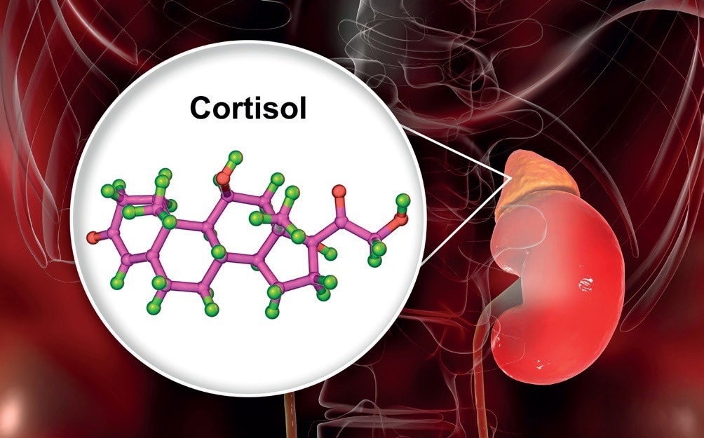
A more accurate diagnosis of Cushing’s syndrome

Cortisol is a steroid hormone of the glucocorticoid class made by the adrenal glands.

Image Credit: Shutterstock/Kateryna Kon

LDDST

  • Overnight protocol: 1 mg of dexamethasone is administered at 11:00 pm, and the serum cortisol levels are measured at 8:00 am the following morning.
  • Two-day protocol: serum cortisol levels are measured at 8:00 am and 0.5 mg of dexamethasone is administered every six hours (9:00 am, 3:00 pm, 9:00 pm, 3:00 am) for two days, totalling 4 mg. Serum cortisol levels are then measured at 9:00 am, six hours after the last dose has been delivered.

HDDST

  • Overnight protocol: baseline serum cortisol or 24-hour urinary free cortisol (UFC) is measured in the morning, and 8 mg of dexamethasone is given at 11.00 pm. Cortisol level in blood is then measured at 8.00 am the following morning.
  • Two-day protocol: Baseline serum cortisol or 24-hour UFC is measured at 8:00 am; 2 mg of dexamethasone is administered every six hours (9:00 am, 3:00 pm, 9:00 pm, 3:00 am) for two days, totaling 16 mg, in tandem with the collection of urine for UFC measurements. Serum cortisol levels are measured at 9:00 am, six hours after the last dose.

Patients whose pituitary glands produce excessive amounts of ACTH will exhibit an abnormal response to the low-dose test but a normal reaction to the high-dose test.

During the LDDST, cortisol levels should decrease following the administration of dexamethasone, and a cut-off value of below 18 ng/mL is recommended to distinguish a healthy response from an unhealthy one.

For the HDDST, a decrease in urine-free cortisol (UFC) or serum cortisol greater than 50% indicates the presence of ACTH-dependent Cushing’s syndrome. This rule applies to both the overnight LDDST and the two-day HDDST methods.

Measuring cortisol levels

Chemiluminescence immunoassay (CLIA) is a widely used method for measuring cortisol and other steroids due to its simplicity, automation, and good sensitivity.

However, it has some drawbacks, including cross-reactivity leading to overestimation of target analyte levels, non-standardization of kits, and the inability to measure more than one analyte per analysis. This is particularly problematic since studies indicate that measuring dexamethasone in combination with cortisol can reduce the number of false-positive DST results and improve interpretability. 3,4,5

Liquid chromatography-tandem mass spectrometry (LC-MS/MS) has emerged as a popular alternative to CLIA for DSTs due to its ability to measure multiple analytes simultaneously and its superior specificity.

Analytes are separated via LC, and their concentrations are measured by MS, with triple quadrupole MS configurations commonly used for this purpose. This technique provides the ability to measure multiple analytes simultaneously, along with higher accuracy and sensitivity than CLIA.

Increased ease of use and accuracy

In the Division of Endocrinology, Diabetes, and Metabolism at the University of Turin, a team has developed an LC-MS/MS technique for simultaneous quantifying cortisol, cortisone, dexamethasone, and six other exogenous corticosteroids in serum/plasma samples.6

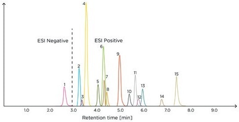
This method can be readily applied in any clinical laboratory equipped with a mass spectrometer and is effective in DSTs, enabling precise measurements of the target analytes in a single chromatographic run (Figure 1).

Sample preparation (1 Hour)

  1. Dilute 200 μL of the serum/plasma sample with 200 μL of water.
  2. Perform supported liquid extraction, manually transferring 400 μL of sample to a microplate.
  3. Apply positive pressure using Tecan Resolvex® A200 automated positive pressure processor.
  4. Elute with 700 μL of methyl tert-butyl ether.
  5. Evaporate and reconstitute in H2 O/MeOH (1:1, v/v).
  6. Agitate.

LC-MS/MS analysis (10 Minutes)

  • LC column: C18 (100 × 2.1 mm, 1.7 μm)
  • Flow rate: 400 μL/min
  • Temperature: 30 °C
  • Injection volume: 20 μL
  • Mobile phase A: H2O + 0.2 mM ammonium fluoride
  • Mobile phase B: acetonitrile
  • Elution programme: Table 1.

The study demonstrated a strong correlation between the results obtained from the newly developed LC-MS/MS method and those obtained using a commercially available CE IVD-marked Steroid Panel LC-MS* kit (Tecan).

The Tecan kit enables simultaneous dexamethasone, cortisol, and cortisone measurement and includes all the necessary components for easy implementation, such as calibrators and controls. The samples are prepared using solid-phase extraction (SPE), which can be semi-automatically performed on a Resolvex® A200 positive pressure processor (Tecan). The kit can measure 15 other steroids in the core steroid metabolism pathway due to the effectiveness of the SPE process.

Table 1. LC gradient elution programme. Source: Tecan

Time (min) Mobile phase A (%) Mobile phase B (%)
0 90 10
0.5 65 35
4.5 65 35
4.51 35 65
6.0 2 98
8.0 2 98
8.01 90 10
10.0 90 10

* In USA: for research use only. Not for use in diagnostic procedures. Product availability and regulatory status may vary from country to country. Consult with your Tecan associate for further information.
A more accurate diagnosis of Cushing’s syndrome

Figure 1. Example chromatogram of the Steroid Panel LC-MS internal standard – run 1. ESI, electrospray ionization; 1, aldosterone; 2, cortisone; 3, dehydro-epiandrosterone sulfate; 4, cortisol; 5, 21-deoxycortisol; 6, corticosterone; 7, dexamethasone; 8, 11-deoxycortisol; 9, androstenedione; 10, 11-deoxycorticosterone; 11, testosterone; 12, dehydroepiandrosterone; 13, 17-hydroxyprogesterone; 14, dihydrotestosterone; 15, progesterone.

Image Credit: Tecan

Summary

Early diagnosis of Cushing’s syndrome is critical to prevent potentially fatal complications. A reliable method for reducing the number of false positives in DSTs involves the simultaneous measurement of cortisol and dexamethasone levels, which can be accurately achieved using LC-MS/MS.

The LC-MS/MS method described in this article enables the simultaneous measurement of multiple analytes, such as cortisol, cortisone, and dexamethasone, in serum or plasma.

This analytical approach can provide clinical laboratories with a straightforward method for performing DSTs, and the commercially available kit can ensure consistent and dependable results.

References and further reading

  1. Cushing’s syndrome [website]. National Institute of Diabetes and Digestive and Kidney Diseases 2018 (https://www.niddk.nih.gov/health-information/endocrine-diseases/cushings-syndrome).
  2. Dogra P, Vijayashankar NP. Dexamethasone suppression test. StatPearls 2022, 8 August (https://www.ncbi.nlm.nih.gov/books/NBK542317 ).
  3. Ceccato F, Artusi C, Barbot M, et al. Dexamethasone measurement during low-dose suppression test for suspected hypercortisolism: threshold development with and validation. J Endocrinol Invest 2020;43(8):1105–1113. doi: 10.1007/s40618-020-01197-6.
  4. Roper SM. Yield of serum dexamethasone measurement for reducing false-positive results of low-dose dexamethasone suppression testing. J Appl Lab Med 2021;6(2):480–485. doi: 10.1093/jalm/jfaa193.
  5. Fleseriu M, Auchus R, Bancos I, et al. Consensus on diagnosis and management of Cushing’s disease: a guideline update. Lancet Diabetes Endocrinol 2021;9(12):847–875. doi: 10.1016/S2213- 8587(21)00235-7.
  6. Ponzetto F, Parasiliti-Caprino M, Settanni F, et al. Simultaneous measurement of cortisol, cortisone, dexamethasone and additional exogenous corticosteroids by rapid and sensitive LC-MS/MS analysis. Molecules 2022;28(1):248. doi: 10.3390/molecules28010248.

From https://www.news-medical.net/whitepaper/20240524/A-more-accurate-diagnosis-of-Cushinge28099s-syndrome.aspx

Cushing’s Syndrome Epidemiology

By Yolanda Smith, BPharm

Cushing’s syndrome is considered to be a rare disorder that results from prolonged exposure to glucocorticoids. However, there are few epidemiological studies to provide adequate data to describe the incidence and prevalence of the condition accurately. Most cases are diagnosed between the ages of 20 and 50, although any individual may be affected at any age.

The presentation of the symptoms of Cushing’s syndrome can vary greatly. In addition, many of the symptoms overlap with those caused by other health conditions, such as metabolic syndrome and polycystic ovary syndrome. This can make the diagnosis of the condition difficult. It is also difficult to establish epidemiological trends in Cushing’s syndrome, because not all cases of the disease are diagnosed. However, it is important that diagnosis is made as soon as possible, because early diagnosis and treatment of the condition are associated with improved morbidity and mortality rates.

Population-based Studies

There are several population-based studies that have reported the incidence and mortality rates of Cushing’s syndrome in certain populations over a discrete period of time.

A study in Denmark followed 166 patients with Cushing’s syndrome for 11 years, finding an incidence of 2 cases per million population per year. Of the 166 patients, 139 had benign disease. There was a mortality rate of 16.5% in the follow-up period of 8 years, with most deaths occurring in the year after the initial diagnosis, often before the initiation of treatment. The causes of death of patients with Cushing’s syndrome in the study included severe infections, cardiac rupture, stroke and suicide.

A study in Spain found 49 cases of Cushing’s syndrome over a period of 18 years, with an incidence of 2.4 cases per million inhabitants per year and a prevalence of 39.1 cases per million. The standard mortality ratio in this study was 3.8, in addition to an increase in morbidity rates.

Incidence

A low incidence of endogenous Cushing’s syndrome was established by the population-based studies outlined above, corresponding to approximately 2 cases per million. Some studies have an estimated incidence as low as 0.7 people per million.

However, the incidence of subclinical Cushing’s syndrome may be underestimated in certain population groups, such as those with osteoporosis, uncontrolled diabetes mellitus or hypertension. For example, of 90 obese patients with uncontrolled diabetes mellitus in one study, three had Cushing’s syndrome. This yielded a prevalence of 3.3%, which is considerably higher than the incidence reported in the population-based studies. However, these findings should be supported by larger studies.

Females are more likely to be affected by Cushing’s syndrome than males, with a risk ratio of approximately 3:1. There does not appear to be a genetic link that involves an ethnic susceptibility to the condition.

Treatment Outcomes

Surgery is the first-line treatment option for most cases of overt disease and remission is achieved in the majority of patients, approximately 65-85%. However, for up to 1 in 5 patients the condition recurs, and the risk does not appear to level off, even after 20 years of follow-up.

The risk of mortality for individuals with Cushing’s syndrome is estimated to be 2-3 times higher than that of the general population, based on epidemiological studies.

Reviewed by Dr Liji Thomas, MD.

From http://www.news-medical.net/health/Cushings-Syndrome-Epidemiology.aspx

Endocrine Society experts call for expanded screening for primary aldosteronism

Washington, DC–The Endocrine Society today issued a Clinical Practice Guideline calling on physicians to ramp up screening for primary aldosteronism, a common cause of high blood pressure.

People with primary aldosteronism face a higher risk of developing cardiovascular disease and dying from it than other people with high blood pressure. As many as one in ten people with high blood pressure may have primary aldosteronism. Uncontrolled high blood pressure can put these individuals at risk for stroke, heart attack, heart failure or kidney failure.

The guideline, entitled “The Management of Primary Aldosteronism: Case Detection, Diagnosis, and Treatment: An Endocrine Society Clinical Practice Guideline,” was published online and will appear in the May 2016 print issue of The Journal of Clinical Endocrinology & Metabolism (JCEM), a publication of the Endocrine Society. The guideline updates recommendations from the Society’s 2008 guideline on primary aldosteronism.

“In the past eight years, we have come to recognize that primary aldosteronism, despite being quite common, frequently goes undiagnosed and untreated,” said John W. Funder, MD, PhD, of the Hudson Institute of Medical Research in Clayton, Australia, and chair of the task force that authored the guideline. “This is a major public health issue. Many people with primary aldosteronism are never screened due to the associated costs. Better screening processes are needed to ensure no person suffering from primary aldosteronism and the resulting risks of uncontrolled high blood pressure goes untreated.”

Primary aldosteronism occurs when the adrenal glands — the small glands located on the top of each kidney – produce too much of the hormone aldosterone. This causes aldosterone, which helps balance levels of sodium and potassium, to build up in the body. The resulting excess sodium can lead to a rise in blood pressure.

The Endocrine Society recommends primary aldosterone screening for people who meet one of the following criteria:

  • Those who have sustained blood pressure above 150/100 in three separate measurements taken on different days;
  • People who have hypertension resistant to three conventional antihypertensive drugs;
  • People whose hypertension is controlled with four or more medications;
  • People with hypertension and low levels of potassium in the blood;
  • Those who have hypertension and a mass on the adrenal gland called an adrenal incidentaloma;
  • People with both hypertension and sleep apnea;
  • People with hypertension and a family history of early-onset hypertension or stroke before age 40; and
  • All hypertensive first-degree relatives of patients with primary aldosteronism.

Other recommendations from the guideline include:

  • The plasma aldosterone-to-renin ratio (ARR) test should be used to screen for primary aldosteronism.
  • All patients diagnosed with primary aldosteronism should undergo a CT scan of the adrenal glands to screen for a rare cancer called adrenocortical carcinoma.
  • When patients choose to treat the condition by having one adrenal gland surgically removed, an experienced radiologist should take blood samples from each adrenal vein and have them analyzed. This procedure, called adrenal vein sampling, is the gold standard for determining whether one or both adrenal glands is producing excess aldosterone.
  • For people with primary aldosteronism caused by overactivity in one adrenal gland, the recommended course of treatment is minimally invasive surgery to remove that adrenal gland.
  • For patients who are unable or unwilling to have surgery, medical treatment including a mineralocorticoid receptor (MR) agonist is the preferred treatment option.

###

The Hormone Health Network offers resources on primary aldosteronism athttp://www.hormone.org/questions-and-answers/2012/primary-aldosteronism.

Other members of the Endocrine Society task force that developed this guideline include: Robert M. Carey, of the University of Virginia Health System in Charlottesville, VA; Franco Mantero of the University of Padova in Padua, Italy; M. Hassan Murad of the Mayo Clinic in Rochester, MN; Martin Reincke of the Klinikum of the Ludwig-Maximilians-University of Munich in München, Bavaria, Germany; Hirotaka Shibata of Oita University in Oita, Japan; Michael Stowasser of the University of Queensland in Brisbane, Australia; and William F. Young, Jr. of the Mayo Clinic in Rochester, MN.

The Society established the Clinical Practice Guideline Program to provide endocrinologists and other clinicians with evidence-based recommendations in the diagnosis and treatment of endocrine-related conditions. Each guideline is created by a task force of topic-related experts in the field. Task forces rely on evidence-based reviews of the literature in the development of guideline recommendations. The Endocrine Society does not solicit or accept corporate support for its guidelines. All Clinical Practice Guidelines are supported entirely by Society funds.

The Clinical Practice Guideline was co-sponsored by the American Heart Association, the American Association of Endocrine Surgeons, the European Society of Endocrinology, the European Society of Hypertension, the International Association of Endocrine Surgeons, the International Society of Hypertension, the Japan Endocrine Society and The Japanese Society of Hypertension.

The guideline was published online at http://press.endocrine.org/doi/10.1210/jc.2015-4061, ahead of print.

Endocrinologists are at the core of solving the most pressing health problems of our time, from diabetes and obesity to infertility, bone health, and hormone-related cancers. The Endocrine Society is the world’s oldest and largest organization of scientists devoted to hormone research and physicians who care for people with hormone-related conditions.

The Society, which is celebrating its centennial in 2016, has more than 18,000 members, including scientists, physicians, educators, nurses and students in 122 countries. To learn more about the Society and the field of endocrinology, visit our site at http://www.endocrine.org. Follow us on Twitter at @TheEndoSociety and @EndoMedia.

Disclaimer: AAAS and EurekAlert! are not responsible for the accuracy of news releases posted to EurekAlert! by contributing institutions or for the use of any information through the EurekAlert system.

From http://www.eurekalert.org/pub_releases/2016-04/tes-ese042616.php

Cushing’s Awareness Challenge: Day 13

robin-complications

What can I say?  Robin’s images area always right on!

My only addition to this list would be the ongoing daily fatigue.  Maybe that’s not really a complication but more of an annoyance.

My bone loss has stopped  (or  slowed down some) over  the years since my pituitary surgery in 1987 but I doubt that I’ll ever gain back any height!

Luckily, I haven’t had a stroke – knock on wood!

Cushing’s – the “gift” that keeps on giving!

maryo colorful zebra


Cushing’s Awareness Challenge: Day 10

robin-tests

Gee, I’m an underachiever. LOL I only had one IPSS and one pituitary surgery.

While I was at NIH, my MRIs still showed nothing, so they did an Inferior Petrosal Sinus Sampling Test. That scared me more than the prospect of surgery. (This test carries the risk of stroke and uncontrollable bleeding from the incision points.) Catheters were fed from my groin area to my pituitary gland and dye was injected. I could watch the whole procedure on monitors.

I could not move during this test or for several hours afterwards to prevent uncontrollable bleeding from a major artery. The test did show where the tumor probably was located.

Also done were more sophisticated dexamethasone suppression tests where drugs were administered by IV and blood was drawn every hour (they put a heplock in my arm so they didn’t have to keep sticking me). I got to go home for a weekend and then went back for the surgery – the Transsphenoidal Resection. I fully expected to die during surgery (and didn’t care if I did) so I signed my will and wrote last letters to those I wanted to say goodbye to.

During the time I was home just before surgery, a college classmate of mine (I didn’t know her) did die at NIH of a Cushing’s-related problem. I’m so glad I didn’t find out until a couple months later!

maryo colorful zebra