Clinical Features and Treatment Options for Pediatric Adrenal Incidentalomas

Abstract

Background

The aim of this study was to investigate the clinical features and treatment options for pediatric adrenal incidentalomas(AIs) to guide the diagnosis and treatment of these tumors.

Methods

The clinical data of AI patients admitted to our hospital between December 2016 and December 2022 were collected and retrospectively analyzed. All patients were divided into neonatal and nonneonatal groups according to their age at the time of the initial consultation.

Results

In the neonatal group, 13 patients were observed and followed up, and the masses completely disappeared in 8 patients and were significantly reduced in size in 5 patients compared with the previous findings. Four patients ultimately underwent surgery, and the postoperative pathological diagnosis was neuroblastoma in three patients and teratoma in one patient. In the nonneonatal group, there were 18 cases of benign tumors, including 9 cases of ganglioneuroma, 2 cases of adrenocortical adenoma, 2 cases of adrenal cyst, 2 cases of teratoma, 1 case of pheochromocytoma, 1 case of nerve sheath tumor, and 1 case of adrenal hemorrhage; and 20 cases of malignant tumors, including 10 cases of neuroblastoma, 9 cases of ganglioneuroblastoma, and 1 case of adrenocortical carcinoma.

Conclusions

Neuroblastoma is the most common type of nonneonatal AI, and detailed laboratory investigations and imaging studies are recommended for aggressive evaluation and treatment in this population. The rate of spontaneous regression of AI is high in neonates, and close observation is feasible if the tumor is small, confined to the adrenal gland and has no distant metastasis.

Peer Review reports

Background

The incidence of adrenal incidentaloma (AI) is increasing due to the increased frequency of imaging and improved imaging sensitivity [1]. AI is relatively common in adults, and several organizations, such as the American Association of Clinical Endocrinologists/American Association of Endocrine Surgeons and the European Society Endocrinology, have proposed specific protocols to guide the evaluation, treatment, and follow-up management of AI in adults [2]. Although AI, a nonfunctioning adrenocortical adenoma, is most common in adults, neuroblastoma is the most common incidental tumor of the adrenal gland in children. In addition, in the neonatal period, which is a more complex stage of childhood, the biology of adrenal masses found in this age group is also more specific, and the nature of these masses can range from spontaneous regression to rapid progression to aggressive disease with metastatic dissemination and even death. Given that AI is the most common malignant tumor, the management of AI in children cannot be simply based on the measurements used in adult AI. In this study, we retrospectively analyzed the clinical data of pediatric AI patients in a single center to investigate the clinical characteristics and management of AI in children.

Methods

A total of 66 children with adrenal tumors were diagnosed and treated at the Department of Urology of the Children’s Hospital of Nanjing Medical University from December 2016 to December 2022. A total of 55 cases were detected during physical examination, or the patients were diagnosed and received treatment for diseases other than adrenal disease after excluding adrenal tumors detected due to typical clinical manifestations or signs such as centripetal obesity and precocious puberty. Research protocols involving human materials were approved by the Medical Ethics Committee of the Children’s Hospital of Nanjing Medical University. All clinical information, radiological diagnosis, laboratory test results, intervention results, and follow-up data were collected from the department’s database.

All the children underwent ultrasonography and CT scanning, and 11 children underwent MRI. In addition to routine tests such as blood routine and biochemical indexes, the examination and evaluation of adrenal endocrine hormones and tumor markers included (1) plasma cortisol and ACTH levels, (2) plasma catecholamine and metabolite determination, (3) plasma renin and plasma aldosterone, (4) urinary vanillylmandelic acid/homovanillic acid(VMA/HVA), and (5) AFP, CEA, NSE, and CA19-9. Five patients underwent a low-dose dexamethasone suppression test. Seventeen of the 55 patients were treated with watch-waiting therapy, 4 of the 17 ultimately underwent surgery, 4 of the 38 patients underwent tumor biopsy, and 34 underwent adrenalectomy.

The data were analyzed using Graph Pad Prism 8. The measurement data are expressed as ‾x ± sd. The maximum diameter of the tumors, age of the patients with benign and malignant tumors, and maximum diameter of the tumors between the laparoscopic surgery group and the open surgery group were compared using paired t tests, and the percentages of the count data were compared using Fisher’s exact test.

Results

In this study, all patients were divided into two groups according to their age at the time of consultation: the neonate group and the nonneonate group.

Neonate group:

There were 7 male and 10 female patients, 7 of whom were diagnosed via prenatal examination and 10 of whom were diagnosed after birth. Five patients were diagnosed with lesions on the left side, 12 patients were diagnosed with lesions on the right side, and the maximal diameters of the masses ranged from 16 to 48 mm. The characteristics of the AIs in the neonate group are presented in Table 1.

Table 1 Characteristics of AI in the neonates group

Among the 17 patients, 8 had cystic masses with a maximum diameter of 1648 mm, 5 had cystic-solid masses with a maximum diameter of 3339 mm, and 4 had solid masses with a maximum diameter of 1845 mm. Two patients with solid adrenal gland masses suggested by CT scan had obvious elevations in serum NSE and maximum diameters of 44 and 45 mm, respectively. These patients underwent adrenal tumor resection, and the pathology diagnosed that they had neuroblastomas(NB). In one patient, the right adrenal gland was 26 × 24 × 27 mm in size with slightly elevated echogenicity at 38 weeks after delivery, and the mass increased to a size of 40 × 39 × 29 mm according to the 1-month postnatal review. MRI suggested that the adrenal gland tumor was associated with liver metastasis, and the pathology of the tumor suggested that it was NB associated with liver metastasis after surgical resection (stage 4 S, FH). One child was found to have 25 × 24 × 14 mm cystic echoes in the left adrenal region during an obstetric examination, and ultrasound revealed 18 × 11 mm cystic solid echoes 5 days after birth. Ultrasound revealed 24 × 15 mm cystic solid echoes at 2 months. Serum NSE and urinary VMA were normal, and the tumor was excised due to the request of the parents. Pathology suggested a teratoma in the postoperative period. A total of 13 children did not receive surgical treatment or regular review via ultrasound, serum NSE or urine VMA. The follow-up time ranged from 1 to 31 months, with a mean of 9.04 ± 7.61 months. Eight patients had complete swelling, and 5 patients were significantly younger than the previous patients. Nonneonate group:

There were 24 male and 14 female patients in the nonneonate group; 24 patients had lesions on the left side, 14 patients had lesions on the right side, and the maximal diameters of the masses ranged from 17 to 131 mm. Most of these tumors were found during routine physical examinations or incidentally during examinations performed for various complaints, such as gastrointestinal symptoms, respiratory symptoms, or other related conditions. As shown in Table 2, abdominal pain was the most common risk factor (44.7%) for clinical onset, followed by routine physical examination and examination for respiratory symptoms.

Table 2 Clinical presentations leading to discovery of AI in non-neonate group

Among the 38 patients, 10 had NBs with maximum diameters ranging from 20 to 131 mm, 9 had ganglion cell neuroblastomas with maximum diameters ranging from 33.6 to 92 mm, 9 had ganglion cell neuromas with maximum diameters ranging from 33 to 62 mm, 2 had adrenal adenomas with maximum diameters ranging from 17 to 70 mm, 1 had a cortical carcinoma with a maximum diameter of 72 mm, 2 had adrenal cysts with maximum diameters ranging from 26 to 29 mm, 2 had mature teratomas with maximum diameters of 34 and 40 mm, 1 had a pheochromocytoma with a diameter of 29 mm, 1 had a nerve sheath tumor with a diameter of 29 mm, and 1 patient with postoperative pathological confirmation of partial hemorrhagic necrosis of the adrenal gland had focal calcification with a maximum diameter of 25 mm (Table 3).

Table 3 Distribution of different pathologies among AI with various sizes in non-neonate group

The mean age of children with malignant tumors was significantly lower than that of children with benign tumors (57.95 ± 37.20 months vs. 105.0 ± 23.85 months; t = 4.582, P < 0.0001). The maximum diameter of malignant tumors ranged from 20 to 131 mm, while that of benign tumors ranged from 17 to 72 mm, and the maximum diameter of malignant tumors was significantly greater than that of benign tumors (65.15 ± 27.61 mm v 37.59 ± 12.98 mm; t = 3.863, P = 0.0004). Four biopsies, 5 laparoscopic adrenal tumor resections and 11 open adrenal tumor resections were performed for malignant tumors, and 16 laparoscopic adrenal tumor resections and 2 open procedures were performed for benign tumors. The maximum diameter of the tumors ranged from 17 to 62 mm in 21 children who underwent laparoscopic surgery and from 34 to 99 mm in 13 children who underwent open resection; there was a statistically significant difference in the maximum diameter of the tumors between the laparoscopic surgery group and the open surgery group (35.63 ± 10.36 mm v 66.42 ± 20.60 mm; t = 5.798, P < 0.0001).

Of the 42 children with definitive pathologic diagnoses at surgery, 23 had malignant tumors, and 19 had benign tumors. There were 15 malignant tumors with calcification on imaging and 5 benign tumors. The percentage of malignant tumors with calcifications in was significantly greater than that of benign tumors (65.22% v 26.32%; P = 0.0157). In the present study, all the children underwent CT, and 31 patients had postoperative pathological confirmation of NB. A total of 26 patients were considered to have neurogenic tumors according to preoperative CT, for a diagnostic compliance rate of 83.97%. Three children were considered to have cortical adenomas by preoperative CT, and 1 was ultimately diagnosed by postoperative pathology, for a diagnostic compliance rate of 33.33%. For 4 patients with teratomas and adrenal cysts, the CT findings were consistent with the postoperative pathology. According to our research, NB 9-110HU, GNB 15-39HU, GB 19-38HU, ACA 8HU, adrenal cyst 8HU, and cellular achwannoma 17HU.

Discussion

According to the clinical practice guidelines developed by the European Society of Endocrinology and European Network for the Study of Adrenal Tumors, AI is an adrenal mass incidentally detected on imaging not performed for a suspected adrenal disease [3]. The prevalence of AI is approximately 4%, and the incidence increases with age [4]. Most adult AIs are nonfunctioning benign adrenal adenomas (up to 75%), while others include functioning adrenal adenomas, pheochromocytomas, and adrenocortical carcinomas [5]. In contrast to the disease spectrum of adult AI cases, NB is the most common tumor type among children with AI, and benign cortical adenomas, which account for the vast majority of adult AI, accounting for less than 0.5% of cases in children [6]. According to several guidelines, urgent assessment of an AI is recommended in children because of a greater likelihood of malignancy [37].

When an adult patient is initially diagnosed with AI, it should be clear whether the lesion is malignant and functional. In several studies, the use of noncontrast CT has been recommended as the initial imaging method for adrenal incidentaloma; a CT attenuation value ≤ 10 HU is used as the diagnostic criterion for benign adenomas; and these methods have a specificity of 71-79% and a sensitivity of 96-98% [89]. A CT scan of tumors with diameters greater than 4 to 6 cm, irregular margins or heterogeneity, a CT attenuation value greater than 10 HU, or a relative contrast enhancement washout of less than 40% 10 or 15 min after administration of contrast media on enhanced CT is considered to indicate potential malignancy [7]. As the most common AI in children, NB often appears as a soft tissue mass with uneven density on CT, often accompanied by high-density calcified shadows, low-density cystic lesions or necrotic areas. CT scans can easily identify more typical NBs, and for those AIs that do not show typical calcified shadows on CT, it is sometimes difficult to differentiate neurogenic tumors from adenomas. In these patients, except for the 1 patient with adrenal cysts who had a CT value of 8 HU, very few of the remaining AI patients had a CT value less than 10 HU. Therefore, the CT value cannot be used simply as a criterion for determining the benign or malignant nature of AI, and additional imaging examinations, such as CT enhancement, MRI, and FDG-PET if necessary, should be performed immediately for AI in children.

Initial hormonal testing is also needed for functional assessment, and aldosterone secretion should also be assessed when the patient is hypertensive or hypokalemic [7]. Patients with AI who are not suitable for surgery should be observed during the follow-up period, and if abnormal adrenal secretion is detected or suggestive of malignancy during this period, prompt adrenal tumor resection is needed. For adult patients with AI, laparoscopic adrenal tumor resection is one of the most effective treatments that has comparative advantages in terms of hospitalization time and postoperative recovery speed; however, there is still some controversy over whether to perform laparoscopic surgery for some malignant tumors with large diameters, especially adrenocortical carcinomas, and some studies have shown that patients who undergo laparoscopic surgery are more prone to peritoneal seeding of tumors [10].

The maximum diameter of an adult AI is a predictor of malignancy, and a study by the National Italian Study Group on Adrenal Tumors, which included 887 AIs, showed that adrenocortical carcinoma was significantly correlated with the size of the mass, and the sensitivity of detecting adrenocortical carcinoma with a threshold of 4 cm was 93% [11]. According to the National Institutes of Health, patients with tumors larger than 6 cm should undergo surgical treatment, while patients with tumors smaller than 4 cm should closely monitored; for patients with tumors between 4 and 6 cm, the choice of whether to be monitored or surgically treated can be based on other indicators, such as imaging [12]. A diameter of 4 cm is not the initial threshold for determining the benign or malignant nature of a mass in children.

In a study of 26 children with AI, Masiakos et al. reported that 9 of 18 benign lesions had a maximal diameter less than 5 cm, 4 of 8 malignant lesions had a maximal diameters less than 5 cm, and 2 had a diameter less than 3 cm. The mean maximal diameter of benign lesions was 4.2 ± 1.7 cm, whereas the mean maximum diameter of malignant lesions was 5.1 ± 2.3 cm. There was no statistically significant difference between the two comparisons; therefore, this study concluded that children with AI diameters less than 5 cm cannot be treated expectantly [6]. Additionally, this study revealed that malignant lesions occurred significantly more frequently than benign lesions in younger children (mean age 1.7 ± 1.8 years v 7.8 ± 5.9 years; P = 0.02).

In the nonneonatal group of this study, 20 patients with malignant tumors had maximum diameters ranging from 20 to 131 mm, 10 had malignant tumors larger than 60 mm, and 3 had tumors smaller than 40 cm; 18 patients with benign tumors had maximum diameters ranging from 17 to 70 mm, 5 had diameters ranging from 40 to 60 mm, and 5 had diameters larger than 60 mm. Therefore, it is not recommended to use the size of the largest diameter of the tumor to decide whether to wait and observe or intervene surgically for children with AI. Instead, it is necessary to consider the age of the child; laboratory test results, such as whether the tumor indices are elevated or not; whether the tumor has an endocrine function; etc.; and imaging test results to make comprehensive judgments and decisions. Preoperative aggressive evaluation and prompt surgical treatment are recommended for nonneonatal pediatric AI patients.

Adrenal hematoma and NBs are the most common types of adrenal area masses in children, while pheochromocytoma, adrenal cyst, and teratoma are rarer masses [13]. In clinical practice, adrenal hematoma and NB are sometimes difficult to differentiate, especially when adrenal masses are found during the prenatal examination and neonatal period, and such children need to be managed with caution. The Children’s Oncology Group (COG ANBL00B1) implemented the watchful waiting treatment for children under 6 months of age with a solid adrenal mass < 3.1 cm in diameter (or a cystic mass < 5 cm) without evidence of distant metastasis, and if there is a > 50% increase in the adrenal mass volume, there is no return to the baseline VMA or HVA levels, or if there is a > 50% increase in the urinary VMA/HVA ratio or an inversion, surgical resection should be performed [14]. Eighty-three children in this study underwent expectant observation, 16 of whom ultimately underwent surgical resection (8 with INSS stage 1 NB, 1 with INSS stage 2B, 1 with INSS stage 4 S, 2 with low-grade adrenocortical neoplasm, 2 with adrenal hemorrhage, and 2 with extralobar pulmonary sequestration). Most of the children who were observed had a reduced adrenal mass volume. Of the 56 patients who completed the final 90 weeks of expectant observation, 27 (48%) had no residual mass, 13 (23%) had a residual mass volume of 0–1 ml, 8 (14%) had a mass volume of 1–2 ml, and 8 (14%) had a volume of > 2 ml; ultimately, 71% of the residual masses had a volume ≤ 1 ml and 86% had a residual volume ≤ 2 ml. In this study, a total of 16 patients were included in the watchful waiting treatment group; 3 patients underwent surgical treatment during the follow-up period, and 13 patients ultimately completed watchful waiting treatment. After 1–31 months of follow-up, 8 patients’ swelling completely disappeared, and 5 patients’ swelling significantly decreased. After strict screening for indications and thorough follow-up review, AIs in the neonatal period can be subjected to watchful waiting treatment, and satisfactory results can be achieved.

For benign adrenal tumors, laparoscopic surgery is superior to open surgery in terms of successful resection, whereas the feasibility of minimally invasive surgery for AI with preoperative suspicion of malignancy is controversial. The European Cooperative Study Group for Pediatric Rare Tumors recommends that minimally invasive surgery be considered only for early childhood tumors and should be limited to small, localized tumors; additionally, imaging should suggest no invasion of surrounding tissue structures or lymph nodes; and this strategy requires surgeons with extensive experience in oncologic and adrenal surgery [15]. NB is the most common pediatric AI, and open tumor resection remains the mainstay of treatment. For small, early tumors without evidence of invasion on preoperative examination, laparoscopic resection may be considered if the principles of oncologic surgery can be adhered to. If the patient responds to chemotherapy, the decision to perform laparoscopic tumor resection can also be re-evaluated after chemotherapy. According to the current study, the recurrence and mortality rates of laparoscopic surgery are comparable to those of open surgery [1617]. The relative contraindications for laparoscopic NB resection include a tumor diameter greater than 6 cm, venous dilatation, and the involvement of adjacent organs or blood vessels [18]. Patients who undergo open adrenalectomy have higher overall survival and recurrence-free survival rates than patients who undergo laparoscopic adrenalectomy [19]. Open adrenalectomy remains the gold standard for surgical resection of adrenocortical carcinoma, whereas laparoscopic adrenalectomy should be reserved for highly selected patients and performed by surgeons with appropriate expertise [20].

Cortical tumors are particularly rare among children with AIs and are sometimes not clearly distinguishable from neurogenic tumors on preoperative imaging; in such patients, the presence of subclinical Cushing’s syndrome needs to be carefully evaluated preoperatively; otherwise, a perioperative adrenal crisis may occur [21]. In patients in whom the possibility of an adrenocortical tumor was considered preoperatively, the assessment for subclinical Cushing’s syndrome mainly involved assessing the serum dehydroepiandrosterone sulfate level and performing an overnight dexamethasone suppression test.

A procedure for evaluating pediatric AI is shown in Fig. 1. Imaging is the first step in the evaluation of AI in children. CT can be used to clarify the nature of most tumors. MRI can be used to evaluate imaging risk factors (IDRFs) for NB. Bone marrow cytomorphology is recommended for all children with AI, along with microscopic residual neuroblastoma testing and further bone scanning if the bone marrow examination is positive. In addition, serum tumor marker levels and other relevant tests should be performed, and hormone levels should be evaluated. If adrenal adenomas cannot be completely excluded during the preoperative examination, a 1 mg overnight dexamethasone suppression test should be performed to exclude subclinical Cushing’s syndrome. In patients with hypertensive hypokalemia, the presence of aldosteronism should be evaluated by testing plasma aldosterone concentrations and plasma renin activity. Adrenal masses found in the neonatal period can be observed if the tumor is small, confined to the adrenal gland and shows no evidence of distant metastasis, while tumors that increase significantly in size during the follow-up period or that are associated with persistently elevated tumor markers require aggressive surgical treatment.

Fig. 1

figure 1

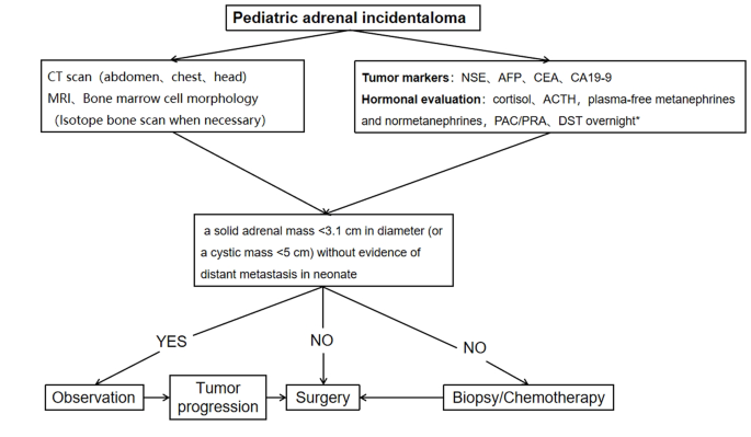
Algorithm for the evaluation and management of a pediatric adrenal incidentaloma. *DST overnight :20µg/kg dexamethasoneweight ˂40 kg,1 mg dexamethasone if ≥ 40 kg. CT = computed tomographic;MRI = magnetic resonance imaging;NSE = neuron-specific enolase;AFP = alpha-fetoprotein;CEA = carcinoembryonic antigen;CA19-9 = cancerantigen19-9;ACTH = adrenocorticotropic hormone;PAC = plasma aldosterone concentration; PRA = plasma renin activity;DST = dexamethasone suppression test

Data availability

The datasets analyzed during the current study are not public, but are available from the corresponding author on reasonable request.

Abbreviations

CT:
computed tomographic
MRI:
magnetic resonance imaging
ACTH:
adrenocorticotropic hormone
VMA:
vanillylmandelic acid
HVA:
homovanillic Acid
AFP:
alpha-fetoprotein
CEA:
carcinoembryonic antigen
NSE:
neuron-specific enolase
CA19-9:
cancerantigen19-9
FH:
favorable histology
HU:
Hounsfiled Unit
COG:
Children’s Oncology Group
INSS:
International Neuroblastoma Staging System

References

  1. Barzon L, Sonino N, Fallo F, Palu G, Boscaro M. Prevalence and natural history of adrenal incidentalomas. Eur J Endocrinol. 2003;149(4):273–85.

    Article CAS PubMed Google Scholar

  2. Maas M, Nassiri N, Bhanvadia S, Carmichael JD, Duddalwar V, Daneshmand S. Discrepancies in the recommendedmanagement of adrenalincidentalomas by variousguidelines. J Urol. 2021;205(1):52–9.

    Article PubMed Google Scholar

  3. Fassnacht M, Tsagarakis S, Terzolo M, et al. European Society of Endocrinology clinical practice guidelines on the management of adrenal incidentalomas, in collaboration with the European network for the study of adrenal tumors. Eur J Endocrinol. 2023;189(1):G1–42.

    Article PubMed Google Scholar

  4. Young WFJr. Clinical practice. The incidentally discovered adrenal mass. N Engl J Med. 2007;356(6):601–10.

    Article Google Scholar

  5. Rowe NE, Kumar R, Schieda N, et al. Diagnosis, management, and follow-up of the incidentallydiscoveredadrenalmass: CUAguidelineendorsed by the AUA. J Urol. 2023;210(4):590–9.

    Article PubMed Google Scholar

  6. Masiakos PT, Gerstle JT, Cheang T, Viero S, Kim PC, Wales P. Is surgery necessary for incidentally discovered adrenal masses in children?J. Pediatr Surg. 2004;39(5):754–8.

    Article Google Scholar

  7. Lee JM, Kim MK, Ko SH et al. Clinical guidelines for the management of adrenal incidentaloma. Endocrinol Metab. 2017;32(2).

  8. Terzolo M, Stigliano A, Chiodini I, et al. AME position statement on adrenal incidentaloma. Eur J Endocrinol. 2011;164(6):851–70.

    Article CAS PubMed Google Scholar

  9. Boland GW, Blake MA, Hahn PF, Mayo-Smith WW. Incidental adrenal lesions: principles, techniques, and algorithms for imaging characterization. Radiology. 2008;249(3):756–75.

    Article PubMed Google Scholar

  10. Payabyab EC, Balasubramaniam S, Edgerly M, et al. Adrenocortical cancer: a molecularlycomplexdiseasewheresurgerymatters. Clin Cancer Res. 2016;22(20):4989–5000.

    Article CAS PubMed Google Scholar

  11. Angeli A, Osella G, Alì A, Terzolo M. Adrenal incidentaloma: an overview of clinical and epidemiological data from the National Italian Study Group. Horm Res. 1997;47(4–6):279–83.

    Article CAS PubMed Google Scholar

  12. Grumbach MM, Biller BM, Braunstein GD, et al. Management of the clinically inapparent adrenal mass (incidentaloma). Ann Intern Med. 2003;138(5):424–9.

    Article PubMed Google Scholar

  13. Zhang K, Zhang Y, Zhang Y, Chao M. A retrospective analysis of the clinical characteristics of 207 hospitalized children with adrenal masses. Front Pediatr. 2023;11:1215095.

    Article PubMed PubMed Central Google Scholar

  14. Nuchtern JG, London WB, Barnewolt CE, et al. A prospective study of expectant observation as primary therapy for neuroblastoma in young infants: a Children‘s oncology group study. Ann Surg. 2012;256(4):573–80.

    Article PubMed Google Scholar

  15. Virgone C, Roganovic J, Vorwerk P, et al. Adrenocortical tumours in children and adolescents: the EXPeRT/PARTNER diagnostic and therapeutic recommendations. Pediatr Blood Cancer. 2021;68(suppl 4):e29025.

    Article PubMed Google Scholar

  16. Chang S, Lin Y, Yang S, et al. Safety and feasibility of laparoscopic resection of abdominal neuroblastoma without image-defined risk factors: a single-center experience. World J Surg Oncol. 2023;21(1):113.

    Article PubMed PubMed Central Google Scholar

  17. Zenitani M, Yoshida M, Matsumoto S, et al. Feasibility and safety of laparoscopic tumor resection in children with abdominal neuroblastomas. Pediatr Surg Int. 2023;39(1):91.

    Article PubMed Google Scholar

  18. International Pediatric Endosurgery Group. IPEG guidelines for the surgical treatment of adrenal masses in children. J Laparoendosc Adv Surg Tech A. 2010;20(2):vii–ix.

    Google Scholar

  19. Nakanishi H, Miangul S, Wang R, et al. Open versuslaparoscopicsurgery in the management of adrenocorticalcarcinoma: a systematicreview and meta-analysis. Ann Surg Oncol. 2023;30(2):994–1005.

    Article PubMed Google Scholar

  20. Gaillard M, Razafinimanana M, Challine A, et al. Laparoscopic or openadrenalectomy for stage I-IIadrenocorticalcarcinoma: a retrospectivestudy. J Clin Med. 2023;12(11):3698.

    Article PubMed PubMed Central Google Scholar

  21. Utsumi T, Iijima S, Sugizaki Y, et al. Laparoscopic adrenalectomy for adrenal tumors with endocrine activity: perioperative management pathways for reduced complications and improved outcomes. Int J Urol. 2023;30(10):818–26.

    Article CAS PubMed Google Scholar

Download references

Acknowledgements

We would like to express our deepest gratitude to all the patients and their parents who participated in this study. Their patience and cooperation were instrumental to the success of this research. We thank our colleagues in the Department of Radiology for their invaluable contributions in diagnosing and monitoring the progression of adrenal incidentalomas. We sincerely appreciate the hard work of the pathologists in diagnosing and classifying tumors, which laid the foundation for our study. Finally, we would like to thank our institution for providing the necessary resources and an enabling environment to conduct this research.

Funding

Not applicable.

Author information

Authors and Affiliations

  1. Department of Urology, Children’s Hospital of Nanjing Medical University, 72 Guangzhou Road, Nanjing, 210008, Jiangsu, China

    Xiaojiang Zhu, Saisai Liu, Yimin Yuan, Nannan Gu, Jintong Sha, Yunfei Guo & Yongji Deng

Contributions

X.J.Z. and Y.J.D designed the study; S.S.L., Y.M.Y., N.N.G., and J.T.S. carried out the study and collected important data; X.J.Z. analysed data and wrote the manuscript; Y.F.G. and Y.J.D.gave us a lot of very good advices and technical support; All authors read and approved the final manuscript.

Corresponding author

Correspondence to Yongji Deng.

Ethics declarations

Competing interests

The authors declare no competing interests.

Ethics approval and consent to participate

Ethics approval for this study was granted by the Ethics Committee of Children’s Hospital of Nanjing Medical University. Informed written consent was obtained from all the guardians of the children and we co-signed the informed consent form with their parents before the study. We confirmed that all methods were performed in accordance with relevant guidelines and regulations.

Conflict of interest

There are no conflicts of interest.

Additional information

Publisher’s Note

Springer Nature remains neutral with regard to jurisdictional claims in published maps and institutional affiliations.

Asymptomatic Pheochromocytoma Associated with MEN Syndrome and Subclinical Cushing’s Syndrome

Abstract

Introduction and importance

Pheochromocytoma and Cushing’s syndrome are rare endocrine conditions caused by tumors in the adrenal gland. These conditions are classified under Multiple Endocrine Neoplasia (MEN) syndrome, characterized by the development of multiple tumors in the endocrine system. However, diagnosing these conditions can be challenging as they often lack clear symptoms, requiring careful evaluation, monitoring, and treatment to prevent complications.

Case presentation

A 23-year-old male recently presented with right-sided abdominal fullness and lipoma-like masses on the torso. Over a span of six months, the abdominal mass nearly doubled in size, accompanied by elevated levels of catecholaminescortisolparathyroid hormone (PTH), and calcitonin. Surprisingly, the patient remained asymptomatic despite these abnormal lab values. CT imaging revealed a substantial increase in the size of the mass in the right adrenal gland, from 6 × 7 cm to approximately 11.2 × 10.2 × 9 cm.

Clinical discussion

Pheochromocytoma secretes catecholamines and often leads to hypertension and related symptoms. Interestingly, most individuals with pheochromocytoma do not exhibit obvious symptoms, necessitating blood and urine tests, along with imaging studies, for accurate diagnosis. The size of the tumor does not necessarily indicate the severity of symptoms. MEN-2, a genetic syndrome, is characterized by pheochromocytoma, medullary thyroid carcinoma, and hyperparathyroidism. Additionally, methods for diagnosing Cushing’s syndrome, caused by excess cortisol production, are discussed.

Conclusion

Early diagnosis and genetic counseling are crucial in preventing complications associated with these conditions. By identifying them, appropriate treatment can be ensured for positive outcomes of patients and their families.

Keywords

Pheochromocytoma
Multiple Endocrine Neoplasia (MEN) syndrome
Cushing’s syndrome
Rare Case Report

Abbreviations

CT

computed tomography

MRI

Magnetic resonance imaging

USG

Ultrasonography

131I-MIBG

iodine 131 labeled meta-iodobenzylganidine

RAAS

Renin-angiotensin-aldosterone system

    1. Introduction

    Pheochromocytoma are catecholamine secreting tumors of chromaffin cells of adrenal medulla. It can be found anywhere in the body, with the majority being intra-abdominal and those other than adrenal medulla are referred to as paragangliomas [1,2]. Pheochromocytoma typically secretes norepinephrine and epinephrine, with norepinephrine being the primary catecholamine. However, some tumors may only secrete one of the two, and rarely, some may secrete dopamine or dopa [3].

    Vast majority >90 % of adrenal neoplasms are benign non-functional adenomas [4].About 10 % of pheochromocytomas are malignant and 10 % of cases are found on both sides. Additionally, approximately 40 % of pheochromocytomas are caused by genetic factors and can be associated with inherited syndromes [5].

    Pheochromocytoma is found to be associated with MEN-2. MEN-2 is a hereditary genetic condition that is caused by a de novo mutation in the RET gene. It is inherited in an autosomal dominant fashion and is mainly characterized by medullary thyroid carcinoma, pheochromocytoma and parathyroid adenoma or hyperplasia [6].

    MEN syndrome can be MEN-1, MEN-2A and MEN-2B. MEN-1 is characterized by pituitary tumors (prolactin or growth hormone), pancreatic endocrine tumors and parathyroid adenomas. Additionally, other tumors such as foregut carcinoidsadrenocortical adenomas, meningioma, lipomas, angiofibromas and collagenomas may also occur in MEN-1. MEN-2A is characterized by medullary thyroid carcinoma, pheochromocytoma, and parathyroid adenoma/hyperplasia; it can also be associated with cutaneous lichen amyloidosis and Hirschsprung disease. On the other hand, MEN-2B is characterized by familial medullary thyroid cancer, pheochromocytoma, mucosal neuromasgastrointestinal tract issues, musculoskeletal and spinal problems. [7].

    Cushing syndrome results from hypercortisolism and is characterized by hypertension, weight gain, easy bruising, and central obesity [4]. Cushing’s disease refers to ACTH-dependent cortisol excess caused by a pituitary adenoma, while ACTH-independent cortisol excess due to non-pituitary causes such as excess use of glucocorticoids, adrenal adenoma, hyperplasia, or carcinoma is referred to as Cushing syndrome [8].

    This case report has been written according to the SCARE checklist [9].

    2. Case presentation

    A 23-year-old male presented to our surgery department with the chief complaint of right sided abdominal fullness for six months. According to the patient a mass was incidentally reported six months back while he was under-evaluation for mild trauma due to road traffic accident. Six months back, the mass was approximately 6 × 7 cm, while at the time of presentation to our department the mass was approximately 11.2 × 10.2 × 9 cm (CT abdomen) which was globular in shape, had regular margin, and moved with respiration. He had no history of hypertension, headache, palpitation, sweating, pallor, recent weight loss, abdominal pain, psychological disturbance, dizzinessloss of consciousness, dark color urine, burning micturition, had normal bowel and bladder habit.

    Past history and family history were insignificant. He was not under any long-term medication and no known drug allergies. He occasionally smokes and consumes alcohol.

    On physical examination at the time of presentation, multiple soft, mobile, painless, subcutaneous nodules like lipoma were present over the torso. His height was 176.8 cm, weight 68 kg, BMI 21.8 kg/m2 (body mass index). He had blood pressure of 110/70 mm of Hg taken in left arm at sitting position, heart rate of 62 beats/min, respiratory rate of 24/min, temperature of 96.6 °F, SPO2 of 98 % at right hand. A mass was palpable on the right side of abdomen, otherwise abdomen was soft, non-tender, normal bowel sound was present. Chest, cardiac and neurologic examinations were all normal.

    Initial laboratory evaluation revealed 24 h. urine metanephrine of 5415 μg/24 h (normal: 25–312 μg/24 h.); 24 h. urine VMA of 32.2 mg/24 h. (normal: <13.60 mg/24 h.); serum cortisol of 535.16 nmol/l after overnight low dose dexamethasone(1 mg) suppression test (normal: <50 nmol/l);24 h. Urine free cortisol of 526.61 nmol/24 h. (normal: 30–145 nmol/24 h) PTH(intact) of 89.2 pg./ml (normal: 15–65 pg./ml); serum calcitonin of 15.2 pg./ml (normal: ≤8.4 pg./ml); serum CEA of 4.72 ng/ml (normal: 0.0–4.4 ng/ml); serum DHEA of 1.19 ng/ml (normal: 1.7–6.1 ng/ml). Baseline investigation: Hematology, urine routine/microscopic, electrolytes were within the normal range.

    Additional laboratory findings were as in the Table 1.

    Table 1.

    Lab evaluation Result Reference Unit
    Metanephrine, urine 24 h 5415 25–312 μg/24 h
    VMA, urine 24 h 32.2 <13.60 mg/24 h
    VMA, urine 12.88 ng/l
    Cortisol, serum, overnight DST 535.16 <50 nmol/l
    Cortisol, urine 24 h 526.61 30–145 nmol/24 h
    ACTH, complete 28.3 7.2–63.3 pg/ml
    DHEA, serum 1.19 1.7–6.1 ng/ml
    CEA, serum 4.72 0.0–4.4 ng/ml
    Phosphorus, serum 3.0 2.5–4.5 mg/dl
    Albumin, serum 5.2 3.5–5.2 g/dl
    Calcitonin, serum 15.2 ≤8.4 pg/ml
    Calcium, serum 8.94 8.6–10.0 mg/dl
    PTH (intact) 89.2 15–65 pg/ml
    aldosterone 8.7 7.0–30 g/dl
    Plasma rennin activity 1.42 0.10–6.56 ng/ml/h
    Aldosterone-rennin ratio 6.13 ≤20
    Creatinine, urine 36 mg/dl

    DST – dexamethasone suppression test; VMA – vanilmandelic acid; ACTH – adrenocorticotropic hormone; DHEA – dehydroepiandrosterone; CEA – carcino-embryonic-antigen; PTH – parathyroid hormone.

    2.1. USG abdomen

    USG abdomen (Fig. 1Fig. 2) showed well defined mixed echoic area in Right adrenal region measuring 12.7 × 10.7 cm in size. There was presence of internal vascularity with multiple foci of cystic compound. The lesion displaced the right kidney inferiorly.

    Fig. 1

    1. Download : Download high-res image (102KB)
    2. Download : Download full-size image

    Fig. 1. USG abdomen.

    Fig. 2

    1. Download : Download high-res image (106KB)
    2. Download : Download full-size image

    Fig. 2. USG abdomen.

    2.2. Plane and contrast CT scan of abdomen

    Plane and contrast CT scan of Abdomen (Fig. 3) showed approximately 11.2 × 10.2 × 9 cm sized, relatively well defined heterogeneous soft tissue density lesion with well-defined enhancing wall in right adrenal region. Non-enhancing areas were noted within the mass suggestive of necrosis. Few calcific foci were noted within the mass with no obvious hemorrhagic component. The lesion showed heterogeneous enhancement post contrast image.

    Fig. 3

    1. Download : Download high-res image (192KB)
    2. Download : Download full-size image

    Fig. 3. CT abdomen.

    After all the workup patient was given diagnosis of right sided Pheochromocytoma associated with MEN syndrome, with ACTH-independent Cushing’s syndrome and right adrenalectomy was performed.

    2.3. Pathology report

    2.3.1. Gross descriptions

    The specimen was globular mass measuring 14.5 × 10 cm, with smooth outer surface. On sectioning, the mass was well circumscribed, soft and yellow-brown, predominantly solid with cyst formation. The size of cyst ranges from 0.3 to 3.5 cm in diameter. Areas of hemorrhages were noted.

    2.3.2. Microscopic description

    Section showed tumor cells arranged in well-defined nests (Zellballen), alveolar and diffuse pattern with intervening fibrovascular stroma. The cells were intermediate to large sized, polygonal with finely granular amphophilic cytoplasm. The nuclei showed mild to moderate pleomorphism and were round to ovoid, with prominent nuclei noted. No capsular invasion, vascular invasion and necrosis. Areas of hemorrhage were seen. Mitosis 0–1/10 high power field was noted (Figs. 4 and 5).

    Unlabelled Image

    1. Download : Download high-res image (1MB)
    2. Download : Download full-size image

    Fig.a Diffuse Zellbalen pattern with intervening fibrous stroma.

    Fig.b Mild to moderate pleomorphic nuclei with abundant hemorrhage.

    Fig.c Low power field with intact capsule.

    Figs. 4 and 5

    1. Download : Download high-res image (352KB)
    2. Download : Download full-size image

    Figs. 4 and 5. Fig. 4 Intra-operative resection of tumor; Fig. 5 tumor after resection.

    3. Discussion

    In Pheochromocytoma activation of the alpha-one adrenergic receptor by catecholamine in the vascular bed causes vasoconstriction and leads to a rise in blood pressure. Similarly, activation of the beta-one receptor in the heart enhances the chronotropic and inotropic effect of the myocardium, leading to an increase in heart rate and cardiac output. In addition, activation of the beta-one receptor in the juxtaglomerular cells of the kidney activates the RAAS system. These receptor activation result in cardiovascular and sympathetic changes, such as hypertension, palpitation, headache, sweating, trembling, and anxiety [10].

    In Pheochromocytoma, the patient may have a 10-fold increase in plasma catecholamines, but the hemodynamic response can still fall within the normal range due to desensitization of the cardiovascular system. When catecholamine levels are elevated for a prolonged period, the alpha-one receptors in blood vessels may be down-regulated, making norepinephrine unresponsive in raising peripheral vascular resistance, which can lead to normal blood pressure. Similarly, a marked decrease in beta-one receptors in the heart could explain the normal heart rate, which was observed in our asymptomatic patient with Pheochromocytoma [11].

    Sometimes in asymptomatic patients, the size of the tumor tends to be larger than in those with hyperfunctioning tumors [12]. However, medical interventions such as surgery, anesthesia inductionintravenous urography contrast, or manipulation of the tumor can trigger adrenergic and hypertensive crises, so biopsy is usually contraindicated in pheochromocytoma [13].

    The diagnosis of pheochromocytoma is typically based on measuring plasma and urinary levels of catecholamines and their derivatives such as metanephrine and vanillylmandelic acid. The most reliable test is the measurement of urinary metanephrine as its excretion levels are relatively higher [13,14]. The combination of 131I-MIBG scintigraphy along with diagnostic urinary and blood tests can further enhance the sensitivity of the test. Specifically, the urinary normetanephrine test is considered the most sensitive single test for detecting Pheochromocytoma [15,16].

    In addition to a 24-h urine test and blood test, if the lab results are positive for Pheochromocytoma or paragangliomas, further diagnostic tests may be recommended, such as a CT scanMRI, m-iodobenzylganidine (MIBG) imaging, or positron emission tomography (PET) [16,17]. In our patient 24 h. urine metanephrine of 5415 μg/24 h (normal: 25–312 μg/24 h.); 24 h. urine VMA of 32.2 mg/24 h. (normal: <13.60 mg/24 h.) and imaging confirmation of right adrenal mass lead to the diagnosis of right sided pheochromocytoma.

    Our patient with pheochromocytoma was tested for parathyroid hormone and calcitonin due to the association of pheochromocytoma with MEN-2 [18]. MEN-2 can be diagnosed biochemically by measuring the baseline levels of calcitonin, parathyroid hormone and serum calcium along with blood tests for catecholamines and their metabolites to detect pheochromocytoma [19]. In our patient, multiple soft, mobile, painless, subcutaneous nodules like lipoma were present over the torso(MEN-1) and high levels of parathyroid hormone and calcitonin were detected(MEN-2). These findings can be correlated with MEN syndrome.

    USG of the neck revealed no abnormalities of thyroid and parathyroid gland in our patient so prophylactic thyroidectomy was not done, instead he was counseled for follow up if any symptoms or thyroid swelling appears.

    The diagnosis of Cushing’s syndrome typically involves measuring the levels of 24-h urine free cortisol and assessing the suppression of cortisol in response to a 1 mg overnight dexamethasone test. If cortisol levels remain elevated despite the test, the next step is to measure serum ACTH levels. If ACTH levels are suppressed, it suggests an ACTH-independent cause of Cushing’s syndrome, while elevated ACTH levels suggest an ACTH-dependent cause. Further evaluation may include a CT scan of the chest, abdomen, and pelvis to identify potential ectopic sources, as well as an MRI of the pituitary gland [8]. Our patient had a high level of 24 h. urine free cortisol of 526.61 nmol/24 h (reference range: 30–145 nmol/24 h) and serum cortisol of 535.16 nmol/L(reference range: <50 nmol/L) after overnight 1 mg dexamethasone suppression test, but normal level of ACTH of 28.3 pg./ml (reference range: 7.2–63.1 ng/ml), this suggests the diagnosis of ACTH independent Cushing’s syndrome.

    4. Conclusion

    Large Pheochromocytoma patients can be asymptomatic and can present in association with other endocrine disorders. So proper evaluation is necessary to find out associated conditions and manage accordingly to prevent the possible outcomes.

    Patient consent

    Written, informed consent was obtained from the patient for the publication of the report.

    Ethical approval

    It is exempted at my institution. We don’t need to take approval from ethical committee for case report.

    Funding

    N/A.

    Author contribution

    Conceptualization: Sanjit Kumar Shah.

    Clinical diagnosis and patient management: Mahipendra Tiwari.

    Microscopic slide preparation: Sneh Acharya.

    Writing original draft: Sanjit Kumar Shah and Avish Shah.

    All authors were involved in reviewing, editing, supervision and in preparing the final

    manuscript.

    Guarantor

    Guarantor: Sanjit Kumar Shah

    Email: sanjitshah023@gmail.com

    Conflict of interest statement

    N/A.

    References

    Association of Chronic Central Serous Chorioretinopathy with Subclinical Cushing’s Syndrome

    https://doi.org/10.1016/j.ajoc.2022.101455

    Abstract

    Purpose

    To report the clinical course of a patient with central serous chorioretinopathy (CSCR) secondary to subclinical hypercortisolism before and after adrenalectomy.

    Observations

    A 50-year-old female patient with multifocal, chronic CSCR was found to have an adrenal incidentaloma and was diagnosed with subclinical hypercortisolism. Patient elected to undergo minimally-invasive adrenalectomy and presented at 3 months after surgery without subretinal fluid.

    Conclusions and Importance

    Subclinical Cushing’s Syndrome (SCS) may present an underrecognized risk factor for developing chronic CSCR. Further investigation is needed to determine the threshold of visual comorbidity that may influence surgical management.

    Keywords

    Central serous chorioretinopathy
    Subclinical Cushing’s syndrome
    Hypercortisolism
    Adrenalectomy
    Retina
    Surgical intervention

    1. Introduction

    Central serous chorioretinopathy (CSCR) is characterized by accumulation of fluid in the subretinal or sub-RPE space, often with consequential visual impairment. Chronic CSCR has been reported as a manifestation of hypercortisolism due to Cushing’s syndrome and subclinical hypercortisolism.1,2 However, the latter is often underrecognized owing to its inherently subtle nature and the optimal threshold for intervention based on associated comorbidities remains unclear. Herein we report the clinical course of a patient with CSCR secondary to subclinical hypercortisolism before and after adrenalectomy.

    2. Case report

    A 50-year-old female presented with blurred, discolored spots in the right eye for several months. Her past medical history included mild hypertension treated with amlodipine. Past ocular and family history were noncontributory.

    On exam, Snellen visual acuity was 20/50 OD, 20/25 OS. Her pupils, intraocular pressure, and anterior segment exam were within normal limits. Dilated fundus exam revealed bilateral, multifocal areas of subretinal fluid and mottled pigmentary changes (Fig. 1A). Optical coherence tomography confirmed areas of subretinal fluid and other areas of outer retinal atrophy (Fig. 1B). Fundus autofluorescence revealed areas of hyperautofluorescence that highlighted the fundoscopic findings (Fig. 1C). Fluorescein angiography showed multifocal areas of expansile dot leakage (Fig. 1D). Altogether these findings were consistent with multifocal, chronic CSCR.

    Fig. 1

    1. Download : Download high-res image (1MB)
    2. Download : Download full-size image

    Fig. 1. Multimodal imaging of bilateral multifocal central serous chorioretinopathy. Fundus photographs reveal multifocal subretinal fluid and pigmentary changes (Fig. 1A). Optical coherence tomography demonstrates subretinal fluid and outer retinal atrophy (Fig. 1B). Areas of hyperautofluorescence highlight the fundoscopic findings of subretinal fluid (Fig. 1C). Fluorescein angiography showing multiple areas of expansile dot leakage (Fig. 1D).

    On further clinical follow-up, an adrenal incidentaloma (AI) was detected when the patient underwent imaging for back pain. Subsequently she saw an endocrinologist; she had a normal serum cortisol, but low ACTH and an abnormal dexamethasone suppression test. This led to a diagnosis of subclinical hypercortisolism and provided a reason for her hypertension and chronic CSCR.

    Since the blur and relative scotomata interfered with her daily activities, she elected to try eplerenone, which yielded a moderate but suboptimal therapeutic response at 50 mg daily. While contemplating photodynamic therapy, in discussion with her endocrinologist, the patient opted to undergo minimally-invasive adrenalectomy. At last follow-up 3 months after surgery and 6 years after her initial presentation, she has been off eplerenone and without subretinal fluid (Fig. 2).

    Fig. 2

    1. Download : Download high-res image (1MB)
    2. Download : Download full-size image

    Fig. 2. Optical coherence tomography imaging at presentation and at last follow-up 3 months after adrenalectomy. There is a significant improvement in subretinal fluid in both eyes, though outer retinal irregularity remains.

    3. Discussion

    CSCR has previously been associated with many risk factors including exposure to excess steroid. A recent study analyzing a nationally representative dataset of 35,000 patients found that patients with CSCR had a higher odds of Cushing’s syndrome (OR 2.19, 95% CI 1.33 to 3.59, p = 0.002) than exposure to exogenous steroids (OR 1.14, 95% CI 1.09 to 1.19, p < 0.001)1 Our case highlights the importance of thorough medication reconciliation and careful consideration of comorbid conditions in patients with chronic CSCR.

    In recent years, subtle endogenous hypercortisolism, termed subclinical Cushing’s syndrome (SCS), has been of particular interest in the endocrinology literature because it can be a challenging diagnosis and the most appropriate management remains controversial.3 In general, SCS is comprised of: 1) the presence of an adrenal incidentaloma or mass, 2) biochemical confirmation of cortisol excess, and 3) no classic phenotypic manifestations of Cushing’s syndrome.4 Since adrenal incidentaloma has an estimated prevalence of 1–8% of the population,5 it is quite possible that SCS is an underrecognized risk factor for CSCR.

    SCS may potentiate metabolic syndrome and osteoporosis; studies have found that surgical resection of adrenal incidentalomas improve weight, blood pressure, and glucose control. Consequently, some authors recommend those with SCS-associated comorbidities be considered for resection.6 An important consideration in these patients is how visual comorbidity factors into intervention. In our patient’s case, the recurrent CSCR, hypertension, and increased risk of metabolic syndrome were sufficient reasons to elect to have surgery.

    4. Conclusion

    In summary, SCS is a condition of subtle cortisol dysregulation that may represent an underrecognized risk factor for chronic CSCR. Further investigation is needed to determine the threshold of visual comorbidity that may influence surgical management.

    Patient consent

    Consent to publish the case report was not obtained. This report does not contain any personal information that could lead to the identification of the patient.

    Acknowledgments and Disclosures

    Grant support was from the J. Arch McNamara Retina Research Fund. The following authors have no financial disclosures: RRS, AS, AC All authors attest that they meet the current ICMJE criteria for Authorship. No other contributions to acknowledge.

    References

    © 2022 The Authors. Published by Elsevier Inc.

    Severe Infection Including Disseminated Herpes Zoster Triggered by Subclinical Cushing’s Disease

    Abstract

    Background

    Subclinical Cushing’s disease (SCD) is defined by corticotroph adenoma-induced mild hypercortisolism without typical physical features of Cushing’s disease. Infection is an important complication associated with mortality in Cushing’s disease, while no reports on infection in SCD are available. To make clinicians aware of the risk of infection in SCD, we report a case of SCD with disseminated herpes zoster (DHZ) with the mortal outcome.

    Case presentation

    An 83-year-old Japanese woman was diagnosed with SCD, treated with cabergoline in the outpatient. She was hospitalized for acute pyelonephritis, and her fever gradually resolved with antibiotics. However, herpes zoster appeared on her chest, and the eruptions rapidly spread over the body. She suddenly went into cardiopulmonary arrest and died. Autopsy demonstrated adrenocorticotropic hormone-positive pituitary adenoma, renal abscess, and DHZ.

    Conclusions

    As immunosuppression caused by SCD may be one of the triggers of severe infection, the patients with SCD should be assessed not only for the metabolic but also for the immunodeficient status.

    Read the rest of the article at https://bmcendocrdisord.biomedcentral.com/articles/10.1186/s12902-021-00757-y

    Clinical Characterization of Patients With Primary Aldosteronism Plus Subclinical Cushing’s Syndrome


    Published: 

    Abstract

    Background

    Primary aldosteronism (PA) plus subclinical Cushing’s syndrome (SCS), PASCS, has occasionally been reported. We aimed to clinically characterize patients with PASCS who are poorly profiled.

    Methods

    A population-based, retrospective, single-center, observational study was conducted in 71 patients (age, 58.2 ± 11.2 years; 24 males and 47 females) who developed PA (n = 45), SCS (n = 12), or PASCS (n = 14). The main outcome measures were the proportion of patients with diabetes mellitus (DM), serum potassium concentration, and maximum tumor diameter (MTD) on the computed tomography (CT) scans.

    Results

    The proportion of DM patients was significantly greater in the PASCS group than in the PA group (50.0% vs. 13.9%, p <  0.05), without a significant difference between the PASCS and SCS groups. Serum potassium concentration was significantly lower in the PASCS group than in the SCS group (3.2 ± 0.8 mEq/L vs. 4.0 ± 0.5 mEq/L; p <  0.01), without a significant difference between the PASCS and PA groups. Among the 3 study groups of patients who had a unilateral adrenal tumor, MTD was significantly greater in the PASCS group than in the PA group (2.7 ± 0.1 cm vs. 1.4 ± 0.1 cm; p <  0.001), without a significant difference between the PASCS and SCS groups.

    Conclusions

    Any reference criteria were not obtained that surely distinguish patients with PASCS from those with PA or SCS. However, clinicians should suspect the presence of concurrent SCS in patients with PA when detecting a relatively large adrenal tumor on the CT scans.

    Background

    Primary aldosteronism (PA), an adrenocortical disorder caused by an adrenal tumor that overproduces aldosterone, accounts for 5 to 15% of patients with hypertension [1]. Cushing’s syndrome (CS), an endocrinopathy resulting from the prolonged, excessive adrenocortical secretion of cortisol, falls roughly into the following 2 categories: adrenocorticotropic hormone (ACTH)-dependent CS and ACTH-independent CS; the former includes Cushing’s disease that is primarily caused by a pituitary ACTH-secreting tumor and ectopic ACTH syndrome resulting from extrapituitary ACTH-secreting tumors (eg, bronchial carcinoid) [2], while the latter is usually caused by unilateral adenomas or carcinomas that provoke the autonomous adrenal cortical secretion [3]. Subclinical Cushing’s syndrome (SCS), an ill-defined endocrine disorder leading to the ACTH-independent secretion of cortisol from an adrenal adenoma that is not fully restrained by pituitary feedback [4], is known to cause hypertension, glucose intolerance, and dyslipidemia [5].

    The concurrence of clinically overt hyperaldosteronism and subclinical hypercortisolism is rare in PA patients [6]. To date, a few number of studies have examined the clinicopathological features of patients with PA plus SCS (PASCS), the incidences of which have ranged between about 10 and 20% [78]. Lower plasma ACTH levels and a greater tumor size were found in patients with PASCS than in patients with PA alone [8]. In the clinical settings, we rarely encounter PASCS patients who show a small adrenal tumor on the computed tomography (CT) scans and/or do not have a low plasma ACTH level in blood samples collected in the early morning. To examine the clinical features of PASCS patients in the present study, we compared clinical, laboratory, and imaging characteristics among patients with PA, SCS, or PASCS.

    Methods

    Patients

    We conducted a population-based, retrospective, single-center, observational study in 187 patients (119 with PA, 54 with SCS, and 14 with PASCS) at Saitama Medical University Hospital, Saitama, Japan, between January 1999 and December 2016. Hypertensive patients with suspected PA or SCS, as well as normotensive or hypertensive patients with an adrenal incidentaloma were referred to our hospital. A total of 116 patients were excluded from the study: 31 who were diagnosed with PA or SCS only because tests required to definitely diagnose these endocrinopathies were not conducted; 61 who failed to meet the new Japanese diagnostic criteria of SCS [9]; 1 who failed to meet the new Korean diagnostic criteria of subclinical hypercortisolism [10]; and 23 who failed to meet the Japanese [11] and United States [12] diagnostic criteria of PA. Therefore, we investigated 71 patients who were definitely diagnosed with PA and/or SCS (45 with PA, 12 with SCS, and 14 with PASCS). This study was approved by the institutional review board of Saitama Medical University. Patients provided written informed consent to the use of their clinical and laboratory data in the study.

    Diagnosis of PA and SCS

    Hormones required for the diagnosis of PA and SCS were assayed according to the procedures described in the pertinent guidelines [911]. Serum cortisol and plasma ACTH levels were determined by electrochemiluminescence immunoassay, plasma aldosterone concentration (PAC) and plasma renin activity (PRA) by radioimmunoassay, and serum dehydroepiandrosterone sulfate (DHEAS) level by chemiluminescent enzyme immunoassay (SRL Inc., Tokyo, Japan). Blood samples were collected in the early morning (7 a.m. to 9 a.m.). PA was suspected when detecting elevated PAC (≥ 150 pg/mL), low PRA (≤ 1.0 ng/mL/hr), and/or the elevated aldosterone-to-renin ratio (> 200). We conducted the following 3 challenge tests in accordance with the Japanese guidelines of PA [11]: captopril challenge test, furosemide upright posture challenge test, and ACTH challenge test. PA was diagnosed when at least 1 of these 3 challenge tests afforded results compatible with the disease. Furthermore, we also referred to the American guideline of PA [12] for selecting only patients who met the diagnostic criteria for PA. Prior to the confirmatory tests, patients had not received any antihypertensive drugs for at least 2 weeks except for those with severe hypertension treated with calcium-channel blockers and/or α-blockers. Adrenal venous sampling (AVS), whose usefulness was well documented in the Japanese and United States guidelines [11,12,13], was conducted in all of patients who had PA or PASCS to make the differential diagnosis of uni- or bilateral aldosterone hypersecretion.

    The low-dose (1-mg) dexamethasone suppression test (DST) and the corticotropin-releasing hormone (CRH) challenge test were conducted, and the diurnal rhythms of cortisol were also determined—all for the diagnosis of SCS. Moreover, the high-dose (8-mg) DST was also conducted to rule out ACTH-dependent CS. Test results were assessed in accordance with the diagnostic criteria advocated by the Japan Endocrine Society [9] to make the definite diagnosis of SCS. Concretely, patients were required to meet the requisites 1–3)—1) presence of an adrenal incidentaloma; 2) lack of characteristic features of Cushing’s syndrome; and 3) normal basal serum cortisol levels, as well as to have either of the requisites 4–6)—4) the cutoff value of serum cortisol level for the diagnosis of SCS was ≥ 5 μg/dL after the 1-mg DST, 5) the cutoff value of serum cortisol level for the diagnosis of SCS was ≥ 3 μg/dL after the 1-mg DST, and at least 1 of “Low plasma levels of ACTH in the early morning,” “No diurnal changes in serum cortisol levels,” “Unilateral uptake on adrenal scintigraphy,” “Low serum levels of DHEAS,” or the presence of “Transient adrenal insufficiency or atrophy of the attached normal adrenal cortex after removal of the adrenal tumor,” or 6) the cutoff value of serum cortisol level for the diagnosis of SCS was ≥ 1.8 μg/dL after the 1-mg DST, with the presence of “Low plasma levels of ACTH in the early morning” and “No diurnal changes in serum cortisol levels,” or the presence of “Transient adrenal insufficiency or atrophy of the attached normal adrenal cortex after removal of the adrenal tumor.” In the present study, we examined only patients who met the requisites 1–3) and either 1 of the requisites 4–6) as patients with SCS. All patients underwent 128-slice CT of the adrenal glands. 131I-adosterol adrenal scintigraphy was conducted in all of patients who had SCS or PASCS to specify the laterality of the adrenal tumor. Consequently, 7 of 12 patients with SCS and 8 of 14 patients with PASCS underwent adrenalectomy. Postsurgical histopathological examination confirmed cortisol hypersecretion based on the atrophy of the normal area adjacent to the adenoma of the removed adrenal gland [9].

    Study outcome measures

    At the initial visit, all patients were checked up for their age and sex. Systolic blood pressure (SBP), diastolic blood pressure (DBP), and the outcome measures listed in Table 1 were examined in untreated patients. At the time of admission to the hospital for making the definite diagnosis, height and body weight were measured to calculate body mass index (BMI). In the early morning of the next day of admission to the hospital, blood pressures were measured. Blood samples were collected to determine PAC, PRA, as well as plasma ACTH, serum cortisol, and serum DHEAS levels. The laterality of the adrenal tumor was confirmed based on the results from AVS and/or CT. The Hounsfield number and MTD of adrenal tumors were determined on the CT scans.

    Table 1 Clinical, laboratory, and imaging characteristics of untreated patients with PA, SCS, or PASCS

    The following terms were defined for PASCS: hypertension, SBP ≥ 140 mmHg and/or DBP ≥ 90 mmHg [14]; diabetes mellitus (DM), an fasting plasma glucose (FPG) level ≥ 126 mg/dL, a 2-h plasma glucose level ≥ 200 mg/dL in the 75-g oral glucose tolerance test, and/or a serum hemoglobin A1c (HbA1c) level ≥ 6.5% in national glycohemoglobin standardization program [15]; and dyslipidemia, a serum triglyceride (TG) level ≥ 150 mg/dL, a serum high-density lipoprotein cholesterol (HDL-C) level < 40 mg/dL, or a serum low-density lipoprotein cholesterol (LDL-C) level ≥ 140 mg/dL [16]. To specify the source of aldosterone hypersecretion by AVS, the following diagnostic criteria were used: 1) the laterality ratio (LR) and the contralaterality ratio (CR) calculated before and after the ACTH challenge test in reference to the Japanese guidelines of PA [11]; 2) the absolute PAC value of ≥ 14,000 pg/mL in reference to the articles of Ohmura [17] and Makita [18]; and 3) the aldosterone ratio of the right and left adrenal veins. According to the Japanese guidelines of PA [11], an LR of > 4 and a CR of < 1 after the ACTH challenge test were used as the cutoff values. Tumor laterality was determined based on a CR of < 1 and the absolute PAC value of ≥ 14,000 pg/mL when the ACTH challenge test indicated an LR of 2 to 4 or a discrepancy occurred in tumor laterality before and after the ACTH challenge test. Since serum cortisol levels considerably differed in the adrenal veins of PASCS patients, the adrenal gland secreting cortisol predominantly was determined based on the aldosterone ratio and on the right-to-left ratio of aldosterone and cortisol in the adrenal veins in reference to the article of Hiraishi et al. [8]. Moreover, tumor laterality was determined based on the results from 131I-adosterol adrenal scintigraphy and on the absolute value of PAC in reference to the articles of Funder et al. [12] and Minami et al. [13]. We did not measure plasma metanephrine concentrations, although the measurement thereof is useful for determining the need for AVS [19] in patients with the suspected concurrence of aldosterone and cortisol hypersecretion.

    Statistical analyses

    Continuous and categorical variables were analyzed according to the one-way analysis of variance and Fisher’s exact test, respectively. Two of the 3 study groups were analyzed according to Student’s t-test. Bonferroni’s correction was applied to the p values from Student’s t-test or Fisher’s exact test in multiple comparisons between 2 among the 3 study groups. Blood steroid profiles were compared between 2 groups according to Student’s t-test or the Mann-Whitney U-test.

    In addition, the multiple linear regression analysis adjusted for age, sex, and BMI was performed to examine differences in MTD and serum potassium concentration among the PA, SCS, and PASCS groups. MTD was not measured in 1 of 42 patients in the PA group who had a unilateral adrenal tumor. Therefore, the data from the patient were excluded as the missing data.

    A value of p <  0.05 was considered statistically significant. The JMP software version 9.0 (SAS Institute, Cary, NC, USA) was used to make all statistical analyses except multiple linear regression analysis that was performed using the STATA software version 14 (Stata Corp, College Station, TX, USA).

    Results

    Study population

    The clinical, laboratory, and imaging characteristics of 71 patients are shown in Table 1. Mean age was 58.2 ± 11.2 years, females (n = 47, 66.2%) were predominant, and mean BMI was 25.2 ± 4.5 kg/m2. No significant difference was found in age, sex, and BMI among the PA, SCS, and PASCS groups (Table 1). SBP and DBP of patients with untreated hypertension were 165.6 ± 26.1 mmHg and 96.0 ± 13.6 mmHg, respectively, in the PA group in contrast to 145.6 ± 26.9 mmHg and 80.0 ± 12.7 mmHg, respectively, in the SCS groups. DBP was significantly greater (p <  0.01) in the PA group than in the SCS group.

    Comorbidities are shown in Table 1. Hypertension occurred in 45 (100%), 9 (75.0%), and 13 (92.9%) patients in the PA, SCS, and PASCS groups, respectively. The proportion of patients with hypertension was significantly greater (p <  0.05) in the PA group than in the SCS group; however, no significant difference was found between the PASCS group and the PA group. Notably, the incidence of hypertension was 100% in patients with PA. DM occurred in 6 (14.0%), 6 (50.0%), and 7 (50.0%) patients in the PA, SCS, and PASCS groups, respectively. The proportion of DM patients was significantly greater (p <  0.05) in the PASCS group than in the PA group. Dyslipidemia occurred in 25 (56.8%), 10 (83.3%), and 9 (64.3%) patients in the PA, SCS, and PASCS groups, respectively; however, no significant difference was found among these study groups.

    Results from laboratory tests are shown in Table 1. FPG was greater not statistically but numerically in the PASCS group than in the PA group (131.6 ± 52.1 mg/dL vs. 103.8 ± 28.5 mg/dL; p = 0.09). On the other hand, FPG was statistically greater in the SCS group than in the PA group (150.0 ± 60.7 mg/dL vs. 103.8 ± 28.5 mg/dL; p <  0.01). HbA1c was greater not statistically but numerically in the PASCS group than in the PA group (6.5 ± 2.1% vs. 5.7 ± 0.9%; p = 0.21). On the other hand, HbA1c was significantly greater in the SCS group than in the PA group (7.3 ± 2.2% vs. 5.7 ± 0.9%; p <  0.01). Serum potassium concentration was significantly lower in the PA group than in the SCS group (3.3 ± 0.7 mEq/L vs. 4.0 ± 0.5 mEq/L; p <  0.01) and in the PASCS group than in the SCS group (3.2 ± 0.8 mEq/L vs. 4.0 ± 0.5 mEq/L; p <  0.01). No significant difference was found in serum potassium concentration between the PA group and the PASCS group. Serum alkaline phosphatase (ALP) level was significantly greater in the PASCS group than in the PA group (279.1 ± 105.4 U/L vs. 212.3 ± 46.3 U/L; p <  0.01). No significant difference was found in serum ALP level between the SCS group and the PASCS group.

    Subsequently, differences in CT Hounsfield units and MTD of adrenal tumors among the 3 study groups were examined with respect to 65 patients who had a unilateral adrenal tumor (Table 2). MTD on the CT scans was significantly greater in the PASCS group than in the PA group (2.7 ± 0.1 cm vs. 1.3 ± 0.1 cm; p <  0.001) and was also greater in the SCS group than in the PA group (2.7 ± 0.2 cm vs. 1.3 ± 0.1 cm; p <  0.001). No significant difference was found in MTD between the SCS group and the PASCS group. MTD was significantly smaller in the PA group than in the other 2 groups, was second smallest in the SCS group, and was largest in the PASCS group (Table 2). MTD ranged as follows: 0.3–2.2 cm, 1.8–3.5 cm, and 1.1–5.0 cm in the PA, SCS, and PASCS groups, respectively (Fig. 1).

    Table 2 Maximum tumor diameters and computed tomography Hounsfield units of adrenal tumors in patients who had a unilateral adrenal tumor
    Fig. 1
    figure1

    Maximum tumor diameters in patients with PA, SCS, or PASCS who had a unilateral adrenal tumor. PA, primary aldosteronism; SCS, subclinical Cushing’s syndrome, PASCS, primary aldosteronism plus subclinical Cushing’s syndrome

    The blood steroid profiles of patients with PA or PASCS are shown in Table 3. PAC was significantly greater in the PASCS group than in the PA group (255.0 [713.3–153.5] vs. 208.0 [273.0–159.8]; p <  0.005). No significant difference was found in PRA in the morning, while the PAC/PRA ratio was significantly greater in the PASCS group than in the PA group (1450.0 [5010.0–529.4] vs. 1258.3 [1956.3–643.1]; p <  0.005). The PAC/PRA ratio in the captopril challenge test was significantly greater in the PASCS group than in the PA group (3028.5 ± 3648.9 vs. 730.7 ± 745.7; p <  0.001) as with PAC in the captopril challenge test (348.6 ± 340.1 vs. 149.0 ± 94.2; p <  0.005). Serum cortisol level was significantly greater in the PASCS group than in the PA group (16.4 ± 6.6 μg/dL vs. 12.4 ± 4.3 μg/dL; p <  0.05). The mean serum cortisol level was 17.8 ± 5.9 μg/dL in the SCS group and was not significantly greater in the SCS group than in the PASCS group (17.8 ± 5.9 μg/dL vs. 16.4 ± 6.6 μg/dL; p = 0.49). No significant difference was found in plasma ACTH and serum DHEAS levels in the early morning; however, these variables were not significantly lower in the PASCS than in the PA group (p = 0.29 for ACTH and p = 0.40 for DHEAS). On the other hand, the peak plasma ACTH levels in the CRH challenge test were significantly lower in the PASCS group than in the PA group (18.9 ± 8.9 vs. 57.1 ± 10.8; p <  0.005) (Table 3) and were not significantly greater in the SCS group than in the PASCS group (15.3 ± 5.6 μg/dL vs. 18.9 ± 8.9 μg/dL; p = 0.64).

    Table 3 Blood steroid profiles of patients with PA or PASCS

    Multiple linear regression analysis on MTD and serum potassium concentration with respect to patients in the PA, SCS, and PASCS groups who had a unilateral adrenal tumor

    MTD was significantly greater in the PASCS and SCS groups than in the PA group with respect to patients who had a unilateral adrenal tumor (Table 2). Therefore, we conducted a multiple linear regression analysis adjusted for age, sex, and BMI to examine differences in MTD among the PA, SCS, and PASCS groups. Consequently, MTD was significantly smaller in the PA group than in the PASCS group (difference, – 1.19 cm; 95% CI, – 1.66 to – 0.72 cm). However, no significant difference was found in MTD between the SCS group and the PASCS group (Table 4). Serum potassium concentration was significantly greater in the SCS group than in the PASCS group (difference, 0.97 mEq/L; 95% CI, 0.38 to 1.54 mEq/L). However, no significant difference was found in serum potassium concentration between the PASCS group and the PA group (Table 4).

    Table 4 Multiple regression analysis on maximum tumor diameter and serum potassium concentration with respect to patients in the PA, SCS, and PASCS groups who had a unilateral adrenal tumor (n = 65)

    The cutoff value of 2.4 cm for tumor size seemed to produce the largest proportion of classified patients (91.0%). Patients with PA who had a tumor size of > 2.4 cm almost certainly had the elements of PASCS (specificity 100%). Namely, the sensitivity and specificity were calculated to be 58.0 and 100%, respectively, when the cutoff point for tumor diameter was set to 2.4 cm. The odds ratio for tumor diameter when comparing PA with PASCS was 0.06 (95% CI, 0.006–0.261).

    Discussion

    We found several clinical and laboratory differences between patients with PASCS and patients with either PA or SCS. Regarding the impact of PA and SCS on glucose metabolism, the risk of developing DM in SCS is enhanced by the overproduction of cortisol that leads to increased gluconeogenesis [20]. Moreover, the risk is also enhanced by PA through 1) a hypokalemia-induced decrease in initial pancreatic insulin release and 2) a reduction in insulin sensitivity [21,22,23]. Hypokalemia is caused by the mineralocorticoid receptor-mediated overexcretion of potassium from the kidneys in both hypercortisolism and hyperaldosteronism [122425]. Serum potassium concentration decreased significantly in the PA group than in the SCS group (p <  0.01). Similarly, the concurrence of PA and SCS significantly decreased serum potassium concentration against the SCS group (p <  0.01), but not the PA group. Of special note was the fact that the PASCS group involving both hyperaldosteronism and hypercortisolism did not show any greater decrease in serum potassium concentration as compared with the PA group. The mineralocorticoid receptors (MRs) bind both mineralocorticoids and glucocorticoids with high affinity (deoxycorticosterone = corticosterone ≥ aldosterone = cortisol) [26]. On the other hand, a cortisol-degrading enzyme—11β-hydroxysteroid dehydrogenase type 2 (11β-HSD2)—is expressed in renal epithelial cells and regulates the binding of aldosterone to the MRs by impeding cortisol binding to the MRs through the inactivation of cortisol to cortisone [2627]. Namely, this physiological event explains the MR-mediated renal excretion of potassium that is enhanced by both cortisol and aldosterone. We hypothesize that the renal potassium excretion-enhancing activity is greater for aldosterone than for cortisol due to the 11β-HSD2-induced, extensive inactivation of cortisol and that the hyperaldosteronism-enhanced renal excretion of potassium in patients with PASCS becomes more apparent, with the less effect of hypercortisolism on renal potassium excretion. Zallocchi et al. [28] described that renal 11β-HSD2 activity is regulated by glucocorticoids, is downregulated following adrenalectomy, and is restored by corticosterone replacement. These findings lead us to hypothesize that 11β-HSD2 may suppress the binding of corticosteroids to the MRs almost completely in subclinical hypercortisolism or that the expression/activity of renal 11β-HSD2 may be increased in PA. However, these hypotheses require further research for its demonstration.

    The proportion of DM patients increased significantly in the PASCS group than in the PA group (p <  0.05), which is in line with a previous study that described abnormal glucose metabolism in PA patients with cortisol hypersecretion [29]. Hyperaldosteronism found in patients with PA also induces abnormal glucose metabolism [21,22,23], although being less intense as compared with hypercortisolism found in patients with SCS. The proportions of DM patients in the PA and SCS groups increased, which resulted to nullify a statistically significant difference in the proportion of DM patients between the 2 study groups. The fact that the risk for DM is increased in PA patients with mild glucocorticoid excess has been reported [30,31,32]; the finding was also described in Japanese patients with PA and patients with PASCS [33].

    Interestingly, patients with PASCS involving hypercortisolism- and hyperaldosteronism-induced hypokalemia showed neither additive or synergic impact on abnormal glucose metabolism contrary to our prediction. The proportion of DM patients was comparable between the PASCS group and the SCS group. However, the reason for these findings is unknown, awaiting the further accumulation of clinical evidence.

    MTD was significantly smaller (p <  0.001) in the PA group than in the PASCS or SCS group, and multiple regression analysis on MTD revealed that MTD was significantly larger by 1.2 cm in the PASCS group than in the PA group (p <  0.001). Previous studies [834] examined the clinical characteristics of patients with PA or PASCS and described significant differences in MTD between the 2 study groups. Their results were concordant with and support our results that indicated no significant difference in MTD between the PASCS group and the SCS group.

    Most of previous clinical studies in patients with SCS have described adrenal tumors of ≥ 2 cm in diameter [3536]. In addition, an adrenal adenoma causing the overproduction of both cortisol and aldosterone is considered to have a ≥ 2.5 cm diameter [34]. In the present study, however, the adrenal tumor was smaller in both patients with SCS and patients with PASCS. Concretely, the smallest MTD was 1.1 cm in patients with PASCS (Fig. 1). None of patients, who had PA and an adrenal tumor < 1 cm in diameter, developed SCS. Therefore, the dexamethasone suppression test may not be required for them.

    Regarding bone metabolism impairment in SCS, the risk of developing osteoporosis is enhanced by the overproduction of cortisol in SCS [3738]. On the other hand, hyperaldosteronism is also known to increase the risk for osteoporosis [39]. SCS and PA are the risk factors for a reduction in BMD and an increase in vertebral fracture [37,38,39]. In the present study, serum ALP level was significantly greater in the PASCS group than in the PA group (p <  0.01). No significant difference was found in serum ALP level between the SCS group and the PASCS group. If this ALP represents bone alkaline phosphatase (BAP), the deleterious effects of hyperaldosteronism on bone metabolism might be masked by the severe abnormalities of bone metabolism caused by hypercortisolism in patients with PASCS. However, the relevant effects are difficult to assess by means of bone metabolism markers [eg, BAP] in patients with hypercortisolism as found in SCS [37]. Unfortunately, we neither used bone metabolism markers, nor measured BMD. Therefore, we will intend to investigate these variables in the future.

    Limitations

    The present study has several limitations. First, the study was retrospective in design and had a relatively small number of patients. Therefore, selection bias and sampling bias cannot be discarded. Second, not all patients underwent AVS or had a histopathological diagnosis. Patients, to whom challenge tests for either PA or SCS were conducted, were not included in the present study. Hence, the number of patients resulted to be relatively small. Third, the lack of data in the present study impeded the analysis of BMD and bone metabolism markers. Fourth, 131I-adosterol adrenal scintigraphy is not only useful for the diagnosis of SCS, but also is a very important imaging modality to predict postsurgical hypoadrenalism [40]. However, we could not investigate the latter.

    Conclusions

    We could not obtain any reference criteria to surely distinguish patients with concurrent endocrinopathies from those with a single endocrinopathy. However, clinicians should suspect the presence of concurrent SCS in patients with PA when detecting an adrenal tumor (≥ 1 cm in diameter) on the CT scans.

    Availability of data and materials

    The datasets analyzed during the current study are available from the corresponding author on a reasonable request.

    Abbreviations

    ACTH:
    Adrenocorticotropic hormone
    ALP:
    Alkaline phosphatase
    BMI:
    Body mass index;
    CRH:
    corticotropin-releasing hormone
    CT:
    computed tomography
    DBP:
    Diastolic blood pressure
    DHEAS:
    Dehydroepiandrosterone sulfate
    FPG:
    Fasting plasma glucose
    HbA1c:
    Hemoglobin A1c
    HDL-C:
    High-density lipoprotein cholesterol
    HU:
    Hounsfield unit
    LDL-C:
    Low-density lipoprotein cholesterol
    MTD:
    Maximum tumor diameter
    NGSP:
    National glycohemoglobin standardization program
    PA:
    Primary aldosteronism
    PAC:
    Plasma aldosterone concentration
    PASCS:
    Primary aldosteronism plus subclinical Cushing’s syndrome
    PRA:
    Plasma renin activity
    SBP:
    Systolic blood pressure
    SCS:
    Subclinical Cushing’s syndrome
    TG:
    Triglyceride
    UA:
    Uric acid

    References

    1. 1.

      Mulatero P, Stowasser M, Loh KC, Fardella CE, Gordon RD, Mosso L, et al. Increased diagnosis of primary aldosteronism, including surgically correctable forms, in centers from five continents. J Clin Endocrinol Metab. 2004;89:1045–50.

    2. 2.

      Kageyama K, Oki Y, Sakihara S, Nigawara T, Terui K, Suda T. Evaluation of the diagnostic criteria for Cushing’s disease in Japan. Endocr J. 2013;60:127–35.

    3. 3.

      Newell-Price J, Bertagna X, Grossman AB, Nieman LK. Cushing’s syndrome. Lancet. 2006;367:1605–17.

    4. 4.

      Terzolo M, Pia A, Reimondo G. Subclinical Cushing’s syndrome: definition and management. Clin Endocrinol. 2012;76:12–8.

    5. 5.

      Iacobone M, Citton M, Scarpa M, Viel G, Boscaro M, Nitti D. Systematic review of surgical treatment of subclinical Cushing’s syndrome. Br J Surg. 2015;102:318–30.

    6. 6.

      Fallo F, Bertello C, Tizzani D, Fassina A, Boulkroun S, Sonino N, et al. Concurrent primary aldosteronism and subclinical cortisol hypersecretion: a prospective study. J Hypertens. 2011;29:1773–7.

    7. 7.

      Piaditis GP, Kaltsas GA, Androulakis II, Gouli A, Makras P, Papadogias D, et al. High prevalence of autonomous cortisol and aldosterone secretion from adrenal adenomas. Clin Endocrinol. 2009;71:772–8.

    8. 8.

      Hiraishi K, Yoshimoto T, Tsuchiya K, Minami I, Doi M, Izumiyama H, et al. Clinicopathological features of primary aldosteronism associated with subclinical Cushing’s syndrome. Endocr J. 2011;58:543–51.

    9. 9.

      Yanase T, Oki Y, Katabami T, Otsuki M, Kageyama K, Tanaka T, et al. New diagnostic criteria of adrenal subclinical Cushing’s syndrome: opinion from the Japan Endocrine Society. Endocr J. 2018;65:383–93.

    10. 10.

      Lee SH, Song KH, Kim J, Park S, Ahn SH, Kim H, et al. New diagnostic criteria for subclinical hypercortisolism using postsurgical hypocortisolism: the co-work of adrenal research study. Clin Endocrinol. 2017;86:10–8.

    11. 11.

      Nishikawa T, Omura M, Satoh F, Shibata H, Takahashi K, Tamura N, et al. Task force committee on primary Aldosteronism, the Japan Endocrine Society guidelines for the diagnosis and treatment of primary aldosteronism—the Japan Endocrine Society 2009. Endocr J. 2011;58:711–21.

    12. 12.

      Funder JW, Carey RM, Mantero F, Murad MH, Reincke M, Shibata H, et al. The management of primary aldosteronism: Case detection, diagnosis, and treatment: an Endocrine Society clinical practice guideline. J Clin Endocrinol Metab. 2016;101:1889–916.

    13. 13.

      Minami I, Yoshimoto T, Hirono Y, Izumiyama H, Doi M, Hirata Y. Diagnostic accuracy of adrenal venous sampling in comparison with other parameters in primary aldosteronism. Endocr J. 2008;55:839–46.

    14. 14.

      Ogihara T, Saruta T, Rakugi H, Fujimoto A, Ueshima K, Yasuno S, et al. CASE-J trial group, relationship between the achieved blood pressure and the incidence of cardiovascular events in Japanese hypertensive patients with complications: a sub-analysis of the CASE-J trial. Hypertens Res. 2009;32:248–54.

    15. 15.

      Committee of the Japan Diabetes Society on the Diagnostic Criteria of Diabetes Mellitus, Seino Y, Nanjo K, Tajima N, Kadowaki T, Kashiwagi A, Araki E, et al. Report of the committee on the classification and diagnostic criteria of diabetes mellitus. J Diabetes Investig. 2010;1:212–28.

    16. 16.

      Teramoto T, Sasaki J, Ueshima H, Egusa G, Kinoshita M, Shimamoto K, et al. Japan atherosclerosis society (JAS) Committee for Epidemiology and Clinical Management of atherosclerosis. Diagnostic criteria for dyslipidemia, executive summary of Japan atherosclerosis society (JAS) guideline for diagnosis and prevention of atherosclerotic cardiovascular diseases for Japanese. J Atheroscler Thromb. 2007;14:155–8.

    17. 17.

      Omura M, Saito J, Yamaguchi K, Kakuta Y, Nishikawa T. Prospective study on the prevalence of secondary hypertension among hypertensive patients visiting a general outpatient clinic in Japan. Hypertens Res. 2004;27:193–202.

    18. 18.

      Makita K, Nishimoto K, Kiriyama-Kitamoto K, Karashima S, Seki T, Yasuda M, et al. A novel method: super-selective adrenal venous sampling. J Vis Exp. 2017;127:55716.

    19. 19.

      Dekkers T, Deinum J, Schultzekool LJ, Blondin D, Vonend O, Hermus AR, et al. Plasma metanephrine for assessing the selectivity of adrenal venous sampling. Hypertension. 2013;62:1152–7.

    20. 20.

      Bancos I, Alahdab F, Crowley RK, Chortis V, Delivanis DA, Erickson D, et al. Therapy of endocrine disease: improvement of cardiovascular risk factors after adrenalectomy in patients with adrenal tumors and subclinical Cushing’s syndrome: a systematic review and meta-analysis. Eur J Endocrinol. 2016;175:R283–95.

    21. 21.

      Corry DB, Tuck ML. The effect of aldosterone on glucose metabolism. Curr Hypertens Rep. 2003;5:106–9.

    22. 22.

      Remde H, Hanslik G, Rayes N, Quinkler M. Glucose metabolism in primary aldosteronism. Horm Metab Res. 2015;47:987–93.

    23. 23.

      Watanabe D, Yatabe M, Ichihara A. Evaluation of insulin sensitivity and secretion in primary aldosteronism. Clin Exp Hypertens. 2016;38:613–7.

    24. 24.

      Young WF. Primary aldosteronism: renaissance of a syndrome. Clin Endocrinol. 2007;66:607–18.

    25. 25.

      Pivonello R, Isidori AM, De Martino MC, Newell-Price J, Biller BM, Colao A. Complications of Cushing’s syndrome: state of the art. Lancet Diabetes Endocrinol. 2016;4:611–29.

    26. 26.

      Funder JW. Mineralocorticoid receptors: distribution and activation. Heart Fail Rev. 2005;10:15–22.

    27. 27.

      Ferrari P, Sansonnens A, Dick B, Frey FJ. In vivo 11beta-HSD-2 activity: variability, salt-sensitivity, and effect of licorice. Hypertension. 2001;38:1330–6.

    28. 28.

      Zallocchi ML, Matkovic L, Calvo JC, Damasco MC. Adrenal gland involvement in the regulation of renal 11beta-hydroxysteroid dehydrogenase 2. J Cell Biochem. 2004;92:591–602.

    29. 29.

      Gerards J, Heinrich DA, Adolf C, Meisinger C, Rathmann W, Sturm L, et al. Impaired glucose metabolism in primary aldosteronism is associated with cortisol co-secretion. J Clin Endocrinol Metab. 2019. https://doi.org/10.1210/jc.2019-00299 [Epub ahead of print].

    30. 30.

      Wu VC, Chueh SJ, Chen L, Chang CH, Hu YH, Lin YH, et al. Risk of new-onset diabetes mellitus in primary aldosteronism: a population study over 5 years. J Hypertens. 2017;35:1698–708.

    31. 31.

      Arlt W, Lang K, Sitch AJ, Dietz AS, Rhayem Y, Bancos I, et al. Steroid metabolome analysis reveals prevalent glucocorticoid excess in primary aldosteronism. JCI Insight. 2017;2:e93136.

    32. 32.

      Beuschlein F, Reincke M, Arlt W. The impact of Connshing’s syndrome – mild cortisol excess in primary aldosteronism drives diabetes risk. J Hypertens. 2017;35:2548.

    33. 33.

      Akehi Y, Yanase T, Motonaga R, Umakoshi H, Tsuiki M, Takeda Y, et al. High prevalence of diabetes in patients with primary aldosteronism (PA) associated with subclinical hypercortisolism and prediabetes more prevalent in bilateral than unilateral PA: a large, multicenter cohort study in Japan. Diabetes Care. 2019;42:938–45.

    34. 34.

      Späth M, Korovkin S, Antke C, Anlauf M, Willenberg HS. Aldosterone- and cortisol-co-secreting adrenal tumors: the lost subtype of primary aldosteronism. Eur J Endocrinol. 2011;164:447–55.

    35. 35.

      Maehana T, Tanaka T, Itoh N, Masumori N, Tsukamoto T. Clinical outcomes of surgical treatment and longitudinal non-surgical observation of patients with subclinical Cushing’s syndrome and nonfunctioning adrenocortical adenoma. Indian J Urol. 2012;28:179–83.

    36. 36.

      Rockall AG, Babar SA, Sohaib SA, Isidori AM, Diaz-Cano S, Monson JP, et al. CT and MR imaging of the adrenal glands in ACTH-independent Cushing syndrome. Radiographics. 2004;24:435–52.

    37. 37.

      Chiodini I, Vainicher CE, Morelli V, Palmieri S, Cairoli E, Salcuni AS, et al. Mechanisms in endocrinology: endogenous subclinical hypercortisolism and bone: a clinical review. Eur J Endocrinol. 2016;175:R265–82.

    38. 38.

      Hardy RS, Zhou H, Seibel MJ, Cooper MS. Glucocorticoids and bone: consequences of endogenous and exogenous excess and replacement therapy. Endocr Rev. 2018;39:519–48.

    39. 39.

      Asbach E, Bekeran M, Reincke M. Parathyroid gland function in primary aldosteronism. Horm Metab Res. 2015;47:994–9.

    40. 40.

      Ricciato MP, Di Donna V, Perotti G, Pontecorvi A, Bellantone R, Corsello SM. The role of adrenal scintigraphy in the diagnosis of subclinical Cushing’s syndrome and the prediction of post-surgical hypoadrenalism. World J Surg. 2014;38:1328–35.

    Download references

    Acknowledgments

    The authors would like to express their gratitude Kazuyuki Inoue, MD and Takujiro Iuchi, MD for their role in the data collection. The authors also thank Satoshi Sakima, MD, for valuable discussions about the manuscript.

    Funding

    No funding was obtained for this study.

    Author information

    SY analyzed and interpreted the data, drafted, and finalized the manuscript. YK performed statistical analyses, YH, YK, SI, YO, MI, II, AS, and MN contributed to the discussion and critically revised the manuscript, AS and MN are taking full responsibility for the work as a whole. All authors read and approved the final manuscript.

    Correspondence to Shigemitsu Yasuda.

    Ethics declarations

    Ethics approval and consent to participate

    All participants gave written informed consent. The present study followed the recommendations of the Declaration of Helsinki and was approved by the ethics committee of Saitama Medical University (18049.01).

    Consent for publication

    This manuscript does not report personal data such as individual details, images or videos; therefore, consent for publication is not applicable.

    Competing interests

    The authors declare that they have no conflict of interest.

    Additional information

    Publisher’s Note

    Springer Nature remains neutral with regard to jurisdictional claims in published maps and institutional affiliations.

    Rights and permissions

    Open Access This article is distributed under the terms of the Creative Commons Attribution 4.0 International License (http://creativecommons.org/licenses/by/4.0/), which permits unrestricted use, distribution, and reproduction in any medium, provided you give appropriate credit to the original author(s) and the source, provide a link to the Creative Commons license, and indicate if changes were made. The Creative Commons Public Domain Dedication waiver (http://creativecommons.org/publicdomain/zero/1.0/) applies to the data made available in this article, unless otherwise stated.

    Reprints and Permissions

    About this article

    Verify currency and authenticity via CrossMark

    Cite this article

    Yasuda, S., Hikima, Y., Kabeya, Y. et al. Clinical characterization of patients with primary aldosteronism plus subclinical Cushing’s syndrome. BMC Endocr Disord 20, 9 (2020). https://doi.org/10.1186/s12902-020-0490-0

    Download citation

    Share this article

    Anyone you share the following link with will be able to read this content:

    Get shareable link

    Keywords

    • Primary aldosteronism
    • Subclinical Cushing’s syndrome
    • Adrenal tumor
    • Maximum tumor diameter
    • Diabetes mellitus
    • Serum potassium