Cushing’s: Update on signs, symptoms and biochemical screening

10.1530/EJE-15-0464

  1. Lynnette Nieman

+Author Affiliations


  1. L Nieman, RBMB, NIH, Bethesda, 20817-1109, United States
  1. Correspondence: Lynnette Nieman, Email: niemanl@mail.nih.gov

Abstract

Endogenous pathologic hypercortisolism, or Cushing’s syndrome, is associated with poor quality of life, morbidity and increased mortality. Early diagnosis may mitigate against this natural history of the disorder.

The clinical presentation of Cushing’s syndrome varies, in part related to the extent and duration of cortisol excess. When hypercortisolism is severe, its signs and symptoms are unmistakable. However, most of the signs and symptoms of Cushing’s syndrome are common in the general population (e.g. hypertension and weight gain) and not all are present in every patient.

In addition to classical features of glucocorticoid excess, such as proximal muscle weakness and wide purple striae, patients may present with the associated co-morbidities that are caused by hypercortisolism. These include cardiovascular disease, thromboembolic disease, psychiatric and cognitive deficits, and infections. As a result, internists and generalists must consider Cushing’s syndrome as a cause, and endocrinologists should search for and treat these co-morbidities.

Recommended tests to screen for Cushing’s syndrome include 1 mg dexamethasone suppression, urine free cortisol and late night salivary cortisol. These may be slightly elevated in patients with physiologic hypercortisolism, which should be excluded, along with exogenous glucocorticoid use. Each screening test has caveats and the choice of tests should be individualized based on each patient’s characteristics and lifestyle.

The objective of this review was to update the readership on the clinical and biochemical features of Cushing’s syndrome that are useful when evaluating patients for this diagnosis.

Read the entire manuscript at http://www.eje-online.org/content/early/2015/07/08/EJE-15-0464.full.pdf+html

The New Molecular Landscape of Cushing’s Disease

Silviu Sbiera#Timo Deutschbein#Isabel Weigand, Martin Reincke, Martin FassnachtcorrespondenceBruno Allolio
#These authors contributed equally to this work.
A few days after acceptance of this manuscript, Bruno Allolio passed away.

Cushing’s disease (CD) is caused by corticotropin-secreting pituitary adenomas and results in substantial morbidity and mortality. Its molecular basis has remained poorly understood until the past few years, when several proteins and genes [such as testicular orphan nuclear receptor 4 (TR4) and heat shock protein 90 (HSP90)] were found to play key roles in the disease. Most recently, mutations in the gene of ubiquitin-specific peptidase 8 (USP8) increasing its deubiquination activity were discovered in a high percentage of corticotroph adenomas. Here, we will discuss emerging insights in the molecular alterations that finally result in CD. The therapeutic potential of these findings needs to be carefully evaluated in the near future, hopefully resulting in new treatment options for this devastating disorder.

Trends

Transsphenoidal surgery and radiotherapy are the treatment of choice in CD. However, despite high initial remission rates, a significant percentage of patients relapse.

Owing to the poor understanding of the pathophysiology of CD, drug therapy is still limited and often only ameliorates the clinical manifestations through blocking of ACTH release or adrenal cortisol synthesis.

Recent research has identified several important proteins (e.g., EGFR, HSP90, TR4, and AVPR1b) whose deregulation is associated with CD and may therefore represent potential therapeutic targets.

Frequent, novel mutations in the USP8 gene that are associated with corticotroph pituitary adenomas were recently discovered that result in reduced EGFR degradation and increased POMC activation in vitro.

Keywords:

Cushing’s disease, pituitary, gene expression, epidermal growth factor receptor, ubiquitin-specific peptidase 8, 14-3-3 proteins

The entire article is available by subscription only.  More information here.

Urinary free cortisol measurement most accurate first-line test for Cushing’s syndrome diagnosis

ufc


Ceccato F, et al. J Clin Endocrinol Metab. 2015;doi:10.1210/jc.2015-2507.

Measuring 24-hour urinary free cortisol with liquid chromatography-mass spectrometry is the most accurate first-line diagnostic tool for diagnosing Cushing’s syndrome in adults, according to research published in The Journal of Clinical Endocrinology & Metabolism.

Filippo Ceccato, MD, of the University Hospital of Padova, Italy, and colleagues analyzed data from 137 adults from 2012 to 2014 (108 women; mean age, 41 years) with clinical conditions suggestive of hypercortisolism. Within the cohort, 38 had a confirmed diagnosis of Cushing’s syndrome (27 women); 99 did not have the diagnosis. In all patients, researchers measured 24-hour urinary free cortisol with liquid chromatography-tandem mass spectrometry (LC-MS/MS), late-night salivary cortisol with a radio-immunometric method and serum cortisol with a 1-mg dexamethasone suppression test. Researchers performed all three tests on patients within 2 weeks to avoid fluctuations in cortisol production.

Researchers found that using LC-MS/MS to measure urinary free cortisol revealed both a combined higher positive ratio (10.7) and a lower negative likelihood ratio (0.03) among the three first-line tests.

For the 1-mg dexamethasone suppression test, researchers found a cutoff of 138 nmol/L revealed the best specificity (97%), whereas the 50 nmol/L cutoff confirmed the best sensitivity (100%). For the late-night salivary cortisol test, researchers found a cutoff of 14.46 provided a sensitivity of 84% and specificity of 89%. For urinary free cortisol, a cutoff of 170 nmol during 24 hours provided a sensitivity of 97% and specificity of 91%.

After using a receiver operating characteristic (ROC)-contrast analysis to compare the power of each test alone and combined with one another, the urinary free cortisol assay was at least as good as all the other possible combinations, according to researchers.

“This result is rather surprising because some authors have recently advocated replacing [the urinary free cortisol] assay with other tests,” the researchers wrote. “Our findings go against such a hypothesis, probably because we used LC-MS/MS in our routine clinical practice for all patients, meaning that high [urinary free cortisol] concentrations pointed to a high likelihood of [Cushing’s syndrome].”

Researchers also observed higher urinary free cortisol levels in men with Cushing’s syndrome, as well as greater cortisol suppression in the 1-mg dexamethasone suppression test in women, but noted that sex did not affect the diagnostic accuracy of tests.

“Choosing between valid tests for ruling out [Cushing’s syndrome] in high-risk populations requires an understanding of their diagnostic performance in different clinical settings,” the researchers wrote. “We recommend measuring [urinary free cortisol] with LC-MS/MS as the first-line screening test for the diagnosis of [Cushing’s syndrome], and then confirming hypercortisolism with the 1-mg [dexamethasone suppression test] or late-night salivary cortisol assay.” – by Regina Schaffer

Disclosure: The researchers report no relevant financial disclosures.

From http://www.healio.com/endocrinology/adrenal/news/online/%7B1851a57b-4e76-4c5d-ad7e-ef217c2a2336%7D/urinary-free-cortisol-measurement-most-accurate-first-line-test-for-cushings-syndrome-diagnosis

A Single Midnight Serum Cortisol Measurement Distinguishes Cushing’s Syndrome from Pseudo-Cushing States

Address all correspondence and requests for reprints to: Dimitris A. Papanicolaou, M.D., Developmental Endocrinology Branch, National Institute of Child Health and Human Development, National Institutes of Health, Building 10, Room 10N262, 10 Center Drive, MSC 1862, Bethesda, Maryland 20892-1862. E-mail: papanicd@cc1.nichd.nih.gov.
Received: October 22, 1997
Accepted: January 05, 1998
First Published Online: July 01, 2013

Cushing’s syndrome (CS) may be difficult to distinguish from pseudo-Cushing states (PCS) based on physical findings or urinary glucocorticoid excretion. As the lack of diurnal variation in serum cortisol is characteristic of CS, we studied whether diurnal cortisol determinations could discriminate CS from PCS. Two hundred and sixty-three patients were evaluated: 240 had CS, and 23 had PCS. Urine was collected for 24 h for measurement of cortisol and 17-hydroxycorticosteroids (17OHCS). Blood was drawn at 2300, 2330, 0000, 0030, and 0100 h and at 0600, 0630, 0700, 0730, and 0800 h the next morning for serum cortisol determination. The main outcome measure was the sensitivity of these parameters for the diagnosis of CS at 100% specificity. A midnight cortisol value greater than 7.5 μg/dL correctly identified 225 of 234 patients with CS and all PCS patients. This sensitivity (96%) was superior to that obtained for any other measure, including urinary cortisol (45%), 17OHCS (22%), any other individual cortisol time point (10–92%), the morning (23%) or the evening (93%) cortisol mean, and the ratio (11%) of morning to evening values. We conclude that at 100% specificity, a single serum cortisol value above 7.5 μg/dL at midnight discriminates CS from PCS with higher sensitivity than 24-h urinary cortisol or 17OHCS, or other individual or combined measures of serum cortisol.

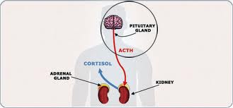
OVERPRODUCTION of cortisol is the biochemical hallmark of Cushing’s syndrome (CS) regardless of its etiology and is evidenced by increased urinary cortisol excretion, and a decrease in the circadian variation of serum cortisol (1).

Pseudo-Cushing states (PCS), as the name implies, share many of the features of Cushing’s syndrome, including cortisol overproduction. The hypercortisolism of PCS is hypothesized to be caused by increased activity of the CRH neuron, which, in turn, stimulates ACTH production and release (2). PCS are a heterogeneous group of disorders, including chronic alcoholism and alcohol withdrawal syndrome (3, 4), major depression (5), poorly controlled diabetes mellitus (6, 7), and obesity (8). Additionally, transient hypercortisolism may be associated with less obvious psychiatric conditions (e.g. anxiety) in patients with clinical features reminiscent of CS, such as obesity and hypertension, which are common in the general population. The substantial overlap in urinary free cortisol (UFC) excretion and clinical features between some patients with CS and those with PCS can make it difficult to distinguish between the two conditions (9). Thus, although persistent elevation of 24-h UFC in the presence of unequivocal signs of CS (particularly classic moon facies, prominent centripetal obesity, severe proximal muscle weakness, and violaceous striae) suggest the diagnosis of CS, patients with less obvious signs pose a diagnostic dilemma.

Several tests have been proposed to diagnose CS, including 24-h UFC measurements, the 1-mg overnight dexamethasone suppression test (DST) (10), the 2-day DST (1), and the dexamethasone-CRH (Dex-CRH) stimulation test (8). Each has drawbacks. Twenty-four-hour urinary collections are inconvenient and often incomplete. The 1-mg overnight DST is commonly used as a screening test to exclude the diagnosis of CS. This test has two caveats. First, a criterion for the level of serum cortisol suppression to exclude CS has not been developed using modern RIAs. Second, although the test has a false negative rate of only 2%, it has a significant false positive rate, especially in chronically ill (23%) or obese patients (13%) (11) and in patients with major depression (43%) or other psychiatric disorders (8–41%) (12). Even in normal individuals, the test may be consistent with CS in up to 30% (9).

Similarly, the 2-mg 2-day DST, often used as a confirmatory diagnostic test, has a diagnostic accuracy of only 71% (8). An additional problem is the variable metabolic clearance of dexamethasone (13), which is especially problematic in patients receiving medications that induce the cytochrome P450-related enzymes (e.g.phenytoin, rifampin, and phenobarbital) (14) or in patients with renal or hepatic failure. In such cases, neither DST gives reliable results. Finally, the drawbacks of 24-h urine collections apply to the DST as well.

We previously determined that the dexamethasone-CRH test has a diagnostic accuracy of 98% in the distinction of CS from PCS (8, 15). However, although accurate, this test has the drawbacks related to dexamethasone clearance, as discussed above.

Physiological cortisol secretion is characterized by circadian rhythmicity. Serum cortisol concentration reaches its zenith in the morning (0600–0800 h) and its nadir in the night during the first half of normal sleep. Krieger et al. defined the normal circadian rhythm of plasma corticosteroid levels as the pattern where all plasma glucocorticoid levels from 1600–2400 h were 75% or less of the 0800 h value (16). As previous studies have found that obese individuals retain a normal circadian cortisol rhythm (17), we hypothesized that differences in circadian plasma cortisol values would distinguish CS from PCS. To test this hypothesis, we prospectively measured serum cortisol values during the normal nadir and zenith periods in patients being evaluated for CS.

Read the entire study at http://press.endocrine.org/doi/10.1210/jcem.83.4.4733?url_ver=Z39.88-2003&rfr_id=ori%3Arid%3Acrossref.org&rfr_dat=cr_pub%3Dpubmed

Narrowing in on Pituitary Tumors

0276f-pituitary-gland


As many as 20 percent of people may have a benign cyst or tumor in their pituitary gland. The vast majority of pituitary tumors are noncancerous, but can cause headaches and profound fatigue, and can also disrupt hormone function.

Currently, surgeons rely on radiologic images and MRIs to gather information about the size and shape of the tumor, but the resolution of such imaging technologies is limited, and additional surgeries to remove more of the tumor may be needed if a patient’s symptoms persist. In a new study published in the Proceedings of the National Academy of Sciences on July 27, investigators from Brigham and Women’s Hospital (BWH) present a new technique that could help surgeons more precisely define the locations of tumors in near real-time.

The new strategy uses a visualization technique (matrix-assisted laser desorption/ionization mass spectrometry imaging – MALDI MSI) that can analyze specific hormones, including growth hormone and prolactin, in tissue. In the newly published study, the researchers find that it’s possible to use MALDI MSI to determine the composition of such hormones in a pituitary sample in less than 30 minutes. This could give surgeons critical information to help distinguish tumor from normal gland.

“Our work is driven by a clinical need: we’ve developed a test specifically tailored for the needs of our neurosurgeon colleagues,” said corresponding author Nathalie Agar, PhD, director of the Surgical Molecular Imaging Laboratory in the Department of Neurosurgery at BWH. “A surgeon may sacrifice half of the pituitary gland in an effort to get the tumor out. Without a tool to distinguish healthy tissue from tumor, it’s hard to know in real-time if the surgery was a success. With this technology, in under 30 minutes a surgeon will be able to know if a sample contains normal pituitary tissue or a pituitary tumor.”

“Patients show up with the clinical symptoms of a pituitary tumor, but the tumor itself may not be visible on an MRI,” said co-author Edward Laws, MD, director of the Pituitary and Neuroendocrine Center at BWH. “This technique, which maps out where excess concentrations of hormone levels are located, has the potential to allow us to confirm that we’ve removed the abnormal tissue.”

“Evaluating whether a piece of pituitary tissue is abnormal can be challenging on frozen section,” said co-author Sandro Santagata, MD, PhD, of BWH’s Department of Pathology. “This approach has wonderful potential for enhancing our diagnostic capabilities. It is clearly an important step toward providing intra-operative molecular characterization of pituitary tissues.”

To test the technique, the research team analyzed hormone levels in 45 pituitary tumors and six normal pituitary gland samples, finding a distinct protein signature unique to the normal or tumor sample.

Mass spectrometry, a technique for measuring chemicals present in a sample, is currently used in the operating room to help inform clinical decisions, but up until now, the focus has been on small molecules – metabolites, fatty acids and lipids – using a different type of approach. By analyzing proteins, MALDI MSI offers a way to visualize hormone levels.

Current methods used to detect hormone levels take too long to fit the time constraints of surgical intervention. Surgeons must either remove a larger amount of potentially healthy pituitary gland or perform follow up surgery if the tumor has not been fully removed.

“We’re hoping that techniques like this one will help move the field toward more precise surgery: surgery that not only removes all of the tumor but also preserves the healthy tissue as much as possible,” said Agar.

In the next phase of their work, Agar and her colleagues plan to test out the technique in BWH’s AMIGO suite and analyze the impact of the technique on clinical decision making.

Other researchers who contributed to this study include David Calligaris, Daniel R. Feldman, Isaiah Norton, Olutayo Olubiyi, Armen N. Changelian, Revaz Machaidze, Matthew L. Vestal and Ian F. Dunn.

This work was funded in part by US National Institute of Health (NIH) Director’s New Innovator Award (1DP2OD007383-01 to N.Y.R.A.), U.S. Army Medical Research/CIMIT (2010A052245), the National Center for Image Guided Therapy grant P41RR019703, NIH K08NS064168, the Pediatric Low Grade Astrocytoma Program at Dana-Farber Cancer Institute, the Brain Science Foundation and the Daniel E. Ponton fund for the Neurosciences at BWH.

Brigham and Women’s Hospital 2015 | 75 Francis Street, Boston MA 02115 | 617-732-5500

From http://www.healthcanal.com/cancers/65676-narrowing-in-on-pituitary-tumors.html