Is Cushing Syndrome More Common in the US Than We Think?

I think members of the Cushing’s Help boards have been saying this forever!  Cushing’s isn’t all that rare.  Just rarely diagnosed,


BOSTON — The prevalence of Cushing syndrome (CS) in the United States may be considerably higher than currently appreciated, new data from a single US institution suggest.

In contrast to estimates of 1 to 3 cases per million patient-years from population-based European studies, researchers at the University of Wisconsin, Milwaukee, estimated that the incidence of CS in Wisconsin is a minimum of 7.2 cases per million patient-years. What’s more, contrary to all previous studies, they found that adrenal Cushing syndrome was more common than pituitary adrenocorticotropic hormone (ACTH)– secreting tumors (Cushing disease), and that fewer than half of individuals with adrenal Cushing syndrome had classic physical features of hypercortisolism, such as weight gain, round face, excessive hair growth, and stretch marks.

“Cases are absolutely being missed…. Clinicians should realize that cortisol excess is not rare. It may not be common, but it needs to be considered in patients with any constellation of features that are seen in cortisol excess,” study investigator Ty B. Carroll, MD, Associate Professor of Medicine, Endocrinology and Molecular Medicine, and the Endocrine Fellowship Program Director at Medical College of Wisconsin in Milwaukee, told Medscape Medical News.

There are several contributing factors, he noted, “including the obesity and diabetes epidemics which make some clinical features of cortisol excess more common and less notable. Providers get used to seeing patients with some features of cortisol excess and don’t think to screen. The consequence of this is more difficult-to-control diabetes and hypertension, more advance metabolic bone disease, and likely more advanced cardiovascular disease, all resulting from extended exposure to cortisol excess,” he said.

Are Milder Cases the Ones Being Missed?

Asked to comment, session moderator Sharon L. Wardlaw, MD, professor of medicine at Columbia University College of Physicians and Surgeons, New York City, said “When we talk about Cushing [syndrome], we usually think of pituitary ACTH as more [common], followed by adrenal adenomas, and then ectopic. But they’re seeing more adrenal adenoma…we are probably diagnosing this a little more now.”

She also suggested that the Wisconsin group may have a lower threshold for diagnosing the milder cortisol elevation seen with adrenal Cushing syndrome. “If you screen for Cushing with a dexamethasone suppression test…[i]f you have autonomous secretion by the adrenal, you don’t suppress as much…. When you measure 24-hour urinary cortisol, it may be normal. So you’re in this in-between [state]…. Maybe in Wisconsin they’re diagnosing it more. Or, maybe it’s just being underdiagnosed in other places.”

She also pointed out that “you can’t diagnose it unless you think of it. I’m not so sure that with these mild cases it’s so much that it’s more common, but maybe it’s like thyroid nodules, where we didn’t know about it until everybody started getting all of these CT scans. We’re now seeing all these incidental thyroid nodules…I don’t think we’re missing florid Cushing.”

However, Wardlaw said, it’s probably worthwhile to detect even milder hypercortisolism because it could still have long-term damaging effects, including osteoporosis, muscle weakness, glucose intolerance, and frailty. “You could do something about it and normalize it if you found it. I think that would be the reason to do it.”

Is Wisconsin Representative of Cushing Everywhere?

Carroll presented the findings at the annual meeting of the Endocrine Society. He began by noting that most of the previous CS incidence studies, with estimates of 1.2-3.2 cases per million per year, come from European data published from 1994 to 2019 and collected as far back as 1955. The method of acquisition of patients and the definitions of confirmed cases varied widely in those studies, which reported CS etiologies of ACTH-secreting neoplasms (pituitary or ectopic) in 75%-85% and adrenal-dependent cortisol excess in 15%-20%.

The current study included data from clinic records between May 1, 2017, and December 31, 2022, of Wisconsin residents newly diagnosed with and treated for CS. The CS diagnosis was established with standard guideline-supported biochemical testing and appropriate imaging. Patients with exogenous and non-neoplastic hypercortisolism and those who did not receive therapy for CS were excluded.

A total of 185 patients (73% female, 27% male) were identified from 27 of the total 72 counties in Wisconsin, representing a population of 4.5 million. On the basis of the total 5.9 million population of Wisconsin, the incidence of CS in the state works out to 7.2 cases per million population per year, Carroll said.

However, data from the Wisconsin Hospital Association show that the University of Wisconsin’s Milwaukee facility treated just about half of patients in the state who are discharged from the hospital with a diagnosis of CS during 2019-2023. “So…that means that an actual or approximate incidence of 14-15 cases per million per year rather than the 7.2 cases that we produce,” he said.

Etiologies were 60% adrenal (111 patients), 36.8% pituitary (68 patients), and 3.2% ectopic (6 patients). Those proportions were similar between genders.

On biochemical testing, values for late-night salivary cortisol, dexamethasone suppression, and urinary free cortisol were highest for the ectopic group (3.189 µg/dL, 42.5 µg/dL, and 1514.2 µg/24 h, respectively) and lowest for the adrenal group (0.236 µg/dL, 6.5 µg/dL, and 64.2 µg/24 h, respectively). All differences between groups were highly statistically significant, at P < .0001, Carroll noted.

Classic physical features of CS were present in 91% of people with pituitary CS and 100% of those ectopic CS but just 44% of individuals with adrenal CS. “We found that adrenal-dependent disease was the most common form of Cushing syndrome. It frequently presented without classic physical features that may be due to the milder biochemical presentation,” he concluded.

Carroll reports consulting and investigator fees from Corcept Therapeutics. Wardlaw has no disclosures. 

Miriam E. Tucker is a freelance journalist based in the Washington DC area. She is a regular contributor to Medscape, with other work appearing in The Washington Post, NPR’s Shots blog, and Diatribe. She is on X (formerly Twitter) @MiriamETucker.

1

Credit

Lead image: Designer491/Dreamstime

Medscape Medical News © 2024 WebMD, LLCSend comments and news tips to news@medscape.net.

Cite this: Is Cushing Syndrome More Common in the US Than We Think? – Medscape – June 07, 2024.

Repeat Endoscopic Endonasal Transsphenoidal Surgery for Residual or Recurrent Cushing’s Disease: Safety, Feasibility, And Success

Abstract

Purpose

The success and outcomes of repeat endoscopic transsphenoidal surgery (ETS) for residual or recurrent Cushing’s disease (CD) are underreported in the literature. This study aims to address this gap by assessing the safety, feasibility, and efficacy of repeat ETS in these patients.

Methods

A retrospective analysis was conducted on 56 patients who underwent a total of 65 repeat ETS performed by a single neurosurgeon between January 2006 and December 2020. Data including demographic, clinical, laboratory, radiological, and operative details were collected from electronic medical records. Logistic regression was utilized to identify potential predictors associated with sustained remission.

Results

Among the cases, 40 (61.5%) had previously undergone microscopic surgery, while 25 (38.5%) had prior endoscopic procedures. Remission was achieved in 47 (83.9%) patients after the first repeat ETS, with an additional 9 (16.1%) achieving remission after the second repeat procedure. During an average follow-up period of 97.25 months, the recurrence rate post repeat surgery was 6.38%. Sustained remission was achieved in 48 patients (85.7%), with 44 after the first repeat ETS and 4 following the second repeat ETS. Complications included transient diabetes insipidus (DI) in 5 (7.6%) patients, permanent (DI) in 2 (3%) patients, and one case (1.5%) of panhypopituitarism. Three patients (4.6%) experienced rhinorrhea necessitating reoperation. A serum cortisol level > 5 µg/dL on postoperative day 1 was associated with a reduced likelihood of sustained remission.

Conclusion

Repeat ETS is a safe and effective treatment option for residual or recurrent CD with satisfactory remission rates and low rates of complications.

Introduction

Cushing’s disease (CD) arises from an adrenocorticotropic hormone (ACTH)-secreting pituitary adenoma, leading to excessive endogenous glucocorticoid production [1]. The reported incidence of CD varies from 0.7 to 2.4 cases per million individuals annually [2,3,4,5,6]. Hypercortisolism impacts every bodily system and is linked to elevated morbidity and mortality risks [78]. Therefore, prompt CD diagnosis and management are crucial to enhance patient outcomes.

Transsphenoidal surgery remains the primary treatment for CD, and have been associated with satisfactory remission rates ranging from 65 to 94% [2359,10,11]. Two surgical techniques are utilized: microscopic and endoscopic approaches. While both methods are effective, studies indicate that endoscopic transsphenoidal surgery (ETS) offers higher rates of complete tumor removal and lower complication rates [12,13,14]. ETS holds advantages over microscopic transsphenoidal surgery (MTS) due to superior tumor visualization, especially for laterally invasive tumors and macroadenomas [15]. Since its introduction in 1997, ETS has gained popularity and is now the standard surgical approach for managing CD [16].

Remission rates post-ETS for CD treatment range from 77 to 90% [17,18,19,20,21,22]. Despite ETS’s technical benefits and favorable outcomes, recurrence rates for Cushing’s disease after successful ETS range between 5.6% and 22.8% [17182223]. Reoperating for residual or recurrent CD presents challenges due to altered surgical landmarks and scar tissue formation from previous surgeries, potentially elevating morbidity, and mortality risks [2425]. Limited literature exists on the success and outcomes of repeat endoscopic transsphenoidal surgery for residual or recurrent CD. This study aims to address this gap by assessing the safety, feasibility, and efficacy of repeat ETS in patients with residual or recurrent Cushing’s disease.

Methods

Study design

This is a retrospective cohort study of repeat endoscopic transsphenoidal surgery for residual or recurrent Cushing’s disease. All patients underwent endoscopic endonasal transsphenoidal surgery by the senior author between 2006 and 2020. The study protocol was approved by the local ethics committee for clinical studies.

Patient selection

The study participants were selected based on specific inclusion and exclusion criteria. Inclusion criteria were as follows: (i) a confirmed diagnosis of Cushing’s disease, (ii) prior transsphenoidal surgery, and (iii) confirmation of residual or recurrent CD through clinical, laboratory, and/or imaging assessments. Exclusion criteria included: (i) prior craniotomy without transsphenoidal surgery, (ii) previous radiotherapy before reoperation, (iii) inaccessible clinical, laboratory, or radiological data, and (iv) follow-up duration of less than 6 months.

Diagnostic criteria

Each patient underwent thorough screening for active Cushing’s disease. An increased 24-hour urine cortisol level > 45 µg/day or a serum fasting cortisol level exceeding 1.8 µg/dl following a low-dose (2 mg) dexamethasone suppression test was deemed abnormal. Subsequently, a high-dose (8 mg) dexamethasone test was administered, and a reduction of 50% or more from the baseline value was indicative of active Cushing’s disease. Due to the technical limitations of the institution that the research has been done, late-night salivary cortisol tests were not performed. Early remission was characterized by a fasting serum cortisol level below 5 µg/dl on the 1st and 7th postoperative days. Patients displaying a serum cortisol level below 1.8 µg/dl after the low-dose dexamethasone suppression test or those requiring continued corticosteroid replacement post-surgery were considered to maintain remission. The presence of a residual adenoma on postoperative magnetic resonance imaging (MRI) confirmed residual disease.

Routine follow-up protocol

Patients were evaluated for Cushing’s disease symptoms before surgery and monitored at 6 months after surgery, as well as during yearly check-ups for any changes in their condition. Fasting serum ACTH and cortisol levels were measured in the morning before surgery, on the 1st and 7th days after surgery, at the 1st, 3rd, and 6th months, and during yearly follow-up appointments. Prior to surgery, all patients underwent contrast-enhanced pituitary MRI and paranasal sinus CT scans. Follow-up pituitary MRI scans were conducted on the 1st day, at 3 and 12 months after surgery, and then annually thereafter.

Data collection

Data from electronic medical records were gathered, encompassing demographic, clinical, laboratory, radiological, and operative details. Laboratory assessments comprised an anterior pituitary hormone panel (Follicle-stimulating hormone [FSH], Luteinizing hormone [LH], Thyroid-stimulating hormone [TSH], Prolactin [PRL], Growth hormone [GH]), serum electrolytes, preoperative and postoperative serum ACTH, and cortisol levels. Patient records, along with CT and MRI scans, were scrutinized to document preoperative tumor characteristics such as size, multifocality, relationship with the cavernous sinus, Hardy-Wilson classification of sellar destruction, and suprasellar extension. Tumors larger than 10 mm were classified as macroadenomas. The operative database was examined to collect data on previous surgeries, including the number and dates of prior procedures, as well as the surgical techniques utilized. Outcome measures comprised remission rates and surgical complications.

Statistical analysis

Statistical analysis was conducted utilizing SPSS 23.0 software (IBM, New York). Two-group comparisons were performed using Chi-square and Fisher’s exact tests for categorical variables and Student’s t-test for continuous variables. Categorical variables were presented as numbers and percentages, while continuous variables were presented as means ± SD or median [IQR]. Logistic regression was performed to investigate potential predictors linked to sustained remission. A p-value of < 0.05 was deemed statistically significant.

Results

Baseline characteristics

Supplementary File 1 displays the demographic characteristics of the patient cohort.

A retrospective analysis was conducted on 190 patients who underwent a total of 212 operations for CD at our department between January 2006 and December 2020. Among them, 56 patients, comprising 65 repeat endonasal transsphenoidal surgeries due to either recurrence (n = 18, 27.7%) or residual disease (n = 47, 72.3%), were identified. The majority of patients were female (n = 48, 85.7%), with a mean age of 37.6 ± 12.4 years. Of the 56 patients, 43 (76.8%) were referred from another institution. Most patients (n = 42, 75%) had undergone only one prior surgery, while 12 patients (21.4%) had a history of two previous surgeries, and 2 patients (3.6%) had undergone three prior surgeries before referral to our center. The average follow-up duration since the first repeat ETS was 97.2 ± 36.8 months. The mean time to recurrence was 80.2 ± 61.1 months (median 75 months, range 23.2 to 103.5 months).

Hormonal data

Table 1 depicts the preoperative and postoperative serum ACTH and cortisol levels. The average preoperative serum cortisol levels for the entire patient cohort stood at 18.7 ± 11.1 µg/dL (median 17, range 12-24.6). The median preoperative 24-hour urine free cortisol level was 237 µg /day [188.5–425.5]. On the initial postoperative day, the mean serum cortisol levels for all patients were 13.4 ± 13.8 µg/dL (median 6.4, range 1.7–21). In 46.2% of cases (n = 30), cortisol levels on the first postoperative day were below 5 µg/dL (< 2 µg/dL in 33.8%). A comparison of the mean preoperative and postoperative serum ACTH and cortisol levels between the groups with residual disease and recurrence is detailed in Table 1.

Table 1 Cohort overview and comparison of recurrence and residual disease groups

Radiological findings

In the entire case cohort, there were 41 microadenomas (63.1%) and 24 macroadenomas (36.9%). Fifteen cases (23.1%) exhibited bifocal adenomas. Adenoma extension into the cavernous sinuses, indicated by cavernous sinus wall displacement, was present in 21 cases (32.3%), while invasion into the cavernous sinuses was observed in 10 cases (15.4%). Based on the Hardy-Wilson Classification, there were 38 Grade I adenomas (58.5%), 16 Grade II adenomas (24.6%), 6 Grade III adenomas (9.2%), and 5 Grade IV adenomas (7.7%). Thirty patients (46.2%) presented with Stage A adenoma, 7 (10.8%) with Stage B adenoma, 2 (3.1%) with Stage C adenoma, 1 (1.5%) with Stage D adenoma, and 25 (38.5%) with Stage E adenoma. As indicated in Table 1, there were no statistically significant differences between patients with residual disease and recurrence concerning radiological findings.

Surgical characteristics

A single surgeon conducted all 65 reoperations. Among these, 47 patients (72.3%) underwent repeat ETS due to residual disease, while 18 (27.7%) did so due to recurrence. The previous surgical technique was microscopic in 40 cases (61.5%) and endoscopic in 25 cases (38.5%). Microscopic transsphenoidal surgeries were exclusively performed at other institutions. There was a notable disparity between patients with residual disease and recurrence regarding the technique of the previous surgery. Residual disease occurrence following endoscopic transsphenoidal surgery was less frequent (n = 11/25, 44%) compared to after microscopic transsphenoidal surgery (n = 36/40, 90%; p < 0.001) (Table 1). Immunohistochemical staining of the specimens indicated that 55 cases (85%) exhibited ACTH-positive adenoma. Nevertheless, all patients with a negative pathology at the repeat surgery had a confirmed ACTH-adenoma at the first surgery. Of the 10 patients (15%) with a negative ACTH-positive adenoma pathology, two patients underwent inferior petrosal sinus sampling (IPSS) previously and were confirmed to have CD. Remaining patients did not undergo an additional inferior petrosal sinus sampling (IPSS) because all functional test results indicated a central source and MRI confirmed pituitary microadenoma in all cases. Notably, there are studies reporting that IPSS may not be required in patients with a sellar mass and a biochemical testing suggestive of CD [2627]. Additionally, we also explored both sides of the pituitary and confirmed the adenoma intraoperatively. Therefore, negative pathology in the repeat surgery is most likely due to sampling error.

Outcomes

As depicted in Fig. 1, among the 56 patients, 47 (83.9%) experienced initial remission following the first repeat ETS, while 9 (16.1%) still had residual adenoma. Within the group achieving initial remission, 44 patients (93.6%) maintained remission without the need for further surgeries, while 3 (6.4%) experienced recurrence during follow-up and required a second repeat ETS.

Fig. 1
figure 1

Outcomes of repeat endoscopic transsphenoidal surgery for residual or recurrent Cushing’s disease

Among the 9 patients with residual disease after the first repeat ETS, 1 (11.1%) opted to defer further treatment, 1 (11.1%) received radiotherapy, 1 (11.1%) chose adrenalectomy, and 6 (66.7%) underwent a second repeat ETS. Of the 9 patients who underwent a second repeat ETS due to residual disease or recurrence, 4 (44.4%) sustained remission, 5 (55.6%) still had residual disease, but 3 of them deferred further treatment, 1 received radiotherapy, while 1 achieved remission after adrenalectomy. Overall, 78.5% (n = 51) of the entire case cohort achieved remission following repeat ETS. Representative cases are presented in Fig. 2.

Fig. 2
figure 2

Case 1: Preoperative and postoperative magnetic resonance imaging (MRI) scans of a 49-year-old female who underwent repeat endoscopic transsphenoidal surgery (ETS) due to recurrent Cushing’s disease and achieved remission. The patient underwent initial surgery 14 years ago at an outside institution. Preoperative T2 (A), and T1 contrast-enhanced (B) MRI scans demonstrate a right-sided pituitary adenoma. Postoperative T2 (C), and T1 contrast-enhanced (D) MRI scans demonstrate total resection of the adenoma. Case 2: Preoperative and postoperative magnetic resonance imaging (MRI) scans of a 53-year-old female who underwent repeat endoscopic transsphenoidal surgery (ETS) due to recurrent Cushing’s disease and achieved remission. The patient underwent initial surgery 3 years ago at an outside institution. Preoperative T2 (E), and T1 contrast-enhanced (F) MRI scans demonstrate a left-sided pituitary adenoma, in close relation to ICA. Postoperative T2 (G), and T1 contrast-enhanced (H) MRI scans demonstrate total resection of the adenoma

Transient diabetes insipidus (DI) developed in 5 patients (7.6%), while 2 (3%) experienced permanent DI following repeat ETS. Intraoperative cerebrospinal fluid (CSF) leak occurred in 20 operations (30.7%). Three patients (4.6%) developed rhinorrhea and required reoperation. Five patients (7.6%) developed prolactin deficiency, 3 patients (4.6%) had GH deficiency, and another 3 patients (4.6%) had TSH deficiency requiring thyroxine replacement. Four patients (6.2%) had combined deficiencies in TSH, FSH, LH and prolactin, while one patient (1.5%) developed panhypopituitarism following the second repeat ETS.

Factors predisposing to unsuccessful repeat endoscopic transsphenoidal surgery

Among the 42 patients who underwent repeat ETS for residual disease, 9 (21.4%) still had residual disease after the first repeat ETS. We conducted a multivariable logistic regression analysis to explore potential risk factors for unsuccessful repeat ETS. However, the analysis did not reveal any significant association between the success of repeat ETS and factors such as extension or invasion into cavernous sinuses, sellar or parasellar extension, or tumor size (Supplementary File 1).

Potential predictors of sustained remission

We conducted a multivariable logistic regression analysis to investigate possible predictors of sustained remission. The variables included in the analysis are detailed in Table 5. The results indicated that having a serum cortisol level exceeding 5 µg/dL on postoperative day 1 was linked to a decreased likelihood of achieving sustained remission (Odds ratio [OR] 0.09, 95% confidence interval [CI] 0.01–0.52, p = 0.006) (Table 2).

Table 2 Logistic regression analysis of potential predictors for continued remission

Discussion

Transsphenoidal surgery remains the established standard for treating Cushing’s disease, with demonstrated remission rates ranging from 65 to 94%, contingent upon the surgeon’s expertise and remission criteria [2359,10,11]. The advent of endoscopic techniques has notably augmented this approach, offering wider visibility, reduced nasal trauma, and shorter hospital stays [16252829]. While the effectiveness of ETS in managing CD is well-documented, literature on its efficacy in treating residual or recurrent cases is limited. Our study addresses this gap by assessing the safety, feasibility, and outcomes of repeat ETS for patients with persistent or recurrent Cushing’s disease.

In our study, 56 patients underwent 65 repeat ETS procedures for residual or recurrent Cushing’s disease. Mean follow-up duration was 97.2 ± 36.8 months, which is one of the longest follow-up durations that has been reported following repeat endoscopic transsphenoidal surgery [530,31,32]. Of these patients, 40 (61.5%) had previously undergone microscopic surgery, while 25 (38.5%) had undergone prior endoscopic procedures. Importantly, a notable difference emerged between patients with residual disease and those experiencing recurrence regarding the prior surgical approach, with residual disease being less frequent after endoscopic surgery compared to microscopic surgery (p < 0.001). This variance was expected, as numerous studies have indicated that ETS yields a higher rate of complete resection compared to MTS [12,13,14].

After the first repeat ETS, 47 patients (83.9%) achieved remission, and 78.5% (n = 44) of them maintained remission at a mean follow-up of 97.2 months without requiring additional surgery. Limited data exists regarding the remission rates of CD following repeat transsphenoidal surgery, with reported rates ranging from 28.9 to 73% [333435]. Burke et al. reported an immediate remission rate of 86.7% and a continued remission rate of 73.3% at follow-up after repeat ETS [36]. Among our patients who achieved remission after successful repeat ETS, 3 individuals (6.38%, n = 3/47) experienced recurrence after the first repeat ETS, with a mean time to recurrence of 45.6 months. The rates of CD recurrence following reoperation vary, with documented rates ranging between 22% and 63.2% [3738]. In our study, 9 patients required a second repeat ETS due to residual disease or recurrence. Of these, 4 (44.4%) achieved continued remission following the second repeat ETS, while 5 (55.6%) had residual disease; however, 4 of them deferred further treatment, and 1 achieved remission after adrenalectomy. In total, 47 patients (83.9%) in the entire patient cohort achieved remission following endoscopic transsphenoidal surgery and did not require further intervention.

Within our case cohort, among the 42 patients who underwent repeat ETS for residual disease, 9 individuals (21.4%) continued to exhibit residual disease following the first repeat ETS. We did not establish a significant association between the success of repeat ETS and factors such as extension or invasion into cavernous sinuses, sellar or parasellar extension, or tumor size.

The degree of hypocortisolism following transsphenoidal surgery is considered a potential indicator of remission in the postoperative period [3]. Numerous studies have indicated that patients with subnormal postoperative cortisol levels tend to experience a lower recurrence rate compared to those with normal or supranormal levels, although consensus on the precise cutoff level remains elusive [30,31,3239]. In a retrospective study involving 52 patients with CD, researchers reported a 100% positive predictive value of a postoperative nadir cortisol level < 2 µg/dL for achieving remission [5]. Additionally, Esposito et al. observed that a morning serum cortisol level ≤ 5 µg/dL on postoperative day 1 or 2 appears to serve as a reliable predictor of remission [11]. In our investigation, logistic regression analysis revealed that patients with a serum cortisol level > 5 µg/dL on postoperative day 1 were less inclined to achieve continued remission compared to those with a serum cortisol level < 5 µg/dL on postoperative day 1.

Repeat transsphenoidal surgery presents unique challenges due to distorted surgical landmarks and the presence of scar tissue from prior procedures, often resulting in lower cure rates and increased morbidity risk [242528]. Non-surgical options such as radiotherapy and radiosurgery have been considered as an effective treatment option for recurrent or residual CD due to low rates of morbidity and acceptable remission rates [2840]. However, our findings suggest that the outcomes and complication rates associated with repeat ETS are comparable to primary ETS for CD and superior to other non-surgical options for residual or recurrent CD. Within our patient cohort, 5 (7.6%) individuals experienced transient diabetes insipidus (DI), while 2 (3%) developed permanent DI. Additionally, one patient (1.5%) experienced panhypopituitarism following the second repeat ETS. Similarly, various studies have reported DI rates ranging from 2 to 13% and panhypopituitarism rates between 2% and 9.7% [252841,42,43]. In our series, 3 (5.3%) patients developed rhinorrhea and required reoperation, consistent with reported rates of postoperative CSF leak ranging from 1 to 5% following repeat endoscopic transsphenoidal surgery for residual or recurrent pituitary tumors [252844]. While radiotherapy and radiosurgery are options for patients who have failed transsphenoidal surgery or experienced recurrence, the literature suggests remission rates ranging from 46 to 84%, with several studies indicating high recurrence rates (25-50%) following radiotherapy [4045,46,47]. In our study, among 56 patients, 47 (83.9%) achieved remission following the first repeat ETS, while 4 (17.8%) achieved remission after the second repeat ETS. Over a mean follow-up duration of 97.25 months, our recurrence rate following repeat ETS was 27.7%, with a mean time to recurrence of 45.62 months.

At our institution, we adhere to a specific algorithm (Fig. 3) for managing Cushing’s disease patients and implement a meticulous protocol for individuals undergoing repeat ETS for residual or recurrent CD. A thorough clinical and radiological assessment is conducted for all patients before surgery. Detailed radiological evaluation is particularly essential to identify any distortions in surgical landmarks from prior procedures, such as the course of sphenoidal septa and the location of the sellar floor opening, as well as other potential aberrations like internal carotid artery and optic nerve dehiscence. Imaging techniques should encompass dynamic pituitary MRI with and without contrast and paranasal CT scans. Our objective is to achieve extensive exposure during surgery, which is especially critical for managing bifocal adenomas or adenomas with cavernous sinus invasion or extension. The expanded visual field also facilitates the visualization of concealed parts of the adenoma, allowing the surgeon to achieve complete resection, which may be challenging or even impossible with limited exposure. We employ a multilayer closure technique to prevent CSF leaks, and if necessary, utilize a vascularized pedicled nasoseptal flap (Hadad-Bassagasteguy flap).

Fig. 3
figure 3

Specific algorithm for the management of Cushing’s disease patients

In summary, our findings suggest that in the hands of experienced surgeons, repeat ETS represents a safe and effective treatment option for managing residual or recurrent Cushing’s disease.

Strengths and limitations

Our study represents one of the largest case series in the literature examining the safety, feasibility, and efficacy of repeat ETS for managing recurrent or residual CD. Our findings underscore the safety and efficacy of repeat ETS in experienced centers, showcasing satisfactory remission rates and minimal complications. However, it is important to acknowledge the retrospective nature of our study, which inherently introduces potential biases such as selection bias. Lastly, our study exclusively focuses on patients undergoing surgical intervention for recurrent or residual CD, limiting our ability to compare the effectiveness of surgical treatment with alternative modalities like radiotherapy or radiosurgery.

Conclusion

Our study underscores the efficacy and safety of repeat endoscopic transsphenoidal surgery in managing residual or recurrent Cushing’s disease. Remarkably, 82.1% of patients achieved remission after their first reoperation, aligning closely with reported remission rates following primary endoscopic transsphenoidal surgery. Furthermore, the complication rates observed in our cohort were consistent with documented rates for both primary and repeat transsphenoidal surgeries. Notably, patients with serum cortisol levels < 5 µg/dL are more likely to maintain remission. Overall, our findings emphasize that in the hands of experienced surgeons, repeat endoscopic transsphenoidal surgery emerges as a reliable and safe treatment modality for residual or recurrent Cushing’s disease, offering satisfactory remission rates and minimal complications.

Data availability

No datasets were generated or analysed during the current study.

Abbreviations

ACTH:
adrenocorticotropic hormone
CD:
Cushing’s disease
CT:
computed tomography
DI:
diabetes insipidus
ETS:
endoscopic endonasal transsphenoidal surgery
MRI:
magnetic resonance imaging
MTS:
microscopic transsphenoidal surgery

References

  1. Cushing H (1994) The basophil adenomas of the pituitary body and their clinical manifestations (pituitary basophilism). 1932. Obes Res 2(5):486–508

    Article CAS PubMed Google Scholar

  2. Aranda G, Enseñat J, Mora M, Puig-Domingo M, Martínez de Osaba MJ, Casals G et al (2015) Long-term remission and recurrence rate in a cohort of Cushing’s disease: the need for long-term follow-up. Pituitary 18(1):142–149

    Article CAS PubMed Google Scholar

  3. Pendharkar AV, Sussman ES, Ho AL, Hayden Gephart MG, Katznelson L (2015) Cushing’s disease: predicting long-term remission after surgical treatment. Neurosurg Focus 38(2):E13

    Article PubMed Google Scholar

  4. Etxabe J, Vazquez JA (1994) Morbidity and mortality in Cushing’s disease: an epidemiological approach. Clin Endocrinol (Oxf) 40(4):479–484

    Article CAS PubMed Google Scholar

  5. Hameed N, Yedinak CG, Brzana J, Gultekin SH, Coppa ND, Dogan A et al (2013) Remission rate after transsphenoidal surgery in patients with pathologically confirmed Cushing’s disease, the role of cortisol, ACTH assessment and immediate reoperation: a large single center experience. Pituitary 16(4):452–458

    Article CAS PubMed Google Scholar

  6. Acebes JJ, Martino J, Masuet C, Montanya E, Soler J (2007) Early post-operative ACTH and cortisol as predictors of remission in Cushing’s disease. Acta Neurochir (Wien). ;149(5):471-7; discussion 7–9

  7. Fernandez-Rodriguez E, Stewart PM, Cooper MS (2009) The pituitary-adrenal axis and body composition. Pituitary 12(2):105–115

    Article CAS PubMed Google Scholar

  8. van Haalen FM, Broersen LH, Jorgensen JO, Pereira AM, Dekkers OM (2015) Management of endocrine disease: mortality remains increased in Cushing’s disease despite biochemical remission: a systematic review and meta-analysis. Eur J Endocrinol 172(4):R143–R149

    Article PubMed Google Scholar

  9. Locatelli M, Vance ML, Laws ER (2005) Clinical review: the strategy of immediate reoperation for transsphenoidal surgery for Cushing’s disease. J Clin Endocrinol Metab 90(9):5478–5482

    Article CAS PubMed Google Scholar

  10. Blevins LS Jr., Christy JH, Khajavi M, Tindall GT (1998) Outcomes of therapy for Cushing’s disease due to adrenocorticotropin-secreting pituitary macroadenomas. J Clin Endocrinol Metab 83(1):63–67

    CAS PubMed Google Scholar

  11. Esposito F, Dusick JR, Cohan P, Moftakhar P, McArthur D, Wang C et al (2006) Clinical review: early morning cortisol levels as a predictor of remission after transsphenoidal surgery for Cushing’s disease. J Clin Endocrinol Metab 91(1):7–13

    Article CAS PubMed Google Scholar

  12. Guo S, Wang Z, Kang X, Xin W, Li X (2021) A Meta-analysis of endoscopic vs. microscopic transsphenoidal surgery for non-functioning and functioning pituitary adenomas: comparisons of efficacy and safety. Front Neurol. ;12

  13. Gao Y, Zhong C, Wang Y, Xu S, Guo Y, Dai C et al (2014) Endoscopic versus microscopic transsphenoidal pituitary adenoma surgery: a meta-analysis. World J Surg Oncol 12:94

    Article PubMed PubMed Central Google Scholar

  14. Yu SY, Du Q, Yao SY, Zhang KN, Wang J, Zhu Z, Jiang XB (2018) Outcomes of endoscopic and microscopic transsphenoidal surgery on non-functioning pituitary adenomas: a systematic review and meta-analysis. J Cell Mol Med 22(3):2023–2027

    Article PubMed PubMed Central Google Scholar

  15. Broersen LHA, van Haalen FM, Biermasz NR, Lobatto DJ, Verstegen MJT, van Furth WR et al (2019) Microscopic versus endoscopic transsphenoidal surgery in the Leiden cohort treated for Cushing’s disease: surgical outcome, mortality, and complications. Orphanet J Rare Dis 14(1):64

    Article PubMed PubMed Central Google Scholar

  16. Jho HD, Carrau RL (1997) Endoscopic endonasal transsphenoidal surgery: experience with 50 patients. J Neurosurg 87(1):44–51

    Article CAS PubMed Google Scholar

  17. Berker M, Işikay I, Berker D, Bayraktar M, Gürlek A (2014) Early promising results for the endoscopic surgical treatment of Cushing’s disease. Neurosurg Rev 37:105-114

  18. Netea-Maier RT, van Lindert EJ, den Heijer M, van der Eerden A, Pieters GF, Sweep CG et al (2006) Transsphenoidal pituitary surgery via the endoscopic technique: results in 35 consecutive patients with Cushing’s disease. Eur J Endocrinol 154(5):675–684

    Article CAS PubMed Google Scholar

  19. Dehdashti AR, Gentili F (2007) Current state of the art in the diagnosis and surgical treatment of cushing disease: early experience with a purely endoscopic endonasal technique. Neurosurg Focus 23(3):E9

    Article PubMed Google Scholar

  20. Starke RM, Reames DL, Chen CJ, Laws ER, Jane JA (2013) Jr. Endoscopic transsphenoidal surgery for cushing disease: techniques, outcomes, and predictors of remission. Neurosurgery 72(2):240–247 discussion 7

    Article PubMed Google Scholar

  21. Sarkar S, Rajaratnam S, Chacko G, Mani S, Hesargatta AS, Chacko AG (2016) Pure endoscopic transsphenoidal surgery for functional pituitary adenomas: outcomes with Cushing’s disease. Acta Neurochir (Wien) 158(1):77–86 discussion

    Article PubMed Google Scholar

  22. Cebula H, Baussart B, Villa C, Assié G, Boulin A, Foubert L et al (2017) Efficacy of endoscopic endonasal transsphenoidal surgery for Cushing’s disease in 230 patients with positive and negative MRI. Acta Neurochir (Wien) 159(7):1227–1236

    Article PubMed Google Scholar

  23. Wagenmakers MA, Boogaarts HD, Roerink SH, Timmers HJ, Stikkelbroeck NM, Smit JW et al (2013) Endoscopic transsphenoidal pituitary surgery: a good and safe primary treatment option for Cushing’s disease, even in case of macroadenomas or invasive adenomas. Eur J Endocrinol 169(3):329–337

    Article CAS PubMed Google Scholar

  24. Esquenazi Y, Essayed WI, Singh H, Mauer E, Ahmed M, Christos PJ, Schwartz TH (2017) Endoscopic endonasal Versus Microscopic Transsphenoidal surgery for recurrent and/or residual pituitary adenomas. World Neurosurg 101:186–195

    Article PubMed PubMed Central Google Scholar

  25. Cavallo LM, Solari D, Tasiou A, Esposito F, de Angelis M, D’Enza AI, Cappabianca P (2013) Endoscopic endonasal transsphenoidal removal of recurrent and regrowing pituitary adenomas: experience on a 59-patient series. World Neurosurg 80(3–4):342–350

    Article PubMed Google Scholar

  26. Perlman JE, Johnston PC, Hui F et al (2021) Pitfalls in performing and interpreting Inferior Petrosal Sinus Sampling: personal experience and literature review. J Clin Endocrinol Metab 106(5):e1953–e1967. https://doi.org/10.1210/clinem/dgab012

    Article PubMed PubMed Central Google Scholar

  27. Yogi-Morren D, Habra MA, Faiman C et al (2015) Pituitary MRI findings in patients with pituitary and ectopic ACTH-dependent cushing syndrome: does a 6-mm pituitary tumor size cut-off value exclude ectopic ACTH syndrome? Endocr Pract 21(10):1098–1103

    Article Google Scholar

  28. Negm HM, Al-Mahfoudh R, Pai M, Singh H, Cohen S, Dhandapani S et al (2017) Reoperative endoscopic endonasal surgery for residual or recurrent pituitary adenomas. J Neurosurg 127(2):397–408

    Article PubMed Google Scholar

  29. Cooke RS, Jones RA (1994) Experience with the direct transnasal transsphenoidal approach to the pituitary fossa. Br J Neurosurg 8(2):193–196

    Article CAS PubMed Google Scholar

  30. Chee GH, Mathias DB, James RA, Kendall-Taylor P (2001) Transsphenoidal pituitary surgery in Cushing’s disease: can we predict outcome? Clin Endocrinol (Oxf) 54(5):617–626

    Article CAS PubMed Google Scholar

  31. Atkinson AB, Kennedy A, Wiggam MI, McCance DR, Sheridan B (2005) Long-term remission rates after pituitary surgery for Cushing’s disease: the need for long-term surveillance. Clin Endocrinol (Oxf) 63(5):549–559

    Article PubMed Google Scholar

  32. Yap LB, Turner HE, Adams CB, Wass JA (2002) Undetectable postoperative cortisol does not always predict long-term remission in Cushing’s disease: a single centre audit. Clin Endocrinol (Oxf) 56(1):25–31

    Article CAS PubMed Google Scholar

  33. Liu X, Dai C, Bao X, Deng K, Yao Y, Sun B et al (2021) Treatment and outcomes of recurrent/persistent Cushing’s disease: a single-center experience. Ann Palliat Med 10(3):2494–2504

    Article PubMed Google Scholar

  34. Valderrábano P, Aller J, García-Valdecasas L, García-Uría J, Martín L, Palacios N, Estrada J (2014) Results of repeated transsphenoidal surgery in Cushing’s disease. Long-term follow-up. Endocrinol Nutr 61(4):176–183

    Article PubMed Google Scholar

  35. Wang B, Zheng S, Ren J, Zhong Z, Jiang H, Sun Q et al (2022) Reoperation for Recurrent and Persistent Cushing’s Disease without Visible MRI Findings. J Clin Med 11(22): 6848

  36. Burke WT, Penn DL, Repetti CS, Iuliano S, Laws ERJ (2019) Outcomes after repeat transsphenoidal surgery for recurrent Cushing Disease: updated. Neurosurgery 85(6):E1030–E6

    Article PubMed Google Scholar

  37. Bakiri F, Tatai S, Aouali R, Semrouni M, Derome P, Chitour F, Benmiloud M (1996) Treatment of Cushing’s disease by transsphenoidal, pituitary microsurgery: prognosis factors and long-term follow-up. J Endocrinol Invest 19(9):572–580

    Article CAS PubMed Google Scholar

  38. Dickerman RD, Oldfield EH (2002) Basis of persistent and recurrent cushing disease: an analysis of findings at repeated pituitary surgery. J Neurosurg 97(6):1343–1349

    Article PubMed Google Scholar

  39. Patil CG, Prevedello DM, Lad SP, Vance ML, Thorner MO, Katznelson L, Laws ER (2008) Jr. Late recurrences of Cushing’s disease after initial successful transsphenoidal surgery. J Clin Endocrinol Metab 93(2):358–362

    Article CAS PubMed Google Scholar

  40. Castinetti F, Brue T, Ragnarsson O (2019) Radiotherapy as a tool for the treatment of Cushing’s disease. Eur J Endocrinol 180(5):D9–d18

    Article PubMed Google Scholar

  41. Tajudeen BA, Mundi J, Suh JD, Bergsneider M, Wang MB (2015) Endoscopic endonasal surgery for recurrent pituitary tumors: technical challenges to the surgical approach. J Neurol Surg B Skull Base 76(1):50–56

    Article PubMed Google Scholar

  42. Hwang JM, Kim YH, Kim JW, Kim DG, Jung HW, Chung YS (2013) Feasibility of endoscopic endonasal approach for recurrent pituitary adenomas after microscopic trans-sphenoidal approach. J Korean Neurosurg Soc 54(4):317–322

    Article PubMed PubMed Central Google Scholar

  43. Berker M, Hazer DB, Yücel T, Gürlek A, Cila A, Aldur M, Onerci M (2012) Complications of endoscopic surgery of the pituitary adenomas: analysis of 570 patients and review of the literature. Pituitary 15(3):288–300

    Article PubMed Google Scholar

  44. Rudnik A, Zawadzki T, Wojtacha M, Bazowski P, Zubgałuszka-Ignasiak B, Duda I (2005) [Endoscopic transsphenoidal treatment of pituitary adenomas]. Neurol Neurochir Pol 39(1):17–23 discussion 4–5

    PubMed Google Scholar

  45. Minniti G, Osti M, Jaffrain-Rea ML, Esposito V, Cantore G, Maurizi Enrici R (2007) Long-term follow-up results of postoperative radiation therapy for Cushing’s disease. J Neurooncol 84(1):79–84

    Article PubMed Google Scholar

  46. Estrada J, Boronat M, Mielgo M, Magallón R, Millan I, Díez S et al (1997) The long-term outcome of pituitary irradiation after unsuccessful transsphenoidal surgery in Cushing’s disease. N Engl J Med 336(3):172–177

    Article CAS PubMed Google Scholar

  47. Littley MD, Shalet SM, Beardwell CG, Ahmed SR, Sutton ML (1990) Long-term follow-up of low-dose external pituitary irradiation for Cushing’s disease. Clin Endocrinol (Oxf) 33(4):445–455

    Article CAS PubMed Google Scholar

Download references

Acknowledgements

Not applicable.

Funding

The authors declare that no funds, grants, or other support were received during the preparation of this manuscript.

Open access funding provided by the Scientific and Technological Research Council of Türkiye (TÜBİTAK).

Author information

Authors and Affiliations

  1. Department of Neurosurgery, Faculty of Medicine, Hacettepe University, Ankara, Turkey

    Sahin Hanalioglu, Muhammet Enes Gurses, Neslihan Nisa Gecici, Baylar Baylarov & Ilkay Isikay

  2. Department of Neurosurgery, Miller School of Medicine, University of Miami, Miami, FL, USA

    Muhammet Enes Gurses

  3. Department of Endocrinology and Metabolism, Faculty of Medicine, Hacettepe University, Ankara, Turkey

    Alper Gürlek

  4. Department of Neurosurgery, Hacettepe University School of Medicine, Sihhiye, Ankara, 06230, Turkey

    Mustafa Berker

Contributions

Conceptualization: S.H, M.B; Methodology: S.H, M.E.G, N.N.G; Formal analysis and investigation: M.E.G, N.N.G, B.B; Writing – original draft preparation: N.N.G; Writing – review and editing: S.H, M.E.G, B.B, I.I, A.G, M.B; Supervision: S.H, I.I, A.G, M.B.

Corresponding author

Correspondence to Mustafa Berker.

Ethics declarations

Ethical approval

This study is approved by the ethics committee of the hospital where the research was conducted and informed consent is obtained from patients.

Competing interests

The authors declare no competing interests.

Additional information

Publisher’s Note

Springer Nature remains neutral with regard to jurisdictional claims in published maps and institutional affiliations.

Electronic supplementary material

Below is the link to the electronic supplementary material.

About this article

Cite this article

Hanalioglu, S., Gurses, M.E., Gecici, N.N. et al. Repeat endoscopic endonasal transsphenoidal surgery for residual or recurrent cushing’s disease: safety, feasibility, and success. Pituitary (2024). https://doi.org/10.1007/s11102-024-01396-x

Download citation

Repeat Endoscopic Endonasal Transsphenoidal Surgery for Residual or Recurrent Cushing’s Disease: Safety, Feasibility, and Success

Abstract

Purpose

The success and outcomes of repeat endoscopic transsphenoidal surgery (ETS) for residual or recurrent Cushing’s disease (CD) are underreported in the literature. This study aims to address this gap by assessing the safety, feasibility, and efficacy of repeat ETS in these patients.

Methods

A retrospective analysis was conducted on 56 patients who underwent a total of 65 repeat ETS performed by a single neurosurgeon between January 2006 and December 2020. Data including demographic, clinical, laboratory, radiological, and operational details were collected from electronic medical records. Logistic regression was used to identify potential predictors associated with sustained remission.

Results

Among the cases, 40 (61.5%) had previously undergone microscopic surgery, while 25 (38.5%) had prior endoscopic procedures. Remission was achieved in 47 (83.9%) patients after the first repeat ETS, with an additional 9 (16.1%) achieving remission after the second repeat procedure. During an average follow-up period of 97.25 months, the recurrence rate post repeat surgery was 6.38%. Sustained remission was achieved in 48 patients (85.7%), with 44 after the first repeat ETS and 4 following the second repeat ETS. Complications included transient diabetes insipidus (DI) in 5 (7.6%) patients, permanent (DI) in 2 (3%) patients, and one case (1.5%) of panhypopituitarism. Three patients (4.6%) experienced rhinorrhea requiring reoperation. A serum cortisol level > 5 µg/dL on postoperative day 1 was associated with a reduced likelihood of sustained remission.

Conclusion

Repeat ETS is a safe and effective treatment option for residual or recurrent CD with satisfactory remission rates and low rates of complications.

Introduction

Cushing’s disease (CD) arises from an adrenocorticotropic hormone (ACTH)-secreting pituitary adenoma, leading to excessive endogenous glucocorticoid production [ 1 ]. The reported incidence of CD varies from 0.7 to 2.4 cases per million individuals annually [ 2 ‐ 6 ]. Hypercortisolism impacts every bodily system and is linked to increased morbidity and mortality risks [ 7 , 8 ]. Therefore, prompt CD diagnosis and management are crucial to enhance patient outcomes.
Transsphenoidal surgery remains the primary treatment for CD, and has been associated with satisfactory remission rates ranging from 65 to 94% [ 2 , 3 , 5 , 9 ‐ 11 ]. Two surgical techniques are utilized: microscopic and endoscopic approaches. While both methods are effective, studies indicate that endoscopic transsphenoidal surgery (ETS) offers higher rates of complete tumor removal and lower complication rates [ 12 ‐ 14 ]. ETS holds advantages over microscopic transsphenoidal surgery (MTS) due to superior tumor visualization, especially for laterally invasive tumors and macroadenomas [ 15 ]. Since its introduction in 1997, ETS has gained popularity and is now the standard surgical approach for managing CD [ 16 ].
Remission rates post-ETS for CD treatment range from 77 to 90% [ 17 ‐ 22 ]. Despite ETS’s technical benefits and favorable outcomes, recurrence rates for Cushing’s disease after successful ETS range between 5.6% and 22.8% [ 17 , 18 , 22 , 23 ]. Reoperating for residual or recurrent CD presents challenges due to altered surgical landmarks and scar tissue formation from previous surgeries, potentially elevating morbidity, and mortality risks [ 24 , 25 ]. Limited literature exists on the success and outcomes of repeat endoscopic transsphenoidal surgery for residual or recurrent CD. This study aims to address this gap by assessing the safety, feasibility, and efficacy of repeat ETS in patients with residual or recurrent Cushing’s disease.

Methods

Study design

This is a retrospective cohort study of repeat endoscopic transsphenoidal surgery for residual or recurrent Cushing’s disease. All patients underwent endoscopic endonasal transsphenoidal surgery by the senior author between 2006 and 2020. The study protocol was approved by the local ethics committee for clinical studies.

Patient selection

The study participants were selected based on specific inclusion and exclusion criteria. Inclusion criteria were as follows: (i) a confirmed diagnosis of Cushing’s disease, (ii) prior transsphenoidal surgery, and (iii) confirmation of residual or recurrent CD through clinical, laboratory, and/or imaging assessments. Exclusion criteria included: (i) prior craniotomy without transsphenoidal surgery, (ii) previous radiotherapy before reoperation, (iii) inaccessible clinical, laboratory, or radiological data, and (iv) follow-up duration of less than 6 months.

Diagnostic criteria

Each patient underwent thorough screening for active Cushing’s disease. An increased 24-hour urine cortisol level > 45 µg/day or a serum fasting cortisol level exceeding 1.8 µg/dl following a low-dose (2 mg) dexamethasone suppression test was deemed abnormal. Subsequently, a high-dose (8 mg) dexamethasone test was administered, and a reduction of 50% or more from the baseline value was indicative of active Cushing’s disease. Due to the technical limitations of the institution that the research has been done, late-night salivary cortisol tests were not performed. Early remission was characterized by a fasting serum cortisol level below 5 µg/dl on the 1st and 7th postoperative days. Patients displaying a serum cortisol level below 1.8 µg/dl after the low-dose dexamethasone suppression test or those requiring continued corticosteroid replacement post-surgery were considered to maintain remission. The presence of a residual adenoma on postoperative magnetic resonance imaging (MRI) confirmed residual disease.

Routine follow-up protocol

Patients were evaluated for Cushing’s disease symptoms before surgery and monitored at 6 months after surgery, as well as during annual check-ups for any changes in their condition. Fasting serum ACTH and cortisol levels were measured in the morning before surgery, on the 1st and 7th days after surgery, at the 1st, 3rd, and 6th months, and during annual follow-up appointments. Prior to surgery, all patients underwent contrast-enhanced pituitary MRI and paranasal sinus CT scans. Follow-up pituitary MRI scans were conducted on the 1st day, at 3 and 12 months after surgery, and then annually thereafter.

Data collection

Data from electronic medical records were gathered, encompassing demographic, clinical, laboratory, radiological, and operational details. Laboratory assessments comprised an anterior pituitary hormone panel (Follicle-stimulating hormone [FSH], Luteinizing hormone [LH], Thyroid-stimulating hormone [TSH], Prolactin [PRL], Growth hormone [GH]), serum electrolytes, preoperative and postoperative serum ACTH, and cortisol levels. Patient records, along with CT and MRI scans, were scrutinized to document preoperative tumor characteristics such as size, multifocality, relationship with the cavernous sinus, Hardy-Wilson classification of sellar destruction, and suprasellar extension. Tumors larger than 10 mm were classified as macroadenomas. The operational database was examined to collect data on previous surgeries, including the number and dates of prior procedures, as well as the surgical techniques utilized. Outcome measures included remission rates and surgical complications.

Statistical analysis

Statistical analysis was conducted utilizing SPSS 23.0 software (IBM, New York). Two-group comparisons were performed using Chi-square and Fisher’s exact tests for categorical variables and Student’s t-test for continuous variables. Categorical variables were presented as numbers and percentages, while continuous variables were presented as means ± SD or median [IQR]. Logistic regression was performed to investigate potential predictors linked to sustained remission. A p-value of < 0.05 was considered statistically significant.

Results

Baseline characteristics

Supplementary File 1 displays the demographic characteristics of the patient cohort.
A retrospective analysis was conducted on 190 patients who underwent a total of 212 operations for CD at our department between January 2006 and December 2020. Among them, 56 patients, comprising 65 repeat endonasal transsphenoidal surgeries due to either recurrence ( n  = 18, 27.7% ) or residual disease ( n  = 47, 72.3%), were identified. The majority of patients were female ( n  = 48, 85.7%), with a mean age of 37.6 ± 12.4 years. Of the 56 patients, 43 (76.8%) were referred from another institution. Most patients ( n  = 42, 75%) had undergone only one prior surgery, while 12 patients (21.4%) had a history of two previous surgeries, and 2 patients (3.6%) had undergone three prior surgeries before referral to our center. The average follow-up duration since the first repeat ETS was 97.2 ± 36.8 months. The mean time to recurrence was 80.2 ± 61.1 months (median 75 months, range 23.2 to 103.5 months).

Hormonal data

Table  1 depicts the preoperative and postoperative serum ACTH and cortisol levels. The average preoperative serum cortisol levels for the entire patient cohort stood at 18.7 ± 11.1 µg/dL (median 17, range 12-24.6). The median preoperative 24-hour urine free cortisol level was 237 µg/day [188.5–425.5]. On the initial postoperative day, the mean serum cortisol levels for all patients were 13.4 ± 13.8 µg/dL (median 6.4, range 1.7–21). In 46.2% of cases ( n  = 30), cortisol levels on the first postoperative day were below 5 µg/dL (< 2 µg/dL in 33.8%). A comparison of the mean preoperative and postoperative serum ACTH and cortisol levels between the groups with residual disease and recurrence is detailed in Table  1 .
Table 1

Cohort overview and comparison of recurrence and residual disease groups
variable
Total ( n  = 65)
Residual disease ( n  = 47)
Recurrence ( n  = 18)
p -value
Technique of the previous surgery
< 0.001
 MTS
40 (61.5)
36 (76.6)
4 (22.2)
 ETS
25 (38.5)
11 (23.4)
14 (77.8)
Tumor size
 Microadenoma
41 (63.1)
30 (63.8)
11 (61.1)
0.839
 Macroadenoma
24 (36.9)
17 (36.2)
7 (38.9)
Multifocality
 Unifocal
50 (76.9)
37 (78.7)
13 (72.2)
0.743
 Bifocal
15 (23.1)
10 (21.3)
5 (27.8)
Relation to cavernous sinus
 Extension
21 (32.3)
15 (31.9)
6 (33.3)
0.589
 invasion
10 (15.4)
6 (12.8)
4 (22.2)
 No relationship
34 (52.3)
26 (55.3)
8 (44.4)
Hardy-Wilson Classification
0.339
 Degrees
  I
38 (58.5)
25 (59.5)
8 (57.1)
  II
16 (24.6)
8 (19)
5 (5)
  III
6 (9.2)
6 (14.3)
1 (7.1)
  IV
5 (7.7)
3 (7.1)
0 (0)
 stage
0.443
  A
30 (46.2)
19 (45.2)
7 (50)
  b
7 (10.8)
4 (9.5)
3 (21.4)
  C
2 (3.1)
2 (4.8)
0 (0)
  D
1 (1.5)
0 (0)
0 (0)
  E
25 (38.5)
17 (40.5)
4 (28.6)
Laboratory values
 Preoperative serum ACTH (pg/mL)
182.71 ± 577.08
60.5 [37.15–104.5]
220.7 ± 675.73
83.5 ± 61.7
0.395
 Preoperative serum cortisol (µg/dL)
18.75 ± 11.16
17 [12-24.65]
19.18 ± 12.11
17.64 ± 8.39
0.621
 Postoperative serum ACTH (pg/mL)
43.29 ± 50.2
25.5 [15.8–53.7]
43.07 ± 45.42
43.94 ± 63.96
0.953
 Postoperative serum cortisol (µg/dL)
13.41 ± 13.85
6.45 [1.77–21.01]
14.62 ± 14.52
10.25 ± 11.7
0.259
POD 1 Cortisol levels
0.700
 >5 µg/dL
35 (53.8)
26 (55.3)
9 (50)
 ≤5 µg/dL
30 (46.2)
21 (44.7)
9 (50)
Tumor pathology
0.198
 ACTH + adenoma
55 (85)
40 (85.1)
15 (83.3)
 Crooke degeneration
2 (3)
1 (2.1)
1 (5.6)
 Pituitary hyperplasia
2 (3)
1 (2.1)
1 (5.6)
 Normal pituitary tissue
6 (9)
5 (10.6)
1 (5.6)
Result of reoperation
0.740
 Remission
51 (78.5)
36 (76.6)
15 (83.3)
 Residual disease
14 (21.5)
11 (23.4)
3 (16.7)
Values ​​are shown as number (%), mean ± SD or median [IQR] unless otherwise indicated
Abbreviations MTS, microscopic transsphenoidal surgery; ETS, endoscopic transsphenoidal surgery; ACTH, adrenocorticotropic hormone; POD 1, postoperative day 1

Radiological findings

In the entire case cohort, there were 41 microadenomas (63.1%) and 24 macroadenomas (36.9%). Fifteen cases (23.1%) exhibited bifocal adenomas. Adenoma extension into the cavernous sinuses, indicated by cavernous sinus wall displacement, was present in 21 cases (32.3%), while invasion into the cavernous sinuses was observed in 10 cases (15.4%). Based on the Hardy-Wilson Classification, there were 38 Grade I adenomas (58.5%), 16 Grade II adenomas (24.6%), 6 Grade III adenomas (9.2%), and 5 Grade IV adenomas (7.7%). Thirty patients (46.2%) presented with Stage A adenoma, 7 (10.8%) with Stage B adenoma, 2 (3.1%) with Stage C adenoma, 1 (1.5%) with Stage D adenoma, and 25 (38.5%) with Stage E adenoma. As indicated in Table  1 , there were no statistically significant differences between patients with residual disease and recurrence concerning radiological findings.

Surgical characteristics

A single surgeon conducted all 65 reoperations. Among these, 47 patients (72.3%) underwent repeat ETS due to residual disease, while 18 (27.7%) did so due to recurrence. The previous surgical technique was microscopic in 40 cases (61.5%) and endoscopic in 25 cases (38.5%). Microscopic transsphenoidal surgeries were exclusively performed at other institutions. There was a notable disparity between patients with residual disease and recurrence regarding the technique of the previous surgery. Residual disease occurrence following endoscopic transsphenoidal surgery was less frequent ( n  = 11/25, 44%) compared to after microscopic transsphenoidal surgery ( n  = 36/40, 90%; p  < 0.001) (Table  1 ). Immunohistochemical staining of the specimens indicated that 55 cases (85%) exhibited ACTH-positive adenoma. Nevertheless, all patients with a negative pathology at the repeat surgery had a confirmed ACTH adenoma at the first surgery. Of the 10 patients (15%) with a negative ACTH-positive adenoma pathology, two patients underwent inferior petrosal sinus sampling (IPSS) previously and were confirmed to have CD. Remaining patients did not undergo an additional inferior petrosal sinus sampling (IPSS) because all functional test results indicated a central source and MRI confirmed pituitary microadenoma in all cases. Notably, there are studies reporting that IPSS may not be required in patients with a sellar mass and a biochemical testing suggestive of CD [ 26 , 27 ]. Additionally, we also explored both sides of the pituitary and confirmed the adenoma intraoperatively. Therefore, negative pathology in the repeat surgery is most likely due to sampling error.

Outcomes

As depicted in Fig.  1 , among the 56 patients, 47 (83.9%) experienced initial remission following the first repeat ETS, while 9 (16.1%) still had residual adenoma. Within the group achieving initial remission, 44 patients (93.6%) maintained remission without the need for further surgeries, while 3 (6.4%) experienced recurrence during follow-up and required a second repeat ETS.

https://static-content.springer.com/image/art%3A10.1007%2Fs11102-024-01396-x/MediaObjects/11102_2024_1396_Fig1_HTML.png

Fig. 1

Outcomes of repeat endoscopic transsphenoidal surgery for residual or recurrent Cushing’s disease
Among the 9 patients with residual disease after the first repeat ETS, 1 (11.1%) opted to defer further treatment, 1 (11.1%) received radiotherapy, 1 (11.1%) chose adrenalectomy, and 6 (66.7%) underwent a second repeat ETS. Of the 9 patients who underwent a second repeat ETS due to residual disease or recurrence, 4 (44.4%) sustained remission, 5 (55.6%) still had residual disease, but 3 of them deferred further treatment, 1 received radiotherapy, while 1 achieved remission after adrenalectomy. Overall, 78.5% ( n  = 51) of the entire case cohort achieved remission following repeat ETS. Representative cases are presented in Fig.  2 .

https://static-content.springer.com/image/art%3A10.1007%2Fs11102-024-01396-x/MediaObjects/11102_2024_1396_Fig2_HTML.png

Fig. 2

Case 1: Preoperative and postoperative magnetic resonance imaging (MRI) scans of a 49-year-old female who underwent repeat endoscopic transsphenoidal surgery (ETS) due to recurrent Cushing’s disease and achieved remission. The patient underwent initial surgery 14 years ago at an outside institution. Preoperative T2 ( A ), and T1 contrast-enhanced ( B ) MRI scans demonstrate a right-sided pituitary adenoma. Postoperative T2 ( C ), and T1 contrast-enhanced ( D ) MRI scans demonstrate total resection of the adenoma. Case 2: Preoperative and postoperative magnetic resonance imaging (MRI) scans of a 53-year-old female who underwent repeat endoscopic transsphenoidal surgery (ETS) due to recurrent Cushing’s disease and achieved remission. The patient underwent initial surgery 3 years ago at an outside institution. Preoperative T2 ( E ), and T1 contrast-enhanced ( F ) MRI scans demonstrate a left-sided pituitary adenoma, in close relation to ICA. Postoperative T2 ( G ), and T1 contrast-enhanced ( H ) MRI scans demonstrate total resection of the adenoma
Transient diabetes insipidus (DI) developed in 5 patients (7.6%), while 2 (3%) experienced permanent DI following repeat ETS. Intraoperative cerebrospinal fluid (CSF) leak occurred in 20 operations (30.7%). Three patients (4.6%) developed rhinorrhea and required reoperation. Five patients (7.6%) developed prolactin deficiency, 3 patients (4.6%) had GH deficiency, and another 3 patients (4.6%) had TSH deficiency requiring thyroxine replacement. Four patients (6.2%) had combined deficiencies in TSH, FSH, LH and prolactin, while one patient (1.5%) developed panhypopituitarism following the second repeat ETS.

Factors predisposing to unsuccessful repeat endoscopic transsphenoidal surgery

Among the 42 patients who underwent repeat ETS for residual disease, 9 (21.4%) still had residual disease after the first repeat ETS. We conducted a multivariable logistic regression analysis to explore potential risk factors for unsuccessful repeat ETS. However, the analysis did not reveal any significant association between the success of repeat ETS and factors such as extension or invasion into cavernous sinuses, sellar or parasellar extension, or tumor size (Supplementary File 1 ).

Potential predictors of sustained remission

We conducted a multivariable logistic regression analysis to investigate possible predictors of sustained remission. The variables included in the analysis are detailed in Table 5. The results indicated that having a serum cortisol level exceeding 5 µg/dL on postoperative day 1 was linked to a decreased likelihood of achieving sustained remission (odds ratio [OR] 0.09, 95% confidence interval [CI] 0.01–0.52, p  = 0.006) (Table  2 ).
Table 2

Logistic regression analysis of potential predictors for continued remission
variable
OR (95% CI)
p -value
Age
1.003 (0.94–1.06)
0.913
Gender
 Female
Reference
 times
0.43 (0.06–2.88)
0.387
Indication for repeat ETS
 Residual disease
Reference
 Recurrence
1.2 (0.25–5.68)
0.812
Tumor size
 Microadenoma
Reference
 Macroadenoma
0.94 (0.18–4.79)
0.948
Relation to cavernous sinus
 No relation
Reference
 Extension invasion
0 (0)
0.999
Hardy-Wilson Classification
 Degrees
  I-II
Reference
  III-IV
3.2 (0.3-34.06)
0.334
 stage
  AC
Reference
  EN
0 (0)
0.999
POD 1 Cortisol levels
 ≤5 µg/dL
Reference
 >5 µg/dL
0.09 (0.01–0.52)
0.006
Abbreviations ETS, endoscopic transsphenoidal surgery; POD 1, postoperative day 1

Discussion

Transsphenoidal surgery remains the established standard for treating Cushing’s disease, with demonstrated remission rates ranging from 65 to 94%, contingent upon the surgeon’s expertise and remission criteria [ 2 , 3 , 5 , 9 ‐ 11 ]. The advent of endoscopic techniques has significantly augmented this approach, offering greater visibility, reduced nasal trauma, and shorter hospital stays [ 16 , 25 , 28 , 29 ]. While the effectiveness of ETS in managing CD is well-documented, literature on its efficacy in treating residual or recurrent cases is limited. Our study addresses this gap by assessing the safety, feasibility, and outcomes of repeat ETS for patients with persistent or recurrent Cushing’s disease.
In our study, 56 patients underwent 65 repeat ETS procedures for residual or recurrent Cushing’s disease. Mean follow-up duration was 97.2 ± 36.8 months, which is one of the longest follow-up durations that has been reported following repeat endoscopic transsphenoidal surgery [ 5 , 30 ‐ 32 ]. Of these patients, 40 (61.5%) had previously undergone microscopic surgery, while 25 (38.5%) had undergone prior endoscopic procedures. Importantly, a notable difference emerged between patients with residual disease and those experiencing recurrence regarding the prior surgical approach, with residual disease being less frequent after endoscopic surgery compared to microscopic surgery ( p  < 0.001). This variance was expected, as numerous studies have indicated that ETS yields a higher rate of complete resection compared to MTS [ 12 ‐ 14 ].
After the first repeat ETS, 47 patients (83.9%) achieved remission, and 78.5% ( n  = 44) of them maintained remission at a mean follow-up of 97.2 months without requiring additional surgery. Limited data exists regarding the remission rates of CD following repeat transsphenoidal surgery, with reported rates ranging from 28.9 to 73% [ 33 , 34 , 35 ]. Burke et al. reported an immediate remission rate of 86.7% and a continued remission rate of 73.3% at follow-up after repeat ETS [ 36 ]. Among our patients who achieved remission after successful repeat ETS, 3 individuals (6.38%, n  = 3/47) experienced recurrence after the first repeat ETS, with a mean time to recurrence of 45.6 months. The rates of CD recurrence following reoperation vary, with documented rates ranging between 22% and 63.2% [ 37 , 38 ]. In our study, 9 patients required a second repeat ETS due to residual disease or recurrence. Of these, 4 (44.4%) achieved continued remission following the second repeat ETS, while 5 (55.6%) had residual disease; however, 4 of them deferred further treatment, and 1 achieved remission after adrenalectomy. In total, 47 patients (83.9%) in the entire patient cohort achieved remission following endoscopic transsphenoidal surgery and did not require further intervention.
Within our case cohort, among the 42 patients who underwent repeat ETS for residual disease, 9 individuals (21.4%) continued to exhibit residual disease following the first repeat ETS. We did not establish a significant association between the success of repeat ETS and factors such as extension or invasion into cavernous sinuses, sellar or parasellar extension, or tumor size.
The degree of hypocortisolism following transsphenoidal surgery is considered a potential indicator of remission in the postoperative period [ 3 ]. Numerous studies have indicated that patients with subnormal postoperative cortisol levels tend to experience a lower recurrence rate compared to those with normal or supranormal levels, although consensus on the precise cutoff level remains elusive [ 30 ‐ 32 , 39 ]. In a retrospective study involving 52 patients with CD, researchers reported a 100% positive predictive value of a postoperative nadir cortisol level < 2 µg/dL for achieving remission [ 5 ]. Additionally, Esposito et al. observed that a morning serum cortisol level ≤ 5 µg/dL on postoperative day 1 or 2 appears to serve as a reliable predictor of remission [ 11 ]. In our investigation, logistic regression analysis revealed that patients with a serum cortisol level > 5 µg/dL on postoperative day 1 were less inclined to achieve continued remission compared to those with a serum cortisol level < 5 µg/dL on postoperative day 1.
Repeat transsphenoidal surgery presents unique challenges due to distorted surgical landmarks and the presence of scar tissue from prior procedures, often resulting in lower cure rates and increased morbidity risk [ 24 , 25 , 28 ]. Non-surgical options such as radiotherapy and radiosurgery have been considered as an effective treatment option for recurrent or residual CD due to low rates of morbidity and acceptable remission rates [ 28 , 40 ]. However, our findings suggest that the outcomes and complication rates associated with repeat ETS are comparable to primary ETS for CD and superior to other non-surgical options for residual or recurrent CD. Within our patient cohort, 5 (7.6%) individuals experienced transient diabetes insipidus (DI), while 2 (3%) developed permanent DI. Additionally, one patient (1.5%) experienced panhypopituitarism following the second repeat ETS. Similarly, various studies have reported DI rates ranging from 2 to 13% and panhypopituitarism rates between 2% and 9.7% [ 25 , 28 , 41 ‐ 43 ]. In our series, 3 (5.3%) patients developed rhinorrhea and required reoperation, consistent with reported rates of postoperative CSF leak ranging from 1 to 5% following repeat endoscopic transsphenoidal surgery for residual or recurrent pituitary tumors [ 25 , 28 , 44 ]. While radiotherapy and radiosurgery are options for patients who have failed transsphenoidal surgery or experienced recurrence, the literature suggests remission rates ranging from 46 to 84%, with several studies indicating high recurrence rates (25-50%) following radiotherapy [ 40 , 45 ‐ 47 ]. In our study, among 56 patients, 47 (83.9%) achieved remission following the first repeat ETS, while 4 (17.8%) achieved remission after the second repeat ETS. Over a mean follow-up duration of 97.25 months, our recurrence rate following repeat ETS was 27.7%, with a mean time to recurrence of 45.62 months.
At our institution, we adhere to a specific algorithm (Fig.  3 ) for managing Cushing’s disease patients and implement a meticulous protocol for individuals undergoing repeat ETS for residual or recurrent CD. A thorough clinical and radiological assessment is conducted for all patients before surgery. Detailed radiological evaluation is particularly essential to identify any distortions in surgical landmarks from prior procedures, such as the course of sphenoidal septa and the location of the sellar floor opening, as well as other potential aberrations like internal carotid artery and optic nerve dehiscence. Imaging techniques should encompass dynamic pituitary MRI with and without contrast and paranasal CT scans. Our objective is to achieve extensive exposure during surgery, which is especially critical for managing bifocal adenomas or adenomas with cavernous sinus invasion or extension. The expanded visual field also facilitates the visualization of concealed parts of the adenoma, allowing the surgeon to achieve complete resection, which may be challenging or even impossible with limited exposure. We employ a multilayer closure technique to prevent CSF leaks, and if necessary, utilize a vascularized pedicled nasoseptal flap (Hadad-Bassagasteguy flap).

https://static-content.springer.com/image/art%3A10.1007%2Fs11102-024-01396-x/MediaObjects/11102_2024_1396_Fig3_HTML.png

Fig. 3

Specific algorithm for the management of Cushing’s disease patients
In summary, our findings suggest that in the hands of experienced surgeons, repeat ETS represents a safe and effective treatment option for managing residual or recurrent Cushing’s disease.

Strengths and limitations

Our study represents one of the largest case series in the literature examining the safety, feasibility, and efficacy of repeat ETS for managing recurrent or residual CD. Our findings underscore the safety and efficacy of repeat ETS in experienced centers, showcasing satisfactory remission rates and minimal complications. However, it is important to acknowledge the retrospective nature of our study, which inherently introduces potential biases such as selection bias. Lastly, our study exclusively focuses on patients undergoing surgical intervention for recurrent or residual CD, limiting our ability to compare the effectiveness of surgical treatment with alternative modalities like radiotherapy or radiosurgery.

Conclusion

Our study underscores the efficacy and safety of repeat endoscopic transsphenoidal surgery in managing residual or recurrent Cushing’s disease. Remarkably, 82.1% of patients achieved remission after their first reoperation, aligning closely with reported remission rates following primary endoscopic transsphenoidal surgery. Furthermore, the complication rates observed in our cohort were consistent with documented rates for both primary and repeat transsphenoidal surgeries. Notably, patients with serum cortisol levels < 5 µg/dL are more likely to maintain remission. Overall, our findings emphasize that in the hands of experienced surgeons, repeat endoscopic transsphenoidal surgery emerges as a reliable and safe treatment modality for residual or recurrent Cushing’s disease, offering satisfactory remission rates and minimal complications.

Acknowledgments

Not applicable.

Declarations

Ethical approval

This study is approved by the ethics committee of the hospital where the research was conducted and informed consent is obtained from patients.

Competing interests

The authors declare no competing interests.
Open Access This article is licensed under a Creative Commons Attribution 4.0 International License, which permits use, sharing, adaptation, distribution and reproduction in any medium or format, as long as you give appropriate credit to the original author(s) and the source, provide a link to the Creative Commons license, and indicate if changes were made. The images or other third party material in this article are included in the article’s Creative Commons license, unless indicated otherwise in a credit line to the material. If material is not included in the article’s Creative Commons license and your intended use is not permitted by statutory regulation or exceeds the permitted use, you will need to obtain permission directly from the copyright holder. To view a copy of this license, visit http://​creativecommons.​org/​licenses/​by/​4.​0/​ .

Publisher’s Note

Springer Nature remains neutral with regard to jurisdictional claims in published maps and institutional affiliations.


From Knee Pain Consultation to Pituitary Surgery: The Challenge of Cushing Disease Diagnosis

Abstract

Cushing syndrome (CS) is a rare endocrinological disorder resulting from chronic exposure to excessive cortisol. The term Cushing disease is used specifically when this is caused by excessive secretion of adrenocorticotropic hormone (ACTH) by a pituitary tumor, usually an adenoma. This disease is associated with a poor prognosis, and if left untreated, it has an estimated 5-year survival rate of 50%. We present the case of a 66-year-old female patient who received a referral to endocrinology for an evaluation of obesity due to right knee arthropathy. Taking into consideration her age, she was screened for osteoporosis, with results that showed diminished bone density. Considering this, combined with other clinical features of the patient, suspicion turned toward hypercortisolism. Laboratory findings suggested that the CS was ACTH-dependent and originated in the pituitary gland. After a second look at the magnetic resonance imaging results, a 4-mm lesion was identified on the pituitary gland, prompting a transsphenoidal resection of the pituitary adenoma.

Introduction

Chronic excessive exposure to glucocorticoids leads to the diverse clinical manifestations of Cushing syndrome (CS), which has an annual incidence ranging from 1.8 to 3.2 cases per million individuals [1]. The syndrome’s signs and symptoms are not pathognomonic, and some of its primary manifestations, such as obesity, hypertension, and glucose metabolism alterations, are prevalent in the general population [2], making diagnosis challenging. Endogenous CS falls into 2 categories: adrenocorticotropic hormone (ACTH)-dependent (80%-85% of cases), mostly due to a pituitary adenoma, or ACTH-independent (15%-20% of cases), typically caused by adrenal adenomas or hyperplasia [3]. Cushing disease (CD) represents a specific form of CS, characterized by the presence of an ACTH-secreting pituitary tumor [1]. Untreated CD is associated with high morbidity and mortality compared to the general population [1], with a 50% survival rate at 5 years [2]. However, surgical removal of a pituitary adenoma can result in complete remission, with mortality rates similar to those of the general population [2]. This article aims to highlight the challenges of suspecting and diagnosing CD and to discuss the current management options for this rare condition.

Case Presentation

A 66-year-old woman received a referral to endocrinology for an evaluation of obesity due to right knee arthropathy. During physical examination, she exhibited a body mass index of 34.3 kg/m2, blood pressure of 180/100, a history of non-insulin-requiring type 2 diabetes mellitus with glycated hemoglobin (HbA1c) of 6.9% (nondiabetic: < 5.7%; prediabetic: 5.7% to 6.4%; diabetic: ≥ 6.5%) and hypertension. Additionally, the patient complained of proximal weakness in all 4 limbs.

Diagnostic Assessment

Upon admission, densitometry revealed osteoporosis with T scores of −2.7 in the lumbar spine and −2.8 in the femoral neck. Hypercortisolism was suspected due to concomitant arterial hypertension, central obesity, muscle weakness, and osteoporosis. Physical examination did not reveal characteristic signs of hypercortisolism, such as skin bruises, flushing, or reddish-purple striae. Late-night salivary cortisol (LNSC) screening yielded a value of 8.98 nmol/L (0.3255 mcg/dL) (reference value [RV] 0.8-2.7 nmol/L [0.029-0.101 mcg/dL]) and ACTH of 38.1 pg/mL (8.4 pmol/L) (RV 2-11 pmol/L [9-52 pg/mL]). A low-dose dexamethasone suppression test (LDDST) was performed (cutoff value 1.8 mcg/dL [49 nmol/L]), with cortisol levels of 7.98 mcg/dL (220 nmol/L) at 24 hours and 20.31 mcg/dL (560 nmol/L) at 48 hours. Subsequently, a high-dose dexamethasone suppression test (HDDST) was conducted using a dose of 2 mg every 6 hours for 2 days, for a total dose of 16 mg, revealing cortisol levels of 0.0220 nmol/L (0.08 ng/mL) at 24 hours and 0.0560 nmol/L (0.0203 ng/mL) at 48 hours, alongside 24-hour urine cortisol of 0.8745 nmol/L (0.317 ng/mL) (RV 30-145 nmol/24 hours [approximately 11-53 μg/24 hours]) [4].

These findings indicated the presence of endogenous ACTH-dependent hypercortisolism of pituitary origin. Consequently, magnetic resonance imaging (MRI) was requested, but the results showed no abnormalities. Considering ectopic ACTH production often occurs in the lung, a high-resolution chest computed tomography scan was performed, revealing no lesions.

Treatment

Upon reassessment, the MRI revealed a 4-mm adenoma, prompting the decision to proceed with transsphenoidal resection of the pituitary adenoma.

Outcome and Follow-Up

The histological analysis revealed positive staining for CAM5.2, chromogranin, synaptophysin, and ACTH, with Ki67 staining at 1%. At the 1-month follow-up assessment, ACTH levels were 3.8 pmol/L (17.2 pg/mL) and morning cortisol was 115.8621 nmol/L (4.2 mcg/dL) (RV 5-25 mcg/dL or 140-690 nmol/L). Somatomedin C was measured at 85 ng/mL (RV 70-267 ng/mL) and prolactin at 3.5 ng/mL (RV 4-25 ng/mL). At the 1-year follow-up, the patient exhibited a satisfactory postoperative recovery. However, she developed diabetes insipidus and secondary hypothyroidism. Arterial hypertension persisted. Recent laboratory results indicated a glycated hemoglobin (HbA1c) level of 5.4%. Medications at the time of follow-up included prednisolone 5 milligrams a day, desmopressin 60 to 120 micrograms every 12 hours, losartan potassium 50 milligrams every 12 hours, and levothyroxine 88 micrograms a day.

Discussion

CD is associated with high mortality, primarily attributable to cardiovascular outcomes and comorbidities such as metabolic and skeletal disorders, infections, and psychiatric disorders [1]. The low incidence of CD in the context of the high prevalence of chronic noncommunicable diseases makes early diagnosis a challenge [2]. This case is relevant for reviewing the diagnostic approach process and highlighting the impact of the availability bias, which tends to prioritize more common diagnoses over rare diseases. Despite the absence of typical symptoms, a timely diagnosis was achieved.

Once exogenous CS is ruled out, laboratory testing must focus on detecting endogenous hypercortisolism to prevent misdiagnosis and inappropriate treatment [5]. Screening methods include 24-hour urinary free cortisol (UFC) for total cortisol load, while circadian rhythm and hypothalamic-pituitary-adrenal (HPA) axis function may be evaluated using midnight serum cortisol and LNSC [5]. An early hallmark of endogenous CS is the disruption of physiological circadian cortisol patterns, characterized by a constant cortisol level throughout the day or no significant decrease [2]. Measuring LNSC has proven to be useful in identifying these patients. The LNSC performed on the patient yielded a high result.

To assess HPA axis suppressibility, tests such as the overnight and the standard 2-day LDDST [5] use dexamethasone, a potent synthetic corticosteroid with high glucocorticoid receptor affinity and prolonged action, with minimal interference with cortisol measurement [6]. In a normal HPA axis, cortisol exerts negative feedback, inhibiting the secretion of corticotropin-releasing-hormone (CRH) and ACTH. Exogenous corticosteroids suppress CRH and ACTH secretion, resulting in decreased synthesis and secretion of cortisol. In pathological hypercortisolism, the HPA axis becomes partially or entirely resistant to feedback inhibition by exogenous steroids [56]. The LDDST involves the administration of 0.5 mg of dexamethasone orally every 6 hours for 2 days, with a total dose of 4 mg. A blood sample is drawn 6 hours after the last administered dose [6]. Following the LDDST, the patient did not demonstrate suppression of endogenous corticosteroid production.

After diagnosing CS, the next step in the diagnostic pathway involves categorizing it as ACTH-independent vs ACTH-dependent. ACTH-independent cases exhibit low or undetectable ACTH levels, pointing to adrenal origin. The underlying principle is that excess ACTH production in CD can be partially or completely suppressed by high doses of dexamethasone, a response not observed in ectopic tumors [6]. In this case, the patient presented with an ACTH of 38.1 pg/mL (8.4 pmol/L), indicative of ACTH-dependent CD.

Traditionally, measuring cortisol levels and conducting pituitary imaging are standard practices for diagnosis. Recent advances propose alternative diagnostic methods such as positron emission tomography (PET) scans and corticotropin-releasing factor (CRF) tests [7]. PET scans, utilizing radioactive tracers, offer a view of metabolic activity in the adrenal glands and pituitary region, aiding in the identification of abnormalities associated with CD. Unfortunately, the availability of the aforementioned tests in the country is limited.

Once ACTH-dependent hypercortisolism is confirmed, identifying the source becomes crucial. A HDDST is instrumental in distinguishing between a pituitary and an ectopic source of ACTH overproduction [26]. The HDDST involves administering 8 mg of dexamethasone either overnight or as a 2-day test. In this case, the patient received 2 mg of dexamethasone orally every 6 hours for 2 days, totaling a dose of 16 mg. Simultaneously, a urine sample for UFC is collected during dexamethasone administration. The HDDST suppressed endogenous cortisol production in the patient, suggesting a pituitary origin.

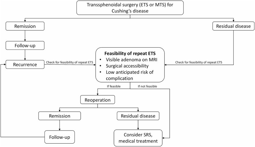
In ACTH-dependent hypercortisolism, CD is the predominant cause, followed by ectopic ACTH syndrome and, less frequently, an ectopic CRH-secreting tumor [35]. With the pretest probability for pituitary origin exceeding 80%, the next diagnostic step is typically an MRI of the pituitary region. However, the visualization of microadenomas on MRI ranges from 50% to 70%, requiring further testing if results are negative or inconclusive [5]. Initial testing of our patient revealed no pituitary lesions. Following a pituitary location, ACTH-secreting tumors may be found in the lungs. Thus, a high-resolution chest computed tomography scan was performed, which yielded negative findings. Healthcare professionals must keep these detection rates in mind. In instances of high clinical suspicion, repeating or reassessing tests and imaging may be warranted [3], as in our case, ultimately leading to the discovery of a 4-mm pituitary adenoma.

It is fundamental to mention that the Endocrine Society Clinical Practice Guideline on Treatment of CS recommends that, when possible, all patients presenting with ACTH-dependent CS and lacking an evident causal neoplasm should be directed to an experienced center capable of conducting inferior petrosal sinus sampling to differentiate between pituitary and nonpituitary or ectopic cause [8]. However, in this instance, such a referral was regrettably hindered by logistical constraints.

Regarding patient outcomes and monitoring in CD, there is no consensus on defining remission criteria following tumor resection. Prolonged hypercortisolism results in suppression of corticotropes, resulting in low levels of ACTH and cortisol after surgical intervention. Typically, remission is identified by morning serum cortisol values below 5 µg/dL (138 nmol/L) or UFC levels between 28 and 56 nmol/d (10-20 µg/d) within 7 days after surgical intervention. In our case, the patient’s morning serum cortisol was 115.8621 nmol/L (4.2 µg/dL), indicating remission. Remission rates in adults are reported at 73% to 76% in selectively resected microadenomas and at 43% in macroadenomas [8], highlighting the need for regular follow-up visits to detect recurrence.

Following the surgery, the patient experienced diabetes insipidus, a relatively common postoperative occurrence, albeit usually transient [8]. It is recommended to monitor serum sodium levels during the first 5 to 14 days postsurgery for early detection and management. Additionally, pituitary deficiencies may manifest following surgery. In this patient, prolactin levels were compromised, potentially impacting sexual response. However, postoperative somatomedin levels were normal, and gonadotropins were not measured due to the patient’s age group, as no additional clinical decisions were anticipated based on those results. Secondary hypothyroidism was diagnosed postoperatively.

Moving forward, it is important to emphasize certain clinical signs and symptoms for diagnosing CD. The combination of low bone mineral density (Likelihood Ratio [LR] +21.33), central obesity (LR +3.10), and arterial hypertension (LR + 2.29) [9] has a higher positive LR than some symptoms considered “characteristic,” such as reddish-purple striae, plethora, proximal muscle weakness, and unexplained bruising [210]. It is essential to give relevance to the signs the patient may present, emphasizing signs that have been proven to have an increased odds ratio (OR) such as osteoporosis (OR 3.8), myopathies (OR 6.0), metabolic syndrome (OR 2.7) and adrenal adenoma (OR 2.4) [9‐11]. The simultaneous development and worsening of these conditions should raise suspicion for underlying issues. Understanding the evolving nature of CD signs highlights the importance of vigilance during medical examinations, prioritizing the diagnostic focus, and enabling prompt initiation of treatment.

Recognizing the overlap of certain clinical features in CS is fundamental to achieving a timely diagnosis.

Learning Points

  • CS diagnosis is challenging due to the absence of pathognomonic signs and symptoms and the overlap of features present in many pathologies, such as metabolic syndrome.
  • Early detection of CS is crucial, given its association with high morbidity and mortality resulting from chronic exposure to glucocorticoids.
  • Recognizing the combination of low bone mineral density, obesity, hypertension, and diabetes as valuable clinical indicators is key in identifying CS.
  • Interdisciplinary collaboration is essential to achieve a comprehensive diagnostic approach.

Acknowledgments

We extend our gratitude to Pontificia Universidad Javeriana in Bogotá for providing essential resources and facilities that contributed to the successful completion of this case report. Special acknowledgment is reserved for the anonymous reviewers, whose insightful feedback significantly enhanced the quality of this manuscript during the peer-review process. Their contributions are sincerely appreciated.

Contributors

All authors made individual contributions to authorship. A.B.O. was involved in the diagnosis and management of this patient. M.A.G., J.M.H., and A.B.O. were involved in manuscript drafting and editing. All authors reviewed and approved the final draft.

Funding

This research received no public or commercial funding.

Disclosures

The authors declare that they have no conflicts of interest related to the current study.

Informed Patient Consent for Publication

Signed informed consent could not be obtained from the patient or a proxy but has been approved by the treating institution.

Data Availability Statement

Restrictions apply to the availability of some or all data generated or analyzed during this study to preserve patient confidentiality or because they were used under license. The corresponding author will on request detail the restrictions and any conditions under which access to some data may be provided.

References

1

Hakami
OA

,

Ahmed
S

,

Karavitaki
N

.

Epidemiology and mortality of Cushing’s syndrome

.

Best Pract Res Clin Endocrinol Metab

.

2021

;

35

(

1

):

101521

.

2

Nieman
LK

,

Biller
BMK

,

Findling
JW

, et al.

The diagnosis of Cushing’s syndrome: an endocrine society clinical practice guideline

.

J Clin Endocrinol Metab

.

2008

;

93

(

5

):

1526

1540

.

3

Gutiérrez Restrepo
J

,

Latorre Sierra
G

,

Campuzano Maya
G

.

Síndrome de cushing

.

Med Lab

.

2009

;

15

:

411

430

.

4

Petersenn
S

,

Newell-Price
J

,

Findling
JW

, et al.

High variability in baseline urinary free cortisol values in patients with Cushing’s disease

.

Clin Endocrinol (Oxf)

.

2014

;

80

(

2

):

261

269

.

5

Lila
AR

,

Sarathi
V

,

Jagtap
VS

,

Bandgar
T

,

Menon
P

,

Shah
NS

.

Cushing’s syndrome: stepwise approach to diagnosis

.

Indian J Endocrinol Metab

.

2011

;

15

(

Suppl4

):

S317

S321

.

6

Dogra
P

,

Vijayashankar
NP

.

Dexamethasone suppression test. In: StatPearls StatPearls Publishing; 2024

. Accessed January 29, 2024. http://www.ncbi.nlm.nih.gov/books/NBK542317/

7

Müller
OA

,

Dörr
HG

,

Hagen
B

,

Stalla
GK

,

von Werder
K

.

Corticotropin releasing factor (CRF)-stimulation test in normal controls and patients with disturbances of the hypothalamo-pituitary-adrenal axis

.

Klin Wochenschr

.

1982

;

60

(

24

):

1485

1491

.

8

Nieman
LK

,

Biller
BMK

,

Findling
JW

, et al.

Treatment of Cushing’s syndrome: an endocrine society clinical practice guideline

.

J Clin Endocrinol Metab

.

2015

;

100

(

8

):

2807

2831

.

9

Aron
DC

.

Cushing’s syndrome: why is diagnosis so difficult?
Rev Endocr Metab Disord

.

2010

;

11

(

2

):

105

116

.

10

Braun
LT

,

Vogel
F

,

Zopp
S

, et al.

Whom should we screen for cushing syndrome? the Endocrine Society practice guideline recommendations 2008 revisited

.

J Clin Endocrinol Metab

.

2022

;

107

(

9

):

e3723

e3730

.

11

Schneider
HJ

,

Dimopoulou
C

,

Stalla
GK

,

Reincke
M

,

Schopohl
J

.

Discriminatory value of signs and symptoms in Cushing’s syndrome revisited: what has changed in 30 years?
Clin Endocrinol (Oxf)

.

2013

;

78

(

1

):

153

154

.

Abbreviations


  • ACTH

    adrenocorticotropic hormone

  • CD

    Cushing disease

  • CRH

    corticotropin-releasing hormone

  • CS

    Cushing syndrome

  • HDDST

    high-dose dexamethasone suppression test

  • HPA

    hypothalamic-pituitary-adrenal

  • LDDST

    low-dose dexamethasone suppression test

  • LNSC

    late-night salivary cortisol

  • MRI

    magnetic resonance imaging

  • OR

    odds ratio

  • RV

    reference value

  • UFC

    urinary free cortisol

© The Author(s) 2024. Published by Oxford University Press on behalf of the Endocrine Society.
This is an Open Access article distributed under the terms of the Creative Commons Attribution License (https://creativecommons.org/licenses/by/4.0/), which permits unrestricted reuse, distribution, and reproduction in any medium, provided the original work is properly cited.

Day 15, Cushing’s Awareness Challenge

Today’s Cushing’s Awareness Challenge post is about kidney cancer (renal cell carcinoma). You might wonder how in the world this is related to Cushing’s. I think it is, either directly or indirectly.

I alluded to this a couple days ago when I said:

I finally started the Growth Hormone December 7, 2004.
Was the hassle and 3 year wait worth it?
Stay tuned for tomorrow, April 15, 2016 when all will be revealed.

So, as I said, I started Growth Hormone for my panhypopituitarism on December 7, 2004.  I took it for a while but never really felt any better, no more energy, no weight loss.  Sigh.

April 14 2006 I went back to the endo and found out that the arginine test that was done in 2004 was done incorrectly. The directions were written unclearly and the test run incorrectly, not just for me but for everyone who had this test done there for a couple years. My endo discovered this when he was writing up a research paper and went to the lab to check on something.

So, I went off GH again for 2 weeks, then was retested. The “good news” was that the arginine test is only 90 minutes now instead of 3 hours.

Wow, what a nightmare my arginine retest started! I went back for that Thursday, April 27, 2006. Although the test was shorter, I got back to my hotel and just slept and slept. I was so glad that I hadn’t decided to go right home after the test.

Friday I felt fine and drove back home, no problem. I picked up my husband for a biopsy he was having and took him to an outpatient surgical center. While I was there waiting for the biopsy to be completed, I started noticing blood in my urine and major abdominal cramps.

There were signs all over that no cellphones were allowed so I sat in the restroom (I had to be in there a lot, anyway!) and I left messages for several of my doctors on what I should do. It was Friday afternoon and most of them were gone 😦  I finally decided to see my PCP after I got my husband home.

When Tom was done with his testing, his doctor took one look at me and asked if I wanted an ambulance. I said no, that I thought I could make it to the emergency room ok – Tom couldn’t drive because of the anaesthetic they had given him. I barely made it to the ER and left the car with Tom to park. Tom’s doctor followed us to the ER and instantly became my new doctor.

They took me in pretty fast since I was in so much pain, and had the blood in my urine. At first, they thought it was a kidney stone. After a CT scan, my new doctor said that, yes, I had a kidney stone but it wasn’t the worst of my problems, that I had kidney cancer. Wow, what a surprise that was! I was admitted to that hospital, had more CT scans, MRIs, bone scans, they looked everywhere.

My new “instant doctor” felt that he wasn’t up to the challenge of my surgery, so he called in someone else.  My next new “instant doctor” came to see me in the ER in the middle of the night.  He patted my hand, like a loving grandfather might and said “At least you won’t have to do chemotherapy”.  And I felt so reassured.

It wasn’t until later, much after my surgery, that I found out that there was no chemo yet that worked for my cancer.  I was so thankful for the way he told me.  I would have really freaked out if he’d said that nothing they had was strong enough!

My open radical nephrectomy was May 9, 2006 in another hospital from the one where the initial diagnosis was made. My surgeon felt that he needed a specialist from that hospital because he believed preop that my tumor had invaded into the vena cava because of its appearance on the various scans. Luckily, that was not the case.

My entire left kidney and the encapsulated cancer (10 pounds worth!) were removed, along with my left adrenal gland and some lymph nodes. Although the cancer (renal cell carcinoma AKA RCC) was very close to hemorrhaging, the surgeon believed he got it all.

He said I was so lucky. If the surgery had been delayed any longer, the outcome would have been much different. I will be repeating the CT scans every 3 months, just to be sure that there is no cancer hiding anywhere. As it turns out, I can never say I’m cured, just NED (no evidence of disease). This thing can recur at any time, anywhere in my body.

I credit the arginine re-test with somehow aggravating my kidneys and revealing this cancer. Before the test, I had no clue that there was any problem. The arginine test showed that my IGF is still low but due to the kidney cancer I couldn’t take my growth hormone for another 5 years – so the test was useless anyway, except to hasten this newest diagnosis.

So… either Growth Hormone helped my cancer grow or testing for it revealed a cancer I might not have learned about until later.

My five years are up now.  When I was 10 years free of this cancer my kidney surgeon *thought* it would be ok to try the growth hormone again.  I was a little leery about this, especially where I didn’t notice that much improvement.

What to do?

BTW, I decided to…