Avascular Necrosis in Patients With Cushing Syndrome

Abstract

Cushing syndrome (CS) results from prolonged exposure to excess glucocorticoids, leading to a range of clinical manifestations including avascular necrosis (AVN), a rare complication of CS. Although AVN is often associated with exogenous glucocorticoid treatment, it can occur in endogenous CS but may be unrecognized because of its rarity and possibly from a subclinical presentation. We describe a case of a 71-year-old male with florid Cushing disease who initially presented with bilateral hip AVN and later developed bilateral shoulder AVN despite achieving biochemical remission following transsphenoidal surgery and adjuvant stereotactic photon radiosurgery. AVN in endogenous CS is underreported, and guidance on routine screening is lacking. Our case underscores the importance of considering AVN in patients with CS, especially in those with persistent or recurrent joint symptoms and markedly elevated cortisol levels. Early detection of AVN is crucial as it can lead to irreversible joint damage and disability if untreated. Screening strategies should be explored to identify high-risk patients who are diagnosed with CS for timely intervention, thereby preventing long-term morbidity associated with AVN.

Introduction

Cushing syndrome (CS) results from prolonged exposure to excess glucocorticoids, either from exogenous glucocorticoids or endogenous sources. In endogenous CS, hypercortisolism may be due to an ACTH-dependent process, most often from a corticotroph adenoma in Cushing disease (CD) or from ectopic ACTH secretion from neuroendocrine tumors or other solid tumors such as small cell lung carcinoma. On the other hand, ACTH-independent CS is mainly driven from adrenal pathology including adrenal adenomas, adrenocortical carcinomas, adrenal hyperplasia, and primary pigmented micronodular disease [1]. The presenting symptoms and signs of CS include hypertension, diabetes mellitus, weight gain, facial plethora, dorsocervical fat pads, muscle weakness, and osteoporosis, most of which may be detected on physical examination or diagnosed biochemically. A less common symptom is avascular necrosis (AVN) of bone tissue [12], which can present with pain or point tenderness of the hip or other joints as well as present subclinically [3].

AVN of the hip results from compromised blood supply to the bone tissue and usually impacts the hips and shoulders. This leads to necrosis of hematopoietic cells, adipocytes, and osteocytes. Subsequently, bone repair processes are activated, with differentiation of mesenchymal cells into osteoblasts to build new bone and hematopoietic stem cells into osteoclasts to remove necrotic tissue. However, because of impaired bone resorption and formation, subchondral fractures eventually occur [4]. Exogenous glucocorticoid treatment is 1 of the most common causes of AVN and may account for up to 38% of atraumatic AVN and is dose dependent [5]. Glucocorticoid treatment is theorized to cause AVN through increased systemic lipids, leading to compromised perfusion to the femoral head resulting from fat emboli or external lipocyte compression, as well as alterations in the inflammatory cytokines resulting in osteoclast activation and osteoblast apoptosis [46]. Compared to exogenous glucocorticoid treatment, AVN caused by endogenous hypercortisolism is not frequently reported nor is it screened for on diagnosis of CS.

We describe a patient who presented with bilateral hip AVN in the context of florid CD. We aim to highlight this presenting feature to heighten awareness for screening for this progressive condition, which can potentially lead to joint damage, loss of mobility, and long-term disability.

Case Presentation

A 71-year-old male with medical history of active tobacco use and obstructive sleep apnea was diagnosed with new-onset hypertension during an annual health visit. He was started on antihypertensive medications (losartan, hydrochlorothiazide, and spironolactone) by his primary care doctor, but the hypertension remained uncontrolled. Over the course of 2 months, the patient developed progressive lower extremity edema and was started on furosemide, which led to hypokalemia and was subsequently discontinued. He clinically deteriorated, with progressive anasarca and dyspnea, and then developed acute left eye ptosis and diplopia and was admitted to the hospital. The patient also endorsed irritability, mood swings, easy bruising, low libido, increased appetite, 30-lb weight gain, and bilateral hip pain.

Diagnostic Assessment

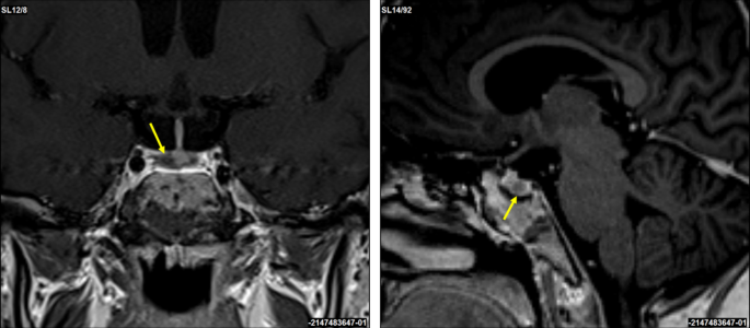
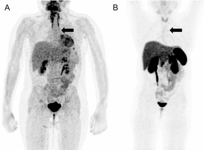
Physical examination was significant for oral candidiasis, dorsocervical fat pad, facial plethora, proximal muscle weakness, and bilateral hip tenderness. Testing confirmed ACTH-dependent CS with elevated 24-hour urine free cortisol of 1116 μg/24 hours (30788.21 nmol/24 hours) and 1171.9 μg/24 hours (32330.38 nmol/24 hours) (normal reference range, 3.5-45 μg/24 hours; 96.56-1241.46 nmol/24 hours) and ACTH of 173 pg/mL (38.06 pmol/L) and 112 pg/mL (24.64 pmol/L) (normal reference range, 7.2-63 pg/mL; 1.58-13.86 pmol/L) on 2 separate occasions. He had hypogonadotropic hypogonadism with total testosterone levels of 41 ng/dL (1.42 nmol/L) (normal reference range, 250-1100 ng/dL; 8.68-38.17 nmol/mL) and suppressed LH and FSH at <0.2 mIU/mL (<0.2 IU/L) (normal reference range, 0.6-12.1; 0.6-12/1.1 IU/L) and 0.2 mIU/mL (<0.2 IU/L) (normal reference range, 1.0-12.0 2 mIU/mL; 1.0-12.0 2 IU/L) respectively, whereas the remaining pituitary hormones were normal, although IGF-1 was low normal at 66 ng/mL (8.65 nmol/L) (normal reference range, 7.2-63 pg/mL; 1.58-13.86 pmol/L). He also had new-onset diabetes mellitus with glycated hemoglobin of 8% (<5.7%) (Table 1). Imaging of the lungs showed a 15-mm solid noncalcified nodule in the posterior right upper lobe concerning for neoplasm. Pituitary magnetic resonance imaging (MRI) revealed a 16 × 20 × 16 mm macroadenoma invading the left cavernous sinus (Fig. 1). Additionally, pelvis computed tomography (CT) scan demonstrated bilateral avascular necrosis of the capital femoral epiphysis without evidence of fracture or subchondral collapse (Fig. 2A and 2B).

Pituitary magnetic resonance imaging (MRI) with gadolinium, using T1-weighted, turbo spin-echo revealed sequence revealed a 16 × 20 × 16 mm macroadenoma invading the left cavernous sinus (white arrow).

Figure 1.

Pituitary magnetic resonance imaging (MRI) with gadolinium, using T1-weighted, turbo spin-echo revealed sequence revealed a 16 × 20 × 16 mm macroadenoma invading the left cavernous sinus (white arrow).

Coronal inversion recovery image bilateral hips demonstrates geographic lesions bilateral femoral heads with serpentine borders consistent with bilateral femoral head bone infarcts. No subchondral collapse or arthritic changes identified (A). Axial proton density with fat saturation image bilateral hips demonstrates geographic lesions bilateral femoral heads with serpentine borders consistent with bilateral femoral head bone infarcts. No subchondral collapse or arthritic changes identified (B). Coronal T1 image of the right shoulder demonstrates geographic lesion medial humeral head with serpentine border consistent with bone infarct. No subchondral collapse or arthritic changes identified (C). Coronal T1 image of the left shoulder demonstrates geographic lesion medial humeral head with serpentine border consistent with bone infarct. No subchondral collapse or arthritic changes identified (D) (white arrows).

Figure 2.

Coronal inversion recovery image bilateral hips demonstrates geographic lesions bilateral femoral heads with serpentine borders consistent with bilateral femoral head bone infarcts. No subchondral collapse or arthritic changes identified (A). Axial proton density with fat saturation image bilateral hips demonstrates geographic lesions bilateral femoral heads with serpentine borders consistent with bilateral femoral head bone infarcts. No subchondral collapse or arthritic changes identified (B). Coronal T1 image of the right shoulder demonstrates geographic lesion medial humeral head with serpentine border consistent with bone infarct. No subchondral collapse or arthritic changes identified (C). Coronal T1 image of the left shoulder demonstrates geographic lesion medial humeral head with serpentine border consistent with bone infarct. No subchondral collapse or arthritic changes identified (D) (white arrows).

Table 1.

Laboratory evaluation of the patient at presentation

Lab Value Reference Range
Conventional units (Système International units)
ACTH 173 pg/mL (38.06 pmol/L) 7.2-63 pg/mL (1.58-13.86 pmol/L)
24-h urine free cortisol 1116 μg/24 h (30,788.21 nmol/24 h) 4.0-55.0 μg/24 h (110.35-1517.34 nmol/24 h)
Total testosterone 41 ng/mL (1.42 nmol/L) 250-1100 ng/mL (8.68-38.17 nmol/L)
Free testosterone 12.3 pg/mL (0.07 nmol/L) 30.0-135.0 pg/mL (0.17-0.79 nmol/L)
LH <0.2 mIU/mL (<0.2 IU/L) 0.6-12.1 mIU/mL (0.6-12.1 IU/L)
FSH 0.2 mIU/mL (0.2 IU/L) 1-12 mIU/mL (1-12 IU/L)
Prolactin 9.6 ng/mL (9.6 μg/L) 3.5-19.4 ng/mL (3.5-19.4 μg/L)
TSH 0.746 mIU/L 0.450-5.330 mIU/L
Free T4 0.66 ng/dL (8.49 pmol/L) 0.61-1.60 ng/dL (7.85-20.59 pmol/L
IGF-1
Z score
66 ng/mL (8.65 nmol/L)
−0.9
34-245 ng/mL (4.45-32.09 nmol/L)
−2.0 to +2.0
HbA1c 8.2% <5.7%

Abbreviations: Hb A1c, hemoglobin A1C.

Treatment

Prophylactic treatment was started with subcutaneous heparin for anticoagulation and trimethoprim-sulfamethoxazole for opportunistic infections. Orthopedic evaluation did not recommend acute intervention for the hip AVN. Given the pituitary macroadenoma on imaging and left cranial nerve VI palsy, it was determined that the patient likely had CD, so he underwent transsphenoidal surgery. Surgical pathology confirmed the adenoma was ACTH positive, sparsely granulated, with Ki-67 index of 4%, and without increased mitotic activity (Fig. 3).

Hematoxylin and eosin (A) and adrenocorticotropic hormone (B) stained sections show oval nuclei with “salt and pepper” chromatin and granular, ACTH-positive cytoplasm. Original magnification 250×.

Figure 3.

Hematoxylin and eosin (A) and adrenocorticotropic hormone (B) stained sections show oval nuclei with “salt and pepper” chromatin and granular, ACTH-positive cytoplasm. Original magnification 250×.

Outcome and Follow-up

Due to ongoing hypercortisolism (Table 2) and residual tumor in the left cavernous sinus, the patient underwent adjuvant treatment with stereotactic photon radiosurgery at a dose of 13 Gy targeted to the left cavernous sinus and was started on osilodrostat, an oral, reversible inhibitor of 11β-hydroxylase that drives the final step of cortisol synthesis and aldosterone synthase, which converts 11-deoxycorticosterone to aldosterone [7]. The starting dose of osilodrostat was 2 mg twice per day. As the patient developed nausea, lack of appetite, and malaise with decreasing cortisol levels, osilodrostat was reduced to 1 mg daily, and he was started on hydrocortisone replacement therapy on week 11 postoperatively (Table 3). Ultimately, both osilodrostat and hydrocortisone were discontinued following normalization of cortisol levels. Regarding the rest of the hormonal deficiencies, his total testosterone and IGF-1 levels improved to levels of 483 ng/dL (16.76 nmol/L) and 99 (12.97 nmol/L), respectively, and he did not require hormone replacement therapy. Clinically, the patient improved with resolution of his hypertension and diabetes and achieved a 38-lb weight loss. Additionally, his diplopia improved and his hip pain resolved without any restriction in mobility. However, 1 year postoperatively, the patient developed bilateral shoulder pain. MRI of the shoulders demonstrated subchondral changes in the right humeral head (Fig. 2C) and a linear area of subchondral change involving the left humeral head (Fig. 2D) consistent with AVN, as well as a bilateral high-grade supraspinatus tear and acromioclavicular joint osteoarthritis. He was treated with an intraarticular methylprednisolone 40-mg injection to both shoulders, with subsequent improvement of the pain and joint mobility. He also underwent a coronary artery bypass graft surgery for 3-vessel disease. The patient has otherwise maintained normal urine and salivary cortisol levels off osilodrostat or hydrocortisone, and 1 year after surgery, the ACTH (cosyntropin) stimulation test was normal. The pulmonary nodule has remained stable on serial imaging.

Table 2.

Postoperative cortisol and ACTH levels

Postoperative day
Lab Reference Range Conventional units (Système International units) 1 2 2 3 4 5
Morning cortisol 3.7-19.4 μg/dL (102.08- 535.21 nmol/L) 26 μg/dL (717.29 nmol/L) 21.5 μg/dL (593.14 nmol/L) 6 μg/dL (165.53 nmol/L) 8.1 μg/dL (223.46 nmol/L) 16.4 μg/dL (452.44 nmol/L) 21.5 μg/dL (593.14 nmol/L)
ACTH 7.2-63.3 pg/mL (1.58- 13.93 pmol/L) 72 pg/mL (15.84 pmol/L) 62 pg/mL (13.64 pmol/L)

Table 3.

Titration of osilodrostat treatment based on cortisol levels

Postoperative week
Lab Reference range Conventional units (Système International units) 8 9 11 13 15 18 22 24
ACTH 7.2-63.3 pg/mL (1.58-13.93 pmol/L) 95.6 pg/mL (21.03 pmol/L) 131 pg/mL (28.82 pmol/L) 58.8 pg/mL (12.94 pmol/L) 79.3 pg/mL (17.45 pmol/L) 79.9 pg/mL (17.58 pmol/L) 73.4 pg/mL (16.15 pmol/L) 62 pg/mL (13.64 pmol/L) 71.5 pg/mL (15.73 pmol/L)
Morning cortisol 3.7-19.4 μg/dL (102.08-535.21 nmol/L) 23.9 μg/dL (659.35 nmol/L) 18.8 μg/dL (518.65 nmol/L) 6.6 μg/dL (182.08 nmol/L) 4.5 μg/dL (124.15 nmol/L) 3.3 μg/dL (91.04 nmol/L) 2.4 μg/dL (66.21) nmol/L 8.2 μg/dL (226.22. nmol/L) 4.1 μg/dL (113.11 nmol/L)
LNSC <0.010-0.090 μg/dL (0.28-2.48 nmol/L) 0.615 μg/dL (16.97 nmol/L) 0.058 μg/dL (1.60 nmol/L) 0.041 μg/dL (1.13 nmol/L) 0.041 μg/dL (1.13 nmol/L)
UFC, 24-h 5-64 μg/24 h (137.94-1765.63 nmol/24 h) 246 μg/24 h (6786.65 nmol/24 h) 226 μg/24 h (6234.89 nmol/24 h) 2 μg/24 h (55.18. nmol/24 h)
Osilodrostat dose 2 mg BID 2 mg BID 2 mg AM
3 mg PM
2 mg BID 2 mg AM
1 mg PM
1 mg BID 1 mg daily Oslidrostat discontinued

Abbreviations: BID, twice per day; LNSC, late night salivary cortisol; UFC, urine free cortisol.

Discussion

Our patient exhibited pronounced hypercortisolism secondary to CD, with bilateral hip AVN as 1 of the presenting symptoms. Despite achieving biochemical remission of the disease and resolution of other associated symptoms, the patient was later diagnosed with bilateral shoulder AVN.

AVN caused by endogenous hypercortisolism is seldom documented, and routine screening for it is not typically conducted during the diagnosis of CS. However, AVN has been reported to be a presenting symptom in several case reports or may manifest years after the initial diagnosis [8]. Reported causes of AVN in endogenous CS include pituitary adenomas, adrenal adenomas or carcinomas, adrenal hyperplasia, or neuroendocrine tumors [8‐23] (Table 4), with some cases of AVN associated with severe hypercortisolism [1015]. Other risk factors associated with AVN include hip trauma, femoral fractures, hip dislocation, systemic lupus erythematosus in the setting of concomitant corticosteroid treatments, or vasculitis, sickle cell disease, hypercoagulability, Gaucher disease, hyperlipidemia or hypertriglyceridemia, hyperuricemia, hematological malignancies, antiretroviral medications, alcohol use, and exogenous steroid treatment [4]. Our patient had no history of hip trauma or other aforementioned comorbidities. Furthermore, during presentation, his lipid levels were normal, with low-density lipoprotein cholesterol of 89 mg/dL (<130 mg/dL) and triglycerides of 97 mg/dL (<150 mg/dL). Therefore, it is likely that his bilateral hip and shoulder AVN was caused by severe endogenous hypercortisolism.

Table 4.

Published cases of avascular necrosis in patients with endogenous hypercortisolism

First author, year Age (y)/sex Time of diagnosis in relation to CS diagnosis AVN related symptoms Imaging modality Imaging description Diagnosis Treatment
Salazar D, 2021 [15] 38 F 3 y prior to diagnosis Right hip pain MRI
  • Right hip joint effusion and synovitis
  • Flattening of the femoral head-Subcortical edema
Adrenal adenoma Right hip arthroplasty
Madell SH, 1964 [16] 41 F 1 month before diagnosis Right shoulder pain X-ray
  • Increased density of the right humeral head with spotty areas of radiolucency
  • Early flattening and beginning of fragmentation
Adrenal adenoma Osteotomy
Anand A, 2022 [21] 47 M Bilateral hip pain MRI
  • Necrosis of bilateral femur heads
adrenocortical carcinoma
Belmahi N, 2018 [9] 28 F Progressive limping and right hip pain MRI
  • Right femoral head AVN
Pituitary adenoma Right total hip replacement
Wicks I, 1987 [10] 39 M 18 months before diagnosis Progressive hip pain and stiffens X-ray
Bone scan
  • Lucent and sclerotic regions within flattened femoral heads
  • Some loss of articular cartilage
Pituitary adenoma Conservative management
Koch C, 1999 [11] 30 F Sudden onset of severe left hip pain MRI
  • Abnormal high intensity signal changes in the bone marrow of the left femoral head
  • Joint effusion
  • Stage 2 AVN
Pituitary adenoma Immediate core decompression surgery with decongestion of the left femoral head
Premkumar M, 2013 [12] 26 F 2 y after pituitary surgery for Cushing, while on replacement steroid therapy Progressive bilateral hip pain resulting in difficulty in walking MRI
  • Bilateral multiple bony infarcts in the proximal femur and distal femur
  • Femoral head collapse fractures -Stage 2 avascular necrosis
Pituitary adenoma
Bauddh N, 2022 [13] 24 M 2 y prior to diagnosis Progressive left hip pain and difficulty in walking X-ray
MRI
  • Left femoral head AVN
Pituitary adenoma Planned for surgery of hip AVN
Joseph A, 2022 [14] 21 F 1 y prior to diagnosis Bilateral hip joint pain X-ray
MRI
  • Ill-defined mixed sclerotic and lytic pattern of the femoral heads
  • Cortical disruption of the round contour
  • Low signal intensity in the subchondral region of the femoral necks on T1-weighted images
Pituitary adenoma Planned for total hip replacement.
Bisphosphonates.
Pazderska A, 2016 [19] 36 F Right leg pain MRI
  • Bilateral AVN of the femoral heads
  • Left femoral head with early bone fragmentation
Bilateral primary pigmented micronodular adrenal disease Spontaneous healing of AVN after adrenalectomy.
Papadakis G, 2017 [22] 55 F MRI
PET/CT 68Ga-DOTATATE
  • Bilateral AVN
  • Bone marrow edema extending to the intertrochanteric area
  • Mild subchondral femoral head collapse of the left hip
  • Increased activity in bilateral femoral heads and in the bone marrow consistent with edema
  • Mild left femoral head collapse
Ectopic ACTH- secreting tumor
Phillips K, 1986 [8] 24 F 4.5 y after diagnosis Right femoral AVN X-ray
  • Flattening and sclerosis of femoral head
Cushing disease
25 F 4 y after diagnosis Right femoral AVN
  • Subchondral lucency
43 F 8 mo after diagnosis Right humeral AVN
  • Sclerosis and flattening of articular surface of humeral head
61 F 11 y after diagnosis Left femoral AVN and bilateral humeral heads
  • Cortical indistinctness and subchondral lucency
  • Left humeral head flattening and sclerosis
Cerletty J, 1973 [20] 54 M 3 mo before diagnosis Right femoral head fracture X-ray
  • Bilateral subchondral sclerosis of the femoral heads
  • Some narrowing of the joint space on the left
  • Infraction of the margin of the right femoral head
  • Femoral neck fracture.
Bilateral adrenal cortical hyperplasia Total hip joint arthroplasty
Ha J-S, 2019 [18] 36 F 2 y before diagnosis 2 mo left hip restricted range of motion X-ray
MRI
  • Right femoral head with areas of hyperlucency and surrounding sclerosis
  • Subtle changes in the shape of the articular surface
  • Bilateral femoral head osteonecrosis -Increased amount of joint fluid and bone marrow edema in the left hip
  • Right femoral head necrosis
Adrenal cortical adenoma Total hip replacement
Takada, J, 2004 [17] 55 F Intense right hip pain and a limp MRI
  • Low-intensity band on T1-weighted images
  • Stage 2 AVN.
Adrenal adenoma Total hip arthroplasty
Modlinger RS, 1972 [23] 69 F Increased pain of right shoulder X-ray
  • Bilateral shoulders with aseptic necrosis of the humeral heads
Ectopic ACTH secretion NET form pancreatic tumor

Abbreviations: AVN, avascular necrosis; F, female; M, male; MRI, magnetic resonance imaging; NET, neuroendocrine tumor.

AVN can result in irreversible femoral head collapse, leading to severe limitation in movement, reduced joint functionality, and decreased quality of life [24]. Initially, patients may be asymptomatic or endorse nonspecific pain when presenting with AVN and may not be diagnosed until an advanced stage when they develop more severe pain and disability [25]. In a meta-analysis assessing the prevalence of AVN in patients with systemic lupus erythematosus, including those who received corticosteroid treatment, asymptomatic AVN was detected in 29% of patients and symptomatic disease was noted in 9% [26]. AVN can diagnosed with MRI or CT imaging. Although noncontrast MRI has higher sensitivity and specificity in detecting early stages of the disease, CT is comparable to MRI in more advanced stages. Ancillary imaging modalities include plain radiography, positron emission tomography, and bone scan [27].

Staging of AVN relies on radiologic features and size of lesions. In earlier stages, imaging can be normal (stage 0) or with subtle abnormalities on MRI or bone scan and normal radiography (stage 1). As the disease progresses, structural changes, including cystic and sclerotic changes (stage 2), subchondral collapse (stage 3), flattening of the femoral head (stage 4), joint narrowing and acetabular changes (stage 5), and, finally, advanced degenerative changes (stage 6) can be detected on most imaging modalities.

Management of early stages of AVN includes observation or conservative weight-bearing management, medical therapy with bisphosphonates, anticoagulation therapy, statins, and vasodilators. Invasive procedures such as mesenchymal stem cells implantation, osteotomy, surgical joint decompression, and total hip replacement are reserved for more advanced stages [28]. Indeed, AVN accounts for approximately 10% of total hip replacements in the United States [29]. Staging has prognostic implications for treatment options and disease outcomes. Early-stage disease, when diagnosed and treated, can often regress, and be cured. Conservative measures, medical treatment, biophysical stimulation, extracorporeal shockwave therapy, or core decompression, can prevent femoral head collapse and further hip arthroplasty. On the other hand, late-stage disease, characterized by joint collapse, is irreversible and often requires joint replacement [30].

Although actual prevalence rates of AVN in endogenous CS is unknown, one should consider screening for AVN in this high-risk population, particularly in patients showing markedly elevated cortisol levels, as in our case. Such an approach would facilitate the early identification of individuals who would benefit from earlier medical or surgical interventions, thereby preventing permanent joint destruction and chronic disability.

Learning Points

  • AVN can be a complication of endogenous hypercortisolism.
  • AVN may present asymptomatically or with nonspecific symptoms such as joint pain.
  • AVN can affect multiple joints, including hips and shoulders, and its early diagnosis relies on MRI or CT imaging.
  • Early detection and intervention for AVN are crucial to prevent irreversible joint damage and disability.
  • Screening for AVN in patients with CS should be considered to enable timely intervention and prevent long-term complications, particularly in patients with hip or shoulder pain and severe hypercortisolism.

Contributors

All authors made individual contributions to authorship. N.T. and O.C. were involved in the diagnosis and management of the patient and manuscript submission. S.B. was involved in the histopathology section and preparation of histology images. T.L. was involved in the interpretation and preparation of the radiology images. A.N.M. was responsible for the patient’s surgery and treatment plan. All authors reviewed and approved the final draft.

Funding

No public or commercial funding.

Disclosures

Dr. Odelia Cooper is an Editorial Board member for JCEM Case Reports and played no role in the journal’s evaluation of the manuscript. There are no other disclosures to declare.

Informed Patient Consent for Publication

Signed informed consent obtained directly from patient.

Data Availability Statement

Data sharing is not applicable to this article as no datasets were generated or analyzed during the current study.

Abbreviations

  • AVN

    avascular necrosis

  • CD

    Cushing disease

  • CS

    Cushing syndrome

  • CT

    computed tomography

  • MRI

    magnetic resonance imaging

© The Author(s) 2025. Published by Oxford University Press on behalf of the Endocrine Society.
This is an Open Access article distributed under the terms of the Creative Commons Attribution License (https://creativecommons.org/licenses/by/4.0/), which permits unrestricted reuse, distribution, and reproduction in any medium, provided the original work is properly cited. See the journal About page for additional terms.

Oncocytic Pituicytoma in a Patient with Cushing’s Disease

The final, formatted version of the article will be published soon.

1) Background: Posterior pituitary tumors (PPTs) are extremely rare, with fewer than 400 cases reported to date. In 2022, the WHO classified four types of tumors originating from the posterior pituitary: traditional pituicytoma, oncocytic pituicytoma, granular pituicytoma, and ependymal pituicytoma. To our knowledge, only one subject with coexistence of Cushing’s disease and oncocytic pituicytoma (spindle cell oncocytoma) has been reported, but the clinical features of this patient were not described in detail.

2) Case presentation: We presented a case of a patient with Cushing’s syndrome and a pituitary mass. Transsphenoidal surgery was performed, and pathologic examination revealed two distinct tumors: a corticotroph adenoma with a diameter of less than 2mm and a larger oncocytic pituicytoma. Post-surgery serum cortisol was 51 nmol/L, indicating complete remission. Corticotroph adenoma or corticotroph hyperplasia were identified after surgery in less than half of the subjects with Cushing’s disease and PPT. (3)

Conclusions: Our study indicates that Cushing’s disease in patients with PPT may be caused by the existence of collision lesions, with corticotroph adenoma or hyperplasia being difficult to detect due to their small dimensions.

Keywords: Cushing’s disease, oncocytic pituicytoma, Spindle cell oncocytoma, pituitary adenoma, Posterior pituitary tumors

Received: 27 Aug 2024; Accepted: 17 Feb 2025.

Copyright: © 2025 Li, Chen, Tan, Yu, Tang, Cai and Li. This is an open-access article distributed under the terms of the Creative Commons Attribution License (CC BY). The use, distribution or reproduction in other forums is permitted, provided the original author(s) or licensor are credited and that the original publication in this journal is cited, in accordance with accepted academic practice. No use, distribution or reproduction is permitted which does not comply with these terms.

* Correspondence:
Huiwen Tan, Department of Endocrinology and Metabolism, West China Hospital of Sichuan University, Chengdu, China
Ying Tang, Department of Pathology, West China Hospital, Sichuan University, Chengdu, Sichuan Province, China
Bowen Cai, Department of Neurosurgery, West China Hospital, Sichuan University, Chengdu, Sichuan Province, China
Jianwei Li, Department of Endocrinology and Metabolism, West China Hospital of Sichuan University, Chengdu, China

Disclaimer: All claims expressed in this article are solely those of the authors and do not necessarily represent those of their affiliated organizations, or those of the publisher, the editors and the reviewers. Any product that may be evaluated in this article or claim that may be made by its manufacturer is not guaranteed or endorsed by the publisher.


From https://www.frontiersin.org/journals/endocrinology/articles/10.3389/fendo.2025.1487120/abstract


Ectopic Adrenocorticotrophic Hormone Syndrome in a 10-Year-Old Girl With a Thymic Neuroendocrine Tumor

Abstract

Background

Thymic neuroendocrine tumor as a cause of Cushing syndrome is extremely rare in children.

Case presentation

We report a case of a 10-year-old girl who presented with typical symptoms and signs of hypercortisolemia, including bone fractures, growth retardation, and kidney stones. The patient was managed with oral ketoconazole, during which she experienced adrenal insufficiency, possibly due to either cyclic adrenocorticotropic hormone (ACTH) secretion or concurrent COVID-19 infection. The patient underwent a diagnostic work-up which indicated the possibility of an ACTH-secreting pituitary neuroendocrine tumor. However, after a transsphenoidal surgery, the diagnosis was not confirmed on histopathological examination. Subsequent bilateral inferior petrosal sinus sampling showed strong indications of the presence of ectopic ACTH syndrome. Detailed rereading of functional imaging studies, including 18F-FDG PET/MRI and 68Ga DOTATOC PET/CT, ultimately identified a small lesion in the thymus. The patient underwent videothoracoscopic thymectomy that confirmed a neuroendocrine tumor with ACTH positivity on histopathological examination.

Conclusion

This case presents some unique challenges related to the diagnosis, management, and treatment of thymic neuroendocrine tumor in a child. We can conclude that ketoconazole treatment was effective in managing hypercortisolemia in our patient. Further, a combination of functional imaging studies can be a useful tool in locating the source of ectopic ACTH secretion. Lastly, in cases of discrepancy in the results of stimulation tests, bilateral inferior petrosal sinus sampling is highly recommended to differentiate between Cushing disease and ectopic ACTH syndrome.

Peer Review reports

Background

In children above seven years of age, the majority of pediatric Cushing syndrome (CS) cases are caused by a pituitary neuroendocrine tumors (PitNET). However, a differential diagnosis of hypercortisolemia in children is often challenging concerning the interpretation of stimulation tests and the fact that up to 50% of PitNET may not be detected on magnetic resonance imaging (MRI) [1]. An ectopic adrenocorticotropic hormone (ACTH) syndrome (EAS) is extremely rare in children. Its diagnosis is often missed or confused with Cushing disease (CD) [2]. Most ACTH-secreting tumors originate from bronchial or thymic neuroendocrine tumors (NETs), or less commonly, from NETs in other locations. To diagnose EAS, specific functional imaging studies are often indicated to elucidate the source of ACTH production.

Pharmacotherapy may be used before surgery to control hypercortisolemia and its symptoms/signs, or in patients in whom the source of hypercortisolism has not been found (e.g., EAS), or surgery failed. Ketoconazole or metyrapone, as adrenal steroidogenesis blockers, were found to be very efficient, although they exhibit side effects [3].

Furthermore, cyclic secretion of ACTH followed by fluctuating plasma cortisol levels is extremely rare in children, including those with EAS [45]. Therefore, in cyclic EAS, the use of steroid inhibitors or acute illness or trauma can be associated with adrenal insufficiency, which can be life-threatening. Here we describe the clinical features, laboratory and radiological investigations, results, management, and clinical outcome of a 10-year-old girl with a thymic NET presenting with ACTH secretion.

Case presentation

A 10-year-old girl was acutely admitted to our university hospital for evaluation of facial edema and macroscopic hematuria in May 2021. A day before admission, she presented to the emergency room for dysuria, pollakiuria, nausea, and pain in her right lower back. Over the past year she had experienced excessive weight gain with increased appetite and growth retardation (Fig. 1). Her height over three years had shifted from the 34th to the 13th centile (Fig. 1). Her parents noticed facial changes, pubic hair development, increased irritability, and moodiness.

Fig. 1

figure 1

Body weight, body height, and body mass index development of the case patient. The black arrow indicates the first presentation, the blue arrow indicates the start of ketoconazole treatment and the yellow arrow indicates the time of thymectomy. Mid-parental height is indicated by the green line

At admission, she was found to have a moon face with a plethora, few acne spots on forehead, as well as facial puffiness. In contrast to slim extremities, an abnormal fat accumulation was observed in the abdomen. Purple striae were present on abdomen and thighs. She did not present with any bruising, proximal myopathy, or edema. On physical examination, she was prepubertal, height was 135 cm (13th centile), and weight was 37 kg (69th centile) with a BMI of 20.4 kg/m2 (90th centile). She developed persistent hypertension. Her past medical history was uneventful except for two fractures of her upper left extremity after minimal trips one and three years ago, both treated with a caste. Apart from hypothyroidism on the maternal side, there was no history of endocrine abnormalities or tumors in the family.

In the emergency room, the patient was started on sulfonamide, pain medication, and intravenous (IV) fluids. Her hypertensive crises were treated orally with angiotensin-converting enzyme inhibitor or with a combination of adrenergic antagonists and serotonin agonists administered IV. Hypokalemia had initially been treated with IV infusion and then with oral potassium supplements. A low serum phosphate concentration required IV management. The initial investigation carried out in the emergency room found hematuria with trace proteinuria. Kidney ultrasound showed a 5 mm stone in her right ureter with a 20 mm hydronephrosis. She did not pass any kidney stones, however, fine white sand urine analysis reported 100% brushite stone.

Hypercortisolemia was confirmed by repeatedly increased 24-hour urinary free cortisol (UFC), (5011.9 nmol/day, normal range 79.0-590.0 nmol/day). Her midnight cortisol levels were elevated (961 nmol/l, normal range 68.2–537 nmol/l). There was no suppression of serum cortisol after 1 mg overnight dexamethasone suppression test (DST) or after low-dose DST (LDDST). An increased morning plasma ACTH (30.9 pmol/l, normal range 1.6–13.9 pmol/) suggested ACTH-dependent hypercortisolemia. There was no evidence of a PitNET on a 1T contrast-enhanced MRI. The high-dose DST (HDDST) did not induce cortisol suppression (cortisol 1112 nmol/l at 23:00, cortisol 1338 nmol/l at 8:00). Apart from the kidney stone, a contrast-enhanced computed tomography (CT) of her neck, chest, and abdomen/pelvis did not detect any lesion. Various tumor markers were negative and the concentration of chromogranin A was also normal.

A corticotropin-releasing hormone (CRH) stimulation test induced an increase in serum cortisol by 32% at 30 min and ACTH concentration by 67% at 15 min (Table 1). A 3T contrast-enhanced MRI scan of the brain identified a 3 × 2 mm lesion in the lateral right side of the pituitary gland (Fig. 2). An investigation of other pituitary hormones was unremarkable. Apart from low serum potassium (minimal level of 2.8 mmol/l; normal range 3.3–4.7 mmol/l) and phosphate (0.94 mmol/l; normal range 1.28–1.82 mmol/l) concentrations, electrolytes were normal. The bone mineral density assessed by whole dual-energy X-ray absorptiometry was normal.

Fig. 2

figure 2

Coronal and sagittal 3T contrast-enhanced brain MRI scans. A suspected 3 × 2 mm lesion in the lateral right side of the pituitary gland (yellow arrows)

The patient was presented at the multidisciplinary tumor board and it was decided that she undergoes transsphenoidal surgery for the pituitary lesion. No PitNET was detected on histopathological examination and no favorable biochemical changes were noted after surgery. After the patient recovered from surgery, subsequent bilateral inferior petrosal sinus sampling (BIPSS) confirmed EAS as the maximum ratio of central to peripheral ACTH concentrations was only 1.7. During the investigation for tumor localization, she was started on ketoconazole treatment (300 mg/day) to alleviate symptoms and signs of hypercortisolism. Treatment with ketoconazole had a beneficial effect on patient health (Fig. 1). There was a weight loss of 2 kg in a month, a disappearance of facial plethora, and a decrease in vigorous appetite. Her liver function tests remained within the normal range.

Table 1 Result of corticotropin-releasing hormone stimulation test

The 24-hour UFC excretion normalized three weeks after ketoconazole initiation. However, six weeks after continuing ketoconazole therapy (400 mg/day), the patient complained of nausea, vomiting, and diarrhea. She was found to have adrenal insufficiency with a low morning serum cortisol of 10.70 nmol/l (normal range 68.2–537 nmol/l) and salivary cortisol concentrations < 1.5 nmol/l (normal range 1.7–29 nmol/l). She was also found to be positive for COVID-19 infection. Ketoconazole treatment was stopped and our patient was educated to take stress steroids in case of persisting or worsening symptoms. Her clinical status gradually improved and steroids were not required.

Meanwhile, whole-body fluorine-18 fluorodeoxyglucose positron emission tomography (18F-FDG PET)/MRI was performed with no obvious hypermetabolic lesion suspicious of a tumor. No obvious accumulation was detected on 68Ga-DOTATOC PET/CT images (Fig. 3). However, a subsequent careful and detailed re-review of the images detected a discrete lesion on 18F-FDG PET/MRI and 68Ga-DOTATOC PET/CT scans in the left anterior mediastinum, in the thymus (Fig. 4).

Fig. 3

figure 3

18F-FDG PET/MRI (A) and 68Ga-DOTATOC (B) PET/CT scans. Whole body MIP reconstructions. Subtle correspondent focal hyperactivity in the left mediastinum (black arrow). The 18F-FDG PET/MRI image courtesy of Prof. Jiri Ferda, MD, PhD, Clinic of the Imaging Methods, University Hospital Plzen, Czech Republic

Fig. 4

figure 4

Axial slices of PET/MRI (AC) and 68Ga-DOTATOC (DF) PET/CT scans. Subtle correspondent focal hyperactivity in the left mediastinum (white arrow). No obvious finding on MRI (C) and CT (F) scans. The FDG PET/MRI image courtesy of Prof. Jiri Ferda, MD, PhD, Clinic of the Imaging Methods, University Hospital Plzen, Czech Republic

Three weeks after the episode of adrenal insufficiency and being off ketoconazole treatment, our patient´s pre-surgery laboratory tests showed slightly low morning cortisol 132 nmol/l with surprisingly normal ACTH 2.96 pmol/l (normal range 1.6–13.9 pmol/). Given the upcoming surgery, she was initiated on a maintenance dose of hydrocortisone (15 mg daily = 12.5 mg/m2/day). Further improvement of cushingoid characteristics (improvement of facial plethora and moon face, weight loss) was noticed. Our patient underwent videothoracoscopic surgery, and a hyperplastic thymus of 80 × 70 × 15 mm with a 4 mm nodule was successfully removed. Tumor immunohistochemistry was positive for ACTH, chromogranin A, CD56, and synaptophysin. Histopathological findings were consistent with a well-differentiated NET grade 1. A subsequent genetic screening did not detect any pathogenic variant in the MEN1 gene.

After surgery, hydrocortisone was switched to a stress dose and gradually decreased to a maintenance dose. Antihypertensive medication was stopped and further weight loss was observed after thymectomy. Within a few weeks after the thoracic surgery, the patient entered puberty, her mood improved significantly, and potassium supplements were stopped. Finally, hydrocortisone treatment was stopped ten months after thymectomy.

Discussion and conclusions

The case presented here demonstrates a particularly challenging work-up of the pediatric patient with the diagnosis of CS caused by EAS due to thymic NET. Differentiating CD and EAS can sometimes be difficult, including the use of various laboratory and stimulation tests and their interpretation, as well as proper, often challenging, reading of functional imaging modalities, especially if a discrete lesion is present at an unusual location [1]. When using established criteria for Cushing disease (for the CRH test an increase of cortisol and/or ACTH by ≥ 20% or ≥ 35%, respectively, and a ≥ 50% suppression of cortisol for the HDDST) our patient presented discordant results. The CRH stimulation test induced an increase in cortisol by 32% and ACTH by 67% and the 3T MRI pointed to the right-side pituitary lesion, both to yield false positive results. The HDDST, on the other hand, did not induce cortisol suppression and was against characteristic findings for CD. We did not proceed with desmopressin testing, which also induces an excess ACTH and cortisol response in CD patients and has rarely been used in pediatric patients, except in those with extremely difficult venous access [6]. Recently published articles investigated the reliability of CRH stimulation tests and HDDST and both concluded that the CRH test has greater specificity than HDDST [78]. Elenius et al. suggested optimal response criteria as a ≥ 40% increase of ACTH and/or cortisol (cortisol as the most specific measure of CD) during the CRH test and a ≥ 69% suppression of serum cortisol during HDDST [7]. Using these criteria, the CD would be excluded in our patient. To demonstrate that the proposed thresholds for the test interpretation widely differ, Detomas et al. proposed a ≥ 12% cortisol increase and ≥ 31% ACTH increase during the CRH test to confirm CD [8].

The fact that up to 50% of PitNET may not be detected on MRI [1] and that more than 20% of patients with EAS are reported to have pituitary incidentalomas [9] makes MRI somewhat unreliable in differentiating CD and EAS. However, finally, well-established and generally reliable BIPSS in our patient supported the diagnosis of EAS. Thus, BIPSS is considered a gold standard to differentiate between CD and EAS; however, it can still provide false negative results in cyclic CS if performed in the trough phase [10] or in vascular anomalies or false positive results as in a recent case of orbital EAS [11].

In children, the presence of thymus tissue may be misinterpreted as normal. Among other reports of thymic NET [12], Hanson et al. reported a case of a prepubertal boy in whom a small thymic NET was initially treated as normal thymus tissue on CT [13]. In our case, initially, the lesion was not detected on the 18F-FDG and 68Ga-DOTATOC PET scans. A small thymic NET was visible only after a detailed and careful re-reading of both PET scans. Although somatostatin receptor (SSR) PET imaging may be helpful in identifying ectopic CRH- or ACTH-producing tumors, there are still some limitations [13]. For example, in the study by Wannachalee et al., 68Ga-DOTATATE identified suspected primary lesions causing ECS in 65% of patients with previously occult tumors and was therefore concluded as a sensitive method for primary as well as metastatic tumors [14]. In our patient, the final correct diagnosis was based on the results of both PET scans. This is in full support of the article published by Liu et al. who concluded that 18F-FDG and SSR PET scans are complementary in determining the proper localization of ectopic ACTH production [15]. Additionally, it is worth noting that not all NETs stain positively for ACTH which may present a burden in its identification.

To control hypercortisolemia, both ketoconazole and metyrapone were considered in our patient. Due to the side effects of metyrapone on blood pressure, ketoconazole was started as a preferred option in our pediatric patient. A retrospective multicenter study concluded that ketoconazole treatment is effective with acceptable side effects, with no fatal hepatitis and adrenal insufficiency in 5.4% of patients [3]. During ketoconazole treatment, our patient developed adrenal insufficiency; however, it is impossible to conclude whether this was solely due to ketoconazole treatment or whether an ongoing COVID-19 infection contributed to the adrenal insufficiency or whether this was caused by a phase of lower or no ACTH secretion from the tumor often seen in patients with cyclic ACTH secretion. The patient’s cyclic ACTH secretion is highly probable since her morning cortisol was slightly lower and ACTH was normal, even after being off ketoconazole treatment for 3 weeks.

When retrospectively and carefully reviewing all approaches to the diagnostic and management care of our pediatric patient, it would be essential to proceed to BIPSS before any pituitary surgery, especially when obtaining discrepant results from stimulation tests, as well as detecting a discrete pituitary lesion ( 6 mm) as recommended by the current guidelines [16]. This was our first experience using ketoconazole in a young child, and although this treatment was associated with very good outcomes in treating hypercortisolemia, close monitoring, and family education on signs and symptoms of adrenal insufficiency are essential to recognizing adrenal insufficiency promptly in any patient with EAS, especially those presenting also with some other comorbidities or stress, here COVID-19 infection.

In conclusion, the pediatric patient here presenting with EAS caused by thymic NET needs very careful assessment including whether cyclic CS is present, the outline of a good management plan to use all tests appropriately and in the correct sequence, monitoring carefully for any signs or symptoms of adrenal insufficiency, and apply appropriate imaging studies, with experienced radiologists providing accurate readings. Furthermore, ketoconazole treatment was found to be effective in reducing the symptoms and signs of CS in this pediatric patient. Finally, due to the rarity of this disease and the challenging work-up, we suggest that a multidisciplinary team of experienced physicians in CS management is highly recommended.

Data availability

No datasets were generated or analysed during the current study.

Abbreviations

ACTH:
Adrenocorticotrophic hormone
BIPSS:
Bilateral inferior petrosal sinus sampling
CD:
Cushing disease
CRH:
Corticotropin-releasing hormone
CS:
Cushing syndrome
CT:
Computed tomography
DST:
Dexamethasone suppression test
EAS:
Ectopic adrenocorticotropic hormone syndrome
18F-FDG PET:
Fluorine-18 fluorodeoxyglucose positron emission tomography
HDDST:
High-dose dexamethasone suppression test
IV:
Intravenous
LDDST:
Low-dose dexamethasone suppression test
NET:
Neuroendocrine tumor
PitNET:
Pituitary neuroendocrine tumor
UFC:
Urinary free cortisol

References

  1. Streuli R. A rare case of an ACTH/CRH co-secreting midgut neuroendocrine tumor mimicking Cushing’s disease. Endocrinol Diabetes Metab Case Rep. 2017;2017:17–58. ,Krull I, Brändle M, et al.

    PubMed PubMed Central Google Scholar

  2. Karageorgiadis AS, Papadakis GZ, Biro J, et al. Ectopic adrenocorticotropic hormone and corticotropin-releasing hormone co-secreting tumors in children and adolescents causing cushing syndrome: a diagnostic dilemma and how to solve it. J Clin Endocrinol Metab. 2015;100(1):141–8.

    Article CAS PubMed Google Scholar

  3. Castinetti F, Guignat L, Giraud P, et al. Ketoconazole in Cushing’s disease: is it worth a try? J Clin Endocrinol Metab. 2014;99(5):1623–30.

    Article CAS PubMed Google Scholar

  4. Mi Q, Yin M-Z, Gao Y-J et al. Thymic atypical carcinoid with cyclical Cushing’s syndrome in a 7-year-old boy: a case report and review of the literature. Intern Med. 2014;4(5).

  5. Moszczyńska E, Pasternak-Pietrzak K, Prokop-Piotrkowska M, et al. Ectopic ACTH production by thymic and appendiceal neuroendocrine tumors – two case reports. J Pediatr Endocrinol Metab. 2020;34(1):141–6.

    Article PubMed Google Scholar

  6. Crock PA, Ludecke DK, Knappe UJ, et al. A personal series of 100 children operated for Cushing’s disease (CD): optimizing minimally invasive diagnosis and transnasal surgery to achieve nearly 100% remission including reoperations. J Pediatr Endocrinol Metab. 2018;31(9):1023–31.

    Article CAS PubMed Google Scholar

  7. Elenius H, McGlotten R, Nieman LK. Ovine CRH stimulation and 8 mg dexamethasone suppression tests in 323 patients with ACTH-dependent Cushing’s syndrome. J Clin Endocrinol Metab. 2023;109(1):e189–189.

    Article Google Scholar

  8. Detomas M, Ritzel K, Nasi-Kordhishti I, et al. Outcome of CRH stimulation test and overnight 8 mg dexamethasone suppression test in 469 patients with ACTH-dependent Cushing’s syndrome. Front Endocrinol (Lausanne). 2022;13:955945.

    Article PubMed Google Scholar

  9. Yogi-Morren D, Habra MA, Faiman C, et al. Pituitary MRI findings in patients with pituitary and ectopic ACTH-dependent Cushing syndrome: does a 6-mm pituitary tumor size cut-off value exclude ectopic ACTH syndrome? Endocr Pract. 2015;21(10):1098–103.

    Article PubMed Google Scholar

  10. Albani A, Berr CM, Beuschlein F, et al. A pitfall of bilateral inferior petrosal sinus sampling in cyclic Cushing’s syndrome. BMC Endocr Disord. 2019;19(1):105.

    Article PubMed PubMed Central Google Scholar

  11. Tan H, Chen D, Yu Y, et al. Unusual ectopic ACTH syndrome in a patient with orbital neuroendocrine tumor, resulted false-positive outcome of BIPSS: a case report. BMC Endocr Disord. 2020;20(1):116.

    Article CAS PubMed PubMed Central Google Scholar

  12. Ahmed MF, Ahmed S, Abdussalam A, et al. A rare case of ectopic adrenocorticotropic hormone syndrome (EAS) in an adolescent girl with a thymic neuroendocrine tumour. Cureus. 2024;16(8):e66615.

    PubMed PubMed Central Google Scholar

  13. Hanson JA, Sohaib SA, Newell-Price J, et al. Computed tomography appearance of the thymus and anterior mediastinum in active Cushing’s syndrome. J Clin Endocrinol Metab. 1999;84:602–5.

    CAS PubMed Google Scholar

  14. Wannachalee T, Turcu AF, Bancos I, et al. The clinical impact of [68 Ga]-DOTATATE PET/CT for the diagnosis and management of ectopic adrenocorticotropic hormone – secreting Tumours. Clin Endocrinol (Oxf). 2019;91(2):288–94.

    Article PubMed Google Scholar

  15. Liu Q, Zang J, Yang Y, et al. Head-to-head comparison of 68Ga-DOTATATE PET/CT and 18F-FDG PET/CT in localizing tumors with ectopic adrenocorticotropic hormone secretion: a prospective study. Eur J Nucl Med Mol Imaging. 2021;48(13):4386–95.

    Article CAS PubMed Google Scholar

  16. Flesiriu M, Auchus R, Bancos I, et al. Consensus on diagnosis and management of Cushing’s disease: a guideline update. Lancet Diabetes Endocrinol. 2021;9(12):847–75.

    Article Google Scholar

Download references

Acknowledgements

The authors thank all the colleagues from the Thomayer University Hospital and Military University Hospital who were involved in the inpatient care of this patient.

Funding

This work was supported by the Charles University research program Cooperatio Pediatrics, Charles University, Third Faculty of Medicine, Prague.

Author information

Authors and Affiliations

  1. Department of Children and Adolescents, Third Faculty of Medicine, Charles University, University Hospital Kralovske Vinohrady, Šrobárova 50, Prague, 100 34, Czech Republic

    Irena Aldhoon-Hainerová

  2. Department of Pediatrics, Thomayer University Hospital, Prague, Czech Republic

    Irena Aldhoon-Hainerová

  3. Department of Medicine, Military University Hospital, Prague, Czech Republic

    Mikuláš Kosák

  4. Third Department of Medicine, First Faculty of Medicine, Charles University, Prague, Czech Republic

    Michal Kršek

  5. Institute of Nuclear Medicine, First Faculty of Medicine, Charles University, General University Hospital, Prague, Czech Republic

    David Zogala

  6. Developmental Endocrinology, Metabolism, Genetics and Endocrine Oncology Affinity Group, Eunice Kennedy Shriver NICHD, NIH, Bethesda, MD, USA

    Karel Pacak

Contributions

All authors made individual contributions to the authorship. IAH, MK, MK, and DZ were involved in the diagnosis and management of this patient. DZ was responsible for the patient´s imaging studies. IAH wrote the first draft of the manuscript. KP revised the manuscript critically. All authors reviewed and approved the final draft.

Corresponding author

Correspondence to Irena Aldhoon-Hainerová.

Ethics declarations

Ethics approval and consent to participate

Not applicable.

Consent for publication

Signed informed consent was obtained from the patient and the patient´s parents for the publication of this case report and accompanying images.

Competing interests

The authors declare no competing interests.

Additional information

Publisher’s note

Springer Nature remains neutral with regard to jurisdictional claims in published maps and institutional affiliations.

https://bmcendocrdisord.biomedcentral.com/articles/10.1186/s12902-024-01756-5

Iatrogenic Cushing Syndrome and Adrenal Suppression Presenting as Perimenopause

JCEM Case Reports, Volume 2, Issue 11, November 2024, luae183, https://doi.org/10.1210/jcemcr/luae183

Abstract

Secondary adrenal insufficiency is a life-threatening condition that may arise in the setting of iatrogenic Cushing syndrome. Intra-articular corticosteroid injections (IACs) are a standard treatment for osteoarthritis, and they carry a high risk of secondary central adrenal suppression (SAI). We present the case of a 43-year-old woman who was referred to reproductive endocrinology for evaluation of abnormal uterine bleeding with a provisional diagnosis of perimenopause. She reported new-onset type 2 diabetes mellitus, abdominal striae, hot flashes, and irregular menses. Laboratory evaluation revealed iatrogenic Cushing syndrome and SAI attributable to prolonged use of therapeutic IACs for osteoarthritis. Treatment included hydrocortisone replacement and discontinuation of IACs followed by hydrocortisone taper over the following 16 months that resulted in the return of endogenous ovarian and adrenal function. This case demonstrates the many hazards of prolonged IAC use, including suppression of ovarian and adrenal function and iatrogenic SAI.

Introduction

Intra-articular corticosteroid injections (IACs) are commonly used for the treatment of symptomatic osteoarthritis [1]. Synovial injections carry the highest risk of secondary central adrenal suppression (SAI) [2-5]. Further, exogenous glucocorticoid administration may also result in secondary Cushing syndrome. Symptoms associated with exogenous glucocorticoid administration vary significantly, and misdiagnosis is common [67]. Here, we present a case of exogenous IAC use resulting in SAI and Cushing syndrome in a 43-year-old woman who was referred for evaluation and treatment of abnormal uterine bleeding with a provisional diagnosis of perimenopause.

Case Presentation

A 43-year-old woman with a past medical history of fibromyalgia, osteoarthritis, bursitis, asthma, gastroesophageal reflux, and diabetes was referred to reproductive endocrinology with a chief complaint of hot flashes for over 2 years and a presumptive diagnosis of perimenopause. Approximately 2 years before the onset of her symptoms, she reported irregular menses, followed by 11 months of amenorrhea, then 3 menstrual intervals with prolonged bleeding lasting 45, 34, and 65 days, respectively. She reported menarche at 11 years old, regular menstrual cycles until the last 2 years, and 4 pregnancies that were spontaneously conceived. She delivered 3 liveborn term children and had one spontaneous miscarriage. Her only complication of pregnancy was gestational hypertension during her last pregnancy that occurred 9 years prior when she was 34 years old.

In addition to menstrual irregularity, she also reported hot flashes, increasing truncal weight gain over the last 5 years, new-onset diabetes mellitus, and hypertension. Eighteen months prior to referral, she had an endometrial biopsy, which demonstrated secretory endometrium without hyperplasia, and cervical cancer screening was negative.

She initially reported the following medications: inhaled fluticasone/propionate + salmeterol 232 mcg + 14 mcg as needed and albuterol 108 mcg as needed. Her daily medications were glimepiride 1 mg, furosemide 20 mg, omeprazole 20 mg, montelukast 10 mg, azelastine hydrochloride 137 mcg, ertugliflozin 5 mg, and tiotropium bromide 2.5 mg. Importantly, she did not report IAC treatments.

Diagnostic Assessment

Initial physical examination showed height of 160 cm, weight of 103.4 kg, body mass index (BMI) of 46 kg/m2, and blood pressure (BP) of 128/80. Physical exam was significant for round facies with plethora, bilateral dorsocervical neck fat pads, and violaceous striae on her abdomen and upper arms (Fig. 1). The patient ambulated with a cane and reported severe bilateral proximal leg atrophy and weakness.


Abdominal and upper extremity striae prior to treatment with truncal obesity immediately before (A) and 1 year after initial diagnosis (B).

Figure 1.

Abdominal and upper extremity striae prior to treatment with truncal obesity immediately before (A) and 1 year after initial diagnosis (B).

A laboratory evaluation was recommended but was not initially completed. She was scheduled for a transvaginal ultrasound that required prior authorization; the pelvic ultrasound showed a heterogeneous and thickened anterior uterine wall, suggestive of adenomyosis, with a posterior intramural fibroid measuring 15 × 15 mm and an anterior intramural fibroid measuring 15 × 8 mm. Endometrial lining was thin at 5 mm. Both ovaries were small, without masses or antral follicles. Three-dimensional reconstruction showed a normal uterine cavity with some heterogeneity of the endometrial lining but no discrete masses suggestive of polyps or intracavitary fibroids as the cause of irregular bleeding. Upon additional questioning, she acknowledged receiving bilateral shoulder, hip, and knee injections of triamcinolone 80 mg every 2 to 3 months to each joint for about 5 years. Table 1 shows the initial laboratory evaluation and includes age-appropriate low ovarian reserve as evidenced by anti-Müllerian hormone (AMH), secondary hypothalamic hypogonadism, diabetes mellitus, and central adrenal suppression. Of note, the diabetes mellitus developed after 3 years of IAC use. Additional diagnostic assessment for adrenal insufficiency by synacthen testing was scheduled, however, the patient declined further investigation.

Initial laboratory values at presentation

Result Reference range
Basic metabolic panel
 Sodium 141 mEq/L; 141 mmol/L 135 to 145 mEq/L; 135 to 145 mmol/L
 Potassium 3.7 mEq/L; 3.7 mmol/L 3.7 to 5.2 mEq/L; 3.7 to 5.20 mmol/L
 Chloride 104 mEq/L; 104 mmol/L 96 to 106 mEq/L; 96 to 106 mmol/L
 Carbon dioxide 25 mEq/L; 25 mmol/L 23 to 29 mEq/L; 23 to 29 mmol/L
 Creatinine 0.42 mg/dL; 37.14 µmol/L 0.6 to 1.3 mg/dL; 53 to 114.9 µmol/L
 Urea nitrogen 14 mg/dL; 5 mmol/L 6 to 20 mg/dL; 2.14 to 7.14 mmol/L
Adrenal function
 Cortisol 0.8 µg/dL; 22.07 nmol/L 4-22 µg/dL; 138-635 nmol/L
 ACTH <5 pg/mL; <1 pmol/L 6-50 pg/mL; 5.5-22 pmol/L
 DHEAS 8 mcg/dL; 0.02 µmol/L 15-205 mcg/dL; 1.36-6.78 µmol/L
Endocrine function
 HbA1c 8.5% <5.7%
 Random glucose 124 mg/dL; 6.9 mmol/L 80-100 mg/dL; 4.4-5.5 mmol/L
 TSH 1.74 mIU/L 0.5-5 mIU/L
 tT4 10.5 µg/dL; 135.2 nmol/L 5.0-12.0 µg/dL; 57-148 nmol/L
 Free T4 index 2.6 ng/dL; 33.4 pmol/L 0.7-1.9 ng/dL; 12-30 pmol/L
 tT3 165 ng/dL; 2.5 nmol/L 60-180 ng/dL; 0.9-2.8 nmol/L
 TPO antibody Negative n/a
Ovarian function
 FSH 5.6 IU/L 4.5-21.5 IU/L
 LH 2.9 IU/L 5-25 IU/L
 Progesterone <0.5 ng/mL; 1.6 nmol/L Varies
 Estradiol 21 pg/mL; 77.1 pmol/L Varies
 AMH 1.1 ng/mL; 7.9 pmol/L 1.0-3.0 ng/mL; 2.15-48.91 pmol/L

Abbreviations: ACTH, adrenocorticotropic hormone; AMH, anti-Müllerian hormone; DHEAS, dehydroepiandrosterone sulfate; eGFR, estimated glomerular filtration rate; FSH, follicle-stimulating hormone; HbA1c, hemoglobin A1C; LH, luteinizing hormone; TPO antibody, thyroid peroxidase antibody; TSH, thyroid stimulating hormone; tT4, total thyroxine.

Treatment

The patient was immediately started on hydrocortisone 10 mg twice daily for glucocorticoid replacement, which was gradually reduced to 5 mg daily each morning at 16 months. Endocrine function testing was trended over the following months as replacement cortisone therapy was tapered.

Outcome and Follow-Up

Within 6 months of replacement and cessation of IACs, hot flashes ceased, and she reported regular menses. She lost 6.8 kg, her truncal obesity and striae significantly improved (Fig. 1), and she could now ambulate without assistance. Her glycated hemoglobin (HbA1c) level decreased from 8.5% to 6.8%. Fourteen months after her initial diagnosis and cessation of IAC, laboratory studies demonstrated partial recovery of adrenal and ovarian function and improved metabolism, as evidenced by increases in morning cortisol, adrenocorticotropic hormone (ACTH), and dehydroepiandrosterone sulfate (DHEAS), and decreased HbA1c. At 16 months, she had a return of ovulatory ovarian function.

Discussion

Cortisol is the main glucocorticoid secreted by human adrenal glands. The secretion pattern is precisely regulated by an integrated limbic-hypothalamic-pituitary (LHP) drive with the physiologic goal of homeostasis [1]. Conditions that result in deviations in glucocorticoid concentrations carry a variety of consequences. Our patient was referred because of a provisional diagnosis of abnormal uterine bleeding and perimenopause, which distracted from recognition of iatrogenic Cushing syndrome and secondary central adrenal suppression. This patient vignette underscores the importance of explicitly asking patients about nonoral medications, as patients may not report their use.

Exogenous administration of long-acting synthetic glucocorticoids may suppress adrenal function via negative feedback at the limbic and hypothalamic levels, which was reflected in this patient by undetectable ACTH and low cortisol levels (Table 1). In addition, excess glucocorticoid levels result in other neuroendocrine concomitants, including suppression of gonadotropin-releasing hormone (GnRH) drive that results in hypothalamic hypogonadism [89], decreased luteinizing hormone (LH) and follicle-stimulating hormone (FSH) levels, and anovulation despite AMH levels indicating residual ovarian reserve [10]. The clinical phenotype is variable and reflects individual glucocorticoid receptor sensitivities [9].

Regardless of cause, Cushing syndrome often presents with hallmark features of central obesity, violaceous striae, easy bruising, round facies, and nuchal adiposity with lower limb muscle atrophy and loss of strength [11]. Additionally, glucocorticoid excess causes insulin resistance and metabolic syndrome [8]. Truncal obesity is a common presenting symptom of excess cortisol. Cortisol inhibits metabolic response to insulin centrally and peripherally and increases gluconeogenesis, which together predispose to and cause diabetes [10].

Exogenous use of injectable glucocorticoids carries the highest risk of adrenal suppression when compared to other routes of exogenous steroids [5]. Patients typically report fatigue, malaise, and gastrointestinal complaints. Oligomenorrhea is a common presenting complaint in women, as was the case in our patient. Hyponatremia, water retention, and hypotension may occur in SAI because of endogenous glucocorticoid deficiency. A thorough laboratory evaluation in this patient revealed low LH, FSH, estradiol, and progesterone levels, indicating hypothalamic hypogonadism and not perimenopause/menopause [12] and low levels of cortisol, ACTH, and DHEAS confirmed SIA [10].

Adrenal insufficiency can be a life-threatening condition that requires supplementation with glucocorticoids [101314]. A review of patients diagnosed with SAI suggested tapering of hydrocortisone before discontinuing all replacement therapy and revealed most patients recover without the need for exogenous steroids after 2 years from diagnosis [14]. ACTH stimulation testing may indicate the return of adrenal function [1415]. Our patient showed increased ACTH, cortisol, and DHEAS at 14 months. Ovulatory ovarian function, indicated by progesterone < 5 ng/mL (< 1.59 nmol/L) (Table 2), returned at 16 months after cessation of IACs. The improvement in adrenal and ovarian function following cessation of IACs and tapering of hydrocortisone replacement therapy was accompanied by decreased HbA1c, weight loss, truncal obesity, and stria, and increased muscle strength scalp hair.


Table 2.

Endocrine lab results 7 years prior, at presentation (T0), and over the next 16 months

Analyte Reference range 7 years prior T0 1 month 7 months 13 months 14 months 16 months
DHEAS 15-205 µg/dL; 1.36-6.78 nmol/L 8 µg/dL; 0.22 nmol/L 5 µg/dL;
0.14 nmol/L
6 µg/dL;
0.16 nmol/L
22 µg/dL; 0.59 nmol/L 28 µg/dL; 0.76 nmol/L 24 µg/dL; 0.65 nmol/L
Cortisol 4-22 µg/dL; 138-635 nmol/L 0.9 µg/dL;
24.83 nmol/L
5.8 µg/dL;
160.01 nmol/L
3.0 µg/dL;
82.76 nmol/L
3.9 µg/dL;
107.59 nmol/L
11.2 µg/dL;
308.99 nmol/L
12.6 µg/dL;
347.61 nmol/L
ACTH 6-50 pg/mL; 5.5-22 pmol/L <5 pg/mL;<1.10 pmol/L <5 pg/mL;<1.10 pmol/L <5 pg/mL;<1.10 pmol/L <5 pg/mL;<1.10 pmol/L 11 pg/mL;
2.42 pmol/L
10 pg/mL;
2.20 pmol/L
HbA1c <5.7% 5.0% 8.5% 8.5% 7.8% 5.8% 5.7% 5.7%
LH 5-25 IU/L 5.8 IU/L 2.9 IU/L 3.3 IU/L 5.2 IU/L 5.7 IU/L
FSH 4.5-21.5 IU/L 6.2 IU/L 5.6 IU/L 2.0 IU/L 3.5 IU/L 1.3 IU/L
Estradiol Varies 21 pg/mL;
77.09 pmol/L
74 pg/mL;
271.65 pmol/L
101 pg/mL;
370.77 pmol/L
121 pg/mL;
444.19 pmol/L
Progesterone Varies <0.5 ng/mL;<1.59 nmol/L <0.5 ng/mL;<1.59 nmol/L <0.5 ng/mL;<1.59 nmol/L 6.6 ng/mL;
20.99 nmol/L

Abbreviations: ACTH, adrenocorticotropic hormone, DHEAS, dehydroepiandrosterone sulfate, FSH, follicle-stimulating hormone, LH, luteinizing hormone, T0, time at presentation.

In conclusion, exogenous glucocorticoids, specifically intra-articular injections, carry the highest potential for iatrogenic Cushing syndrome and secondary adrenal insufficiency. Glucocorticoid excess has a variable presentation that often obscures diagnosis. As this scenario demonstrates, careful physical and laboratory assessment and tapering of hydrocortisone replacement eventually can lead to restoration of adrenal, ovarian, and metabolic function and improved associated symptoms.

Learning Points

  • Exogenous intra-articular glucocorticoid use may suppress adrenal and ovarian function via central suppression of ACTH and GnRH.
  • Cushing syndrome presents with a broad spectrum of signs and symptoms that may be mistaken for individual conditions, such as perimenopause and isolated diabetes mellitus.
  • Exogenous steroid use may lead to Cushing syndrome and subsequent adrenal insufficiency, which is life-threatening.
  • Treatment of adrenal insufficiency with a long-term exogenous glucocorticoid taper allows for subsequent return of adrenal and ovarian function.

Contributors

All authors contributed to authorship. S.L.B. was involved in the diagnosis and management of the patient, and manuscript editing. S.A. was involved in patient follow-up and manuscript development. J.M.G. was responsible for manuscript development and editing. All authors reviewed and approved the final draft.

Funding

None declared.

Disclosures

S.L.B. reports ClearBlue Medical Advisory Board, 2019—present

Emblem Medical Advisory Board, Amazon Services, 2022—present

Medscape, 2023

Myovant, May 2023

Omnicuris, 2023

Sage Therapeutics and Biogen Global Medical, Zuranolone OB/GYN Providers Advisory Board, Dec 2022, March 2023

Member, Board of Trustees, Salem Academy and College, Salem, NC: 2018-present (Gratis)

Informed Patient Consent for Publication

Signed informed consent obtained directly from the patient.

Data Availability Statement

Originally data generated and analyzed in this case are reported and included in this article.

References

1

Johnston
PC

,

Lansang
MC

,

Chatterjee
S

,

Kennedy
L

.

Intra-articular glucocorticoid injections and their effect on hypothalamic-pituitary-adrenal (HPA)-axis function

.

Endocrine

.

2015

;

48

(

2

):

410

416

.

2

Stout
A

,

Friedly
J

,

Standaert
CJ

.

Systemic absorption and side effects of locally injected glucocorticoids

.

PM R

.

2019

;

11

(

4

):

409

419

.

3

Prete
A

,

Bancos
I

.

Glucocorticoid induced adrenal insufficiency

.

BMJ

.

2021

;

374

:

n1380

.

4

Herman
JP

,

McKlveen
JM

,

Ghosal
S

, et al.

Regulation of the hypothalamic-pituitary-adrenocortical stress response

.

Compr Physiol

.

2016

;

6

(

2

):

603

621

.

5

Broersen
LH

,

Pereira
AM

,

Jørgensen
JO

,

Dekkers
OM

.

Adrenal insufficiency in corticosteroids use: systematic review and meta-analysis

.

J Clin Endocrinol Metab

.

2015

;

100

(

6

):

2171

2180

.

6

Tan
JW

,

Majumdar
SK

.

Development and resolution of secondary adrenal insufficiency after an intra-articular steroid injection

.

Case Rep Endocrinol

.

2022

;

2022

:

4798466

.

7

Colpitts
L

,

Murray
TB

,

Tahhan
SG

,

Boggs
JP

.

Iatrogenic cushing syndrome in a 47-year-old HIV-positive woman on ritonavir and inhaled budesonide

.

J Int Assoc Provid AIDS Care

.

2017

;

16

(

6

):

531

534

.

8

Lee
SM

,

Hahm
JR

,

Jung
TS

, et al.

A case of Cushing’s syndrome presenting as endometrial hyperplasia

.

Korean J Intern Med

.

2008

;

23

(

1

):

49

52

.

9

Yesiladali
M

,

Yazici
MGK

,

Attar
E

,

Kelestimur
F

.

Differentiating polycystic ovary syndrome from adrenal disorders

.

Diagnostics (Basel)

.

2022

;

12

(

9

):

2045

.

10

Raff
H

,

Sharma
ST

,

Nieman
LK

.

Physiological basis for the etiology, diagnosis, and treatment of adrenal disorders: Cushing’s syndrome, adrenal insufficiency, and congenital adrenal hyperplasia

.

Compr Physiol

.

2014

;

4

(

2

):

739

769

.

11

Unuane
D

,

Tournaye
H

,

Velkeniers
B

,

Poppe
K

.

Endocrine disorders & female infertility

.

Best Pract Res Clin Endocrinol Metab

.

2011

;

25

(

6

):

861

873

.

12

Peacock
K

,

Carlson
K

,

Ketvertis
KM.

Menopause.

StatPearls

.

StatPearls Publishing, Copyright © 2024, StatPearls Publishing LLC.

,

2024

.

13

Foisy
MM

,

Yakiwchuk
EM

,

Chiu
I

,

Singh
AE

.

Adrenal suppression and Cushing’s syndrome secondary to an interaction between ritonavir and fluticasone: a review of the literature

.

HIV Med

.

2008

;

9

(

6

):

389

396

.

14

Draoui
N

,

Alla
A

,

Derkaoui
N

, et al.

Assessing recovery of adrenal function in glucocorticoid-treated patients: our strategy for screening and management

.

Ann Med Surg (Lond)

.

2022

;

78

:

103710

.

15

Joseph
RM

,

Hunter
AL

,

Ray
DW

,

Dixon
WG

.

Systemic glucocorticoid therapy and adrenal insufficiency in adults: a systematic review

.

Semin Arthritis Rheum

.

2016

;

46

(

1

):

133

141

.

Abbreviations


  • ACTH

    adrenocorticotropic hormone

  • AMH

    anti-Müllerian hormone

  • DHEAS

    dehydroepiandrosterone sulfate

  • FSH

    follicle-stimulating hormone

  • HbA1c

    glycated hemoglobin

  • IAC

    intra-articular corticosteroid

  • LH

    luteinizing hormone

  • SAI

    secondary central adrenal suppression

Published by Oxford University Press on behalf of the Endocrine Society 2024.
This work is written by (a) US Government employee(s) and is in the public domain in the US. See the journal About page for additional terms.

Cushing’s Syndrome Masquerading as Fibromyalgia: A Case Series

​Abstract

Three young female patients with a history of generalized body pain were diagnosed with fibromyalgia. They visited several specialities which related patients’ symptoms to their previous diagnosis of fibromyalgia and were treated symptomatically. These patients developed a multitude of clinical features including fractures, hypertension, abnormal weight gain, proximal myopathic pain and bruising. They were seen by rheumatologists whose assessment was that their clinical features were not entirely due to fibromyalgia and suspected that patients have a possible underlying endocrine cause. Patients were referred to an endocrinologist for further tests with suspicion of Cushing’s syndrome. Laboratory tests and imaging confirmed a diagnosis of Cushing’s syndrome. Two of them had adrenal adenoma and one had iatrogenic corticosteroid use. These cases emphasize the need for thorough clinical evaluation for patients who are thought to have fibromyalgia. Fibromyalgia is a diagnosis of exclusion.

Introduction

Fibromyalgia is a chronic functional neurosensory disorder characterized by diffuse musculoskeletal pain, fatigue, and insomnia [1]. The exact cause is yet to be understood and the diagnosis relies solely on the patient’s history as physical examination, imaging, and laboratory tests are usually normal making it a diagnosis of exclusion.

Cushing’s syndrome is an endocrine disorder caused by an increase in cortisol level in the body due to either exogenous glucocorticoid administration or endogenous overproduction of cortisol due to adrenal adenoma, pituitary adenoma, or ectopic paraneoplastic foci [2].

Patients may present with central obesity, easily bruised skin, purple abdominal striae, osteoporosis and pathological fractures, secondary hypertension, hyperglycemia, fatigue, and proximal muscle weakness.

We herein report three cases of patients who had diffuse muscle pain and were misdiagnosed as fibromyalgia without ruling out endocrinological causes such as Cushing’s syndrome which they were found to have.

Case Presentation

Case report 1

A 38-year-old Egyptian female with a history of fibromyalgia presented to the urgent care in November 2020 with right little toe pain and swelling after hitting it against the wall. She had a fracture of the distal phalanx of the fifth toe (Figure 1) and was managed conservatively.

X-ray-of-right-foot-showed-fracture-at-the-distal-phalanx-of-fifth-toe-with-suspected-intra-articular-extension
Figure 1: X-ray of right foot showed fracture at the distal phalanx of fifth toe with suspected intra-articular extension

In January 2022, she presented to her gynaecologist with headache, body swelling and was found to be hypertensive (156/105mmHg). She was referred to cardiology for management of hypertension, who recommended keeping a blood pressure (BP) diary with one-week follow-up as her BP was high on one occasion only.

In May 2022, she visited an internist because of easy bruising for six years in both lower limbs and history of bleeding following dental procedure. She was also complaining of gaining weight (15 kg over seven months). Investigations including coagulation profile, serum electrolyte, blood glucose, liver enzymes, and autoimmune antibodies were ordered, and they were normal. Patient was reassured and was diagnosed as purpura simplex.

In September 2022, she had a visit to the cardiologist after she was diagnosed with hypertension in Egypt and was on ramipril (2.5mg) and torsemide (10mg). The cardiologist continued ramipril and discontinued torsemide. The cardiologist referred her to internal medicine because of her history of fibromyalgia, and review of her prescribed medications from Egypt which included duloxetine, hydroxychloroquine (HCQ), and melatonin.

She had multiple visits to internists between September 2022 and March 2023 with complaints of body swelling, generalized joint stiffness, hip pain, proximal myopathic pain when lifting arms or standing up with oral ulcers and small reddish-purple spots just beneath the skin’s surface most likely purpura simplex. Laboratory tests were ordered, and they showed she had low serum potassium and positive antinuclear antibody (ANA) titer (DFS-70 pattern). Also, she had negative rheumatoid factor (RF), extractable nuclear antigen (ENA) panel, antineutrophil cytoplasmic antibodies (ANCA) and anti-cyclic citrullinated peptide (CCP) with normal C-reactive protein (CRP) and erythrocyte sedimentation rate (ESR). She was given potassium supplements and magnesium. During her visits she was prescribed various medications for fibromyalgia including duloxetine, amitriptyline, and tramadol. She also developed back pain and her MRI of sacroiliac joints showed signs of left-sided linear sacrum fracture, crescentic subchondral edema in the right femoral head suggestive of avascular necrosis (AVN) and narrowing of L5/S1 intervertebral disc space with degenerative changes (Figure 2).

MRI-sacroiliac-joints-showed-left-sided-linear-sacrum-fracture
Figure 2: MRI sacroiliac joints showed left-sided linear sacrum fracture

She then visited an orthopedic surgeon in April 2023 with back and right hip pain. The orthopedic doctor thought that her symptoms and signs were not entirely consistent with fibromyalgia, and she was referred to rheumatology for further review.

On rheumatology review she gave a history of whole-body pain, back pain, severe right hip pain, two fractures (left foot and sacrum), hypertension, hypokalaemia, amenorrhea for 18 months, weight gain (of 15 kg over seven months) and skin bruising. Laboratory tests showed negative autoimmune tests, low serum potassium, high alkaline phosphatase (ALP), normal parathyroid hormone (PTH), Mg, vitamin D and calcium. She was referred to internal medicine for low serum potassium, with suspicion of adrenocortical excess.

Her internist suspected Cushing’s syndrome as her physical examination showed that she was obese with florid purple striae on the trunk and arms in addition to proximal muscle weakness . He then ordered investigations that showed low adrenocorticotropic hormone (ACTH) using electrochemiluminescence immunoassay (ECLIA) of <1 pg/mL (normal range 7.2-63.3 pg/mL), and high serum cortisol using chemiluminescence microparticles immunoassay (CMIA) at 5 pm of 604.03 nmol/L (normal range 79.0-478 nmol/L). Her cortisol before 10 am that was collected at 9:02 am was 623.91 nmol/L (normal range 101-536 nmol/L). In view of these values, she was referred to the endocrinologist. Serum aldosterone, renin, and their ratio were all normal. 24-hour urinary cortisol was inconclusive because of low volume of urine. Luteinizing hormone (LH), follicle stimulating hormone (FSH), thyroid stimulating hormone (TSH), prolactin, metanephrines and normetanephrines were normal. It was planned to do overnight dexamethasone suppression tests (ODST), but patient travelled to Egypt.

CT abdomen showed a 3.2×2×3 cm well-defined lesion arising from the junction between the arms of the right adrenal gland showing inhomogeneous density with inhomogeneous enhancement after IV contrast administration with delayed washout, the maximum enhancement after the IV contrast administration at the portal phase about 55 Hounsfield units (HU) indicating a right adrenal adenoma (Figure 3). CT sacrum showed fragmented fracture inferior ramus of right pubic bone associated with callus formation and significant fragmented fracture lateral part of superior ramus of right pubic bone associated with callus formation (Figure 4). MRI hips showed avascular necrosis of the right femur head (stage II according to Ficat and Arlet classification) (Figure 5), which was treated with core decompression surgery.

CT-adrenal-showed-a-3.2×2×3-cm-well-defined-inhomogeneous-density-lesion-arising-from-the-junction-between-the-arms-of-the-right-adrenal-gland-consistent-with-adrenal-adenoma
Figure 3: CT adrenal showed a 3.2×2×3 cm well-defined inhomogeneous density lesion arising from the junction between the arms of the right adrenal gland consistent with adrenal adenoma
CT-pelvis-showed-fragmented-fracture-at-the-inferior-and-superior-ramus-of-right-pubic-bone-associated-with-callus-formation.-Subcortical-ill-defined-lytic-area-is-noted-at-the-right-humeral-head-surrounded-with-sclerotic-reaction-could-be-due-to-avascular-necrosis-(AVN)
Figure 4: CT pelvis showed fragmented fracture at the inferior and superior ramus of right pubic bone associated with callus formation. Subcortical ill-defined lytic area is noted at the right humeral head surrounded with sclerotic reaction could be due to avascular necrosis (AVN)
MRI-of-the-pelvis-showed-subcortical-geographic-area-at-the-right-femoral-head-with-inhomogeneous-signal-intensity-(edematous-and-sclerotic-changes)-mostly-due-to-avascular-necrosis-(stage-II-according-to-Ficat-and-Arlet-classification)
Figure 5: MRI of the pelvis showed subcortical geographic area at the right femoral head with inhomogeneous signal intensity (edematous and sclerotic changes) mostly due to avascular necrosis (stage II according to Ficat and Arlet classification)

She had the surgery to remove the adrenal adenoma in Egypt and histopathology confirmed the diagnosis. She was then started on corticosteroids as she had low serum cortisone levels after her surgery. Currently she is also taking duloxetine and calcium/vitamin D. She developed a fracture at the right femoral neck after a fall and had hip replacement in Egypt (Figure 6).

X-ray-of-the-right-hip-joint-showed-signs-of-right-hip-joint-replacement
Figure 6: X-ray of the right hip joint showed signs of right hip joint replacement

Case report 2

A 47-year-old Bangladesh female presented with a complex array of symptoms initially suggestive of fibromyalgia. The patient reported chronic widespread muscle and joint pain, with identification of approximately eight tender points during examination. These symptoms, coupled with fatigue, were initially thought to be fibromyalgia due to their nonspecific nature. Subsequently, the patient started to have multiple bone fractures. In total she had six fractures over one year including fractures of the superior and inferior pubic ramus on the left side, right metatarsal bone fracture, fracture of the left proximal shaft of the fifth metatarsal, fractures of the shafts of the third and fourth left metatarsal. She has been reviewed by multiple physicians. A deeper look at her medical history revealed that despite the absence of overt Cushingoid features, she has several medical problems, including newly diagnosed hypertension and type 2 diabetes mellitus (hemoglobin A1C (HbA1C) 7.3%), raising the possibility of an underlying endocrine disorder. Psychiatric concerns involve a history of anxiety, insomnia, and major depressive disorder, with medication adjustments made independently. In addition, the patient reported irregular menstrual cycles, further complicating the clinical picture. Subtle signs such as unexplained central weight gain and telangiectasia prompted further endocrine evaluation.

Elevated morning cortisol levels and non-suppressed cortisol on an overnight 1 mg dexamethasone suppression test with high am cortisol, low am ACTH, ODST showed non-suppressed cortisol >400, and >500 on two occasions, and 24-hour urine free cortisol is high = 483 nmol (28-138). Adrenal CT without contrast revealed a well-defined heterogeneous isodense-to-hypodense lesion in the left adrenal gland, measuring 3.2 x 2.4 cm with a density of 16 HU, indicative of an adrenal adenoma. Imaging also identified old fractures of the left 10th rib and transverse processes of L1 and L4, which were previously undocumented and suggested underlying bone fragility.

The combination of subtle endocrine symptoms, nonspecific musculoskeletal pain, and psychological components initially led to a misdiagnosis of fibromyalgia. However further endocrine investigation confirmed Cushing’s syndrome due to an adrenal adenoma (Figure 7).

CT-adrenal-showed-a-3.2-x-2.4-cm-well-defined-hypodense-lesion-in-left-adrenal-gland
Figure 7: CT adrenal showed a 3.2 x 2.4 cm well-defined hypodense lesion in left adrenal gland

The patient underwent successful laparoscopic removal of the left adrenal adenoma. Post-operatively, the patient developed adrenal insufficiency, necessitating a carefully managed hydrocortisone tapering regimen. Management of diabetes, hypertension, and psychiatric symptoms continued, with adjustments anticipated in response to changes in endocrine status post-adrenectomy. The patient was started on calcium and vitamin D supplementation to address the secondary osteoporosis.

Case report 3

A 35-year-old Emirati woman with a medical history of hypothyroidism, asthma, obstructive sleep apnea, scoliosis, secondary degenerative lumbosacral changes from a previous accident, and migraines sought consultation at the Department of Rheumatology.

She reported a two-year history of polyarthralgia, proximal muscle weakness, profound fatigue, and peripheral edema. BP was 148/88. Physical examination revealed a round face, dorsocervical fat pad, central obesity, and puffy hands and feet.

Laboratories revealed hemoglobin (Hb) 13 g/l, creatinine kinase (CK) normal, while CRP was high (7 mg/l). Weakly positive anti-NOR 90 antibodies were found and noted to have unclear etiology with no clinical manifestation of scleroderma. Vitamin D deficiency was corrected (level: 47 nmol/L, normal range 50-150 nmol/L), and hypothyroidism medication was adjusted (TSH 7.7 IU/L, T4 9, normal range 12-22).

Despite extensive evaluations, including bilateral hands and feet X-rays, MRI of the hand, PET scan and laboratory assessments, the etiology of her symptoms remained elusive. Following a provisional diagnosis of fibromyalgia, the patient was managed symptomatically with medications, including pregabalin, amitriptyline, and duloxetine for one year. However, her symptoms persisted.

Further investigations revealed low serum cortisol levels: a morning cortisol level of 20 nmol/l (64-536), ACTH <0.3 pg/ml (1.6-13.9), and a 24-hour urine cortisol level of 11 nmol (28-138 nmol). Dual-energy X-ray absorptiometry (DEXA) scan demonstrated low bone mineral density with highest value at the lumbar sites (L2-L4), with a T-score of -2.4. Upon detailed review, it was noted that the individual had a history of frequent injections in both sacroiliac and lumbar facet joints, as well as trigger point injections ranging from 80-120 mg, administered every two to three months over a period of two years. Given the overall picture, with adequate adrenal response to synacthen test (the synacthen test results were as follows: baseline ACTH level was 1.2 pmol/L, rising to 0.8 pmol/L at 30 minutes and 0.4 pmol/L at 60 minutes; corresponding cortisol levels were 52 nmol/L at baseline, increasing to 433 nmol/L at 30 minutes and 472 nmol/L at 60 minutes), this was correlated with the diagnosis of iatrogenic Cushing’s syndrome.

A summary of the cases is in Table 1, and the timeline of the cases is in Table 2.

Case Age Gender BMI Steroid (Exogenous vs Endogenous) HTN DM Hyperlipidemia Psychiatric symptoms Fracture Abnormal Test Results Treatment
Case 1 38 F 31.4 Endogenous- adrenal adenoma Yes No  No No Four fractures Low potassium, low ACTH (<1pg/mL), high serum cortisol (604.03 nmol/L) Adrenal adenoma surgical resection
Case 2 48 F 26 Endogenous- adrenal adenoma Yes Yes  Yes Depression on Rx Six fractures Low ACTH (<0.3 pmol/L), high serum cortisol (1104 nmol/L), 24-hour urine free cortisol is high = 483 nmol (28-138) Adrenal adenoma surgical resection
Case 3 35 F 38 Exogenous Yes No No Depression and anxiety on Rx Low serum cortisol 20 nmol/l (64-536), low ACTH <0.3 pg/ml (1.6-13.9), 24-hour urine cortisol 11 nmol (28-138). Refrain from injection
Table 1: Summary of patients with Cushing syndrome who presented with fibromyalgia

F: female, HTN: Hypertension, DM: Diabetes Mellitus, Rx: Treatment, ACTH: Adrenocorticotropic hormone

Case Timeline of clinical features Final diagnosis date
Case 1 Bruises, myalgia, body pain since 2016; headache, body swelling since 2020; hypertension since 2021; hip pain since Jan 2022; fractured toe in Nov 2022; fracture of pubic rami discovered incidentally in April 2023; avascular necrosis of right hip in April 2023 May 2023 she was diagnosed with Cushing syndrome due to adrenal adenoma
Case 2 Widespread muscle and joint pain in 2017; hypertension and type 2 diabetes mellitus in 2019; multiple fractures in 2020-2021; anxiety, insomnia, and major depressive illness in 2020; menstrual irregularities in July 2021 November 2021 she was diagnosed with Cushing syndrome due to adrenal adenoma
Case 3 Polyarthralgia, proximal muscle weakness, profound fatigue, and peripheral oedema in 2021-2023; depression and anxiety in 2022; hypertension in 2023; low bone mineral density in 2023 June 2023 exogenous Cushing syndrome
Table 2: Timeline of the three cases

Discussion

Fibromyalgia is a multifactorial painful body disorder with several hypotheses regarding its etiology and pathophysiology such as increased pain sensitivity, neuroendocrine axis dysregulation, hypermobile joints, poor physical fitness, as well as genetic predisposition and environmental triggers [3].

Fibromyalgia and Cushing’s syndrome are distinct medical conditions, but they can share some common symptoms such as fatigue, muscle weakness, mood changes, sleep disturbances, and memory deficits. Because of the multiple symptoms that are present in both, a patient could be misdiagnosed with fibromyalgia instead of Cushing’s syndrome if proper history-taking, physical examination and relevant investigation are not pursued. Fibromyalgia is a diagnosis of exclusion, so effort should be made to look for any possible cause of the patient’s symptoms before making a diagnosis of fibromyalgia. According to the American College of Rheumatology, a patient must satisfy these three conditions to be diagnosed with fibromyalgia: widespread pain index (WPI) ≥7 and symptom severity (SS) scale score ≥5 or WPI 3-6 and SS scale score ≥9, symptoms have been present at a similar level for at least three months, and the patient does not have a disorder that would otherwise explain the pain [4].

According to the 2008 Endocrine Society guidelines, Cushing syndrome’s diagnosis is made by lab tests that show consistently high production of cortisol using 24-hour urine free cortisol level, low-dose (1mg) dexamethasone suppression test, or late-night salivary or serum cortisol [5].

A literature review was performed using PubMed and Google Scholar databases. Search terms included “fibromyalgia” and “Cushing’s syndrome” to which five results were shown. Out of the five results, only one case report had slight relevance to our two cases which was about a 39-year-old woman previously diagnosed with Cushing’s disease who developed fibromyalgia [1]. Unlike our cases, she was already diagnosed with Cushing’s disease. Several cases of iatrogenic Cushing’s syndrome are widely recognized [6-10]. Although intra-articular corticosteroid injections are uncommon causes, they are becoming increasingly recognized especially in patients who have received multiple or relatively high doses [11-13].

Our patients saw different physicians from various specialties and had multiple hospital visits over two to three years. They were originally diagnosed with fibromyalgia. Despite a multitude of other symptoms and signs such as fractures, weight gain, amenorrhea, easy bruising, and hypertension, the initial diagnosis of fibromyalgia was carried forward by multiple physicians without proper re-evaluation, resulting in only symptomatic treatment. These cases highlight the importance of thorough clinical evaluation and a holistic approach to patients who present with fibromyalgia symptoms even if a previous diagnosis of fibromyalgia has been made.

Conclusions

These cases underscore the challenges in differentiating Cushing’s syndrome from other conditions, particularly when presenting with nonspecific symptoms similar to fibromyalgia. Heightened clinical suspicion, thorough evaluation, and consideration of medication histories are essential. A high index of suspicion, combined with targeted radiological and biochemical testing, is crucial for accurate diagnosis and effective management.

References

  1. Ohara N, Katada S, Yamada T, et al.: Fibromyalgia in a patient with Cushing’s disease accompanied by central hypothyroidism. Intern Med. 2016, 55:3185-90. 10.2169/internalmedicine.55.5926
  2. Sharma ST, Nieman LK, Feelders RA: Cushing’s syndrome: epidemiology and developments in disease management. Clin Epidemiol. 2015, 7:281-93. 10.2147/CLEP.S44336
  3. Coles ML, Weissmann R, Uziel Y: Juvenile primary fibromyalgia syndrome: epidemiology, etiology, pathogenesis, clinical manifestations and diagnosis. Pediatr Rheumatol Online J. 2021, 19:22. 10.1186/s12969-021-00493-6
  4. Wolfe F, Clauw DJ, Fitzcharles MA, et al.: The American College of Rheumatology preliminary diagnostic criteria for fibromyalgia and measurement of symptom severity. Arthritis Care Res (Hoboken). 2010, 62:600-10. 10.1002/acr.20140
  5. Nieman LK, Biller BM, Findling JW, Newell-Price J, Savage MO, Stewart PM, Montori VM: The diagnosis of Cushing’s syndrome: an Endocrine Society Clinical Practice Guideline. J Clin Endocrinol Metab. 2008, 93:1526-40. 10.1210/jc.2008-0125
  6. Psomadakis C, Tweddell R, Lewis F: Too much of a good thing? Iatrogenic Cushing syndrome secondary to excessive topical steroid use in lichen sclerosus. Clin Exp Dermatol. 2023, 48:429-30. 10.1093/ced/llac097
  7. Jones W, Chastain CA, Wright PW: Iatrogenic cushing syndrome secondary to a probable interaction between voriconazole and budesonide. Pharmacotherapy. 2014, 34:e116-9. 10.1002/phar.1432
  8. Fredman R, Tenenhaus M: Cushing’s syndrome after intralesional triamcinolone acetonide: a systematic review of the literature and multinational survey. Burns. 2013, 39:549-57. 10.1016/j.burns.2012.09.020
  9. Sadarangani S, Berg ML, Mauck W, Rizza S: Iatrogenic cushing syndrome secondary to ritonavir-epidural triamcinolone interaction: an illustrative case and review. Interdiscip Perspect Infect Dis. 2014, 2014:849432. 10.1155/2014/849432
  10. Sukhumthammarat W, Putthapiban P, Sriphrapradang 😄 Local injection of triamcinolone acetonide: a forgotten aetiology of Cushing’s syndrome. J Clin Diagn Res. 2017, 11:OR01-2. 10.7860/JCDR/2017/27238.10091
  11. Tan JW, Majumdar SK: Development and resolution of secondary adrenal insufficiency after an intra-articular steroid injection. Case Rep Endocrinol. 2022, 2022:4798466. 10.1155/2022/4798466
  12. Alidoost M, Conte GA, Agarwal K, Carson MP, Lann D, Marchesani 😧 Iatrogenic Cushing’s syndrome following intra-articular triamcinolone injection in an HIV-infected patient on cobicistat presenting as a pulmonary embolism: case report and literature review. Int Med Case Rep J. 2020, 13:229-35. 10.2147/IMCRJ.S254461
  13. Kumar S, Singh RJ, Reed AM, Lteif AN: Cushing’s syndrome after intra-articular and intradermal administration of triamcinolone acetonide in three pediatric patients. Pediatrics. 2004, 113:1820-4. 10.1542/peds.113.6.1820


From https://www.cureus.com/articles/264073-cushings-syndrome-masquerading-as-fibromyalgia-a-case-series#!/