Reoperation for Recurrent and Persistent Cushing’s Disease without Visible MRI Findings

Abstract

Purpose: Transsphenoidal surgery is the first-line treatment for Cushing’s disease (CD), even with negative preoperative magnetic resonance imaging (MRI) results. Some patients with persistent or recurring hypercortisolism have negative MRI findings after the initial surgery. We aimed to analyze the efficacy of repeat surgery in two groups of patients and determine if there is an association between positive MRI findings and early remission. Patients and Methods: Clinical, imaging, and biochemical information of 42 patients who underwent repeat surgery by a single neurosurgeon between 2002 and 2021 was retrospectively analyzed. We compared the endocrinological, histopathological, and surgical outcomes before and after repeat surgery among 14 CD patients with negative MRI findings and 28 patients with positive MRI findings. Results: Immediate remission was achieved in 29 patients (69.0%) who underwent repeat surgery. Among all patients, 28 (66.7%) had MRI findings consistent with solid lesions. There was no significant difference in remission rates between the recurrence and persistence groups (77.8% vs. 57.1%, odds ratio = 2.625, 95% confidence interval = 0.651 to 10.586). Patients in remission after repeat surgery were not associated with positive MRI findings (odds ratio = 3.667, 95% confidence interval = 0.920 to 14.622). Conclusions: In terms of recurrence, repeat surgery in patients with either positive or negative MRI findings showed reasonable remission rates. For persistent disease with positive MRI findings, repeat surgery is still an option; however, more solid evidence is needed to determine if negative MRI findings are predictors for failed reoperations for persistent hypercortisolism.

Keywords: 

Cushing’s diseaseMRIpersistencerecurrencerepeat surgery

1. Introduction

Transsphenoidal pituitary surgery is the primary treatment choice for patients with Cushing’s Disease (CD), which has a reported remission rate of 70% to 90% [1,2]. However, hypercortisolism persists in some of these surgical patients and recurs in 3–29% of patients, even in those who have benefited from remission for more than a decade [3,4].

In cases in which the primary surgery failed, serval treatments are considered, including reoperation, medication, conventional radiotherapy, radiosurgery, and bilateral adrenalectomy [4]. With remission rates as high as 87% [5], reoperation is a feasible option worth considering. Although some studies have concentrated on the risk factors and long-term outcomes of repeated transsphenoidal surgery [6,7], the necessity of reoperation in patients with varied clinical, imaging, and pathological characteristics has not been adequately discussed. Reoperation is considered when lesions remain visible on magnetic resonance imaging (MRI), given that tumor removal will likely lead to remission, even if it is located in the cavernous sinus [8]. Nevertheless, the incidence of positive MRI findings is typically low in CD patients with either recurrent or persistent disease [5,9,10,11]. Furthermore, MRI has limitations in revealing the accurate structures of the operated area due to distorted anatomy related to the formation of granulation tissue and inflammatory changes after the initial surgery [12]. Unlike the considerable remission rate achieved after the first operation despite negative MRI findings [1], the decision to perform a second operation without visible lesions detected on MRI is challenging for neurosurgeons. These uncertainties emphasize the importance of discussing the risk factors and the necessity of repeat surgery, especially for patients with negative radiological results.

Our retrospective study aimed to ascertain the treatment preference for reoperation in patients with persistent and recurrent CD and evaluate the significance of MRI findings for selecting patients that are likely to benefit from reoperation. Furthermore, we aimed to provide a reference for surgeons in making decisions on repeat surgical intervention for patients who are most likely to benefit, thereby improving the remission rates associated with reoperation.

2. Patients and Methods

We retrospectively identified patients with CD treated with repeated transsphenoidal surgery between 2002 and 2021 at our institution. Patients with three or more pituitary surgeries were excluded from the present study. The preoperative and postoperative evaluations of the first surgeries are shown in Table 1. All patients fulfilled the following inclusion criteria: persistent hypercortisolism after initial surgery or recurrence after remission with a period of normocortisolism or adrenal insufficiency.

Table 1. Preoperative characteristics of the initial surgery.

Table

This study included 42 patients aged 44.4 ± 14.6 years at the time of the repeat operation (Table S1). The median interval between the two operations was 43 months (interquartile range [IQR] = 18–90). The median follow-up duration after the second operation was 15.5 months (IQR = 4–59).

2.1. Diagnosis

The diagnostic criteria for recurrence in the present study included new onset or recurrence of symptoms, clinical features, serum cortisol level, 24 h urinary-free cortisol (UFC) level, and biochemical tests (low-dose dexamethasone suppression test and high-dose dexamethasone suppression test (HDDST)), which are frequently used to define CD remission, recurrence, and persistence. An algorithm that is currently used in biochemical assessment and management of recurrent and persistent disease is shown in Figure 1. All tests were performed in a College of American Pathologists-accredited laboratory (No. 7217913). Serum cortisol and UFC were examined using an Access Immunoassay System (Beckman Coulter Inc., Fullerton, CA, USA). The normal ranges were 6.7–22.6 µg/dL and 21–111 µg/24 h, respectively. Plasma adrenocorticotropic hormone (ACTH) levels were measured using an ELSA-ACTH immunoradiometric method (Cisbio Bioassays, Codolet, France). The normal range was 12–78 pg/mL. A serum cortisol value of less than 5 μg/dL was considered to indicate remission. Patients who were not considered to be in remission were discharged and routinely evaluated 6 months after surgery for possible delayed remission. Patients were administered oral cortisone and gradually withdrawn to a physiologic replacement dose after 1 month. The yearly follow-up visit included physical examinations and serum cortisol, UFC, and plasma ACTH assessments. MRI was not performed routinely after surgery unless persistent or recurrent hypercortisolism was confirmed biochemically, as postoperative imaging may not be reliably interpreted for hormone-active pituitary adenoma.

Jcm 11 06848 g001 550

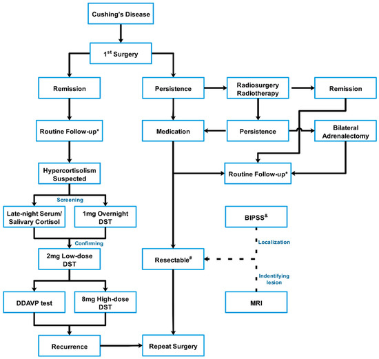
Figure 1. Algorithm of the biochemical assessment and treatment of persistent and recurrent Cushing’s disease.

Contrast-enhanced pituitary MRI at our center was conducted to facilitate diagnosis and surgical planning using a superconducting magnet 1.5/3.0 Tesla scanner (SIGNA; GE Healthcare, Chicago, IL, USA). Before gadolinium injection (0.01 mmol/kg gadopentetate dimeglumine; Magnevist, Berlex Laboratories, Inc., Montville, NJ, USA), T1-weighted spin echo and T2-weighted turbo spin echo images were obtained in the coronal and sagittal planes. Beginning simultaneously with gadolinium injection, coronal and sagittal T1-weighted spin echo images were obtained 2 min after the injection. Imaging studies were independently reviewed by a neuroradiologist, endocrinologist, and the patient’s neurosurgeon. Pituitary imaging prior to the first surgery performed outside of our center was acquired and re-interpreted by the same team. Full agreement was reached on the positive nature of the MRI findings. Otherwise, when MRI findings appeared normal or interpretation was ambiguous, the MRI findings were considered negative.

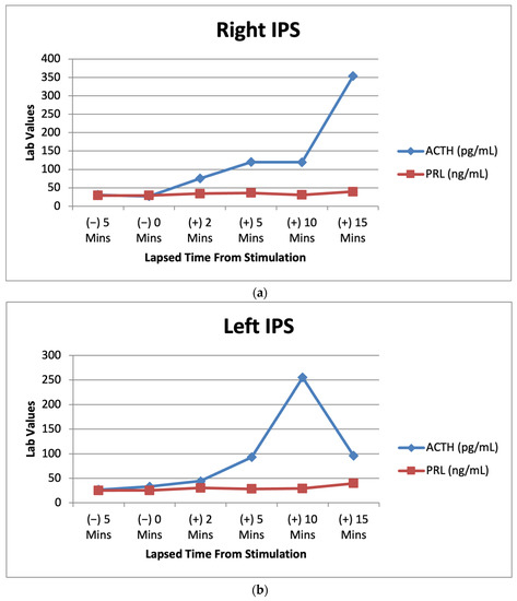
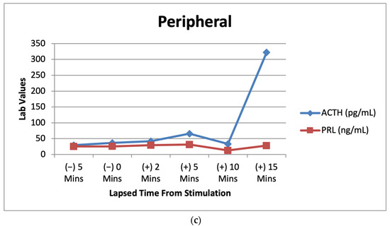
Meanwhile, bilateral inferior petrosal sinus sampling (BIPSS) with or without vasopressin (available after 2015) stimulation was performed in nine patients who experienced recurrence but lacked initially positive ACTH staining on the first histological examination to reconfirm whether the Cushing’s syndrome diagnosis was pituitary-dependent. Two patients were evaluated by BIPSS, although the initial pathology was positive. Regarding persistent disease, among eight patients without positive ACTH staining in their first pathological assessment, five were confirmed by positive BIPSS results and five were confirmed by visible radiological lesions. Only one patient with negative ACTH-staining adenoma underwent repeat surgery with either negative BIPSS results or negative imaging findings.

2.2. Surgical Procedure

The same surgeon performed surgery on all patients via the mononostril transsphenoidal approach under a microscope or endoscope (available from December 2015). The initial location prior to the first operation did not guide the resection during repeat surgery. For each patient with positive MRI results, the imaging-identified areas for adenoma were biopsied as frozen sections for the initial pathological evaluation. Subsequent resection with a rim of pituitary tissue around the tumor cavity was conducted to confirm neoplasm-free margins. No further exploration was performed before frozen pathology confirmation was available unless the BIPSS result showed an increased ACTH level on the other side.

For invisible tumors on MRI, the dura mater was opened widely to facilitate exploration of the whole gland, starting from the initial location on MRI before the first surgery or the side with the higher ACTH level in the BIPSS, if available. If no obvious tumor was identified on this side by the neurosurgeon intraoperatively, half of the gland was resected using the guidance of BIPSS lateralization.

If a tumor was frozen pathologically and identified after half of the gland was removed, the residual gland remained unresected and was only gently explored and sampled in the most suspected area. In some circumstances in which the frozen section was negative, it was subjected to a subtotal adenohypophysectomy of the intermediate lobe and neurohypophysis.

If invasive adenoma characteristics were also identified, the involved dura and medial wall of the cavernous sinus were resected or coagulated. A sample was collected for postoperative pathological confirmation, if available.

2.3. Outcome

Patients were defined as being in remission with an immediate postoperative serum cortisol nadir <5 μg/dL or 24 h UFC at a normal level [13]. Persistent hypercortisolism was defined as an increased postoperative UFC level, while recurrent hypercortisolism was defined as a reappearance of hypercortisolism after a period of normocortisolism or adrenal insufficiency.

2.4. Statistical Analysis

Descriptive statistics are presented as means ± standard deviations when normally distributed or medians and ranges when not normally distributed to describe patient outcome measures and incidence of remission among the study population. Statistical significance was set at a p value < 0.05. Fisher’s exact test was used to compare proportions of categorical measures between groups. All analyses were conducted using Instat (GraphPad Software, San Diego, CA, USA).

3. Results

3.1. Patient Characteristics

The basic information and perioperative evaluations of the two operations are shown in Table 1 and Table S1. Among all 27 recurrent cases, the preoperative MRI before the first operation showed a definite pituitary adenoma. The other 12 patients with persistent hypercortisolism had positive MRI findings before the first surgery. The remaining three patients with negative radiographic findings were diagnosed with CD and underwent the first transsphenoidal surgery (TSS) based on their endocrinological results.

For patients with confirmed persistent or recurrent CD, the imaging findings prior to the second operation of 14 individuals were negative (no solid evidence of tumors), and 28 clearly had positive results for the presence of a solid lesion. All patients who underwent a second surgery for recurrent or persistent hypercortisolism after the initial surgery were endocrinologically re-evaluated before the repeat surgery. There were 38 cases with positive HDDST results among 42 patients. BIPSS was performed in 18 patients with only one that did not reach the criteria of pituitary origin.

3.2. Outcome

In our study, 29 of 42 patients (69.0%, 22 recurrent and 7 persistent cases of CD) were in remission after the repeat operation without additional therapy during follow-up (Table S1). At follow-up, compared with patients with persistent disease, the recurrence group had a higher remission rate, although the difference was not significant (77.8% [21/27] vs. 57.1% [8/15]; p > 0.05; odds ratio = 2.625, 95% confidence interval = 0.651 to 10.586). Negative preoperative MRI findings were not associated with lower odds of immediate remission after repeat surgery (p > 0.05; odds ratio = 3.667, 95% confidence interval = 0.920 to 14.622; Table 2).

Table 2. The remission rate of the recurrent and persistent hypercortisolism patients with or without positive MRI findings.

Table

3.3. Association between Outcomes and MRI Findings

The remission rates of the persistent and recurrent disease groups with positive and negative MRI findings prior to the second procedure are shown in Table 2. Twenty-nine patients whose MRI findings revealed the existence of pituitary adenomas achieved successful outcomes after reoperation (Representative case, #19, Figure 2). The other seven patients who experienced recurrent or persistent hypercortisolism without clear imaging evidence of tumor appearance also benefited from reoperation (Representative case, #11, Figure 3).

Jcm 11 06848 g002 550

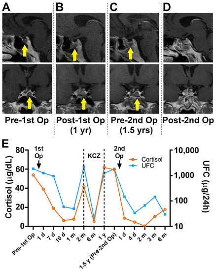
Figure 2. Preoperative and postoperative MR images of the two operations (AD) demonstrate an in situ relapsed intrasellar mass (yellow arrow). Biochemical results obtained before and after the operations (E) show the tumor-related hormone change. KCZ, ketoconazole; MR, magnetic resonance.

Jcm 11 06848 g003 550

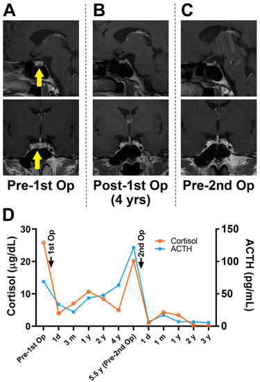
Figure 3. MR images (A) demonstrated a pituitary microadenoma on the left side (yellow arrow) before the first operation but not at the subsequent follow-ups (B,C). The biochemical results obtained before the second operation (D) revealed hypercortisolism indicating relapse without obvious MRI confirmation. MR, magnetic resonance; MRI, magnetic resonance imaging.

3.4. Pathology

Respectively, 15/27 (55.6%) and 7/15 (46.7%) patients with recurrent and persistent hypercortisolism had ACTH-positive staining in the first pathological findings. Among patients who achieved remission after the second operation, 20 of 29 patients had confirmed adenoma with positive ACTH pathological staining, while 3 patients with adenoma were ACTH-negative. There were five patients that did not achieve remission even though they had positive ACTH-staining adenoma in the second pathological examination. Meanwhile, five patients achieved remission, although no adenomas were found in their pathological specimens. Overall, positive pathology after either the initial or repeated surgery was not a significant predictor for remission after the second surgery.

3.5. Complications

Four of forty-two patients experienced major postoperative complications and underwent medical or surgical interventions. Most patients recovered well after the second operation, except in one case with persistent hypercortisolism, where a severe intracranial infection led to death. Another three cases with cerebral spinal fluid leakage related to the second operation were successfully surgically repaired afterwards.

Hypopituitarism was a common complication in this subgroup of CD. All of the patients in remission after the second TSS underwent glucocorticoid replacement therapy (hydrocortisone or cortisone), adjusted according to the 24 h UFC. A total of 20 patients (20/29, 68.9%) underwent thyroxine replacement therapy. Three patients (3/29, 10.3%) had permanent diabetes insipidus. In the non-remission group, five patients (5/13, 38.5%) experienced hypothyroidism, and two patients (2/13, 15.4%) had permanent diabetes insipidus.

4. Discussion

In the present study, we reported outcomes for 42 patients undergoing repeat TSS for recurrent and persistent disease in which an overall remission rate of 69.0% was achieved. Immediate remission rates after reoperation for recurrence have been reported in the literature up to 87% [13,14], which is similar to those of other second-line therapies such as radiation therapy and medical treatment. The CD recurrence rate after the initial TSS is reportedly 10–25% with a follow-up time of 10 years [15,16,17]. Ram et al. reported that surgeons performed a second TSS immediately after the first TSS when the postoperative serum cortisol level did not meet the standard level of remission. With an interval time of 1 to 6 weeks, 71% of patients with persistent disease achieved immediate remission, and 53% (9/17) achieved long-term remission [13]. Another study showed a remission rate of 70% with reoperation performed within 10 days [18]. A second TSS reportedly leads an additional 8% of patients to long-term CD remission [3]. Recurrence groups had slightly higher remission rates, which are insignificant when compared with persistent groups in the present study. Similar findings are demonstrated in the study by Ram et al. implicating that failure of the initial surgery suggested that the patient was more difficult to treat successfully with surgery than most patients with recurrence [13]. Therefore, the selection criteria for potential patients and reoperation strategies require further discussion.

4.1. Surgical Strategy

The surgical strategy for the initial CD surgery varies depending on the major concerns of different pituitary surgeons. Some surgeons intend to preserve more normal gland tissue during surgery while others chase higher remission rates. Selective adenectomy is a reasonable choice for visible tumors. Several authors adopted a slightly extended resection with a rim or sometimes 2–3 mm of like-normal tissue around the tumor, which could be considered a partial hypophysectomy [19,20]. A hemi-hypophysectomy is more common in cases in which no tumor was identified during the operation, and the MRI or BIPSS results indicated remarkable lateralization of the tumor origin [21]. Wide exploration of the contralateral side should also be conducted in cases in which BIPSS results are inconsistent with the MRI findings, which may help identify tiny tumors. More extensive procedures, including subtotal or sometimes total pituitary gland resection, have been performed to maximize remission rates up to 75.9–81.8% [20,22], which may be a reasonable recommendation when imaging/intraoperative findings are not definitive, considering the negative impacts on reoperated patients with persistent hypercortisolism rather than hypopituitarism. Interestingly, pathological confirmation rates are fairly low in cases with extended resection even though they show high remission rates. There seems to be a current trend of surgeons performing a partial hypophysectomy, as a total hypophysectomy can lead to hypopituitarism [5,22,23], given that it may not obviously increase remission rates and may decrease quality of life [24].

4.2. MRI Findings

Regarding radiological findings, we emphasize that negative MRI findings do not necessarily indicate the inexistence of pituitary adenomas or negative pathological results. A number of cases in the study by Wagenmakers et al. showed that remission achieved after repeated transsphenoidal surgery was not predictable by positive MRI findings before the first or second operation [10]. Preoperative MRI provides a reference for the diagnosis of pituitary adenomas, although it has a limited predictive function for patient prognosis [9], especially for the repeat operation in which the original anatomical structure was more or less destroyed in the initial surgery. A positive MRI finding before the second operation should promote confidence in surgeons. The remission rate after reoperation with positive MRI findings was reportedly as high as 72.7% [10]. According to our study, the two positive-MRI groups with different initial surgical outcomes showed higher remission rates, albeit insignificantly. Positive MRI findings suggest better endocrinological outcomes may be achieved by a second operation in both recurrent and persistent disease groups compared with patients with negative imaging findings. An excellent remission rate (more than 80%) was achieved in the recurrent group with positive MRI findings, thus encouraging a repeat TSS. An acceptable remission rate (over 60%) close to those of alternative treatment options was observed in the recurrent group with negative MRI findings, as well as the persistent group with positive MRI findings. We noted that one patient with persistent CD and negative MRI findings achieved remission after reoperation. Therefore, whether a second surgical treatment is beneficial for these patients should be carefully considered.

Regarding the recurrent or persistent cases of CD, patients underwent an initial surgery, and we regarded the MRI findings as a possible method to assist in decision making. A second operation is considered when visible lesions remain on MRI under the assumption that removal of the residual tumor leads to remission of the disease. Meanwhile, some recurrent and persistent patients with negative MRI findings also benefited from reoperation. Furthermore, MRI has its limitations in revealing the accurate structures of the originally operated area. The distortion and cicatrization from the previous operation and material packing in the sellar region lead to confusion [12,25]. Unlike the considerable remission rate achieved after the initial operation despite negative MRI findings, reoperation without certain lesion detection on MRI is associated with dissatisfactory remission rates [1], similar to the results of our study. Nevertheless, Knappe and Lüdecke [9] presented a different opinion regarding the significance of MRI findings and reported that it was not usually helpful for determining therapeutic strategies due to its low incidence of detecting existing microadenomas (missed diagnosis in 38–70% of cases). However, the BIPSS results in these cases in which MRI revealed no definitive information on tumors are therefore critical for surgeons to ascertain the pituitary origin of the disease, although another study suggested that MRI and BIPSS do not help locate recurrent tumors [10]. MRI may not help identify tumors in the cavernous sinus or other parasellar regions.

4.3. Pathology

We compared the pathological results and remission situations of recurrent patients and persistent patients and failed to find any relationship between pathological results and remission expectations. These findings are supported by the findings of Ram et al. [13], in which no tumors were found in 11 of 17 patients during the second procedure, and 6 of 11 patients achieved remission. In a series by Locatelli et al. [11], no tumors were found in 8 of 12 patients during the second operation, and 5 had surgical remissions. Even in cases of remission, the positive rate of pathological exams was not as high as expected. There was no significant difference in remission rates between patients grouped by pathological results or one-to-one correspondence between histopathological confirmation and surgical outcomes [11]. To date, little evidence supports the prediction of reoperation outcomes by either of the two pathology results.

4.4. Other Considerations and Factors

In patients with recurrent and persistent hypercortisolism after their first operation, it was difficult to identify solid lesions on MRI compared with the initial preoperative scans. Notably, BIPSS may provide more information, especially for patients who did not undergo this test before the first operation. Moreover, it may help avoid unnecessary repeat TSS in patients with persistent hypercortisolism by revealing false positives for pituitary ACTH overproduction. BIPSS results have the potential to not only confirm the pituitary origin of the condition (despite the fact that the first histological examination did not show ACTH-positive staining) but also to guide exploration and decision making for a hemi-hypophysectomy or accessing the cavernous sinus, especially for patients without obvious tumors identified intraoperatively. Careful dissection is highly recommended on the side of the obviously lateralized BIPSS results, which sometimes also indicate cavernous sinus invasion not shown on MRI and the necessity of opening the medical wall to achieve extended exploration. The predictive value of BIPSS lateralization in repeated surgery requires further investigation, although it is not optimal in native patients with CD [26].

According to a study by Lonser et al. [27], over 20% of CD patients had cavernous sinus invasion that was confirmed histologically. The authors advocated for complete resection, including the invaded sella dura and medial cavernous sinus wall by an experienced surgeon’s hands. Notably, endoscopy with magnification and lighting provides a panoramic view to facilitate extended exploration of the sella, including the cavernous sinus, compared with the microscope-based approach. Micko et al. demonstrated that an endoscope allows for a radical inspection of the entire medial wall of the cavernous sinus [28] and increases the lateral angle of visualizations to facilitate differentiation between tumor tissues and other tissues. These advantages over the microscopic transsphenoidal approach are critical for recurrent and unremitted cases; however, further studies with larger sample sizes are needed to verify this conclusion.

4.5. Other Adjunctive Treatments to Repeat Surgery

Previous studies have noted that ketoconazole may contribute to enhanced tumor appearance on MRI to facilitate pituitary resection in some circumstances [29]. Castinetti et al. reported that visible lesions may be identified on MRI in one-third of patients who were administered ketoconazole [30].

In the literature, reoperation for persistent cases without visible lesions on MRI is rarely satisfactory [31], although these patients may benefit from radiosurgery using the entire sellar region as the therapeutic target [32]. The hormonal normalization was achieved after radiosurgery in half of the cases, including those with negative MRI findings [33]. In general, the radiosurgery outcomes and the less commonly used radiotherapy are more favorable, particularly in MRI-negative cases with persistent hypercortisolism compared with repeat surgery, with potentially fewer complications and a shorter length of hospital stay [34,35]. Salvage TSS for refractory CD after radiation therapy has rarely been reported [36] owing to the difficulty of disrupting surgical landmarks, the formation of scar tissue, and the effects of preoperative radiotherapy [34].

Bilateral adrenalectomy is generally considered the ultima ratio in patients who fail to respond to other treatment options. However, patients who undergo bilateral adrenalectomy will require lifelong surveillance of the corticotroph tumor’s progression, which may lead to Nelson’s syndrome, via MRI and ACTH measurements. Most experts agree that selective transsphenoidal adenomectomy should be recommended as the first-line therapy in patients with Nelson’s syndrome before extrasellar expansion of the tumor occurs [37].

4.6. Limitations

Similar to previous studies, our sample size was not large enough to conduct powerful statistical analyses. Some patients lost during follow-up limited the evaluation of long-term outcomes in the current study. We observed a trend in the predictable values of positive preoperative MRI findings, which is not enough evidence to support an apparent relationship. A potential weakness of the present study is that the outcome was only focused on the biochemical benefits of remission after surgical intervention, possibly leading to an underestimation of the risks of hypopituitarism and decreased quality of life. Indeed, larger case series are needed to further investigate the potential predictive factors and best surgical strategy.

5. Conclusions

Patients with initial surgical treatment may experience hypercortisolism without positive MRI findings in both recurrent and persistent disease. Our findings suggest that for most patients who experience recurrent or persistent CD, reoperation should be an option even with negative MRI findings. However, further comprehensive investigation on recurrent or persistent CD patients is required. Larger groups of surgically treated CD patients with long follow-up periods should be evaluated to improve reoperation outcomes and determine the appropriate selection criteria for repeat surgery, especially for persistent CD patients.

Supplementary Materials

The following supporting information can be downloaded at: https://www.mdpi.com/article/10.3390/jcm11226848/s1, Table S1. Preoperative and postoperative evaluation of the repeated surgery of 42 patients.

Author Contributions

B.W. and Y.S. contributed to the study’s conception and design. S.Z. drafted the manuscript. J.R., Z.Z., H.J., Q.S., T.S. and W.W. contributed to data acquisition, analysis, and interpretation. B.W. and Y.S. critically revised the manuscript for important intellectual content. Y.S. and L.B. accept final responsibility for this article. All authors have read and agreed to the published version of the manuscript.

Funding

This work was supported in part by the National Natural Science Foundation of China (82000751) and the Shanghai Sailing Program (20YF1438900).

Institutional Review Board Statement

This study involving human participants was conducted in accordance with the 1964 Helsinki declaration and its later amendments or comparable ethical standards and was approved by the Ruijin Hospital Ethics Committee of Shanghai Jiao Tong University School of Medicine (approval number 2020-64).

Informed Consent Statement

The need for individual consent was waived by the Ethics Committee owing to the retrospective nature of the study.

Data Availability Statement

All data generated or analyzed during this study are included in this article. Further enquiries may be directed to the corresponding authors.

Conflicts of Interest

The authors have no relevant financial or non-financial interests to disclose.

References

  1. Sun, Y.; Sun, Q.; Fan, C.; Shen, J.; Zhao, W.; Guo, Y.; Su, T.; Wang, W.; Ning, G.; Bian, L. Diagnosis and therapy for Cushing’s disease with negative dynamic MRI finding: A single-centre experience. Clin. Endocrinol. 2012, 76, 868–876. [Google Scholar] [CrossRef]
  2. Tritos, N.A.; Biller, B.M.K. Current management of Cushing’s disease. J. Intern. Med. 2019, 286, 526–541. [Google Scholar] [CrossRef] [PubMed]
  3. Dimopoulou, C.; Schopohl, J.; Rachinger, W.; Buchfelder, M.; Honegger, J.; Reincke, M.; Stalla, G.K. Long-term remission and recurrence rates after first and second transsphenoidal surgery for Cushing’s disease: Care reality in the Munich Metropolitan Region. Eur. J. Endocrinol. 2014, 170, 283–292. [Google Scholar] [CrossRef] [PubMed]
  4. Rutkowski, M.J.; Flanigan, P.M.; Aghi, M.K. Update on the management of recurrent Cushing’s disease. Neurosurg. Focus 2015, 38, E16. [Google Scholar] [CrossRef] [PubMed]
  5. Burke, W.T.; Penn, D.L.; Repetti, C.S.; Iuliano, S.; Laws, E.R. Outcomes After Repeat Transsphenoidal Surgery for Recurrent Cushing Disease: Updated. Neurosurgery 2019, 85, E1030-e6. [Google Scholar] [CrossRef] [PubMed]
  6. Alexandraki, K.I.; Kaltsas, G.A.; Isidori, A.M.; Storr, H.L.; Afshar, F.; Sabin, I.; Akker, S.A.; Chew, S.L.; Drake, W.M.; Monson, J.P.; et al. Long-term remission and recurrence rates in Cushing’s disease: Predictive factors in a single-centre study. Eur. J. Endocrinol. 2013, 168, 639–648. [Google Scholar] [CrossRef]
  7. Espinosa-de-Los-Monteros, A.L.; Sosa-Eroza, E.; Espinosa, E.; Mendoza, V.; Arreola, R.; Mercado, M. Long-term outcome of the different treatment alternatives for recurrent and persistent cushing disease. Endocr. Pract. 2017, 23, 759–767. [Google Scholar] [CrossRef] [PubMed]
  8. Mastorakos, P.; Taylor, D.G.; Chen, C.-J.; Buell, T.; Donahue, J.H.; Jane, J.A. Prediction of cavernous sinus invasion in patients with Cushing’s disease by magnetic resonance imaging. J. Neurosurg. 2018, 130, 1593–1598. [Google Scholar] [CrossRef]
  9. Knappe, U.J.; Lüdecke, D.K. Persistent and recurrent hypercortisolism after transsphenoidal surgery for Cushing’s disease. Acta Neurochir. Suppl. 1996, 65, 31–34. [Google Scholar] [CrossRef]
  10. Wagenmakers, M.A.; Netea-Maier, R.T.; van Lindert, E.J.; Timmers, H.J.; Grotenhuis, J.A.; Hermus, A.R. Repeated transsphenoidal pituitary surgery (TS) via the endoscopic technique: A good therapeutic option for recurrent or persistent Cushing’s disease (CD). Clin. Endocrinol. 2009, 70, 274–280. [Google Scholar] [CrossRef]
  11. Locatelli, M.; Vance, M.L.; Laws, E.R. Clinical review: The strategy of immediate reoperation for transsphenoidal surgery for Cushing’s disease. J. Clin. Endocrinol. Metab. 2005, 90, 5478–5482. [Google Scholar] [CrossRef] [PubMed]
  12. Wang, F.; Zhou, T.; Wei, S.; Meng, X.; Zhang, J.; Hou, Y.; Sun, G. Endoscopic endonasal transsphenoidal surgery of 1166 pituitary adenomas. Surg. Endosc. 2015, 29, 1270–1280. [Google Scholar] [CrossRef]
  13. Ram, Z.; Nieman, L.K.; Cutler, G.B., Jr.; Chrousos, G.P.; Doppman, J.L.; Oldfield, E.H. Early repeat surgery for persistent Cushing’s disease. J. Neurosurg. 1994, 80, 37–45. [Google Scholar] [CrossRef] [PubMed]
  14. Aranda, G.; Enseñat, J.; Mora, M.; Puig-Domingo, M.; Martínez de Osaba, M.J.; Casals, G.; Verger, E.; Ribalta, M.T.; Hanzu, F.A.; Halperin, I. Long-term remission and recurrence rate in a cohort of Cushing’s disease: The need for long-term follow-up. Pituitary 2015, 18, 142–149. [Google Scholar] [CrossRef] [PubMed]
  15. Swearingen, B.; Biller, B.M.; Barker, F.G., 2nd; Katznelson, L.; Grinspoon, S.; Klibanski, A.; Zervas, N.T. Long-term mortality after transsphenoidal surgery for Cushing disease. Ann. Intern. Med. 1999, 130, 821–824. [Google Scholar] [CrossRef]
  16. Atkinson, A.B.; Kennedy, A.; Wiggam, M.I.; McCance, D.R.; Sheridan, B. Long-term remission rates after pituitary surgery for Cushing’s disease: The need for long-term surveillance. Clin. Endocrinol. 2005, 63, 549–559. [Google Scholar] [CrossRef]
  17. Patil, C.G.; Prevedello, D.M.; Lad, S.P.; Vance, M.L.; Thorner, M.O.; Katznelson, L.; Laws, E.R., Jr. Late recurrences of Cushing’s disease after initial successful transsphenoidal surgery. J. Clin. Endocrinol. Metab. 2008, 93, 358–362. [Google Scholar] [CrossRef]
  18. Trainer, P.J.; Lawrie, H.S.; Verhelst, J.; Howlett, T.A.; Lowe, D.G.; Grossman, A.B.; Savage, M.O.; Afshar, F.; Besser, G.M. Transsphenoidal resection in Cushing’s disease: Undetectable serum cortisol as the definition of successful treatment. Clin. Endocrinol. 1993, 38, 73–78. [Google Scholar] [CrossRef]
  19. Guilhaume, B.; Bertagna, X.; Thomsen, M.; Bricaire, C.; Vila-Porcile, E.; Olivier, L.; Racadot, J.; Derome, P.; Laudat, M.H.; Girard, F. Transsphenoidal pituitary surgery for the treatment of Cushing’s disease: Results in 64 patients and long term follow-up studies. J. Clin. Endocrinol. Metab. 1988, 66, 1056–1064. [Google Scholar] [CrossRef]
  20. Hammer, G.D.; Tyrrell, J.B.; Lamborn, K.R.; Applebury, C.B.; Hannegan, E.T.; Bell, S.; Rahl, R.; Lu, A.; Wilson, C.B. Transsphenoidal microsurgery for Cushing’s disease: Initial outcome and long-term results. J. Clin. Endocrinol. Metab. 2004, 89, 6348–6357. [Google Scholar] [CrossRef]
  21. Bakiri, F.; Tatai, S.; Aouali, R.; Semrouni, M.; Derome, P.; Chitour, F.; Benmiloud, M. Treatment of Cushing’s disease by transsphenoidal, pituitary microsurgery: Prognosis factors and long-term follow-up. J. Endocrinol. Investig. 1996, 19, 572–580. [Google Scholar] [CrossRef] [PubMed]
  22. Carr, S.B.; Kleinschmidt-DeMasters, B.K.; Kerr, J.M.; Kiseljak-Vassiliades, K.; Wierman, M.E.; Lillehei, K.O. Negative surgical exploration in patients with Cushing’s disease: Benefit of two-thirds gland resection on remission rate and a review of the literature. J. Neurosurg. 2018, 129, 1260–1267. [Google Scholar] [CrossRef] [PubMed]
  23. Rees, D.A.; Hanna, F.W.; Davies, J.S.; Mills, R.G.; Vafidis, J.; Scanlon, M.F. Long-term follow-up results of transsphenoidal surgery for Cushing’s disease in a single centre using strict criteria for remission. Clin. Endocrinol. 2002, 56, 541–551. [Google Scholar] [CrossRef] [PubMed]
  24. Santos, A.; Resmini, E.; Gómez-Ansón, B.; Crespo, I.; Granell, E.; Valassi, E.; Pires, P.; Vives-Gilabert, Y.; Martínez-Momblán, M.A.; de Juan, M.; et al. Cardiovascular risk and white matter lesions after endocrine control of Cushing’s syndrome. Eur. J. Endocrinol 2015, 173, 765–775. [Google Scholar] [CrossRef]
  25. Abe, T.; Tanioka, D.; Sugiyama, K.; Kawamo, M.; Murakami, K.; Izumiyama, H. Electromagnetic field system for transsphenoidal surgery on recurrent pituitary lesions—Technical note. Surg. Neurol. 2007, 67, 40–44; discussion 44–45. [Google Scholar] [CrossRef]
  26. Deipolyi, A.; Bailin, A.; Hirsch, J.A.; Walker, T.G.; Oklu, R. Bilateral inferior petrosal sinus sampling: Experience in 327 patients. J. Neurointerventional Surg. 2017, 9, 196–199. [Google Scholar] [CrossRef]
  27. Lonser, R.R.; Ksendzovsky, A.; Wind, J.J.; Vortmeyer, A.O.; Oldfield, E.H. Prospective evaluation of the characteristics and incidence of adenoma-associated dural invasion in Cushing disease. J. Neurosurg. 2012, 116, 272–279. [Google Scholar] [CrossRef]
  28. Micko, A.S.; Wöhrer, A.; Wolfsberger, S.; Knosp, E. Invasion of the cavernous sinus space in pituitary adenomas: Endoscopic verification and its correlation with an MRI-based classification. J. Neurosurg. 2015, 122, 803–811. [Google Scholar] [CrossRef] [PubMed]
  29. Lau, D.; Rutledge, C.; Aghi, M.K. Cushing’s disease: Current medical therapies and molecular insights guiding future therapies. Neurosurg. Focus 2015, 38, E11. [Google Scholar] [CrossRef]
  30. Castinetti, F.; Morange, I.; Jaquet, P.; Conte-Devolx, B.; Brue, T. Ketoconazole revisited: A preoperative or postoperative treatment in Cushing’s disease. Eur. J. Endocrinol. 2008, 158, 91–99. [Google Scholar] [CrossRef]
  31. Valderrábano, P.; Aller, J.; García-Valdecasas, L.; García-Uría, J.; Martín, L.; Palacios, N.; Estrada, J. Results of repeated transsphenoidal surgery in Cushing’s disease. Long-term follow-up. Endocrinol. Nutr. 2014, 61, 176–183. [Google Scholar] [CrossRef] [PubMed]
  32. Sheehan, J.M.; Vance, M.L.; Sheehan, J.P.; Ellegala, D.B.; Laws, E.R., Jr. Radiosurgery for Cushing’s disease after failed transsphenoidal surgery. J. Neurosurg. 2000, 93, 738–742. [Google Scholar] [CrossRef] [PubMed]
  33. Jagannathan, J.; Sheehan, J.P.; Jane, J.A. Evaluation and management of Cushing syndrome in cases of negative sellar magnetic resonance imaging. Neurosurg. Focus 2007, 23, E3. [Google Scholar] [CrossRef] [PubMed]
  34. Benveniste, R.J.; King, W.A.; Walsh, J.; Lee, J.S.; Delman, B.N.; Post, K.D. Repeated transsphenoidal surgery to treat recurrent or residual pituitary adenoma. J. Neurosurg. 2005, 102, 1004–1012. [Google Scholar] [CrossRef]
  35. Jahangiri, A.; Wagner, J.; Han, S.W.; Zygourakis, C.C.; Han, S.J.; Tran, M.T.; Miller, L.M.; Tom, M.W.; Kunwar, S.; Blevins, L.S., Jr.; et al. Morbidity of repeat transsphenoidal surgery assessed in more than 1000 operations. J. Neurosurg. 2014, 121, 67–74. [Google Scholar] [CrossRef]
  36. McCollough, W.M.; Marcus, R.B., Jr.; Rhoton, A.L., Jr.; Ballinger, W.E.; Million, R.R. Long-term follow-up of radiotherapy for pituitary adenoma: The absence of late recurrence after greater than or equal to 4500 cGy. Int. J. Radiat. Oncol. Biol. Phys. 1991, 21, 607–614. [Google Scholar] [CrossRef]
  37. Reincke, M.; Albani, A.; Assie, G.; Bancos, I.; Brue, T.; Buchfelder, M.; Chabre, O.; Ceccato, F.; Daniele, A.; Detomas, M.; et al. Corticotroph tumor progression after bilateral adrenalectomy (Nelson’s syndrome): Systematic review and expert consensus recommendations. Eur. J. Endocrinol. 2021, 184, P1–P16. [Google Scholar] [CrossRef]
Publisher’s Note: MDPI stays neutral with regard to jurisdictional claims in published maps and institutional affiliations.

© 2022 by the authors. Licensee MDPI, Basel, Switzerland. This article is an open access article distributed under the terms and conditions of the Creative Commons Attribution (CC BY) license (https://creativecommons.org/licenses/by/4.0/).

Share and Cite


MDPI and ACS Style

Wang, B.; Zheng, S.; Ren, J.; Zhong, Z.; Jiang, H.; Sun, Q.; Su, T.; Wang, W.; Sun, Y.; Bian, L. Reoperation for Recurrent and Persistent Cushing’s Disease without Visible MRI Findings. J. Clin. Med. 202211, 6848. https://doi.org/10.3390/jcm11226848

AMA Style

Wang B, Zheng S, Ren J, Zhong Z, Jiang H, Sun Q, Su T, Wang W, Sun Y, Bian L. Reoperation for Recurrent and Persistent Cushing’s Disease without Visible MRI Findings. Journal of Clinical Medicine. 2022; 11(22):6848. https://doi.org/10.3390/jcm11226848Chicago/Turabian Style

Wang, Baofeng, Shuying Zheng, Jie Ren, Zhihong Zhong, Hong Jiang, Qingfang Sun, Tingwei Su, Weiqing Wang, Yuhao Sun, and Liuguan Bian. 2022. “Reoperation for Recurrent and Persistent Cushing’s Disease without Visible MRI Findings” Journal of Clinical Medicine 11, no. 22: 6848. https://doi.org/10.3390/jcm11226848

Find Other Styles

Note that from the first issue of 2016, MDPI journals use article numbers instead of page numbers. See further details here.

Article Metrics

Citations

No citations were found for this article, but you may check on Google Scholar

Article Access Statistics

Article access statisticsArticle Views20. Nov21. Nov22. Nov23. Nov24. Nov25. Nov26. Nov27. Nov28. Nov29. Nov30. Nov1. Dec2. Dec3. Dec4. Dec5. Dec6. Dec7. Dec8. Dec9. Dec10. Dec0100200300400500

For more information on the journal statistics, click here.

Multiple requests from the same IP address are counted as one view.

From https://www.mdpi.com/2077-0383/11/22/6848

(Korlym) Corcept settles patent dispute with Hikma over Cushing’s syndrome drug

  • Corcept Therapeutics (NASDAQ:CORT) announced an agreement with Hikma Pharmaceuticals (OTCPK:HKMPF) USA on Thursday to resolve the ongoing patent lawsuit related to Korlym, an oral therapy indicated for patients with Cushing’s syndrome.
  • The litigation was filed in the New Jersey district court in 2021, shortly after Hikma (OTCPK:HKMPF) informed Corcept (CORT) about its submission of an Abbreviated New Drug Application (ANDA) seeking FDA approval for a generic version of Korlym.
  • Per the terms of the settlement, Corcept (CORT) has allowed Hikma (OTCPK:HKMPF) the rights to market a generic version of Korlym from Oct. 01, 2034, or earlier subject to certain conditions.
  • The companies plan to submit the agreement for the review of the U.S. Federal Trade Commission (FTC) and the United States Department of Justice (DOJ).
  • A similar patent lawsuit filed by Corcept (CORT) against the U.S. unit of Teva Pharmaceutical (TEVA) remains pending.
  • Thanks mainly to higher sales volumes of Korlym, Corcept (CORT) added $366.0M net product revenue in 2021 with ~3% YoY growth.


    • From

https://seekingalpha.com/news/3915421-cort-stock-on-watch-after-patent-settlement-with-hikma-for-korlym

Ketogenic Diet Initially Masks Symptoms of Hypercortisolism in Cushing’s Disease

Abstract

Cushing’s syndrome (CS) is a diagnosis used to describe multiple causes of serum hypercortisolism. Cushing’s disease (CD), the most common endogenous subtype of CS, is characterized by hypercortisolism due to a pituitary tumor secreting adrenocorticotropic hormone (ACTH). A variety of tests are used to diagnose and differentiate between CD and CS. Hypercortisolism has been found to cause many metabolic abnormalities including hypertension, hyperlipidemia, impaired glucose tolerance, and central adiposity. Literature shows that many of the symptoms of hypercortisolism can improve with a low carb (LC) diet, which consists of consuming <30 g of total carbohydrates per day. Here, we describe the case of a patient with CD who presented with obesity, hypertension, striae and bruising, who initially improved some of his symptoms by implementing a LC diet. Ultimately, as his symptoms persisted, a diagnosis of CD was made. It is imperative that practitioners realize that diseases typically associated with poor lifestyle choices, like obesity and hypertension, can often have alternative causes. The goal of this case report is to provide insight on the efficacy of nutrition, specifically a LC diet, on reducing metabolic derangements associated with CD. Additionally, we will discuss the importance of maintaining a high index of suspicion for CD, especially in those with resistant hypertension, obesity and pre-diabetes/diabetes.

1. Introduction

Cushing’s syndrome (CS) is a rare disorder of hypercortisolism related to exposure to high levels of cortisol (>20 mcg/dL between 0600–0800 or >10 mcg/dL after 1600) for an extended period [1,2]. CS affects 10 to 15 people per million and is more common among those with diabetes, hypertension, and obesity [3]. The metabolic derangements associated with CS include visceral obesity, elevated blood pressure, dyslipidemia, type II diabetes mellitus (T2DM) and insulin resistance [4]. CS physical exam findings include round face, dorsal fat pad, central obesity, abdominal striae, acne, and ecchymosis [3]. Other symptoms associated with CS include low libido, headache, change in menses, depression and lethargy [2,3,5]. The most common features of CS are weight gain, which is found in 82% of cases, and hypertension, which is found in 50–85% of cases [6]. CS can be caused by exogenous glucocorticoids, known as iatrogenic CS, ectopic ACTH secretion (EAS) from sources like a small cell lung cancer or adrenal adenoma, known as EAS CS, or excess production of ACTH from a pituitary tumor, known as CD [3]. In CD, ACTH subsequently causes increased production of cortisol from the adrenal glands. CD accounts for 80–85% of endogenous cases of CS [3]. Other conditions including alcoholism, depression, severe obesity, bulimia and anorexia nervosa can lead to a Cushing-like state, although are not considered true CS [3]. Many studies have demonstrated that LC diets can ameliorate some of the most common metabolic derangements seen in CD, namely hyperglycemia, weight gain, hypertension and insulin resistance.
A LC diet is a general term for diets which lower the total carbohydrates consumed per day [4]. A ketogenic diet is a subtype of LC that is described as having even fewer carbohydrates, typically less than 30 g/day. By reducing carbohydrate intake and thus limiting insulin production, the body achieves ketosis by producing an elevated number of ketones including β-hydroxybutyric acid, acetoacetic acid, and acetone, in the blood [7]. A carnivore diet, a specific type of a ketogenic diet, is defined as mainly eating animal food such as meat, poultry, eggs and fish. Contrarily, a standard American diet (SAD) is defined as a diet high in processed foods, carbs, added sugars, refined fats, and highly processed dairy products [8]. There are several therapeutic applications for LC diets that are currently supported by strong evidence. These include weight loss, cardiovascular disease, T2DM, and epilepsy. LC diets have clinical utility for acne, cancer, polycystic ovary syndrome (PCOS), and neurologic deficits [9].
In this case report, the patient endorsed initially starting a LC diet to address weight gain and high blood sugars that he noted on a glucometer. The patient noted a 35 pounds (lbs.) weight loss over the first 1.5 years on his LC diet, as well as improved blood pressure and in his overall health. He then adopted a carnivore diet but found that weight loss was difficult to maintain, although his body composition continued to improveand his clothes fit better. Later, he noted that his blood pressure would at times be poorly controlled despite multiple medications and strict dietary adherence. The patient reported “being in despair” and “not trusting his doctors” because they did not understand how much his diet had helped him. Despite strict adherence, his symptoms of insulin resistance and hypertension persisted. In this report, we will describe how his symptoms of CD were ameliorated by the ketogenic diet. This case report also highlights that when patients are unable to overcome hormonal pathology, clinicians should not blame patients for lack of adherence to a diet, but instead understand the need to evaluate for complex pathology.

2. Detailed Case Description

A male patient in his thirties, of Asian descent, had a past medical history of easy bruising, central obesity, headaches, hematuria, and hypertension and past family medical history of hypertension in his father and brother. In 2015, he was at his heaviest weight of 179 lbs. with a body mass index (BMI) of 28 kg/m2, placing him in the overweight category (25.0–29.9 kg/m2). At that time the patient reported he was following a SAD diet and was active throughout the day. The patient stated he ate a diet of vegetables, fruits and carbohydrates, but he was not able to lose weight. The patient stated that he switched to a LC diet, to address weight gain and hyperglycemia, and he reported that he lost approximately 35 lbs. in 1.5 years. The patient described his LC diet as eating green leafy vegetables, low carb fruits, fish, poultry, beef and dairy products. The patient then later switched to a carnivore diet. He noted despite aggressively adhering to his diet, that his weight-loss had plateaued, although his waist circumference continued to decrease. The patient noted his carnivore diet consisted of eating a variety of different meats, poultry, fish and eggs.
The metabolic markers seen in Table 1 were obtained after the patient had started a carnivore diet. The patient’s blood glucose levels decreased overtime despite impaired glucose metabolism being a known side effect of hypercortisolism [4]. The patient’s high-density lipoprotein (HDL) remained in a healthy range (40–59 mg/dL) and his triglycerides stayed in an optimal range (<100 mg/dL), despite dyslipidemia being a complication of CD [4]. When the patient was consuming a SAD diet, he was not under the care of a physician and was unable to provide us with previous biomarkers.
Table 1. Patient’s metabolic markers on a carnivore diet. Glucose (70 to 99 mg/dL), total cholesterol (desirable <200 mg/dL, borderline high 200–239 mg/dL, high >239 mg/dL), triglycerides (optimal: <100 mg/dL), HDL (low male: <40 mg/dL), low density lipoprotein (LDL) (Optimal: <100 mg/dL).
Table
Despite strict adherence to his diet and initial improvement in his weight, his blood pressure and his blood sugar levels, in October of 2021 the patient was admitted to the hospital for hypertensive urgency, with a blood pressure of 216/155. His complaints at the time were unexplained ecchymosis, hematuria and significant headaches that were resistant to Excedrin (acetaminophen-aspirin-caffeine) use. At the hospital, the patient underwent a computed tomography (CT) scan of the head and radiograph of the chest, and both images were negative for acute pathology. During his hospital admission, the patient denied any changes in vision, chest pain or edema of the legs. Ultimately, the patient was told to eat a low-salt diet and to follow-up with a cardiologist. At discharge, the patient was placed on hydrochlorothiazide, labetalol, amlodipine and lisinopril. The patient was then seen by his primary care physician in November of 2021 and his urinalysis at that time showed 30 mg/mL (Negative/Trace) of protein in his urine, without hematuria. The patient’s primary care physician discontinued his hydrochlorothiazide and started the patient on furosemide. Additionally, the primary care physician reinforced cutting out salt and limiting his calories to prevent any further weight gain, which his physician explained would contribute further to his hypertension. He was referred to hematology and oncology in November of 2021 for his symptoms of hematuria and abnormal ecchymosis to his abdomen, thighs and arms. The patient’s coagulation and platelet counts were normal, and his symptoms were noted to be improving. His hematuria and ecchymosis were attributed to his significant Excedrin use from the past 1–2 months, secondary to his headaches, and their anti-platelet effect. It was noted that the patient had significant hemolysis during his hospital admission. However, in his follow up examination, there were no signs of hemolysis, and it was attributed to his hypertensive urgency. Again, a low-salt, calorie-limited diet was recommended. The patient was referred to cardiology where he was evaluated for secondary hypertension, because despite his weight loss and his strict adherence to his diet, his blood pressure was still uncontrolled on multiple medications. He had a normal echocardiogram and renal ultrasound which showed no signs of renal artery stenosis bilaterally. At that time the patient’s serum renin, aldosterone and urine metanephrine levels were all normal. His cardiologist increased his lisinopril, and continued him on amlodipine, furosemide and labetalol and reinforced the recommendations of lowering his salt and preventing weight gain.
The patient first contacted our office in January of 2022. At that time his blood pressure was noted to be 160/120 despite being compliant with current blood pressure medications. The patient reported strict adherence to his carnivore diet by sharing his well-documented meals on his social media accounts. Given the persistent symptoms, despite his significant change in diet and weight loss, we were concerned that a hormonal etiology may be driving his symptoms. The patient was seen in-person, in our office, in March of 2022. At the request of the patient, we again reviewed his social media profile to assess his meal choices and diet. While the patient was eager to show us his carnivore meals, what we incidentally noted in his photos was despite weight loss and strict diet adherence, he had developed moon facies (Figure 1a,b). On the physical exam, we noted his prominent abdominal striae (Figure 2). Several screening tests for Cushing’s syndrome were ordered. A midnight salivary cortisol was ordered, with values of 0.884 ug/dL (<0.122 ug/dL) and 0.986 ug/dL (<0.122 ug/dL) and a urinary free cortisol excretion (UFC) was ordered, with values of 8.8 ug/L (5–64 ug/L). At this point our suspicion was confirmed that the patient had inappropriately elevated cortisol.
Metabolites 12 01033 g001 550
Figure 1. The patient’s progression of moon facies, (a) photo from 2019 after initial weight loss (b) photo from office visit in 2022.
Metabolites 12 01033 g002 550
Figure 2. The arrows demonstrate early striae visualized on the lower abdomen bilaterally, unclear in image due to poor office lighting.
Based on screening tests and significant physical exam findings, we referred the patient to endocrinology for a low dose dexamethasone suppression test (DST). They performed a low dose DST revealing a dehydroepiandrosterone (DHEA) of 678 ug/dL (89–427 ug/dL) and ACTH of 23.9 pg/mL (7.2–63.3 pg/mL). The low dose DST and midnight salivary cortisol were both positive indicating hypercortisolism. To begin determining the source of hypercortisolism, the plasma ACTH was evaluated and was 27.2 pg/mL (7.2–63.3 pg/mL). While ACTH was within normal range, a plasma ACTH > 20 pg/mL is suggestive of ACTH-dependent CS, so a magnetic resonance imaging (MRI) of the brain was ordered [2]. The MRI revealed a 4 mm heterogeneous lesion in the central pituitary gland which is suspicious of a cystic microadenoma. To confirm that a pituitary tumor was the cause of the patient’s increased cortisol, the patient was sent for inferior petrosal sinus sampling (IPSS). The results of the IPSS indicated an increase in ACTH in both inferior petrosal sinuses and peripheral after corticotropin-releasing hormone (CRH) stimulation (Figure 3a–c), which was consistent with hypercortisolism.
Metabolites 12 01033 g003a 550Metabolites 12 01033 g003b 550
Figure 3. (a) Right IPS venous sampling values for ACTH and prolactin after CRH stimulation over multiple time intervals. (b) Left IPS venous sampling values for ACTH and prolactin after CRH stimulation over multiple time intervals. (c) Peripheral sampling values for ACTH and prolactin after CRH stimulation over multiple time intervals.
Lab results from the patient’s IPSS venous sampling can be seen above. The graphs depict the lab values of ACTH (7.2–63.3 pg/mL) and prolactin (PRL) (2.1–17.7 ng/mL) before and after CRH stimulation during IPSS. PRL acts as a baseline to indicate successful catheterization in the procedure [10].
Using the ACTH levels from our patient’s IPSS we calculated a ratio of inferior petrosal sinus to peripheral (IPS:P). These results can be seen below (Table 2). The right IPS:P was calculated as 3.60 at 10 min and the left IPS:P as 7.65 at 10 min. These ratios confirmed that the hypercortisolism was due to the pituitary tumor, as it is higher than the 3:1 ratio necessary for diagnosis of CD [11]. The patient is currently scheduled to undergo surgical resection of the pituitary microadenoma.
Table 2. Right and left petrosal sinus to peripheral serum ACTH ratios.
Table

3. Clinical Evaluation for CS

In this case, the patient presented with uncontrolled hypertension, weight gain despite a strict diet, hyperglycemia, abdominal striae and moon facies. Despite evaluation, both inpatient and outpatient, a diagnosis of CS was not yet explored. When CS is suspected based on clinical findings, the use of exogenous steroids must first be excluded as it is the most common cause of hypercortisolism [3]. If there is still concern for CS, there are three screening tests that can be done which are sensitive but not specific for hypercortisolism. The screening tests include: a 24-h UFC, 2 late night salivary cortisol tests, low dose (1 g) DST [3]. To establish the preliminary diagnosis of hypercortisolism two screening tests must be abnormal [2].
The first step to determine the cause of hypercortisolism is to measure the plasma level of ACTH. Low values of ACTH < 5 pg/mL indicate the cause is likely ACTH-independent CS and imaging of the adrenal glands is warranted as there is a high suspicion of an adrenal adenoma [2,3]. When the serum ACTH is elevated >/20 pg/mL it is likely an ACTH-dependent form of CS [2]. To further evaluate an ACTH-dependent hypercortisolism, an MRI should be obtained as there is high suspicion that the elevated cortisol is coming from a pituitary adenoma. If there is a pituitary mass >6 mm there is a strong indication for the diagnosis of CD [2]. However, pituitary tumors can be quite small and can be missed on MRIs in 20–58% of patients with CD [2]. If there is still a high suspicion of CD with an inconclusive MRI, a high dose DST (8 g) is done. Patients with CD should not respond and their ACTH and DHEA, a steroid precursor, should remain high. Similarly, CRH stimulation test is done and patients with CD should have an increase in ACTH and/or cortisol within 45 min of CRH being given. If the patient has a positive high-dose DST, CRH-stimulation test and an MRI with a pituitary tumor >6 mm no further testing is needed as it is likely the patient has CD [2]. If either of those tests are abnormal, the MRI shows a pituitary tumor < 6 mm, or there is diagnostic ambiguity, the patient should undergo IPSS with ACTH measurements before and after the administration of CRH [4]. IPSS is the gold standard for determining the source of ACTH secretion and confirming CD. In this invasive procedure, ACTH, prolactin, and cortisol levels are sampled prior to CRH stimulation and after CRH stimulation. PRL acts as a baseline to indicate successful catheterization in the procedure [12]. To confirm CD, a ratio of IPS:P is calculated for values prior to and after CRH stimulation. A peak ratio greater than 2.0 before CRH stimulation or a peak ratio greater than 3.0 after CRH stimulation is indicative of CD. In comparing the right and left petrosal sinus sample, an IPS:P ratio greater than 1.4 suggests adenoma lateralization. However, due to high variability, IPSS should not be used for diagnosing lateralization [13].

4. Discussion

Surgical intervention remains the primary treatment for CD [4]. However, remission is not guaranteed as symptoms and metabolic diseases have been shown to persist afterwards. In the literature it has been shown that nutrition can have a powerful impact on suppressing, or even reversing metabolic disorders and comorbidities associated with CD. A LC diet has been shown to promote significant weight loss, reduce hypertension, improve dyslipidemia, reverse T2DM and improve cortisol levels (2, 14–15, 18–21).
There are reports of weight loss on a LC diet in the literature. A LC significantly reduced weight and BMI of 30 male subjects [14]. In a group of 120 participants over 24 weeks who followed a LC versus low fat (LF) diet, showed a greater weight loss in the LC group vs. the LF group [15]. Patients diagnosed and treated for CD found that their weight remained largely unchanged even after treatment [6]. In many cases, surgical treatment does not always resolve the associated comorbidity of central adiposity in CD. In such cases, a LC diet can be used before, during and after treatment, as an adjunct, to decrease associated weight gain and comorbidities.
Nutritional intervention can be a powerful adjunct to reduce comorbidities associated with CD. As seen in this case report, the patient’s symptoms of CD, especially hypertension and weight gain, improved with dietary changes despite him having a pituitary microadenoma. Multiple studies showed that a LC diet was able to decrease blood pressure parameters. In a group of 120 participants over 24 weeks who followed a LC versus a LF diet showed a greater decrease in both systolic and diastolic blood pressure in the LC group vs. the LF group [15]. Other literature which studied the effect of a LC diet on hypertension demonstrated the reduction of blood pressure and is thought to be due to ketogenesis. It is thought the production of ketones have a natriuretic effect on the body therefore lowering systemic blood pressure [16].
A LC diet improves lipid profiles and inflammatory markers associated with metabolic syndrome [14]. Literature shows that a LC diet has a greater impact on decreasing triglyceride levels and increasing HDL levels, when compared to a LF diet [15]. Triglyceride levels in patients in CD remission remained high [17]. Therefore, it can be hypothesized that a LC diet would be beneficial, in addition to standard CD treatment, to lower the associated comorbidity of hypertriglyceridemia and metabolic syndrome.
Insulin resistance, a precursor to T2DM, is a common comorbidity of hypercortisolism which can be treated with a LC diet. One study showed that in subjects with T2DM, a decrease in A1c and a reduction in antidiabetic therapy were seen with consumption of a LC diet [18]. Additionally, a cohort of 9 participants following a LC diet were able to collectively lower their A1c on average by 1% while concurrently discontinuing various antidiabetic therapies including insulin [19].
Literature shows that a LC diet can minimize systemic cortisol levels through various mechanisms. Current treatment of CD includes medications which block cortisol production and/or cortisol secretion [2]. LC can imitate similar results seen through medication intervention for CD. Carbohydrate restriction can lower cortisol levels, as carbohydrates stimulate adrenal cortisol secretion and extra-adrenal cortisol regeneration [4]. A ketogenic diet can lower the level of ghrelin, a peptide produced in the stomach that has orexigenic properties [20,21]. Literature shows that ghrelin increases levels of serum cortisol [22]. Therefore, implementing a ketogenic diet would decrease ghrelin, and subsequently minimize the effects of increased ghrelin on serum cortisol. A LC diet decreases visceral fat which itself is an endocrine organ and can increase the synthesis of cortisol [14]. Therefore, decreasing visceral fat also decreases the production of cortisol. A LC was shown to significantly reduced weight, BMI and cortisol levels of 30 obese male subjects [14]. Further, a LC diet excludes foods with a high glycemic index which cause increased stress on the body which subsequently leads to the activation of the hypothalamic-pituitary-axis which causes increased levels of cortisol [14].
This case report illustrated how a LC diet was initially successful at ameliorating the patient’s associated symptoms of hypertension and obesity, making his diagnosis of CD go undetected. Literature shows that while the prevalence of CS on average is a fraction of a percent, it is much higher among patients with poorly controlled diabetes, hypertension and early onset osteoporosis [3]. Two hundred patients with diabetes mellitus were studied and 5.5% were found to have CS [23]. Another study discovered that in subjects with CD, 36.4% were found to have hyperlipidemia, 73.1% with hypertension, and 70.2% with impaired glucose metabolism [17]. It can be concluded that a higher index of suspicion and lower threshold for screening for CS may be necessary in obese and diabetic patient populations. A lower threshold for screening can allow for earlier diagnosis for many patients, and therefore provide better outcomes for those diagnosed with CS.
It is important for clinicians to consider alternative pathology for patients combating metabolic derangements. As depicted in this case, the patient lost 35 lbs. while on a LC diet, despite having hypercortisolism, presumably for months to years prior to the diagnosis of his condition. The patient noted a tendency to gain weight, have elevated blood sugar and blood pressure which prompted him to begin self-treatment with increasingly strict carbohydrate restriction. The patient was able to keep his symptoms of hypercortisolism managed, potentially making the diagnosis difficult for his team of clinicians. From a diagnostic perspective, it’s important to understand that strict dietary adherence can have profound impacts on even the most severe hormonal pathology. Ultimately, this case serves as a reminder of the power of nutrition to address metabolic derangements and simultaneously as a reminder to diagnosticians to never rely on lack of dietary adherence as a reason for persistent metabolic symptoms. The reflexive advice to “not gain weight” and “lower salt intake” in retrospect appears both dogmatic and careless. In this case, the patient had seen several doctors and was even hospitalized and yet his disease state remained unclear and the dietary messaging cursory.

5. Conclusions

Many chronic diseases, including diabetes, hypertension and obesity, are generally thought to be caused by dietary and lifestyle choices. However, as exemplified in this report underlying medical problems, such as endocrine disorders, can be the cause of such metabolic derangements. It is critical that practitioners consider other causes of metabolic derangements, as assuming that they are due to poor dietary adherence, can allow them to go undiagnosed. While there is extensive literature on LC diets and their effect on the metabolic derangements associated with hypercortisolism, there needs to be further research on LC as an adjunctive therapy to conventional CD treatment. Ultimately, nutrition can have a powerful impact on suppressing, or even reversing metabolic disorders. As depicted in this case study, a LC diet is powerful enough to temporarily suppress symptoms of CD.

Author Contributions

M.K.D., E.-C.P.-M. and T.K. equally contributed to this case report. All authors have read and agreed to the published version of the manuscript.

Funding

This research received no external funding.

Institutional Review Board Statement

Not applicable.

Informed Consent Statement

Written informed consent has been obtained from the patient to publish this paper.

Data Availability Statement

The data presented in this study are available in article.

Acknowledgments

We would like to thank our patients and the Society of Metabolic Health Practitioners.

Conflicts of Interest

T.K. is an unpaid member of the Board of Directors of the Society of Metabolic Health Practitioners and a producer of podcasts on health and nutrition, with all proceeds donated to humanitarian charities; his spouse has ownership interest in a food company. The other author reports no conflicts of interest.

References

  1. Nieman, L.K. UpToDate. Available online: https://www.uptodate.com/contents/measurement-of-cortisol-in-serum-and-saliva?search=cortisol%20level&source=search_result&selectedTitle=1~150&usage_type=default&display_rank=1 (accessed on 27 September 2022).
  2. Feelders, R.; Sharma, S.; Nieman, L. Cushing’s Syndrome: Epidemiology and Developments in Disease Management. Clin. Epidemiol. 2015, 7, 281. [Google Scholar] [CrossRef] [PubMed]
  3. Guaraldi, F.; Salvatori, R. Cushing Syndrome: Maybe Not so Uncommon of an Endocrine Disease. J. Am. Board Fam. Med. 2012, 25, 199–208. [Google Scholar] [CrossRef] [PubMed]
  4. Guarnotta, V.; Emanuele, F.; Amodei, R.; Giordano, C. Very Low-Calorie Ketogenic Diet: A Potential Application in the Treatment of Hypercortisolism Comorbidities. Nutrients 2022, 14, 2388. [Google Scholar] [CrossRef] [PubMed]
  5. Nieman, L.K. UpToDate. Available online: https://www.uptodate.com/contents/epidemiology-and-clinical-manifestations-of-cushings-syndrome?search=cushings%20diagnosis%20symptoms&source=search_result&selectedTitle=2~150&usage_type=default&display_rank=2 (accessed on 27 September 2022).
  6. Schernthaner-Reiter, M.H.; Siess, C.; Gessl, A.; Scheuba, C.; Wolfsberger, S.; Riss, P.; Knosp, E.; Luger, A.; Vila, G. Factors Predicting Long-Term Comorbidities in Patients with Cushing’s Syndrome in Remission. Endocrine 2018, 64, 157–168. [Google Scholar] [CrossRef] [PubMed]
  7. Giordano, C.; MarchiÃ2, M.; Timofeeva, E.; Biagini, G. Neuroactive Peptides as Putative Mediators of Antiepileptic Ketogenic Diets. Front. Neurol. 2014, 5, 63. [Google Scholar] [CrossRef]
  8. Standard American Diet (SAD). Available online: https://piviohealth.com/knowledge-bank/glossary/standard-american-diet-sad/ (accessed on 2 October 2022).
  9. Paoli, A.; Rubini, A.; Volek, J.S.; Grimaldi, K.A. Beyond Weight Loss: A Review of the Therapeutic Uses of Very-Low-Carbohydrate (Ketogenic) Diets. Eur. J. Clin. Nutr. 2013, 67, 789–796. [Google Scholar] [CrossRef] [PubMed]
  10. Sharma, S.T.; Nieman, L.K. Is Prolactin Measurement of Value during Inferior Petrosal Sinus Sampling in Patients with ACTH-Dependent Cushing’s Syndrome? J. Endocrinol. Investig. 2013, 36, 1112–1116. [Google Scholar] [CrossRef]
  11. Kline, G.; Chin, A.C. Chapter 5—Adrenal disorders. In Endocrine Biomarkers: Clinical Aspects and Laboratory Determination; Elsevier: Amsterdam, The Netherlands, 2017; Available online: https://www.sciencedirect.com/science/article/pii/B9780128034125000057 (accessed on 18 October 2022).
  12. Ghorbani, M.; Akbari, H.; Griessenauer, C.J.; Wipplinger, C.; Dastmalchi, A.; Malek, M.; Heydari, I.; Mollahoseini, R.; Khamseh, M.E. Lateralization of Inferior Petrosal Sinus Sampling in Cushing’s Disease Correlates with Cavernous Sinus Venous Drainage Patterns, but Not Tumor Lateralization. Heliyon 2020, 6, e05299. [Google Scholar] [CrossRef]
  13. Knecht, L. Inferior Petrosal Sinus Sampling in the Diagnosis of Cushing’s Disease. Available online: https://csrf.net/doctors-articles/inferior-petrosal-sinus-sampling-diagnosis-cushings-disease/ (accessed on 18 October 2022).
  14. Polito, R.; Messina, G.; Valenzano, A.; Scarinci, A.; Villano, I.; Monda, M.; Cibelli, G.; Porro, C.; Pisanelli, D.; Monda, V.; et al. The Role of Very Low Calorie Ketogenic Diet in Sympathetic Activation through Cortisol Secretion in Male Obese Population. J. Clin. Med. 2021, 10, 4230. [Google Scholar] [CrossRef] [PubMed]
  15. Yancy, W.S.; Olsen, M.K.; Guyton, J.R.; Bakst, R.P.; Westman, E.C. A Low-Carbohydrate, Ketogenic Diet versus a Low-Fat Diet to Treat Obesity and Hyperlipidemia. Ann. Intern. Med. 2004, 140, 769. [Google Scholar] [CrossRef] [PubMed]
  16. Khan, S.S.; Ning, H.; Wilkins, J.T.; Allen, N.; Carnethon, M.; Berry, J.D.; Sweis, R.N.; Lloyd-Jones, D.M. Association of Body Mass Index with Lifetime Risk of Cardiovascular Disease and Compression of Morbidity. JAMA Cardiol. 2018, 3, 280–287. [Google Scholar] [CrossRef]
  17. Sun, X.; Feng, M.; Lu, L.; Zhao, Z.; Bao, X.; Deng, K.; Yao, Y.; Zhu, H.; Wang, R. Lipid Abnormalities in Patients with Cushing’s Disease and Its Relationship with Impaired Glucose Metabolism. Front. Endocrinol. 2021, 11, 600323. [Google Scholar] [CrossRef] [PubMed]
  18. Bolla, A.; Caretto, A.; Laurenzi, A.; Scavini, M.; Piemonti, L. Low-Carb and Ketogenic Diets in Type 1 and Type 2 Diabetes. Nutrients 2019, 11, 962. [Google Scholar] [CrossRef] [PubMed]
  19. Norwitz, N.G.; Soto-Mota, A.; Kalayjian, T. A Company Is Only as Healthy as Its Workers: A 6-Month Metabolic Health Management Pilot Program Improves Employee Health and Contributes to Cost Savings. Metabolites 2022, 12, 848. [Google Scholar] [CrossRef] [PubMed]
  20. Ebbeling, C.B.; Feldman, H.A.; Klein, G.L.; Wong, J.M.W.; Bielak, L.; Steltz, S.K.; Luoto, P.K.; Wolfe, R.R.; Wong, W.W.; Ludwig, D.S. Effects of a Low Carbohydrate Diet on Energy Expenditure during Weight Loss Maintenance: Randomized Trial. BMJ 2018, 363, k4583. [Google Scholar] [CrossRef] [PubMed]
  21. Marchiò, M.; Roli, L.; Lucchi, C.; Costa, A.M.; Borghi, M.; Iughetti, L.; Trenti, T.; Guerra, A.; Biagini, G. Ghrelin Plasma Levels after 1 Year of Ketogenic Diet in Children with Refractory Epilepsy. Front. Nutr. 2019, 6, 112. [Google Scholar] [CrossRef] [PubMed]
  22. Kärkkäinen, O.; Farokhnia, M.; Klåvus, A.; Auriola, S.; Lehtonen, M.; Deschaine, S.L.; Piacentino, D.; Abshire, K.M.; Jackson, S.N.; Leggio, L. Effect of Intravenous Ghrelin Administration, Combined with Alcohol, on Circulating Metabolome in Heavy Drinking Individuals with Alcohol Use Disorder. Alcohol. Clin. Exp. Res. 2021, 45, 2207–2216. [Google Scholar] [CrossRef] [PubMed]
  23. Catargi, B.; Rigalleau, V.; Poussin, A.; Ronci-Chaix, N.; Bex, V.; Vergnot, V.; Gin, H.; Roger, P.; Tabarin, A. Occult Cushing’s Syndrome in Type-2 Diabetes. Available online: https://academic.oup.com/jcem/article/88/12/5808/2661485 (accessed on 27 September 2022).
Publisher’s Note: MDPI stays neutral with regard to jurisdictional claims in published maps and institutional affiliations.

Recurrent Neuroendocrine Tumor of the Cervix Presenting With Ectopic Cushing’s Syndrome

Abstract

Neuroendocrine carcinomas (NEC) of the cervix are a rare disease entity and account for only 1-2% of cervical carcinomas. The small-cell variant is the most common, with a worse prognosis and a higher rate of lymphatic and hematogenous metastases when compared with other subtypes of NEC. The diagnosis is usually made when the extra-pelvic disease is already apparent. Cushing’s syndrome due to adrenocorticotropic hormone (ACTH)-secreting tumors of the cervix is exceedingly rare. To date, there have been no reported cases in the literature of Cushing’s syndrome induced by the recurrence of metastases years after the initial diagnosis. This is a case of recurrent small-cell neuroendocrine carcinoma of the cervix presenting with Cushing’s syndrome five years after her original diagnosis. We present here the workup, management, and follow-up of this patient, including multisystemic, coordinated medical care.

Introduction

Neuroendocrine carcinomas (NECs) are heterogenous groups of tumors derived from neuroendocrine cells. NECs of the cervix are rare and account for 1-2% of all cervical carcinomas, with the small-cell variant being the most common [1,2]. Small-cell NECs have a high rate of lymphatic and hematogenous metastasis even when the carcinoma is limited to the cervix. Patients usually present at a late stage, with the extra-pelvic disease being apparent at the time of diagnosis [2]. Among the different histologic variants of NEC of the cervix, the small-cell variant has the highest rate of recurrence [3]. Adrenocorticotropic hormone (ACTH)-secreting tumors of the cervix are rare [4]. We present a case of recurrent metastatic NEC of the cervix five years after the original diagnosis of NEC of the cervix, now presenting with Cushing’s syndrome [1,2].

Case Presentation

A 39-year-old female with a history of recurrent small-cell cervical cancer presented to the emergency department (ED) of our hospital with complaints of weight gain, generalized facial edema, lightheadedness, tingling sensation of her entire face, bilateral leg edema, and abdominal distention.

Her problems started a month prior to her ED visit, when she started to complain of abdominal distention. She had a computed tomography (CT) abdomen with contrast, which revealed evidence of metastatic disease, including multiple large liver lesions (Figure 1). Subsequently, she had a positron emission tomography (PET) scan, which confirmed the presence of hypermetabolic lesions in the right peritonsillar tissue, liver, right lower quadrant of the abdomen, and bilateral pulmonary nodules with lymphadenopathy in the left hilum (Figure 2). A liver biopsy was done, with the final pathology consistent with recurrent NEC of the cervix. She was started on cisplatin, etoposide, and atezolizumab by gynecologic oncology but started to develop facial swelling and progressive abdominal distention, prompting this ED consult and subsequent admission.

Abdomial-CT-with-contrast-done-one-month-prior-showed-evidence-of-metastatic-disease-including-multiple-large-liver-lesions.
Figure 1: Abdomial CT with contrast done one month prior showed evidence of metastatic disease including multiple large liver lesions.
PET/CT-demonstrated-the-presence-of-hypermetabolic-lesions-in-the-liver-and-right-lower-quadrant-of-the-abdomen.
Figure 2: PET/CT demonstrated the presence of hypermetabolic lesions in the liver and right lower quadrant of the abdomen.

She had a significant medical history of being diagnosed with cervical cancer (FIGO stage 1B2 NEC) five years prior by gynecologic oncology, at which time she underwent concurrent chemo-radiation followed by surgical assessment of her pelvic lymph nodes with robotic pelvic lymph node dissection and bilateral ovarian transposition to avoid premature menopause. She was subsequently treated with cisplatin and pelvic radiation. She had a follow-up cervical biopsy several months after chemotherapy, which showed persistent NEC, but her PET scan showed no evidence of metastatic disease. After undergoing a robotic total laparoscopic hysterectomy, the final pathology showed a persistent microscopic focus of NEC of the cervix with negative margins. She received adjuvant chemotherapy with cisplatin and etoposide for six cycles with regular follow-up pap smears and annual PET scans, with no evidence of recurrence for five years.

On admission, her vital signs were: blood pressure = 129/79 mm Hg, pulse rate = 85/min, respiratory rate = 18/min, and temperature = 98.5 °F (36.9 °C). Her physical examination was notable for moon facies (a noticeable change from her pictures as recent as two months prior), supraclavicular and dorsocervical fat pads, multiple bruises on her arms, edema of her face and legs, acne of her face and neck, and hair growth of her chin area. No purple striae were seen on the abdomen.

Laboratory tests revealed leukopenia and thrombocytopenia (which were attributed to her chemotherapy), recently diagnosed diabetes (occasional hyperglycemia and HbA1c 7.7%), and electrolyte imbalances (hypokalemia and hypophosphatemia) (Table 1).

Sodium 142 mEq/L (135–145 mEq/L)
Potassium 2.0 mEq/L (3.5–5.0 mEq/L)
Chloride 98 mEq/L (98–108 mEq/L)
CO2 35 mEq/L (21–32 mEq/L)
Anion gap 9 mEq/L (8–16 mEq/L)
BUN 14 mg/dL (7–13 mEq/L)
Creatinine 1.13 mg/dL (0.6–1.1 mg/dL)
Glucose 460 mg/dL (74–100 mg/dL)
Calcium 7.8 mg/dL (8.5–10.1 mg/dL)
Phosphorous 1.0 mg/dL (2.5–4.5 mg/dL)
Albumin 2.5 mg/dL (3.1–4.5 mg/dL)
AST 43 U/L (15–27 U/L)
ALT 76 U/L (12–78 U/L)
White blood cell count 0.6 k/cmm (4.5–10.0 k/cmm)
Red blood cell count 3.55 million cells/μL (3.7–5 × 2)
Hemoglobin 11.9 g/dL (12.0–16.0)
Hematocrit 34.3% (35.0–47.0)
Platelet 45 k/cmm (150–440 k/cmm)
Table 1: Initial laboratory work showed leukopenia, thrombocytopenia, hyperglycemia, hypokalemia, and hypophosphatemia.

AST: aspartate aminotransferase, CO2: carbon dioxide, BUN: blood urea nitrogen, ALT: alanine aminotransferase.

Her chest X-ray showed bilateral pleural effusions. Magnetic resonance imaging (MRI) of the brain showed no evidence of pituitary masses, abnormalities, or metastatic disease in the brain. A CT of the chest showed new bilateral non-calcified lung nodules when compared to the previous PET scan, pathologic-sized left hilar adenopathy, and multiple peripherally enhancing hepatic nodules and masses (Figure 3). The adrenal glands were unremarkable. Workup for facial swelling and bilateral leg edema showed no evidence of superior vena cava (SVC) syndrome on both her chest CT and transthoracic echocardiogram.

Contrast-enhanced-chest-CT-showing-bilateral-noncalcified-lung-nodules.
Figure 3: Contrast-enhanced chest CT showing bilateral noncalcified lung nodules.

She was admitted to the intensive care unit (ICU) and started on empiric antibiotics and filgrastim for neutropenia. Replacement therapy for both hypokalemia and hypophosphatemia was given. After both electrolytes were normalized, the patient was started on basal-bolus insulin therapy.

Based on her clinic presentation of excessive weight gain, new-onset hyperglycemia, hypertension with hypokalemia, and a history of NEC, suspicion of Cushing’s syndrome was high. Further workup showed elevated serum cortisol after 1 mg overnight dexamethasone suppression, elevated 24-hour urine cortisol, and elevated midnight salivary cortisol, which confirmed Cushing’s syndrome (Table 2). ACTH was also elevated, but dehydroepiandrosterone sulfate (DHEAS) was normal. Thyroid function tests showed a slightly low free thyroxine, but this was attributed to an acute illness.

HgbA1C 7.7% (4.0-6.0%)
ACTH 1207 pg/mL (7.2–63.3 pg/mL)
24-hour urine cortisol 7070 μg/24 hr (6–42 μg/24 hr)
Salivary cortisol >1.000 μg /dL (0.025–0.600 μg/dL)
Serum cortisol after 1 mg overnight dexamethasone suppression 143.0 μg/dL (3.1–16.7 μg/dL)
Total testosterone 77 ng/dL (14–76 ng/dL)
DHEAS 250.0 μg/dL (57.3–279.2 μg/dL)
Chromogranin A 970.9 ng/mL (0.0–101.8 ng/mL)
TSH 0.572 mIU/L (0.358–3.74mIU/L)
Free T4 0.70 ng/dl (0.76–1.46) ng/dl
Table 2: Work up showed elevated ACTH, elevated 24-hour urine cortisol, elevated salivary cortisol, and elevated serum cortisol after 1 mg overnight dexamethasone suppression test.

HgbA1C: hemoglobin A1C; ACTH: adrenocorticotropic hormone; DHEAS: dehydroepiandrosterone sulfate; TSH: thyroid stimulating hormone; free T4: free thyroxine.

A diagnosis of Cushing’s syndrome due to metastatic small-cell neuroendocrine carcinoma of the cervix was assumed. A bilateral adrenalectomy, which is the definitive treatment of hypercortisolism when surgical removal of the source of excess ACTH is done, was not done because gynecologic oncology wanted to treat her with chemotherapy urgently due to her metastases and the nature of the disease and felt that surgery and recovery would delay the start of chemotherapy. Ketoconazole was felt to be a poor choice in the setting of liver metastases with worsening liver function tests. The patient was thus started on mifepristone 300 mg daily, as it is indicated for hypercortisolism secondary to endogenous Cushing’s syndrome with diabetes. Nephrology was consulted, and potassium supplementation was transitioned to oral potassium chloride 40 meq tablets four times a day; spironolactone 50 mg twice daily was added for the hypokalemia and hypertension, which occurred after the patient started bevacizumab. Hypokalemia is a common side effect of mifepristone therapy due to the glucocorticoid receptor blockade, which leads to cortisol’s spillover effect on unopposed mineralocorticoid receptors. She was discharged home with a basal-bolus insulin regimen.

Her posthospitalization course was complicated by compression fractures of her lumbar spine one week after discharge with no history of falls. An MRI of the spine showed chronic compression fractures of the T11-L3 vertebral bodies with no evidence of osseous metastatic disease. Dual-energy X-ray absorptiometry (DXA) scan interpretation demonstrated osteoporosis. Vertebral fracture assessment showed morphometric fractures in the lower thoracic and upper lumbar vertebrae. She was subsequently treated with IV administration of 5 mg of zoledronic acid. She was also readmitted multiple times after her initial admission due to the patient’s developing neutropenic fever, which was treated with filgrastim and antibiotics.

After starting mifepristone, her glycemic control improved to the point that insulin therapy could be subsequently discontinued. Her liver enzymes normalized, and ketoconazole was subsequently added for adjunct therapy to treat hypercortisolism, but the dose could not be optimized due to persistently elevated liver function tests. Hypokalemia management and resistant hypertension were additional challenges encountered by this patient.

At her follow-up visits, she had notably lost weight with the improvement of her leg edema. She continued to follow up with a nephrologist on an outpatient basis, and her normal potassium levels were normal on 40 meq of oral potassium chloride tablets four times a day and spironolactone 150 mg twice a day. She was followed up closely by her gynecologic oncologist and was on bevacizumab, topotecan, and paclitaxel before her unfortunate demise a few months later.

Discussion

Cushing’s syndrome due to ectopic ACTH secretion only represents 9-18% of cases. Most primary endocrine tumors responsible for ectopic ACTH secretion are located in the chest [5]. Abdominal and retroperitoneal neuroendocrine tumors are the second- and third-most reported sites [5]. Neuroendocrine tumors of the cervix are incredibly rare [6-9].

A unique feature of this case is that the patient presented with Cushing’s syndrome due to neuroendocrine tumor metastases found five years after the primary site of the tumor was resected. For this patient, a biopsy of the liver confirmed a metastatic neuroendocrine tumor, but it is unknown if the other sites of metastases are implicated in the production of excess ACTH.

The management of this disease focuses on controlling hypercortisolism, consequent hyperglycemia, and hypokalemia. Surgical excision of ACTH-secreting neuroendocrine tumors is the most effective, but in cases where that is not possible, bilateral adrenalectomy and medical treatment are the next best treatments for this disease entity [10]. For this patient, bilateral adrenalectomy was not done as gynecologic oncology wanted to treat her with chemotherapy urgently due to the metastases and nature of the disease and felt that surgery and recovery would delay the start of chemotherapy.

We provided medical management for the patient’s hypercortisolism. Pharmacological therapy for hypercortisolism can be categorized into immediate-acting steroidogenesis inhibitors (metyrapone, ketoconazole, and etomidate), slow-acting cortisol-lowering drugs (mitotane), and glucocorticoid receptor antagonists (mifepristone) [5]. We initially chose mifepristone because it is indicated in patients with type 2 diabetes mellitus and could be given safely despite the patient’s worsening liver function levels [11].

As demonstrated, the management of recurrent hypokalemia proved challenging in this patient. The phenomenon is well known to be induced by ectopic ACTH. Several mechanisms contribute to this. Activation of renal tubular type 1 (mineralocorticoid) receptors by cortisol is thought to be the mechanism that applies mainly to patients with severe hypercortisolism due to ectopic ACTH secretion. Additionally, there may also be an increase in the production of renin substrate from the liver. The high serum cortisol concentrations may not be completely inactivated by 11β-hydroxysteroid dehydrogenase type 2 in the kidney and overwhelm its ability to convert cortisol to cortisone, resulting in activation of mineralocorticoid receptors resulting in potassium loss in the distal tubules [12]. Hypokalemia may also result from adrenal hypersecretion of mineralocorticoids, such as deoxycorticosterone and corticosterone. This can also be amplified by mifepristone, as it is a glucocorticoid receptor antagonist that increases circulating cortisol levels [12].

Complications such as hypokalemia, hyperglycemia, acute respiratory distress syndrome, infections, muscle wasting, hypertension, and bone fractures can occur and can arise at any time throughout the course of the disease when urine-free cortisol is fivefold or more above the upper limit of normal [5]. Ketoconazole was initially considered for medical treatment, but due to mildly elevated liver enzymes during the initial presentation, we decided to use mifepristone instead. A small cohort study showed that severe hypercortisolism and increased baseline transaminase levels could be due to cortisol-induced hepatic steatosis [13]. Later in her course, ketoconazole was added to her mifepristone therapy to decrease adrenal cortisol production. Unfortunately, her dose could not be increased due to the patient’s persistently elevated liver enzymes.

Recurrent pancytopenia due to chemotherapy contributed to the protracted nature of this patient’s clinical course. Due to cortisol’s immunosuppressive and anti-inflammatory effects, opportunistic infections can arise [14]. Since her initial hospitalization, she has been readmitted several times due to neutropenic fever, which was treated with filgrastim and antibiotics.

Conclusions

Ectopic Cushing’s syndrome due to metastatic neuroendocrine small-cell carcinoma is a rare condition with a poor prognosis. The options for treatment are few and not necessarily curative. There needs to be increased awareness of this serious and rare complication. Managing the condition can be a challenge and requires a multidisciplinary team approach to improve outcomes.


References

  1. Cohen JG, Kapp DS, Shin JY, et al.: Small cell carcinoma of the cervix: treatment and survival outcomes of 188 patients. Am J Obstet Gynecol. 2010, 203:347.e1-6. 10.1016/j.ajog.2010.04.019
  2. Salvo G, Gonzalez Martin A, Gonzales NR, Frumovitz M: Updates and management algorithm for neuroendocrine tumors of the uterine cervix. Int J Gynecol Cancer. 2019, 29:986-95. 10.1136/ijgc-2019-000504
  3. Stecklein SR, Jhingran A, Burzawa J, Ramalingam P, Klopp AH, Eifel PJ, Frumovitz M: Patterns of recurrence and survival in neuroendocrine cervical cancer. Gynecol Oncol. 2016, 143:552-7. 10.1016/j.ygyno.2016.09.011
  4. Chen J, Macdonald OK, Gaffney DK: Incidence, mortality, and prognostic factors of small cell carcinoma of the cervix. Obstet Gynecol. 2008, 111:1394-402. 10.1097/AOG.0b013e318173570b
  5. Young J, Haissaguerre M, Viera-Pinto O, Chabre O, Baudin E, Tabarin A: Management of Endocrine Disease: Cushing’s syndrome due to ectopic ACTH secretion: an expert operational opinion. Eur J Endocrinol. 2020, 182:R29-58. 10.1530/EJE-19-0877
  6. Hashi A, Yasumizu T, Yoda I, et al.: A case of small cell carcinoma of the uterine cervix presenting Cushing’s syndrome. Gynecol Oncol. 1996, 61:427-31. 10.1006/gyno.1996.0168
  7. Iemura K, Sonoda T, Hayakawa A, et al.: Small cell carcinoma of the uterine cervix showing Cushing’s syndrome caused by ectopic adrenocorticotropin hormone production. Jpn J Clin Oncol. 1991, 21:293-8.
  8. Barghouthi N, Perini J, Cheng J: Ectopic adrenocorticotropic hormone production: a case of neuroendocrine cervical small cell carcinoma presenting as Cushing syndrome. AACE Clin Case Rep. 2018, 4:e367-e369. 10.4158/ACCR-2018-0080
  9. Di Filippo L, Vitali G, Taccagni G, Pedica F, Guaschino G, Bosi E, Martinenghi S: Cervix neuroendocrine carcinoma presenting with severe hypokalemia and Cushing’s syndrome. Endocrine. 2020, 67:318-20. 10.1007/s12020-020-02202-x
  10. Ilias I, Torpy DJ, Pacak K, Mullen N, Wesley RA, Nieman LK: Cushing’s syndrome due to ectopic corticotropin secretion: twenty years’ experience at the National Institutes of Health. J Clin Endocrinol Metab. 2005, 90:4955-62. 10.1210/jc.2004-2527
  11. Biller BM, Grossman AB, Stewart PM, et al.: Treatment of adrenocorticotropin-dependent Cushing’s syndrome: a consensus statement. J Clin Endocrinol Metab. 2008, 93:2454-62. 10.1210/jc.2007-2734
  12. Fleseriu M, Biller BM, Findling JW, Molitch ME, Schteingart DE, Gross 😄 Mifepristone, a glucocorticoid receptor antagonist, produces clinical and metabolic benefits in patients with Cushing’s syndrome. J Clin Endocrinol Metab. 2012, 97:2039-49. 10.1210/jc.2011-3350
  13. Young J, Bertherat J, Vantyghem MC, Chabre O, Senoussi S, Chadarevian R, Castinetti F: Hepatic safety of ketoconazole in Cushing’s syndrome: results of a Compassionate Use Programme in France. Eur J Endocrinol. 2018, 178:447-58. 10.1530/EJE-17-0886
  14. Sarlis NJ, Chanock SJ, Nieman LK: Cortisolemic indices predict severe infections in Cushing syndrome due to ectopic production of adrenocorticotropin. J Clin Endocrinol Metab. 2000, 85:42-47. 10.1210/jcem.85.1.6294


From https://www.cureus.com/articles/111698-recurrent-neuroendocrine-tumor-of-the-cervix-presenting-with-ectopic-cushings-syndrome

Levoketoconazole Treatment in Endogenous Cushing’s Syndrome

Objective: This extended evaluation (EE) of the SONICS study assessed effects of levoketoconazole for an additional 6 months following open-label, 6-month maintenance treatment in endogenous Cushing’s syndrome.

Design/Methods: SONICS included dose-titration (150–600 mg BID), 6-month maintenance, and 6-month EE phases. Exploratory efficacy assessments were performed at Months 9 and 12 (relative to start of maintenance). For pituitary MRI in patients with Cushing’s disease, a threshold of ≥2 mm denoted change from baseline in largest tumor diameter.

Results: Sixty patients entered EE at Month 6; 61% (33/54 with data) exhibited normal mean urinary free cortisol (mUFC). At Months 9 and 12, respectively, 55% (27/49) and 41% (18/44) of patients with data had normal mUFC. Mean fasting glucose, total and LDL-cholesterol, body weight, body mass index, abdominal girth, hirsutism, CushingQoL, and BDI-II scores improved from study baseline at Months 9 and 12. Forty-six patients completed Month 12; 4 (6.7%) discontinued during EE due to adverse events. The most common adverse events in EE were arthralgia, headache, hypokalemia, and QT prolongation (6.7% each). No patient experienced ALT or AST >3× ULN, QTcF interval >460 msec, or adrenal insufficiency during EE. Of 31 patients with tumor measurements at baseline and Month 12 or follow-up, largest tumor diameter was stable in 27 (87%) patients, decreased in 1, and increased in 3 (largest increase 4 mm).

Conclusion: In the first long-term levoketoconazole study, continued treatment through 12-month maintenance period sustained the early clinical and biochemical benefits in most patients completing EE, without new adverse effects.

Read the whole article at https://eje.bioscientifica.com/configurable/content/journals$002feje$002faop$002feje-22-0506$002feje-22-0506.xml?t%3Aac=journals%24002feje%24002faop%24002feje-22-0506%24002feje-22-0506.xml&body=pdf-45566