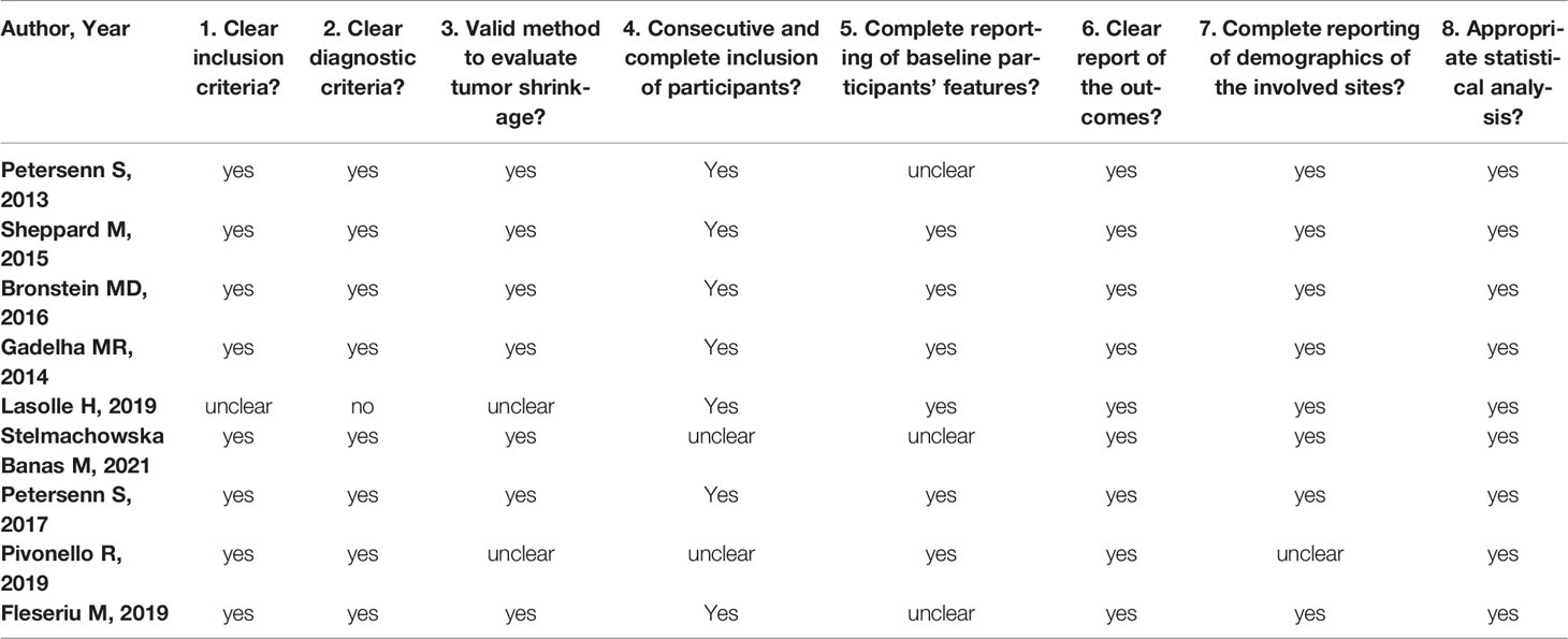
Characterization of Adrenal miRNA-Based Dysregulations in Cushing’s Syndrome

Abstract

MiRNAs are important epigenetic players with tissue- and disease-specific effects. In this study, our aim was to investigate the putative differential expression of miRNAs in adrenal tissues from different forms of Cushing’s syndrome (CS). For this, miRNA-based next-generation sequencing was performed in adrenal tissues taken from patients with ACTH-independent cortisol-producing adrenocortical adenomas (CPA), from patients with ACTH-dependent pituitary Cushing’s disease (CD) after bilateral adrenalectomy, and from control subjects. A confirmatory QPCR was also performed in adrenals from patients with other CS subtypes, such as primary bilateral macronodular hyperplasia and ectopic CS. Sequencing revealed significant differences in the miRNA profiles of CD and CPA. QPCR revealed the upregulated expression of miR-1247-5p in CPA and PBMAH (log2 fold change > 2.5, p < 0.05). MiR-379-5p was found to be upregulated in PBMAH and CD (log2 fold change > 1.8, p < 0.05). Analyses of miR-1247-5p and miR-379-5p expression in the adrenals of mice which had been exposed to short-term ACTH stimulation showed no influence on the adrenal miRNA expression profiles. For miRNA-specific target prediction, RNA-seq data from the adrenals of CPA, PBMAH, and control samples were analyzed with different bioinformatic platforms. The analyses revealed that both miR-1247-5p and miR-379-5p target specific genes in the WNT signaling pathway. In conclusion, this study identified distinct adrenal miRNAs as being associated with CS subtypes.

1. Introduction

Cushing’s syndrome (CS) results from the excessive secretion of cortisol, leading to visceral obesity, resistance to insulin, osteoporosis, and altered lipid and glucose metabolism [1,2]. Excessive production of cortisol by the adrenal glands can be either ACTH-dependent or -independent. In the majority of patients, hypercortisolism is due to ACTH secretion by corticotroph adenomas of the pituitary gland (Cushing’s disease, CD) or by ectopic tumors [3]. Approximately 20% of cases are ACTH-independent, where cortisol is secreted autonomously by the adrenal cortex. The pathology of ACTH-independent cases is diverse; they are most often caused by unilateral cortisol-producing adrenocortical adenomas (CPA). Rare causes are cortisol-secreting adrenocortical carcinomas (ACC), primary bilateral macronodular adrenocortical hyperplasia (PBMAH), bilateral CPAs, and primary pigmented nodular adrenal disease (PPNAD) [4,5]. Irrespective of the subtype, prolonged exposure to cortisol in CS is associated with increased mortality and cardiovascular morbidity in its patients [6]. Treatment is based on the underlying cause of hypercortisolism, with pituitary surgery or adrenalectomy being the preferred choice. Medical therapy options in CS are few and consist of pituitary-directed drugs, steroid synthesis inhibitors, and glucocorticoid receptor antagonists [7]. For the timely diagnosis and targeted management of CS and its subtypes, a comprehensive understanding of cortisol secretion, in terms of canonical signaling pathways as well as upstream epigenetic factors, is needed.
MiRNA molecules have emerged as key epigenetic players in the transcriptional regulation of cortisol production. Briefly, the deletion of Dicer in adrenals, a key miRNA processing enzyme, revealed diverse expression changes in miRNAs along with related changes in steroidogenic enzymes such as Cyp11b1 [8]. Furthermore, key enzymes in the cortisol biosynthesis pathway, namely Cyp11a1, Cyp21a1, Cyp17a1, Cyp11b1, and Cyp11b2, were also found to be regulated by various miRNAs (miRNA-24, miRNA-125a-5p, miRNA-125b-5p, and miRNA-320a-3p) in in vitro studies [9]. Consequently, various studies have also characterized miRNA expression profiles in CS subtypes. Importantly, miRNA expression in the corticotropinomas of CD patients was found to vary according to USP8 mutation status [10]. Other studies have also identified specific miRNA candidates and associated target genes in the adrenals of patients with PPNAD [11], PBMAH [12,13], and massive macronodular adrenocortical disease [14]. Interestingly, no common miRNA candidates were found among these studies, indicating the specificity of miRNAs to the different underlying pathologies in CS.
There are limited studies directly comparing miRNA expression profiles of ACTH-dependent and ACTH-independent CS patients. Consequently, in our previous study, we found differences in expression profiles when comparing circulating miRNAs in CD and CPA patients [15]. We hypothesized that the presence of ACTH possibly influences the miRNA profile in serum due to the upstream differential expression in the origin tissues. In this study, we aim to further explore this hypothesis by comparing the miRNA expression profile of adrenal tissues in ACTH-dependent and ACTH-independent CS. In brief, miRNA specific sequencing was performed in two prevalent subtypes of CS: in CD, the most prevalent ACTH-dependent form; and in CPA, the most prevalent ACTH-independent form. Specific miRNA candidates related to each subtype were further validated in other forms of CS. To further investigate our hypothesis, the response of miRNA candidates following ACTH stimulation was assessed in mice, and the expression of miRNAs in murine adrenals was subsequently investigated. Finally, an extensive targeted gene analysis was performed based on in silico predictions, RNA-seq data, and luciferase assays.

2. Results

2.1. Differentially Expressed miRNAs

NGS revealed differentially expressed miRNAs between the different groups analyzed (Figure 1). CD and CPA taken together as CS showed a differentially expressed profile (42 significant miRNAs) in comparison to controls. Moreover, individually, CPA and CD were found to show a significantly different expression profile in comparison to controls (n = 38 and n = 17 miRNAs, respectively). Interestingly, there were no significantly upregulated genes in the adrenals of patients with CD in comparison to the control adrenals. A comparative analysis of the top significant miRNAs (log2 fold change (log2 FC) > 1.25 & p < 0.005) between the two groups was performed and the representative Venn diagrams are given in Figure 2. Briefly, miR-1247-5p, miR-139-3p, and miR-503-5p were significantly upregulated in CPA, in comparison to both CD and controls. Furthermore, miR-150-5p was specifically upregulated in CPA as compared to CD. Several miRNAs (miR-486-5p, miR-551b-3p, miR-144-5p, miR-144-3p, and miR-363-3p) were found to be significantly downregulated in the groups of CPA and CD in comparison to controls. MiR-19a-3p and miR-873-5p were found to be commonly downregulated in CPA in comparison to both CD and controls. Principal component analyses based on miRNA sequencing did not identify any major clusters among the samples. Furthermore, the miRNA profile was not different among the CPA samples based on the mutation status of PRKACA (Supplementary Materials Figure S1).
Ijms 23 07676 g001 550
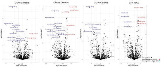
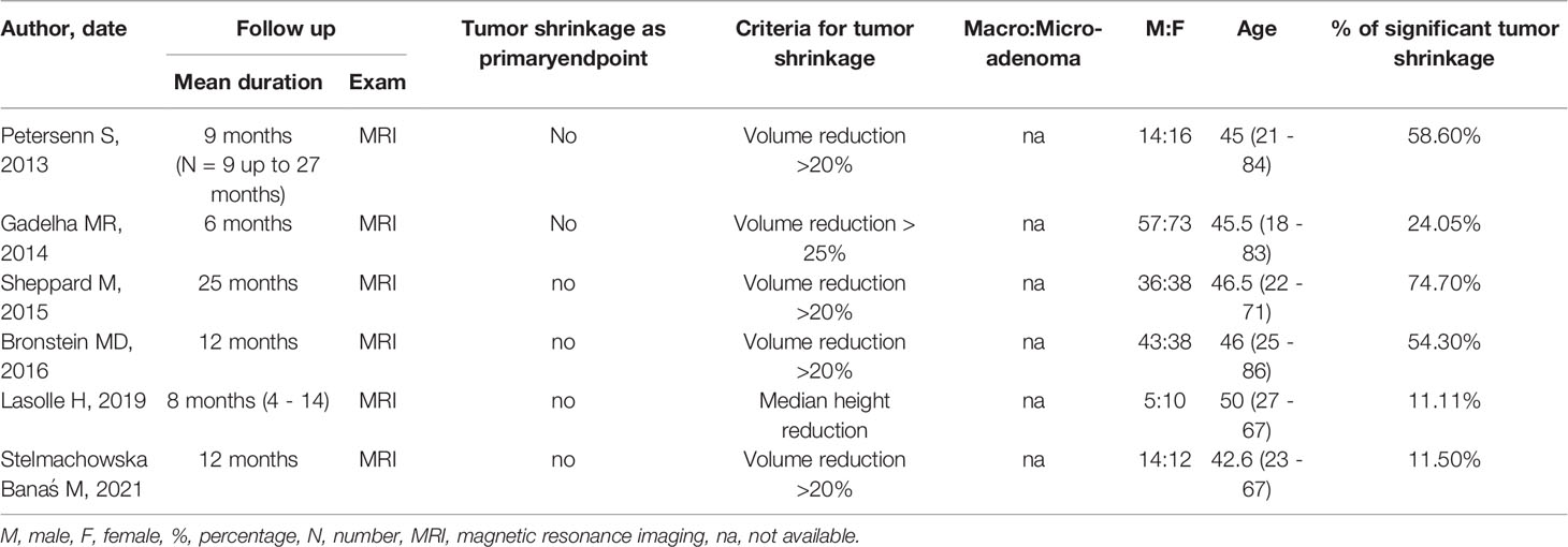
Figure 1. Differentially expressed miRNAs from sequencing. Volcano plot showing the relationship between fold change (log2 fold change) and statistical significance (−log10 p value). The red points in the plot represent significantly upregulated miRNAs, while blue points represent significantly downregulated miRNAs. CPA, cortisol producing adenoma; CD, Cushing’s disease; Cushing’s syndrome represents CPA and CD, taken together.
Ijms 23 07676 g002 550
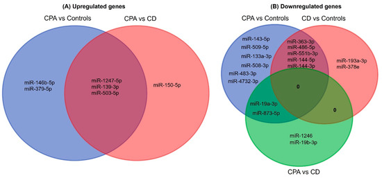
Figure 2. Venn analyses of the common significant miRNAs from each group. The significantly expressed miRNAs from each sequencing analysis were shortlisted and compared between the groups. CPA, cortisol producing adenoma; CD, Cushing’s disease.

2.2. Validation and Selection of Candidate miRNAs

For validation by QPCR, the most significant differentially expressed miRNAs (log2 FC > 1.25 & p < 0.005) among the groups were chosen (Table S1). According to the current knowledge, upregulated miRNAs are known to contribute more to pathology than downregulated miRNAs [16]. Since the total number of significantly upregulated miRNAs was six, all these miRNAs were chosen for validation. Contrarily, 25 miRNAs were significantly downregulated among the groups. In particular, miR-486-5p, miR-551b-3p, miR-144-5p, miR-144-3p, and miR-363-3p were found to be commonly downregulated in the CS group in comparison to controls; therefore, these miRNAs were chosen for validation.
Among the upregulated miRNA candidates, miR-1247-5p QPCR expression confirmed the NGS data (Figure 3A, Table S1). Moreover, miR-150-5p and miR-139-3p were upregulated in CPA specifically in comparison to CD, and miR-379-5p was upregulated in CD in comparison to controls by QPCR. In the case of downregulated genes, none of the selected miRNAs could be confirmed by QPCR (Figure 3B). Thus, analysis of the six upregulated and five downregulated miRNAs from NGS yielded two significantly upregulated miRNA candidates, miR-1247-5p in CPA and miR-379-5p in CD, when compared to controls. These miRNA candidates were taken up for further QPCR validation in an independent cohort of other subtypes of CS (Figure 4), namely ACTH-dependent ectopic CS (n = 3) and ACTH-independent PBMAH (n = 10). The QPCR analysis in the other subtypes revealed miR-1247-5p to be consistently upregulated in ACTH-independent CS (PBMAH and CPA) in comparison to ACTH-dependent CS (CD and ectopic CS) and controls. On the other hand, miR-379-5p was upregulated in CD and PBMAH in comparison to controls.
Ijms 23 07676 g003 550
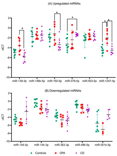
Figure 3. QPCR analyses of significant miRNAs from sequencing analyses. Data are represented as mean ± standard deviation (SD) of −dCT values: (A) Expression analysis of significantly upregulated miRNAs; (B) Expression analysis of common significantly downregulated miRNAs. Housekeeping gene: miR-16-5p. Statistics: ANOVA test with Bonferroni correction to detect significant differences between patient groups with at least a significance of p-value < 0.05 (*).
Ijms 23 07676 g004 550
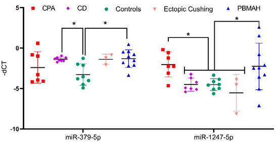
Figure 4. QPCR analyses of significantly upregulated miRNAs from validation QPCR. Data are represented as mean ± standard deviation (SD) of −dCT values. Housekeeping gene: miR-16-5p. Statistics: ANOVA test with Bonferroni correction to detect significant differences between patient groups with at least a significance of p-value < 0.05 (*).

2.3. In Vivo Assessment of ACTH-Independent miR-1247-5p

To analyze the influence of ACTH on miRNA expression, the expression of miR-1247-5p and miR-379-5p were assessed in the adrenal tissues of ACTH stimulated mice at different time points. For this analysis, miR-96-5p was taken as a positive control, as it has previously been reported to be differentially expressed in ACTH stimulated mice [17]. The analyses revealed that the expression of miR-1247-5p and miR-379-5p did not change at different timepoints of the ACTH stimulation (Figure 5). Meanwhile, the positive control of mir-96-5p showed a dynamic expression pattern with upregulation after 10 min, followed by downregulation at the subsequent 30 and 60 min time points, in concordance with previously reported findings [18].
Ijms 23 07676 g005 550
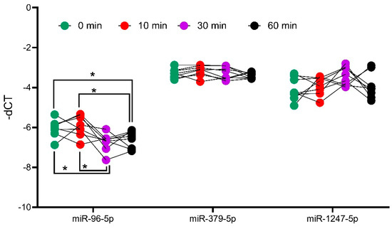
Figure 5. Analysis of miRNA expression in ACTH stimulated mice tissue. QPCR analyses of positive controls, miR-96-5p, and candidates miR-379-5p and miR-1247-5p. Mice were injected with ACTH, and adrenals were collected at different timepoints to assess the impact of ACTH on miRNA expression. Data are represented as mean ± standard deviation (SD) of −dCT values. Housekeeping gene: miR-26a-5p. Statistics: ANOVA test with Bonferroni correction to detect significant differences between patient groups with at least a significance of p-value < 0.05 (*).

2.4. In Silico Analyses of miRNA Targets

Two diverse approaches were employed for a comprehensive in silico analysis of the miRNA targets. First, the predicted targets of miR-1247-5p and miR-379-5p were taken from the TargetScan database, which identified miRNA–mRNA target pairs based on sequence analyses [19]. The expression status of these targets was then checked in the RNA sequencing data from CPA vs. controls (miR-1247-5p) and PBMAH vs. controls (miR-379-5p). Targets that showed significant expression changes in the sequencing data were shortlisted (Figure 6A). Among the 1061 predicted miR-1247-5p targets, 28 genes were found to show significant expression changes in CPA (20 upregulated, 8 downregulated). On the other hand, for 124 predicted miR-379-5p targets, 23 genes were found to show significant expression changes in PBMAH (20 upregulated, 3 downregulated). Interestingly, the selected targets were found to be unique for each miRNA, except for FICD (FIC domain protein adenylyltransferase) (Figure 6B).
Ijms 23 07676 g006 550
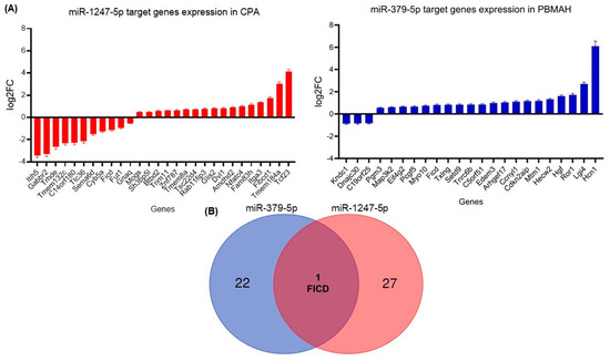
Figure 6. (A) Differentially expressed target genes of miRNAs from sequencing. Data are represented as log2 fold change in comparison to the controls. Statistics: ANOVA test with Bonferroni correction to detect significant differences between patient groups with at least a significance of p-value < 0.05. (B) Venn analyses of common significant miRNA target genes and related pathways. The significantly expressed targets from each sequencing analysis were shortlisted and compared between the groups. Predicted pathways of the targets from the Panther database were shortlisted and compared between the groups.

2.5. In Vitro Analyses of miR-1247-5p Targets

For in vitro analyses, we focused on downregulated targets, as we expect our upregulated miRNA candidates to cause a downregulation of the target mRNAs. For our downregulated mRNAs, only targets of miR-1247-5p were found to have published links to CS, namely Cyb5a, Gabbr2, and Gnaq (Table 1). Therefore, these three targets were then verified by QPCR in the groups of CPA, CD, PBMAH, ectopic CS, and controls (Figure 6). Only Cyb5A was found to be significantly downregulated in ACTH-dependent forms (ectopic CS and CD) as well as in ACTH-independent CS (PBMAH and CPA) in comparison to controls. Consequently, to assess whether Cyb5a is indeed regulated by miR-1247-5p, a dual luciferase assay was performed. To prove our hypothesis, treatment of Cyb5a-WT cells with miR-1247-5p mimic was expected to lead to a reduced luminescence, whereas no effects were expected in cells treated with the miR-1247-5p inhibitor or the Cyb5a-mutant (with a mutation in the miR-1247-5p binding site). As shown in Figure 7, transfection of miR-1247-5p significantly reduced luminescence from Cyb5a-WT in comparison to cells transfected with Cyb5a-WT and miR-1247-5p inhibitors. However, these predicted binding results were not found to be specific, as there were no significant differences when compared to wells transfected with Cyb5a-WT alone (Figure 8). Consecutively, when the mutated Cyb5a-Mut were transfected along with the mimics and inhibitors, no significant differences in luminescence were observed in the transfected population. Thus, direct interaction between miR-1247-5p and its predicted target gene Cyb5A could not be conclusively proven using this luciferase assay.
Ijms 23 07676 g007 550
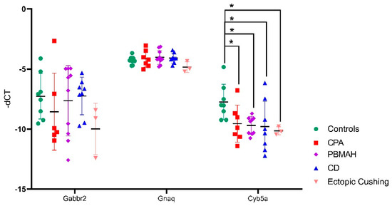
Figure 7. QPCR analyses of the top predicted targets of miR-1247-5p. Data are represented as mean ± standard deviation (SD) of −dCT values. Housekeeping gene: PPIA. Statistics: ANOVA test with Bonferroni correction to detect significant differences between patient groups with at least a significance of p-value < 0.05 (*).
Ijms 23 07676 g008 550
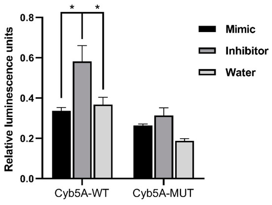
Figure 8. Results of dual luminescence assay on cells transfected with miR-1247-5p mimics and related controls. Cells were transfected with plasmids containing either the WT or Mut miRNA binding sequence in Cyb5a. Either miR-1247-5p mimics or miR-1247-5p inhibitors were transfected together with the respective plasmids. Data are represented as mean ± standard error of mean (SEM) of relative luminescence unit values. Statistics: ANOVA test with Bonferroni correction to detect significant differences between patient groups with at least a significance of p value < 0.05 (*).
Table 1. Analysis of the predicted targets of miR-1247-5p and their expression levels in comparison to controls (log2 fold change). Published literature on target genes in reference to CS is highlighted in bold.
Table

2.6. Pathway Analyses of miRNA Targets

For the pathway analysis (Reactome) we used the 28 predicted miRNA-1247-5p targets and the 23 predicted miRNA-379-5p targets from TargetScan, which were significantly differently expressed in the RNA-seq (Figure 6). Concurrently, the pathways commonly enriched by both miRNAs included the WNT signaling pathway and N-acetyl-glucosamine synthesis (Figure 9A). As a complementary approach, in silico analyses were also performed based on the targets from miRTarBase. In this database, targets are shortlisted based on published experimental results. In this analysis, miR-1247-5p (n = 21) and miR-379-5p targets (n = 85) were unique. While the validated targets of miR-379-5p were found to show significant changes in expression in the RNA-seq data from PBMAH (n = 12), none of the validated miR-1247-5p targets were found to show significant expression changes in the RNA-seq data from CPA. Therefore, all the validated targets of the miRNAs were subjected to pathway analyses (Panther). Interestingly, the WNT signaling pathway was also found to be commonly regulated by both miRNAs using this approach (Figure 9B). Finally, the expression status of target genes related to WNT signaling pathways were checked in our RNA-seq data (Figure S2). Given the upregulated status of the miRNAs, a downregulated expression of its target genes was expected. However, a significantly upregulated expression was observed for DVL1 in CPA in comparison to controls and for ROR1 in PBMAH in comparison to controls.
Ijms 23 07676 g009 550
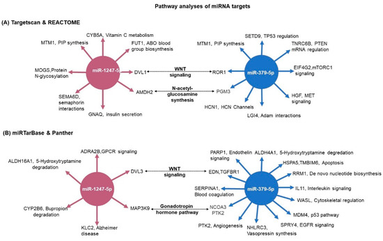
Figure 9. Pathway analyses of miRNA target genes. (A) The predicted targets were matched with the RNA-seq expression data. For miR-1247-5p, CPA vs. controls expression data; and for miR-379-5p, PBMAH vs. controls expression data. The significantly expressed target genes were then subjected to pathway analyses by Reactome. The significantly enriched pathway networks (p < 0.05) and their related genes are given. (B) The experimentally validated target genes from miRTarBase were analyzed for their role in pathways by the Panther database. The target genes and their related pathways are given. The commonly represented pathways are marked in bold.

3. Discussion

MiRNAs are fine regulators of both physiology and pathology and have diverse roles as diagnostic biomarkers as well as therapeutic targets. While circulating miRNAs have been investigated as potential biomarkers for hypercortisolism in CS subtypes (36), comprehensive analyses of their pathological role in CS subtypes have not yet been performed. This study hoped to uncover the pathological role of miRNAs in different CS subtypes as well as identify unique epigenetic targets contributing to hypercortisolism in these subtypes. As such, miRNA sequencing was performed in the ACTH-independent CPA and ACTH-dependent CD, with additional QPCR validation in PBMAH and ectopic CS. As expected, miRNA expression profiles in CD and CPA were very different.

3.1. ACTH-Independent Upregulated miRNAs in CS

Among the analyzed miRNAs, only miR-1247-5p and miR-379-5p showed the most prominent changes in expression levels. Briefly, miR-1247-5p was significantly upregulated in ACTH-independent forms of CS, CPA, and PBMAH (Figure 1 and Figure 3) while miR-379-5p was found to be upregulated in CD and PBMAH, in comparison to controls. Even though CD and PBMAH represent CS subtypes with different ACTH dependence, albeit both with hyperplastic tissue, it is interesting to find a shared miRNA expression status. Concurrently, miRNAs have been identified as dynamic players in regulating the acute effect of ACTH on adrenal steroidogenesis in in vivo murine studies [20,21]. Further diverse miRNAs have been characterized to regulate steroidogenesis in ACTH and dexamethasone treated rats [22] (suppressed ACTH) as well as in in vitro studies [20]. It is possible that miR-379-5p contributes to the adrenal hyperplasia present in both PBMAH and CD by targeting specific genes related to hyperplasia, and miR-1247-5p by contributing to cortisol production independent of ACTH regulation in CPA and PBMAH.
Interestingly, the miRNA candidates (mir-1247-5p and miR-379-5p) in our study have not been previously characterized in any of these studies. Furthermore, the expression of mir-1247-5p and miR-379-5p were found to be independent of ACTH stimulation, underlying their role in CS independently of the HPA axis control and postulating specific regulatory processes.

3.2. Target Genes of miRNAs in CS

Initially, we focused on the selection of known CS specific target genes that could be directly repressed by miRNAs, thereby contributing to pathology. The predicted target genes of miR-1247-5p and miR-379-5p were assessed for their downregulated expression status in the RNA-seq data. Among the selected target genes, only Cyb5a was found to be significantly downregulated in all forms of CS (Figure 6). Cytochrome b5 (CYB5A) is a marker of the zona reticularis and is an important regulator of androstenedione production [23,24]. Based on its role in adrenal steroidogenesis, it is possible that Cyb5a is downregulated by miR1247-5p. To prove our hypothesis, a dual luciferase assay was performed in HELA cell line to identify a direct interaction between Cyb5a and miR-1247-5p (Figure 7). Unfortunately, a direct interaction could not be proven, indicating that miR-1247-5p perhaps regulates its target genes in different ways.

3.3. Pathway Analyses of miRNA Targets

To identify miRNA specific regulatory processes, comprehensive target and pathway analyses were performed. Independent pathway analyses of the respective targets with two different databases of Reactome and Panther showed the WNT signaling pathway as a common targeted pathway of both mir-1247-5p and miR-379-5p (Figure 8). The WNT signaling pathway represents a crucial regulator in diverse developmental as well as pathological processes with tissue-specific effects [25,26]. Consequently, the WNT pathway has been largely characterized as one of the dysregulated pathophysiological mechanisms in CPA. Mutations in PRKACA, one of the WNT signaling proteins, are present in approximately 40% of CPA [27]. In the case of CD, dysregulated WNT signaling has been characterized as promoting proliferation in ACTH-secreting pituitary adenomas [28]. Moreover, activating mutations in beta catenin, one of the WNT signaling pathways, has been characterized as driving adrenal hyperplasia through both proliferation-dependent and -independent mechanisms [29]. Thus, it could be hypothesized that by targeting specific genes in the pathway, miRNAs drive specific pathophysiological processes in diverse CS subtypes.

3.4. MiRNA Target Genes in WNT Signaling

DVL1 (TargetScan) and DVL3 (miRTar) are the shortlisted target genes of miR-1247-5p in the WNT signaling pathway. These genes are members of canonical WNT pathways and, importantly, activation of the cytoplasmic effector Dishevelled (Dvl) is a critical step in WNT/β-catenin signaling initiation [30,31]. Interestingly, no difference in DVL1 and DVL3 gene expression was found in the analyses of ACTH-secreting pituitary adenomas [32]. Therefore, it could be possible that DVL1 and DVL3 are only targeted by miR-1247-5p specifically in the adrenal of CPA and PBMAH patients, leading to its characterized tumor progression. EDN1, TGFBR1 (TargetScan), and ROR1 (miRTar) were the target genes of miR-379-5p related to the WNT pathway. In epithelial ovarian cancer, Endothelin-1 (EDN-1) was found to regulate the epithelial–mesenchymal transition (EMT) and a chemoresistant phenotype [33]. In the case of receptor tyrosine kinase-like orphan receptor 1 (ROR1), higher expression of the gene was associated with a poor prognosis in ovarian cancer [34]. Concurrently, suppression of TGFBR1-mediated signaling by conditional knockout in mice was found to drive the pathogenesis of endometrial hyperplasia, independent of the influence of ovarian hormones [35]. Therefore, it could be hypothesized that the dysregulated expression of these factors in adrenals could trigger similar hyperplastic effects mediated by these factors, as in ovarian tissues.

3.5. Bottlenecks and Future Outlook

Interestingly, among these genes, only DVL1 and ROR1 were found to be significantly upregulated in the RNA-seq data (Figure S2). The major regulatory role of miRNAs in gene expression come from their ability to repress gene expression at the level of transcription and translation. There are also reports of miRNA-mediated gene upregulation; however, the physiological evidence of the role is still unresolved [36]. Therefore, it is interesting to see the selected targets of miR-1247-5p and miR-379-5p upregulated. Moreover, it should be noted that most of the experimentally validated miRNA targets were identified by CLIP methods [37]. Crosslinking immunoprecipitation (CLIP) are binding assays that provide genome-wide maps of potential miRNA-target gene interactions based on sequencing. Moreover, these assays do not make functional predictions on the outcome of miRNA binding, and neither do upregulation or downregulation [38,39]. Therefore, in our current experimental setting, we could only identify potential miRNA target genes and speculate on their pathological role based on the published literature and in silico analyses. Furthermore, extensive mechanistic analyses based on these potential targets could help in elaborating the specific epigenetic pathways that fine-tune CS pathology in different subtypes.

4. Materials and Methods

4.1. Sample Collection and Ethics Approval

All patients were registered in the German Cushing’s Registry, the ENS@T or/and NeoExNET databases (project number protocol code 379-10 and 152-10). The study was approved by the Ethics Committee of the University of Munich. All experiments were performed according to relevant guidelines and protocols, and written informed consent was obtained from all patients involved. The adrenal samples used in the sequencing (miRNA and RNA) belong to the same patient.
For miRNA-specific next-generation sequencing (NGS), a total of 19 adrenocortical tissue samples were used. The cohort consisted of the following patient groups: ACTH-independent CPA, n = 7; ACTH-dependent hypertrophic adrenals of CD patients after bilateral adrenalectomy, n = 8; normal adjacent adrenal tissue from patients with pheochromocytoma as controls, n = 8. For QPCR validation, the patient groups included adrenal tissue from ACTH-independent PBMAH, n = 10, and ACTH-dependent ectopic CS, n = 3.
In the case of mRNA sequencing, a total of 23 adrenocortical tissue samples were used. This includes the following patient groups: CPA, n = 7; PBMAH, n = 8; normal adjacent adrenal tissue from patients with pheochromocytoma as controls, n = 8.
The clinical characteristics of the patients are given in Table 2. Furthermore, of the eight CPA samples in the study, three of them carried known somatic driver mutations in the PRKACA gene and in the ten PBMAH samples, two carried germline mutations in the ARMC5 gene.
Table 2. Clinical characteristics of the patient groups. Data are given as median with 25th and 75th percentiles in brackets. CPA, cortisol producing adenoma; CD, Cushing’s disease.
Table
The adrenal tissues were stored at −80 °C. Total RNA isolation was carried out from all adrenal cortex samples by an RNeasy Tissue Kit (Qiagen, Hilden, Germany). The isolated RNA was kept frozen at −80 °C until further use.

4.2. MiRNA and RNA Sequencing

RNA integrity and the absence of contaminating DNA were confirmed by Bioanalyzer RNA Nano (Agilent Technologies, Santa Clara, CA, USA) and by Qubit DNA High sensitivity kits, respectively. Sequencing libraries were prepared using the Illumina TruSeq Small RNA Library Preparation Kit. NGS was performed on 2 lanes of an Illumina HiSeq2500 (Illumina, CA, USA) multiplexing all samples (paired end read, 50 bp). The quality of sequencing reads was verified using FastQC0.11.5 (http://www.bioinformatics.babraham.ac.uk/projects/fastqc, date last accessed: 13 March 2020) before and after trimming. Adapters were trimmed using cutadapt [40]. Reads with <15 bp and >40 bp insert sequences were discarded. An alignment of reads was performed using miRBase V21 [41,42] with sRNAbench [43]. EdgeR and DeSeq in R were used for further analyses [44,45]. MiRNAs with at least 5 raw counts per library were included. RNA-seq was performed by Qiagen, Hilden, Germany. For mRNA, sequencing was performed on Illumina NextSeq (single end read, 75 bp). Adapter and quality trimming were performed by the “Trim Reads” tool from CLC Genomics Workbench. Furthermore, reads were trimmed based on quality scores. The QC reports were generated by the “QC for Sequencing Reads” tool from CLC Genomics Workbench. Read mapping and gene quantification were performed by the “RNA-seq Analysis” tool from CLC Genomics Workbench [46]. The miRNA-seq data generated in this study have been submitted to the NCBI (PRJNA847385).

4.3. Validation of Individual miRNAs

MiRNAs and genes significantly differentially expressed by NGS were validated by QPCR. Reverse transcription of miRNA-specific cDNA was performed by using the TaqMan MicroRNA Reverse Transcription Kit (Thermo Fisher Scientific, Munich, Germany), and the reverse transcription of RNA genes was done by using the Superscript VILO cDNA synthesis Kit (Thermo Fisher Scientific, Munich, Germany). 50 ng of RNA was used for each of the reverse transcription reactions. Quantitative real-time PCR was performed using the TaqMan Fast Universal PCR Master Mix (2×) (Thermo Fisher Scientific, Munich, Germany) on a Quantstudio 7 Flex Real-Time PCR System (Thermo Fisher Scientific, Munich, Germany) in accordance with the manufacturer’s protocol. All QPCR reactions were performed in a final reaction volume of 20 μL and with 1 μL of 1:5 diluted cDNA. Negative control reactions contained no cDNA templates. Gene expression was quantified using the relative quantification method by normalization with reference gene [47]. Statistical analysis using the bestkeeper tool was used to compare and select the best reference gene with stable expression across the human adrenal samples [48]. Reference genes with significantly different Ct values (p-value < 0.01) between the samples were excluded. Furthermore, primer efficiency and the associated correlation coefficient R2 of the selected reference gene were determined by the standard curve method in serially diluted cDNA samples [49]. In the case of miRNA reference genes, miR-16-5p [48,50,51] and RNU6B [52] previously used in similar studies were compared. MiR-16-5p was found to show the most stable expression levels across the samples with a p-value of 0.452 in comparison to RNU6B which had a p-value of 0.001. In the case of RNA reference genes, PPIA [53] and GAPDH [54] were compared. Here, PPIA was found to show the most stable expression levels across the samples with a p-value of 0.019 in comparison to GAPDH which had a p-value of 0.003. Therefore, these genes were used for the normalization of miRNA and RNA expression in human adrenal samples.

4.4. Target Screening

In silico prediction of the possible miRNA targets was performed using the miRNA target database, TargetScan, and miRTarBase [19,37]. The top predicted targets were further screened based on their expression status in the RNA-seq data from the adrenocortical tissues of CPA, PBMAH, and controls (unpublished data). Pathway analyses of the targets were performed using Reactome [55] and Panther [56] online platforms. The selected downregulated targets were analyzed by QPCR in the adrenocortical samples to confirm their expression status. The successfully validated candidates were then analyzed for regulation by the miRNA using a dual luciferase assay [57].

4.5. Dual Luciferase Assay

The interaction between the predicted 3′-UTR region of Cyb5a and miR-1247-5p was detected using a luciferase activity assay. The 3′UTR sequences of Cyb5a (129 bp) containing the predicted miR-1247-5p binding sites (psiCHECK-2 Cyb5a 3′UTR WT) were cloned into the psiCHECK-2 vector (Promega, Fitchburg, WI, USA). A QuikChange Site-Directed Mutagenesis kit (Agilent Technologies, CA, USA) was used to mutate the miR-1247-5p binding site (psiCHECK-2 Cyb5a 3′UTR mutant) according to the manufacturer’s protocol. All the sequences were verified by Sanger sequencing. Then, 200 ng of the plasmid was used for each transfection. Synthetic miR-1247-5p mimics and specific oligonucleotides that inhibit endogenous miR-1247-5p (miR-1247-5p inhibitors) were purchased from Promega and 100 nmol of the molecules were used for each transfection according to the manufacturer’s protocol. For the assay, HeLa cells were seeded in 96-well plates and incubated for 24 h. The following day, cells were transfected using the following different conditions: (1) psiCHECK-2 Cyb5a 3′UTR WT + miR-1247-5p mimic; (2) psiCHECK-2 Cyb5a 3′UTR WT + miR-1247-5p inhibitor; (3) psiCHECK-2 Cyb5a 3′UTR WT + water; (4) psiCHECK-2 Cyb5a 3′UTR mutant + miR-1247-5p mimic; (5) psiCHECK-2 Cyb5a 3′UTR mutant + miR-1247-5p inhibitor; (6) psiCHECK-2 Cyb5a 3′UTR mutant + water. Forty-eight hours later, luciferase activity in the cells was measured using the dual luciferase assay system (Promega, Fitchburg, WI, USA) in accordance with the manufacturer’s instructions. Renilla luciferase activity was normalized to firefly luciferase activity. Each treatment was performed in triplicate. Any interaction between the cloned gene, Cyb5a (WT and mutant), and miR-1247-5p mimic is accompanied by a decrease in luminescence. This decrease in luminescence would not be observed when the plasmids are transfected with the miR-1247-5p inhibitor, indicating that observed luminescence differences are caused by specific interactions between the plasmid and the miR-1247-5p mimic. Transfection of the plasmid with water corrects any background interactions between the cloned gene and endogenous miRNAs in the culture.

4.6. In Vivo ACTH Stimulation

Experiments were performed on 13-week-old C57BL/6 J female mice (Janvier, Le Genest-Saint-Isle, France). Mice were intraperitoneally injected with 1 mg/kg of ACTH (Sigma Aldrich, Munich, Germany) and adrenals were collected after 10, 30, and 60 min of injections. In addition, control adrenals were collected from mice at baseline conditions (0 min). Mice were killed by cervical dislocation and adrenals were isolated, snap-frozen in liquid nitrogen, and stored at −80 °C for later RNA extraction. MiR-26a was taken as a housekeeping gene in the QPCR [58]. All mice were maintained in accordance with facility guidelines on animal welfare and approved by Landesdirektion Sachsen, Chemnitz, Germany.

4.7. Statistical Analysis and Software

R version 3.6.1 was used for the statistical analyses. To identify RNAs differentially expressed, a generalized linear model (GLM, a flexible generalization of ordinary linear regression that allows for variables that have distribution patterns other than a normal distribution) in the software package edgeR (Empirical Analysis of DGE in R) was employed to calculate p-values [45,59]. p-values were adjusted using the Benjamin–Hochberg false discovery rate (FDR) procedure [60]. Disease groups were compared using the unpaired Mann–Whitney test, and to decrease the false discovery rate a corrected p-value was calculated using the Benjamin–Hochberg method. p adjusted < 0.05 and log2 fold change >1.25 was applied as the cut-off for significance for NGS data. GraphPad Prism Version 8 was used for the statistical analysis of QPCR. To calculate differential gene expression, the dCt method (delta Ct (cycle threshold) value equals target miRNA’s Ct minus housekeeping miRNA’s Ct) was used (Microsoft Excel 2016, Microsoft, Redmond, WA, USA). For QPCR, an ANOVA test with Bonferroni correction was used [61] to assess significance; p-values < 0.05 were considered significant.

5. Conclusions

In conclusion, while comprehensive information regarding the role of miRNAs in acute and chronic phases of steroidogenesis is available, there is little known about the pathological independent role of miRNAs in the pathology of CS. In our study, we have described ACTH-independent miR-1247-5p and miR-379-5p expression in CS for the first time. Thus, by regulating different genes in the WNT signaling pathway, the miRNAs may individually contribute to the Cushing’s pathology in specific subtypes.

Supplementary Materials

The following supporting information can be downloaded at: https://www.mdpi.com/article/10.3390/ijms23147676/s1.

Author Contributions

Conceptualization, S.V., A.C. and A.R.; methodology, S.V., R.Z. and M.E.; software, S.V. and M.E.; validation, R.Z., A.O., D.W. and B.W.; formal analysis, S.V.; investigation, S.V., R.Z., M.E., A.O. and D.W.; resources, A.C., B.W., M.R. and A.R.; data curation, S.V. and R.Z.; writing—original draft preparation, S.V., R.Z. and A.R.; writing—review and editing, S.S., M.R. and A.R.; visualization, S.V.; supervision, A.R.; project administration, A.R.; funding acquisition, A.C., S.S., M.R. and A.R. All authors have read and agreed to the published version of the manuscript.

Funding

This work was supported by a grant from the Deutsche Forschungsgemeinschaft (DFG) (within the CRC/Transregio 205/1 “The Adrenal: Central Relay in Health and Disease”) to A.C., B.W., S.S., M.R. and A.R., and individual grant SB 52/1-1 to S.S. This work is part of the German Cushing’s Registry CUSTODES and has been supported by a grant from the Else Kröner-Fresenius Stiftung to MR (2012_A103 and 2015_A228). A.R. was supported by the FöFoLe Program of the Ludwig Maximilian University, Munich. We thank I. Shapiro, A. Parl, C. Kühne, and S. Zopp for their technical support.

Institutional Review Board Statement

The study was conducted according to the guidelines of the Declaration of Helsinki and approved by the Ethics Committee of the Ludwig Maximilian University, Munich (protocol code 379-10, 152-10 and 20 July2021).

Informed Consent Statement

Informed consent was obtained from all subjects involved in the study.

Data Availability Statement

The miRNA-seq data generated in this study have been submitted to the NCBI (PRJNA847385).

Conflicts of Interest

The authors declare no conflict of interest.

References

  1. Kotłowska, A.; Puzyn, T.; Sworczak, K.; Stepnowski, P.; Szefer, P. Metabolomic biomarkers in urine of cushing’s syndrome pa-tients. Int. J. Mol. Sci. 2017, 18, 294. [Google Scholar] [CrossRef] [PubMed][Green Version]
  2. Valassi, E.; Tabarin, A.; Brue, T.; Feelders, R.A.; Reincke, M.; Netea-Maier, R.; Toth, M.; Zacharieva, S.; Webb, S.M.; Tsagarakis, S.; et al. High mortality within 90 days of diagnosis in patients with Cushing’s syndrome: Results from the ERCUSYN registry. Eur. J. Endocrinol. 2019, 181, 461–472. [Google Scholar] [CrossRef]
  3. Stratakis, C. Cushing syndrome caused by adrenocortical tumors and hyperplasias (corticotropin-independent Cushing syn-drome). Endocr. Dev. 2008, 13, 117–132. [Google Scholar]
  4. Jarial, K.D.S.; Walia, R.; Nahar, U.; Bhansali, A. Primary bilateral adrenal nodular disease with Cushing’s syndrome: Varying aeti-ology. BMJ. Case. Rep. 2017, 2017, bcr2017220154. [Google Scholar] [CrossRef] [PubMed]
  5. Kamilaris, C.D.C.; Stratakis, C.A.; Hannah-Shmouni, F. Molecular Genetic and Genomic Alterations in Cushing’s Syndrome and Primary Aldosteronism. Front. Endocrinol. 2021, 12, 142. [Google Scholar] [CrossRef] [PubMed]
  6. Feelders, R.A.; Pulgar, S.J.; Kempel, A.; Pereira, A.M. The burden of Cushing’s disease: Clinical and health-related quality of life aspects. Eur. J. Endocrinol. 2012, 167, 311–326. [Google Scholar] [CrossRef] [PubMed][Green Version]
  7. Feelders, R.A.; Newell-Price, J.; Pivonello, R.; Nieman, L.K.; Hofland, L.J.; Lacroix, A. Advances in the medical treatment of Cush-ing’s syndrome. Lancet Diabetes Endocrinol. 2019, 7, 300–312. [Google Scholar] [CrossRef]
  8. Krill, K.T.; Gurdziel, K.; Heaton, J.H.; Simon, D.P.; Hammer, G.D. Dicer Deficiency Reveals MicroRNAs Predicted to Control Gene Expression in the Developing Adrenal Cortex. Mol. Endocrinol. 2013, 27, 754–768. [Google Scholar] [CrossRef]
  9. Robertson, S.; Diver, L.A.; Alvarez-Madrazo, S.; Livie, C.; Ejaz, A.; Fraser, R.; Connell, J.M.; MacKenzie, S.M.; Davies, E. Regulation of Corticosteroidogenic Genes by MicroRNAs. Int. J. Endocrinol. 2017, 2017, 2021903. [Google Scholar] [CrossRef][Green Version]
  10. Bujko, M.; Kober, P.; Boresowicz, J.; Rusetska, N.; Zeber-Lubecka, N.; Paziewska, A.; Pekul, M.; Zielinski, G.; Styk, A.; Kunicki, J.; et al. Differential microRNA Expression in USP8-Mutated and Wild-Type Corticotroph Pituitary Tumors Reflect the Difference in Protein Ubiquitination Processes. J. Clin. Med. 2021, 10, 375. [Google Scholar] [CrossRef]
  11. Iliopoulos, D.; Bimpaki, E.I.; Nesterova, M.; Stratakis, C.A. MicroRNA Signature of Primary Pigmented Nodular Adrenocortical Disease: Clinical Correlations and Regulation of Wnt Signaling. Cancer Res. 2009, 69, 3278–3282. [Google Scholar] [CrossRef] [PubMed][Green Version]
  12. Tan, X.-G.; Zhu, J.; Cui, L. MicroRNA expression signature and target prediction in familial and sporadic primary macronodular adrenal hyperplasia (PMAH). BMC Endocr. Disord. 2022, 22, 11. [Google Scholar] [CrossRef]
  13. Vaczlavik, A.; Bouys, L.; Violon, F.; Giannone, G.; Jouinot, A.; Armignacco, R.; Cavalcante, I.P.; Berthon, A.; Letouzé, E.; Vaduva, P.; et al. KDM1A inactivation causes hereditary food-dependent Cushing syndrome. Genet. Med. 2021, 24, 374–383. [Google Scholar] [CrossRef]
  14. Bimpaki, E.I.; Iliopoulos, D.; Moraitis, A.; Stratakis, C.A. MicroRNA signature in massive macronodular adrenocortical disease and implications for adrenocortical tumorigenesis. Clin. Endocrinol. 2010, 72, 744–751. [Google Scholar] [CrossRef]
  15. Vetrivel, S.; Zhang, R.; Engel, M.; Altieri, B.; Braun, L.; Osswald, A.; Bidlingmaier, M.; Fassnacht, M.; Beuschlein, F.; Reincke, M.; et al. Circulating microRNA Expression in Cushing’s Syndrome. Front. Endocrinol. 2021, 12, 10. [Google Scholar] [CrossRef] [PubMed]
  16. O’Brien, J.; Hayder, H.; Zayed, Y.; Peng, C. Overview of microRNA biogenesis, mechanisms of actions, and circulation. Front. Endocrinol. 2018, 9, 402. [Google Scholar] [CrossRef][Green Version]
  17. Butz, H.; Patócs, A. MicroRNAs in endocrine tumors. Electron. J. Int. Fed. Clin. Chem. Lab. Med. 2019, 30, 146–164. [Google Scholar]
  18. Riester, A.; Issler, O.; Spyroglou, A.; Rodrig, S.H.; Chen, A.; Beuschlein, F. ACTH-Dependent Regulation of MicroRNA As Endogenous Modulators of Glucocorticoid Receptor Expression in the Adrenal Gland. Endocrinology 2012, 153, 212–222. [Google Scholar] [CrossRef][Green Version]
  19. Huang, X.; Zhong, R.; He, X.; Deng, Q.; Peng, X.; Li, J.; Luo, X. Investigations on the mechanism of progesterone in inhibiting endo-metrial cancer cell cycle and viability via regulation of long noncoding RNA NEAT1/microRNA-146b-5p mediated Wnt/β-catenin signaling. IUBMB Life 2019, 71, 223–234. [Google Scholar] [CrossRef][Green Version]
  20. Azhar, S.; Dong, D.; Shen, W.-J.; Hu, Z.; Kraemer, F.B. The role of miRNAs in regulating adrenal and gonadal steroidogenesis. J. Mol. Endocrinol. 2020, 64, R21–R43. [Google Scholar] [CrossRef]
  21. Allen, M.J.; Sharma, S. Physiology, Adrenocorticotropic Hormone (ACTH). StatPearls 2021. Available online: https://www.ncbi.nlm.nih.gov/books/NBK500031/ (accessed on 8 December 2021).
  22. Hu, Z.; Shen, W.-J.; Cortez, Y.; Tang, X.; Liu, L.-F.; Kraemer, F.B.; Azhar, S. Hormonal Regulation of MicroRNA Expression in Steroid Producing Cells of the Ovary, Testis and Adrenal Gland. PLoS ONE 2013, 8, e78040. [Google Scholar] [CrossRef][Green Version]
  23. Ghayee, H.K.; Rege, J.; Watumull, L.M.; Nwariaku, F.E.; Carrick, K.S.; Rainey, W.E.; Miller, W.L.; Auchus, R.J. Clinical, biochemical, and molecular characterization of macronodular adrenocortical hyperplasia of the zona reticularis: A new syndrome. J. Clin. Endocrinol. Metab. 2011, 96, E243–E250. [Google Scholar] [CrossRef] [PubMed][Green Version]
  24. Nakamura, Y.; Fujishima, F.; Hui, X.-G.; Felizola, S.J.A.; Shibahara, Y.; Akahira, J.-I.; McNamara, K.M.; Rainey, W.E.; Sasano, H. 3βHSD and CYB5A double positive adrenocortical cells during adrenal development/aging. Endocr. Res. 2015, 40, 8–13. [Google Scholar] [CrossRef] [PubMed][Green Version]
  25. Ng, L.F.; Kaur, P.; Bunnag, N.; Suresh, J.; Sung, I.C.H.; Tan, Q.H.; Gruber, J.; Tolwinski, N.S. WNT Signaling in Disease. Cells 2019, 8, 826. [Google Scholar] [CrossRef][Green Version]
  26. Song, J.L.; Nigam, P.; Tektas, S.S.; Selva, E. microRNA regulation of Wnt signaling pathways in development and disease. Cell. Signal. 2015, 27, 1380–1391. [Google Scholar] [CrossRef][Green Version]
  27. Beuschlein, F.; Fassnacht, M.; Assié, G.; Calebiro, D.; Stratakis, C.A.; Osswald, A.; Ronchi, C.L.; Wieland, T.; Sbiera, S.; Faucz, F.R.; et al. Constitutive Activation of PKA Catalytic Subunit in Adrenal Cushing’s Syndrome. N. Engl. J. Med. 2014, 370, 1019–1028. [Google Scholar] [CrossRef][Green Version]
  28. Ren, J.; Jian, F.; Jiang, H.; Sun, Y.; Pan, S.; Gu, C.; Chen, X.; Wang, W.; Ning, G.; Bian, L.; et al. Decreased expression of SFRP2 promotes development of the pituitary corticotroph adenoma by upregulating Wnt signaling. Int. J. Oncol. 2018, 52, 1934–1946. [Google Scholar] [CrossRef][Green Version]
  29. Pignatti, E.; Leng, S.; Yuchi, Y.; Borges, K.S.; Guagliardo, N.A.; Shah, M.S.; Ruiz-Babot, G.; Kariyawasam, D.; Taketo, M.M.; Miao, J.; et al. Beta-Catenin Causes Adrenal Hyperplasia by Blocking Zonal Transdifferentiation. Cell Rep. 2020, 31, 107524. [Google Scholar] [CrossRef]
  30. Grumolato, L.; Liu, G.; Mong, P.; Mudbhary, R.; Biswas, R.; Arroyave, R.; Vijayakumar, S.; Economides, A.N.; Aaronson, S.A. Canonical and noncanonical Wnts use a common mechanism to activate completely unrelated coreceptors. Genes Dev. 2010, 24, 2517–2530. [Google Scholar] [CrossRef][Green Version]
  31. Tauriello, D.V.F.; Jordens, I.; Kirchner, K.; Slootstra, J.W.; Kruitwagen, T.; Bouwman, B.A.M.; Noutsou, M.; Rüdiger, S.G.D.; Schwamborn, K.; Schambony, A.; et al. Wnt/β-catenin signaling requires interaction of the Dishevelled DEP domain and C terminus with a discontinuous motif in Frizzled. Proc. Natl. Acad. Sci. USA 2012, 109, E812–E820. [Google Scholar] [CrossRef][Green Version]
  32. Colli, L.M.; Saggioro, F.; Neder Serafini, L.; Camargo, R.C.; Machado, H.; Moreira, A.C.; Antonini, S.R.; De Castro, M. Components of the Canonical and Non-Canonical Wnt Pathways Are Not Mis-Expressed in Pituitary Tumors. PLoS ONE 2013, 8, e62424. [Google Scholar] [CrossRef] [PubMed][Green Version]
  33. Rosanò, L.; Cianfrocca, R.; Tocci, P.; Spinella, F.; Di Castro, V.; Caprara, V.; Semprucci, E.; Ferrandina, G.; Natali, P.G.; Bagnato, A. En-dothelin A receptor/β-arrestin signaling to the Wnt pathway renders ovarian cancer cells resistant to chemotherapy. Cancer Res. 2014, 74, 7453–7464. [Google Scholar] [CrossRef] [PubMed][Green Version]
  34. Zhang, H.; Qiu, J.; Ye, C.; Yang, D.; Gao, L.; Su, Y.; Tang, X.; Xu, N.; Zhang, D.; Xiong, L.; et al. ROR1 expression correlated with poor clinical outcome in human ovarian cancer. Sci. Rep. 2014, 4, 5811. [Google Scholar] [CrossRef] [PubMed][Green Version]
  35. Gao, Y.; Li, S.; Li, Q. Uterine epithelial cell proliferation and endometrial hyperplasia: Evidence from a mouse model. Mol. Hum. Reprod. 2014, 20, 776–786. [Google Scholar] [CrossRef]
  36. Orang, A.V.; Safaralizadeh, R.; Kazemzadeh-Bavili, M. Mechanisms of miRNA-Mediated Gene Regulation from Common Downregulation to mRNA-Specific Upregulation. Int. J. Genom. 2014, 2014, 970607. [Google Scholar]
  37. Huang, H.Y.; Lin, Y.C.D.; Li, J.; Huang, K.Y.; Shrestha, S.; Hong, H.C.; Tang, Y.; Chen, Y.G.; Jin, C.N.; Yu, Y.; et al. miRTarBase 2020: Up-dates to the experimentally validated microRNA–target interaction database. Nucleic Acids Res. 2020, 48, D148–D154. [Google Scholar] [CrossRef] [PubMed][Green Version]
  38. Liu, W.; Wang, X. Prediction of functional microRNA targets by integrative modeling of microRNA binding and target expres-sion data. Genome Biol. 2019, 20, 18. [Google Scholar] [CrossRef] [PubMed]
  39. Wang, X. Improving microRNA target prediction by modeling with unambiguously identified microRNA-target pairs from CLIP-ligation studies. Bioinformatics 2016, 32, 1316–1322. [Google Scholar] [CrossRef]
  40. Martin, M. Cutadapt removes adapter sequences from high-throughput sequencing reads. EMBnet. J. 2011, 17, 10–12. [Google Scholar] [CrossRef]
  41. Kozomara, A.; Griffiths-Jones, S. miRBase: Annotating high confidence microRNAs using deep sequencing data. Nucleic Acids Res. 2014, 42, D68–D73. [Google Scholar] [CrossRef][Green Version]
  42. Griffiths-Jones, S. miRBase: The MicroRNA Sequence Database. Methods Mol. Biol. 2006, 342, 129–138. [Google Scholar] [PubMed]
  43. Aparicio-Puerta, E.; Lebrón, R.; Rueda, A.; Gómez-Martín, C.; Giannoukakos, S.; Jáspez, D.; Medina, J.M.; Zubković, A.; Jurak, I.; Fromm, B.; et al. sRNAbench and sRNAtoolbox 2019: Intuitive fast small RNA profiling and differential expression. Nucleic Acids Res. 2019, 47, W530–W535. [Google Scholar] [CrossRef] [PubMed][Green Version]
  44. Anders, S.; Huber, W. Differential expression analysis for sequence count data. Genome Biol. 2010, 11, R106. [Google Scholar] [CrossRef] [PubMed][Green Version]
  45. Robinson, M.D.; McCarthy, D.J.; Smyth, G.K. EdgeR: A Bioconductor package for differential expression analysis of digital gene expression data. Bioinformatics 2010, 26, 139–140. [Google Scholar] [CrossRef][Green Version]
  46. Liu, C.-H.; Di, Y.P. Analysis of RNA Sequencing Data Using CLC Genomics Workbench. Methods Mol. Biol. 2020, 2102, 61–113. [Google Scholar] [CrossRef]
  47. Pfaffl, M.W.; Tichopad, A.; Prgomet, C.; Neuvians, T.P. Determination of stable housekeeping genes, differentially regulated target genes and sample integrity: BestKeeper–Excel-based tool using pair-wise correlations. Biotechnol. Lett. 2004, 26, 509–515. [Google Scholar] [CrossRef]
  48. Wang, X.; Zhang, X.; Yuan, J.; Wu, J.; Deng, X.; Peng, J.; Wang, S.; Yang, C.; Ge, J.; Zou, Y. Evaluation of the performance of serum miRNAs as normalizers in microRNA studies focused on cardiovascular disease. J. Thorac. Dis. 2018, 10, 2599–2607. [Google Scholar] [CrossRef]
  49. Geigges, M.; Gubser, P.M.; Unterstab, G.; Lecoultre, Y.; Paro, R.; Hess, C. Reference Genes for Expression Studies in Human CD8 + Naïve and Effector Memory T Cells under Resting and Activating Conditions. Sci. Rep. 2021, 10, 9411. [Google Scholar] [CrossRef]
  50. Song, J.; Bai, Z.; Han, W.; Zhang, J.; Meng, H.; Bi, J.; Ma, X.; Han, S.; Zhang, Z. Identification of Suitable Reference Genes for qPCR Analysis of Serum microRNA in Gastric Cancer Patients. Dig. Dis. Sci. 2011, 57, 897–904. [Google Scholar] [CrossRef]
  51. Szabó, D.R.; Luconi, M.; Szabó, P.M.; Tóth, M.; Szücs, N.; Horányi, J.; Nagy, Z.; Mannelli, M.; Patócs, A.; Rácz, K.; et al. Analysis of cir-culating microRNAs in adrenocortical tumors. Lab. Investig. 2014, 94, 331–339. [Google Scholar] [CrossRef][Green Version]
  52. Butz, H.; Mészáros, K.; Likó, I.; Patocs, A. Wnt-Signaling Regulated by Glucocorticoid-Induced miRNAs. Int. J. Mol. Sci. 2021, 22, 11778. [Google Scholar] [CrossRef] [PubMed]
  53. Muñoz, J.J.; Anauate, A.C.; Amaral, A.G.; Ferreira, F.M.; Watanabe, E.H.; Meca, R.; Ormanji, M.S.; Boim, M.A.; Onuchic, L.F.; Heilberg, I.P. Ppia is the most stable housekeeping gene for qRT-PCR normalization in kidneys of three Pkd1-deficient mouse models. Sci. Rep. 2021, 11, 19798. [Google Scholar] [CrossRef] [PubMed]
  54. Xia, X.; Liu, Y.; Liu, L.; Chen, Y.; Wang, H. Selection and verification of the combination of reference genes for RT-qPCR analysis in rat adrenal gland development. J. Steroid. Biochem. Mol. Biol. 2021, 208, 105821. [Google Scholar] [CrossRef] [PubMed]
  55. Gillespie, M.; Jassal, B.; Stephan, R.; Milacic, M.; Rothfels, K.; Senff-Ribeiro, A.; Griss, J.; Sevilla, C.; Matthews, L.; Gong, C.; et al. The reactome pathway knowledgebase 2022. Nucleic Acids Res. 2022, 50, D687–D692. [Google Scholar] [CrossRef]
  56. Mi, H.; Ebert, D.; Muruganujan, A.; Mills, C.; Albou, L.-P.; Mushayamaha, T.; Thomas, P.D. PANTHER version 16: A revised family classification, tree-based classification tool, enhancer regions and extensive API. Nucleic Acids Res. 2021, 49, D394–D403. [Google Scholar] [CrossRef]
  57. Wu, T.; Lin, Y.; Xie, Z. MicroRNA-1247 inhibits cell proliferation by directly targeting ZNF346 in childhood neuroblastoma. Biol. Res. 2018, 51, 13. [Google Scholar] [CrossRef][Green Version]
  58. Muñoz, J.J.; Anauate, A.; Amaral, A.G.; Ferreira, F.M.; Meca, R.; Ormanji, M.S.; Boim, M.A.; Onuchic, L.F.; Heilberg, I.P. Identification of housekeeping genes for microRNA expression analysis in kidney tissues of Pkd1 deficient mouse models. Sci. Rep. 2020, 10, 231. [Google Scholar] [CrossRef][Green Version]
  59. Love, M.I.; Huber, W.; Anders, S. Moderated estimation of fold change and dispersion for RNA-seq data with DESeq2. Genome Biol. 2014, 15, 550. [Google Scholar] [CrossRef][Green Version]
  60. Hu, Z.; Gao, S.; Lindberg, D.; Panja, D.; Wakabayashi, Y.; Li, K.; Kleinman, J.E.; Zhu, J.; Li, Z. Temporal dynamics of miRNAs in human DLPFC and its association with miRNA dysregulation in schizophrenia. Transl. Psychiatry 2019, 9, 196. [Google Scholar] [CrossRef]
  61. Esteva-Socias, M.; Gómez-Romano, F.; Carrillo-Ávila, J.A.; Sánchez-Navarro, A.L.; Villena, C. Impact of different stabilization methods on RT-qPCR results using human lung tissue samples. Sci. Rep. 2020, 10, 3579. [Google Scholar] [CrossRef]
Publisher’s Note: MDPI stays neutral with regard to jurisdictional claims in published maps and institutional affiliations.

Pasireotide-Induced Shrinkage in GH and ACTH Secreting Pituitary Adenoma

Introduction: Pasireotide (PAS) is a novel somatostatin receptor ligands (SRL), used in controlling hormonal hypersecretion in both acromegaly and Cushing’s Disease (CD). In previous studies and meta-analysis, first-generation SRLs were reported to be able to induce significant tumor shrinkage only in somatotroph adenomas. This systematic review and meta-analysis aim to summarize the effect of PAS on the shrinkage of the pituitary adenomas in patients with acromegaly or CD.

Materials and methods: We searched the Medline database for original studies in patients with acromegaly or CD receiving PAS as monotherapy, that assessed the proportion of significant tumor shrinkage in their series. After data extraction and analysis, a random-effect model was used to estimate pooled effects. Quality assessment was performed with a modified Joanna Briggs’s Institute tool and the risk of publication bias was addressed through Egger’s regression and the three-parameter selection model.

Results: The electronic search identified 179 and 122 articles respectively for acromegaly and CD. After study selection, six studies considering patients with acromegaly and three with CD fulfilled the eligibility criteria. Overall, 37.7% (95%CI: [18.7%; 61.5%]) of acromegalic patients and 41.2% (95%CI: [22.9%; 62.3%]) of CD patients achieved significant tumor shrinkage. We identified high heterogeneity, especially in acromegaly (I2 of 90% for acromegaly and 47% for CD), according to the low number of studies included.

Discussion: PAS treatment is effective in reducing tumor size, especially in acromegalic patients. This result strengthens the role of PAS treatment in pituitary adenomas, particularly in those with an invasive behavior, with progressive growth and/or extrasellar extension, with a low likelihood of surgical gross-total removal, or with large postoperative residual tissue.

Systematic Review Registration: https://www.crd.york.ac.uk/prospero/display_record.php?ID=CRD42022328152, identifier CRD42022328152

Introduction

Pasireotide (PAS) is a novel somatostatin receptor ligand (SRL) with a high affinity for the somatostatin receptor (SSR) type 5 (12). Somatotroph adenomas are usually responsive to first-generation SRLs (octreotide and lanreotide), as they are able to reduce growth hormone (GH) secretion through SSR type 2 (3). In the flow-chart of acromegaly treatment, PAS is suggested in case of resistance to first-generation SRLs, as SSR type 5 is also abundantly expressed in GH-secreting adenomas (3). A phase III study with PAS long-acting release (LAR) proved its efficacy in first-generation SRLs-resistant acromegalic patients after 6 months (4). In the extension study (Colao A et al.), 37% of patients achieved normalization of insulin-like growth factor 1 (IGF-1) and/or GH levels <1 µg/L, considering both those performing the extension treatment and those crossing over from the first-generation SRL-control group to the PAS LAR group. Nearly two-thirds of responses were achieved after at least 6 months of treatment. Up-titration of the dose from 40 to 60 mg monthly enriched the number of responders, suggesting that the PAS LAR effect may be both time- and dose-dependent (5). Concomitant improvement in signs and symptoms has also been confirmed in other series (69).

SSR type 5 is the predominant isoform in human corticotroph adenomas, since it is not down-regulated by high cortisol levels, as SSR type 2 does. Therefore, PAS is the only SRL available in patients with Cushing’s Disease (CD) (2). In a phase III study, subcutaneous (s.c.) PAS proved to be effective in normalizing urinary free cortisol (respectively in 13% and 25% of patients taking 600 µg or 900 µg bis-in-die for 12 months) (10), achieving significant clinical improvement (11). In the same clinical setting, PAS LAR showed similar efficacy and safety profiles (12). These benefits could be maintained for up to 5 years in an extension study (1314). In a recent meta-analysis, PAS treatment provided disease control in 44% of 522 patients with CD (15). Patients harbouring USP-8 mutations demonstrated an increased SSR type 5 expression in the corticotroph adenoma, increasing the likelihood of a positive response to PAS therapy (16). The safety profile of PAS is similar to that of first-generation SRLs, except for a significant worsening in glucose homeostasis (17).

Despite the normalization of hormonal excess, another goal of the medical treatment in GH-secreting pituitary adenomas is the reduction of the size of the adenoma (18). First-generation SRLs proved to be effective in achieving tumor shrinkage in acromegaly: Endocrine Society clinical practice guidelines suggested their role as primary therapy in poor surgical candidates and in those with extrasellar extension without chiasmal compression (18). Cozzi et al. reported in a large prospective cohort of acromegalic patients a significant Octreotide-induced tumor shrinkage in 82% of those receiving SRL as first-line treatment; most of them exhibited an early shrinkage with a progressive trend in reduction later on (19). A meta-analysis of 41 studies reported a significant tumor shrinkage in 50% of included patients (20). Data from the primary treatment with once-monthly lanreotide in surgical naïve patients demonstrated its efficacy in reducing tumor volume, achieving significant tumor shrinkage in 63% of them (21). Hypo-intensity on T2-weighted sequences at baseline magnetic resonance imaging (MRI) seems to predict tumor volume reduction during first-generation SRLs treatment (22). Regarding patients with CD, most patients presented a microadenoma, usually not aggressive or invasive: only in selected cases tumor shrinkage is an aim to achieve in patients with corticotropinoma.

As available data are scarce (or limited to selected studies), and the issue of pituitary adenoma shrinkage is of primary importance in the management of tumors that cannot be addressed through surgery, the aim of this systematic review and meta-analysis is to summarize available data regarding the effect of PAS on tumor size.

Materials and Methods

We used the Population-Intervention-Comparison-Outcome (PICO) model to formulate the research questions for the systematic review (23), as summarised in Figure 1. The systematic review and meta-analysis were conducted and are reported according to the Preferred Reporting Items for Systematic Reviews and Meta-Analysis of Diagnostic Test Accuracy Studies (PRISMA-DTA) statement (24). We registered the protocol on the International Prospective Register of Systematic Reviews database (PROSPERO, https://www.crd.york.ac.uk/PROSPERO, number CRD42022328152).

Figure 1
www.frontiersin.orgFIGURE 1 PICO (Population-Intervention-Comparison-Outcome) model design to our study.

Search Strategy

An extensive Medline search was performed for the research question by two of the authors (F.C. and A.M.) independently, discrepancies were resolved by discussion. The literature search was performed up to January 2022, no language restriction was applied. Research included the following keywords: 1) (“acromegalies” [All Fields] OR “acromegaly”[MeSH Terms] OR “acromegaly”[All Fields]) AND (“pasireotide”[Supplementary Concept] OR “pasireotide”[All Fields]); 2) (“pituitary ACTH hypersecretion”[MeSH Terms] OR (“pituitary”[All Fields] AND “ACTH”[All Fields] AND “hypersecretion”[All Fields]) OR “pituitary ACTH hypersecretion”[All Fields]) AND (“pasireotide”[Supplementary Concept] OR “pasireotide”[All Fields]).

Inclusion and exclusion criteria were specified in advance and protocol-defined, in order to avoid methodological bias for post-hoc analysis. The searches were designed to select all types of studies (retrospective, observational, controlled, randomized, and non-randomized) conducted in patients with acromegaly or CD treated with PAS as monotherapy; the assessment of the proportion of significant tumor shrinkage was an inclusion criterion. Search terms were linked to Medical Subject Headings when possible. Keywords and free words were used simultaneously. Additional articles were identified with manual searches and included a thorough review of other meta-analyses, review articles, and relevant references. Consolidation of studies was performed with Mendeley Desktop 1.19.8.

Study Selection

We included all original research studies conducted in adult patients that underwent PAS treatment used as monotherapy (s.c. bis-in-die and intramuscular once/monthly), that provided sufficient information about tumor size reduction during treatment. In case of overlapping cohorts of patients (as clinical trials with core and extension phases), we included only the extension study, in order to select those patients with measurable tumor shrinkage after long-term treatment. Local reports regarding patients involved in multicenter trials were excluded from the analysis, as they had been already considered in the larger series. Reviewers were not blinded to the authors or journals when screening articles.

Data Extraction and Quality Assessment

Two authors (F.C. and A.M.) read the included papers and extracted independently relevant data, any disagreements were resolved by discussion. If data were not clear from the original manuscript, the authors of the primary study were contacted to clarify the doubts.

Contents of data extraction in the selected paper included: name of the first author, year of publication, setting (referral centre, academic hospital, mono- or multi-centric collection), type of treatment, its dose schedule and duration, pituitary imaging method during follow- up, the endpoint type regarding adenoma size analysis (i.e. primary vs exploratory). When data were reported for each patient or for subgroups, a global percentage of significant tumor shrinkage was calculated considering all subjects involved in the study.

To assess the risk of bias in the included studies, the critical appraisal tool from Joanna Briggs’s Institute (JBI) was adapted to evaluate those considered in our metanalysis (25). Among the items proposed, we selected the most appropriate to our setting: 1. Were the inclusion criteria clearly identified? 2. Were diagnostic criteria for acromegaly or CD well defined? 3. Were valid methods applied to evaluate tumor shrinkage? 4. Was the inclusion of participants consecutive and complete? 5. Was the reporting of baseline participants’ features (demographic and clinical) complete? 6. Was the report of the outcomes clear? 7. Was the report of demographics of the involved sites complete? 8. Was statistical analysis appropriate? For each aspect we assigned as possible choices of answer: yes, no or unclear.

Data Synthesis and Analysis

A qualitative synthesis was performed summarizing the study design and population characteristics (age, male to female ratio, macro- to micro-adenoma ratio, prior treatments).

A random-effect model was used to estimate pooled effects. Forest plots for percentages of significant tumor size reduction were generated to visualize heterogeneity among the studies. In order to assess publication bias, despite the low number of articles considered, we performed funnel plot and asymmetry analysis adjusted for the low number of studies (an Egger’s regression test and a three-parameter selection model where two tailed p < 0.05 was considered statistically significant). The I2 test was conducted to analyze the heterogeneity between studies: an I2 >50% indicated a between-study heterogeneity.

Statistical analyses were performed with R: R-4.1.2 for Windows 10 (32/64 bit) released 2021-11-01 and R studio desktop RStudio Desktop 1.4.1717 for Windows 10 64 bit (R Foundation for Statistical Computing, Vienna, Austria, URL https://www.R-project.org/).

Results

Study Selection

The study selection process for acromegaly is depicted in Figure 2. The electronic search revealed 179 articles, with one duplicate (N = 178). After the first screening, 141 articles did not meet the eligibility criteria and were discarded. The full-text examination of the remaining studies excluded additional 31 articles: 27 did not provide adequate data about tumor size, two represented the core phase of an extension study, another one referred to a subset of patients from a larger study, and the last one did not provide sufficient data about the percentage of tumor size reduction. Thus, six studies fulfilling eligibility criteria (reported in Tables 12), were selected for data extraction and analysis.

Figure 2
www.frontiersin.orgFIGURE 2 Search strategy for acromegaly. * Petersenn S, 2010 (PAS sc, phase II) and Colao A, 2014 (PAS LAR). ** Shimon I, 2015 (PAS LAR). *** Tahara S, 2019 (PAS LAR, phase II). PAS, pasireotide, sc, subcutaneous, LAR, long-acting release.

Table 1
www.frontiersin.orgTABLE 1 Studies considered for the metanalysis in acromegaly.

Table 2
www.frontiersin.orgTABLE 2 Studies considered for the metanalysis in acromegaly.

The study selection process for CD is depicted in Figure 3. The electronic search revealed 122 articles; an additional one had been included post-hoc, through reference analysis of selected articles (N = 123). After the first screening, 91 articles did not meet the eligibility criteria and were discarded. The full-text examination of the remaining studies excluded 29 more articles: 23 did not provide sufficient data on tumor shrinkage, two of them represented the core phase of extension studies, two referred to subsets of patients included in a larger study and two did not provide sufficient data regarding the percentage of tumor size reduction. Thus, three studies fulfilling eligibility criteria (reported in Tables 34) were selected for data extraction and analysis.

Figure 3
www.frontiersin.orgFIGURE 3 Search strategy for Cushing’s Disease. * Lacroix A, 2018 (PAS LAR, phase III) and Lacroix A, 2020 (PAS sc, phase III post-hoc analysis). ** Simeoli C, 2014 (PAS sc) and Colao A 2012 (PAS sc, phase III). *** Daniel E, 2018 (PAS sc and LAR) and Trementino L, 2016 (PAS sc). PAS, pasireotide, sc, subcutaneous, LAR, long acting release.

Table 3
www.frontiersin.orgTABLE 3 Studies considered for the metanalysis in Cushing’s Disease.

Table 4
www.frontiersin.orgTABLE 4 Studies considered for the metanalysis in Cushing’s Disease.

Study Characteristics

Four multi- and two mono-centric studies in patients with acromegaly were considered and analyzed, all presenting a prospective design. Tumor size analysis was not one of the primary endpoints in any of the considered studies; from an initial overall recruitment of 358 patients, only 265 were included for tumor size reduction analysis. Most patients had previously undergone different treatments (Table 1). All studies, except one, used PAS LAR, dose titration was allowed in all trials. Median follow-up ranged from 6 to 25 months; MRI was performed to evaluate tumor size reduction and the criteria for considering it significant was mainly based on tumor volume analysis, except for Lasolle H et al. which considered median height reduction (26). Data from the PAOLA study provided separate percentages of significant tumor shrinkage for PAS at 40 mg or 60 mg once monthly; considering that respectively 12 and 7 patients showed a reduction >25%, a significant shrinkage was reported in 19 out of 79 considered cases (24%) (4). Stelmachowska-Banás et al. described one patient with McCune-Albright’s syndrome presenting with pituitary hyperplasia, without a visible adenoma at MRI; as its pituitary volume decreased during treatment, the patient was included in the group with significant tumor shrinkage (27). No study provided information about macro- to micro-adenoma ratio. Data regarding age and male to female ratio are also reported in Table 2.

Three studies including patients with CD met the eligibility criteria (Tables 34); all of them presented a multicentre prospective design, recruiting 139 patients, most of them assuming PAS as a second-line treatment, after a surgical failure. For tumor shrinkage analysis, a subgroup of 34 patients was considered, taking s.c. PAS bis-in-die in two studies and PAS LAR in the third; in all cases titration was admitted. Tumor size analysis was a secondary endpoint in all three studies. Follow-up ranged from 6 to 60 months; tumor size assessment was performed with pituitary MRI. Only Pivonello et al. evaluated maximum diameter, instead of tumor volume changes (28). The population analyzed for tumor shrinkage mainly presented with a microadenoma. Data regarding age and gender are reported in Table 4. In the trial reported by Petersenn S et al., we arbitrarily fixed the criterion to define a significant tumor volume reduction (at least 25% of the baseline size of the pituitary adenoma), and the proportion of responders was calculated from the supplementary materials accordingly (3/6 = 50%) (13). Pivonello et al. separated patients exhibiting mild-moderate from those with severe hypercortisolism; we considered them together for tumor size analysis obtaining an overall proportion of significant size reduction of 21.4% (3 out of 14 subjects) (28).

Risk of Bias

The evaluation of the risk of bias performed with the adapted JBI tool is reported in Table 5. All studies presented clear diagnostic and inclusion criteria, except that of Lasolle H et al. (26). Although all papers reported a valid tool for tumor shrinkage analysis (MRI), two of them did not analyse tumor volume and did not provide a clear definition of significant size reduction (2628). Regarding other items, the majority of the considered studies did not appear to present a clear source of bias.

Table 5
www.frontiersin.orgTABLE 5 Evaluation of the risk of bias performed with the adapted Joanna Briggs’s Institute (JBI) tool.

Meta-Analysis

In the six studies considered for acromegaly, 37.7% (95%CI: [18.7%; 61.5%]) of patients demonstrated a significant tumor size reduction (Figure 4). As expected, heterogeneity in tumor reduction between studies was high (I2 = 90%). We attempted to address publication bias despite the low-number of studies (Figure 6A): Egger’s regression test did not indicate the presence of funnel plot asymmetry (intercept = -3.15 with 95%CI: [-10.17; 3.85], t = -0.883, p = 0.427) and the three-parameter selection model performed for p < 0.05 (and p < 0.1 as a sensitivity analysis) suggested absence of publication bias (28).

Figure 4
www.frontiersin.orgFIGURE 4 Pooled effect for the proportion of responders (i.e. presenting significant tumor shrinkage) in acromegaly. CI, confidence interval.

In the three studies considered for CD, 41,2% (95%CI: [22.9%; 62.3%]) of patients overall demonstrated a significant tumor size reduction (Figure 5). The heterogeneity in tumor reduction between the studies represented by I2 amounted to 47%. Publication bias analysis (Figure 6B) was performed using Egger’s regression test (intercept = -1.828 with 95%CI: [-14.53; 10.88], t = -0.282, p = 0.825) without evidence of asymmetry. The three-parameter selection model on the contrary could not be performed due to the small number of studies.

Figure 5
www.frontiersin.orgFIGURE 5 Pooled effect for the proportion of responders (i.e. presenting significant tumor shrinkage) in Cushing’s Disease. CI, confidence interval.

Figure 6
www.frontiersin.orgFIGURE 6 (A) Funnel plot assessing publication bias for Acromegaly. (B) Funnel plot assessing publication bias for Cushing’s Disease.

Discussion

The biochemical efficacy of medical treatment with PAS in GH- or ACTH-secreting pituitary adenomas has been described in previous metanalyses for acromegaly (2930) and CD (15), the latter also exploring the clinical benefit. In addition to these reports, this meta-analysis shows that PAS treatment can induce an additional clinically significant tumor shrinkage in approximately 40% of patients.

Acromegaly

Overall, PAS treatment provided tumor shrinkage in 37.7% of the considered patients. A previous metanalysis on octreotide in acromegaly provided a higher percentage of tumor size reduction (over 50%) (20). Nevertheless, since PAS treatment is usually considered as a second- or third-line treatment in the therapeutic flow-chart of acromegaly, the population recruited is mainly composed of patients with first-generation SRL-resistant somatotroph adenomas. This bias in recruited populations of acromegalic patients may explain this difference in the outcome. In a direct comparison, although PAS LAR seemed more effective in achieving biochemical control, both the SRLs, the first- and the second-generation types, achieved similar percentages of tumor shrinkage (67). Moreover, in the crossover extension, the switch from octreotide to PAS was more effective than the reverse schedule, achieving a slightly higher percentage of further significant tumor shrinkage (8). Lasolle et al. reported that the expression of SSR type 5 and the granulation pattern are of limited value for the prediction of PAS responsiveness: 5 out of 9 somatotropinomas in their series were densely granulated (two did not respond to PAS), and the expression of SSR type 5 was modest in one controlled patient (26).

Other than SRLs, a further therapeutic option targeting the somatotroph adenoma is cabergoline, either as monotherapy in mild cases or as an add-on treatment for resistant adenomas (18). In a previous metanalysis, cabergoline in monotherapy resulted less effective than SRLs, achieving tumor shrinkage in about one third of the enrolled patients (31). It should also be mentioned that some studies reported an escape phenomenon from its treatment efficacy (32).

Data coming from the combination of PAS LAR and pegvisomant in acromegaly were not considered in our metanalysis, due to inclusion criteria and variable combination therapy of the two drugs (33). Since some cases of adenoma growth had been reported during pegvisomant use (3435), this combination therapy represents a rational approach, but tumor volume analysis is less reliable, given the purpose of our study. Despite concerns regarding tumor growth, pegvisomant effectiveness in acromegaly is well documented (1829), although the cost of this combination treatment can limit its applicability in real-life practice. Moreover, it is worth mentioning Coopmans and collaborators’ follow-up analysis, suggesting a PAS mediated anti-tumoral effect in acromegaly. During treatment, patients exhibited a significant increase in T2-weighted sequences signal at MRI; moreover, patients exhibiting this MRI characteristic in their adenomas showed a more evident decrease in IGF-1 levels, but not a similar pattern in reduction of pituitary adenoma size (36). This finding may be related to cell degeneration or tumor cell necrosis, without necessarily determining significant tumor size reduction. Further studies, probably with more data coming from histological reports, may be necessary to better understand these findings.

Cushing’s Disease

Overall, PAS treatment provided significant tumor shrinkage in 41.2% of CD patients. Regarding pituitary-directed drugs, at this moment available for CD treatment, the efficacy of cabergoline has been proven in vitro studies, but its efficacy in clinical trials is still debated (1537). In a previous prospective study, cabergoline induced significant tumor shrinkage (defined as tumor volume reduction >20%) in 4 out of 20 (20%) of the patients recruited after 24 months (38). PAS is the only pituitary-directed treatment for this condition approved by Drug Agencies. Although few studies have been considered in this metanalysis, due to the strict inclusion criteria, PAS appears more effective in tumor size reduction versus cabergoline, resulting in a better choice in CD therapy when aiming to control the pituitary adenoma.

In contrast to acromegaly, the majority of CD patients present a microadenoma, suggesting that tumor size might be a less relevant issue during medical treatment, even if the “cure” of the disease may forecast the resolution of the adenoma. Besides, up to 30% of CD patients, depending also on MRI accuracy and neuro-radiologist’s expertise, may present with negative imaging that prevents any evaluation of tumor shrinkage (39). In spite of that, endocrinologists, not so infrequently, deal with aggressive corticotroph adenomas, characterized by invasive local growth and/or resistance to conventional therapies. This challenging entity often requires multidisciplinary expertise to suggest different approaches, including PAS treatment (40). It should be mentioned that some non-pituitary targeting drugs, as inhibitors of cortisol synthesis, have been associated with tumor growth, due to cortisol-ACTH negative feedback. In particular, during osilodrostat treatment in a phase III study, four recruited patients discontinued osilodrostat after a significant increase in tumor volume (two with micro- and two with macro-adenomas 41), and this growth had also been described during ketoconazole and mitotane treatments (42). Thus, it may be speculated that PAS could provide a rational approach as an combination treatment with steroidogenesis inhibitors. Moreover, after bilateral adrenalectomy, pituitary adenoma tumor size is of the utmost importance, as patients may be at risk of developing a progression of the adenoma, the so-called Nelson’s syndrome. In a prospective study from Daniel E et al., PAS proved to be also effective in this setting, reducing ACTH levels and stabilizing the residual tumor over a treatment period of 7 months (43). Further studies, with longer treatment observation, may reveal whether PAS may achieve significant tumor shrinkage in these patients, as suggested by previous case reports in literature (4445).

Conclusion

The main limitation of our study resides in the scarce literature provided up to now (260 patients with acromegaly and 34 with CD), in the different therapy schedules and different criteria for tumor shrinkage in the selected study (largest tumor diameter vs a selected percentage of reduction). Moreover, in none of the study tumor reduction was one of the primary endpoints, and surgery was performed before PAS in most patients (78-88% of CD and 43-96% of acromegaly).

PAS is a novel compound, with a rising role in the treatment of secreting pituitary adenomas. Thus, this topic might be amplified with more data coming from further clinical studies, as real-life studies, possibly also addressing markers predictive of response to this treatment (e.g., expression of SSR type 2 and type 5 or somatic mutations in USP8 at tissue level of ACTH-secreting adenomas). Nevertheless, we can already state that PAS treatment is effective in reducing tumor size, especially in acromegaly. Our results strengthen the role of PAS treatment in somatotroph and corticotroph adenomas, especially when tumor volume is a relevant issue (i.e. tumor progression, extrasellar invasion) (1839), as a neoadjuvant treatment before surgery or as tailored treatment, alone or in combination, in persistent disease or when surgery is not feasible. Future research aiming to characterize markers predictive of response could help to identify optimal candidates for this treatment.

Data Availability Statement

The original contributions presented in the study are included in the article. Further inquiries can be directed to the corresponding author.

Ethics Statement

Informed consent was obtained from all subjects participating in the studies analyzed.

Author Contributions

Authors involved contributed to research as reported: literature search (FC, AM), preparation of original draft (FC, AM, MB, LD), literature review (CS, FC, AM, MF), manuscript editing (CS, FC, AM, MB, LD, MF) and supervision (RM, CS). All authors approved the final version of the paper.

Conflict of Interest

The authors declare that the research was conducted in the absence of any commercial or financial relationships that could be construed as a potential conflict of interest.

Publisher’s Note

All claims expressed in this article are solely those of the authors and do not necessarily represent those of their affiliated organizations, or those of the publisher, the editors and the reviewers. Any product that may be evaluated in this article, or claim that may be made by its manufacturer, is not guaranteed or endorsed by the publisher.

References

1. Chinezu L, Vasiljevic A, Jouanneau E, François P, Borda A, Trouillas J, et al. Expression of Somatostatin Receptors, SSTR2A and SSTR5, in 108 Endocrine Pituitary Tumors Using Immunohistochemical Detection With New Specific Monoclonal Antibodies. Hum Pathol (2014) 45(1):71–7. doi: 10.1016/j.humpath.2013.08.007

PubMed Abstract | CrossRef Full Text | Google Scholar

2. Ceccato F, Scaroni C, Boscaro M. Clinical Use of Pasireotide for Cushing’s Disease in Adults. Ther Clin Risk Manage (2015) 11:425–34. doi: 10.2147/TCRM.S37314

CrossRef Full Text | Google Scholar

3. Melmed S, Colao A, Barkan A, Molitch M, Grossman AB, Kleinberg D, et al. Guidelines for Acromegaly Management: An Update. J Clin Endocrinol Metab (2009) 94(5):1509–17. doi: 10.1210/jc.2008-2421

PubMed Abstract | CrossRef Full Text | Google Scholar

4. Gadelha MR, Bronstein MD, Brue T, Coculescu M, Fleseriu M, Guitelman M, et al. Pasireotide Versus Continued Treatment With Octreotide or Lanreotide in Patients With Inadequately Controlled Acromegaly (PAOLA): A Randomised, Phase 3 Trial. Lancet Diabetes Endocrinol (2014) 2(11):875–84. doi: 10.1016/S2213-8587(14)70169-X

PubMed Abstract | CrossRef Full Text | Google Scholar

5. Colao A, Bronstein MD, Brue T, De Marinis L, Fleseriu M, Guitelman M, et al. Pasireotide for Acromegaly: Long-Term Outcomes From an Extension to the Phase III PAOLA Study. Eur J Endocrinol (2020) 182(6):583. doi: 10.1530/EJE-19-0762

PubMed Abstract | CrossRef Full Text | Google Scholar

6. Colao A, Bronstein MD, Freda P, Gu F, Shen CC, Gadelha M, et al. Pasireotide Versus Octreotide in Acromegaly: A Head-to-Head Superiority Study. J Clin Endocrinol Metab (2014) 99(3):791–9. doi: 10.1210/jc.2013-2480

PubMed Abstract | CrossRef Full Text | Google Scholar

7. Sheppard M, Bronstein MD, Freda P, Serri O, De Marinis L, Naves L, et al. Pasireotide LAR Maintains Inhibition of GH and IGF-1 in Patients With Acromegaly for Up to 25 Months: Results From the Blinded Extension Phase of a Randomized, Double-Blind, Multicenter, Phase III Study. [Published Correction Appears in Pituitary]. (2015) 18(3):385–94. doi: 10.1007/s11102-014-0585-6

CrossRef Full Text | Google Scholar

8. Bronstein MD, Fleseriu M, Neggers S, Colao A, Sheppard M, Gu F, et al. Switching Patients With Acromegaly From Octreotide to Pasireotide Improves Biochemical Control: Crossover Extension to a Randomized, Double-Blind, Phase III Study. BMC Endocr Disord (2016) 16:16. doi: 10.1186/s12902-016-0096-8

PubMed Abstract | CrossRef Full Text | Google Scholar

9. Gadelha M, Bex M, Colao A, Pedroza García EM, Poiana C, Jimenez-Sanchez M, et al. Evaluation of the Efficacy and Safety of Switching to Pasireotide in Patients With Acromegaly Inadequately Controlled With First-Generation Somatostatin Analogs. Front Endocrinol (Lausanne). (2020) 10:931. doi: 10.3389/fendo.2019.00931

PubMed Abstract | CrossRef Full Text | Google Scholar

10. Colao A, Petersenn S, Newell-Price J, Findling JW, Gu F, Maldonado M, et al. A 12-Month Phase 3 Study of Pasireotide in Cushing’s Disease. N Engl J Med (2012) 366(10):914–24. doi: 10.1056/NEJMoa1105743

PubMed Abstract | CrossRef Full Text | Google Scholar

11. Pivonello R, Petersenn S, Newell-Price J, Findling JW, Gu F, Maldonado M, et al. Pasireotide Treatment Significantly Improves Clinical Signs and Symptoms in Patients With Cushing’s Disease: Results From a Phase III Study. Clin Endocrinol (Oxf). (2014) 81(3):408–17. doi: 10.1111/cen.12431

PubMed Abstract | CrossRef Full Text | Google Scholar

12. Lacroix A, Gu F, Gallardo W, Pivonello R, Yu Y, Witek P, et al. Efficacy and Safety of Once-Monthly Pasireotide in Cushing’s Disease: A 12 Month Clinical Trial. Lancet Diabetes Endocrinol (2018) 6(1):17–26. doi: 10.1016/S2213-8587(17)30326-1

PubMed Abstract | CrossRef Full Text | Google Scholar

13. Petersenn S, Salgado LR, Schopohl J, Portocarrero-Ortiz L, Arnaldi G, Lacroix A, et al. Long-Term Treatment of Cushing’s Disease With Pasireotide: 5-Year Results From an Open-Label Extension Study of a Phase III Trial. Endocrine. (2017) 57(1):156–65. doi: 10.1007/s12020-017-1316-3

PubMed Abstract | CrossRef Full Text | Google Scholar

14. Fleseriu M, Petersenn S, Biller BMK, Kadioglu P, De Block C, T’Sjoen G, et al. Long-Term Efficacy and Safety of Once-Monthly Pasireotide in Cushing’s Disease: A Phase III Extension Study. Clin Endocrinol (Oxf). (2019) 91(6):776–85. doi: 10.1111/cen.14081

PubMed Abstract | CrossRef Full Text | Google Scholar

15. Simões Corrêa Galendi J, Correa Neto ANS, Demetres M, Boguszewski CL, Nogueira VDSN. Effectiveness of Medical Treatment of Cushing’s Disease: A Systematic Review and Meta-Analysis. Front Endocrinol (Lausanne). (2021) 12:732240. doi: 10.3389/fendo.2021.732240

PubMed Abstract | CrossRef Full Text | Google Scholar

16. Hayashi K, Inoshita N, Kawaguchi K, Ibrahim Ardisasmita A, Suzuki H, Fukuhara N, et al. The USP8 Mutational Status may Predict Drug Susceptibility in Corticotroph Adenomas of Cushing’s Disease. Eur J Endocrinol (2016) 174(2):213–26. doi: 10.1530/EJE-15-0689

PubMed Abstract | CrossRef Full Text | Google Scholar

17. Samson SL, Gu F, Feldt-Rasmussen U, Zhang S, Yu Y, Witek P, et al. Managing Pasireotide-Associated Hyperglycemia: A Randomized, Open-Label, Phase IV Study. Pituitary. (2021) 24(6):887–903. doi: 10.1007/s11102-021-01161-4

PubMed Abstract | CrossRef Full Text | Google Scholar

18. Katznelson L, Laws ER Jr, Melmed S, Molitch ME, Murad MH, Utz A, et al. Acromegaly: An Endocrine Society Clinical Practice Guideline. J Clin Endocrinol Metab (2014) 99(11):3933–51. doi: 10.1210/jc.2014-2700

PubMed Abstract | CrossRef Full Text | Google Scholar

19. Cozzi R, Montini M, Attanasio R, Albizzi M, Lasio G, Lodrini S, et al. Primary Treatment of Acromegaly With Octreotide LAR: A Long-Term (Up to Nine Years) Prospective Study of its Efficacy in the Control of Disease Activity and Tumor Shrinkage. J Clin Endocrinol Metab (2006) 91(4):1397–403. doi: 10.1210/jc.2005-2347

PubMed Abstract | CrossRef Full Text | Google Scholar

20. Giustina A, Mazziotti G, Torri V, Spinello M, Floriani I, Melmed S. Meta-Analysis on the Effects of Octreotide on Tumor Mass in Acromegaly. PloS One (2012) 7(5):e36411. doi: 10.1371/journal.pone.0036411

PubMed Abstract | CrossRef Full Text | Google Scholar

21. Caron PJ, Bevan JS, Petersenn S, Flanagan D, Tabarin A, Prévost G, et al. Tumor Shrinkage With Lanreotide Autogel 120 Mg as Primary Therapy in Acromegaly: Results of a Prospective Multicenter Clinical Trial. J Clin Endocrinol Metab (2014) 99(4):1282–90. doi: 10.1210/jc.2013-3318

PubMed Abstract | CrossRef Full Text | Google Scholar

22. Potorac I, Petrossians P, Daly AF, Alexopoulou O, Borot S, Sahnoun-Fathallah M, et al. T2-Weighted MRI Signal Predicts Hormone and Tumor Responses to Somatostatin Analogs in Acromegaly. Endocr Relat Cancer. (2016) 23(11):871–81. doi: 10.1530/ERC-16-0356

PubMed Abstract | CrossRef Full Text | Google Scholar

23. Schardt C, Adams MB, Owens T, Keitz S, Fontelo P. Utilization of the PICO Framework to Improve Searching PubMed for Clinical Questions. BMC Med Inform Decis Mak. (2007) 7:16. doi: 10.1186/1472-6947-7-16

PubMed Abstract | CrossRef Full Text | Google Scholar

24. McInnes MDF, Moher D, Thombs BD, McGrath TA, Bossuyt PM. Preferred Reporting Items for a Systematic Review and Meta-Analysis of Diagnostic Test Accuracy Studies: The PRISMA-DTA Statement [Published Correction Appears in JAMA. 2019 Nov 26;322(20):2026]. JAMA. (2018) 319(4):388–96. doi: 10.1001/jama.2017.19163

CrossRef Full Text | Google Scholar

25. Munn Z, Barker TH, Moola S, Tufanaru C, Stern C, McArthur A, et al. Methodological Quality of Case Series Studies: An Introduction to the JBI Critical Appraisal Tool. JBI Evid Synth. (2020) 18(10):2127–33. doi: 10.11124/JBISRIR-D-19-00099

PubMed Abstract | CrossRef Full Text | Google Scholar

26. Lasolle H, Ferriere A, Vasiljevic A, Eimer S, Nunes ML, Tabarin A. Pasireotide-LAR in Acromegaly Patients Treated With a Combination Therapy: A Real-Life Study. Endocr Connect. (2019) 8(10):1383–94. doi: 10.1530/EC-19-0332

PubMed Abstract | CrossRef Full Text | Google Scholar

27. Stelmachowska-Banaś M, Czajka-Oraniec I, Tomasik A, Zgliczyński W. Real-World Experience With Pasireotide-LAR in Resistant Acromegaly: A Single Center 1-Year Observation. Pituitary. (2022) 25(1):180–90. doi: 10.1007/s11102-021-01185-w

PubMed Abstract | CrossRef Full Text | Google Scholar

28. Pivonello R, Arnaldi G, Scaroni C, Giordano C, Cannavò S, Iacuaniello D, et al. The Medical Treatment With Pasireotide in Cushing’s Disease: An Italian Multicentre Experience Based on “Real-World Evidence”. Endocrine. (2019) 64(3):657–72. doi: 10.1007/s12020-018-1818-7

PubMed Abstract | CrossRef Full Text | Google Scholar

29. Leonart LP, Ferreira VL, Tonin FS, Fernandez-Llimos F, Pontarolo R. Medical Treatments for Acromegaly: A Systematic Review and Network Meta-Analysis. Value Health (2018) 21(7):874–80. doi: 10.1016/j.jval.2017.12.014”

PubMed Abstract | CrossRef Full Text | Google Scholar

30. Qiao N, He M, Shen M, Zhang Q, Zhang Z, Shou X, et al. Comparative Efficacy Of Medical Treatment For Acromegaly: A Systematic Review And Network Meta-Analysis Of Integrated Randomized Trials And Observational Studies. Endocr Pract (2020) 26(4):454–62. doi: 10.4158/EP-2019-0528

PubMed Abstract | CrossRef Full Text | Google Scholar

31. Sandret L, Maison P, Chanson P. Place of Cabergoline in Acromegaly: A Meta-Analysis. J Clin Endocrinol Metab (2011) 96(5):1327–35. doi: 10.1210/jc.2010-2443

PubMed Abstract | CrossRef Full Text | Google Scholar

32. Giraldi EA, Ioachimescu AG. The Role of Dopamine Agonists in Pituitary Adenomas. Endocrinol Metab Clin North Am (2020) 49(3):453–74. doi: 10.1016/j.ecl.2020.05.006

PubMed Abstract | CrossRef Full Text | Google Scholar

33. Muhammad A, van der Lely AJ, Delhanty PJD, Dallenga AHG, Haitsma IK, Janssen JAMJL, et al. Efficacy and Safety of Switching to Pasireotide in Patients With Acromegaly Controlled With Pegvisomant and First-Generation Somatostatin Analogues (PAPE Study). J Clin Endocrinol Metab (2018) 103(2):586–95. doi: 10.1210/jc.2017-02017

PubMed Abstract | CrossRef Full Text | Google Scholar

34. uhk JH, Jung S, Psychogios MN, Göricke S, Hartz S, Schulz-Heise S, et al. Tumor Volume of Growth Hormone-Secreting Pituitary Adenomas During Treatment With Pegvisomant: A Prospective Multicenter Study. J Clin Endocrinol Metab (2010) 95(2):552–8. doi: 10.1210/jc.2009-1239

PubMed Abstract | CrossRef Full Text | Google Scholar

35. Marazuela M, Paniagua AE, Gahete MD, Lucas T, Alvarez-Escolá C, Manzanares R, et al. Somatotroph Tumor Progression During Pegvisomant Therapy: A Clinical and Molecular Study. J Clin Endocrinol Metab (2011) 96(2):E251–9. doi: 10.1210/jc.2010-1742

PubMed Abstract | CrossRef Full Text | Google Scholar

36. Coopmans EC, van der Lely AJ, Schneiders JJ, Neggers SJCMM. Potential Antitumour Activity of Pasireotide on Pituitary Tumours in Acromegaly. Lancet Diabetes Endocrinol (2019) 7(6):425–6. doi: 10.1016/S2213-8587(19)30113-5

PubMed Abstract | CrossRef Full Text | Google Scholar

37. Ferriere A, Cortet C, Chanson P, Delemer B, Caron P, Chabre O, et al. Cabergoline for Cushing’s Disease: A Large Retrospective Multicenter Study. Eur J Endocrinol (2017) 176(3):305–14. doi: 10.1530/EJE-16-0662

PubMed Abstract | CrossRef Full Text | Google Scholar

38. Pivonello R, De Martino MC, Cappabianca P, De Leo M, Faggiano A, Lombardi G, et al. The Medical Treatment of Cushing’s Disease: Effectiveness of Chronic Treatment With the Dopamine Agonist Cabergoline in Patients Unsuccessfully Treated by Surgery. J Clin Endocrinol Metab (2009) 94(1):223–30. doi: 10.1210/jc.2008-1533

PubMed Abstract | CrossRef Full Text | Google Scholar

39. Fleseriu M, Auchus R, Bancos I, Ben-Shlomo A, Bertherat J, Biermasz NR, et al. Consensus on Diagnosis and Management of Cushing’s Disease: A Guideline Update. Lancet Diabetes Endocrinol (2021) 9(12):847–75. doi: 10.1016/S2213-8587(21)00235-7

PubMed Abstract | CrossRef Full Text | Google Scholar

40. Ceccato F, Lombardi G, Manara R, Emanuelli E, Denaro L, Milanese L, et al. Temozolomide and Pasireotide Treatment for Aggressive Pituitary Adenoma: Expertise at a Tertiary Care Center. J Neurooncol. (2015) 122(1):189–96. doi: 10.1007/s11060-014-1702-0

PubMed Abstract | CrossRef Full Text | Google Scholar

41. Pivonello R, Fleseriu M, Newell-Price J, Bertagna X, Findling J, Shimatsu A, et al. Efficacy and Safety of Osilodrostat in Patients With Cushing’s Disease (LINC 3): A Multicentre Phase III Study With a Double-Blind, Randomised Withdrawal Phase. Lancet Diabetes Endocrinol (2020) 8(9):748–61. doi: 10.1016/S2213-8587(20)30240-0

PubMed Abstract | CrossRef Full Text | Google Scholar

42. Varlamov EV, Han AJ, Fleseriu M. Updates in Adrenal Steroidogenesis Inhibitors for Cushing’s Syndrome – A Practical Guide. Best Pract Res Clin Endocrinol Metab (2021) 35(1):101490. doi: 10.1016/j.beem.2021.101490

PubMed Abstract | CrossRef Full Text | Google Scholar

43. Daniel E, Debono M, Caunt S, Girio-Fragkoulakis C, Walters SJ, Akker SA, et al. A Prospective Longitudinal Study of Pasireotide in Nelson’s Syndrome. Pituitary. (2018) 21(3):247–55. doi: 10.1007/s11102-017-0853-3

PubMed Abstract | CrossRef Full Text | Google Scholar

44. Katznelson L. Sustained Improvements in Plasma ACTH and Clinical Status in a Patient With Nelson’s Syndrome Treated With Pasireotide LAR, a Multireceptor Somatostatin Analog. J Clin Endocrinol Metab (2013) 98(5):1803–7. doi: 10.1210/jc.2013-1497

PubMed Abstract | CrossRef Full Text | Google Scholar

45. He X, Spencer-Segal JL. Rapid Response of Nelson’s Syndrome to Pasireotide in Radiotherapy-Naive Patient. Clin Diabetes Endocrinol (2020) 6(1):22. doi: 10.1186/s40842-020-00110-7

PubMed Abstract | CrossRef Full Text | Google Scholar

Keywords: pasireotide, cushing, acromegaly, tumor volume, tumor size

Citation: Mondin A, Manara R, Voltan G, Tizianel I, Denaro L, Ferrari M, Barbot M, Scaroni C and Ceccato F (2022) Pasireotide-Induced Shrinkage in GH and ACTH Secreting Pituitary Adenoma: A Systematic Review and Meta-Analysis. Front. Endocrinol. 13:935759. doi: 10.3389/fendo.2022.935759

Received: 04 May 2022; Accepted: 06 June 2022;
Published: 01 July 2022.

Edited by:

Mohammad E. Khamseh, Iran University of Medical Sciences, Iran

Reviewed by:

Rosa Paragliola, Catholic University of the Sacred Heart, Rome, Italy
Marek Bolanowski, Wroclaw Medical University, Poland
Adriana G Ioachimescu, Emory University, United States

Copyright © 2022 Mondin, Manara, Voltan, Tizianel, Denaro, Ferrari, Barbot, Scaroni and Ceccato. This is an open-access article distributed under the terms of the Creative Commons Attribution License (CC BY). The use, distribution or reproduction in other forums is permitted, provided the original author(s) and the copyright owner(s) are credited and that the original publication in this journal is cited, in accordance with accepted academic practice. No use, distribution or reproduction is permitted which does not comply with these terms.

*Correspondence: Filippo Ceccato, filippo.ceccato@unipd.it

ORCID: Alessandro Mondin, orcid.org/0000-0002-6046-5198
Renzo Manara, orcid.org/0000-0002-5130-3971
Giacomo Voltan, orcid.org/0000-0002-3628-0492
Irene Tizianel, orcid.org/0000-0003-4092-5107
Luca Denaro, orcid.org/0000-0002-2529-6149
Marco Ferrari, orcid.org/0000-0002-4023-0121
Mattia Barbot, orcid.org/0000-0002-1081-5727
Carla Scaroni, orcid.org/0000-0001-9396-3815
Filippo Ceccato, orcid.org/0000-0003-1456-8716

Disclaimer: All claims expressed in this article are solely those of the authors and do not necessarily represent those of their affiliated organizations, or those of the publisher, the editors and the reviewers. Any product that may be evaluated in this article or claim that may be made by its manufacturer is not guaranteed or endorsed by the publisher.

From https://www.frontiersin.org/articles/10.3389/fendo.2022.935759/full

Subclinical Hemorrhage of ACTH-secreting Pituitary Adenomas in Children and Adolescents Changes Their Biochemical Profile

Journal of the Endocrine Society, Volume 6, Issue 7, July 2022, bvac080, https://doi.org/10.1210/jendso/bvac080

Abstract

Context

Subclinical pituitary hemorrhage, necrosis, and/or cystic degeneration (SPH) presents mainly in large tumors and prolactinomas. The characteristics of patients with Cushing disease (CD) and SPH are not known.

Objective

To determine if SPH affects the presentation and biochemical profile of young patients with CD.

Methods

Pediatric and adolescent patients who were diagnosed with CD between 2005 and 2021 and available magnetic resonance imaging images were evaluated for SPH. The clinical and biochemical characteristics of patients with and without SPH were compared.

Results

Evidence of possible SPH was present in 12 out of 170 imaging studies (7.1%). Patients with and without SPH had similar age at diagnosis and sex distribution but differed in disease duration (median duration: 1.0 year [1.0-2.0] in the SPH group vs 2.5 years [1.5-3.0] in the non-SPH group, P = .014). When comparing their biochemical evaluation, patients with SPH had higher levels of morning adrenocorticotropin (ACTH) (60.8 pg/mL [43.5-80.3]) compared to patients without SPH (39.4 pg/mL [28.2-53.2], P = .016) and the degree of cortisol reduction after overnight high dose (8 mg or weight-based equivalent) dexamethasone was lower (–58.0% [–85.4 to –49.7]) compared to patients without SPH (85.8 [–90.5 to –76.8], P = .035). The presence of SPH did not affect the odds of remission after surgery or the risk of recurrence after initial remission.

Conclusion

SPH in ACTH-secreting pituitary adenomas may affect their biochemical response during endocrine evaluations. They may, for example, fail to suppress to dexamethasone which can complicate diagnosis. Thus, SPH should be mentioned on imaging and taken into consideration in the work up of pediatric patients with CD.

Acute hemorrhage or necrosis of pituitary adenomas (PAs), defined as pituitary apoplexy, is a rare life-threatening condition that requires emergent neurosurgical evaluation [1]. However, subclinical hemorrhage, necrosis, intratumor cystic degeneration, and/or infarct of PAs, herein all events included in the term subclinical pituitary hemorrhage (SPH) for brevity, may occur in up to 7% to 22% of all pituitary tumors [2-9]. SPH is not associated with significant clinical symptoms and is often discovered at the time of routine diagnostic evaluation [2-9].

Previous studies suggested that SPH is more common in large tumors, prolactin-secreting or nonfunctioning PAs, while other factors, such as initiation or withdrawal of treatment with dopamine agonists, use of anticoagulants and others, have also been hypothesized to be involved in this process [56]. Overall, adrenocorticotropin (ACTH)-secreting PAs represent small percentage of SPH (0%-3.2% of cases reported) [3568]. Although pituitary apoplexy is associated with pituitary hormone deficiencies, SPH has a lower if any effect on the function of the remaining pituitary gland when it occurs in nonfunctioning adenomas [34].

The diagnosis of Cushing syndrome (CS) involves elaborate and time-dependent tests that are based on cortisol secretion and its regulation by ACTH [10]. Furthermore, the differential diagnosis of ACTH-dependent causes between ACTH-secreting PAs (Cushing disease, CD) and ectopic ACTH secretion is based on several dynamic tests, such as corticotropin-releasing hormone (CRH) stimulation and dexamethasone suppression [11]. The biochemical profile of corticotropinomas with SPH to both baseline and dynamic endocrine tests is not known.

Materials and Methods

Participants

Individuals enrolled under the protocol 97-CH-0076 (ClinicalTrials.gov identifier NCT00001595) at the Eunice Kennedy Shriver National Institute of Child Health and Human Development (NICHD), from 2005 to 2020 with confirmed diagnosis of CD, were screened for eligibility in the study. Pediatric and adolescent patients (diagnosis age < 21 years) with imaging studies available before any surgical intervention were included in the analysis. Patients with previous surgery of the pituitary gland or who were evaluated during recurrence were excluded from the study since postoperative changes make imaging findings difficult to distinguish from true SPH, and biochemical presentation at recurrence often differs in severity from initial diagnosis. CS diagnosis was based on criteria defined by the Endocrine Society guidelines and adjusted for the pediatric and adolescent population as needed (abnormal measurements in at least 2 of the following criteria: elevated 24 hour urinary free cortisol [UFC], elevated midnight serum cortisol [> 4.4 mcg/dL in children or > 7.5 mcg/dL for patients age > 18 years], and/or failure to suppress cortisol to 1 mg [or weight-based equivalent dose] overnight dexamethasone suppression test [postdexamethasone cortisol > 1.8 mcg/dL]). CD diagnosis was based on postoperative histologic confirmation of ACTH-secreting PA in most cases, or remission after pituitary surgery even if histologic report failed to identify a PA in the studied material. Remission was defined as postoperative cortisol nadir of less than 2 mcg/dL and/or clinical/biochemical remission during follow-up.

Informed consent was obtained from parents and assent from patients if developmentally appropriate. Study procedures were approved by the NICHD and/or central National Institutes of Health Institutional Review Board.

Magnetic Resonance Imaging Scans

SPH was defined as minimal or no clinical symptoms reported by the patients (apart from those commonly associated with hypercortisolemia) and magnetic resonance imaging (MRI) findings consistent with hemorrhage, intratumor infarction, and/or intratumor cyst formation (suggesting old infarcts) based on the radiologist and the principal investigator’s (C.A.S.) assessment [1213]. MRI scans were performed based on standard clinical protocols as previously described [14]. Briefly, MRIs at the National Institutes of Health were performed before and after intravenous administration of gadolinium contrast material, with a gradient echo sequence and thin slices (≤ 1.5 mm). MRIs were performed in either a 1.5 Tesla or 3 Tesla MR machine from various manufacturers over time.

Clinical and Biochemical Data

Clinical and biochemical data were extracted from electronic medical records. Tumor size was recorded as the maximum dimension retrieved from the histology report. In cases where histologic report was not available, failed to identify a PA in the studied material, or if the histology report recorded only dimensions on fragments of the tumor, the maximum dimension was retrieved from the MRI images. If the MRI was negative and the histology was negative (but the patient achieved remission after surgery), the tumor size was recorded as missing.

Serum cortisol and plasma ACTH levels were calculated as the average value of the corresponding levels performed at 23:30 h and 00:00 h (reported as midnight values) and 07:30 h and 08:00 h (reported as morning values). Twenty-four–hour UFC was calculated as the average of the first 2 or 3 samples reported in the electronic medical records. Given the possible differences in the assays and reported reference range for UFC, we calculated the increase of UFC based on the upper limit of normal according to the following formula: UFC fold change = UFC/upper limit of the reference range. Serum cortisol was measured with solid-phase, competitive chemiluminescent enzyme immunoassay on a Siemens Immulite 2500 analyzer. Plasma ACTH was measured with a chemiluminescence immunoassay on a Siemens Immulite 200 XPi analyzer. UFC was measured with a chemiluminescent enzyme immunoassay until 2011 and with high-performance liquid chromatography–tandem mass spectrometry since 2011. High-dose dexamethasone suppression test was performed as previously described. Briefly, oral dexamethasone (120 mcg/kg, max 8 mg) was administered at 23:00 and cortisol was measured before (8 AM the day of administration) and after (9 AM the day after dexamethasone administration). The change of cortisol was calculated as: 100* [(postdexamethasone cortisol – predexamethasone cortisol)/predexamethasone cortisol]. Levels of cortisol lower than the lower limit of detection of the assay (< 1 mcg/dL) were substituted with the intermediate value (0.5) for all analyses. CRH stimulation test was performed as previously described. Briefly, an intravenous catheter was placed in the forearm the night before testing; the patient was fasting and lying in bed, and ovine CRH (oCRH) was administered (1 mcg/kg, max 100 mcg). Samples for cortisol and ACTH were taken at –5, 0, 15, 30, and 45 minutes after the administration of oCRH. The response to the last was expressed as the percentage change from baseline by subtracting the pretest cortisol and ACTH values from the posttest values and dividing by the former. The mean cortisol increase was estimated at 30 and 45 minutes from baseline. For ACTH the mean increase was estimated at 15 and 30 minutes after the administration of oCRH. CRH stimulation test was not performed after the discontinuation of oCRH by the company in November 2020.

Statistical Analysis

Categorical data are described as counts (proportions) and were compared between groups using the Fisher exact test. Fisher odds ratio (OR) was used to assess the odds of remission based on the presence of SPH and is presented as OR and 95% CI.

Continuous data were checked for normality and not normally distributed data are presented as median (first quartile to third quartile) and were compared between 2 groups using Wilcoxon rank-sum test. The Cox proportional hazards test was used to assess the risk of recurrence based on the presence of SPH and is presented as hazard ratio (HR) and 95% CI. Statistical analyses were performed in R.

Results

Clinical Data

Out of 170 patients with available MRI before first surgery, 12 patients had evidence of possible SPH (7.1%) (Table 1). Various MRI findings were noted (Fig. 1), most commonly hyperintense lesions in T1 and T2 precontrast images (Fig. 1A and 1B), while some patients had intratumor heterogeneity suggestive of cystic formation (Fig. 1C-1F). As expected, the tumor size of patients with SPH as noted in histology reports or MRI images was higher than that in patients without SPH (median size: 8.5 mm [7.0-11.25] in the SPH group vs 5.4 mm [4-8] in the non-SPH group; P < .001).


Table 1.

Characteristics of patients with and without subclinical pituitary hemorrhage, necrosis, and/or cystic degeneration

No SPH (N = 158) SPH (N = 12) P
Age at diagnosis, y 13.0 (10.6 to 15.4) 12.5 (10.6 to 15.6) .95
Sex
 Female 89 (56.3%) 5 (41.7%) .49
 Male 69 (43.7%) 7 (58.3%)
Disease duration, y 2.50 (1.5 to 3.0)
n = 138
1.00 (1.0 to 2.0)
n = 10
.014
Morning cortisol, mcg/dL 16.2 (12.6 to 20.4)
n = 139
18.4 (13.7 to 27.5)
n = 10
.28
Midnight cortisol, mcg/dL 14.0 (10.7 to 19.7)
n = 133
16.3 (11.0 to 23.0)
n = 9
.52
UFC fold change 4.89 (2.51 to 8.50)
n = 131
8.81 (6.86 to 9.42)
n = 9
.18
Morning ACTH, pg/mL 39.4 (28.2 to 53.2)
n = 143
60.8 (43.5 to 80.3)
n = 10
.016
Cortisol change during CRH stimulation test, % 68.7 (33.3 to 111)
n = 128
46.0 (–7.46 to 90.7)
n = 8
.22
ACTH change during CRH stimulation test, % 145 (59.6 to 260)
n = 127
103 (–5.75 to 278)
n = 8
.55
Cortisol change during high-dose dexamethasone suppression test, % –85.8 (–90.5 to –76.8)
n = 120
–58.0 (–85.4 to –49.7)
n = 9
.035
Remission
 Yes 127 (80.4%) 10 (83.3%) .99
 No 25 (15.8%) 2 (16.7%)

Number in each cell reports the number of patients with available results.

Abbreviations: ACTH, adrenocorticotropin; CRH, corticotropin-releasing hormone; SPH, subclinical pituitary hemorrhage, necrosis, and/or cystic degeneration; UFC, urinary free cortisol.


Figure 1.

Imaging findings in patients with subclinical pituitary hemorrhage, necrosis, and/or cystic degeneration. A, T1, and B, T2 magnetic resonance imaging (MRI) scans of the same patient showing area of high intensity inside the tumor suggesting acute/subacute episode. C, T2, and D, T1 MRI scans of the same patient showing heterogeneity in a large tumor with high-intensity areas suggesting blood-filled cavities and/or necrosis. E, T1, and F, T2 MRI scans of the same patient showing fluid inside the tumor.

Imaging findings in patients with subclinical pituitary hemorrhage, necrosis, and/or cystic degeneration. A, T1, and B, T2 magnetic resonance imaging (MRI) scans of the same patient showing area of high intensity inside the tumor suggesting acute/subacute episode. C, T2, and D, T1 MRI scans of the same patient showing heterogeneity in a large tumor with high-intensity areas suggesting blood-filled cavities and/or necrosis. E, T1, and F, T2 MRI scans of the same patient showing fluid inside the tumor.

Patients with and without SPH were similar in age (median age: 12.5 years [10.6-15.6] in the SPH group vs 13.0 years [10.6-15.4] in the non-SPH group; P = .95) and sex distribution (n of female = 5 (41.7%) in the SPH group vs 89 (56.3%) in the non-SPH group; P = .49). Patients with SPH had a shorter duration of disease as noted by changes in their growth chart parameters (median duration: 1.0 [1.0-2.0] year in the SPH group vs 2.5 [1.5-3.0] years in the non-SPH group; P = .014).

Patients in the 2 groups did not differ on their anthropometric characteristics, including height and body mass index z scores(P > .05). They also had similar blood pressure parameters and did not differ in terms of the frequency of hypertension diagnosis. No patient was on anticoagulation treatment nor had received radiation at the time of the MRI. One patient in the SPH group had a history of lower leg deep vein thrombosis and was previously on low-heparin therapy, but he had stopped treatment at least 6 months before the MRI.

Biochemical Evaluation of Hypercortisolemia

Morning and midnight serum cortisol and 24-hour UFC levels were similar in both groups, but patients with SPH had higher levels of morning ACTH (60.8 pg/mL [43.5-80.3]) compared to patients without SPH (39.4 pg/mL [28.2-53.2]; P = .016). Changes in cortisol and ACTH levels during the CRH stimulation test were similar between the 2 groups, but patients with SPH who underwent the overnight high-dose dexamethasone suppression test (n = 8) had lower suppression of cortisol after dexamethasone (–58.0% [–85.4 to –49.7]) compared to patients without SPH (n = 120) (–86.0% [–90.5 to –76.7]; P = .035) (Fig. 2). When the cutoff of suppression of more than 69% was considered, patients with SPH had a lower chance of suppressing more than 69% compared to patients without SPH (OR: 0.18; 95% CI, 0.03-0.95).


Figure 2.

Cortisol levels before and after high-dose dexamethasone suppression test in patients with and without subclinical pituitary hemorrhage, necrosis, and/or cystic degeneration (SPH).

 Cortisol levels before and after high-dose dexamethasone suppression test in patients with and without subclinical pituitary hemorrhage, necrosis, and/or cystic degeneration (SPH).

Remission After Surgical Treatment and Risk for Recurrence

The chance of immediate postoperative remission after surgery was similar in patients with and without SPH. For patients with initial remission and follow-up of at least 3 months, analysis of the risk of recurrence did not show a difference in recurrence rate between the 2 groups (HR: 1.12; 95% CI, 0.13-9.4, in the SPH group compared to the non-SPH group, adjusting for the neurosurgeon).

Discussion

SPH is often an incidental finding in the imaging evaluation of patients with PAs. The frequency of SPH in patients with CD is low (7.1% in our study) but these patients may differ in terms of their history of shorter duration of symptoms and the biochemical evaluation. More specifically, patients with CD and SPH showed higher ACTH levels and lower suppression of cortisol to high-dose dexamethasone. This, however, did not affect their prognosis in terms of immediate postoperative remission and long-term risk of recurrence.

SPH has been mainly studied in cohorts of patients with various types of PAs. From these studies important conclusions have been made suggesting that the risk of SPH is higher in patients with large nonfunctioning PAs or prolactinomas. Other risk factors for pituitary apoplexy are thought to be size of the adenoma, change in size, initiation, and withdrawal of dopamine agonists, type of dopamine agonist, use of anticoagulants, diabetes mellitus, hypertension, head trauma, radiotherapy, and preceding dynamic endocrine testing.

Patients with CD often represent a small portion of these cohorts, and to our knowledge there is no study to investigate how these patients respond to stimulation/suppression tests. For that reason, we evaluated these findings in our cohort of only patients with ACTH-secreting adenomas. As most of our referrals involve pediatric patients, we limited our cohort only to patients diagnosed at younger than 21 years to have a more homogeneous group.

The pathogenesis of pituitary apoplexy has been hypothesized to lie in more friable vessels in PAs, which, while the tumor increases in size, are more susceptible to rupture or cause surrounding feeding vessels to extend and bleed [715]. CS, because of the coexisting hypercortisolemia, leads independently to endothelial dysfunction and coagulation defects, which are often apparent as easy bruising, thrombotic events, and other signs. However, review of the literature and the estimated frequency of SPH in our cohort suggest that patients with CD are not at increased risk for SPH, potentially related to the relatively small adenomas present in these patients [1617].

The main difference of patients with CD and SPH compared to those without lies in their biochemical testing, more characteristically in lower suppression to dexamethasone. The overnight high-dose dexamethasone suppression test was originally designed to differentiate various types of CS [18]. Although originally described as a highly accurate test, in clinical practice, cutoffs of 50% to 80% have shown variable sensitivity and specificity and certain centers opt not to use this test unless all other diagnostic evaluations yield confounding results [19-21]. In previous studies a threshold of suppression of more than 69% showed the highest accuracy (sensitivity: 71%, specificity: 100%), and we have incorporated this in our diagnostic algorithm (acknowledging the limitations of the test) [2223]. In our analysis, patients with SPH had lower suppression of cortisol under the effect of high-dose dexamethasone and a higher chance of not passing the aforementioned threshold. The mechanism for the lower suppression to dexamethasone of these tumors may be due to lower vascular circulation of dexamethasone at the level of the tumor, and/or the lower sensitivity of necrotic cells to the negative feedback by circulating glucocorticoids.

A limitation of this study was that the diagnosis of SPH was based on MRI findings. However, MRI sequences and machines differed between patients and over time. Thus, although large hemorrhagic/necrotic lesions are probably accurately identified, it is possible that smaller lesions are misclassified as negative; the effect of smaller hemorrhagic areas to the biochemical testing may however be smaller as well. Further, the MRIs were not read by a central radiologist, but rather from the radiologist on call at each time point, and this could lead to discrepancies in readings. In addition, our cohort’s data may not be generalized to the pediatric or adult CD population, as often our referrals consist of patients with difficult to treat, small, or otherwise unusual tumors.

In conclusion, SPH may be incidentally identified in up to 7% of patients with CD. Patients with CD and SPH may differ in terms of their response to endocrine tests, and this finding should be incorporated in their evaluation.

Abbreviations


  • ACTH

    adrenocorticotropin

  • CD

    Cushing disease

  • CRH

    corticotropin-releasing hormone

  • CS

    Cushing syndrome

  • HR

    hazard ratio

  • MRI

    magnetic resonance imaging

  • NICHD

    Eunice Kennedy Shriver National Institute of Child Health and Human Development

  • OR

    odds ratio

  • PA

    pituitary adenoma

  • SPH

    subclinical pituitary hemorrhage

  • UFC

    urinary free cortisol

Financial Support

This work was supported by the intramural research program of the Eunice Kennedy Shriver NICHD, NIH, Bethesda, MD 20892, USA.

Disclosures

Dr Stratakis holds patents on the function of the PRKAR1APDE11A, and GPR101 genes and related issues; his laboratory has also received research funding on the GPR101 gene, and on abnormal growth hormone secretion and its treatment by Pfizer, Inc. He is currently employed by ELPEN, SA and has been consulting for Lundbeck Pharmaceuticals and Sync, SA. The other authors have nothing to disclose.

Data Availability

Some or all datasets generated during and/or analyzed during the current study are not publicly available but may be available from the corresponding author on reasonable request.

Published by Oxford University Press on behalf of the Endocrine Society 2022.
This work is written by (a) US Government employee(s) and is in the public domain in the US.
Published by Oxford University Press on behalf of the Endocrine Society 2022.

Long-term Outcomes and Complications from Endoscopic Versus Microscopic Transsphenoidal Surgery for Cushing’s Disease – a 15-year Single-center Study

Abstract

Background

Endoscopic endonasal surgery is the main transsphenoidal approach for pituitary surgery in many centers, however few studies compare the endoscopic and microscopic surgical approach with regard to long-term follow-up. This single-center study aimed to compare the two techniques over 15 years.

Methods

Medical records and magnetic resonance images from 40 patients with primary transsphenoidal surgery for Cushing’s disease at Sahlgrenska University Hospital between 2003 and 2018 were reviewed. Fourteen patients who underwent microscopic surgery and 26 patients who underwent endoscopic surgery were included in this study.

Results

In the microscopic group, 12 of 14 patients achieved endocrine remission, compared to 19 of 26 patients in the endoscopic group (n. s.). Three patients in each group developed a late recurrence. Complications were seen in 5 patients in the microscopic group and in 8 patients in the endoscopic group (n. s.). No serious complications, such as carotid artery damage, cerebrovascular fluid leakage, epistaxis, or meningitis, occurred in any group. The postoperative hospital stay was shorter in the endoscopic than the microscopic group.

Conclusion

Endoscopic endonasal surgery for Cushing’s disease showed no difference in remission, recurrence, and complication rates compared to the microscopic approach. The endoscopic group had a shorter postoperative hospital stay than the microscopic group, which in part may be due to the minimal invasiveness of the endoscopic approach.

References (0)

Cited by (0)

Conflicts of interest

The authors have no conflicts of interest.

Author statements

Conceptualization: D. Farahmand, E. Backlund, O. Ragnarsson and P. Trimpou

Data curation: Dan Farahmand, Erica Backlund, J. Carlqvist, T. Skoglund, T. Hallén, O. Ragnarsson, P. Trimpou.

Formal Analysis: D. Farahmand, E. Backlund

Funding acquisition: D. Farahmand

Investigation: D. Farahmand, E. Backlund, O. Ragnarsson and P. Trimpou

Methodology: D. Farahmand, E. Backlund, O. Ragnarsson and P. Trimpou

Project administration: D. Farahmand, E. Backlund, O. Ragnarsson and P. Trimpou

Supervision: D. Farahmand

Writing – original draft: Penelope Trimpou

Writing – review & editing: E. Backlund, O. Ragnarsson, T. Skoglund, T. Hallén, G. Gudnadottir, J. Carlqvist and D. Farahmand.

View full text

From https://www.sciencedirect.com/science/article/abs/pii/S1878875022009640

MRI-negative Cushing’s Disease

Background

Cushing’s disease (CD) is among the most common etiologies of hypercortisolism. Magnetic resonance imaging (MRI) is often utilized in the diagnosis of CD, however, up to 64% of adrenocorticotropic hormone (ACTH)-producing pituitary microadenomas are undetectable on MRI. We report 15 cases of MRI negative CD who underwent surgical resection utilizing a purely endoscopic endonasal approach.

Methods

Endoscopic endonasal transsphenoidal surgery (EETS) was performed on 134 CD cases by a single surgeon. Fifteen cases met inclusion criteria: no conclusive MRI studies and no previous surgical treatment. Data collected included signs/symptoms, pre- and post-operative hormone levels, and complications resulting from surgical or medical management. Data regarding tumor diameter, location, and tumor residue/recurrence was obtained from both pre- and post-operative MRI. Immunohistochemistry was performed to assess for tumor hormone secretion.

Results

Aside from a statistically significant difference (P = 0.001) in histopathological results between patients with negative and positive MRI, there were no statistically significant difference between these two groups in any other demographic or clinical data point. Inferior petrosal sinus sampling (IPSS) with desmopressin (DDAVP®) administration was performed on the 15 patients with inconclusive MRIs to identify the origin of ACTH hypersecretion via a central/peripheral (C/P) ratio. IPSS in seven, five and three patients showed right, left, and central side lateralization, respectively. With a mean follow-up of 5.5 years, among MRI-negative patients, 14 (93%) and 12 patients (80%) achieved early and long-term remission, respectively. In the MRI-positive cohort, over a mean follow-up of 4.8 years, 113 patients (94.9%) and 102 patients (85.7%) achieved initial and long-term remission, respectively.

Conclusions

Surgical management of MRI-negative/inconclusive Cushing’s disease is challenging scenario requiring a multidisciplinary approach. An experienced neurosurgeon, in collaboration with a dedicated endocrinologist, should identify the most likely location of the adenoma utilizing IPSS findings, followed by careful surgical exploration of the pituitary to identify the adenoma.

Peer Review reports

Introduction

Cushing’s disease (CD) is the most common cause of hypercortisolism [1]. Left untreated, CD can result in multiple complications, most often cardiovascular disease or infection, and has a mortality rate 1.7–4.8-times higher than the general population [2,3,4]. Although MRI is the imaging modality of choice for identifying these tumors, imaging is often inconclusive [5].

Prior studies have shown that adrenocorticotropic hormone (ACTH)-producing pituitary microadenomas are undetectable on MRI in 36–64% of cases [5]. However, the development and widespread utilization of 3-T MRI (3TMRI) has led to much higher tumor detection rates [67]. With a negative predictive value of approximately 19–94% and variable sensitivity and specificity, anywhere from 4 to 54% of MRIs are incorrectly reported, especially in the setting of ACTH-secreting pituitary adenomas [89]. With such variation in radiographic appearance, reliance on imaging for the management of CD patients can cause significant uncertainty for neurosurgeons and endocrinologists alike.

The choice approach in the surgical management of these adenomas is via an endoscopic endonasal transsphenoidal surgery (EETS) [21011], resulting in overall post-operative remission rates of 64–93% globally and 50–71% for cases without a conclusive MRI [12,13,14,15]. Inconclusive MRIs pose a significant challenge in the surgical management of CD, with the decision to pursue surgery for MRI-negative CD remaining highly controversial [8101416]. In this study, we report 15 cases of CD without positive MRIs who underwent adenoma resection via EETS.

Patients, materials and methods

Patients population

Between January 2005 and December 2018, EETS was performed in 134 CD cases by a single surgeon at Loghman hakim and Erfan hospitals. Of those patients, 15 cases met inclusion criteria: inconclusive MRI studies and no prior surgical treatment. The population consisted of 12 women (mean age 32.5 years; range 14–65 years) and 3 men (mean age 35 years; range 22–60 years). Data collected included signs/symptoms, pre- and post-operative hormone levels, and complications resulting from surgical or medical management. Data regarding tumor diameter, location, and tumor residue/recurrence was obtained from both pre- and post-operative MRIs. Immunohistochemistry was performed to assess for tumor hormone secretion.

Ethics approval and consent to participate

All procedures performed in this study involving human participants were in accordance with the ethical standards and approved by the Shahid Beheshti Medical University (SBMU) Ethical Committee and the 1964 Helsinki Declaration and its later amendments or comparable ethical standards. Also, a written informed consent was obtained from all subjects (or their parent or legal guardian in the case of children under 16).

Imaging

All patients underwent pre- and post-operative dynamic pituitary MRI via a superconducting 1.5-T scanner. Prior to gadolinium injection, T1-weighted Spin Echo (SE) and T2-weighted turbo SE images, followed by coronal dynamic acquisition (T1-weighted turbo SE), were obtained in the coronal plane using the following protocol: TR/TE, 400/20 ms; 288 · 192 matrix; two excitations; 18 · 18 cm field of view (FOV); 3 mm in thickness with 0.3-mm intersection gap. Afterwards, with simultaneous gadolinium injection, coronal and sagittal T1-weighted SE images were obtained 2 minutes following injection. All images were independently reviewed by both a radiologist and a neurosurgeon.

MRIs studies were categorized into direct and indirect signs of CD. Direct signs consisted of any inhomogeneity found in the pituitary, such as a lesions with diminished enhancement. Indirect signs included pituitary stalk deviation and bulging or erosion of the sellar contour. MRI studies were considered negative (normal) if no direct or indirect signs were identified.

In some cases, small lesions with diameters under 6 mm may be seen on MRI however are not considered indicative of CD due to the high prevalence of incidentalomas in this region. MRIs in which these lesions were present were classified as inconclusive.

Any uncertainty in interpreting the MRIs by any of the reviewers resulted in exclusion of the image from this study.

Pre-operative endocrine examination

All cases were ACTH-dependent Cushing syndrome showing clinical features including weight gain, proximal myopathy, and wide base purple striae. Furthermore, all cases demonstrated laboratory abnormalities consistent with CD, including increased 24-hour urinary free cortisol (UFC) excretion, loss of the cortisol circadian rhythm, high basal ACTH level, failure of low-dose dexamethasone to suppress cortisol secretion in addition to serum suppression or 24-hour UFC after high-dose dexamethasone. Additionally, pre- and post-operative levels of anterior pituitary hormone including prolactin, growth hormone (GH), insulin-like growth factor I (IGF-I), thyroid stimulating hormone (TSH), free/total Triiodothyronine (T3)/ Thyroxine (T4), follicle-stimulating hormone (FSH), Luteinizing hormone (LH), and free/total testosterone (men) or estradiol (premenopausal women) were measured.

The 15 cases of MRI negative CD were diagnosed and categorized according to their endocrine profile in order to distinguish the ACTH-dependent CD from pseudo-cushing syndrome.

Bilateral inferior petrosal sinus sampling (BIPSS)

All 15 cases of MRI-negative ACTH-dependent Cushing’s syndrome underwent bilateral inferior petrosal sinus sampling (BIPSS). To confirm that the elevated ACTH secretion originated from the pituitary, BIPSS was simultaneously performed with central/peripheral (C/P) ACTH gradient measurement, utilizing the calculations described by Oldfield et al. [17].

No significant complications occurred in performing the procedures. A petrosal to peripheral ACTH ratio ≥ 2.0 in the basal state, a peak ratio ≥ 3.0 after desmopressin (DDAVP®) administration, or a normalized IPS:P ratio > 0.8 were considered diagnostic of CD. Additionally, tumor lateralization was specified when the interpetrosal gradient ratio of ACTH was ≥1.4 [18].

Endoscopic Endonasal Transsphenoidal surgical approach

All patients underwent surgery by a single neurosurgeon and otolaryngologist (ENT) with extensive experience in pituitary tumor excision via EETS. Exposure to the sellar floor was provided by an ENT surgeon while drilling of the sella was performed by the neurosurgeon. Extensive drilling of the sellar floor laterally up to the carotid artery bilaterally provided a wide view of the medial wall of the cavernous sinus as well as exposure of the anterior and posterior intercavernous sinuses was performed in all cases. The dura was then opened to expose the pituitary gland. Following tumor identification, adenomectomy was performed with selective removal of a rim of normal pituitary tissue. In cases where a tumor was not visualized on initial exposure of the pituitary, the pituitary gland was explored laterally via a horizontal paramedian incision on the IPSS suggesting side. If a tumor was not visualized at this stage, a vertical paramedian incision was then performed. In some cases, a cream-like substance was drained from the pituitary incision. Although this was suspicious of a tumor and tissue biopsy was obtained, it was not considered a definite tumor diagnosis and thus surgical exploration (EXP) was done in the same manner on the other side of the pituitary. In the scenario where no distinct adenoma was found, both sides of the pituitary gland underwent EXP with emphasis on lateralizing sides distinguished by IPSS. However, we did not rely solely on IPSS lateralization, as whole gland EXP was performed in all cases. Although ACTH secreting pituitary adenomas are the most common cause of Cushing syndrome, pituitary adenomas can also be ectopic, forming outside of the sella turcica with no direct connection to the pituitary gland [19]. After EXP of each side of the gland, ipsilateral periglandular inspection with visualization of the medial wall of the cavernous sinus and diaphragm was performed to identify a potential ectopic microadenoma in the periglandular region. Although the exact origin of ectopic ACTH-producing pituitary adenomas is unclear, they likely emerge from remnants of Rathke’s pouch during its development course [20]. As a result, these tumors can be discovered in the nasopharynx, sphenoid sinus, cavernous sinus, clivus, or suprasellar area [21]. Detecting an adenoma at this stage may prevent further unnecessary EXP of pituitary gland. If a visible tumor was still not detected, a vertical medial incision was made on the pituitary gland adjacent to the pituitary stalk and neurohypophysis. If a tumor could not be reliably identified by extensive EXP of the entire pituitary gland or BIPSS failed to localize a pituitary adenoma, we did not progress to performing incomplete or complete hypophysectomy. Figures 1 and 2, respectively, demonstrate the surgical management algorithm and pituitary incisions for MRI-negative CD.

Fig. 1

figure 1

Eight-step MRI negative Cushing’s disease surgical management

Fig. 2

figure 2

Schematic illustration of 8 steps in endoscopic endonasal approach to MRI inconclusive Cushing’s disease (Resembling half Georgia flag)

If an ectopic ACTH-secreting adenoma is not easily found, permanent destructive or ablative surgeries such as bilateral adrenalectomy and hypophysectomy may be required [20]. Despite the danger of Nelson syndrome, bilateral adrenalectomy remains a feasible option in the management of refractory CD [2223].

Histological examination

All intraoperative tissue specimens obtained underwent histological examination by a pathologist. Pituitary specimens were fixed in buffered 10% formalin and embedded in paraffin wax. All specimens were first examined by Hematoxylin and Eosin (H&E) staining to detect regions which had loss of acinar organization. Additionally, reticulin and periodic Acid-Schiff (PAS) staining was implemented for a more accurate histopathologic diagnosis. Immunohistochemistry staining was used to identify cytokeratin and anterior pituitary hormones, including ACTH, in the case of a pituitary adenoma not being detected by H&E staining. The presence of ACTH-secreting cells was examined via immunocytochemistry using specific anti-ACTH antibodies.

Post-operative endocrinologic assessment and follow up

Serum cortisol and ACTH levels were monitored for 2–5 days following surgery. Initial follow-up occurred 2 weeks post-operatively with a subsequent visit occurring 3 months postoperatively, during each visit a complete pituitary hormonal evaluation was performed. This evaluation was repeated every 3 months for up to 2 years and every 6 months after that. An initial postoperative pituitary MRI was typically performed within 3 months after surgery. For patients to be considered to be in initial post-operation remission, a basal plasma cortisol level lower than 140 nmol/L (5 μg/dL) or adequate suppression of plasma cortisol (≤56 nmol/L) (≤1.8 μg/dL) following the 1-mg dexamethasone suppression test was necessary during the first month following surgery. Long term remission was defined as a plasma cortisol lower than 84 nmol/L (3 μg/dL) after a 1-mg dexamethasone suppression test at the final visit. Recurrence was defined as a recurring case of hypercortisolism with insufficient suppression of plasma cortisol (> 140 nmol/L) after a 1-mg dexamethasone suppression test. Clinical criteria for remission included significant symptomatic improvement or resolution without additional therapy (radiotherapy, adrenalectomy). Patients achieving remission had to meet both laboratory and clinical criteria to be classified as such. Glucocorticoids were not given postoperatively except when there was laboratory evidence of hypercortisolism and/or clinical manifestations of glucocorticoid insufficiency. Additionally, 4 to 6 weeks post-operatively, thyroid and gonadal axis function was assessed by measuring free T4, TSH, FSH, and LH levels in addition to end-organ hormones (estradiol in women and testosterone in men).

Statistical analysis

SPSS software (version 26, Chicago, IL) was used to analyze the data. For continuous data, we calculated descriptive statistics, mean and standard deviation (SD), and for categorical variables, frequency and percentages were calculated. The chi-square or Fisher’s exact test was used to analyze categorical data, while the student’s t-test or Mann- Whitney U test was used to analyze continuous variables’ means, depending on the distribution’s normality. Statistical significance was defined by a p value of < 0.05.

Results

Demographic and clinical data of 134 patients with CD who underwent EETS are shown in Table 1. Fifteen (11.2%) of the 134 CD patients who underwent EETS were MRI-negative and 119 patients (88.8%) were MRI positive. The female/male ratio in the MRI-negative group was four to one while this ratio in the MRI-positive cohort was 2.6. With regards to sex distribution, Fisher’s exact test found no statistically significant difference between these two groups (P = 0.565). All patients had clinical manifestation of Cushing’s syndrome including obesity, hirsutism, glucose intolerance, and hypertension. As shown in Table 1, pre-operative ACTH level was 134.02 ± 21.78 ng/l and 151.76 ± 44.17 ng/l in MRI-negative and MRI-positive patients, respectively, and no statistically significant difference was observed between these two groups (P = 0.781). As demonstrated in Table 1, UFC was 462.3 ± 43.98 μg/24 h and 478.4 ± 73.02 in MRI-negative and MRI-positive patients, respectively, and no statistically significant difference was observed between these two groups (P = 0.832).

Table 1 Demographic and clinical data

IPSS with DDAVP® administration was performed on the 15 MRI-negative patients to identify the origin of ACTH hypersecretion via the C/P ratio. Seven patients showed right-sided lateralization and five patients showed left-sided lateralization. In remaining three patients, IPSS did not show an ACTH interpetrosal gradient ratio greater than the cutoff point, which was interpreted as an ACTH hypersecretion with central origin. On EXP, adenomas were found in 2 of the 3 patients, with no adenoma being found in the 3rd. The IPSS results were in concordance with our observations during EXPs in 60% of patients. However, in 13% of patients, no adenoma was detected, and in 26% an adenoma was found on the opposite side of the pituitary where pre-operative IPSS results initially reported a tumor or was suggestive of one being present. In 60% of MRI-negative patients, histological examination demonstrated an adrenocorticotropic pituitary adenoma, but in 40% no adenoma was found after pathological examinations. In MRI-positive patients, positive histology was observed in 112 patients (94.1%), while in 7 patients (5.9%) histopathological studies were negative. Fisher’s exact test revealed that the difference between MRI-negative and MRI-positive patients in terms of histopathological result was statistically significant (P = 0.001).

In all four patients who had discordant IPSS results as well as the patients who had negative or inconclusive findings on EXP, tissue samples were obtained from suspicious sites during EXP and were sent for histopathological examination. Histopathology demonstrated adrenocorticotropic adenoma tissues in 3 of them on the opposite side of the IPSS suggested region, while in 1 of them the histological results were inconclusive. This patient (case 10) achieved initial remission, however she experienced recurrence after 25 months, and similarly to her initial presentation, MRI findings were negative and IPSS suggested right sided lateralization. She underwent revision surgery, and a distinct adenoma was detected on the right side, which was confirmed by histological examination, after which she went into remission following selective adenectomy (Table 2).

Table 2 Presents summary of patients’ demographics, IPSS and surgical exploration results

Among the patients with inconclusive MRI, 14 (93%) achieved initial remission, 12 of which (80%) went on to long term remission with a mean follow up of 5.5 years. Two patients (cases 10 and 11) developed recurrence following initial remission; according to the IPSS suggested side, partial hypophysectomy was performed in both cases however neither was able to achieve remission afterwards. One patient (case 13) was unable to achieve initial remission following the initial surgery and thus required continued medical management. With a mean follow-up of 4.8 years among the 119 patients with positive MRI, 113 patients (94.9%) and 102 patients (85.7%) achieved initial and long-term remission, respectively. There were no statistically significant differences between these two groups in terms of either initial (P = 0.767) or long-term remission (P = 0.457). Among the 102 patients who achieved long-term remission, 12 patients (11.7%) experienced disease recurrence. With regards to recurrence rate, there was no statistically significant difference between patients with either positive or negative MRI (P = 0.542).

In two patients (cases 2 and 6) the adenoma was not found during EXP, however tissue samples obtained from the IPSS suggested side demonstrated adrenocorticotropic pituitary adenoma in both patients on histopathological examination.

Diabetes insipidus (DI) was the most frequent complication associated with CD. Transient DI occurred in seven cases with resolution prior to discharge. There was one case of permanent DI diagnosed in follow-up. Additionally, one patient developed symptomatic adrenal insufficiency requiring glucocorticoid replacement. Two patients developed hypothyroidism requiring hormone replacement. Panhypopituitarism was not seen following the initial surgeries however occurred in one case following revision surgery (partial hypophysectomy) which required hormone replacement therapy. Cerebrospinal fluid (CSF) leak resulting in meningitis was seen in one patient, however no other complications occurred during the post-operative period. None of our patients demonstrated clinical or endocrinological signs of gonadal insufficiency in follow-up aside from the aforementioned case of panhypopituitarism following revision partial hypophysectomy. In the MRI-positive cohort, 51 patients showed transients DI (42.8%), with 4 of the patients (3.4%) experiencing DI till last follow-up. Partial anterior pituitary insufficiency and complete anterior pituitary insufficiency was observed in one (0.8%) and two (1.6%) patients, respectively. Syndrome of inappropriate antidiuretic hormone (SIADH) secretion was observed in 3 patients (2.5%).

Discussion

In this study we present the outcomes of pure endoscopic endonasal surgical treatment of fifteen patients with MRI-negative Cushing’s disease. Due to the arduous nature of treatment in this patient population, we used a precise method of EXP as described above, resulting in initial remission in 93% of patients post-operatively. Based on the work of Bansal et al., patients with a definite adenoma on MRI who underwent microscopic transsphenoidal surgery had a statistically significant greater rate of early remission and lower rates of persistent disease than those with negative/equivocal findings [24]. However, in terms of late remission and recurrence, there was no statistically significant difference between these two groups [24]. Negative/equivocal MRI results and the incidence of macroadenoma, particularly in patients with cavernous sinus invasion, were found to predict poor remission rates [24]. According to some investigations, MRI-negative CD patients had a poorer remission rate [2526]. In other studies, however, there was no statistically significant difference in remission between those who had MRI-negative CD and those who had a MRI-positive CD, which is consistent with our result [1427,28,29,30,31,32]. Recurrence occurred in 2 patients, while 12 patients showed no clinical or endocrinological signs of recurrence during the mean follow-up of 5 years, and one patient did not go to remission. Aside from one CSF leak leading to meningitis and one case of permanent DI, there were no major surgery related complications. Pituitary CD is a common and potentially lethal condition that, if left untreated, can lead to sequelae such as morbid obesity, hypertension, and diabetes mellitus. Diagnosis and treatment of CD is more challenging than other functional pituitary adenomas. Currently, trans-sphenoidal pituitary EXP is considered the standard of care for CD [33,34,35]. CD is typically diagnosed by endocrinologist through clinical symptoms, and supported by laboratory tests such as the 8 AM blood or saliva cortisol level, 24 hours urinary free cortisol level, low- and high-dose dexamethasone suppression tests, and the corticotropin-releasing hormone (CRH) stimulating test [36,37,38]. When ACTH-dependent CD is diagnosed, or clinical signs and symptoms are highly suggestive of it, MRI imaging of the pituitary is often the next step to identify the causative agent i.e., a pituitary adenoma. With regards to pituitary lesions, MRI is considered the most sensitive imaging modality, however reported sensitivity varies significantly between studies, with reported rates ranging from 22 to 92% [39,40,41].

The rate of MRI-negative microadenomas is reported to be between 36 to 63% [5]. Hofmann et al. reported no identified tumor in 49.3% of 270 MRIs [29]. Yamada et al. reported a lower frequency (17%) of MRI-negative CD in their series [42]. In our series, only 15 out of 134 (11.19%) CD patients were MRI-negative. In general, negative-MRIs could be explained by several factors such as field strength, technique (the correct pulse sequence and parameters), radiologist interpretation errors, or tumor size. Identifying tumors smaller than 3 mm in diameter is difficult in MRIs with 2.5- to 3-mm-thick image sections [29]. Dynamic MRI and 3-TMRI can result in a higher sensitivity in identifying ACTH-secreting microadenomas [6743]. In addition, spoiled gradient-recalled echo sequence (SPGR) view can help to increase sensitivity [44]. The relatively low number of negative-MRIs in our study can be attributed to the more extensive review of MRI images, utilization of high-field strength MRI (1.5 T), as well as the implementation of SPGR dynamic studies with 1.5- to 2.0-mm-thick sections, in addition to standard methods. Additionally, assessment of images by experienced pituitary neuroradiologists may have reduced the negative-MRI rate in our series. Although small tumor size is a likely factor in MRI-negative CD, prior studies have reported examples of MRI-negative microadenomas 4-6 mm in size, typically large enough to be easily identified on EXP [42].

If MRI is unable to identify the tumor definitively, the next best step is venous sampling to confirm CD. There are various indication for BIPSS, including patients who have clinical and laboratory findings of CD but normal or inconclusive MRI results [45], cases that do not have a clear hormone test response, or cases where there are inconsistencies between laboratory and imaging results [46]. BIPSS is also recommended by some as standard for any case of confirmed ACTH-dependent Cushing’s syndrome [4748]. In our institution, BIPSS is reserved for MRI-negative Cushing’s patients. Newell-Price et al. reviewed 21 studies with 569 total patients, and found that BIPSS with CRH stimulation had a 96% sensitivity and 100% specificity in separating CD from pseudo-Cushing’s states [49]. Most studies report a 90–100% sensitivity and specificity for BIPSS [50,51,52]. In the majority of cases of CD, a pituitary microadenoma can be found eccentric to one side of the pituitary, having venous drainage directly into the ipsilateral inferior petrosal sinus (IPS) [53].

This phenomenon is the basis for utilizing BIPSS as a means of lateralizing ACTH secreting pituitary tumors. There are many instances where EXP fails to detect a pituitary adenoma, despite conformation of pituitary origin of ACTH secretion via BIPSS. Evidence of lateralization prior to surgery can convince the surgeon to perform a guided hemi hypophysectomy. In our series, the accuracy of BIPSS for lateralizing adenomas was 60%, similar to the reported accuracy in the literature of approximately 70% [17]. Inaccurate lateralization from BIPSS has been attributed to asymmetrical venous drainage with shunting of blood toward the dominant side. Thus, BIPSS appears to be a superior diagnostic tool compared to other means of lateralization, and neurosurgeons should be wary of making operative decisions solely from BIPSS data [49].

The standard of care for MRI-negative CD is highly disputed. There is evidence suggesting surgical exploration is more problematic than watchful waiting [8], or that it is not indicated in MRI-negative CD [54]. Many advancements have led to the widespread adoption of transsphenoidal approach during the last three decades, especially the endoscope [31]. Regardless of the width or depth of access, the endoscopic approach allows the surgeon to have a large panoramic view. Many cases in the literature have reported successfully treating functional pituitary tumors via endoscopic surgery [273155,56,57,58]. The results suggest that they are on par with, if not superior to, traditional microscopic approaches. When patients were operated on utilizing a microscopic technique assisted by a pre-operative ACTH gradient, the overall rate of partial adenomectomy (partial hypophysectomy) was 30%, including 19% in patients with positive MRIs and 40% in those with negative MRIs [28]. However, endoscopic visualization of pituitary adenomas has allowed for the need for partial adenomectomy to be reduced to less than 2%, limiting the damage to the normal pituitary gland during operation [28]. A recently published meta-analysis demonstrated that although there was no statistically significant differences between EETS and microscopic endonasal transsphenoidal surgery in the sub-analysis with regards to recurrence rate, remission rate, and persistence rate, the recurrence rate in the microscopic endonasal transsphenoidal surgery group was almost three times higher than in the EETS group [11]. As a result, EETS appears to be a possible suggested therapeutic method, while more studies are needed to establish the therapeutic method of choice [59].

In general, pituitary surgery is not advisable in cases of MRI-negative CD where IPSS is not able to prove a central origin of ACTH secretion [42]. However, when IPSS demonstrates central ACTH secretion, surgical intervention has been proposed as a first line treatment in MRI-negative CD [25324260]. The outcome of surgical intervention in MRI-negative patients is variable in the literature. Some reports indicated lower remission rate in these patients [4261], while others have concluded that EXP results in greater complications in this population [815]. Additionally, several studies have shown no significant difference in outcomes of pituitary surgery between MRI-negative and MRI-positive patients [142532]. Pivonello et al. found the lack of tumor detection on pre-operative MRI operation to be a negative prognostic factor in surgical management [62]. In the present study, surgery was performed for all MRI-negative Cushing’s patients with positive IPSS results. We achieved 93% initial remission and 80% long term remission rates, comparable to mean remission rates in patients with preoperative identification of tumor, as reported in the literature, ranging from 52.6–100% [62].

Failure to identify an adenoma on EXP or in histologic examination is not uncommon in the surgical management of CD. Intraoperative detection of the adenoma has been shown to be a factor of favorable prognosis [63,64,65]. Similarly, failure to identify an adenoma on histopathology has been found to be a negative prognostic indicator. Specifically, remission rates were significantly lower in cases where no histological tumor identification could be provided [146366]. In our study, two cases revealed no adenoma on EXP, however the tissue samples subsequently obtained from the IPSS suggesting side were consistent with pituitary adenoma on histologic examinations. In six cases, a cream-like substance was identified within the pituitary following incision, however histologic examination failed to demonstrate adrenocorticotropic adenoma in any of them. Nonetheless, 5 of the 6 patients went into remission following surgery, potentially due to the small size of tissue samples obtained which in turn made accurate histopathological assessment more difficult [1467].

In cases where EXP does not result in localization of an adenoma, surgical decision making becomes complicated. Generally, total hypophysectomy is not advisable due to high rates of endocrine complications as well as failing to provide significantly increased remission rates over partial hypophysectomy [6268]. In this scenario, multiple studies have recommended partial hypophysectomy based on IPSS lateralization as the next best step in management [6369]. Carr et al. suggested the advantage of 2/3 gland resection on remission rate in MRI-negative CD [60], but as previously discussed, IPSS may incorrectly lateralize adenomas, and thus surgeons should be hesitant when making decisions regarding tumor lateralization based solely on BIPSS data [1749]. Moreover, both adenomectomy and hypophysectomy are not without risks and potential complications. Surgical aggressiveness is correlated with increased likelihood of pituitary loss-of-function, supported by literature showing that the larger the amount of resection, the higher the rate of hypopituitarism. It has been reported that patients undergoing adenomectomy, hemi-hypophysectomy, and-total hypophysectomy had mean rates of hypopituitarism of 6.6, 20.2, and 80.2%, respectively [637071]. As most CD patients are females of reproductive age, preserving child-bearing capacity is an important consideration, one which results in reluctance to perform hemi-hypophysectomy. In our series, we performed selective adenectomy when distinct adenomas were found, and in the cases where no adenoma was detected, meticulous EXP of pituitary gland bilaterally was performed. Subsequently, if EXP was inconclusive, a vertical median incision was made near the pituitary stalk to explore central part of the gland, which is believed to be the nest for adrenocorticotropic cells. Although an important step in localizing the adenoma, this also likely explains the high rate of postoperative DI in our study. No additional hemi-hypophysectomy was performed during the initial surgery in our study. With this technique, we achieved acceptable results with regards to remission rates, and none of our patients experienced panhypopituitarism in postoperative follow-ups. In one patient where CD recurred 2 years post-operatively, inadequate bony exposure and limited visualization of the medial wall of the right cavernous sinus resulted in failure to identify the adenoma during the initial surgery, further supporting the strategy of creating extensive exposure of the operative field in MRI-negative CD. Another possible reason for recurrence in this patient would be growth of a previously undetected microadenoma.

Conclusion

Surgical treatment of MRI-negative Cushing’s disease is a demanding scenario necessitating multidisciplinary management. An experienced neurosurgeon working in collaboration with an endocrinologist should specify the most likely region of the tumor via IPSS. Additionally, surgical exploration of the pituitary is an invaluable tool in identifying adenomas while reducing the need for aggressive hypophysectomy, thus decreasing the likelihood of complications. Although MRI-negative Cushing’s disease presents significant challenges to neurosurgeons, surgical management remains essential in achieving remission.

Availability of data and materials

The authors confirm that the data supporting the findings of this study are available within the article.

References

  1. Mehta GU, Lonser RR, Oldfield EH. The history of pituitary surgery for Cushing disease: historical vignette. J Neurosurg. 2012;116(2):261–8.

    PubMed Article Google Scholar

  2. Hammer GD, Tyrrell JB, Lamborn KR, Applebury CB, Hannegan ET, Bell S, et al. Transsphenoidal microsurgery for Cushing’s disease: initial outcome and long-term results. J Clin Endocrinol Metabol. 2004;89(12):6348–57.

    CAS Article Google Scholar

  3. Nieman LK. Cushing’s syndrome: update on signs, symptoms and biochemical screening. Eur J Endocrinol. 2015;173(4):M33–8.

    CAS PubMed PubMed Central Article Google Scholar

  4. Swearingen B, Biller BM, Barker FG, Katznelson L, Grinspoon S, Klibanski A, et al. Long-term mortality after transsphenoidal surgery for Cushing disease. Ann Intern Med. 1999;130(10):821–4.

    CAS PubMed Article Google Scholar

  5. Lüdecke DK, Flitsch J, Knappe UJ, Saeger W. Cushing’s disease: a surgical view; this publication is dedicated to our professor emeritus Dr. med. Rudolf Kautzky. J Neuro-Oncol. 2001;54(2):151–66.

    Article Google Scholar

  6. Bartynski WS, Lin L. Dynamic and conventional spin-echo MR of pituitary microlesions. Am J Neuroradiol. 1997;18(5):965–72.

    CAS PubMed PubMed Central Google Scholar

  7. Erickson D, Erickson B, Watson R, Patton A, Atkinson J, Meyer F, et al. 3 tesla magnetic resonance imaging with and without corticotropin releasing hormone stimulation for the detection of microadenomas in Cushing’s syndrome. Clin Endocrinol. 2010;72(6):793–9.

    CAS Article Google Scholar

  8. Semple PL, Vance ML, Findling J, Laws ER Jr. Transsphenoidal surgery for Cushing’s disease: outcome in patients with a normal magnetic resonance imaging scan. Neurosurgery. 2000;46(3):553–9.

    CAS PubMed Article Google Scholar

  9. Tabarin A, Laurent F, Catargi B, Olivier-Puel F, Lescene R, Berge J, et al. Comparative evaluation of conventional and dynamic magnetic resonance imaging of the pituitary gland for the diagnosis of Cushing’s disease. Clin Endocrinol. 1998;49(3):293–300.

    CAS Article Google Scholar

  10. Nieman LK, Biller BM, Findling JW, Murad MH, Newell-Price J, Savage MO, et al. Treatment of Cushing’s syndrome: an endocrine society clinical practice guideline. J Clin Endocrinol Metab. 2015;100(8):2807–31.

    CAS PubMed PubMed Central Article Google Scholar

  11. Sabahi M, Shahbazi T, Maroufi SF, Vidal K, Recinos PF, Kshettry VR, et al. MRI-negative Cushing’s disease: a review on therapeutic management. World Neurosurg. 2022.

  12. Ciric I, Zhao J-C, Du H, Findling JW, Molitch ME, Weiss RE, et al. Transsphenoidal surgery for Cushing disease: experience with 136 patients. Neurosurgery. 2012;70(1):70–81.

    PubMed Article Google Scholar

  13. Netea-Maier R, Van Lindert E, Den Heijer M, Van der Eerden A, Pieters G, Sweep C, et al. Transsphenoidal pituitary surgery via the endoscopic technique: results in 35 consecutive patients with Cushing’s disease. Eur J Endocrinol. 2006;154(5):675–84.

    CAS PubMed Article Google Scholar

  14. Salenave S, Gatta B, Pecheur S, Fo S-G, Visot A, Lasjaunias P, et al. Pituitary magnetic resonance imaging findings do not influence surgical outcome in adrenocorticotropin-secreting microadenomas. J Clin Endocrinol Metab. 2004;89(7):3371–6.

    CAS PubMed Article Google Scholar

  15. Semple PL, Laws ER. Complications in a contemporary series of patients who underwent transsphenoidal surgery for Cushing’s disease. J Neurosurg. 1999;91(2):175–9.

    CAS PubMed Article Google Scholar

  16. Lonser RR, Nieman L, Oldfield EH. Cushing’s disease: pathobiology, diagnosis, and management. J Neurosurg. 2017;126(2):404–17.

    PubMed Article Google Scholar

  17. EH OLDFIELD, ME GIRTON, JL DOPPMAN. Absence of intercavernous venous mixing: evidence supporting lateralization of pituitary microadenomas by venous sampling. J Clin Endocrinol Metab. 1985;61(4):644–7.

    Article Google Scholar

  18. Mamelak AN, Dowd CF, Tyrrell JB, McDonald JF, Wilson CB. Venous angiography is needed to interpret inferior petrosal sinus and cavernous sinus sampling data for lateralizing adrenocorticotropin-secreting adenomas. J Clin Endocrinol Metab. 1996;81(2):475–81.

    CAS PubMed Google Scholar

  19. Thompson LD, Seethala RR, Müller S. Ectopic sphenoid sinus pituitary adenoma (ESSPA) with normal anterior pituitary gland: a clinicopathologic and immunophenotypic study of 32 cases with a comprehensive review of the English literature. Head and neck pathology. 2012;6(1):75–100.

    PubMed PubMed Central Article Google Scholar

  20. Seltzer J, Lucas J, Commins D, Lerner O, Lerner A, Carmichael JD, et al. Ectopic ACTH-secreting pituitary adenoma of the sphenoid sinus: case report of endoscopic endonasal resection and systematic review of the literature. Neurosurg Focus. 2015;38(2):E10.

    PubMed Article Google Scholar

  21. Langford L, Batsakis JG. Pituitary gland involvement of the sinonasal tract. Ann Otology, Rhinology & Laryngology. 1995;104(2):167–9.

    CAS Article Google Scholar

  22. Gurazada K, Ihuoma A, Galloway M, Dorward N, Wilhelm T, Khoo B, et al. Nasally located ectopic ACTH-secreting pituitary adenoma (EAPA) causing Nelson’s syndrome: diagnostic challenges. Pituitary. 2014;17(5):423–9.

    CAS PubMed Article Google Scholar

  23. Pluta RM, Nieman L, Doppman JL, Watson JC, Tresser N, Katz DA, et al. Extrapituitary parasellar microadenoma in Cushing’s disease. J Clin Endocrinol Metabol. 1999;84(8):2912–23.

    CAS Google Scholar

  24. Bansal P, Lila A, Goroshi M, Jadhav S, Lomte N, Thakkar K, et al. Duration of post-operative hypocortisolism predicts sustained remission after pituitary surgery for Cushing’s disease. Endocrine connections. 2017;6(8):625–36.

    CAS PubMed PubMed Central Article Google Scholar

  25. Jagannathan J, Sheehan JP, Jane JA. Evaluation and management of Cushing syndrome in cases of negative sellar magnetic resonance imaging. Neurosurg Focus. 2007;23(3):1–7.

    Article Google Scholar

  26. Pouratian N, Prevedello DM, Jagannathan J, Lopes MB, Vance ML, Laws ER Jr. Outcomes and management of patients with Cushing’s disease without pathological confirmation of tumor resection after transsphenoidal surgery. J Clin Endocrinol Metab. 2007;92(9):3383–8.

    CAS PubMed Article Google Scholar

  27. Cebula H, Baussart B, Villa C, Assié G, Boulin A, Foubert L, et al. Efficacy of endoscopic endonasal transsphenoidal surgery for Cushing’s disease in 230 patients with positive and negative MRI. Acta Neurochir. 2017;159(7):1227–36.

    PubMed Article Google Scholar

  28. Cristante J, Lefournier V, Sturm N, Passagia JG, Gauchez AS, Tahon F, et al. Why we should still treat by neurosurgery patients with Cushing disease and a normal or inconclusive pituitary MRI. J Clin Endocrinol Metab. 2019;104(9):4101–13.

    Article Google Scholar

  29. Hofmann BM, Hlavac M, Martinez R, Buchfelder M, Müller OA, Fahlbusch R. Long-term results after microsurgery for Cushing disease: experience with 426 primary operations over 35 years. J Neurosurg. 2008;108(1):9–18.

    PubMed Article Google Scholar

  30. Testa R, Albiger N, Occhi G, Sanguin F, Scanarini M, Berlucchi S, et al. The usefulness of combined biochemical tests in the diagnosis of Cushing’s disease with negative pituitary magnetic resonance imaging. Eur J Endocrinol. 2007;156(2):241–8.

    CAS PubMed Article Google Scholar

  31. Starke RM, Reames DL, Chen C-J, Laws ER, Jane JA Jr. Endoscopic transsphenoidal surgery for Cushing disease: techniques, outcomes, and predictors of remission. Neurosurgery. 2013;72(2):240–7.

    PubMed Article Google Scholar

  32. Sun Y, Sun Q, Fan C, Shen J, Zhao W, Guo Y, et al. Diagnosis and therapy for Cushing’s disease with negative dynamic MRI finding: a single-Centre experience. Clin Endocrinol. 2012;76(6):868–76.

    Article Google Scholar

  33. Esposito F, Dusick JR, Cohan P, Moftakhar P, McArthur D, Wang C, et al. Early morning cortisol levels as a predictor of remission after transsphenoidal surgery for Cushing’s disease. J Clin Endocrinol Metab. 2006;91(1):7–13.

    CAS PubMed Article Google Scholar

  34. Ram Z, Nieman LK, Cutler GB, Chrousos GP, Doppman JL, Oldfield EH. Early repeat surgery for persistent Cushing’s disease. J Neurosurg. 1994;80(1):37–45.

    CAS PubMed Article Google Scholar

  35. Rollin GAFS, Ferreira NP, Junges M, Gross JL, Czepielewski MA. Dynamics of serum cortisol levels after transsphenoidal surgery in a cohort of patients with Cushing’s disease. J Clin Endocrinol Metab. 2004;89(3):1131–9.

    CAS PubMed Article Google Scholar

  36. Minami I, Tateno T, Yoshimoto T, Doi M, Izumiyama H, Akashi T, et al. Subclinical Cushings disease with amelioration of metabolic comorbidities after removal of pituitary tumor. Intern Med. 2006;45(21):1231–5.

    PubMed Article Google Scholar

  37. Nakane T, Kuwayama A, Watanabe M, Takahashi T, Kato T, Ichihara K, et al. Long term results of transsphenoidal adenomectomy in patients with Cushing’s disease. Neurosurgery. 1987;21(2):218–22.

    CAS PubMed Article Google Scholar

  38. Buchfelder M, Nistor R, Fahlbusch R, Huk WJ. The accuracy of CT and MR evaluation of the Sella turcica for detection of adrenocorticotropic hormone-secreting adenomas in Cushing disease. Am J Neuroradiol. 1993;14(5):1183–90.

    CAS PubMed PubMed Central Google Scholar

  39. Nieman LK. Medical therapy of Cushing’s disease. Pituitary. 2002;5(2):77–82.

    CAS PubMed Article Google Scholar

  40. Escourolle H, Abecassis J, Bertagna X, Guilhaume B, Pariente D, Derome P, et al. Comparison of computerized tomography and magnetic resonance imaging for the examination of the pituitary gland in patients with Cushing’s disease. Clin Endocrinol. 1993;39(3):307–13.

    CAS Article Google Scholar

  41. Findling JW, Doppman JL. Biochemical and radiologic diagnosis of Cushing’s syndrome. Endocrinol Metab Clin N Am. 1994;23(3):511–37.

    CAS Article Google Scholar

  42. Yamada S, Fukuhara N, Nishioka H, Takeshita A, Inoshita N, Ito J, et al. Surgical management and outcomes in patients with Cushing disease with negative pituitary magnetic resonance imaging. World Neurosurg. 2012;77(3–4):525–32.

    PubMed Article Google Scholar

  43. Portocarrero-Ortiz L, Bonifacio-Delgadillo D, Sotomayor-González A, Garcia-Marquez A, Lopez-Serna R. A modified protocol using half-dose gadolinium in dynamic 3-tesla magnetic resonance imaging for detection of ACTH-secreting pituitary tumors. Pituitary. 2010;13(3):230–5.

    CAS PubMed Article Google Scholar

  44. Patronas N, Bulakbasi N, Stratakis CA, Lafferty A, Oldfield EH, Doppman J, et al. Spoiled gradient recalled acquisition in the steady state technique is superior to conventional postcontrast spin echo technique for magnetic resonance imaging detection of adrenocorticotropin-secreting pituitary tumors. J Clin Endocrinol Metabol. 2003;88(4):1565–9.

    CAS Article Google Scholar

  45. Findling JW, Raff H. Cushing’s syndrome: important issues in diagnosis and management. J Clin Endocrinol Metab. 2006;91(10):3746–53.

    CAS PubMed Article Google Scholar

  46. Arnaldi G, Angeli A, Atkinson A, Bertagna X, Cavagnini F, Chrousos G, et al. Diagnosis and complications of Cushing’s syndrome: a consensus statement. J Clin Endocrinol Metab. 2003;88(12):5593–602.

    CAS PubMed Article Google Scholar

  47. McCANCE DR, McILRATH E, McNEILL A, DS GORDON, DR HADDEN, KENNEDY L, et al. Bilateral inferior petrosal sinus sampling as a routine procedure in ACTH-dependent Cushing’s syndrome. Clin Endocrinol. 1989;30(2):157–66.

    CAS Article Google Scholar

  48. JW FINDLING, ME KEHOE, JL SHAKER, RAFF H. Routine inferior petrosal sinus sampling in the differential diagnosis of adrenocorticotropin (ACTH)-dependent Cushing’s syndrome: early recognition of the occult ectopic ACTH syndrome. J Clin Endocrinol Metabol. 1991;73(2):408–13.

    Article Google Scholar

  49. Newell-Price J, Trainer P, Besser M, Grossman A. The diagnosis and differential diagnosis of Cushing’s syndrome and pseudo-Cushing’s states. Endocr Rev. 1998;19(5):647–72.

    CAS PubMed Google Scholar

  50. Machado MC, De Sa SV, Domenice S, Fragoso MCBV, Puglia P Jr, Pereira MAA, et al. The role of desmopressin in bilateral and simultaneous inferior petrosal sinus sampling for differential diagnosis of ACTH-dependent Cushing’s syndrome. Clin Endocrinol. 2007;66(1):136–42.

    CAS Google Scholar

  51. Hernandez I, Espinosa-de-los-Monteros AL, Mendoza V, Cheng S, Molina M, Sosa E, et al. Ectopic ACTH-secreting syndrome: a single center experience report with a high prevalence of occult tumor. Arch Med Res. 2006;37(8):976–80.

    CAS PubMed Article Google Scholar

  52. Invitti C, Giraldi FP, Cavagnini F. Endocrinology SGotHPAAotISo: inferior petrosal sinus sampling in patients with Cushing’s syndrome and contradictory responses to dynamic testing. Clin Endocrinol. 1999;51(2):255–7.

    CAS Article Google Scholar

  53. Obuobie K, Davies J, Ogunko A, Scanlon M. Venous thrombo-embolism following inferior petrosal sinus sampling in Cushing’s disease. J Endocrinol Investig. 2000;s23(8):542–4.

    Article Google Scholar

  54. Barrou Z, Abecassis J, Guilhaume B, Thomopoulos P, Bertagna X, Derome P, et al. Magnetic resonance imaging in Cushing disease. Prediction Surg Results. Presse medicale. 1997;26(1):7.

    CAS Google Scholar

  55. Guaraldi F, Zoli M, Asioli S, Corona G, Gori D, Friso F, et al. Results and predictors of outcome of endoscopic endonasal surgery in Cushing’s disease: 20-year experience of an Italian referral pituitary center. J Endocrinol Investig. 2020;43(10):1463–71.

    CAS Article Google Scholar

  56. Shin S, Gardner P, Ng J, Faraji A, Agarwal N, Chivukula S. Endoscopic endonasal approach for ACTH-secreting pituitary adenomas: outcomes and analysis of remission rates and tumor biochemical activity with respect to tumor invasiveness. World Neurosurg. 2015.

  57. Wagenmakers M, Boogaarts H, Roerink S, Timmers H, Stikkelbroeck N, Smit J, et al. Endoscopic transsphenoidal pituitary surgery: a good and safe primary treatment option for Cushing’s disease, even in case of macroadenomas or invasive adenomas. Eur J Endocrinol. 2013;169(3):329–37.

    CAS PubMed Article Google Scholar

  58. Zhang K, Shen M, Qiao N, Chen Z, He W, Ma Z, et al. Surgical outcomes and multidisciplinary management strategy of Cushing’s disease: a single-center experience in China. Neurosurg Focus. 2020;48(6):E7.

    PubMed Article Google Scholar

  59. Sabahi M, Shahbazi T, Maroufi SF, Vidal K, Recinos P, Kshettry V, et al. MRI-negative Cushing’s disease: a systematic review and Meta-analysis. J Neurological Surg Part B: Skull Base. 2022;83(S 01):A169.

    Google Scholar

  60. Carr SB, Kleinschmidt-DeMasters BK, Kerr JM, Kiseljak-Vassiliades K, Wierman ME, Lillehei KO. Negative surgical exploration in patients with Cushing’s disease: benefit of two-thirds gland resection on remission rate and a review of the literature. J Neurosurg. 2017;129(5):1260–7.

    Article Google Scholar

  61. Bochicchio D, Losa M, Buchfelder M. Factors influencing the immediate and late outcome of Cushing’s disease treated by transsphenoidal surgery: a retrospective study by the European Cushing’s disease survey group. J Clin Endocrinology Metab. 1995;80(11):3114–20.

    CAS Google Scholar

  62. Pivonello R, De Leo M, Cozzolino A, Colao A. The treatment of Cushing’s disease. Endocr Rev. 2015;36(4):385–486.

    CAS PubMed PubMed Central Article Google Scholar

  63. Prevedello DM, Pouratian N, Sherman J, Jane JA, Vance ML, Lopes MB, et al. Management of Cushing’s disease: outcome in patients with microadenoma detected on pituitary magnetic resonance imaging. J Neurosurg. 2008;109(4):751–9.

    PubMed Article Google Scholar

  64. Jagannathan J, Smith R, DeVroom HL, Vortmeyer AO, Stratakis CA, Nieman LK, et al. Outcome of using the histological pseudocapsule as a surgical capsule in Cushing disease. J Neurosurg. 2009;111(3):531–9.

    PubMed PubMed Central Article Google Scholar

  65. Burke C, Adams C, Esiri M, Morris C, Bevan J. Transsphenoidal surgery for Cushing’s disease: does what is removed determine the endocrine outcome? Clin Endocrinol. 1990;33(4):525–37.

    CAS Article Google Scholar

  66. Acebes J, Martino J, Masuet C, Montanya E, Soler J. Early post-operative ACTH and cortisol as predictors of remission in Cushing’s disease. Acta Neurochir. 2007;149(5):471–9.

    CAS PubMed Article Google Scholar

  67. Mampalam TJ, Tyrrell JB, Wilson CB. Transsphenoidal microsurgery for Cushing disease: a report of 216 cases. Ann Intern Med. 1988;109(6):487–93.

    CAS PubMed Article Google Scholar

  68. Tyrrell JB, Brooks RM, Fitzgerald PA, Cofoid PB, Forsham PH, Wilson CB. Cushing’s disease: selective trans-sphenoidal resection of pituitary microadenomas. N Engl J Med. 1978;298(14):753–8.

    CAS PubMed Article Google Scholar

  69. Fomekong E, Maiter D, Grandin C, Raftopoulos C. Outcome of transsphenoidal surgery for Cushing’s disease: a high remission rate in ACTH-secreting macroadenomas. Clin Neurol Neurosurg. 2009;111(5):442–9.

    PubMed Article Google Scholar

  70. Rees D, Hanna F, Davies J, Mills R, Vafidis J, Scanlon M. Long-term follow-up results of transsphenoidal surgery for Cushing’s disease in a single Centre using strict criteria for remission. Clin Endocrinol. 2002;56(4):541–51.

    CAS Article Google Scholar

  71. Trainer P, Lawrie H, Verhelst J, Howlett T, Lowe D, Grossman A, et al. Transsphenoidal resection in Cushing’s disease: undetectable serum cortisol as the definition of successfuI treatment. Clin Endocrinol. 1993;38(1):73–8.

    CAS Article Google Scholar

Download references

Acknowledgments

We are grateful to all those who have helped us to accomplish and fulfil this project.

Funding

None.

Author information

Authors and Affiliations

  1. Department of Neurosurgery, Loghman Hakim Hospital, Shahid Beheshti University of Medical Sciences, Tehran, Iran

    Guive Sharifi, Amir Arsalan Amin & Seyed Ali Mousavinejad

  2. Skull Base Research Center, Loghman Hakim Hospital, Shahid Beheshti University of Medical Sciences, Tehran, Iran

    Guive Sharifi, Amir Arsalan Amin, Nader Akbari Dilmaghani & Seyed Ali Mousavinejad

  3. Neurosurgery Research Group (NRG), Student Research Committee, Hamadan University of Medical Sciences, Hamadan, Iran

    Mohammadmahdi Sabahi

  4. Department of Neurosurgery, Rutgers-New Jersey Medical School, Newark, NJ, USA

    Nikolas B. Echeverry

  5. Department of Otolaryngology, Head and Neck Surgery, Loghman Hakim Hospital, Shahid Beheshti University of Medical Sciences, Tehran, Iran

    Nader Akbari Dilmaghani

  6. Obesity Research Center, Research Institute for Endocrine Sciences, Shahid Beheshti University of Medical Sciences, Tehran, Iran

    Majid Valizadeh

  7. Department of Endocrinology, Loghman Hakim Hospital, Shahid Beheshti Medical University, Tehran, Iran

    Zahra Davoudi

  8. Department of Neurological Surgery, Pauline Braathen Neurological Center, Cleveland Clinic Florida, Weston, Florida, USA

    Badih Adada & Hamid Borghei-Razavi

  9. Cleveland Clinic Lerner College of Medicine of Case Western Reserve University, Director of Minimally Invasive Cranial and Pituitary Surgery Program, Research Director of Neuroscience Institute, Cleveland Clinic Florida Region, 2950 Cleveland Clinic Blvd. Weston, Cleveland, FL, 33331, USA

    Hamid Borghei-Razavi

Contributions

Guive Sharifi, Mohammadmahdi Sabahi and Amirarsalan Amin have given substantial contributions to the conception and the design of the manuscript, Mohammadmahdi Sabahi, Nikolas B. Echeverry, Nader Akbari Dilmaghani, Ali Mousavi Nejad, and Zahra Davoudi to the acquisition, analysis, and interpretation of the data. All authors have participated in drafting the manuscript. Mohammadmahdi Sabahi, Majid Valizadeh, and Badih Adada revised it critically. Hamid Borghei-Razavi supervised this project. All authors read and approved the final version of the manuscript. All authors contributed equally to the manuscript and read and approved the final version of the manuscript.

Corresponding author

Correspondence to Hamid Borghei-Razavi.

Ethics declarations

Ethics approval and consent to participate

All procedures performed in this study involving human participants were in accordance with the ethical standards and approved by the Shahid Beheshti Medical University (SBMU) Ethical Committee and the 1964 Helsinki Declaration and its later amendments or comparable ethical standards. Also, informed consent to participate in this study was obtained from participants included in the (or their parent or legal guardian in the case of children under 16).

Consent for publication

Not applicable.

Competing interests

All authors certify that they have no affiliations with or involvement in any organization or entity with any financial interest (such as honoraria; educational grants; participation in speakers’ bureaus; membership, employment, consultancies, stock ownership, or other equity interest; and expert testimony or patent-licensing arrangements), or non-financial interest (such as personal or professional relationships, affiliations, knowledge or beliefs) in the subject matter or materials discussed in this manuscript.

Additional information

Publisher’s Note

Springer Nature remains neutral with regard to jurisdictional claims in published maps and institutional affiliations.

About this article

Cite this article

Sharifi, G., Amin, A.A., Sabahi, M. et al. MRI-negative Cushing’s Disease: Management Strategy and Outcomes in 15 Cases Utilizing a Pure Endoscopic Endonasal Approach. BMC Endocr Disord 22, 154 (2022). https://doi.org/10.1186/s12902-022-01069-5

Download citation