Severe Osteoporosis in a Young Man with Bilateral Cushing’s Syndrome

Abstract

Background

The diagnosis of Cushing’s syndrome is challenging; however, through the clinical picture and the search for secondary causes of osteoporosis, it was possible to reach the diagnosis of the case reported. There was an independent, symptomatic ACTH hypercortisolism manifested by typical phenotypic changes, severe secondary osteoporosis and arterial hypertension in a young patient.

Case presentation

A 20-year-old Brazilian man with low back pain for 8 months. Radiographs showed fragility fractures in the thoracolumbar spine, and bone densitometry showed osteoporosis, especially when evaluating the Z Score (− 5.6 in the lumbar spine). On physical examination, there were wide violaceous streaks on the upper limbs and abdomen, plethora and fat increase in the temporal facial region, hump, ecchymosis on limbs, hypotrophy of arms and thighs, central obesity and kyphoscoliosis. His blood pressure was 150 × 90 mmHg. Cortisol after 1 mg of dexamethasone (24.1 µg/dL) and after Liddle 1 (28 µg/dL) were not suppressed, despite normal cortisoluria. Tomography showed bilateral adrenal nodules with more severe characteristics. Unfortunately, through the catheterization of adrenal veins, it was not possible to differentiate the nodules due to the achievement of cortisol levels that exceeded the upper limit of the dilution method. Among the hypotheses for the differential diagnosis of bilateral adrenal hyperplasia are primary bilateral macronodular adrenal hyperplasia, McCune–Albright syndrome and isolated bilateral primary pigmented nodular hyperplasia or associated with Carney’s complex. In this case, primary pigmented nodular hyperplasia or carcinoma became important etiological hypotheses when comparing the epidemiology in a young man and the clinical-laboratory-imaging findings of the differential diagnoses. After 6 months of drug inhibition of steroidogenesis, blood pressure control and anti-osteoporotic therapy, the levels and deleterious metabolic effects of hypercortisolism, which could also impair adrenalectomy in the short and long term, were reduced. Left adrenalectomy was chosen, given the possibility of malignancy in a young patient and to avoid unnecessary definitive surgical adrenal insufficiency if the adrenalectomy was bilateral. Anatomopathology of the left gland revealed expansion of the zona fasciculate with multiple nonencapsulated nodules.

Conclusion

The early identification of Cushing’s syndrome, with measures based on the assessment of risks and benefits, remains the best way to prevent its progression and reduce the morbidity of the condition. Despite the unavailability of genetic analysis for a precise etiological definition, it is possible to take efficient measures to avoid future damage.

Peer Review reports

Background

Cushing’s syndrome may be exogenous or endogenous and, in this case, can be ACTH-dependent or independent. In the case reported, there was an independent, symptomatic ACTH hypercortisolism manifested by typical phenotypic changes, severe secondary osteoporosis and arterial hypertension in a young patient. Osteoporosis secondary to hypercortisolism occurs due to chronic reduction in bone formation, loss of osteocytes and increased reabsorption caused by intense binding of cortisol to glucocorticoid receptors present in bone cells [1]. In addition, excess cortisol impairs vitamin D metabolism and reduces endogenous parathyroid hormone secretion, intestinal calcium reabsorption, growth hormone release, and lean body mass [2]. Subclinical Cushing disease occurs in up to 11% of individuals diagnosed with early-onset osteoporosis and 0.5–1% of hypertension patients. [3] A cross-sectional study published in 2023 revealed a prevalence of 81.5% bone loss in 19 patients with Cushing’s syndrome [2]. The prevalence of osteopenia ranges from 60 to 80%, and the prevalence of osteoporosis ranges from 30 to 65% in patients with Cushing’s syndrome. Additionally, the incidence of fragility fractures ranges from 30 to 50% in these patients [4] and is considered the main cause of morbidity affecting the quality of life. The diagnosis is challenging, given the presence of confounding factors; however, through the clinical picture and the search for secondary causes of osteoporosis, it was possible to reach a syndromic diagnosis. Early identification of this syndrome, with measures based on the assessment of risks and benefits, remains the best way to prevent progression and reduce morbidity related to this disease [2].

Case presentation

A 20-year-old Brazilian male patient reported low back pain that had evolved for 8 months, with no related trauma. He sought emergency care and performed spinal radiographs on this occasion (03/2019). Due to the several alterations observed in the images, he was referred to the Orthopedics Service of the Hospital of Federal University of Juiz de Fora, which prescribed orthopedic braces, indicated physical therapy and was referred again to the Osteometabolic Diseases outpatient clinic of the Endocrinology and Rheumatology Services of the Hospital of Federal University of Juiz de Fora on 10/2019.

The radiographs showed a marked reduction in the density of bone structures, scoliotic deviation with convexity toward the left and reduction in the height of the lumbar vertebrae, with partial collapses of the vertebral bodies at the level of T12, L1, L2, L3 and L5, with recent collapses in T12 and L1, suggesting bone fragility fractures. The same can be seen in posterior magnetic resonance imaging (Fig. 1).

Fig. 1

figure 1

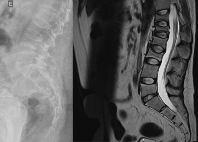
Radiography and Magnetic Resonance Imaging (MRI) of lumbosacral spine in profile

Bone scintigraphy on 08/2019 did not reveal hyper flow or anomalous hyperemia in the topography of the thoracolumbar spine, and in the later images of the exam, there was a greater relative uptake of the tracer in the lumbar spine (vertebrae T10–T12, L2–L4), of nonspecific aspect, questioning the presence of osteoarticular processes or ankylosing spondylitis.

It was also observed in the bone densitometry requested in October 2019, performed by dual-energy X-ray absorptiometry (DXA), low bone mineral density (BMD) in the lumbar spine, femoral neck and total femur, when comparing the results to evaluating the Z Score (Table 1).

Table 1 Dual-energy X-ray absorptiometry (DXA)

Thus, the diagnosis of osteoporosis was established, and treatment with vitamin D 7000 IU per week was started due to vitamin D3 insufficiency associated with the bisphosphonate alendronate 70 mg, also weekly. The patient had a past pathological history of fully treated syphilis (2018) and perianal condyloma with a surgical resection on 09/2017 and 02/2018. In the family history, it was reported that a maternal uncle died of systemic sclerosis. In the social context, the young person denied drinking alcohol and previous or current smoking.

On physical examination, there were no lentiginous skin areas or blue nevi; however, wide violet streaks were observed on the upper limbs and abdomen, with plethora and increased fat in the temporal facial region and hump (Fig. 2a, b), limb ecchymosis, hypotrophy of the arms and thighs, central obesity and kyphoscoliosis. Systemic blood pressure (sitting) was 150 × 90 mmHg, BMI was 26.09 kg/m2, and waist circumference was 99 cm, with no reported reduction in height, maintained at 1.55 m.

Fig. 2

figure 2

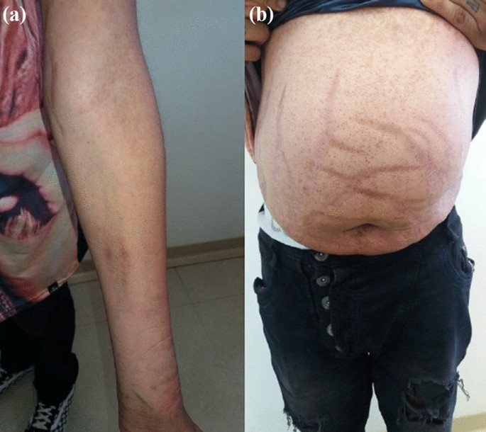
Changes in the physical examination. a Violet streaks on the upper limbs, b Violet streaks on abdomen

An investigation of secondary causes for osteoporosis was initiated, with the following laboratory test results (Table 2).

Table 2 Laboratory tests

Computed tomography of the abdomen with adrenal protocol performed on 08/13/2020 characterized isodense nodular formation in the body of the left adrenal and in the lateral portion of the right adrenal, measuring 1.5 cm and 0.6 cm, respectively. The lesions had attenuation of approximately 30 HU, showing enhancement by intravenous contrast, with an indeterminate washout pattern in the late phase after contrast (< 60%) (Fig. 3).

Fig. 3

figure 3

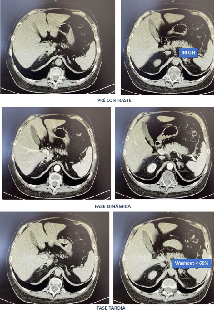
Computed tomography abdomen with adrenal protocol

After contact with the interventional radiology of the Hospital of Federal University of Juiz de Fora, catheterization of adrenal veins was performed on 10/2020; however, it was not possible to perform adequate lesion characterization due to obtaining serum cortisol levels that extrapolated the dilutional upper limit of the method (Table 3).

Table 3 Adrenal catheterization

The calculation of the selectivity index was 6.63 (Reference Value (RV) > 3), confirming the good positioning of the catheter within the vessels during the procedure. The calculated lateralization index was 1.1296 (VR < 3), denoting bilateral hormone production. However, as aldosterone was not collected from a peripheral vein, it was not possible to obtain the contralateral rate and define whether there was contralateral suppression of aldosterone production [5].

Due to pending diagnoses for a better therapeutic decision and Cushing’s syndrome in clear evolution and causing organic damage, it was decided, after catheterization, to make changes in the patient’s drug prescription. Ketoconazole 400 mg per day was started, the dose of vitamin D was increased to 14,000 IU per week, and ramipril 5 mg per day was prescribed due to secondary hypertension. In addition, given the severity of osteoporosis, it was decided to replace previously prescribed alendronate with zoledronic acid.

Magnetic resonance imaging of the upper abdomen was performed on 06/19/2021, which demonstrated lobulated nodular thickening in the left adrenal gland with areas of decreased signal intensity in the T1 out-phase sequence, denoting the presence of fat, and homogeneous enhancement using contrast, measuring approximately 1.7 × 1.5 × 1.3 cm, suggestive of an adenoma. There was also a small nodular thickening in the lateral arm of the right adrenal, measuring approximately 0.8 × 0.6 cm, which was difficult to characterize due to its small dimensions and nonspecific appearance.

PPNAD or carcinoma became an important etiological hypothesis for the case described when comparing the epidemiology in a young man and the clinical-laboratory-imaging findings of the differential diagnoses. According to a dialog with the patient and family, the group of experts opted for unilateral glandular surgical resection on the left side (11/11/2021), where more significant changes were visualized, as there was a possibility of malignancy in a young patient and to avoid a definitive adrenal insufficiency condition because of bilateral adrenalectomy. This would first allow the analysis of the material and follow-up of the evolution of the condition with the permanence of the contralateral gland.

In the macroscopic analysis of the adrenalectomy specimen, adrenal tissue weighing 20 g and measuring 9.3 × 5.5 × 2.0 cm was described, completely surrounded by adipose tissue. The gland has a multinodular surface and varies between 0.2 and 1.6 cm in thickness, showing a cortex of 0.1 cm in thickness and a medulla of 1.5 cm in thickness (Fig. 4).
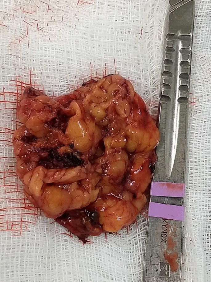
Fig. 4

figure 4

Left adrenal

The microscopic analysis described the expansion of the zona fasciculate, with the formation of multiple nonencapsulated nodules composed of polygonal cells with ample and eosinophilic cytoplasm and frequent depletion of intracytoplasmic lipid content. No areas of necrosis or mitotic activity were observed. The histopathological picture is suggestive of cortical pigmented micronodular hyperplasia of the adrenal gland.

For the final etiological definition and an indication of contralateral adrenalectomy, which could be unnecessary and would avoid chronic corticosteroid therapy, or else, it would be necessary to protect the patient from future complications with the maintenance of the disease in the right adrenal gland, it would be essential to search for mutations in the PRKAR1A, PDE11A, PDE8B and PRKACA genes [15]; however, such genetic analysis is not yet widely available, and the impossibility of carrying it out at the local level did not allow a complete conclusion of the case.

Discussion

Through the clinical picture presented and the research of several secondary causes for osteoporosis, it was possible to arrive at the diagnosis of Cushing syndrome [6]. There was symptomatic independent ACTH hypercortisolism, manifested by typical phenotypic changes, severe secondary osteoporosis, and arterial hypertension in a young patient.

The diagnosis of Cushing’s syndrome is always challenging, given the presence of confounding factors such as the following:

  • Physiological states of hypercortisolism—pseudo Cushing (strenuous exercise, pregnancy, uncontrolled diabetes, sleep apnea, chronic pain, alcohol withdrawal, psychiatric disorders, stress, obesity, glucocorticoid resistance syndromes);
  • Cyclic or mild—subclinical Cushing’s pictures;
  • Frequent and, even unknown, short- and long-term use of corticosteroids under different presentations;
  • Increase in the general population incidence of diabetes and obesity;
  • Screening tests with singularities for collection and individualized for different patient profiles.

It is important to note that the basal morning cortisol measurement is not the ideal test to assess hypercortisolism and is better applied to the assessment of adrenal insufficiency. However, the hypercortisolism of the case was unequivocal, and this test was also shown to be altered several times. As no test is 100% accurate, the current guidelines suggest the use of at least two first-line functional tests that focus on different aspects of the pathophysiology of the hypothalamic‒pituitary‒adrenal axis to confirm the hypercortisolism state: 24-hours cortisol, nocturnal salivary cortisol, morning serum cortisol after suppression with 1 mg of dexamethasone or after Liddle 1. Given that night-time salivary cortisol would require hospitalization, the other suggested tests were chosen, which are easier to perform in this context [78].

Subsequently, tests were performed to determine the cause of hypercortisolism, such as serum ACTH levels and adrenal CT. The suppressed ACTH denoted the independence of its action. CT showed bilateral adrenal nodules with more severe features: solid lesion, attenuation > 10 UI on noncontrast images, and contrast washout speed < 60% in 10 minutes. In this case, it is essential to make a broad clinical decision and dialog with the patient to weigh and understand the risks and benefits of surgical treatment [9].

Among the main diagnostic hypotheses for the differential diagnosis of bilateral adrenal hyperplasia are primary bilateral macronodular adrenal hyperplasia, McCune–Albright syndrome (MAS) and bilateral primary pigmented nodular hyperplasia (PPNAD) isolated or associated with Carney’s complex. Another possibility would be bilateral adrenocorticotropic hormone (ACTH)-dependent macronodular hyperplasia secondary to long-term adrenal stimulation in patients with Cushing’s disease (ACTH-secreting pituitary tumor) or ectopic ACTH production, but the present case did not present with ACTH elevation.

Primary macronodular adrenal hyperplasia (nodules > 1 cm) predominates in women aged 50–60 years and may also be detected in early childhood (before 5 years) in the context of McCune–Albright syndrome. Most cases are considered sporadic; however, there are now several reports of familial cases whose presentation suggests autosomal dominant transmission. Several pathogenic molecular causes were identified in the table, indicating that it is a heterogeneous disease [10]. The pathophysiology occurs through the expression of anomalous ectopic hormone receptors or amplified eutopic receptors in the adrenals. It usually manifests in an insidious and subclinical way, with cortisol secretion mediated through receptors for gastric inhibitory peptide (GIP), vasopressin (ADH), catecholamines, interleukin 1 (IL-1), leptin, luteinizing hormone (LH), serotonin or others. Nodular development is not always synchronous or multiple; thus, hypercortisolism only manifests when there is a considerable increase in the number of adrenocortical cells, with severe steroidogenesis observed by cortisoluria greater than 3 times the upper limit of normal. Patients with mild Cushing’s syndrome should undergo screening protocols to identify aberrant receptors, as this may alter the therapeutic strategy. If there is evidence of abnormal receptors, treatment with beta-blockers is suggested for patients with beta-adrenergic receptors or with gonadotropin-releasing hormone (GnRH) agonists (and sex steroid replacement) for patients with LH/hCG receptors. In patients in whom aberrant hormone receptors are not present or for whom no specific pharmacological blockade is available or effective, the definitive treatment is bilateral adrenalectomy, which is known to make the patient dependent on chronic corticosteroid therapy [11]. Studies have shown the effectiveness of unilateral surgery in the medium and long term, opting for the resection of the adrenal gland of greater volume and nodularity by CT, regardless of the values obtained by catheterization of adrenal veins, but with the possibility of persistence or recurrence in the contralateral gland. Another possibility would be total unilateral adrenalectomy associated with subtotal contralateral adrenalectomy [12].

In McCune–Albright syndrome (MAS), there are activating mutations in the G-protein GNAS1 gene, generating autonomic hyperfunction of several tissues, endocrine or not, and there may be, for example, a constant stimulus similar to ACTH on the adrenal gland. In this case, pituitary levels of ACTH are suppressed, and adrenal adenomas with Cushing’s syndrome appear. Hypercortisolism may occur as an isolated manifestation of the syndrome or be associated with the triad composed of polyostotic fibrous dysplasia, café au lait spots with irregular borders and gonadal hyperfunction with peripheral precocious puberty. The natural history of Cushing’s syndrome in McCune-Albright syndrome (MAS) is heterogeneous, with some children evolving with spontaneous resolution of hypercortisolism, while others have a more severe condition, eventually requiring bilateral adrenalectomy [13].

PPNAD predominates in females, in people younger than 30 years, multiple and small (< 6 mm) bilateral pigmented nodules (surrounded by atrophied cortex), which can reach 1.5 cm in adulthood, with family genetic inheritance (66%) or sporadic inheritance (33%), and as part of the Carney complex reported in 40% of cases. In 70% of cases, inactivating mutations are identified in the PKA regulatory 1-alpha subunit (PRKAR1A), a tumor suppressor gene [14]. Osteoporosis is often associated with this condition [15]. One test that can distinguish patients with PPNAD from other primary adrenocortical lesions is cortisoluria after sequential suppression with low- and high-dose dexamethasone. In contrast to most patients with primary adrenocortical disease, who demonstrate no change in urinary cortisol, 70% of PPNAD patients have a paradoxical increase in urinary cortisol excretion [16]. The treatment of choice for PPNAD is bilateral adrenalectomy due to the high recurrence rate for primary adrenal disease [17].

Carney complex is a multiple neoplastic syndrome with autosomal dominant transmission, characterized by freckle-like cutaneous hyperpigmentation (lentiginosis), endocrine tumors [(PPNAD), testicular and/or thyroid tumors and acromegaly] and nonendocrine tumors, including cutaneous, cardiac, mammary, and osteochondral myxomas, among others. In the above case, the transthoracic echocardiogram of the patient on 03/18/2021 showed cavities of normal dimensions, preserved systolic and diastolic functions, no valve changes and no lentiginous skin areas and blue nevi, making the diagnosis of the syndrome less likely. The definitive diagnosis of Carney requires two or more main manifestations. Several related clinical components may suggest the diagnosis but not define it. The diagnosis can also be made if a key criterion is present and a first-degree relative has Carney or an inactivating mutation of the gene encoding PRKAR1A [18].

The adenoma is usually small in size (< 3 cm), similar to the nodules in this case; however, it is usually unilateral, with an insidious and mild evolution, especially in adult women over 35 years of age, producing only 1 steroid class. Carcinomas are usually large (> 6 cm), and only 10% are bilateral. They should be suspected mainly when the tumor presents with hypercortisolism associated with hyperandrogenism. They have a bimodal age distribution, with peaks in childhood and adolescence, as well as at the end of life [3].

Conclusion

Early identification of Cushing’s syndrome, with measures based on the assessment of risks and benefits, remains the best way to prevent progression and reduce morbidity [2]. After 6 months of drug inhibition of steroidogenesis, blood pressure control and anti-osteoporotic therapy, the objective was to minimize the levels and deleterious metabolic effects of hypercortisolism, which could also harm the surgical procedure in the short and long term through infections, dehiscence, nonimmediate bed mobilization and cardiovascular events. Unilateral adrenalectomy was chosen, given the possibility of malignancy in a young patient and to avoid definitive surgical adrenal insufficiency if the adrenalectomy was bilateral. Despite the unavailability of genetic analysis for a precise etiological definition, it is possible to take efficient measures to avoid unnecessary consequences or damage.

Availability of data and materials

All data generated or analysed during this study are included in this published article [and its Additional file 1]. The datasets generated and/or analysed during the current study are available in the link https://ufjfedubr-my.sharepoint.com/:v:/g/personal/barbara_reis_ufjf_edu_br/EVpIR005sPZGlQvMJhIwSaUB0Hig4KOjhkG4D4cMggUwHA?e=Dk8tng.

Abbreviations

ACTH:
Adrenocorticotropic hormone
PPNAD:
Bilateral primary pigmented nodular hyperplasia
DXA:
Dual energy X-ray absorptiometry
GIP:
Gastric inhibitory peptide
GnRH:
Gonadotropin-releasing hormone
IL-1:
Interleukin 1
BMD:
Low bone mineral density
LH:
Luteinizing hormone
MAS:
McCune–Albright syndrome
PRKAR1A:
PKA regulatory 1-alpha subunit
ADH:
Vasopressin

References

  1. Pedro AO, Plapler PG, Szejnfeld VL. Manual brasileiro de osteoporose: orientações práticas para os profissionais de saúde. 1st ed. São Paulo: Editora Clannad; 2021. ISBN 978-65-89832-00-3.

  2. Naguib R, Elkemary EZ, Elsharkawi KM. The severity of bone loss: a comparison between Cushing’s disease and Cushing’s syndrome. J Endocrinol Metab. 2023;13(1):33–8. https://doi.org/10.14740/jem857.

    Article Google Scholar

  3. Vilar L, et al. Endocrinologia Clínica. 6th ed. Rio de Janeiro: Guanabara Koogan; 2016.

    Google Scholar

  4. Wang D, Dang CX, Hao YX, Yu X, Liu PF, Li JS. Relationship between osteoporosis and Cushing syndrome based on bioinformatics. Medicine (Baltimore). 2022;101(43): e31283.

    Article CAS PubMed Google Scholar

  5. Williams TA, Reincke M. Management of Endocrine Disease: diagnosis and management of primary aldosteronism: the Endocrine Society guideline 2016 revisited. Eur J Endocrinol. 2018;179(1):R19–29. https://doi.org/10.1530/EJE-17-0990.

    Article CAS PubMed Google Scholar

  6. Compston J, Cooper A, Cooper C, Gittoes N, Gregson C, Harvey N, National Osteoporosis Guideline Group (NOGG), et al. UK clinical guideline for the prevention and treatment of osteoporosis. Arch Osteoporos. 2017;12(1):43. https://doi.org/10.1007/s11657-017-0324-5.

    Article CAS PubMed PubMed Central Google Scholar

  7. Nieman LK. Diagnosis of Cushing’s syndrome in the modern era. Endocrinol Metab Clin N Am. 2018;47(2):259–73. https://doi.org/10.1016/j.ecl.2018.02.001.

    Article Google Scholar

  8. Herr K, Muglia VF, Koff WJ, Westphalen AC. Imaging of the adrenal gland lesions. Radiol Bras. 2014;47(4):228–39. https://doi.org/10.1590/0100-3984.2013.1762.

    Article PubMed PubMed Central Google Scholar

  9. Hsiao HP, Kirschner LS, Bourdeau I, Keil MF, Boikos SA, Verma S, et al. Clinical and genetic heterogeneity, overlap with other tumor syndromes, and atypical glucocorticoid hormone secretion in adrenocorticotropin-independent macronodular adrenal hyperplasia compared with other adrenocortical tumors. J Clin Endocrinol Metab. 2009;94(8):2930–7. https://doi.org/10.1210/jc.2009-0516.

    Article CAS PubMed PubMed Central Google Scholar

  10. Mircescu H, Jilwan J, N’Diaye N, et al. Are ectopic or abnormal membrane hormone receptors frequently present in adrenal Cushing’s syndrome? J Clin Endocrinol Metab. 2000;85(10):3531–6. https://doi.org/10.1210/jcem.85.10.6865.

    Article CAS PubMed Google Scholar

  11. Miller BS, Auchus RJ. Evaluation and treatment of patients with hypercortisolism: a review. JAMA Surg. 2020;155(12):1152–9. https://doi.org/10.1001/jamasurg.2020.3280.

    Article PubMed Google Scholar

  12. Haddad NG, Eugster EA. Peripheral precocious puberty including congenital adrenal hyperplasia: causes, consequences, management and outcomes. Best Pract Res Clin Endocrinol Metab. 2019;33(3):101273. https://doi.org/10.1016/j.beem.2019.04.007.

    Article PubMed Google Scholar

  13. Bonnet-Serrano F, Bertherat J. Genetics of tumors of the adrenal cortex. Endocr Relat Cancer. 2018;25(3):R131–52. https://doi.org/10.1530/ERC-17-0361.

    Article CAS PubMed Google Scholar

  14. Carney JA, Young WF Jr. Primary pigmented nodular adrenocortical disease and its associated conditions. Endocrinologist. 1992;2:6.

    Article Google Scholar

  15. Stratakis CA, Sarlis N, Kirschner LS, Carney JA, Doppman JL, Nieman LK, et al. Paradoxical response to dexamethasone in the diagnosis of primary pigmented nodular adrenocortical disease. Ann Intern Med. 1999;131(8):585–91. https://doi.org/10.7326/0003-4819-131-8-199910190-00006.

    Article CAS PubMed Google Scholar

  16. Powell AC, Stratakis CA, Patronas NJ, Steinberg SM, Batista D, Alexander HR, et al. Operative management of Cushing syndrome secondary to micronodular adrenal hyperplasia. Surgery. 2008;143(6):750–8. https://doi.org/10.1016/j.surg.2008.03.022.

    Article PubMed Google Scholar

  17. Almeida MQ, Stratakis CA. Carney complex and other conditions associated with micronodular adrenal hyperplasias. Best Pract Res Clin Endocrinol Metab. 2010;24(6):907–14. https://doi.org/10.1016/j.beem.2010.10.006.

    Article CAS PubMed PubMed Central Google Scholar

  18. Hannah-Shmouni F, Stratakis CA. A gene-based classification of primary adrenocortical hyperplasias. Horm Metab Res. 2020;52(3):133–41. https://doi.org/10.1055/a-1107-2972.

    Article CAS PubMed Google Scholar

Download references

Acknowledgements

Not applicable.

Funding

Not applicable.

Author information

Authors and Affiliations

  1. Serviço de Endocrinologia, Hospital Universitário da Universidade Federal de Juiz de Fora, Juiz de Fora, Minas Gerais, Brazil

    Bárbara Oliveira Reis, Christianne Toledo Sousa Leal, Danielle Guedes Andrade Ezequiel, Ana Carmen dos Santos Ribeiro Simões Juliano, Flávia Lopes de Macedo Veloso, Leila Marcia da Silva, Lize Vargas Ferreira, Mariana Ferreira & Gabriel Zeferino De Oliveira Souza

Contributions

All the authors contributed to the conception and design of the work and have approved the submitted version. All authors read and approved the final manuscript.

Corresponding author

Correspondence to Bárbara Oliveira Reis.

Ethics declarations

Ethics approval and consent to participate

Not applicable.

Consent for publication

Written informed consent was obtained from the patient for publication of this case report and any accompanying images. A copy of the written consent is available for review by the Editor-in-Chief of this journal.

Competing interests

The authors declare that they have no competing interests.

Additional information

Publisher’s Note

Springer Nature remains neutral with regard to jurisdictional claims in published maps and institutional affiliations.

Supplementary Information

Additional file 1. Surgical removal of adrenal gland.

Cushing’s Disease Associated With Partially Empty Sella Turcica Syndrome

Abstract

The association between empty sella turcica (EST) syndrome and Cushing’s disease has been rarely reported. It is plausible to hypothesize that EST syndrome in association with Cushing’s disease can be attributed to intracranial hypertension. In this case report, we present a 47-year-old male patient who presented with weight loss, fatigue, easy bruising, acanthosis nigricans, and skin creases hyperpigmentation. Investigations revealed hypokalemia and confirmed the diagnosis of Cushing’s disease. Magnetic resonance imaging (MRI) brain showed a partial EST syndrome and a new pituitary nodule as compared with previous brain imaging. Transsphenoidal surgery was pursued and was complicated by cerebrospinal fluid leakage. This case reflects the rare association of EST syndrome and Cushing’s disease, suggesting the increased risk of postoperative complications in this setting and the diagnostic challenge that EST syndrome imposes. We review the literature for a possible mechanism of this association.

Introduction

Cushing’s disease is commonly caused by an adrenocorticotropic hormone (ACTH)-producing pituitary adenoma, which can be very challenging to be seen on brain magnetic resonance imaging (MRI) [1]. Empty sella turcica (EST) syndrome is a radiological diagnosis of apparently empty turcica secondary to outpouching of the arachnoid mater into the turcica, which can be attributed to intracranial hypertension (ICHTN). This can make the visual diagnosis of pituitary adenoma even more challenging in clinical practice. ICHTN has been also associated with Cushing’s disease and might explain this infrequent association between EST and Cushing’s disease [1]. EST syndrome can be either partial or complete, primary or secondary and has been seen infrequently with Cushing’s disease. In this setting, not only that it is likely to obscure an underlying pituitary lesion, but also it does contribute to the risk of postoperative complications [2].

Case Presentation

A 47-year-old male presented to the emergency department (ED) with slowly progressive generalized limb muscle weakness affecting both distal and proximal muscles over a few weeks and gait instability for three days prior to presentation. He also reported unintentional 40 pounds weight loss over the previous four months. Past medical history was significant for type II diabetes mellitus, hypothyroidism, hypertension, and dyslipidemia. In the ED, vital signs included a blood pressure of 140/90 mmHg, a heart rate of 66 beats per minute, a respiratory rate of 16 cycles per minute, and SpO2 of 97% on room air. Body mass index has decreased to 22 kg/m2 from a baseline of 26 kg/m2 one month prior. On the physical exam, he exhibited cachexia, easy bruising, acanthosis nigricans, and hyperpigmentation of skin creases. All other systems were negative. Complete metabolic panel and complete blood count were obtained showing hyperglycemia of 311 mg/dl, see Table 1. Further lab evaluation showed elevated salivary cortisol at 2.96 microgram/dl (reference range 0.094-1.551 mcg/dl), elevated 24-hour urinary free cortisol at 156 mcg/24 hour (reference 10-100 mcg/24h), positive overnight dexamethasone suppression test with serum cortisol at 2.8 mcg/dl (reference more than 2 mcg/dl), negative anti-adrenal antibodies, normal aldosterone, and elevated dehydroepiandrostenedione at 401 mcg/dl (reference 32-240 mcg/dl), with lack of suppression of the ACTH level at 35.1 pg/ml (reference 10-60 pg/ml). This confirmed the diagnosis of Cushing’s disease.

Variable Finding Reference
Random glucose 311 Less than 200 mg/dl
Sodium 141 137-145 mmol/L
Potassium 2.5 3.5-5.1 mmol/L
Chloride 96 98-107 mmol/L
Bicarbonate 32 22-30 mmol/L
Blood urea nitrogen 32 9-20 mg/dl
Creatinine 0.52 0.66-1.25 mg/dl
Calcium 8.7 8.6-10.3 mg/dl
Total protein 5.5 6.5-8.5 g/dl
Albumin 3.3 3.5-5 g/dl
Total bilirubin 0.6 0.2-1.3 mg/dl
Alkaline phosphatase 115 38-126 U/L
Aspartate transaminase 17 17-59 U/L
Alanine transaminase 39 Less than 49 U/L
White blood cell count 10×10^3 cells/mcl 4-10×1063 cells/mcl
Hemoglobin 15.3 13.7-17.5 g/dl
Platelet 281 150-400×10^3 cells/mcl
Table 1: Lab Findings

Computed tomography (CT) scan of the head was unremarkable. CT scan of the chest was also unremarkable. CT scan of abdomen and pelvis showed no adrenal mass. Ultrasound of the kidneys was unremarkable. Pituitary MRI brain protocol for adenoma showed a partial EST, shortening within neurohypophysis and a new 10 x 8 x 4 mm nodule along the floor of pituitary sella as compared to MRI four years ago (Figure 1).

Magnetic-Resonance-Imaging-(MRI)-Brain
Figure 1: Magnetic Resonance Imaging (MRI) Brain

MRI brain showing partially empty sella turcica syndrome ( black arrow) with a small nodule at the floor of the turcica (white arrow).

The diagnosis of Cushing’s disease was confirmed, and the patient underwent trans-sphenoidal resection of pituitary adenoma. Histological examination showed positive CAM 5.2, positive chromogranin, and ACTH immunostains. The patient presented to the ED five days after discharge home. He stated that he noticed drainage from the nose that transitioned from bloody to clear fluid and has been increasing in quantity for two days with associated intermittent headaches since the surgery. He was afebrile with stable vital signs. No signs of infection were noted on basic labs. These were significant only for mild asymptomatic hyponatremia of 131 mmol/L and hypokalemia of 3.3 mmol/L. The patient was diagnosed with cerebrospinal fluid (CSF) leakage and had a lumbar drain trial. The trial was unsuccessful after several days, and the patient underwent a transnasal endoscopic repair of CSF rhinorrhea using nasoseptal flaps. At an outpatient follow-up one month and three months after the surgery, prior lab abnormalities including hypokalemia, hyponatremia, and hyperglycemia resolved. No further evidence of CSF leakage was appreciated, and he remained asymptomatic.

Discussion

EST syndrome is characterized by herniation of the subarachnoid space into the intrasellar space with compression of the pituitary gland into the posteroinferior wall [3]. This is likely to obscure the presence of underlying pituitary mass. The incidence of EST syndrome in the general population is estimated at 20%. The association between EST syndrome and Cushing’s disease has been reported infrequently. A retrospective study of 68 patients with Cushing’s disease found that 16% of these have EST syndrome [3].

Cushing’s disease usually results from pituitary adenomas secreting ACTH, and even the smallest microadenomas can produce a systemic disease. These microadenomas can be very difficult to recognize on brain MRI [4]. This is complicated in EST syndrome and even further with the possibility of ectopic ACTH production. A retrospective study of 197 patients diagnosed with Cushing’s disease concluded that EST syndrome is associated with higher prevalence of MRI-negative Cushing’s disease. This was attributed to ICHTN and pituitary gland compression [1]. Although surgery is curative in 70-90% of cases, EST syndrome was found to have higher risk of postoperative complications among those with Cushing’s disease including diabetes insipidus, hypopituitarism, and CSF leakage [3]. This is usually because in the case of MRI-negative Cushing’s disease with total EST syndrome, empiric surgical exploration is sought after inferior petrosal sampling confirms the pituitary origin of excess ACTH, and postoperative remission indicates adequate tumor resection [2]. This entails a higher chance of uncertainty and injury to healthy pituitary tissue.

EST syndrome can be either primarily due to defects in the sellar diaphragm or anatomical variant or secondary to ICHTN. EST syndrome has been reported in association with many conditions associated with elevated intracranial pressure including tumors, thrombosis, meningitis, hydrocephalus, and Arnold-Chiari malformation [5]. Reversal of EST syndrome has been reported in those with idiopathic ICHTN with therapy by acetazolamide, ventriculoperitoneal shunt, and lumbar puncture [6,7]. A study has shown correlation between CSF circulation impairment or blockage and EST syndrome [8]. The incidence of EST syndrome in association with symptomatic intracranial hypertension is variable and ranges from 2.5% for total EST syndrome to 94% for partial EST syndrome [9]. Impaired CSF circulation and dynamics have been reported in 77% of patients with EST syndrome [10]. In addition to intracranial hypertension, EST syndrome has also been described in association with obesity, meningioma, pediatric nevoid basal cell carcinoma, therapy for growth hormone deficiency and even in healthy individuals [9]. Lack of symptoms of intracranial hypertension in this patient does not rule it out as intracranial hypertension in EST syndrome represents a spectrum that ranges from asymptomatic, milder intracranial hypertension to symptomatic intracranial hypertension with headache, visual disturbance, and papilledema [10]. This explains the fact that only 8-14% of EST syndrome progress to symptomatic ICHTN, while symptomatic ICHTN has been associated with EST syndrome in 94% of cases.

ICHTN has been seen in association with disturbance of the hypothalamic-pituitary-adrenal axis. This has been reported after surgical and medical treatment of Cushing’s disease, withdrawal of long-term steroid therapy, initial presentation of Addison’s disease, or relative glucocorticoids deficiency [11]. Cortisol excess increases CSF production and reduces its absorption, hence increasing intracranial pressure [12]. Another possible mechanism is the expression of both mineralocorticoid responsive epithelial sodium channel receptors on the basolateral membrane of the CSF producing epithelial cells of the choroid plexus as well as the expression of 11-beta hydroxysteroid dehydrogenase type 1 enzyme, which is a bidirectional enzyme that mainly functions to convert the inactive cortisone to active cortisol. These mechanisms play a role in maintaining the balance between CSF production and absorption [13,14].

In this case, the patient presented some clinical findings that are rarely associated with Cushing’s disease, combined with a radiological feature that masked the true diagnosis. Our patient presented with significant weight loss, rather than central obesity, which is normally associated with Cushing’s disease. Although possible, the increase in ACTH due to Cushing’s disease is not sufficient to cause hyperpigmentation, which is a classical finding of Addison’s disease, where the entire adrenal cortex is usually affected due to an autoimmune destruction; however, the zona glomerulosa of the adrenal cortex produces aldosterone and its deficiency would lead to hyperkalemia [15]. Our patient presented with both hyperpigmentation and hypokalemia.

Conclusions

EST syndrome is an uncommon radiological finding of apparently EST that has been reported in association with ICHTN. The latter has also been seen in association with Cushing’s disease/syndrome. This is likely to result from glucocorticoid excess-induced change in CSF flow dynamics. EST has been infrequently described in association with Cushing’s disease. This association has a clinical implication as it is likely to obscure the visualization of pituitary lesions responsible for Cushing’s disease, contribute to diagnostic uncertainty, and increase the risk of healthy pituitary tissue injury and the risk of postoperative complications including CSF leakage.

References

  1. Himes BT, Bhargav AG, Brown DA, Kaufmann TJ, Bancos I, Van Gompel JJ: Does pituitary compression/empty sella syndrome contribute to MRI-negative Cushing’s disease? A single-institution experience. Neurosurg Focus. 2020, 48:E3. 10.3171/2020.3.FOCUS2084
  2. Sun Y, Sun Q, Fan C, et al.: Diagnosis and therapy for Cushing’s disease with negative dynamic MRI finding: a single-centre experience. Clin Endocrinol (Oxf). 2012, 76:868-76. 10.1111/j.1365-2265.2011.04279.x
  3. Manavela MP, Goodall CM, Katz SB, Moncet D, Bruno OD: The association of Cushing’s disease and primary empty sella turcica. Pituitary. 2001, 4:145-51. 10.1023/a:1015310806063
  4. Chatain GP, Patronas N, Smirniotopoulos JG, et al.: Potential utility of FLAIR in MRI-negative Cushing’s disease. J Neurosurg. 2018, 129:620-8. 10.3171/2017.4.JNS17234
  5. Friedman DI, Jacobson DM: Diagnostic criteria for idiopathic intracranial hypertension. Neurology. 2002, 59:1492-5. 10.1212/01.wnl.0000029570.69134.1b
  6. Triggiani V, Giagulli VA, Moschetta M, Guastamacchia E: An unusual case of reversible empty sella. Endocr Metab Immune Disord Drug Targets. 2016, 16:154-6. 10.2174/1871530315666151001141507
  7. Wind JJ, Lonser RR, Nieman LK, DeVroom HL, Chang R, Oldfield EH: The lateralization accuracy of inferior petrosal sinus sampling in 501 patients with Cushing’s disease. J Clin Endocrinol Metab. 2013, 98:2285-93. 10.1210/jc.2012-3943
  8. Brismar K, Bergstrand G: CSF circulation in subjects with the empty sella syndrome. Neuroradiology. 1981, 21:167-75. 10.1007/BF00367338
  9. Ranganathan S, Lee SH, Checkver A, Sklar E, Lam BL, Danton GH, Alperin N: Magnetic resonance imaging finding of empty sella in obesity related idiopathic intracranial hypertension is associated with enlarged sella turcica. Neuroradiology. 2013, 55:955-61. 10.1007/s00234-013-1207-0
  10. Maira G, Anile C, Mangiola A: Primary empty sella syndrome in a series of 142 patients. J Neurosurg. 2005, 103:831-6. 10.3171/jns.2005.103.5.0831
  11. Zada G, Tirosh A, Kaiser UB, Laws ER, Woodmansee WW: Cushing’s disease and idiopathic intracranial hypertension: case report and review of underlying pathophysiological mechanisms. J Clin Endocrinol Metab. 2010, 95:4850-4. 10.1210/jc.2010-0896
  12. Sinclair AJ, Ball AK, Burdon MA, Clarke CE, Stewart PM, Curnow SJ, Rauz S: Exploring the pathogenesis of IIH: an inflammatory perspective. J Neuroimmunol. 2008, 201:212-20. 10.1016/j.jneuroim.2008.06.029
  13. Sinclair AJ, Onyimba CU, Khosla P, et al.: Corticosteroids, 11beta-hydroxysteroid dehydrogenase isozymes and the rabbit choroid plexus. J Neuroendocrinol. 2007, 19:614-20. 10.1111/j.1365-2826.2007.01569.x
  14. Amin MS, Wang HW, Reza E, Whitman SC, Tuana BS, Leenen FH: Distribution of epithelial sodium channels and mineralocorticoid receptors in cardiovascular regulatory centers in rat brain. Am J Physiol Regul Integr Comp Physiol. 2005, 289:R1787-97. 10.1152/ajpregu.00063.2005
  15. Stratakis CA: Skin manifestations of Cushing’s syndrome. Rev Endocr Metab Disord. 2016, 17:283-6. 10.1007/s11154-016-9399-3

From https://www.cureus.com/articles/161111-cushings-disease-associated-with-partially-empty-sella-turcica-syndrome-a-case-report#!/

Cushing’s Syndrome is Associated with Early Medical- and Surgical-related Complications Following Total Joint Arthroplasty

I’m glad I didn’t know this before my recent knee surgery!

Abstract

Background

Cushing’s syndrome (CS) is a disorder characterized by exposure to supraphysiologic levels of glucocorticoids. The purpose of this study was to evaluate the association between CS and postoperative complication rates following total joint arthroplasty (TJA).

Methods

Patients diagnosed with CS undergoing TJA for degenerative etiologies were identified from a large national database and matched 1:5 to a control cohort using propensity scoring. Propensity score matching resulted in 1,059 total hip arthroplasty (THA) patients with CS matched to 5,295 control THA patients and 1,561 total knee arthroplasty (TKA) patients with CS matched to 7,805 control TKA patients. Rates of medical complications occurring within 90 days of TJA and surgical-related complications occurring within 1 year of TJA were compared using odds ratios.

Results

The THA patients with CS had higher incidences of pulmonary embolism (Odds Ratio (OR) 2.21, P=0.0026), urinary tract infection (OR 1.29, P=0.0417), pneumonia (OR 1.58, P=0.0071), sepsis (OR 1.89, P=0.0134), periprosthetic joint infection (OR 1.45, P=0.0109), and all-cause revision surgery (OR 1.54, P=0.0036). The TKA patients with CS had significantly higher incidences of urinary tract infection (OR 1.34, P=0.0044), pneumonia (OR 1.62, P=0.0042), and dislocation (OR 2.43, P=0.0049) and a lower incidence of manipulation under anesthesia (MUA) (OR 0.63, P=0.0027).

Conclusion

Cushing’s syndrome is associated with early medical- and surgical-related complications following TJA and a reduced incidence of MUA following TKA.

Introduction

Cushing’s syndrome (CS) is characterized by exposure to supraphysiologic levels of glucocorticoids, whether endogenous or exogenous. Chronic exposure to hypercortisolism can lead to the development of comorbidities known to be risk factors for complications following total joint arthroplasty (TJA) including obesity, hypertension, diabetes, hyperlipidemia, and cerebrovascular disease.[1,2] Hypercortisolism is also a known risk factor for the development of osteonecrosis, and there have been several case reports of this disease being caused by endogenous production of corticosteroids.[3, 4, 5, 6, 7, 8] It can therefore be expected that the incidence of arthroplasty procedures among CS patients is likely higher than the general population. It is important to identify and understand patient specific risk factors for complications following TJA. There has been a major push recently to investigate the association between uncommon disorders and complication rates following TJA in order to risk stratify, counsel, and optimize these patients appropriately.[9, 10, 11, 12, 13, 14, 15]

The typical clinical features of CS include increased central adiposity, purple striae, thin skin, fatigue, and proximal atrophy of the upper and lower limbs.[16,17] The most common etiology of endogenous CS is overproduction of adrenocorticotropic hormone (ACTH) from a pituitary adenoma, although ACTH-independent forms of CS may be caused by overproduction of glucocorticoids from the adrenal glands.[2] First-line laboratory tests for the diagnosis of CS include 24-hour urinary free cortisol, late night salivary cortisol, and the dexamethasone suppression test to determine if the negative feedback of the hypothalamic-pituitary-adrenal axis is functioning appropriately.[16] Hypercortisolism associated with CS is known to have a deleterious effect on bone health by decreasing osteoblast function and increasing bone resorption and has been associated with decreased bone mineral density at various sites in the femur including Ward’s triangle, the femoral neck, and the greater trochanter.[18] The effect of these changes in physiology on outcomes following TJA remains unclear. There are few prior case reports describing arthroplasty procedures for CS patients,[3, 4, 5] with one case report of total hip arthroplasty (THA) for femoral head osteonecrosis complicated by pulmonary thromboembolism requiring a 10-day admission to the ICU.[3] However, no large scale studies to date have investigated complication rates following TJA within this patient population. It is therefore important to better understand the risks associated with this pathology. The purpose of this study was to evaluate the association between CS and postoperative complication rates following TJA. We hypothesized that patients who have CS would have increased incidences of early medical- and surgical-related complications.

Section snippets

Methods

This is a retrospective cohort study utilizing the commercially available M151Ortho database via PearlDiver (PearlDiver Inc., Colorado Springs, Colorado). This database contains deidentified records for 151 million patients in the United States in accordance with the Health Insurance Portability and Accountability Act (HIPAA). Patient records were queried using International Classification of Diseases (ICD) codes and Current Procedural Terminology (CPT) codes. This study was deemed exempt from

Results

The THA patients who had CS had significantly higher 90-day incidences of PE (OR 2.21, P=0.0026), UTI (OR 1.29, P=0.0417), pneumonia (OR 1.58, P=0.0071), and sepsis (OR 1.89, P=0.0134) (Table 2). The TKA patients who had CS had significantly higher 90-day incidences of UTI (OR 1.34, P=0.0044) and pneumonia (OR 1.62, P=0.0042) (Table 3). Regarding surgical-related complications, CS patients undergoing THA had significantly higher incidences of PJI (OR 1.45, P=0.0109) and all-cause revision

Discussion

This study revealed that patients who have CS are at increased risk of developing early postoperative complications following TJA. Understanding this risk profile is important for accurate shared decision making between CS patients and their clinicians. Interestingly, CS seems to influence rates of instability and stiffness following TKA as patients in the test cohort were more likely to sustain a dislocation and less likely to undergo MUA. Rates of infectious medical complications including

Conclusion

Cushing’s syndrome is associated with an increased risk of early infectious complications following TJA including UTI, pneumonia, sepsis, and hip PJI and an increased incidence of dislocation following TKA. Interestingly, CS appears to be protective against arthrofibrosis as patients who have CS had lower incidences of MUA following TKA. Clinicians may be guided by this study to accurately risk stratify and counsel patients with CS prior to undergoing TJA.

References (29)


Catastrophic ACTH-secreting Pheochromocytoma

Abstract

Summary

Cushing’s syndrome due to ectopic adrenocorticotropic hormone (ACTH) secretion (EAS) by a pheochromocytoma is a challenging condition. A woman with hypertension and an anamnestic report of a ‘non-secreting’ left adrenal mass developed uncontrolled blood pressure (BP), hyperglycaemia and severe hypokalaemia. ACTH-dependent severe hypercortisolism was ascertained in the absence of Cushingoid features, and a psycho-organic syndrome developed. Brain imaging revealed a splenial lesion of the corpus callosum and a pituitary microadenoma. The adrenal mass displayed high uptake on both 18F-FDG PET/CT and 68Ga-DOTATOC PET/CT; urinary metanephrine levels were greatly increased. The combination of antihypertensive drugs, high-dose potassium infusion, insulin and steroidogenesis inhibitor normalized BP, metabolic parameters and cortisol levels; laparoscopic left adrenalectomy under intravenous hydrocortisone infusion was performed. On combined histology and immunohistochemistry, an ACTH-secreting pheochromocytoma was diagnosed. The patient’s clinical condition improved and remission of both hypercortisolism and catecholamine hypersecretion ensued. Brain magnetic resonance imaging showed a reduction of the splenial lesion. Off-therapy BP and metabolic parameters remained normal. The patient was discharged on cortisone replacement therapy for post-surgical hypocortisolism. EAS due to pheochromocytoma displays multifaceted clinical features and requires prompt diagnosis and multidisciplinary management in order to overcome the related severe clinical derangements.

Learning points

  • A small but significant number of cases of adrenocorticotropic hormone (ACTH)-dependent Cushing’s syndrome are caused by ectopic ACTH secretion by neuroendocrine tumours, which is usually associated with severe hypercortisolism causing severe clinical and metabolic derangements.
  • Ectopic ACTH secretion by a pheochromocytoma is exceedingly rare but can be life-threatening, owing to the simultaneous excess of both cortisol and catecholamines.
  • The combination of biochemical and hormonal testing and imaging procedures is mandatory for the diagnosis of ectopic ACTH secretion, and in the presence of an adrenal mass, the possibility of an ACTH-secreting pheochromocytoma should be taken into account.
  • Immediate-acting steroidogenesis inhibitors are required for the treatment of hypercortisolism, and catecholamine excess should also be appropriately managed before surgical removal of the tumour.
  • A multidisciplinary approach is required for the treatment of this challenging entity.

Background

Cushing’s syndrome (CS) is a rare endocrine disease characterized by high levels of glucocorticoids; it increases morbidity and mortality due to cardiovascular and infectious diseases (123).

To diagnose CS, adrenocorticotropic hormone (ACTH)-dependent disease must be distinguished from ACTH-independent disease, and pituitary ACTH production from ectopic production. About 20% of ACTH-dependent cases arise from ectopic ACTH secretion (EAS) (234). EAS is most often due to aberrant ACTH production by small-cell lung carcinoma or neuroendocrine tumours originating in the lungs or gastrointestinal tract; this, in turn, strongly increases cortisol production by the adrenal glands (345).

Since the first-line treatment of EAS is the surgical removal of the ectopic ACTH-secreting tumour, its prompt and accurate localization is crucial.

Rapid cortisol reduction by means of immediate-acting steroidogenesis inhibitors (4) is mandatory in order to treat the related endocrine, metabolic and electrolytic derangements. EAS by a pheochromocytoma is exceedingly rare and can be life-threatening.

We describe the case of a woman with hypertension and a known ‘non-secreting’ left adrenal mass, who manifested uncontrolled blood pressure (BP), hyperglycaemia, hypokalaemia and psycho-organic syndrome associated with damage of the splenium of the corpus callosum. These findings were eventually seen to be related to an ACTH-secreting left pheochromocytoma, which was ascertained by hormonal evaluation and morphological and functional imaging assessment and confirmed by histopathology/immunostaining. Hormonal hypersecretion resolved after adrenalectomy and metabolic derangements normalized.

Case presentation

A 72-year-old woman with hypertension was taken to the emergency department because of increased BP (200/100 mm Hg). High BP (190/100 mmHg) was confirmed, whereas oxygen saturation (98%), heart rate (84 bpm) and lung and abdomen examination were normal. Electrocardiogram and chest x-ray were unremarkable. Captopril 50 mg orally, followed by intramuscular clonidine, normalized BP.

The patient looked thin and reported significant weight loss (10 kg) over the previous 6 months; she was on antihypertensive therapy with bisoprolol 5 mg/day and irbesartan 150 mg/day, and ezetimibe 10 mg/day for dyslipidaemia. The patient’s records included a previous diagnosis in another hospital of normofunctioning multinodular goitre and a 2.5 cm-left solid inhomogeneous adrenal mass with well-defined margins, which was found on CT performed 6 years earlier during the work-up for hypertension. On the basis of hormonal data and absent uptake on 123I metaiodobenzylguanidine scintigraphy, the adrenal lesion had been deemed to be non-functioning and follow-up had been advised. Unfortunately, only initial cortisol (15.7 μg/dL) and 24-h urine-free cortisol (UFC) levels (32.5 μg/24 h) were retrievable; both proved normal.

Investigations

Blood chemistry showed neutrophilic leucocytosis, hyperglycaemia with increased glycated haemoglobin, severe hypokalaemia and metabolic alkalosis (Table 1). Potassium infusion (50 mEq in 500 mL saline/24 h) was rapidly started, together with a subcutaneous rapid-acting insulin analogue and prophylactic enoxaparin. The patient experienced mental confusion, hallucinations and restlessness; non-enhanced computed tomography (CT) of the brain revealed a hypodense area of the splenium of the corpus callosum, possibly due to metabolic damage (Fig. 1A).

Figure 1View Full Size
Figure 1

Non-enhanced CT showing a hypodense area of the splenium of the corpus callosum (arrows), without mass effect (A, axial view). Contrast-enhanced MR image showing a hypointense pituitary lesion (arrow) which enhances more slowly than normal pituitary parenchyma, deemed suspicious for microadenoma (B, coronal view). FLAIR MR image showing hyperintense signal of the splenium of the corpus callosum (asterisk), which partially extended to the crux of the left fornix (arrow) (C, axial view). As the lesion showed no restricted diffusion on DWI (D, axial view), an ischaemic lesion was excluded. A progressive reduction in the extension of the hyperintense signal in the splenium of the corpus callosum (arrowheads) and in the crux of the left fornix (arrows) was observed on FLAIR MR images (2 months (E); 3 months (F); axial view). CT, computed tomography; DWI, diffusion-weighted imaging; FLAIR, fluid-attenuated inversion recovery; MR, magnetic resonance.

Citation: Endocrinology, Diabetes & Metabolism Case Reports 2023, 2; 10.1530/EDM-22-0308

Table 1Hormonal and biochemical evaluation of patient throughout hospitalization and follow-up.

Normal range On hospital admission After surgery
10 days 2 months 3 months 6 months 9 months 12 months 16 months
ACTH (pg/mL) 9–52 551 7 37 50 29.5 26 40.9 52
Morning cortisol† (µg/dL) 7–19.2 63.4 14 5.1 3.5 3.8 4.2 7.2 12.8
After 1 mg overnight dexamethasone
 ACTH 583
 Cortisol 60
DHEAS (µg/dL) 9.4–246 201
24-h urinalysis (µg/24 h)
 Adrenaline 0–14.9 95.5
 Noradrenaline 0–66 1133
 Metanephrine 74–297 1927
 Normetanephrine 105–354 1133
Chromogranin A 0–108 290
Renin (supine) (µU/mL) 2.4–29 3.9 14.6
Aldosterone (supine) (ng/dL) 3–15 3.4 12.5
LH (mIU/mL)* > 10 0.3 65.8
FSH (mIU/mL)* > 25 1.9 116
PRL (ng/mL) 3–24 13.7
FT4 (ng/dL) 0.9–1.7 1.1 1.2
FT3 (pg/mL) 1.8–4.6 1.1 2.7
TSH (µU/mL) 0.27–4.2 0.23 1.3
PTH (pg/mL) 15–65 166
Calcium (mg/dL) 8.2–10.2 8.2
Calcitonin (pg/mL) 0–10 1
Glycaemia (mg/dL) 60–110 212 69 73 83
Potassium (mEq/L) 3.5–5 2.4 3.3 3.9 4.2 3.7 5 4.4 3.9
Leucocytes (K/µL) 4.0–9.3 15.13
HbA1c (mmol/mol) 20–42 55 30
HCO3 (mEq/L) 22–26 41.8

*For menopausal age; †07:00–10:00 h.


The patient was transferred to the internal medicine ward. Although potassium infusion was increased to 120 mEq/day, serum levels did not normalize; a mineralocorticoid receptor antagonist (potassium canreonate) was therefore introduced, but the effect was partial. In order to control BP, the irbersartan dose was increased (300 mg/day) and amlodipine (10 mg/day) was added.

The combination of severe hypertension, newly occurring diabetes and resistant hypokalaemia prompted us to hypothesize a common endocrine aetiology.

A thorough hormonal array showed very high ACTH and cortisol levels, whereas supine renin and aldosterone levels were in the low-normal range (Table 1). Since our patient proved repeatedly non-compliant with 24-h urine collection, UFC could not be measured.

After an overnight 1 mg dexamethasone suppression test, cortisol levels remained unchanged, whereas ACTH levels slightly increased (Table 1). Notably, the patient showed no Cushingoid features. Gonadotropin levels were inappropriately low for the patient’s age; FT4 levels were normal, whereas FT3 and thyroid-stimulating hormone (TSH) levels were reduced and calcitonin levels were normal (Table 1). HbA1c levels were increased (Table 1).

Finally, secondary hyperparathyroidism, associated with low-normal calcium levels and reduced vitamin D levels, was found (Table 1).

Brain contrast-enhanced magnetic resonance (MR) imaging revealed a 5-mm median posterior pituitary microadenoma (Fig. 1B) and a hyperintense lesion of the splenium of the corpus callosum (Fig. 1C). Diffusion-weighted MR images of the lesion showed no restricted diffusion (Fig. 1D), thus excluding an ischaemic origin. Petrosal venous sampling for ACTH determination at baseline and after CRH stimulation was excluded, as it was deemed a high-risk procedure, given the patient’s poor condition.

Since the ACTH and cortisol levels were greatly increased and were associated with severe hypokalaemia, EAS was hypothesized; total-body contrast-enhanced CT revealed the left adrenal mass (3 cm), which showed regular margins and heterogeneous enhancement (Fig. 2A and B) and measured 25 Hounsfield units. There was no evidence of adrenal hyperplasia in the contralateral adrenal gland. The adrenal mass showed intense tracer uptake on both 18F-FDG PET/CT (Fig. 2C and D), suggestive of adrenal malignancy or functioning tumour, and 68Ga-DOTATOC PET/CT (Fig. 3), which is characteristic of a neuroendocrine lesion. No other sites of suspicious tracer uptake were detected.

Figure 2View Full Size
Figure 2

Contrast-enhanced abdominal computed tomography showing a 3-cm left adrenal mass (arrow) with well-defined margins and inhomogeneus enhancement, deemed compatible with an adenoma (A, coronal view; B, axial view). The adrenal mass showed high uptake (SUV max: 7.3) on 18F-FDG PET/CT (C, coronal view; D, axial view).

Citation: Endocrinology, Diabetes & Metabolism Case Reports 2023, 2; 10.1530/EDM-22-0308

Figure 3View Full Size
Figure 3

The left adrenal mass displaying very high uptake (SUV max: 40) on 68Ga-DOTATOC PET/CT (arrow, axial view).

Citation: Endocrinology, Diabetes & Metabolism Case Reports 2023, 2; 10.1530/EDM-22-0308


Bisoprolol was withdrawn, and 24-h urinary catecholamine, metanephrine and normetanephrine levels proved significantly increased, as were chromogranin A levels (Table 1). In sum, an ACTH-secreting pheochromocytoma was suspected and the pituitary microadenoma was deemed a likely incidental finding.

The patient’s mental state worsened, fluctuating from sopor to restlessness, which required parenteral neuroleptics and restraint. An electroencephalogram revealed a specific slowdown of cerebral electrical activity. Following rachicentesis, the cerebrospinal fluid showed pleocytosis (lympho-monocytosis), whereas a culture test and polymerase chain reaction for common neurotropic agents were negative. The neurologist hypothesized a psycho-organic syndrome secondary to severe metabolic derangement. Intravenous ampicillin, acyclovir and B vitamins were empirically started. The patient was transferred to the subintensive unit, where a nasogastric tube and central venous catheter were inserted, and enteral nutrition was started.

Treatment

Ketoconazole was started at a dosage of 200 mg twice daily; both cortisol and ACTH levels significantly decreased over a few days (Fig. 4), with a progressive decrease in glucose levels and normalization of potassium levels and BP on therapy. Subsequently, ketoconazole was titrated to 600 mg/day owing to a new increase in cortisol levels, which eventually normalized (Fig. 4). Of note, ACTH levels partially decreased on ketoconazole treatment (Fig. 4).

Figure 4View Full Size
Figure 4

ACTH and cortisol levels throughout the patient’s hospitalization and follow-up.

Citation: Endocrinology, Diabetes & Metabolism Case Reports 2023, 2; 10.1530/EDM-22-0308


Doxazosin 2 mg/day was added and the patient’s systolic BP blood settled at around 100 mm Hg; after a few days, bisoprolol was restarted. Contrast-enhanced MR showed a partial reduction of the hyperintense splenial lesion (Fig. 1E). Despite the severe clinical condition and the high risks of adrenal surgery, the patient’s relatives strongly requested the procedure and laparoscopic left adrenalectomy was planned. Alpha-blocker and fluid infusion were continued, ketoconazole was withdrawn the day before surgery, and a 100 mg IV bolus of hydrocortisone was administered just before the operation, followed by 200 mg/day, at first in continuous infusion, then as a 100 mg bolus every 8 h. After the removal of the left adrenal mass, noradrenaline infusion was required, owing to the occurrence of severe hypotension.

Outcome and follow-up

Pathology revealed a 2.5 cm reddish-brown encapsulated tumour, which was compatible with pheochromocytoma (Fig. 5A and B); ACTH immunostaining was positive in about 30% of tumour cells (Fig. 5C). This confirmed the diagnostic hypothesis of an ACTH-secreting pheochromocytoma. The tumour was stained for Chromogranin A (Fig. 5D). There were no signs of adrenal cortex hyperplasia in the resected gland. Thorough germinal genetic testing, comprising the commonest pheochromocytoma/paraganglioma genes: CDKN1B, KIF1B, MEN1, RET, SDHA, SDHB, SDHC, SDHD, SDHAF2 and TMEM127, was negative.

Figure 5View Full Size
Figure 5

Histological images of adrenal pheochromocytoma: the tumour is composed of well-defined nests of cells (‘zellballen’) (A; haematoxylin-eosin stain (HE), ×20) with pleomorphic nuclei with prominent nucleoli, basophilic or granular amphophilic cytoplasm (B; HE, ×40). The mitotic index was low: 1 mitosis per 30 high-power fields, and Ki-67 was 1%. On immunohistochemistry, cytoplasmatic ACTH staining was found in about 30% of tumour cells (C; ×20), whereas most tumour cells were stained for chromogranin A (D; ×20).

Citation: Endocrinology, Diabetes & Metabolism Case Reports 2023, 2; 10.1530/EDM-22-0308


One week after surgery ACTH levels had dropped to a low-normal value: 7 pg/mL, and cortisol levels (before morning hydrocortisone bolus administration) were normal: 14 µg/dL (Fig. 4). The patient’s clinical status slowly improved and the nasogastric tube was removed; intravenous hydrocortisone was carefully tapered until withdrawal and high-dose oral cortisone acetate (62.5 mg/day) was started. This dose was initially required since BP remained low (systolic: 90 mm Hg); thereafter, cortisone was reduced to 37.5 mg/day. Plasma cortisol levels before morning cortisone administration were reduced (Fig. 4). A new MR of the brain showed a further partial reduction of the splenial lesion (Fig. 1F). The patient was discharged with normal off-therapy BP and metabolic parameters.

During follow-up, she fully recovered, and BP and metabolic parameters remained normal. Gonadotropin levels became adequate for the patient’s age, and TSH and renin/aldosterone levels normalized (Table 1). Hypoadrenalism, however, persisted for more than 1 year; as the last hormonal evaluation, 16 months after surgery, showed normal baseline cortisol levels, the cortisone dose was tapered (12.5 mg/day) and further hormonal examination was scheduled (Table 1). ACTH and cortisol levels throughout the patient’s hospitalization and follow-up are shown in Fig. 4.

Discussion

The diagnosis of EAS is challenging and requires two steps: confirmation of increased ACTH and cortisol levels and anatomic distinction from pituitary sources of ACTH overproduction. Besides metabolic derangements (hyperglycaemia, hypertension), EAS-related severe hypercortisolism may cause profound hypokalaemia (345).

In our patient, the combination of worsening hypertension, newly occurring diabetes and resistant hypokalaemia raised the suspicion of a common endocrine cause.

ACTH-dependent severe hypercortisolism was ascertained, and subsequent brain MR revealed a pituitary microadenoma.

The diagnosis of CS requires the combination of two abnormal test results: 24-h UFC, midnight salivary cortisol and/or abnormal 1 mg dexamethasone suppression testing (26). ACTH evaluation (low/normal-high) is fundamental to tailoring the imaging technique.

The very high cortisol levels found in our patient were unchanged after overnight dexamethasone testing, whereas UFC could not be assessed owing to the lack of compliance with urine collection. The accuracy of the UFC assays, however, may be impaired by cortisol precursors and metabolites. Salivary cortisol assessment was not performed since the specific assay is not available in our hospital.

The combination of ACTH-dependent severe hypercortisolism and hypokalaemia prompted us to suspect EAS. The differential diagnosis between pituitary and ectopic ACTH-dependent CS involves high-dose (8 mg) dexamethasone suppression testing, which has relatively low diagnostic accuracy (6). Owing to the patient’s very high cortisol levels and severe hypokalaemia, this testing was not performed, on account of the risks of administering corticosteroids in a patient already exposed to excessive levels (6). Furthermore, owing to the increase in ACTH levels observed after overnight dexamethasone testing, we postulated the possible occurrence of glucocorticoid-driven positive feedback on ACTH secretion, which has been described in EAS, including cases of pheochromocytoma (7).

Finally, in the case of EAS suspected of being caused by pheochromocytoma, we do not recommend performing high-dose dexamethasone suppression testing, owing to the risk of triggering a catecholaminergic crisis (8).

The dynamic tests commonly used to distinguish patients with EAS from those with Cushing’s disease are the CRH stimulation test and the desmopressin stimulation test, either alone or in combination with CRH testing (6). Owing to the rapid worsening of our patient’s condition, dynamic testing was not done; however, the clinical picture and hormonal/biochemical data were suggestive of EAS.

EAS is mainly (45–50%) due to neuroendocrine tumours, mostly of the lung (small-cell lung cancer and bronchial tumours), thymus or gastrointestinal tract; however, up to 20% of ACTH-secreting tumours remain occult (345).

ACTH-secreting pheochromocytomas are responsible for about 5% of cases of EAS (34910). Indeed, this rate ranges widely, from 2.5% (11) to 15% (12), according to the different case series. Patients with EAS due to pheochromocytoma present with severe CS, overt diabetes mellitus, hypertension and hypokalaemia (3); symptoms of catecholamine excess may be unapparent (3), making the diagnosis more challenging.

A recent review of 99 patients with ACTH- and/or CRH-secreting pheochromocytomas found that the vast majority displayed a Cushingoid phenotype (10); by contrast, another review of 24 patients reported that typical Cushingoid features were observed in only 30% of patients, whereas weight loss was a prevalent clinical finding (13). We hypothesized that the significant weight loss reported by our patient was largely due to the hypermetabolic state induced by catecholamines, which directly reduce visceral and subcutaneous fat, as recently reported (14).

Our patient showed no classic stigmata of CS, owing to the rapid onset of severe hypercortisolism (1013), whereas she had worsening hypertension and newly occurring diabetes mellitus, which were related to both cortisol and catecholamine hypersecretion; hypokalaemia was deemed to be secondary to severe hypercortisolism. Indeed, greatly increased cortisol levels act on the mineralocorticoid receptors of the distal tubule after saturating 11β-hydroxysteroid dehydrogenase type 2, leading to hypokalaemia (4). Consequently, hypokalaemia is much more common (74–95% of patients) in EAS than in classic Cushing’s disease (10%) (3410). This apparent mineralocorticoid excess suppresses renin and aldosterone secretion, as was ascertained in our patient.

In this setting, the most effective way to manage hypokalaemia is to treat the hypercortisolism itself by administering immediate-acting steroidogenesis inhibitors, combined with potassium infusion and a mineralocorticoid receptor-antagonist (e.g. spironolactone) at an appropriate dosage (100–300 mg/day) (4).

In ACTH-secreting pheochromocytoma, cortisol hypersecretion potentiates catecholamine-induced hypertension by stimulating the phenol-etholamine-N-methyl–transferase enzyme, which transforms noradrenaline to adrenaline (4). Indeed, in our patient, the significant ketoconazole-induced reduction in cortisol secretion led to satisfactory BP control on antihypertensive drugs. After the biochemical diagnosis of pheochromocytoma, a selective alpha-blocker was added, and after a few days, a beta-blocker was restarted in order to control reflex tachycardia (15).

Our patient had greatly increased ACTH levels (>500 pg/mL) associated with very high cortisol levels (>60 µg/dL), which, together with the finding of hypokalaemia, prompted us to hypothesize EAS. With regard to these findings, ACTH levels are usually higher (>200 pg/mL) in patients with EAS than in those with CS due to a pituitary adenoma; however, considerable overlapping occurs (31116). Most patients with ACTH-secreting pheochromocytomas in those series had ACTH levels >300 pg/mL, and a few had normal ACTH levels (9), thus complicating the diagnosis. In addition, patients with EAS usually have higher cortisol levels than those with ACTH-secreting adenomas (311).

In our patient, the left adrenal mass was deemed the culprit of EAS, and owing to very high urinary metanephrine levels, a pheochromocytoma was suspected.

It can be assumed that the adrenal tumour, which was anamnestically reported as ‘non-secreting’, but on which only part of the initial hormonal data were available, was actually a pheochromocytoma at the time of the first diagnosis but displayed a silent clinical and hormonal behaviour. The mass subsequently showed significant uptake on both 18F-FDG PET/CT and 68Ga-DOTATOC PET/CT (45). It is claimed that 68Ga-DOTATOC PET/CT provides a high grade (90%) of sensitivity and specificity in the diagnosis of tumours that cause EAS (45); nevertheless, a recent systematic review reported much lower sensitivity (64%), which increased to 76% in histologically confirmed cases (17).

In patients with EAS, immediate-acting steroidogenesis inhibitors are required in order to achieve prompt control of severe hypercortisolism (4). Ketoconazole is one of the drugs of choice since it inhibits adrenal steroidogenesis at several steps. In our patient, ketoconazole rapidly reduced cortisol levels to normal values, without causing hepatic toxicity (4). Moreover, ketoconazole proved effective at a moderate dosage (600 mg/day), which falls within the mean literature range (1819). However, dosages up to 1200–1600 mg/day are sometimes required in severe cases (usually EAS) (1819). Speculatively, our results might reflect an enhanced inhibitory action of ketoconazole at the adrenal level, which was able to override the strong ectopic ACTH stimulation.

In addition, the finding that, following cortisol reduction, ACTH levels paradoxically decreased suggests an additive and direct effect of the drug. This effect has been observed in a few patients with EAS (20) and is supported by in vitro studies showing a direct anti-proliferative and pro-apoptotic effect of ketoconazole on ectopic ACTH secretion by tumours (21). Finally, the reduction in ACTH levels during treatment with steroidogenesis inhibitors prompts us to postulate the presence of glucocorticoid-driven positive feedback on ACTH secretion, as already described in neuroendocrine tumours (72021). The coexistence of EAS and ACTH-producing pituitary adenoma is very rare but must be taken into account. In our case, we deemed the pituitary mass found on MR to be a non-secreting microadenoma. This hypothesis was strengthened by the finding that, following exeresis of the ACTH-secreting pheochromocytoma, ACTH normalized, hypercortisolism vanished and pituitary function recovered. These findings suggest that: (i) altered pituitary function at the baseline was secondary to the inhibitory effect of hypercortisolism; (ii) the excessive production of cortisol was driven by ACTH overproduction outside the pituitary gland, specifically within the adrenal gland tumour.

In our patient, a few days after surgery, morning cortisol levels before hydrocortisone bolus administration were ‘normal’. Owing to both the half-life of hydrocortisone (8–12 h) and the supraphysiological dosage used, it is likely that a residual part of the drug, which cross-reacts in the cortisol assay, was still circulating at the time of blood collection, thus resulting in ‘normal’ cortisol values. Following the switch to oral cortisone, cortisol levels before therapy were low, thus confirming post-surgical hypocortisolism. Hypocortisolism remained throughout the first year after surgery, and glucocorticoid therapy was continued. Sixteen months after surgery, baseline cortisol levels returned to the normal range; cortisone therapy was therefore tapered and a further hormonal check was scheduled. Assessment of the cortisol response to ACTH stimulation testing would be helpful in order to check the resumption of the residual adrenal function.

A peculiar aspect of our case was the occurrence of a psycho-organic syndrome together with the finding of a splenial lesion on brain imaging, which was deemed secondary to metabolic injury. Indeed, the increased cortisol levels present in patients with Cushing’s disease are detrimental to the white matter of the brain, including the corpus collosum, causing subsequent clinical derangements (22).

Besides the direct effects of hypercortisolism, the splenial damage was also probably due to long-standing hypertension, worsened by newly occurring catecholamine hypersecretion and diabetes. Together with the normalization of cortisol and glycaemic levels, and of BP, a partial reduction in the splenial damage was observed on two subsequent MR examinations, and the patient’s neurological condition slowly improved until she fully recovered.

In our patient, thorough germinal genetic testing for the commonest pheochromocytoma/paraganglioma (PPGL) genes proved negative. Since approximately 40% of these tumours have germline mutations, genetic testing is recommended regardless of the patient’s age and family history. In the absence of syndromic, familial or metastatic presentation, the selection of genes for testing may be guided by the tumour location and biochemical phenotype.

Alterations of the PPGL genes can be divided into two groups: 10 genes (RET, VHL, NF1, SDHD, SDHAF2, SDHC, SDHB, SDHA, TMEM127 and MAX) that have well-defined genotype–phenotype correlations, thus allowing to tailor imaging procedures and medical management, and a group of other emerging genes, which lack established genotype–phenotype associations; for patients in whom mutations of genes belonging to this second group are detected, and hence hereditary predisposition is established, only general medical surveillance and family screening can be planned (2324).

In conclusion, our case highlights the importance of investigating patients with hypertension and metabolic derangements such as diabetes and hypokalaemia, since these findings may be a sign of newly occurring EAS, which, in rare cases, may be due to an ACTH-secreting pheochromocytoma. Since the additive effect of cortisol and catecholamine can cause dramatic clinical consequences, the possibility of an ACTH-secreting pheochromocytoma should be taken into account in the presence of an adrenal mass. EAS must be considered an endocrine emergency requiring urgent multi-specialist treatment. Surgery, whenever possible, is usually curative, and anatomic brain damage, as ascertained in our patient, may be at least partially reversible.

Declaration of interest

The authors declare that there is no conflict of interest that could be perceived as prejudicing the impartiality of the research reported.

Funding

This study did not receive any specific grant from any funding agency in the public, commercial or not-for-profit sector. The study was approved by the Local Ethics Committee (no: 732/2022).

Patient consent

The patient provided written informed consent.

Author contribution statement

All authors contributed equally to the conception, writing and editing of the manuscript. L Foppiani took care of the patient during hospitalization and in the outpatient department, performed the metabolic and endocrine work-up, conceived the study, analysed the data and wrote the manuscript. MG Poeta evaluated the patient during hospitalization with regard to neurological problems and planned the related work-up (brain imaging procedures and rachicentesis). M Rutigliani analysed the histological specimens and performed immunohistochemical studies. S Parodi performed CT and MR scans and analysed the related images. U Catrambone performed the left adrenalectomy. L Cavalleri performed general anaesthesia and assisted the patient during the surgical and post-surgical periods. G Antonucci revised the manuscript. P Del Monte helped in the endocrine work-up, in the evaluation of hormonal data and in the revision of the manuscript. A Piccardo performed 18F-FDG PET/CT and analysed the related images.

Acknowledgement

The work of Prof Silvia Morbelli in performing and analysing 68Ga-DOTATOC PET/CT is gratefully acknowledged.

References

From https://edm.bioscientifica.com/view/journals/edm/2023/2/EDM22-0308.xml


Seliciclib for the Treatment of Cushing’s Disease Targeting Pituitary

The following is a summary of “Treatment of Cushing Disease With Pituitary-Targeting Seliciclib,” published in the March 2023 issue of Endocrinology & Metabolism by Liu, et al.


Seliciclib (R-roscovitine) has been shown in preclinical studies to inhibit neoplastic corticotroph proliferation and the production of adrenocorticotropic hormone (ACTH) in the pituitary gland. Therefore, for a study, researchers sought to investigate the effectiveness of seliciclib as a pituitary-targeting treatment for patients with Cushing’s disease (CD).

Two prospective, open-label, phase 2 trials were conducted at a tertiary referral pituitary center. Adult patients with de novo, persistent, or recurrent CD received oral seliciclib 400 mg twice daily for four consecutive days each week for four weeks. The primary endpoint in the single-center study was normalization of 24-hour urinary free cortisol (UFC; ≤ 50 µg/24 hours) at the end of the study, and in the multicenter study, the primary endpoint was UFC normalization or a ≥ 50% reduction in UFC from baseline to the end of the study.

Of the 16 patients who consented, 9 were treated with seliciclib. The mean UFC decreased by 42% from 226.4 ± 140.3 µg/24 hours at baseline to 131.3 ± 114.3 µg/24 hours at the end of the study. The longitudinal model showed significant reductions in UFC from baseline to each treatment week. Three patients achieved a ≥ 50% reduction in UFC (range, 55%-75%), and two exhibited a 48% reduction; none achieved UFC normalization. Plasma ACTH decreased by 19% (P = 0.01) in patients with ≥48% UFC reduction. Three patients developed grade ≤ 2 elevated liver enzymes, anemia, and/or elevated creatinine, resolved with dose interruption/reduction. Two patients developed grade 4 liver-related serious adverse events that resolved within four weeks of seliciclib discontinuation.

The results suggested that seliciclib may target pituitary corticotrophs in CD and reverse hypercortisolism. Although potential liver toxicity of seliciclib resolves with treatment withdrawal, a further determination is required to establish the lowest effective dose.

Source: academic.oup.com/jcem/article-abstract/108/3/726/6754906?redirectedFrom=fulltext