Cushing’s Disease: When Symptoms Are Mistaken for Menopause

After 8 years of menopause-like symptoms, a 62 year old patient discovered she actually had Cushing’s disease. Read about her long journey to get a diagnosis and her success in finding her dream-come-true neurosurgeon.

A Mysterious Decline in Health

When Elisabeth N., 62, started developing symptoms that included obesity, osteoporosis, insomnia, kidney stones and hair loss, she attributed it to what most women her age would: menopause. Back in 2000 she never would have thought those seemingly normal symptoms for a woman her age would lead her to Santa Monica to be treated eight years later by Daniel Kelly, MD., neurosurgeon and director of the Pacific Pituitary Disorders Center at Pacific Neuroscience Institute and Saint John’s Health Center. In fact, it wasn’t till February of 2008 that she learned it could all be caused by something completely different.

A Sister’s Observation Leads to a Breakthrough

“I wouldn’t have known about Cushing’s disease if it weren’t for my youngest sister; I’m 25 years older and so fortunate she has her medical degree,” explained Elisabeth, a kitchen and bath designer in Mesa, Arizona. “We hadn’t seen one another for five years when we visited in February of 2008. My appearance had drastically changed by then. She told me, ‘Don’t be scared, Bethie, but I think you should be tested for Cushing’s.’”

Learning About Cushing’s Disease

cushing's disease symptoms infographic

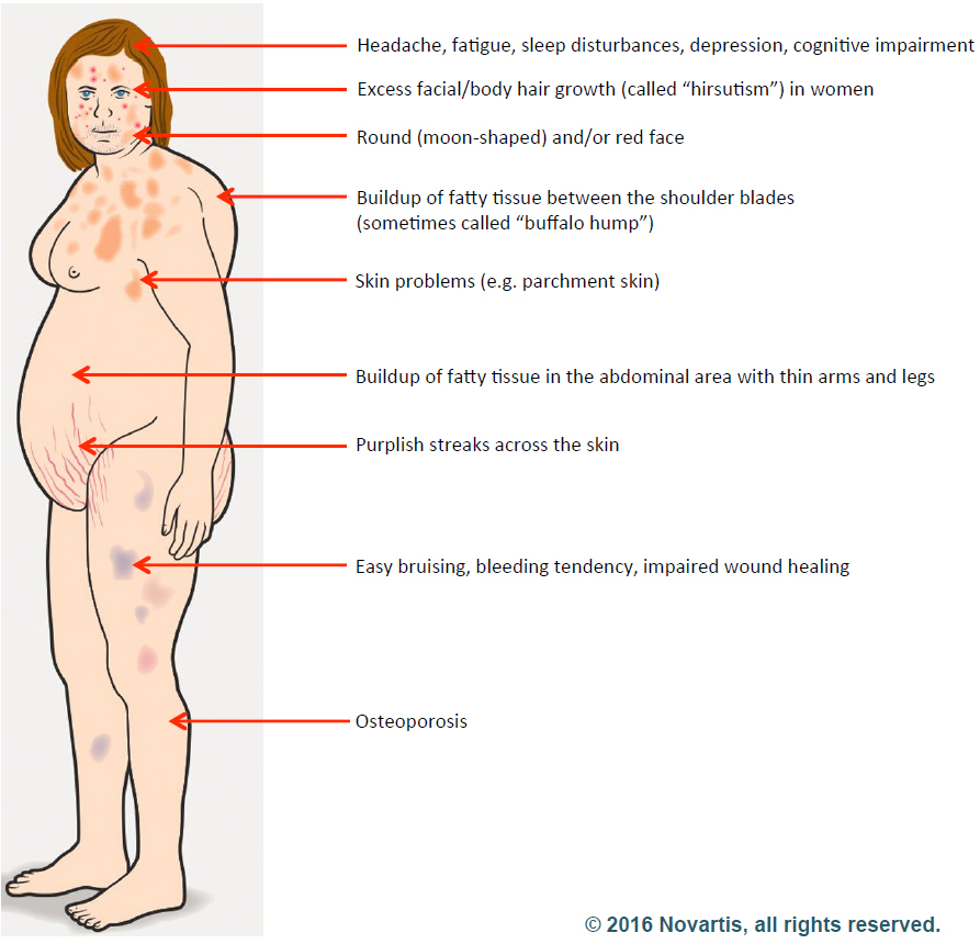
Elisabeth started researching Cushing’s disease right away and was relieved to learn that a cure was possible. Cushing’s is a hormonal disorder caused by high levels of the hormone cortisol. Symptoms include upper-body obesity, fragile skin that bruises easily, weakened bones, severe fatigue, weak muscles, high blood pressure, high blood glucose, increased thirst and urination, depression and a fatty hump between the shoulders. Women can also experience irregular menstrual periods and excess hair growth on their bodies. It can be caused by taking glucocorticoids such as prednisone or if there is a problem with a person’s pituitary gland or hypothalamus.

A Long and Uncertain Testing Journey

Elisabeth immediately set up a doctor’s appointment to get her cortisol and adrenocorticotropic hormone (ACTH – the pituitary hormone that stimulates the adrenal glands to make cortisol) levels tested. Over the next nine months Elisabeth went through several blood, urine, saliva and plasma tests for her cortisol and ACTH production and had an MRI. The tests showed elevated cortisol and ACTH levels but the initial impression was that her levels were not high enough to indicate Cushing’s disease and her pituitary MRI showed no apparent tumor. Elisabeth met with both a pituitary neurosurgeon and an endocrinologist, but both determined her condition not to be Cushing’s.

“My cortisol was not high enough; I wasn’t obese enough; I wasn’t disabled enough; I wasn’t depressed enough,” Elisabeth said.

Finding the Missing Clue

She felt frustrated by the diagnosis and continued to research possibilities online. It was during this research that she stumbled upon an article comparing MRI Tesla strengths. It recommended getting a Tesla 3.0 with contrast to pick up small abnormalities. Elisabeth scheduled a new MRI at the beginning of November. This time the scan detected a 6 mm tumor on the right side of her pituitary gland. Additional hormonal testing also confirmed that she did indeed have high ACTH and cortisol levels consistent with Cushing’s, “I was thrilled to finally have proof I had Cushing’s, but terrified because I knew I’d have to have brain surgery to remove it,” Elisabeth said. “I knew I wanted transsphenoidal surgery – the safest, most successful procedure with the least complications if done by an experienced surgeon.”

Understanding the Endonasal Transsphenoidal Approach

endonasal procedure illustration

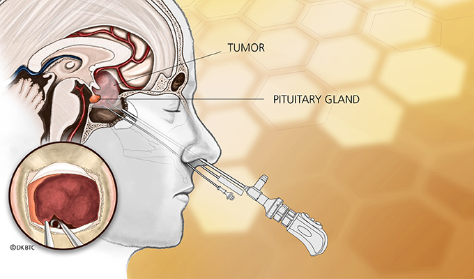
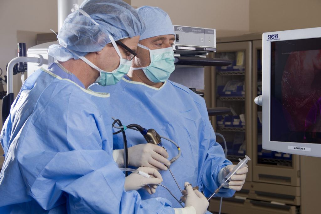
Endonasal transsphenoidal tumor removal, aka endoscopic endonasal approach, such as what Elisabeth needed, is a surgery that uses the nostril as the entry point with visualization from the operating microscope and endoscope. The approach passes through the back of the nasal cavity and into the sphenoid sinus to the skull base without facial incisions, brain retraction or post-operative nasal packing.

“This type of surgery is ideal for removing over 99% of pituitary adenomas, like what Elisabeth had and is considered first-line therapy for patients with Cushing’s disease,” Dr. Kelly explained. “Untreated or incompletely treated, Cushing’s disease is a very serious condition leading to uncontrolled hypertension, diabetes, weight gain and increased mortality.”

Choosing the Right Surgeon

With all the months she had to prepare for a diagnosis, Elisabeth knew exactly what needed to be done.

“I’d known I wanted Dr. Kelly to perform surgery but never imagined it could happen,” Elisabeth said. “I found him online. He’s ranked as one of the world’s top neurosurgeons specializing in this type of surgery. While watching his YouTube videos, I was awed by his kindness, patience, sense of humor, approachability, professionalism and complete lack of arrogance in spite of his fame. I’m still pinching myself that I had the fortune and honor to become one of his patients.”

A Life-Changing Call

Dr. Daniel Kelly and Dr. Chester Griffiths perform surgery

Figuring she didn’t have anything to lose, Elisabeth called Dr. Kelly’s office and asked if he was accepting new patients (he was) and how long was his waiting list (she could see him next week). Elisabeth was astounded. She immediately mailed Dr. Kelly all the test results, films and reports she could gather.

“Two days later, Dr. Kelly personally called and left a message, indicating it appeared there was indeed an ACTH secreting adenoma on my pituitary gland and to call him back,” Elisabeth said. “I was blown away. I’d have expected to win the lottery first.”

Successful Surgery and a New Beginning

Dr. Kelly arranged to perform Elisabeth’s surgery two weeks later on November 26 – the day before Thanksgiving. Her cortisol levels fell dramatically within 24 hours of surgery. She has remained in remission since then. Years after surgery, she continues to feel like a new person and regularly stays in contact with Dr. Kelly and his office staff.

About Dr. Daniel Kelly

Dr. Daniel Kelly, a board-certified neurosurgeon, is the director and one of the founders of the Pacific Neuroscience Institute, director of the Pacific Brain Tumor Center and Pacific Pituitary Disorders Center, and is Professor of Neurosurgery at Saint John’s Cancer Institute at Providence Saint John’s Health Center. Considered to be one of the top neurosurgeons in the US, he is a multiple recipient of the Patients’ Choice Award and Southern California Super Doctors distinction.

Cushing Patients Could Be Diagnosed, Subtyped Using Plasma Steroid Levels

Patients with different subtypes of Cushing’s syndrome (CS) have distinct plasma steroid profiles. This could be used as a test for diagnosis and classification, a German study says.

The study, “Plasma Steroid Metabolome for Diagnosis and Subtyping Patients with Cushing Syndrome,” appeared in the journal Clinical Chemistry.

A quick diagnosis of CS is crucial so that doctors can promptly give therapy. However, diagnosing CS is often complicated by the multiple tests necessary not just to diagnose the disease but also to determine its particular subtype.

Cortisol, which leads to CS when produced at high levels, is a steroid hormone. But while earlier studies were conducted to determine whether patients with different subtypes of CS had distinct steroid profiles, the methods researchers used were cumbersome and have been discontinued for routine use.

Recently, a technique called LC-MS/MS has emerged for multi-steroid profiling in patients with adrenocortical dysfunction such as congenital adrenal hyperplasia, adrenal insufficiency and primary aldosteronism.

Researchers at Germany’s Technische Universität in Dresden used that method to determine whether patients with the three main subtypes of CS (pituitary, ectopic and adrenal) showed differences in plasma steroid profiles. They measured levels of 15 steroids produced by the adrenal glands in single plasma samples collected from 84 patients with confirmed CS and 227 age-matched controls.

They found that CS patients saw huge increases in the plasma steroid levels of 11-deoxycortisol (289%), 21-deoxycortisol (150%), 11-deoxycorticosterone (133%), corticosterone (124%) and cortisol (122%), compared to patients without the disease.

Patients with the ectopic subtype had the biggest jumps in levels of these steroids. However, plasma 18-oxocortisol levels were particularly low in ectopic disease. Other steroids demonstrated considerable variation.

Patients with the adrenal subtype had the lowest concentration of dehydroepiandrosterone (DHEA) and DHEA-SO4, which are androgens. Patients with the ectopic and pituitary subtype had the lowest concentration of aldosterone.

Through the use of 10 selected steroids, patients with different subtypes of CS could be identified almost as closely as with other tests, including the salivary and urinary free cortisol test, the dexamethasone-suppressed cortisol test, and plasma adrenocorticotropin levels. The misclassification rate using steroid levels was 9.5 percent, compared to 5.8 percent in other tests.

“This study using simultaneous LC-MS/MS measurements of 15 adrenal steroids in plasma establishes distinct steroid metabolome profiles that might be useful as a test for CS,” the team concluded, adding that using LC-MS/MS is advantageous, as specimen preparation is simple and the entire panel takes 12 minutes to run. This means it could be offered as a single test for both identification and subtype classification.

From https://cushingsdiseasenews.com/2018/01/02/plasma-steroid-levels-used-screen-diagnosis-subtyping-patients-cushing-syndrome/

Metastatic Pituitary Carcinoma Successfully Treated with Radiation, Chemo.

A man with Cushing’s disease — caused by an adrenocorticotrophic hormone (ACTH)-secreting pituitary adenoma — who later developed metastases in the central nervous system without Cushing’s recurrence, was successfully treated over eight years with radiation and chemotherapy, according to a case report.

The report, “Long-term survival following transformation of an adrenocorticotropic hormone secreting pituitary macroadenoma to a silent corticotroph pituitary carcinoma: Case report,” was published in the journal World Neurosurgery.

Pituitary carcinomas make up only 0.1-0.2% of all pituitary tumors and are characterized by a primary pituitary tumor that metastasizes into cranial, spinal, or systemic locations. Fewer than 200 cases have been reported in the literature.

Most of these carcinomas secrete hormones, with ACTH being the most common. Though the majority of ACTH-secreting carcinomas present with Cushing’s disease, about one-third do not show symptoms of the condition and have normal serum cortisol and ACTH levels. These are called silent corticotroph adenomas and are considered more aggressive.

A research team at the University of Alabama at Birmingham presented the case of a 51-year-old Caucasian man with ACTH-dependent Cushing’s disease. He had undergone an incomplete transsphenoidal (through the nose) resection of an ACTH-secreting pituitary macroadenoma – larger than 10 mm in size – and radiation therapy the year before.

At referral in August 1997, the patient had persistent high cortisol levels and partial hypopituitarism, or pituitary insufficiency. He exhibited Cushing’s symptoms, including facial reddening, moon facies, weight gain above the collarbone, “buffalo hump,” and abdominal stretch marks.

About two years later, the man was weaned off ketoconazole — a medication used to lower cortisol levels — and his cortisol levels had been effectively reduced. He also had no physical manifestations of Cushing’s apart from facial reddening.

In May 2010, the patient reported two episodes of partial seizures, describing two spells of right arm tingling, followed by impaired peripheral vision. Imaging showed a 2.1-by-1-cm mass with an associated cyst within the brain’s right posterior temporal lobe, as well as a 1.8-by-1.2-cm mass at the cervicomedullary junction, which is the region where the brainstem continues as the spinal cord. His right temporal cystic mass was then removed by craniotomy.

A histopathologic analysis was consistent with pituitary carcinoma. Cell morphology was generally similar to the primary pituitary tumor, but cell proliferation was higher. Physical exams showed no recurrence of Cushing’s disease and 24-hour free urinary cortisol was within the normal range.

His cervicomedullary metastasis was treated with radiation therapy in July 2010. He took the oral chemotherapy temozolomide until August 2011, and Avastin (bevacizumab, by Genentech) was administered from September 2010 to November 2012.

At present, the patient continues to undergo annual imaging and laboratory draws. He receives treatment with hydrocortisone, levothyroxine — synthetic thyroid hormone — and testosterone replacement with androgel.

His most recent exam showed no progression over eight years of a small residual right temporal cyst, a residual mass along the pituitary stalk — the connection between the hypothalamus and the pituitary gland — and a small residual mass at the cervicomedullary junction. Lab results continue to show no Cushing’s recurrence.

“Our case is the first to document a patient who initially presented with an endocrinologically active ACTH secreting pituitary adenoma and Cushing’s disease who later developed cranial and spinal metastases without recurrence of Cushing’s disease and transformation to a silent corticotroph pituitary carcinoma,” the scientists wrote.

They added that the report is also the first documenting “8 years of progression-free survival in a patient with pituitary carcinoma treated with radiotherapy, [temozolomide] and bevacizumab.”

Adapted from https://cushingsdiseasenews.com/2019/01/03/successful-treatment-pituitary-carcinoma-radiation-chemo-case-report/

Long-Term Obesity Persists Despite Pituitary Adenoma Treatment In Childhood

Sethi A, et al. Clin Endocrinol. 2019;doi:10.1111/CEN.14146.

January 5, 2020

Obesity is common at diagnosis of pituitary adenoma in childhood and may persist despite successful treatment, according to findings published in Clinical Endocrinology.

“The importance of childhood and adolescent obesity on noncommunicable disease in adult life is well recognized, and in this new cohort of patients, we report that obesity is common at presentation of pituitary adenoma in childhood and that successful treatment is not necessarily associated with weight loss,” Aashish Sethi, MD, MBBS, a pediatric endocrinologist in the department of endocrinology at Alder Hey Children’s Hospital in Liverpool, United Kingdom, and colleagues wrote. “We have reported obesity, and obesity-related morbidity in a mixed cohort of children and young adults previously, but [to] our knowledge, this is the first time this observation has been reported in a purely pediatric cohort.”

In a retrospective study, Sethi and colleagues analyzed clinical and radiological data from 24 white children from Alder Hey Children’s Hospital followed for a median of 3.3 years between 2000 and 2019 (17 girls; mean age at diagnosis, 15 years). Researchers assessed treatment modality (medical, surgical or radiation therapy), pituitary hormone deficiencies and BMI, as well as results of any genetic testing.

Within the cohort, 13 girls had prolactinomas (mean age, 15 years), including 10 macroadenomas between 11 mm and 35 mm in size. Children presented with menstrual disorders (91%), headache (46%), galactorrhea (46%) and obesity (38%). Nine children were treated with cabergoline alone, three also required surgery, and two were treated with the dopamine agonist cabergoline, surgery and radiotherapy.

Five children had Cushing’s disease (mean age, 14 years; two girls), including one macroadenoma. Those with Cushing’s disease presented with obesity (100%), short stature (60%) and headache (40%). Transsphenoidal resection resulted in biochemical cure; however, two patients experienced relapse 3 and 6 years after surgery, respectively, requiring radiotherapy. One patient also required bilateral adrenalectomy.

Six children had a nonfunctioning pituitary adenoma (mean age, 16 years; two girls), including two macroadenomas. These children presented with obesity (67%), visual field defects (50%) and headache (50%). Four required surgical resections, with two experiencing disease recurrence after surgery and requiring radiotherapy.

During the most recent follow-up exam, 13 children (54.1%) had obesity, including 11 who had obesity at diagnosis.

“The persistence of obesity following successful treatment, in patients with normal pituitary function, suggests that mechanisms other than pituitary hormone excess or deficiency may be important,” the researchers wrote. “It further signifies that obesity should be a part of active management in cases of pituitary adenoma from diagnosis.” – by Regina Schaffer

Disclosures: The authors report no relevant financial disclosures.

From https://www.healio.com/endocrinology/adrenal/news/online/%7Bde3fd83b-e8e0-4bea-a6c2-99eb896356ab%7D/long-term-obesity-persists-despite-pituitary-adenoma-treatment-in-childhood

Paraneoplastic Cushing’s Syndrome Due to ACTH-Secreting Acinic Cell Carcinoma of the Parotid Gland: A Rare Case

​​​​​​​​​​​​​​Although acinic cell carcinoma — a cancer of the glands that produce saliva — is usually considered low risk, it can behave aggressively and cause Cushing’s syndrome, according to researchers who described such a case involving a 58-year-old man in Turkey.

They added that if not recognized and treated at an early stage, it can rapidly become life-threatening.

“The rarity of this entity poses a diagnostic challenge,” wrote the group of four researchers from a hospital in Turkey. “Awareness of this association is critical, as early identification and intervention may be lifesaving in selected patients.”

The case was described in a letter to the editor, titled “Paraneoplastic Cushing’s syndrome due to ACTH-secreting acinic cell carcinoma of the parotid gland: A rare case,” in the European Annals of Otorhinolaryngology, Head and Neck Diseases.