Cushing’s and Hairy Nipples

Hairy nipples are a common condition in women. The amount of hair on the nipples varies, but some women find that the hair becomes long, coarse, and dark, which can be distressing.

Hairy nipples are rarely a cause for concern and are usually not a sign of any underlying health issues. However, occasionally they can signify something more serious, in which case, it is essential to consult a doctor.

Almost every part of a person’s skin is covered in hair and hair follicles. On certain parts of the body, such as the top of the head, the hair usually grows longer and thicker, while on other parts, it is thin and transparent.

Fast facts on hairy nipples:

  • It is not known how common hairy nipples are or how many women have them.
  • Many women do not report the condition and instead manage it themselves.
  • It is possible for hair that used to be fine and light to turn coarse and dark with age.

Causes of hairy nipples in women

There are several underlying reasons that might cause nipple hairs to grow. These are:

Cushing’s syndrome

Cushing’s syndrome is another condition caused by hormonal imbalance. When it occurs, there is an excess of cortisol in the body. In this case, a person may experience several symptoms, such as:

  • increased hair growth
  • abnormal menstrual periods
  • high blood pressure
  • a buildup of fat on the chest and tummy, while arms and legs remain slim
  • a buildup of fat on the back of the neck and shoulders
  • a rounded and red, puffy face
  • bruising easily
  • big purple stretch marks
  • weakness in the upper arms and thighs
  • low libido
  • problems with fertility
  • mood swings
  • depression
  • high blood glucose level

Cushing’s syndrome is fairly rare, and the cause is usually associated with taking glucocorticosteroid medicine, rather than the body overproducing the hormone on its own.

It is possible, however, that a tumor in the lung, pituitary gland, or adrenal gland is the cause.

Also:

Hormonal changes and fluctuations

Hormonal changes in women can cause many different symptoms, one of which is changes in nipple hair growth and color.

Some common hormonal changes happen during pregnancy and menopause.

However, hormonal changes can also occur when a woman is in her 20s and 30s, which may cause nipple hair to change appearance or become noticeable for the first time.

Overproduction of male hormones

It is possible for hormonal imbalances to cause hairy nipples. Overproduction of male hormones, including testosterone, can cause hair growth, while other symptoms include:

  • oily skin that can lead to breakouts and acne
  • menstrual periods stopping
  • increase in skeletal muscle mass
  • male pattern baldness, leading to a woman losing hair on her head

If overproduction of male hormones is suspected, it is a good idea to make an appointment with a doctor who can confirm this with a simple test.

Polycystic ovary syndrome

Polycystic ovary syndrome (PCOS) occurs because of a hormonal imbalance. PCOS is a condition that affects the way the ovaries work.

Common symptoms of PCOS are:

  • infertility
  • irregular menstrual periods
  • ovarian cysts
  • excessive hair growth in unusual places, such as the nipples

PCOS is believed to affect around 1 in 5 women.

Medication

The side effects of particular medicines can cause unusual hair growth.

Medicines, such as testosterone, glucocorticosteroids, and certain other immunotherapy drugs may cause hairy nipples.

What are the treatment options?

Treatment for hairy nipples is not usually necessary for health reasons.

However, many women with the condition prefer to try and reduce or get rid of the appearance of hair on their nipples for cosmetic purposes.

There are several methods by which they can try and do this:

Trimming the nipple hair

Trimming the nipple hair may be enough to reduce its appearance. Small nail scissors are ideal, and hair can be cut close to the skin. It is essential to do this carefully and avoid catching the skin.

Trimming will need to be carried out regularly when the hair grows back.

Tweezing the nipple hair

Tweezing nipple hair is an effective way to get rid of unwanted nipple hair. However, this option can be painful as the skin around the nipple area is particularly soft and sensitive.

It is also important to bear in mind that the hair will return, and tweezing the hair increases the risk of infection and ingrown hairs.

Shaving the nipple hair

Shaving is another option to reduce the appearance of nipple hair. However, it is advisable to do so with caution to avoid nicking the sensitive skin.

This option also carries an increased risk of developing ingrown hairs and infection.

Waxing

Sugaring or waxing is a good hair removal option, though either one is likely to be painful. A salon is the best place to get this treatment type, as doing this at home may cause damage to the skin. Infection and ingrown hairs are again a risk.

Laser hair removal

These popular treatments can help to reduce the hair growth and slow or even prevent regrowth for a while. However, they can be painful, too.

Laser treatment is by far the most expensive option, as it will need to be performed by a plastic surgeon or cosmetic dermatologist.

Hormonal treatment

If a hormonal imbalance is the cause of hairy nipples, a doctor may prescribe or adjust a woman’s medication therapy to restore a healthy hormonal balance.

Other treatments and how to choose

The above treatments are all commonly used to remove and reduce nipple hair and usually have minimal side effects.

Bleaching or using hair removal cream to treat the condition, however, is not advised as these methods are usually too harsh for this sensitive area and may cause irritation and damage.

At what point should you see a doctor?

Hairy nipples in women are quite common, and there is usually no need to see a doctor. However, if they are accompanied by any other unusual symptoms, it is a good idea to make an appointment.

A doctor will be able to perform tests to determine whether an underlying cause, such as PCOS, is causing the growth of nipple hair. If so, they will give advice and medication therapy to help manage the condition.

A doctor will also be able to advise how to remove nipple hair safely.

Takeaway

For the majority of women, nipple hair may seem unsightly, but it is not a cause for any concerns about their health.

However, because some medical conditions can cause nipple hair to darken and grow, it is important to see a doctor if any other symptoms are experienced.

Nipple hair can usually be easily treated and managed, should a woman choose to try to remove the hair for cosmetic reasons.

Adapted from https://www.medicalnewstoday.com/articles/320835.php


Reasons You Have Flab Around Your Abdomen

Some diseases and conditions could be responsible for your abdominal fat.
Mita Majumdar | Updated: April 24, 2017 6:15 pm

Visceral fat or unhealthy belly fat that surrounds the liver and other organs in the abdomen puts you at risk for serious health problems, such as, metabolic syndrome, heart disease, and type 2 diabetes. But, what causes your pot belly or beer fat in the first place? The most obvious answers you will get is – ‘You are not exercising enough’, or, ‘you are eating too much of fatty foods or sugary foods’, or ‘you are not eating the right foods’, or ultimately, ‘It’s genetics! You got it from your parents’. All of these reasons are true, of course. However, some diseases/ disorders and conditions, too, could be responsible for your abdominal fat and these have nothing to do with not exercising or not eating right. Following are some of these disorders.

Cushing’s Syndrome

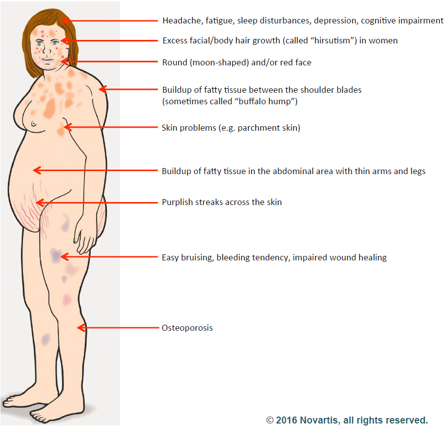
Cushing’s syndrome, also called hypercortisolism, is an endocrine disorder that occurs when your body is exposed to high cortisol levels over a long period of time. It is a treatable disorder, however, if it is chronic, the symptoms can last lifelong.

Symptoms: Symptoms vary according to the severity of the disorder. The characteristic symptoms include –

  • Fatty tissue deposits in the midsection
  • Fatty deposits in the upper back, especially between the shoulders, so that it resembles a hump
  • Puffy face
  • Violaceous stretch marks (pink or purple) on the arms, breast, stomach, and thighs that are more than 1 cm wide. [1]
  • Easy bruising
  • Fatigue
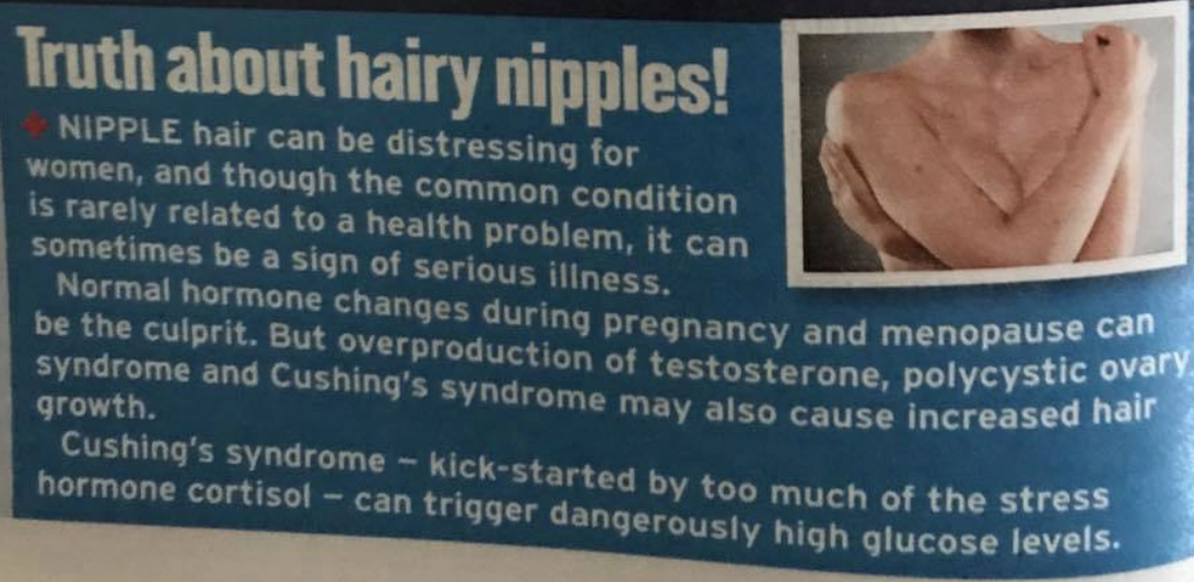
  • Hirsutism and irregularity in menstruation in women
  • Loss of libido and erectile dysfunction in men
  • Cognitive dysfunction, depression, unpredictable emotional outbursts, irritability is present in 70-85 percent of people with Cushing’s syndrome.[1]

Causes:

  • Overuse of corticosteroids
  • Overproduction of cortisol by the adrenal glands

Management:

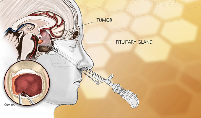
  • Surgery is the first line of treatment for Cushing’s syndrome.
  • Medication include: [2]

a.Pituitary gland directed therapy

b.Adrenal-blocking drugs

c.Glucocorticoid receptor-antagonizing drugs

  • Pituitary radiotherapy

Addison’s disease

Addison’s disease, also called adrenal insufficiency, is a disorder where your adrenal glands produce insufficient hormones, especially, glucocorticoids including cortisol and aldosterone. It is a life-threatening disease that can affect anyone irrespective of their gender or age.

How do glucocorticoids influence abdominal fats? Glucocorticoids including cortisol convert the fats into energy in the liver. They also help your body respond to stress. When sufficient amount of glucocorticoids are not produced by the adrenal glands, the fats accumulate in the abdominal area, and you see it as flab around your middle.

Symptoms:

  • Hyperpigmentation
  • Extreme fatigue
  • Low blood sugar and low blood pressure
  • Salt craving as one of the functions of adrenal glands is to maintain the sodium-potassium balance in the body
  • Nausea, vomiting, abdominal pain
  • Weight loss but gain in abdominal fat

Causes:

  • Insufficient production of adrenal cortex hormones
  • Stopping of prescribed corticosteroids
  • Tuberculosis and other infections of adrenal glands
  • Spread of cancer to the adrenal glands

Management:

  • Oral corticosteroids or corticosteroid injections
  • Intravenous injections of hydrocortisone, saline solution, and dextrose in case of Addisonian crisis

Stress

Chronic stress is a very big cause of belly fat. When you are exposed to stress, a chain reaction starts in the body because of the dysregulation of HPA axis of the neuroendocrine system. HPA axis is a complex interaction between the hypothalamus, pituitary gland, and adrenal glands. The hypothalamus produces a corticotropin releasing hormone (CRH) and vasopressin. These together stimulate the secretion of adrenocorticotropic hormone (ACTH). ACTH is transported by the blood to the adrenal glands, which then produces corticosteroids, mainly, cortisol from cholesterol. One of the functions of cortisol is to signal the body to store fat, and specifically, the fat storage occurs in the abdominal area, where the cortisol receptors are greater. Researchers have found that stress causes hyperactivation of HPA axis, leading to accumulation of fat tissue, especially in the abdomen region.

So, the more and longer you are stressed (or if you are chronically stressed), chances are that you will be carrying more belly fat!

Ascites

Ascites is the buildup of fluid in the abdominal space. Ascites usually occurs in people with cancer, and it is then called malignant ascites. Onset of ascites is generally the terminal phase in cancer. Ascites also occurs in patients with liver cirrhosis, kidney failure, or heart disease.

Symptoms:

The first sign of ascites is an increase in abdominal girth accompanied by weight gain. [4] Although it looks like it is belly fat, it is actually the fluid that causes the bulging.

Other symptoms include:

  • Shortness of breath
  • Nausea and vomiting
  • Swelling in the feet and ankle
  • Decreased appetite, sense of fullness, bloating
  • Fatigue
  • Haemorrhoids

Management:

If the ascites is not causing any discomfort, it may not require any treatment. Treatment of ascites can have many side effects. Talk to your doctor before you go in for management/ treatment.

Abdominal hernia

Abdominal hernia is a swelling or a bulge in the abdominal area where an organ or fatty tissue pushes through a weak spot in the abdominal wall. The abdominal wall is made up of tough connective tissue and tendons that stretch from the ribs to the groin. Depending on the position of the weakness in your abdominal wall, the hernia can be inguinal (groin), femoral (upper thigh), umbilical (belly button), hiatal (upper stomach), or even incisional. Incisional hernia can occur when the intestine pushes through a weak spot at the site of abdominal surgery.

Symptoms:

  • Visible bulge that may or may not cause discomfort
  • Feeling of heaviness in the abdomen
  • Sharp pain when you strain or lift objects

Causes:

  • Constipation and diarrhoea
  • Persistent coughing and sneezing
  • Straining or suddenly lifting a heavy object

Management:

  • Umbilical hernia, common in young children, mostly resolves by itself as the abdominal muscles get stronger.
  • Other abdominal hernia normally do not resolve by themselves. Doctors suggest waiting and watching.
  • If treatment is required, surgery is the only option. Surgery involves pushing the hernia back into the abdomen and repairing the abdominal wall.

Menopause

Menopause is certainly not a disease or a disorder. It is the time in a woman’s life when she stops menstruating and cannot become pregnant because her ovaries stop producing the required amounts of hormones oestrogen and progesterone. A woman reaches menopause when she has not had her periods for 12 months.

Symptoms:

  • Hot flashes and/ or night sweats
  • Vaginal dryness
  • Mood swings
  • Sleep disturbances

It is very common to gain belly fat during menopause. This is because of the low oestrogen levels. Oestrogen seems to influence the distribution of fat in the body, in a way that the fat is redistributed from the hips, buttocks, and thighs to the belly. However, a study published in the journal Metabolism reported that though women did significantly gain belly fat, especially deep inside the belly, relative fat distribution is not significantly different after menopause. [5] But the fact remains that women do gain flab in the abdomen after menopause.

Belly fat can be seriously harmful. If your belly fat is not because of the above-mentioned conditions, you can lose it by adopting a healthy lifestyle that includes sleeping enough, exercising regularly, eating right, and reducing stress.

Reference

  1. Sharma ST, Nieman LK, Feelders RA. Cushing’s syndrome: epidemiology and developments in disease management. Clinical Epidemiology. 2015;7:281-293. doi:10.2147/CLEP.S44336.
  1. Feelders RA, Hofland LJ. Medical treatment of Cushing’s disease. J Clin Endocrinol Metab. 2013;98:425–438.
  1. Kyrou I, Chrousos GP, Tsigos C. Stress, visceral obesity, and metabolic complications. Ann N Y Acad Sci. 2006 Nov;1083:77-110.
  1. Sinicrope FA. Ascites. In: Kufe DW, Pollock RE, Weichselbaum RR, et al., editors. Holland-Frei Cancer Medicine. 6th edition. Hamilton (ON): BC Decker; 2003.
  2. Franklin RM, Ploutz-Snyder L, Kanaley JA. Longitudinal changes in abdominal fat distribution with menopause. Metabolism. 2009 Mar;58(3):311-5. doi: 10.1016/j.metabol.2008.09.030.

Adapted from http://www.thehealthsite.com/diseases-conditions/reasons-you-have-flab-around-your-abdomen-f0417/


Day 19, Cushing’s Awareness Challenge 2016

In case you haven’t guessed it, my cause seems to be Cushing’s Awareness.  I never really decided to devote a good portion of my life to Cushing’s, it just fell into my lap, so to speak – or my laptop.

I had been going along, raising my son, keeping the home-fires burning,  trying to forget all about Cushing’s.  My surgery had been a success, I was in remission, some of the symptoms were still with me but they were more of an annoyance than anything.

I started being a little active online, especially on AOL.  At this time, I started going through real-menopause, not the fake one I had gone through with Cushing’s.  Surprisingly, AOL had a group for Cushing’s people but it wasn’t very active.

What was active, though, was a group called Power Surge (as in I’m not having a hot flash, I’m having a Power Surge).  I became more and more active in that group, helping out where I could, posting a few links here and there.

Around this time I decided to go back to college to get a degree in computer programming but I also wanted a basic website for my piano studio.  I filled out a form on Power Surge to request a quote for building one.  I was very surprised when Power Surge founder/webmaster Alice (AKA Dearest) called me.  I was so nervous.  I’m not a good phone person under the best of circumstances and here she was, calling me!

I had to go to my computer class but I said I’d call when I got back.  Alice showed me how to do some basic web stuff and I was off.  As these things go, the O’Connor Music Studio page grew and grew…  And so did the friendship between Alice and me.  Alice turned out to be the sister I never had, most likely better than any sister I could have had.

In July of 2000, Alice and I were wondering why there weren’t many support groups online (OR off!) for Cushing’s. This thought percolated through my mind for a few hours and I realized that maybe this was my calling. Maybe I should be the one to start a network of support for other “Cushies” to help them empower themselves.

I wanted to educate others about the awful disease that took doctors years of my life to diagnose and treat – even after I gave them the information to diagnose me. I didn’t want anyone else to suffer for years like I did. I wanted doctors to pay more attention to Cushing’s disease.

The first website (http://www.cushings-help.com) went “live” July 21, 2000. It was just a single page of information. The message boards began September 30, 2000 with a simple message board which then led to a larger one, and a larger. Today, in 2012, we have over 8 thousand members. Some “rare disease”!

This was on the intro page of Cushing’s Help until 2013…

I would like to give abundant thanks Alice Lotto Stamm, founder of Power Surge, premier site for midlife women, for giving me the idea to start this site, encouraging me to learn HTML and web design, giving us the use of our first spiffy chatroom, as well as giving me the confidence that I could do this. Alice has helped so many women with Power Surge. I hope that I can emulate her to a smaller degree with this site.

Thanks so much for all your help and support, Alice!

In August 2013 my friend died.  In typical fashion, I started another website

I look around the house and see things that remind me of Alice.  Gifts, print outs, silly stuff, memories, the entire AOL message boards on floppy disks…

Alice, I love you and will miss you always…

MaryOOneRose

Recurrent sellar mass after resection of pituitary macroadenoma

A Puerto Rican woman aged 50 years presented to an ophthalmologist with complaints of vision changes, including difficulty seeing images in her peripheral vision in both eyes and difficulty in color perception. Her medical history was significant for menopause at age 43 years, type 2 diabetes and hypertension. She had no prior history of thyroid disease, changes in her weight, dizziness or lightheadedness, headaches, galactorrhea or growth of her hands or feet.

Formal visual fields showed bitemporal superior quadrantopsia, and she was sent to the ED for further evaluation.

Imaging and laboratory tests

A pituitary protocol MRI was performed that showed a large 3 cm x 2 cm x 2.2 cm mass in the pituitary with mild osseous remodeling of the sella turcica and mass effect on the optic chiasm (Figure 1). The mass was isointense with the brain parenchyma on T1-weighted and T2-weighted images and homogeneously enhanced after IV gadolinium contrast administration.

Baseline laboratory samples drawn at 11 p.m. in the ED showed a cortisol of 16.9 µg/dL (nighttime reference range: 3-16 µg/dL), adrenocorticotropic hormone 65 pg/mL (reference range: 6-50 pg/mL), prolactin 19.4 ng/mL (reference range: 5.2-26.5 ng/mL), thyroid-stimulating hormone 1.36 µIU/mL (reference range: 0.35-4.9 µIU/mL), free thyroxine 0.9 ng/dL (reference range: 0.6-1.8 ng/dL), triiodothyronine 85 ng/dL (reference range: 83-160 ng/dL), follicle-stimulating hormone (FSH) 11.1 mIU/mL (postmenopausal reference range: 26.7- 133.4 mIU/mL) and luteinizing hormone (LH) 1.2 mIU/mL (postmenopausal reference range: 5.2-62 mIU/mL).


Figure 1. T1-weighted MRI images with and without contrast of the pituitary. Coronal (A) and sagittal (C) images showed a large isodense (with brain parenchyma) 3 cm x 2 cm x 2.2 cm mass (red arrow) in the sella with superior extension to the optic chiasm. After gadolinium contrast, coronal (B) and sagittal (D) images show the mass homogenously enhances consistent with a pituitary adenoma.

Images courtesy of Pavani Srimatkandada, MD.

Given the patient’s high nighttime cortisol and adrenocorticotropic hormone (ACTH) levels, she underwent an overnight dexamethasone suppression test with 1 mg dexamethasone. Her morning cortisol was appropriately suppressed to less than 1 µg/dL, excluding Cushing’s disease.

Pituitary adenoma resection

The patient was diagnosed with a nonsecreting pituitary adenoma with suprasellar extension and optic chiasm compression with visual field deficits. The macroadenoma caused an inappropriately normal LH and FSH in a postmenopausal woman consistent with hypogonadotrophic hypogonadism.

She underwent transnasal transsphenoidal resection of the nonsecreting pituitary adenoma. The dural defect caused by the surgery was patched with an abdominal fat graft with a DuraSeal dura patch. A postoperative MRI showed complete resection of the adenoma with no evaluable tumor in the sella (Figure 2). Her postoperative course was complicated by transient diabetes insipidus requiring intermittent desmopressin; however, this resolved before her discharge from the hospital.

Figure 2. T1-weighted MRI images with contrast. Coronal views before (A) and after (B) transphenoidal tumor resection show complete resolution of the enhancing pituitary mass (A; red arrow) that is replaced with a new hypodense mass in the sella (B; yellow arrow). This mass is filled with cerebrospinal fluid with a residual rim of enhancing tissue. This is consistent with the development of a pseudomeningocele in the sella.


Postoperative testing confirmed secondary deficiency of the adrenal, thyroid and ovarian axes requiring hormone therapy. The patient had stable temporal hemianopia in the left eye with improved vision in the right eye.

Recurrent mass detected

One year after surgery, during a routine follow-up appointment, the patient reported no dizziness, lightheadedness, worsening vision changes, rhinorrhea or headache. She had a follow-up MRI of the brain with and without contrast, which showed the interval appearance of a mass in the sella that extended from the sphenoid sinus into the sella and came in contact with the optic nerve (Figure 3).

Figure 3. Axial MRI images of the sella after resection of pituitary adenoma. On T1-weighted images the mass (red arrow) in the sella is hypodense (black) compared with the brain parenchyma. On T2-weighted images, the mass (red arrow) is hyperdense (bright) compared with the brain, consistent with fluid. Cerebrospinal fluid in the sulci on the brain surface and the vitreous fluid within the eye are also hyperintense on T2-weighted images (yellow arrows).


On MRI, the mass was isodense with the cerebral spinal fluid (CSF) with a residual rim of enhancing normal pituitary tissue. This appearance is consistent with the postoperative development of a pseudomeningocele and not a solid mass in the sella (Table).

Pseudomeningoceles are abnormal collections of CSF that communicate with the CSF space around the brain; these occur after brain surgery involving duraplasty (incision and repair of the dura). Unlike meningoceles, pseudomeningoceles are not completely encased by a surrounding membrane, and they communicate with the circulating CSF. Similar to CSF, a pseudomeningocele is hypodense (dark) compared with brain on T1-weighted MRI images and hyperdense (bright) on T2-weight images.


Pseudomeningocele treatment

Treatment may be conservative or may involve neurosurgical repair if symptomatic. Little published data addresses the development of pseudomeningoceles after transsphenoidal pituitary surgery, but this complication occasionally occurs, especially if the dural incision is large. One study noted that pseudomeningoceles are one of the most common complications after suboccipital decompression for Chiari’s malformation, but the effect of this complication is unclear.

Endocrinologists must recognize that recurrent development of pituitary masses after transsphenoidal pituitary adenoma surgery may not represent regrowth of pituitary tissue, but instead development of a meningocele/pseudomeningocele. Pseudomeningocele can be easily confirmed because this fluid collection has very different MRI characteristics than pituitary adenoma (Table). Given that patients may remain asymptomatic after the development of a pseudomeningocele, periodic MRI imaging, hormonal evaluation and ophthalmologic monitoring of visual fields are required after transsphenoidal pituitary surgery.

References:
  • Hernandez Guilabert PM. Poster No C-1330. Presented at: European Society of Radiology; March 7-11, 2013; Vienna.
  • Parker SL, et al. J Neurosurg. 2013;doi:10.3171/2013.8.JNS122106.
For more information:
  • Stephanie L. Lee, MD, PhD, ECNU, is an associate professor of medicine and associate chief, in the Section of Endocrinology, Diabetes and Nutrition at Boston Medical Center. Lee can be reached at Boston Medical Center, 88 E. Newton St., Endocrinology Evans 201, Boston, MA 02118; email: stephanie.lee@bmc.org. Lee reports no relevant financial disclosures.
  • Pavani Srimatkandada, MD, is an endocrinology fellow in the Section of Endocrinology, Diabetes and Nutrition at Boston Medical Center. Srimatkandada can be reached at Boston Medical Center, 88 E. Newton St., Endocrinology Evans 201, Boston, MA 02118. She reports no relevant financial disclosures.

From http://www.healio.com/endocrinology/thyroid/news/print/endocrine-today/%7B82430fb6-bbe4-4908-a389-447eee8cd005%7D/recurrent-sellar-mass-after-resection-of-pituitary-macroadenoma

Day Twenty-nine, Cushing’s Awareness Challenge 2015


In case you haven’t guessed it, my cause seems to be Cushing’s Awareness.  I never really decided to devote a good portion of my life to Cushing’s, it just fell into my lap, so to speak – or my laptop.

I had been going along, raising my son, keeping the home-fires burning,  trying to forget all about Cushing’s.  My surgery had been a success, I was in remission, some of the symptoms were still with me but they were more of an annoyance than anything.

I started being a little active online, especially on AOL.  At this time, I started going through real-menopause, not the fake one I had gone through with Cushing’s.  Surprisingly, AOL had a group for Cushing’s people but it wasn’t very active.

What was active, though, was a group called Power Surge (as in I’m not having a hot flash, I’m having a Power Surge).  I became more and more active in that group, helping out where I could, posting a few links here and there.

Around this time I decided to go back to college to get a degree in computer programming but I also wanted a basic website for my piano studio.  I filled out a form on Power Surge to request a quote for building one.  I was very surprised when Power Surge founder/webmaster Alice (AKA Dearest) called me.  I was so nervous.  I’m not a good phone person under the best of circumstances and here she was, calling me!

I had to go to my computer class but I said I’d call when I got back.  Alice showed me how to do some basic web stuff and I was off.  As these things go, the O’Connor Music Studio page grew and grew…  And so did the friendship between Alice and me.  Alice turned out to be the sister I never had, most likely better than any sister I could have had.

In July of 2000, Alice and I were wondering why there weren’t many support groups online (OR off!) for Cushing’s. This thought percolated through my mind for a few hours and I realized that maybe this was my calling. Maybe I should be the one to start a network of support for other “Cushies” to help them empower themselves.

I wanted to educate others about the awful disease that took doctors years of my life to diagnose and treat – even after I gave them the information to diagnose me. I didn’t want anyone else to suffer for years like I did. I wanted doctors to pay more attention to Cushing’s disease.

The first website (http://www.cushings-help.com) went “live” July 21, 2000. It was just a single page of information. The message boards began September 30, 2000 with a simple message board which then led to a larger one, and a larger. Today, in 2012, we have over 8 thousand members. Some “rare disease”!

This was on the intro page of Cushing’s Help until 2013…

I would like to give abundant thanks Alice Lotto Stamm, founder of Power Surge, premier site for midlife women, for giving me the idea to start this site, encouraging me to learn HTML and web design, giving us the use of our first spiffy chatroom, as well as giving me the confidence that I could do this. Alice has helped so many women with Power Surge. I hope that I can emulate her to a smaller degree with this site.

Thanks so much for all your help and support, Alice!


In August 2013 my friend died.  In typical fashion, I started another website

I look around the house and see things that remind me of Alice.  Gifts, print outs, silly stuff, memories, the entire AOL message boards on floppy disks…

Alice, I love you and will miss you always…

MaryOOneRose

%d bloggers like this: