Day 3: Cushing’s Awareness Challenge

me-tired

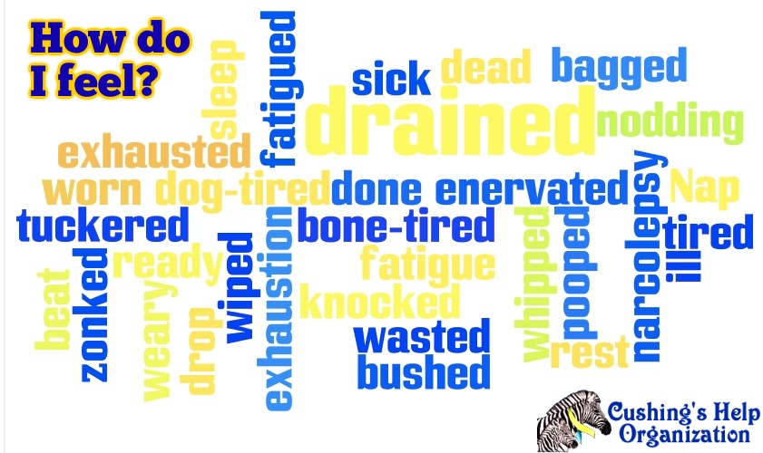
Sleep.  Naps.  Fatigue, Exhaustion.  I still have them all.  I wrote on my bio in 1987 after my pituitary surgery “I am still and always tired and need a nap most days. I do not, however, still need to take whole days off just to sleep.

That seems to be changing back, at least on the weekends.  A recent weekend, both days, I took 7-hour naps each day and I still woke up tired. That’s awfully close to taking a whole day off to sleep again.

In 2006, I flew to Chicago, IL for a Cushing’s weekend in Rockford.  Someone else drove us to Lake Geneva, Wisconsin for the day.  Too much travel, too Cushie, whatever, I was too tired to stay awake.  I actually had put my head down on the dining room table and fallen asleep but our hostess suggested the sofa instead.  Amazing that I traveled that whole distance – and missed the main event 😦

This sleeping thing really impacts my life.  Between piano lessons, I take a nap.  I sleep as late as possible in the mornings and afternoons are pretty much taken up by naps.  I nod off at night during TV. One time I came home between church services and missed the third service because I fell asleep.

I only TiVo old tv shows that I can watch and fall asleep to since I already know the ending.

Since  mid-February, I have been doing physical therapy twice a week for 2 hours at a time for a knee injury (read more about that in Bees Knees).  I come home from that exhausted – and in more pain than I went.  I know it’s working and my knee is getting better, but it’s such a time and energy sapper.  Neither of which I can really spare.

Maybe now that I’m nearly 10  years out from my kidney cancer (May 9, 2006) I could theoretically go back on Growth Hormone again.  My surgeon says he “thinks” it’s ok.  I’m sort of afraid to ask my endo about it, though.  I want to feel better and get the benefits of the GH again but I don’t want any type of cancer again and I certainly can’t afford to lose another kidney.

I’ll probably just muddle through without it.  I always laugh when I see that commercial online for something called Serovital.  I saw it in Costco the other day and it mentions pituitary right on the package.  I wish I could take the people buying this, sit them down and tell them not to mess with their pituitary glands.  But I won’t.  I’ll take a nap instead because I’m feeling so old and weary today, and yesterday.

And tomorrow…

Day 2, Cushing’s Awareness Challenge

The Seven Dwarves of Cushing's

So, these are only seven of the many, many symptoms of Cushing’s.  I had those above – and I often felt like I looked like one of those little bearded dwarves.

Cushing’s affects every part of the body.  It’s not like when I had kidney cancer and only the kidney was affected.

Here are some of the many areas affected.

  • Progressive obesity and skin changes
  • Weight gain and fatty tissue deposits, particularly around the midsection and upper back, in the face (moon face) and between the shoulders (buffalo hump). Some symptoms such as sudden weight gain, are caused by excess cortisol. The excess cortisol in the body does not increase protein and carbohydrate metabolism. It slows or nearly disables metabolism function, which can cause weight gain (fat accumulation) in the buttocks, abdomen, cheeks, neck, or upper back.
  • Loss of muscle mass. Some areas of the body, such as the arms and legs, will remain thin.
  • Pink or purple stretch marks (striae) on the skin of the abdomen, thighs, breasts and arms
  • Thinning, fragile skin that bruises easily
  • Slow healing of cuts, insect bites and infections
  • Acne

Women with Cushing’s syndrome may experience:

  • Thicker or more visible body and facial hair (hirsutism)
  • Irregular or absent menstrual periods

Men with Cushing’s syndrome may experience:

  • Decreased libido
  • Decreased fertility
  • Erectile dysfunction

Other signs and symptoms include:

  • Fatigue
  • Muscle weakness
  • Depression, anxiety and irritability
  • Loss of emotional control
  • Cognitive difficulties
  • New or worsened high blood pressure
  • Glucose intolerance that may lead to diabetes
  • Headache
  • Bone loss, leading to fractures over time
  • Hyperlipidemia (elevated lipids – cholesterol – in the blood stream)
  • Recurrent opportunistic or bacterial infections
Think you have Cushing’s?  Get to a doctor and don’t give up!

MaryO
         MaryO

Day 1: Cushing’s Awareness Challenge

April is always Cushing’s Awareness Challenge month because Dr. Harvey Cushing was born on April 8th, 1869.

30-posts

Thanks to Robin for this wonderful past logo!  I’ve participated in these 30 days for Cushing’s Awareness several times so I’m not quite sure what is left to say this year but I always want to get the word out when I can.

As I see it, there have been some strides the diagnosis or treatment of Cushing’s since last year.  More drug companies are getting involved, more doctors seem to be willing to test, a bit more awareness, maybe.


April Fool's Day

How fitting that this challenge should begin on April Fool’s Day.  So much of Cushing’s  Syndrome/Disease makes us Cushies seem like we’re the April Fool.  Maybe, just maybe, it’s the doctors who are the April Fools…

Doctors tell us Cushing’s is too rare – you couldn’t possibly have it.  April Fools!

All you have to do is exercise and diet.  You’ll feel better.  April Fools!

Those bruises on your legs?  You’re just clumsy. April Fools!

Sorry you’re growing all that hair on your chin.  That happens as you age, you know.  April Fools!

Did you say you sleep all day?  You’re just lazy.  If you exercised more, you’d have more energy. April Fools!

You don’t have stretch marks.  April Fools!

You have stretch marks but they are the wrong [color/length/direction] April Fools!

The hump on the back of your neck is from your poor posture. April Fools!

Your MRI didn’t show a tumor.  You couldn’t have Cushing’s. April Fools!

This is all in your mind.  Take this prescription for antidepressants and go home.  April Fools!

If you have this one surgery, your life will get back to normal within a few months. April Fools!

What?  You had transsphenoidal surgery for Cushing’s?  You wasted your time and money. April Fools!

I am the doctor.  I know everything.  Do not try to find out any information online. You could not have Cushing’s.  It’s too rare…  April FOOL!

All this reminds me of a wonderful video a message board member posted a while ago:

So now – who is the April Fool?  It wasn’t me.  Don’t let it be you, either!

Olfactory Neuroblastoma Causing Cushing’s Syndrome Due to the Ectopic Adrenocorticotropic Hormone (ACTH) Secretion

Abstract

Cushing’s syndrome is a constellation of features occurring due to high blood cortisol levels. We report a case of a 47-year-old male with a history of recurrent olfactory neuroblastoma (ONB). He presented with bilateral lower limb weakness and anosmia and was found to have Cushing’s syndrome due to high adrenocorticotropic hormone (ACTH) levels from an ectopic source, ONB in this case. Serum cortisol and ACTH levels declined after tumor removal.

Introduction

Olfactory neuroblastoma (ONB), or esthesioneuroblastoma, is a rare malignancy arising from neuroepithelium in the upper nasal cavity. It represents approximately 2% of all nasal passage tumors, with an incidence of approximately 0.4 per 2.5 million individuals [1]. ONB shares similar histological features with small round blue cell neoplasms of the nose. Ectopic hormone secretion is a very rare feature associated with these tumors. Five-year overall survival is reported to be between 60% and 80% [2,3]. The age distribution is either in the fifth to sixth decade of life [4,5], or in the second and sixth decades [6].

Features of Cushing’s syndrome (moon face, buffalo hump, central obesity hypertension, fragile skin, easy bruising, fatigue, muscle weakness) are due to high blood cortisol levels [7]. It can be either primary (cortisol-secreting adrenal tumor), secondary (adrenocorticotropic hormone (ACTH)-secreting pituitary tumor, also called Cushing disease), or ectopic ACTH secretion (from a non-pituitary source). All three types share similar features [8].

Ectopic ACTH syndrome (EAS) is due to an extra pituitary tumor, producing ACTH. It accounts for 12-17% of Cushing’s syndrome cases [9]. Most cases of EAS-producing tumors are in the lungs, mediastinum, neuroendocrine tumors of the gastrointestinal tract, and pheochromocytomas [9]. Ectopic ACTH secretion from an ONB is very rare. As of 2015, only 18 cases were reported in the literature [10]. Here, we report such a case.

Case Presentation

Our patient is a 47-year-old Bangladeshi male, with a history of recurrent ONB that was resected twice in the past (transsphenoidal resection in 2016 and 2019) with adjuvant radiotherapy, no chemotherapy was given. He also had diabetes mellitus type 1 (poorly controlled) and hypertension. He presented with bilateral lower limb weakness, anosmia, decreased oral intake, loss of taste for one week, and bilateral submandibular swelling that increased in size gradually over the past two years. There was no history of fever, cough, abdominal pain, or exposure to sick contacts. The patient reported past episodes of similar symptoms, but details are unclear. The patient’s family history is positive for diabetes mellitus type 1 in both parents. Lab tests in the emergency department showed hypokalemia and hyperglycemia as detailed in Table 1. He was admitted for further workup of the above complaints.

Test Patient Results Reference Range Unit Status
Hemoglobin 14.7 13-17 g/dL Normal
White blood cell (WBC) 17.9 4-10 10*9/L High
Neutrophils 15.89 2-7 10*9/L High
Lymphocytes 1.07 1-3 10*9/L Normal
Sodium 141 136-145 mmol/L Normal
Potassium 2.49 3.5-5.1 mmol/L Low (Panic)
Chloride 95 98-107 mmol/L Low
Glucose 6.52 4.11-5.89 mmol/L Elevated
C-reactive protein (CRP) 0.64 Less than 5 mg/L Normal
Erythrocyte sedimentation rate (ESR) 2 0-30 mm/h Normal
Creatinine 73 62-106 µmol/L Normal
Uric acid 197 202.3-416.5 µmol/L Normal
Alanine aminotransferase (ALT) 33.2 0-41 U/L Normal
Aspartate aminotransferase (AST) 18.6 0-40 U/L Normal
International Normalised Ratio (INR) 1.21 0.8-1.2 sec High
Prothrombin time (PT) 15.7 12.3-14.7 sec High
Lactate dehydrogenase (LDH) 491 135-225 U/L High
Thyroid-stimulating hormone (TSH) 0.222 0.27-4.20 mIU/L Low
Adrenocorticotropic hormone (ACTH) 106 ≤50 ng/L Elevated
Cortisol (after dexamethasone suppression) 1750 Morning hours (6-10 am): 172-497 nmol, Afternoon hours (4-8 pm): 74.1-286 nmol nmol/L Elevated (failure of suppression)
24-hour urine cortisol (after dexamethasone suppression) 5959.1 <120 nmol/24 hrs nmol/24hr Elevated (failure of suppression)
Table 1: Results of blood test at the time of hospitalization. Hypokalemia and high values of adrenocorticotropic hormone and cortisol were confirmed.

On examination, the patient’s vital signs were as follows: blood pressure was 154/77 mmHg, heart rate of 60 beats per minute, respiratory rate was 18 breaths per minute, oxygen saturation of 98% on room air, and a temperature of 36.7°C. The patient had a typical Cushingoid appearance with a moon face, buffalo hump, purple striae on the abdomen, central obesity, and hyperpigmentation of the skin. Submandibular lymph nodes were enlarged bilaterally. The examination of the submandibular lymph nodes showed a firm, fixed mass extending from the angle of the mandible to the submental space on the left side. Neurological examination showed weakness in both legs bilaterally (strength 3/5) and anosmia (checked by orthonasal smell test). The rest of the neurological exam was normal.

Laboratory findings revealed (in Table 1) a marked hypokalemia of 2.49 mmol/L and hyperglycemia of 6.52 mmol/L. The serum cortisol level was elevated at 1587 nmol/L. Serum ACTH levels were raised at 106 ng/L (normal value ≤50 ng/L). Moreover, the high-dose dexamethasone suppression test failed to lower the serum ACTH levels and serum and urine cortisol. Serum cortisol level after the suppression test was 1750 nmol/L, while 24-hour urine cortisol after the test was 5959.1 nmol/24hr. Serum ACTH levels after the test also remained high at 100mg/L. This indicated failure of ACTH suppression by high-dose dexamethasone, which points towards ectopic ACTH production. Other blood tests (complete blood count, liver function tests) were insignificant.

A computed tomography scan with contrast (CT scan) of the chest, abdomen, and pelvis, with a special focus on the adrenals, was negative for any malignancy or masses. CT scan of the neck showed bilaterally enlarged submandibular lymph nodes and an enlarged right lobe of the thyroid with nodules. Fine needle aspiration (FNA) of the thyroid nodules revealed a benign nature. Magnetic resonance imaging (MRI) of the brain showed a contrast-enhancing soft tissue lesion (18x18x10mm) in the midline olfactory groove area with extension into the frontal dura and superior sagittal sinus, suggesting recurrence of the previous ONB. There was evidence of previous surgery also. The pituitary gland was normal (Figures 12).

A-brain-MRI-(T1-weighted;-without-contrast;-sagittal-plane)-shows-a-soft-tissue-lesion-located-in-the-midline-olfactory-groove-area.-Dural-surface-with-extension-into-anterior-frontal-dura.
Figure 1: A brain MRI (T1-weighted; without contrast; sagittal plane) shows a soft tissue lesion located in the midline olfactory groove area. Dural surface with extension into anterior frontal dura.

MRI: Magnetic resonance imaging

A-brain-MRI-(T2-weighted;-without-contrast;-axial-plane)-shows-a-soft-tissue-lesion-located-in-the-midline-olfactory-groove-area.
Figure 2: A brain MRI (T2-weighted; without contrast; axial plane) shows a soft tissue lesion located in the midline olfactory groove area.

MRI: Magnetic resonance imaging

Octreotide scintigraphy showed three focal abnormal uptakes in the submandibular cervical nodes. Additionally, there was a moderate abnormal uptake at the midline olfactory groove with bilateral extension (Figure 3).

Whole-body-octreotide-scan-(15-mCi-99mTc-Octreotide-IV)-demonstrates-three-focal-abnormal-uptakes:-the-largest-(5.2-x-2.4-cm)-in-the-left-submandibular-region,-and-two-smaller-ones-on-the-right,-suggestive-of-lymph-node-uptake.-Additional-abnormal-uptake-was-seen-along-the-midline-of-the-olfactory-groove-region-with-bilateral-extension.-No-other-significant-abnormal-uptake-was-identified.
Figure 3: Whole-body octreotide scan (15 mCi 99mTc-Octreotide IV) demonstrates three focal abnormal uptakes: the largest (5.2 x 2.4 cm) in the left submandibular region, and two smaller ones on the right, suggestive of lymph node uptake. Additional abnormal uptake was seen along the midline of the olfactory groove region with bilateral extension. No other significant abnormal uptake was identified.

On microscopic examination, an excisional biopsy after the transcranial resection surgery of the frontal skull base tumor showed nests and lobules of round to oval cells with clear cytoplasm, separated by vascular and hyalinized fibrous stroma (Figures 4A4B). Tumor cells show mild to moderate nuclear pleomorphism, and fine chromatin (Figure 4C). A fibrillary neural matrix is also present. Some mitotic figures can be seen. Immunohistochemical stains revealed positive staining for synaptophysin (Figure 4D) and chromogranin (Figure 4E). Stains for CK (AE1/AE3), CD45, Desmin, and Myogenin are negative. Immunostaining for ACTH was focally positive (Figure 4F), while the specimen of the cervical lymph nodes showed the same staining, indicating metastases. The cytomorphologic and immunophenotypic features observed are consistent with a Hyams grade II ONB, with ectopic ACTH production.

Histopathological-and-immunohistochemical-findings-of-olfactory-neuroblastoma.
Figure 4: Histopathological and immunohistochemical findings of olfactory neuroblastoma.

A (100x magnification) and B (200x magnification) – hematoxylin and eosin (H-E) staining shows cellular nests of round blue cells separated by hyalinized stroma. C (400x magnification) – nuclei show mild to moderate pleomorphism with fine chromatin. D (100x magnification) – an immunohistochemical stain for synaptophysin shows diffuse, strong cytoplasmic positivity within tumor cells. E (200x magnification) – tumor cells are positive for chromogranin. F (400x magnification) – ACTH cytoplasmic expression in tumor cells.

ACTH: adrenocorticotropic hormone

For his resistant hypokalemia, he had to be given intravenous (IV) and oral potassium chloride (KCL) repeatedly. The patient underwent transcranial resection of the frontal skull base tumor. The patient received cefazolin for seven days, and hydrocortisone for four days. After transcranial resection, his cortisol level decreased to 700 nmol/L. Furthermore, ACTH dropped, and serum potassium also normalized. Subsequently, the patient was transferred to the intensive care unit (ICU) for meticulous monitoring and continued care. In the ICU, the patient developed one episode of a generalized tonic-clonic seizure, which aborted spontaneously, and the patient received phenytoin and levetiracetam to prevent other episodes. A right-sided internal jugular vein and left transverse sinus thrombosis were also developed and treated with enoxaparin sodium. Following surgery, his low potassium levels improved, resulting in an improvement in his limb weakness. His other symptoms also gradually improved after surgery. Three weeks following the primary tumor resection, he underwent bilateral neck dissection with right hemithyroidectomy, for removal of the metastases. The patient opted out of chemotherapy and planned for an international transfer to his home country for further management. Other treatments that he received during hospitalization were ceftriaxone, azithromycin, and Augmentin®. Insulin was used to manage his diabetes, perindopril to regulate his blood pressure, and spironolactone to increase potassium retention. Omeprazole was administered to prevent GI bleeding and heartburn/gastroesophageal reflux disease relief after discharge.

Discussion

ONB was first described in 1924, and it is a rare neuroectodermal tumor that accounts for 2% of tumors affecting the nasal cavity [11]. Even though ONB has a good survival rate, long-term follow-up is necessary due to the disease’s high recurrence rate [2]. ONB recurrence has been approximated to range between 30% and 60% after successful treatment of the primary tumor [12]. Recurrent disease is usually locoregional and tends to have a long interval to relapse with a mean of six years [12]. The first reported case of ectopic ACTH syndrome caused by ONB was in 1987 by M Reznik et al., who reported a 48-year-old woman with ONB who developed a Cushing-like syndrome 28 months before her death [13].

The occurrence of Cushing’s syndrome due to ectopic ACTH can occur either in the initial tumor or even years later during its course or after recurrence [3,6,9,14]. Similar to the case of Abe et al. [3], our patient also presented with muscle weakness due to hypokalemia, which is a feature of Cushing’s syndrome. Hypokalemia is present at diagnosis in 64% to 86% of cases of EAS and is resistant to treatment [9,14], as seen in our case. In our patient, the exact time of development of Cushing’s syndrome could not be ascertained due to the non-availability of previous records. However, according to the patient, he started developing abdominal obesity, pigmentation, and buffalo hump in 2021 about two years after his second surgery for ONB.

The distinction between pituitary ACTH and ectopic ACTH involves utilizing CT/MRI of the pituitary, corticotropin-releasing hormone (CRH) stimulation test with petrosal sinus blood sampling, high dose dexamethasone suppression test, and checking serum K+ (more commonly low in ectopic ACTH) [2,15,16]. In our case, a CRH stimulation test was not available but CT/MRI brain, dexamethasone test, low serum potassium, plus the postoperative fall in cortisol levels, all pointed towards an ectopic ACTH source.

Conclusions

In conclusion, this case highlights the rare association between ONB and ectopic ACTH syndrome, which developed after tumor recurrence. The patient’s unique presentation of bilateral lower limb weakness and hypokalemia can cause diagnostic challenges, emphasizing the need for comprehensive diagnostic measures. Surgical intervention proved crucial, with postoperative cortisol values becoming normal, highlighting the efficacy of this approach. The occurrence of ectopic ACTH production in ONB patients, although very rare, is emphasized, so that healthcare professionals who deal with these tumors are aware of this complication. This report contributes valuable insights shedding light on the unique ONB manifestation causing ectopic ACTH syndrome. The ongoing monitoring of the patient’s clinical features will further enrich the understanding of the course of this uncommon phenomenon in the medical literature.

References

  1. Thompson LD: Olfactory neuroblastoma. Head Neck Pathol. 2009, 3:252-9. 10.1007/s12105-009-0125-2
  2. Abdelmeguid AS: Olfactory neuroblastoma. Curr Oncol Rep. 2018, 20:7. 10.1007/s11912-018-0661-6
  3. Abe H, Suwanai H, Kambara N, et al.: A rare case of ectopic adrenocorticotropic hormone syndrome with recurrent olfactory neuroblastoma. Intern Med. 2021, 60:105-9. 10.2169/internalmedicine.2897-19
  4. Yin Z, Wang Y, Wu Y, et al.: Age distribution and age-related outcomes of olfactory neuroblastoma: a population-based analysis. Cancer Manag Res. 2018, 10:1359-64. 10.2147/CMAR.S151945
  5. Platek ME, Merzianu M, Mashtare TL, Popat SR, Rigual NR, Warren GW, Singh AK: Improved survival following surgery and radiation therapy for olfactory neuroblastoma: analysis of the SEER database. Radiat Oncol. 2011, 6:41. 10.1186/1748-717X-6-41
  6. Elkon D, Hightower SI, Lim ML, Cantrell RW, Constable WC: Esthesioneuroblastoma. Cancer. 1979, 44:3-1087. 10.1002/1097-0142(197909)44:3<1087::aid-cncr2820440343>3.0.co;2-a
  7. Nieman LK, Biller BM, Findling JW, Newell-Price J, Savage MO, Stewart PM, Montori VM: The diagnosis of Cushing’s syndrome: an Endocrine Society Clinical Practice Guideline. J Clin Endocrinol Metab. 2008, 93:1526-40. 10.1210/jc.2008-0125
  8. Chabre O: Cushing syndrome: physiopathology, etiology and principles of therapy [Article in French]. Presse Med. 2014, 43:376-92. 10.1016/j.lpm.2014.02.001
  9. Isidori AM, Lenzi A: Ectopic ACTH syndrome. Arq Bras Endocrinol Metabol. 2007, 51:1217-25. 10.1590/s0004-27302007000800007
  10. Kunc M, Gabrych A, Czapiewski P, Sworczak K: Paraneoplastic syndromes in olfactory neuroblastoma. Contemp Oncol (Pozn). 2015, 19:6-16. 10.5114/wo.2015.46283
  11. Finlay JB, Abi Hachem R, Jang DW, Osazuwa-Peters N, Goldstein BJ: Deconstructing olfactory epithelium developmental pathways in olfactory neuroblastoma. Cancer Res Commun. 2023, 3:980-90. 10.1158/2767-9764.CRC-23-0013
  12. Ni G, Pinheiro-Neto CD, Iyoha E, et al.: Recurrent esthesioneuroblastoma: long-term outcomes of salvage therapy. Cancers (Basel). 2023, 15:1506. 10.3390/cancers15051506
  13. Reznik M, Melon J, Lambricht M, Kaschten B, Beckers A: Neuroendocrine tumor of the nasal cavity (esthesioneuroblastoma). Apropos of a case with paraneoplastic Cushing’s syndrome [Article in French]. Ann Pathol. 1987, 7:137-42.
  14. Kadoya M, Kurajoh M, Miyoshi A, et al.: Ectopic adrenocorticotropic hormone syndrome associated with olfactory neuroblastoma: acquirement of adrenocorticotropic hormone expression during disease course as shown by serial immunohistochemistry examinations. J Int Med Res. 2018, 46:4760-8. 10.1177/0300060517754026
  15. Clotman K, Twickler MTB, Dirinck E, et al.: An endocrine picture in disguise: a progressive olfactory neuroblastoma complicated with ectopic Cushing syndrome. AACE Clin Case Rep. 2017, 3:278-83. 10.4158/EP161729.CR
  16. Chung YS, Na M, Ku CR, Kim SH, Kim EH: Adrenocorticotropic hormone-secreting esthesioneuroblastoma with ectopic Cushing’s syndrome. Yonsei Med J. 2020, 61:257-61. 10.3349/ymj.2020.61.3.257

From https://www.cureus.com/articles/226080-olfactory-neuroblastoma-causing-cushings-syndrome-due-to-the-ectopic-adrenocorticotropic-hormone-acth-secretion-a-case-report?score_article=true#!/

Violaceous Abdominal Striae Noted on Physical Examination: A Clue to the Diagnosis of Cushing’s Syndrome

This article costs $70 to buy :(  https://doi.org/10.1136/bcr-2024-259687

Doctors should suspect Cushing’s syndrome when they see patients with purple stretch marks and metabolic conditions such as diabetes, even if those symptoms aren’t the reasons for a medical visit, physicians in Japan wrote in a case study describing how they reached that diagnosis for a woman in her early 30s.