Exogenous Cushing Syndrome and Hip Fracture Due to Over-the-Counter Supplement (Artri King)

Abstract

The most common cause of Cushing syndrome (CS) is exposure to exogenous glucocorticoids. There is an increasing incidence of adulterated over-the-counter (OTC) supplements containing steroids. We present a case of Artri King (AK)-induced CS in a 40-year-old woman who presented with an intertrochanteric fracture of her right femur. Laboratory testing revealed suppressed cortisol and adrenocorticotropic hormone, which was consistent with suppression of the hypothalamic-pituitary-adrenal (HPA) axis. Following the cessation of the AK supplement, the patient’s HPA axis recovered, and the clinical manifestations of CS improved. This case emphasizes the need for better regulation of OTC supplements and the need for cautious use.

Introduction

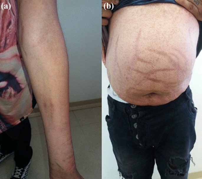
Cushing syndrome (CS) is a condition that occurs because of high blood levels of glucocorticoids (GCs). These patients can present with a variety of systemic signs and symptoms, including truncal obesity, easy bruising of the skin, violaceous abdominal striae, resistant hypertension, dysglycemia, as well as osteoporosis. CS can occur because of adrenal etiologies such as adrenal adenoma, adrenal cancer, or adrenal hyperplasia or from an adrenocorticotropic hormone (ACTH)-producing pituitary adenoma or ectopic tumor. However, the most common cause of CS is the exogenous administration of GCs [1]. While exogenous GCs are often medically prescribed for the treatment of inflammatory conditions, some patients may be accidentally exposed to exogenous GCs from over-the-counter (OTC) supplements. We present a case of a young woman who developed exogenous CS and suffered a hip fracture as a result of taking an OTC supplement, Artri King (AK), adulterated with GCs.

Case Presentation

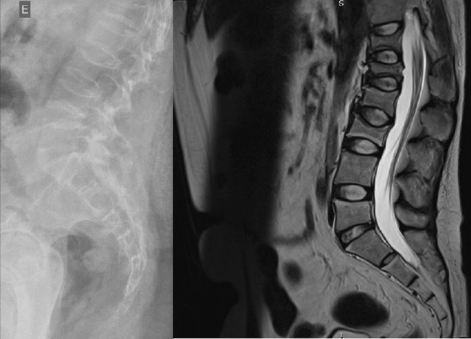
A 40-year-old obese woman presented to the hospital following a fall at home. She reported a snapping noise and sudden right hip pain while trying to stand up, and subsequently fell to the floor. She had noted right-sided hip pain for several days preceding her fall. She was evaluated in the emergency department where computed tomography (CT) imaging of the right lower extremity showed an intertrochanteric fracture of the right femur (Figure 1). The patient underwent open reduction and internal fixation of her right femur. The patient reported an unexplained weight gain of approximately 40 lbs in the preceding five months with a peak weight of 223 lbs (101 kg) and a body mass index (BMI) of 37 kg/m2. The patient denied taking any medications or supplements at the time of hospitalization. The endocrinology team was consulted to evaluate for causes of secondary osteoporosis in this young woman.

A-CT-scan-showing-the-right-intertrochanteric-fracture-of-the-right-femur-(yellow-arrows)
Figure 1: A CT scan showing the right intertrochanteric fracture of the right femur (yellow arrows)

Diagnostic assessment

Her vital signs showed a blood pressure of 142/96 mmHg, heart rate of 68 beats per minute, temperature of 98.1°F (36.7°C), and 98% oxygenation on room air. Physical examination did not reveal abdominal striae or buffalo hump. She did have supraclavicular fat deposition and central obesity. No proximal muscle weakness was present.

Laboratory tests were pertinent for decreased 25-hydroxy vitamin D, increased parathyroid hormone (PTH), and normal calcium (Table 1). These findings were consistent with secondary hyperparathyroidism due to vitamin D deficiency. Dual-energy X-ray absorptiometry (DEXA) scan revealed osteoporosis (Figures 23 and Tables 23). Further testing showed normal thyroid-stimulating hormone (TSH), estradiol, follicle-stimulating hormone (FSH), and luteinizing hormone (LH), thus ruling out hyperthyroidism and primary ovarian insufficiency as possible causes of reduced bone mineral density (Table 1). Random cortisol was checked as hypercortisolism was suspected but it was found to be decreased along with decreased ACTH as well (Table 4). A cosyntropin stimulation test was performed, which showed decreased baseline cortisol with inappropriately decreased cortisol levels at 30 minutes and 60 minutes (Table 5). Given the discordance between the patient’s presentation and the lab results, assay interference was suspected, and further evaluation of the adrenal function was performed. Repeat labs using liquid chromatography-mass spectrometry (LCMS) assay again confirmed persistently low cortisol (Table 4). A 24-hour free urine cortisol was too low to quantify per assay despite the adequate volume. Further evaluation showed overall low adrenal steroids, including deoxycorticosterone, 17-hydroxyprogesterone, androstenedione, 11-deoxycortisol, pregnenolone, dehydroepiandrosterone sulfate, corticosterone, and progesterone.

Lab test Patient’s value Reference range
25-hydroxy vitamin D 12.8 ng/ml 30-100 ng/ml
Parathyroid hormone (PTH) 86.2 pg/ml 10-66 pg/ml
Serum calcium 9.5 ng/dl 8.8-10.5 mg/dl
Thyroid-stimulating hormone (TSH) 2.49 mIU/L 0.36-3.74 mIU/L
Estradiol 57.1 pg/ml 19.8-144.2 pg/ml
Follicle-stimulating hormone (FSH) 5.4 mIU/ml 2.5-10.4 mIU/ml
Luteinizing hormone (LH) 6 mIU/ml 1.9-12.5 mIU/ml
Table 1: Patient’s lab values on admission
Dual-energy-X-ray-absorptiometry-(DEXA)-scan-of-the-femoral-neck-showing-osteopenia
Figure 2: Dual-energy X-ray absorptiometry (DEXA) scan of the femoral neck showing osteopenia
Dual-energy-X-ray-absorptiometry-(DEXA)-scan-of-the-lumbar-spine-showing-osteoporosis
Figure 3: Dual-energy X-ray absorptiometry (DEXA) scan of the lumbar spine showing osteoporosis
Region Area (cm2) Bone mineral content (g) Bone mineral density (g/cm2) T-score Peak reference Z-score Age-matched
Femoral neck 4.76 3.53 0.742 -1.0 87 -0.7 91
Total 33.39 26.14 0.783 -1.3 83 -1.1 85
Table 2: Summary of dual-energy X-ray absorptiometry (DEXA) scan results of the femoral neck
Region Area (cm2) Bone mineral content (g) Bone mineral density (g/cm2) T-score Peak reference Z-score Age-matched
L1 10.79 7.56 0.701 -2.6 71 -2.4 73
L2 11.79 9.06 0.768 -2.4 75 -2.1 77
L3 12.70 9.98 0.786 -2.7 73 -2.4 75
L4 15.57 11.42 0.733 -3.0 69 -2.7 71
Total 50.86 38.03 0.748 -2.7 71 -2.5 73
Table 3: Summary of dual-energy X-ray absorptiometry (DEXA) scan results of the lumbar spine
Lab test Patient’s values while on Artri King Patient’s values four weeks off of Artri King Reference range
Random cortisol (routine assay) <0.64 μg/dL 7.3 μg/dL 5-25 μg/dL
Adrenocorticotropic hormone (ACTH) 1.5 pg/ml 12 pg/ml 7.2-63.3 pg/ml
Random cortisol (using liquid chromatography-mass spectrometry (LCMS) assay) 0.526 μg/dL N/A 5-25 μg/dL
Table 4: Patient’s cortisol and adrenocorticotropic hormone levels before and after stopping Artri King
Cosyntropin stimulation test Patient value Reference range
Baseline cortisol 1.64 μg/dL 5-25 μg/dL
Cortisol after 30 minutes 1.33 μg/dL >18 μg/dL
Cortisol after 60 minutes 6.48 μg/dL >18 μg/dL
Table 5: Results of cosyntropin test while on Artri King

Treatment

She was started on teriparatide as well as vitamin D and calcium supplementation for the treatment of osteoporosis. Based on the aforementioned testing and the apparent symptoms of hypercortisolism, the patient was questioned again about the potential intake of steroids. She then recalled that she had been taking AK, an OTC supplement promoted for joint pain and arthritis. She reported that she had been taking two tablets of the supplement three times a day intermittently for the past three years. The patient neglected to bring it to the medical team’s attention before because she was under the impression that it was a multivitamin and did not have implications on her diagnosis. She was asked to stop the supplement and was educated about potential adrenal insufficiency symptoms and GC withdrawal.

Outcome and follow up

Repeat labs after four weeks off AK showed improved cortisol and ACTH levels indicating recovery of her hypothalamic-pituitary-adrenal (HPA) axis (Table 4). She lost 25 lbs in this time span with lifestyle modification. She continues teriparatide for osteoporosis, and monitoring of her bone mineral density is planned.

Discussion

This patient initially presented with a pathological fracture of her right femoral head. Given her young age, causes of secondary osteoporosis, including CS, were explored. The prevalence of osteoporosis in CS patients is 50% [2]. The effects of GC on bone health have been well studied. The major mechanism by which GC affects bone mineral density is by impairment of bone formation. GCs increase osteoblast and osteocyte apoptosis and decrease osteoblast function through their catabolic effects, which result in a dramatic decrease in bone formation rate. A prolonged lifespan of osteoclasts is observed with GC. A decrease in bone formation markers such as P1NP and osteocalcin has been observed in patients treated with GC [3]. Long-term GC use is associated with increased risk for fractures with a reported global prevalence of fractures of 30-50%. The risk for vertebral fractures is even higher, particularly in the thoracic and lumbar vertebrae. Interestingly, the risk for fracture with GC use peaks early in the course of treatment, often as early as three months into treatment, and declines rapidly after GC discontinuation [4]. An increased fracture risk has been described even with relatively low doses of GC (2.5-7.5 mg of prednisone or other equivalently dosed GC) and even with short-term use of under 30 days [5].

Our patient’s initial labs confirmed adrenal suppression despite our initial suspicion of CS, given her ongoing weight gain, central obesity, and osteoporosis. However, no obvious source of exogenous GC was identified. In most cases, the source of exogenous GC is easily identified through medication reconciliation; however, in our case, the patient was inadvertently exposed to steroids from an unregulated supplement, AK. The supplement’s ingredients were listed as glucosamine, chondroitin, collagen, vitamin C, curcumin, methylsulfonylmethane, nettle, and omega-3 fatty acids, with no mention of any steroid components. In a letter to the editor of the Internal Medicine magazine, several doctors published their concerns about a recent increase in CS cases associated with the use of AK and other similarly unregulated products [6]. Based on our literature search, three similar cases were published [7,8]. The reported cases developed CS after taking Artri King for several months, but none of them presented with a fracture.

A warning by the U.S. Food & Drug Administration (FDA) was issued on April 20, 2022, indicating that FDA laboratory testing of this supplement confirmed the presence of undeclared drug ingredients, including dexamethasone, methocarbamol, and diclofenac. The FDA, however, was unable to confirm the exact amount of dexamethasone that these supplements contained [9]. Adverse events, including liver toxicity and death, were reported by the FDA.

One study revealed that between 2007 and 2016, the FDA had issued more than 700 warnings about the sale of dietary supplements that contained unlisted and potentially dangerous ingredients. The majority of these supplements included those marketed for sexual enhancement, weight loss, or muscle building [10]. This case highlights the risks of undisclosed ingredients in OTC supplements.

Conclusions

In conclusion, we recommend that a thorough reconciliation of medication and supplements be obtained for all patients with CS. Supplements should be stopped and HPA axis testing should be repeated in patients with suspected exogenous GC exposure, even if steroids are not declared in the ingredients. It is also important to monitor such patients for adrenal insufficiency due to GC withdrawal and consider GC tapering if necessary. Our patient showed improvement in cortisol levels with no overt symptoms of adrenal insufficiency without the need for GC therapy. This case demonstrates the first case of AK-induced CS resulting in a pathological fracture. Given the increased use and availability of OTC supplements, this case highlights on the importance of detailed history-taking and the role of supplements in causing CS. This case also stresses the need for further education and counseling of our patients as well as tighter control on the manufacturing and sale of these supplements.

References

  1. Lacroix A, Feelders RA, Stratakis CA, Nieman LK: Cushing’s syndrome. Lancet. 2015, 386:913-27. 10.1016/S0140-6736(14)61375-1
  2. Mancini T, Doga M, Mazziotti G, Giustina A: Cushing’s syndrome and bone. Pituitary. 2004, 7:249-52. 10.1007/s11102-005-1051-2
  3. Briot K, Roux 😄 Glucocorticoid-induced osteoporosis. RMD Open. 2015, 1:e000014. 10.1136/rmdopen-2014-000014
  4. Canalis E, Mazziotti G, Giustina A, Bilezikian JP: Glucocorticoid-induced osteoporosis: pathophysiology and therapy. Osteoporos Int. 2007, 18:1319-28. 10.1007/s00198-007-0394-0
  5. Waljee AK, Rogers MA, Lin P, et al.: Short term use of oral corticosteroids and related harms among adults in the United States: population based cohort study. BMJ. 2017, 357:j1415. 10.1136/bmj.j1415
  6. Del Carpio-Orantes L, Quintín Barrat-Hernández A, Salas-González A: Iatrogenic Cushing syndrome due to fallacious herbal supplements. The case of Ortiga Ajo Rey and Artri King. Med Int Mex. 2021, 37:599-602.
  7. Patel R, Sherf S, Lai NB, Yu R: Exogenous Cushing syndrome caused by a “Herbal” supplement. AACE Clin Case Rep. 2022, 8:239-42. 10.1016/j.aace.2022.08.001
  8. Mikhail N, Kurator K, Martey E, Gaitonde A, Cabrera C, Balingit P: Iatrogenic Cushing’s syndrome caused by adulteration of a health product with dexamethasone. JSM Clin Case Rep. 2022, 3:
  9. U.S. Food and Drug Administration. Public notification: Artri King contains hidden drug ingredients. (2022). Accessed: February 25, 2023: https://www.fda.gov/drugs/medication-health-fraud/public-notification-artri-king-contains-hidden-drug-ingredients.
  10. Tucker J, Fischer T, Upjohn L, Mazzera D, Kumar M: Unapproved pharmaceutical ingredients included in dietary supplements associated with US Food and Drug Administration warnings. JAMA Netw Open. 2018, 1:e183337. 10.1001/jamanetworkopen.2018.3337

From https://www.cureus.com/articles/153927-exogenous-cushing-syndrome-and-hip-fracture-due-to-over-the-counter-supplement-artri-king#!/

BMD may Underestimate Bone Deterioration for Women with Endogenous Cushing’s Syndrome

Nearly one-third of women with endogenous Cushing’s syndrome and normal bone mineral density have a low trabecular bone score, according to study data.

“A large proportion of patients had degraded microarchitecture despite normal BMD,” Hiya Boro, DM, MD, MBBS, consultant in endocrinology, diabetes and metabolism at Aadhar Health Institute in India, and colleagues wrote. “The risk of fracture may be underestimated if BMD alone is measured. Hence, trabecular bone score should be added as a routine complementary tool in the assessment of bone health in patients with Cushing’s syndrome.”

About one-third of women with endogenous Cushing's syndrome have normal BMD and low trabecular bone score. 
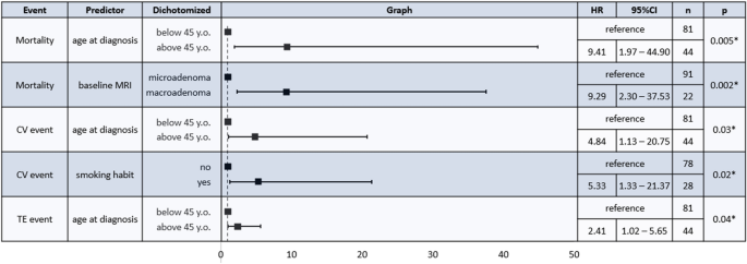
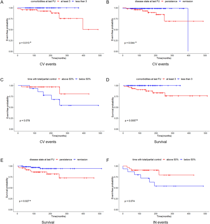
Data were derived from Boro H, et al. Clin Endocrinol. 2023;doi:10.1111/cen.14944.

Researchers conducted a cross-sectional study at a single center in India from March 2018 to August 2019. The study included 40 women with overt endogenous Cushing’s syndrome and 40 healthy sex-matched controls. Seum and salivary cortisol and plasma adrenocorticotropic hormone (ACTH) were measured. Participants were considered ACTH independent if they had a level of less than 2.2 pmol/L. Areal BMD was measured at the lumbar spine, femoral neck, total hip and distal one-third of the nondominant distal radius. Low BMD for age was defined as a z score of less than –2. Trabecular bone score was measured at the lumbar spine. Fully degraded microarchitecture was defined as a trabecular bone score of 1.2 or lower and partial degradation was a trabecular bone score of 1.21 to 1.34.

Of the 40 women with Cushing’s syndrome, 32 were ACTH-dependent and the other eight were ACTH independent. Of the independent group, seven had adrenal adenoma and one had adrenocortical carcinoma.

Women with Cushing’s syndrome had lower BMD at the lumbar spine (0.812 g/cm2 vs. 0.97 g/cm2< .001), femoral neck (0.651 g/cm2 vs. 0.773 g/cm2< .001) and total hip (0.799 g/cm2 vs. 0.9 g/cm2< .001) than the control group.

“No significant difference was noted in the distal radius BMD,” the researchers wrote. “This may be explained by the fact that cortisol excess predominantly affects trabecular rather than cortical bone.”

Absolute trabecular bone score was lower in the Cushing’s syndrome group compared with controls (1.2 vs. 1.361; P < .001). Based on trabecular bone score, 42.5% of women with Cushing’s syndrome had fully degraded bone microarchitecture, 45% had partially degraded microarchitecture and 12.5% had normal microarchitecture.

“In our study, 32.5% of patients had normal BMD with low trabecular bone score, thus highlighting the fact that patients may have normal BMD despite degraded microarchitecture,” the researchers wrote. “As such, assessment of BMD alone may underestimate the risk of fractures in patients with Cushing’s syndrome.”

Complications and Mortality of Cushing’s Disease: Report on Data Collected Over a 20-Year Period at a Referral Centre

Abstract

Context

Cushing’s disease (CD) is rare condition burdened by several systemic complications correlated to higher mortality rates. The primary goal of clinicians is to achieve remission, but it is unclear if treatment can also increase life expectancy.

Aim

To assess the prevalence of cortisol-related complications and mortality in a large cohort of CD patients attending a single referral centre.

Materials and methods

The clinical charts of CD patients attending a referral hospital between 2001 and 2021 were reviewed.

Results

126 CD patients (median age at diagnosis 39 years) were included. At the last examination, 78/126 (61.9%) of the patients were in remission regardless of previous treatment strategies. Patients in remission showed a significant improvement in all the cardiovascular (CV) comorbidities (p < 0.05). The CV events were more frequent in older patients (p = 0.003), smokers and persistent CD groups (p < 0.05). Most of the thromboembolic (TE) and infective events occurred during active stages of the disease. The CV events were the most frequent cause of death. The standardized mortality ratio (SMR) resulted increased in persistent cases at the last follow-up (SMR 4.99, 95%CI [2.15; 9.83], p < 0.001) whilst it was not higher in those in remission (SMR 1.66, 95%CI [0.34; 4.85], p = 0.543) regardless of the timing or number of treatments carried out. A younger age at diagnosis (p = 0.005), a microadenoma (p = 0.002), and remission status at the last follow-up (p = 0.027) all increased survival. Furthermore, an elevated number of comorbidities, in particular arterial hypertension, increased mortality rates.

Conclusions

Patients with active CD presented a poor survival outcome. Remission restored the patients’ life expectancy regardless of the timing or the types of treatments used to achieve it. Persistent CD-related comorbidities remained major risk factors.

Introduction

Cushing’s disease (CD) is the most common cause of endogenous glucocorticoid excess due to uncontrolled adrenocorticotropic hormone (ACTH) secretion from a pituitary adenoma, for the most part a microadenoma [1]. A rare condition with an estimated incidence of 0.6—2.6 cases per million per year, it is burdened by high morbidity and mortality, for the most part linked to cardiovascular (CV) events. This is particularly true for active CD which is characterized by hypertension, diabetes mellitus, obesity and dyslipidaemia. The severity of the clinical picture seems to depend more on the duration of the disease rather than on the degree of cortisol elevation, although other confounding factors may affect the clinical phenotype [2]. Prompt diagnosis and resolution of hypercortisolemia are paramount to revert cortisol-related comorbidities and to improve life expectancy. Although new individualized medical treatment options for CD continue to evolve, transsphenoidal surgery (TSS) remains the first line treatment for potentially operable patients as it is the only treatment that seems to provide a rapid, long-lasting remission. Persistent and recurrent cases are nevertheless major concerns, since up to 50% of cases might require other treatment modalities to achieve disease control and those patients are once again exposed to cortisol excess that can negatively impact their survival [3]. An increased mortality has been noted in patients with active CD, while patients in remission show a markedly lower one. It is still unclear if mortality in these patients is higher than that in the general population. Some studies report a normal life expectancy [4,5,6,7,8] while others describe a persistently higher mortality [9,10,11]. One study reported finding a higher mortality as long as 10 years after remission, and only patients cured by a single TSS showed a normal life expectancy [12].

In view of these considerations, this study was designed to assess the prevalence of cortisol-related comorbidities/complications and mortality in a large group of CD patients attending a tertiary referral centre over the past 20 years. Other study aims were to evaluate the predictors of long-term outcomes and the impact of different treatments on life expectancy in CD patients.

Materials and Methods

One hundred twenty-six CD patients diagnosed between December 2001 and December 2021 were eligible for this monocentric, retrospective, observational study. Hypercortisolism was suspected on the basis of the patient’s clinical features and it was confirmed by appropriate hormonal testing [low dose dexamethasone suppression test (LDDST), 24-h urinary free cortisol (UFC) and late-night salivary cortisol (LNSC)] after excluding the possibility of exogenous glucocorticoid intake from any route [13]. UFC and LNSC were assessed at least in two different samples as recommended [1415].

The diagnosis of ACTH-dependent syndrome was confirmed on the strength of detectable ACTH levels (> 10 ng/L) and appropriate responses to a high dose dexamethasone suppression test (HDDST), corticotrophin releasing hormone (CRH) and/or desmopressin (DDAVP) tests [16]. All the patients underwent a pituitary magnetic resonance imaging (MRI); they also underwent bilateral inferior petrosal sinus sampling (BIPSS) when the results of hormonal tests were ambiguous. The pituitary origin of ACTH secretion was confirmed by biochemical remission after TSS, histology and/or post-operative hypoadrenalism.

The results of clinical, biochemical and radiological tests as well as the treatments performed to control cortisol secretion (surgery, radiotherapy and/or medical therapy), any comorbidities (i.e., arterial hypertension, impaired glucose homeostasis, dyslipidaemia, overweight), any hormone deficiencies, any complications (i.e., CD-related events such as infective, CV and thromboembolic events) and any deaths recorded in the medical charts were collected.

The disease severity at baseline was defined on the basis of the patient’s UFC values as mild (up to two-fold the upper limit of normal – ULN), moderate (between 2 and 5 times the ULN) or severe (over five-fold the ULN).

Patient’s classification on the basis of disease activity are indicated in Supplementary material and methods sections.

The presence of hypertension, glucose metabolism impairment, obesity, dyslipidaemia and hypopituitarism were defined as by specific Guidelines, Supplementary [19,20,21,22,23,24].

The current study was designed in accordance with the principles of the Declaration of Helsinki and approved by the Ethical Committee of the province of Padova (protocol code 236n/AO/22, date of approval 29 April 2022).

The types of CD complications characterizing the patient were classified into three categories: CV, thromboembolic (TE), or infective (IN) events. Depending on the timing of its presentation, an event was classified as occurring: “prior” to diagnosis, “during” active CD or “after” CD remission. Events requiring hospitalization or iv antibiotic administration were registered as IN events. The causes of death were classified under the following headings: CV, infections, cancer, psychiatric complications leading to suicide, TE events or other (the last when none of the previous causes was applicable).

Statistical analysis

Categorical variables were reported as counts or percentages, and quantitative variables as median and interquartile ranges [IQR]. The comparisons between groups were performed with a Mann–Whitney sum rank test for independent quantitative variables; a Wilcoxon signed-rank test was run for dependent quantitative variables. As far as categorical variables were concerned, the McNemar test or a chi-square test were used for paired and unpaired data, respectively.

A Cox regression analysis was performed to evaluate possible predictors for events and mortality based on the assumption of constant hazards over time. As time-dependent variables (e.g., achieving remission) did not meet this assumption, their survival analysis was performed using Kaplan–Meier analysis. Regarding complications, as there is usually a delay in CD diagnosis [25], Kaplan Meier curves for event free probability were calculated beginning 24 months prior to the diagnosis in order to include “prior” events possibly related to cortisol excess in our analysis. Vice versa, survival analysis for mortality was calculated beginning with the CD diagnosis date. Standardized mortality ratio (SMR) was calculated based on indirect age standardization in order to compare the observed deaths in our CD population with the expected number of deaths in the general population [2627]. A Fisher exact test was carried out to assess significant differences with respect to the general population and calculating the 95% confidence interval (95% CI) for SMR.

The threshold for statistical significance was set at p-value < 0.05. Statistical analyses were performed with R: R-4.2.0 for Windows 10 (32/64 bit) released in April 2022 and R studio desktop version 4.2.0 (2022-04-22) for Windows 10 64 bit (R Foundation for Statistical Computing, Vienna, Austria, URL https://www.R-project.org/). An open-source calculator was also used to perform the Fisher exact test (http://www.openepi.com).

Results

Baseline

The data of 167 CD patients attending the Centre between December 2001 and December 2021 were collected. The information regarding 41 patients were not included in the analysis because of insufficient follow-up data (i.e. patients referred for second opinion or for diagnostic workup or those with follow-up < 1 year from first line treatment). The remaining 126 patients presented a median age at diagnosis of 39 [31–50 years]; the female: male ratio was 3:1. The median follow-up was 130.5 months [72.5–201.5]. The patients’ clinical features at the time of diagnosis are outlined in Table 1.

Table 1 The patients’ clinical features at the time of diagnosis

The median UFC levels were 3.2 times the ULN [2–5.6]. Almost half of the cohort presented moderate cortisol excess (45/98, 45.9%), with lower proportions of the patients presenting mild (26/98, 26.5%) and severe disease (22/98, 27.6%).

Most of the patients (91/113, 80.5%) had a microadenoma, including 29/91(31.9%) with negative imaging. The remaining 22 patients (19.5%) had a macroadenoma.

Treatments

Most of the patients underwent TSS as the first line treatment (113/126), only one patient underwent craniotomy. Eight patients received primary medical treatment, three received first-line radiotherapy and one underwent BA soon after diagnosis. Overall, 115 patients underwent pituitary surgery (one patient with a previous unsuccessful pituitary irradiation) and the remission rate was 60.9%. Relapses were observed in 46.7% of the cases after a median time of 56 [29–83] months. The second surgery proved less successful with respect to the first one; the remission rate was 43.2% (16/37); of these, 25% developed recurrence during the follow-up period. The median time to relapse was 66.5 [36–120] months. Only two patients underwent a third surgery; in both cases it was not curative (Supplementary Fig. 1) [27]. A 4th and a 5th TSS were performed in one of these for debulking purposes due to an aggressive pituitary lesion. Surgical remission was not affected by pre-treatment with cortisol-lowering medications neither before the first (p = 1.0) nor the second TSS (p = 0.88). Moreover, hormone control did not improve the surgical outcomes, although a tendency towards a higher remission rate was observed in those patients who showed good disease control before undergoing the second surgery (Supplementary Fig. 2) [27].

Overall, 34 patients received radiotherapy, either the conventional (18.5%) or the stereotactic type (81.5%). Remission was noted in 36.7% (11/30) of the patients with at least a 12-month post-radiotherapy follow-up. As expected, the longer the follow-up, the higher the remission rate; it was 41.67% (10/24) and 46.7% (7/15) at 5 and 10 years, respectively.

Thirteen patients underwent BA and achieved complete remission. Excluding the patients with less than 12 months of follow-up, 4 out of 11 (36.4%) of the patients developed CTP-BADX/NS over a mean follow-up period of 110 [106 -329] months. Three patients out of the 11 were previously irradiated at pituitary level to control cortisol secretion. Four CD patients underwent unilateral adrenalectomy due to a dominant adrenal lesion consistent with chronic ACTH stimulation. Two (50%), harbouring unilateral adenomas larger than 5 cm, achieved remission after surgery; both cases were previously irradiated at the pituitary level.

All but one of the 48 patients with persistent hypercortisolism at the last follow-up were on cortisol lowering medications. The untreated patient had a residual mild cortisol excess after TSS and medical therapy was discontinued because of multiple drug intolerance. At the last follow-up 28 patients were receiving monotherapy, and 19 were receiving combination treatment; 25 patients were receiving steroidogenesis inhibitors, 9 pituitary-target drugs and 13 a combination of the two compounds (Supplementary Table 1) [27]. Most of our patients achieved UFC normalization (complete control in 67.4%, partial control in 22.7%, uncontrolled in 10.9%). Data pertaining to a single patient with renal function impairment who presented falsely low UFC were not included in this analysis. When available, LNSC was restored in 14/41 cases (34.2%). No differences in the patients’ outcomes linked to the type of treatment prescribed (monotherapy vs combination treatment) or its target (adrenal vs pituitary) were found (data not shown).

We also evaluated the extent of cortisol excess throughout the active phase of CD both for the patients presenting persistence at the last available follow-up (n = 48) and for those in remission after multiple therapies (i.e., late remission) (n = 33). As described in the material and methods section, disease activity for each year of active disease was defined on the basis of patients’ UFC levels. A minimum of three UFC measurements were registered every year and the median value was calculated. When data were missing, the patients were considered uncontrolled during that period. The results are reported in Supplementary Table 2 [27]; both the persistence and late remission groups showed UFC levels < 2xULN over more than 50% of the time span evaluated (58.8% and 73.6%, respectively). There was a progressive increase in the proportion of controlled patients over the observation period (Fig. 1).

Fig. 1
figure 1

Percentage of patients controlled during active CD

Comorbidities

The principal CD features at baseline and at the last follow-up examination were evaluated, (Supplementary Table 2). At time of diagnosis, no differences were observed as regards comorbidities between patients who achieved remission and those with persistent disease at baseline, (Supplementary Table 3). The patients in remission at the last examination showed a significant improvement in all the parameters considered; those with persistent CD did not (Table 2).

Table 2 A comparison of Cushing’s disease features at baseline and at the last follow-up examination

As far as hormone deficiencies were concerned, 42/126 (33.3%) of the patients developed at least one deficit due to previous treatments (Supplementary table 4) [27], including hypocortisolism due to BA. Neither the second surgery nor radiotherapy led to an increase in hypopituitarism (Supplementary Fig. 3) [27].

Complications and mortality

As far as CD complications were concerned, 18.3% of the patients had a TE event, 17.5% presented an IN event and 7.1% presented a CV one. Most of the events occurred during an active phase of CD (Table 3). Other concomitant thrombotic risk factors were present in 10/19 (52.6%) of the patients experiencing TE events. TE events were related to surgery (pituitary, adrenal or others) in 5 cases, to post-traumatic fractures in 2, to prolonged immobilization in 2, and to a symptomatic SARS CoV2 infection in one case. IN events affected the respiratory system in 9 cases, the gastro-intestinal tract in 5 cases, the soft tissues in three cases, the central nervous system in 2 cases, the musculoskeletal system in 2 cases and the genitourinary tract in one case.

Table 3 Thromboembolic, infective, and cardiovascular events and their timing (see materials and methods)

Overall, 11 deaths were recorded during the follow-up period (130.5 [72.5–201.5] months). The causes of death were classified as: cardiovascular events (n = 4), infections (n = 2), cancer (n = 2), suicide (n = 1), thromboembolic events (n = 0), others (n = 2; a cerebral haemorrhage in one case and an unknown cause in the other).

Cox regression was performed to evaluate the predictors of events (CV, IN, TE) and mortality (Fig. 2). The older patients presented an increased risk of mortality (HR 9.41, 95%CI [1.97; 44.90], p = 0.005), of CV events (HR 4.84, 95%CI [1.13; 20.75], p = 0.003) and of TE events (HR 2.41, 95%CI [1.02; 5.65], p = 0.04). Similarly, the presence of a macroadenoma at the time of the first MRI was associated with reduced survival (HR 9.29, 95%CI [2.30; 37.53], p = 0.002). Smoking was correlated to CV events (HR 5.33, 95%CI [1.33; 21.37], p = 0.02). Hypercortisolism severity at baseline did not affect the risk of complications or survival. No gender related differences were observed, although a tendency toward more CV events was noted in the males (p = 0.08).

Fig. 2

figure 2

Cox regression analysis for predictors of mortality and cardiovascular, infective or thromboembolic events; only significant results are shown. HR: Hazard ratio; CI: confidence interval; n: number, CV: cardiovascular; TE: thromboembolic. *p < 0.05

Kaplan Meier curves were plotted for complications (CV, IN and TE) and mortality in order to assess time-dependent variables (i.e., the number of comorbidities and the disease status at the last follow-up, the timing of remission and the disease activity in the patients with persistent CD at the last follow-up). We found that persistent disease and multiple comorbidities (at least 3) at the last follow-up were associated with increased CV events (p = 0.044 and p = 0.013, respectively) and mortality (p = 0.027 and p = 0.0057, respectively) (Fig. 3). The timing of remission did not influence the mortality or the risk of complications (data not shown). With regard to the patients with persistence, those presenting total/partial control for more than half of the follow-up period considered tended to have fewer CV and IN events (p = 0.078 and p = 0.074, respectively) (Fig. 3). Similarly, among patients with persistent cortisol excess the impaired circadian rhythm of secretion was associate to TE events and a trend to higher mortality (Supplementary Fig. 4). Sub-analysis of each comorbidity revealed that hypertension played a pivotal role during the follow-up period for CV complications (p = 0.011) and mortality (p = 0.0039). Similarly, dyslipidaemia was related to CV events (p = 0.046) and prediabetes/diabetes were associated to TE events (p = 0.035). A tendency toward increased mortality in the patients with impaired glucose homeostasis at the last follow-up was also noted (p = 0.052) (Data not shown).

Fig. 3

figure 3

Kaplan Meier curves for cardiovascular events based on: A) comorbidities at the last follow-up examination; B) disease status at the last follow-up examination; C) control during active disease for patients presenting persistence at the last follow-up. Kaplan Meier curves for survival plotting: D) comorbidities at the last follow-up examination; E) disease status at the last follow-up examination. Kaplan Meier curves for infective events based on: F) hormone control during active disease of patients presenting persistence at the last follow-up examination. FU: follow-up; CV: cardiovascular; IN: infective. *p < 0.05

The entire CD cohort presented an increased mortality, with a SMR of 3.22 (95%CI [1.70; 5.60], p = 0.002). Mortality was significantly higher in the patients with persistent disease (SMR 4.99, 95%CI [2.15; 9.83], p < 0.001), but it was similar to that of the general population in the patients in remission (SMR 1.66, 95%CI [0.34; 4.85], p = 0.543). The finding was independent of the timing or the modality used to achieve cortisol control; for the early remission group the SMR was 2.15 (95%CI [0.36; 7.11], p = 0.477) and for the late remission group it was 1.14 (95%CI [< 0.01; 5.62], p = 1.0). The length of remission period was 82 [38–139] for the early remission group vs 85 [21–136] for the late remission one.

Discussion

Study findings have confirmed that CD patients have a higher mortality and, as previously observed, the most common cause of death in these patients was, first of all, CV events and, secondly, infections [9]. Although there were no fatal TE events in our cohort, that type of complication was the most frequent one. As expected, the patients with persistent CD presented significantly increased mortality with respect to the general population. At the last follow-up examination the CD patients in remission had a mortality rate that was comparable to that of the general population regardless of the number of treatments needed to achieve remission. The finding is in contrast with the results of a multicentre study examining patients with more than 10 years of remission that reported finding a normal life expectancy only in the patients who achieved an early remission following a single TSS [12]. The better life expectancy in our series may be explained by an extensive use of cortisol-lowering medications in our centre during active phases of CD. There was moreover at least a partial control in the late remission group during over 70% of the years assessed; this might have had a positive effect on the overall survival rate (data not shown). Furthermore, our study considered relatively recent years when significant improvement in timely diagnosis and available medical therapies have been made [9]. Lastly, being monocentric, our study showed a homogenous management of comorbidities that by contrast, is in highly unlikely in a retrospective international study. Since cardiovascular and metabolic risk factors related to cortisol-excess are major determinant of mortality in CD, the latter point is of the outmost importance.

Survival was positively influenced in our cohort by a younger age at diagnosis, the presence of a microadenoma at baseline [9] and a remission status at the last follow-up examination. As expected, an elevated number of comorbidities increased mortality, and as has been previously reported, arterial hypertension, in particular, reduced survival [28]. A tendency toward increased mortality was also noted in connection to impaired glucose homeostasis, but data on this topic are still controversial [810122829].

Cortisol excess atherosclerotic risk leading to CV events are closely liked. Beyond cortisol’s direct action on the tissues, this association is probably related to a clustering of several metabolic complications such as insulin resistance, arterial hypertension, dyslipidaemia and overweight commonly present in CD patients [3031]. Indeed, the patients presenting multiple comorbidities, especially arterial hypertension and dyslipidaemia, showed more CV complications. CV events were also more frequent in the patients with persistent hypercortisolism, and, as observed in general population in the elderly and in the smokers [32].

Older age at the time of diagnosis and dis-glycemia at the last follow-up examination were found to be related to TE events. It was instead impossible to identify predictors of infective complications. Although most TE and IN events occurred during active disease, remission did not significantly reduce these complications. The finding is in line with the data of a recent study focusing on a Swedish population reporting that CD patients present a higher risk of sepsis and thromboembolism even during long term remission [33]. Moreover, it is worthy of note that most of the TE events (52.6%) were accompanied by a concomitant risk factor such as recent surgery. These data highlight the importance of adequate prophylaxis in CD patients facing prothrombotic conditions such as those linked to a perioperative period [334]. Disease severity at the baseline did not affect the patients’ complications or survival; the finding is not entirely surprising as the degree of cortisol excess does not necessarily correlate with the severity of the clinical picture [2].

The patients who achieved remission in our cohort showed an overall improvement in all the cortisol-related comorbidities. Hypertension was the most prevalent complication at the time of diagnosis, while overweight, which persisted in approximately 50% of the cases after remission, became by far the most frequent comorbidity. Glucose homeostasis alterations were the least prevalent at the time of diagnosis, although an underestimation is probable, as only fasting glycaemia or glycosylated haemoglobin were evaluated in most cases and provocative testing for hypercortisolism was not carried out [35].

With regards to demographic features, for the most part our patients were diagnosed during their third/fourth decade of life and they were prevalently female, in line with previous reports [36]. Most cases were due to a pituitary microadenoma (80% of the cases in our patients), including non-visible lesions on the MRI.

As far as treatment was concerned, the remission rate after the first TSS was quite low with respect to what would be expected at a tertiary centre; the finding can be explained by the fact that many of the patients studied had been referred to our unit after undergoing unsuccessful pituitary surgery elsewhere. However, the assessment of surgical performance in various centres goes beyond the aim of the present study. As expected, a second TSS was less successful than the first one, but the rate of success found in our patients was in line with literature data [37]. Although the immediate remission rate after a second TSS was comparable to the long term outcome of radiotherapy, a quarter of the patients experienced a relapse just as they did after the first surgery [17]. Regarding the risk of developing hypopituitarism was concerned, no significant difference was found between the two approaches. These data have confirmed that both re-intervention and radiation treatment can be considered valid second-tier options, and a case by case approach should be adopted. Pre-operative medical treatment with cortisol-lowering medications did not improve the surgical outcomes, regardless of its effectiveness in controlling cortisol excess, in line with data by the European Registry on Cushing’s Syndrome (ERCUSYN) [38].

At the last follow-up examination, no differences in disease control were found when the treatment targets (pituitary vs adrenal) of the patients were compared. A higher control rate of hypercortisolism during active CD was found over time, possibly reflecting better drug dose titration and the widening landscape of available drugs with over two thirds of the patients presented completely controlled UFC at last examination. The fact that only one third of our patients achieved circadian rhythm restoration confirmed the previously reported difficulty in normalizing this parameter [39,40,41]. Interestingly, TE were more frequent when LNSC was uncontrolled and the same tendency was observed for survival, confirming the better outcome of patients with rhythm restoration [8]. Although only the last available value of LNSC was assessed, this finding might potentially turn the spotlight on the importance of LNSC normalization during medical treatment [42], but further studies are required to confirm these data.

In line with previous reports, more than one third of the patients who underwent BA developed CTP-BADX/NS [18]. Although BA seems to immediately control hypercortisolism, this benefit should be carefully weighed against the risk of permanent adrenal insufficiency and CTP-BADX/NS. The patients received minimal doses of glucocorticoid replacement treatments following BA to avoid both over- and under treatment that might negatively impact survival [43], and this might explain why BA was not associated to increased mortality as observed in other series [44]. Unilateral adrenalectomy was performed in selected cases when a large adrenal nodule, probably provoked by chronic ACTH stimulation [45], was found. Interestingly, two patients who had previously undergone radiation treatment of the pituitary achieved disease remission after this surgery. The “transition” from pituitary to adrenal hypercortisolism after long standing ACTH-stimulation on adrenal nodules in CD patients has already been described by other investigators, and it may explain our findings in the patients studied [46].

The study’s retrospective single-centre nature represents its primary limitation. Its other important limitation, the relatively low number of cases and deaths examined, is of course linked to the condition’s rarity. Being a monocentric study does, on the other hand, have its advantages as it ensures that the treatment strategies, comorbidities evaluation and management are homogeneous. Furthermore, data on comorbidities, disease activity, type of cortisol lowering medications and comorbidities are available for most of our cohort. Besides, a potential protective effect of tailored medical therapy to reduce cortisol levels seems to reduce some complications and, to a less extent, overall mortality, especially when circadian cortisol secretion is restored. Further studies are still required to confirmed these latter findings.

To conclude, active CD is characterized by increased morbidity and mortality, but disease remission seems to restore a normal life expectancy regardless of the timing and type of treatment used to achieve it. Thus, our aim as physicians is to pursue this goal by any means. Conversely, persistent cases seem to maintain an increase mortality, despite the use of effective cortisol lowering medications. Clearly persistent CD-related comorbidities require opportune monitoring and prompt management.

Data availability

Raw data are available from the corresponding author upon reasonable request.

References

  1. Barbot M, Zilio M, Scaroni C (2020) Cushing’s syndrome: overview of clinical presentation, diagnostic tools and complications. Best Pract Res ClinEndocrinolMetab 34(2):101380. https://doi.org/10.1016/j.beem.2020.101380

    Article CAS Google Scholar

  2. Guarnotta V, Amato MC, Pivonello R et al (2017) The degree of urinary hypercortisolism is not correlated with the severity of cushing’s syndrome. Endocrine 55(2):564–572. https://doi.org/10.1007/s12020-016-0914-9

    Article CAS PubMed Google Scholar

  3. Fleseriu M, Auchus R, Bancos I et al (2021) Consensus on diagnosis and management of Cushing’s disease: a guideline update. Lancet Diabetes Endocrinol 9(12):847–875. https://doi.org/10.1016/S2213-8587(21)00235-7

    Article PubMed PubMed Central Google Scholar

  4. Jones PS, Swearingen B (2022) Pituitary surgery in Cushing’s disease: first line treatment and role of reoperation. Pituitary 25(5):713–717. https://doi.org/10.1007/s11102-022-01254-8

    Article CAS PubMed Google Scholar

  5. Dekkers OM, Biermasz NR, Pereira AM et al (2007) Mortality in patients treated for Cushing’s disease is increased, compared with patients treated for nonfunctioning pituitary macroadenoma. J ClinEndocrinolMetab 92(3):976–981. https://doi.org/10.1210/jc.2006-2112

    Article CAS Google Scholar

  6. Hassan-Smith ZK, Sherlock M, Reulen RC et al (2012) Outcome of Cushing’s disease following transsphenoidal surgery in a single center over 20 years. J ClinEndocrinolMetab 97(4):1194–1201. https://doi.org/10.1210/jc.2011-2957

    Article CAS Google Scholar

  7. Yaneva M, Kalinov K, Zacharieva S (2013) Mortality in Cushing’s syndrome: data from 386 patients from a single tertiary referral center. Eur J Endocrinol. 169(5):621–627. https://doi.org/10.1530/EJE-13-0320

    Article CAS PubMed Google Scholar

  8. Roldán-Sarmiento P, Lam-Chung CE, Hinojosa-Amaya JM et al (2021) Diabetes, active disease, and afternoon serum cortisol levels predict cushing’s disease mortality: a cohort study. J ClinEndocrinolMetab 106(1):e103–e111. https://doi.org/10.1210/clinem/dgaa774

    Article Google Scholar

  9. Limumpornpetch P, Morgan AW, Tiganescu A et al (2022) The effect of endogenous cushing syndrome on all-cause and cause-specific mortality. J ClinEndocrinolMetab 107(8):2377–2388. https://doi.org/10.1210/clinem/dgac265

    Article Google Scholar

  10. Ragnarsson O, Olsson DS, Papakokkinou E et al (2019) Overall and disease-specific mortality in patients with cushing disease: a swedish nationwide study. J ClinEndocrinolMetab 104(6):2375–2384. https://doi.org/10.1210/jc.2018-02524

    Article Google Scholar

  11. Bengtsson D, Ragnarsson O, Berinder K et al (2022) Increased mortality persists after treatment of cushing’s disease: a matched nationwide cohort study. J Endocr Soc. https://doi.org/10.1210/jendso/bvac045

    Article PubMed PubMed Central Google Scholar

  12. Clayton RN, Jones PW, Reulen RC et al (2016) Mortality in patients with Cushing’s disease more than 10 years after remission: a multicentre, multinational, retrospective cohort study. Lancet Diabetes Endocrinol 4(7):569–576. https://doi.org/10.1016/S2213-8587(16)30005-5

    Article PubMed Google Scholar

  13. Nieman LK, Biller BM, Findling JW et al (2008) The diagnosis of Cushing’s syndrome: an endocrine society clinical practice guideline. J ClinEndocrinolMetab 93(5):1526–1540. https://doi.org/10.1210/jc.2008-0125

    Article CAS Google Scholar

  14. Petersenn S, Newell-Price J, Findling JW, Gu F, Maldonado M, Sen K, Salgado LR, Colao A, Biller BM, Pasireotide B2305 Study Group (2014) High variability in baseline urinary free cortisol values in patients with Cushing’s disease. ClinEndocrinol 80(2):261–9. https://doi.org/10.1111/cen.12259

    Article CAS Google Scholar

  15. Sandouk Z, Johnston P, Bunch D, Wang S, Bena J, Hamrahian A, Kennedy L (2018) Variability of late-night salivary cortisol in cushing disease: a prospective study. J ClinEndocrinolMetab 103(3):983–990. https://doi.org/10.1210/jc.2017-02020

    Article Google Scholar

  16. Barbot M, Trementino L, Zilio M et al (2016) Second-line tests in the differential diagnosis of ACTH-dependent Cushing’s syndrome. Pituitary 19(5):488–495. https://doi.org/10.1007/s11102-016-0729-y

    Article CAS PubMed Google Scholar

  17. Barbot M, Albiger N, Koutroumpi S et al (2013) Predicting late recurrence in surgically treated patients with Cushing’s disease. ClinEndocrinol (Oxf) 79(3):394–401. https://doi.org/10.1111/cen.12133

    Article CAS Google Scholar

  18. Reincke M, Albani A, Assie G et al (2021) Corticotrophtumor progression after bilateral adrenalectomy (Nelson’s syndrome): systematic review and expert consensus recommendations. Eur J Endocrinol 184(3):P1–P16. https://doi.org/10.1530/EJE-20-1088

    Article CAS PubMed PubMed Central Google Scholar

  19. Williams B, Mancia G, Spiering W, AgabitiRosei E, Azizi M, Burnier M, Clement DL, Coca A, de Simone G, Dominiczak A, Kahan T, Mahfoud F, Redon J, Ruilope L, Zanchetti A, Kerins M, Kjeldsen SE, Kreutz R, Laurent S, Lip GYH, McManus R, Narkiewicz K, Ruschitzka F, Schmieder RE, EvgenyShlyakhto CT, Aboyans V, Desormais L, ESC Scientific Document Group (2018) ESC/ESH Guidelines for the management of arterial hypertension: the task force for the management of arterial hypertension of the european society of cardiology (ESC) and the european society of hypertension (ESH). Eur Heart J 39(33):3021–3104. https://doi.org/10.1093/eurheartj/ehy339

    Article PubMed Google Scholar

  20. American Diabetes Association Professional Practice Committee (2022) Classification and diagnosis of diabetes: standards of medical care in diabetes-2022. Diabetes Care. 45(1):S17–S38. https://doi.org/10.2337/dc22-S002

    Article Google Scholar

  21. Yumuk V, Tsigos C, Fried M, Schindler K, Busetto L, Micic D, Toplak H, Obesity Management Task Force of the European Association for the Study of Obesity (2015) European guidelines for obesity management in adults. Obes Facts 8(6):402–24. https://doi.org/10.1159/000442721

    Article PubMed PubMed Central Google Scholar

  22. Expert Panel on Detection Evaluation and Treatment of High Blood Cholesterol in Adults (2001) Executive summary of the third report of the national cholesterol education program (NCEP) expert panel on detection evaluation and treatment of high blood cholesterol in adults (Adult Treatment Panel III). JAMA. 285(19):2486–2497. https://doi.org/10.1001/jama.285.19.2486

    Article Google Scholar

  23. Mach F, Baigent C, Catapano AL et al (2019) ESC/EAS Guidelines for the management of dyslipidaemias: lipid modification to reduce cardiovascular risk. Eur Heart J. 41(1):111–188. https://doi.org/10.1093/eurheartj/ehz455

    Article Google Scholar

  24. Fleseriu M, Hashim IA, Karavitaki N, Melmed S, Murad MH, Salvatori R, Samuels MH (2016) Hormonal replacement in hypopituitarism in adults: an endocrine society clinical practice guideline. J ClinEndocrinolMetab 101(11):3888–3921. https://doi.org/10.1210/jc.2016-2118

    Article CAS Google Scholar

  25. Rubinstein G, Osswald A, Hoster E, Losa M, Elenkova A, Zacharieva S, Machado MC, Hanzu FA, Zopp S, Ritzel K, Riester A, Braun LT, Kreitschmann-Andermahr I, Storr HL, Bansal P, Barahona MJ, Cosaro E, Dogansen SC, Johnston PC, Santos de Oliveira R, Raftopoulos C, Scaroni C, Valassi E, van der Werff SJA, Schopohl J, Beuschlein F, Reincke M (2020) Time to diagnosis in cushing’s syndrome: a meta-analysis based on 5367 patients. J ClinEndocrinolMetab 105(3):12. https://doi.org/10.1210/clinem/dgz136

    Article Google Scholar

  26. BreslowNE DNE (1987) Statistical methods in cancer research. Volume II–The design and analysis of cohort studies. OxfordUniversity Press, New York

    Google Scholar

  27. Mondin A, Ceccato F, Voltan G et al (2023) Treatment complications and mortality of Cushing’s disease: report on data collected over a 20-year period at a referral centre. EJEA. https://doi.org/10.1530/endoabs.90.P416

    Article Google Scholar

  28. Clayton RN, Raskauskiene D, Reulen RC, Jones PW (2011) Mortality and morbidity in Cushing’s disease over 50 years in Stoke-on-Trent, UK: audit and meta-analysis of literature. J ClinEndocrinolMetab 96(3):632–642. https://doi.org/10.1210/jc.2010-1942

    Article CAS Google Scholar

  29. Lambert JK, Goldberg L, Fayngold S, Kostadinov J, Post KD, Geer EB (2013) Predictors of mortality and long-term outcomes in treated Cushing’s disease: a study of 346 patients. J ClinEndocrinolMetab 98(3):1022–1030. https://doi.org/10.1210/jc.2012-2893

    Article CAS Google Scholar

  30. Sharma ST, Nieman LK, Feelders RA (2015) Comorbidities in Cushing’s disease. Pituitary 18(2):188–194. https://doi.org/10.1007/s11102-015-0645-6.PMID:25724314;PMCID:PMC4374115

    Article CAS PubMed PubMed Central Google Scholar

  31. Schernthaner-Reiter MH, Siess C, Gessl A et al (2019) Factors predicting long-term comorbidities in patients with Cushing’s syndrome in remission. Endocrine. 64(1):157–168. https://doi.org/10.1007/s12020-018-1819-6

    Article CAS PubMed Google Scholar

  32. Visseren FLJ, Mach F, Smulders YM et al (2022) 2021 ESC Guidelines on cardiovascular disease prevention in clinical practice: developed by the task Force for cardiovascular disease prevention in clinical practice with representatives of the european society of cardiology and 12 medical societies with the special contribution of the european association of preventive cardiology (EAPC). Rev EspCardiol 75(5):429. https://doi.org/10.1016/j.rec.2022.04.003

    Article Google Scholar

  33. Papakokkinou E, Olsson DS, Chantzichristos D, Dahlqvist P, Segerstedt E, Olsson T, Petersson M, Berinder K, Bensing S, Höybye C, Edén-Engström B, Burman P, Bonelli L, Follin C, Petranek D, Erfurth EM, Wahlberg J, Ekman B, Åkerman AK, Schwarcz E, Bryngelsson IL, Johannsson G, Ragnarsson O (2020) Excess morbidity persists in patients with cushing’s disease during long-term remission: a swedish nationwide study. J ClinEndocrinolMetab. 105(8):291. https://doi.org/10.1210/clinem/dgaa291

    Article Google Scholar

  34. Barbot M, Daidone V, Zilio M, Albiger N, Mazzai L, Sartori MT, Frigo AC, Scanarini M, Denaro L, Boscaro M, Casonato S, Ceccato F, Scaroni C (2015) Perioperative thromboprophylaxis in Cushing’s disease: what we did and what weare doing? Pituitary 18(4):487–493. https://doi.org/10.1007/s11102-014-0600-y

    Article PubMed Google Scholar

  35. Barbot M, Ceccato F, Scaroni C (2018) Diabetes mellitus secondary to cushing’s disease. Front Endocrinol 5(9):284. https://doi.org/10.3389/fendo.2018.00284

    Article Google Scholar

  36. Lacroix A, Feelders RA, StratakisCA NLK (2015) Cushing’s syndrome. Lancet 386(9996):913–927. https://doi.org/10.1016/S0140-6736(14)61375-1

    Article CAS PubMed Google Scholar

  37. Perez-Vega C, Ramos-Fresnedo A, Tripathi S, Domingo RA, Ravindran K, Almeida JP, Peterson J, Trifiletti DM, Chaichana KL, Quinones-Hinojosa A, Samson SL (2022) Treatment of recurrent and persistent Cushing’s disease after first transsphenoidal surgery: lessons learned from an international meta-analysis. Pituitary 25(3):540–549. https://doi.org/10.1007/s11102-022-01215-1

    Article PubMed Google Scholar

  38. Valassi E, Franz H, Brue T, Feelders RA, Netea-Maier R, Tsagarakis S, Webb SM, Yaneva M, Reincke M, Droste M, Komerdus I, Maiter D, Kastelan D, Chanson P, Pfeifer M, Strasburger CJ, Tóth M, Chabre O, Krsek M, Fajardo C, Bolanowski M, Santos A, Trainer PJ, Wass JAH, Tabarin A, ERCUSYN Study Group (2018) Preoperative medical treatment in Cushing’s syndrome: frequency of use and its impact on postoperative assessment: data from ERCUSYN. Eur J Endocrinol 178(4):399–409. https://doi.org/10.1530/EJE-17-0997

    Article CAS PubMed Google Scholar

  39. Barbot M, Albiger N, Ceccato F, Zilio M, Frigo AC, Denaro L, Mantero F, Scaroni C (2014) Combination therapy for Cushing’s disease: effectiveness of two schedules of treatment: should we start with cabergoline or ketoconazole? Pituitary 17(2):109–117. https://doi.org/10.1007/s11102-013-0475-3)

    Article CAS PubMed Google Scholar

  40. van der Pas R, de Bruin C, Pereira AM, Romijn JA, Netea-Maier RT, Hermus AR, Zelissen PM, de Jong FH, van der Lely AJ, de Herder WW, Webb SM, Lamberts SW, Hofland LJ, Feelders RA (2013) Cortisol diurnal rhythm and quality of life after successful medical treatment of Cushing’s disease. Pituitary 16(4):536–544. https://doi.org/10.1007/s11102-012-0452-2

    Article CAS PubMed Google Scholar

  41. Findling JW, Fleseriu M, Newell-Price J, Petersenn S, Pivonello R, Kandra A, Pedroncelli AM, Biller BM (2016) Late-night salivary cortisol may be valuable for assessing treatment response in patients with Cushing’s disease: 12-month. Phase III Pasireotide Study Endocrine 54(2):516–523. https://doi.org/10.1007/s12020-016-0978-6

    Article CAS PubMed Google Scholar

  42. Newell-Price J, Pivonello R, Tabarin A, Fleseriu M, Witek P, Gadelha MR et al (2020) Use of late-night salivary cortisol to monitor response to medical treatment in Cushing’s disease. Eur J Endocrinol 182(2):207–17

    Article CAS PubMed Google Scholar

  43. Bornstein SR, Allolio B, Arlt W et al (2016) Diagnosis and treatment of primary adrenal insufficiency: an endocrine society clinical practice guideline. J ClinEndocrinolMetab 101(2):364–389. https://doi.org/10.1210/jc.2015-1710

    Article CAS Google Scholar

  44. Hakami OA, Ahmed S, Karavitaki N (2021) Epidemiology and mortality of Cushing’s syndrome. Best Pract Res Clin Endocrinol Metab 35(1):101521. https://doi.org/10.1016/j.beem.2021.101521

    Article CAS PubMed Google Scholar

  45. Albiger NM, Occhi G, Sanguin F, Iacobone M, Casarrubea G, Ferasin S, Mantero F, Scaroni C (2011) Adrenal nodules in patients with Cushing’s disease: prevalence, clinical significance and follow-up. J Endocrinol Invest 34(8):e204–e209. https://doi.org/10.3275/7349

    Article CAS PubMed Google Scholar

  46. Di Dalmazi G, Timmers HJLM, Arnaldi G et al (2019) Somatic PRKACA mutations: association with transition from pituitary-dependent to adrenal-dependent cushing syndrome. J ClinEndocrinolMetab 104(11):5651–5657. https://doi.org/10.1210/jc.2018-02209

    Article Google Scholar

Download references

Funding

Open access funding provided by Università degli Studi di Padova within the CRUI-CARE Agreement. The author(s) received no financial support for the research, authorship, and/or publication of this article.

Author information

Authors and Affiliations

  1. Endocrinology Unit, Department of Medicine-DIMED, University Hospital of Padova, Via Ospedale Civile, 105, 35128, Padua, Italy

    Alessandro Mondin, Filippo Ceccato, Giacomo Voltan, Pierluigi Mazzeo, Carla Scaroni & Mattia Barbot

  2. Neuroradiology Unit, University Hospital of Padova, Padua, Italy

    Renzo Manara

  3. Academic Neurosurgery, Department of Neurosciences, University of Padova, Padua, Italy

    Luca Denaro

Contributions

AM and MB wrote the main manuscript text, AM run statistics, AM prepared figures, GV and PM data collection and prepared tables, all authors were involved in patients’ management, CS and MB design the study, FC, CS and MB reviewed the manuscript.

Corresponding author

Correspondence to Mattia Barbot.

Ethics declarations

Competing interests

Authors certify that they have no affiliations with or involvement in any organization or entity with any financial or non-financial interest in the subject matters discussed in this manuscript.

Ethical approval

The current study was designed in accordance with the principles of the Declaration of Helsinki and approved by the Ethical Committee of the province of Padova (protocol code 236n/AO/22, date of approval 29 April 2022).

Additional information

Publisher’s Note

Springer Nature remains neutral with regard to jurisdictional claims in published maps and institutional affiliations.

Supplementary Information

Below is the link to the electronic supplementary material.

Association of IGF-1 Level with Low Bone Mass in Young Patients with Cushing’s Disease

Abstract

Purpose. Few related factors of low bone mass in Cushing’s disease (CD) have been identified so far, and relevant sufficient powered studies in CD patients are rare. On account of the scarcity of data, we performed a well-powered study to identify related factors associated with low bone mass in young CD patients.

Methods. This retrospective study included 153 CD patients (33 males and 120 females, under the age of 50 for men and premenopausal women). Bone mineral density (BMD) of the left hip and lumbar spine was measured by dual energy X-ray absorptiometry (DEXA). In this study, low bone mass was defined when the Z score was −2.0 or lower. Results. Among those CD patients, low bone mass occurred in 74 patients (48.37%). Compared to patients with normal BMD, those patients with low bone mass had a higher level of serum cortisol at midnight (22.31 (17.95-29.62) vs. 17.80 (13.75-22.77), ), testosterone in women (2.10 (1.33–2.89) vs. 1.54 (0.97–2.05), ), higher portion of male (32.43% vs. 11.54%, ) as well as hypertension (76.12% vs. 51.67%, ), and lower IGF-1 index (0.59 (0.43–0.76) vs. 0.79 (0.60–1.02), ). The Z score was positively associated with the IGF-1 index in both the lumbar spine (r = 0.35153, ) and the femoral neck (r = 0.24418, ). The Z score in the femoral neck was negatively associated with osteocalcin (r = −0.22744, ). Compared to the lowest tertile of the IGF-1 index (<0.5563), the patients with the highest tertile of the IGF-1 index (≥0.7993) had a lower prevalence of low bone mass (95% CI 0.02 (0.001–0.50), ), even after adjusting for confounders such as age, gender, duration, BMI, hypertension, serum cortisol at midnight, PTH, and osteocalcin.

Conclusions. The higher IGF-1 index was independently associated with lower prevalence of low bone mass in young CD patients, and IGF-1 might play an important role in the pathogenesis of CD-caused low bone mass.

1. Introduction

Cushing’s disease (CD), caused by an adrenocorticotropic hormone (ACTH)-secreting pituitary tumor, is a rare disease with approximately 1.2 to 2.4 new cases per million people each year [1].

Osteoporosis has been recognized as a serious consequence of endogenous hypercortisolism since the first description in 1932 [2]. The prevalence of osteoporosis is around 38–50%, and the rate of atraumatic compression fractures is 15.8% in CD patients [3]. After cortisol normalization and appropriate treatment, recovery of the bone impairment occurs slowly (6–9 years) and partially [45]. Hypercortisolemia impairs bone quality through multiple mechanisms [6]. Growth hormone (GH) and insulin-like growth factor 1 (IGF-1) play a crucial role in bone growth and development [7]. IGF-1 is considered essential for the longitudinal growth of bone, skeletal maturity, and bone mass acquisition not only during growth but also in the maintenance of bone in adults [8]. Previous research studies revealed that low serum IGF-1 levels were associated with a 40% increased risk of fractures [910], and serum IGF-1 levels could be clinically useful for evaluating the risk of spinal fractures [11]. In Marl Hotta’s research, extremely low or no response of plasma GH to recombinant human growth hormone (hGRH) injection was noted in CD patients. This result suggested that the diminished hGRH-induced GH secretion in patients with Cushing’s syndrome might be caused by the prolonged period of hypercortisolemia [12]. Other surveys indicated that glucocorticoids, suppressing GH–IGF-1 and the hypothalamic-pituitary-gonadal axes, lead to decreased number and dysfunction of osteoblast [13].

However, the exact mechanism is still unclear, and few risk factors for osteoporosis in CD have been identified so far. Until now, relevant and sufficiently powered studies in CD patients have been rare [1415]. Early recognition of the changes in bone mass in CD patients contributes to early diagnosis of bone mass loss and prompt treatment, which could help minimize the incidence of adverse events such as fractures.

On account of the scarcity of data and pressing open questions concerning risk evaluation and management of osteoporosis, we performed a well-powered study to identify the related factors associated with low bone mass in young CD patients at the time of diagnosis.

2. Materials and Methods

2.1. Subjects

This retrospective study enrolled 153 CD patients (33 males and 120 females) from the Department of Endocrinology and Metabolism of Huashan Hospital between January 2010 and February 2021. All subjects were evaluated by the same group of endocrinologists for detailed clinical evaluation. This study, which was in complete adherence to the Declaration of Helsinki, was approved by the Human Investigation Ethics Committee at Huashan Hospital, Fudan University (No. 2017M011). We collected data on demographic characteristics, laboratory tests, and bone mineral density.

Inclusion criteria included the following: (1) willingness to participate in the study; (2) premenopausal women ≥18 years old, men ≥18 years old but younger than 50 years old, and young women (<50 years old) with menstrual abnormalities who were associated with CD after excluding menstrual abnormalities caused by other causes; (3) diagnosis of CD according to the updated diagnostic criteria [16]; and (4) pathological confirmation after transsphenoidal surgery (positive immunochemistry staining with ACTH). Exclusion criteria included Cushing’s syndrome other than pituitary origin.

2.2. Clinical and Biochemical Methods

IGF-1 was measured using the Immulite 2000 enzyme-labeled chemiluminescent assay (Siemens Healthcare Diagnostic, Surrey, UK). Other endocrine hormones, including cortisol (F), 24-hour urinary free cortisol (24hUFC), adrenocorticotropic hormone (ACTH), prolactin (PRL), luteinizing hormone (LH), follicle stimulating hormone (FSH), estrogen (E2), progesterone (P), testosterone (T), thyroid stimulating hormone (TSH), and free thyroxine (FT4), were carried out by the chemiluminescence assay (Advia Centaur CP). Intra-assay and interassay coefficients of variation were less than 8 and 10%, respectively, for the estimation of all hormones.

Bone metabolism markers included osteocalcin (OC), type I procollagen amino-terminal peptide (P1NP), parathyroid hormone (PTH), and 25-hydroxyvitamin D (25(OH)VD), measured in a Roche Cobas e411 analyzer using immunometric assays (Roche Diagnostics, Indianapolis, IN, USA).

The IGF-1 index was defined as the ratio of the measured value to the respective upper limit of the reference range for age and sex. Body mass index (BMI) was calculated using the following formula: weight (kg)/height2 (m2). The bone mineral density (BMD) measuring instrument was Discovery type W dual energy X-ray absorptiometry from the American HOLOGIC company. Quality control tests were conducted every working day. Before examination, the date of birth, height, weight, and menopause date of the examiner were accurately recorded, and then BMD (g/cm2) of the left hip and lumbar spine were measured by DEXA. Z value was used for premenopausal women and men younger than 50 years old, and Z-value = (measured value − mean bone mineral density of peers)/standard deviation of BMD of peers [1718]. In this study, low bone mass was defined as a Z-value of −2.0 or lower.

2.3. Statistical Analysis

The baseline characteristics were compared between CD patients with and without low bone mass by using the Student’s t-test for continuous variables and the χ2 test for category variables. Bone turnover markers, alanine aminotransferase (ALT), triglyceride (TG), IGF-1 index, thyroid stimulating hormone (TSH), free triiodothyronine (FT3), free thyroxine (FT4), testosterone (T), 24 hours of urine cortisol (24 h UFC), and serum cortisol at 8 a.m. (F8 am) and at midnight (F24 pm) were not in normal distribution, so variables mentioned above were Log10-transformed, which could be used as continuous variables during statistical analysis. Participants were categorized into three groups according to tertiles of the IGF-1 index: <0.5986, 0.5986–0.8380, and >0.8380. The linear trend across IGF-1 index tertiles was tested using linear regression analysis for continuous variables and the Cochran–Armitage test for categorical variables. We used a multivariate logistic regression model to identify related factors that are independently associated with the risk of low bone mass. Variables included in the multivariate logistic regression model were selected based on the Spearman rank correlation analysis and established traditional low bone mass risk factors as priors. The results were presented as odds ratios (OR) and the corresponding 95% confidence intervals (CI). Significance tests were two-tailed, with  value <0.05 considered statistically significant for all analyses. Statistical analysis was performed using SAS version 9.3 (SAS Institute Inc, Cary, NC, USA).

3. Results

3.1. The Prevalence of Low Bone Mass in Young Cushing’s Disease Patients

From the inpatient system of Huashan hospital, a total of 153 CD patients under the age of 50 for men and premenopausal women (some with menstrual abnormalities were associated with CD) were included, aged from 13 to 49 years, with an average age of 34.25 ± 8.39 years. There were 33 males (21.57%) and 120 females (78.43%). These CD patients included newly diagnosed CD, recurrences of CD, and CD without remission after treatment. There were no differences in the prevalence of different statuses of CD between the two groups (Table 1).

Table 1 
Clinical and biochemical preoperative characteristics of young Cushing’s disease patients according to status of bone mineral density at diagnosis.

Among these CD patients, low bone mass occurred in 74 patients (48.37%), including 24 men and 50 women. The prevalence of low bone mass was 41.67% and 72.73% in female and male CD patients, respectively, and 42 (56.76%) patients suffered from low bone mass in the lumbar spine only, while 10 (13.51%) patients had low bone mass in the femoral neck only, and 22 (29.73%) patients had low bone mass in both parts.

In female patients with low bone mass, 27 (54%) had low bone mass in the lumbar region only, 9 (18%) in the femoral neck only, and 14 (28%) had low bone mass in both parts. For male patients with low bone mass, 16 (66.67%) patients had low bone mass only in the lumbar region, and the rest (8, 33.33%) had low bone mass in both parts.

Ten patients had a history of fragility fractures (6 ribs, 3 vertebrae, 1 femoral neck, and ribs), and all of them achieved low bone mass in BMD.

3.2. Baseline Characteristics of Cushing’s Disease Patients with and without Low Bone Mass

These CD patients were divided into two groups with and without low bone mass (Table 1). Compared to patients without low bone mass, those low bone mass patients had a higher level of diastolic blood pressure (DBP) (97.07 ± 13.69 vs. 89.76 ± 13.43, ), serum creatinine (66.15 ± 24.33 vs. 55.90 ± 13.35, ), uric acid (0.36 ± 0.10 vs. 0.32 ± 0.10, ), cholesterol (5.57 ± 1.30 vs. 5.06 ± 1.47, ), testosterone in women (2.10 (1.33–2.89) vs. 1.54 (0.97–2.05), ), F24 pm (22.31 (17.95–29.62) vs. 17.80 (13.75–22.77), ), and higher portion of male (32.43% vs. 11.54%, ), as well as hypertension (76.12% vs. 51.67%, ). The low bone mass group had a lower IGF-1 index (0.59 (0.43–0.76) vs. 0.79 (0.60–1.02), ) and FT3 level (3.54 (3.16–4.04) vs. 3.98 (3.47–4.45), ) than those without low bone mass. CD patients without low bone mass were more likely to have serum IGF-1 above the upper limit of the normal reference range (ULN) with age-adjusted (18, 26.87% vs. 3, 4.84%, ). No differences of bone turnover makers were found between the two groups.

3.3. Association between Baseline Characteristics and BMD

Spearman’s rank correlation analysis was used to explore the related factors of low bone mass in young CD patients (Table 2). The results indicated that the Z score in the lumbar spine was positively associated with age at diagnosis (r = 0.18801, ), IGF-1 index (r = 0.35153, ), FT3 level (r = 0.24117, ), estradiol in women (r = 0.2361, ), and occurrence of normal menstruation in females (r = 0.2267, ). Meanwhile, SBP (r = −0.21575, ), DBP (r = −0.32538, ), ALT (r = −0.17477, ), serum creatinine (r = −0.36072, ), cholesterol (r = −0.20205, ), testosterone in women (r = −0.2700, ), F8 am (r = −0.18998, ), and serum cortisol at midnight (r = −0.27273, ) were negatively associated with the Z-score in the lumbar spine. The results also illustrated that the Z-score in the femoral neck was positively associated with BMI (r = 0.33926, ), IGF-1 index (r = 0.24418, ), FT3 level (r = 0.20487, ), and occurrence of normal menstruation in females (r = 0.2393, ). Serum creatinine (r = −0.1932, ), osteocalcin (r = −0.22744, ), and testosterone in women (r = −0.2363, ) were negatively associated with the Z-score in the femoral neck.

Table 2 
Spearman rank correlation of BMD and various variables in Cushing’s disease patients.
3.4. IGF-1 Index and Low Bone Mass

Participants were categorized into the following three groups according to tertiles of the preoperative IGF-1 index: <0.5986 (tertiles 1), 0.5986–0.8380 (tertiles 2), and >0.8380 (tertiles 3). With the IGF-1 index increasing, the level of PTH decreased (54.85 (38.35–66.2), 38.9 (26.6–66.9), 36 (25.5–47.05), and ), while other bone metabolism makers, including PINP, osteocalcin, and 25 (OH) VD, showed no differences among the three groups (Figures 1(a)1(d)). With the increase in the IGF-1 index level, the Z-score of both vertebra lumbalis (tertiles 1: −2.4 (−3.3∼−1.5); tertiles 2: −1.9 (−2.3∼−1.0); tertiles 3: −1.15 (−1.9∼−0.4), ) and the neck of femur (tertiles 1: −1.7 (−2.3∼−0.95); tertiles 2: −1.2 (−1.9∼−0.5); tertiles 3: −1.0 (−1.5∼−0.5), ) increased gradually (Figures 2(a) and 2(b)). Meanwhile, prevalence of low bone mass decreased (68.29%, 53.33%, 23.81%, ) (Figure 3(a)) both in the vertebra lumbalis (63.41%, 48.89%, 16.67%, ) and the neck of femur (32.5%, 11.11%, 11.19%, ), with the increasing of the IGF-1 index level (Figures 3(b) and 3(c)).

Figure 1 
Bone turnover makers in three groups according to tertiles of the preoperative IGF-1 index. Tertiles 1: <0.5986, tertiles 2: 0.5986–0.8380, and tertiles 3 >0.8380. a for PINP; b for osteocalcin; c for PTH; d for VD-OH25. (a) p for trend = 0.2601. (b) p for trend = 0.1310. (c) p for trend = 0.008. (d) p for trend = 0.7956.
Figure 2 
Z-score of both the neck of femur and the vertebra lumbalis in three tertiles of the IGF-1 index. a for the neck of femur; b for the vertebra lumbalis. Tertiles 1: <0.5986, tertiles 2: 0.5986–0.8380, and tertiles 3 >0.8380. (a) p for trend = 0.0148. (b) p for trend < 0.0001.
Figure 3 
Prevalence of low bone mass according to tertiles of the preoperative IGF-1 index. With increment of the IGF-1 index level, prevalence of low bone mass decreased, both in the vertebra lumbalis and neck of femur. Tertiles 1: <0.5986, tertiles 2: 0.5986–0.8380, and tertiles 3 >0.8380. (a) p for trend = 0.0002. (b) p for trend = 0.0169. (c) p for trend < 0.0001.

In the logistic regression analysis of the related factors of low bone mass, most of the potentially relevant factors were put into this model; only the IGF-1 index was still significantly negatively associated with the prevalence of low bone mass after adjusting for covariables. The results indicated that compared to the patients in the lowest tertile of the IGF-1 index (<0.5563), those with the highest tertile of the IGF-1 index (≥0.7993) had a lower prevalence of low bone mass (95% CI 0.16 (0.06–0.41), ). After adjusting for age, gender, and BMI, the patients in the highest tertile of the IGF-1 index still conferred a lower prevalence of low bone mass (95% CI 0.15 (0.06–0.42), ). The association between the IGF-1 index and low bone mass still existed (95% CI 0.02 (0.001–0.5), ) even after adjusting for age, gender, CD duration, BMI, hypertension, dyslipidemia, diabetes, ALT, Scr, FT3, F24 pm, PTH, and osteocalcin (Table 3). In comparison to the reference population, the participants in the middle tertile of the IGF-1 index (0.5563–0.7993) had no different risk of low bone mass.

Table 3 
Association between the preoperative IGF-1 index and the risk of low bone mass.

4. Discussion

Our results revealed that low bone mass occurred in around half of young CD patients, affecting more males than females, and mostly in the lumbar spine. The CD patients in our study had a high prevalence (48.37%) of low bone mass at the baseline. This was in accordance with the findings of previous research, and the reported prevalence of osteoporosis due to excess endogenous cortisol ranges from 22% to 59% [1925]. In this study, CD patients’ lumbar vertebrae were more severely affected than the neck of the femur. It is reported that lumbar vertebrae, containing more trabecular bone than femur neck, were more vulnerable to endogenous cortisol [26].

Our results also indicated that men were more prone to low bone mass than women in CD, which was in accordance with several other studies [232728]; possibly, the deleterious effect of cortisol excess on BMD might overrule the protective effects of sex hormones, and men were more often hypogonadal compared with women in CD patients. In our study, patients with low bone mass had a significantly higher level of F24 pm. Both cortisol levels in the morning and at midnight, were negatively associated with the Z-score of BMD in the lumbar spine at diagnosis. But these results were not seen in the femoral neck at diagnosis. This further indicated that lumbar vertebrae were more vulnerable to endogenous cortisol. BMI was considered to be associated with bone mass [29]. In our study, higher BMI was associated with higher BMD at diagnosis in the femur neck but not in the lumbar vertebrae, consistent with other studies [30].

Interestingly, besides the above known related factors, we also found that a higher level of the IGF-1 index was strongly associated with a lower prevalence of low bone mass, both in the vertebra lumbalis and the neck of the femur, independently of age, gender, duration, BMI, hypertension, dyslipidemia, diabetes, level of ALT, creatinine, FT3, and F24 pm. The IGF-1 index was also positively associated with the BMD Z-score, both in the lumbar spine and the femoral neck. So far, there have been few studies concerning the association between IGF-1 and low bone mass in Cushing’s disease patients. As we know, GH [3132] and IGF-1 [33] have been demonstrated to increase both bone formation (e.g., collagen synthesis) and bone resorption. However, in CD patients, glucocorticoids resulted in decreased number and dysfunction of osteoblasts by inhibiting GH-IGF-1 axes [3435]. In vitro studies suggested that at high concentrations of glucocorticoids, a decreased release of GHRH had been reported [3638]; therefore, GH-IGF-1 axes were inhibited. IGF-1 possessed anabolic mitogenic actions in osteoblasts while reducing the anabolic actions of TGF-β [39]. The decrease in IGF-1 might be a risk factor for low bone mass in CD patients. In vitro studies had also indicated that the suppressive effects of glucocorticoids on osteoblast function can be partially reversed by GH or IGF treatment [8]. In recent years, some studies have also shown that patients with untreated Cushing’s disease may have elevated IGF-1, and mildly elevated IGF-1 in Cushing’s disease does not imply pathological growth hormone excess. Higher IGF-1 levels could predict better outcomes in CD [4041]. Possible mechanisms were not clear, which might involve changes in IGF binding proteins (IGFBPs), interference in IGFBP fragments, IGF-1 synthesis or clearance, and/or the effects of hyperinsulinism induced by excess glucocorticoids. In our study, the results also showed that IGF-1 was an independent protective factor for low bone mass in CD patients.

Our study was one of the few well-powered research studies on the association of IGF-1 levels with low bone mass in young CD patients. These represented important strengths of our study, especially given the rarity of CD. The main limitation of this study was its retrospective nature. This could not prove causality. A prospective study should be conducted to explore the causality between IGF-1 and osteoporosis in CD patients. In addition, this study lacked morphometric data for spinal fractures in all patients, which may underestimate the incidence of fractures and osteoporosis. However, our study indicated that a lower IGF-1 index level was significantly associated with low bone mass in young CD patients, which might provide a new aspect to understand the possible risk factors and mechanism of osteoporosis in CD patients.

In conclusion, our study found that a higher IGF-1 index was independently and significantly associated with decreased prevalence of low bone mass in young CD patients, drawing attention to the role of IGF-1 in the pathogenesis of CD-caused low bone mass and may support the exploration of this pathway in therapeutic agent development in antiosteoporosis in CD.

Data Availability

The data used to support the findings of the study are available on request from the authors.

Additional Points

Through a retrospective study of a large sample of Cushing’s disease (CD) patients from a single center, we found that a higher IGF-1 index was independently associated with a lower prevalence of low bone mass in young CD patients and IGF-1 might play an important role in the pathogenesis of CD-caused low bone mass.

Disclosure

Wanwan Sun and Quanya Sun were the co-first authors.

Conflicts of Interest

The authors declare that they have no conflicts of interest.

Authors’ Contributions

Wanwan Sun analyzed the data and wrote the manuscript. Quanya Sun collected the data. Hongying Ye and Shuo Zhang conducted the study design and quality control. All authors read and approved the final manuscript. Wanwan Sun and Quanya Sun contributed equally to this work.

Acknowledgments

The present study was supported by grants from the initial funding of the Huashan Hospital (2021QD023). The study was also supported by grants from Multidisciplinary Diagnosis and Treatment (MDT) demonstration project in research hospitals (Shanghai Medical College, Fudan University, no: DGF501053-2/014).

References

  1. H. Nishioka and S. Yamada, “Cushing’s disease,” Journal of Clinical Medicine, vol. 8, no. 11, p. 1951, 2019.

    View at: Publisher Site | Google Scholar

  2. H. Cushing, “The basophil adenomas of the pituitary body and their clinical manifestations (pituitary basophilism),” Obesity Research, vol. 2, no. 5, pp. 486–508, 1994.

    View at: Publisher Site | Google Scholar

  3. R. A. Feelders, S. J. Pulgar, A. Kempel, and A. M. Pereira, “Management of endocrine disease: the burden of Cushing’s disease: clinical and health-related quality of life aspects,” European Journal of Endocrinology, vol. 167, no. 3, pp. 311–326, 2012.

    View at: Publisher Site | Google Scholar

  4. R. Pivonello, M. De Leo, A. Cozzolino, and A. Colao, “The treatment of Cushing’s disease,” Endocrine Reviews, vol. 36, no. 4, pp. 385–486, 2015.

    View at: Publisher Site | Google Scholar

  5. R. Pivonello, M. C. De Martino, M. De Leo, C. Simeoli, and A. Colao, “Cushing’s disease: the burden of illness,” Endocrine, vol. 56, no. 1, pp. 10–18, 2017.

    View at: Publisher Site | Google Scholar

  6. R. S. Hardy, H. Zhou, M. J. Seibel, and M. S. Cooper, “Glucocorticoids and bone: consequences of endogenous and exogenous excess and replacement therapy,” Endocrine Reviews, vol. 39, no. 5, pp. 519–548, 2018.

    View at: Publisher Site | Google Scholar

  7. R. Bouillon and A. Prodonova, “Growth hormone deficiency and peak bone mass: laboratory for experimental medicine and Endocrinology, catholic university of leuven, gasthuisberg, leuven, Belgium,” Journal of Pediatric Endocrinology and Metabolism, vol. 13, no. s2, pp. 1327–1342, 2000.

    View at: Publisher Site | Google Scholar

  8. A. Giustina, G. Mazziotti, and E. Canalis, “Growth hormone, insulin-like growth factors, and the skeleton,” Endocrine Reviews, vol. 29, no. 5, pp. 535–559, 2008.

    View at: Publisher Site | Google Scholar

  9. T. Sugimoto, K. Nishiyama, F. Kuribayashi, and K. Chihara, “Serum levels of insulin-like growth factor (IGF) I, IGF-binding protein (IGFBP)-2, and IGFBP-3 in osteoporotic patients with and without spinal fractures,” Journal of Bone and Mineral Research, vol. 12, no. 8, pp. 1272–1279, 1997.

    View at: Publisher Site | Google Scholar

  10. P. Garnero, E. Sornay-Rendu, and P. D. Delmas, “Low serum IGF-1 and occurrence of osteoporotic fractures in postmenopausal women,” The Lancet, vol. 355, no. 9207, pp. 898-899, 2000.

    View at: Publisher Site | Google Scholar

  11. C. Ohlsson, D. Mellström, D. Carlzon et al., “Older men with low serum IGF-1 have an increased risk of incident fractures: the MrOS Sweden study,” Journal of Bone and Mineral Research, vol. 26, no. 4, pp. 865–872, 2011.

    View at: Publisher Site | Google Scholar

  12. M. Hotta, T. Shibasaki, A. Masuda et al., “Effect of human growth hormone-releasing hormone on GH secretion in Cushing’s syndrome and non-endocrine disease patients treated with glucocorticoids,” Life Sciences, vol. 42, no. 9, pp. 979–984, 1988.

    View at: Publisher Site | Google Scholar

  13. L. T. Braun and M. Reincke, “The effect of biochemical remission on bone metabolism in Cushing’s syndrome: a 2-year follow-up study,” Journal of Bone and Mineral Research, vol. 36, no. 11, pp. 2281-2282, 2021.

    View at: Publisher Site | Google Scholar

  14. I. Kanazawa, T. Yamaguchi, M. Yamamoto, M. Yamauchi, S. Yano, and T. Sugimoto, “Serum insulin-like growth factor-I level is associated with the presence of vertebral fractures in postmenopausal women with type 2 diabetes mellitus,” Osteoporosis International, vol. 18, no. 12, pp. 1675–1681, 2007.

    View at: Publisher Site | Google Scholar

  15. A. Scillitani, G. Mazziotti, C. Di Somma et al., “Treatment of skeletal impairment in patients with endogenous hypercortisolism: when and how?” Osteoporosis International, vol. 25, no. 2, pp. 441–446, 2014.

    View at: Publisher Site | Google Scholar

  16. M. Fleseriu, R. Auchus, I. Bancos et al., “Consensus on diagnosis and management of Cushing’s disease: a guideline update,” Lancet Diabetes and Endocrinology, vol. 9, no. 12, pp. 847–875, 2021.

    View at: Publisher Site | Google Scholar

  17. J. M. Liu, D. L. Zhu, Y. M. Mu, and W. B. Xia, “Chinese Society of Osteoporosis and Bone Mineral Research, the Chinese Society of Endocrinology, Chinese Diabetes Society, Chinese Medical Association; Chinese Endocrinologist Association, Chinese Medical Doctor Association,” Management of fracture risk in patients with diabetes-Chinese Expert Consensus Journal of Diabetes, vol. 11, pp. 906–919, 2019.

    View at: Google Scholar

  18. P. M. Camacho, S. M. Petak, N. Binkley et al., “American association of clinical endocrinologists and American college of endocrinology clinical practice guidelines for the diagnosis and treatment of postmenopausal OSTEOPOROSIS-2020 update,” Endocrine Practice, vol. 26, no. 1, pp. 1–46, 2020.

    View at: Publisher Site | Google Scholar

  19. C. V. dos Santos, L. Vieira Neto, M. Madeira et al., “Bone density and microarchitecture in endogenous hypercortisolism,” Clinical Endocrinology, vol. 83, no. 4, pp. 468–474, 2015.

    View at: Publisher Site | Google Scholar

  20. N. Ohmori, K. Nomura, K. Ohmori, Y. Kato, T. Itoh, and K. Takano, “Osteoporosis is more prevalent in adrenal than in pituitary Cushing’s syndrome,” Endocrine Journal, vol. 50, no. 1, pp. 1–7, 2003.

    View at: Publisher Site | Google Scholar

  21. M. E. Randazzo, E. Grossrubatscher, P. Dalino Ciaramella, A. Vanzulli, and P. Loli, “Spontaneous recovery of bone mass after cure of endogenous hypercortisolism,” Pituitary, vol. 15, no. 2, pp. 193–201, 2012.

    View at: Publisher Site | Google Scholar

  22. L. Tauchmanovà, R. Pivonello, C. Di Somma et al., “Bone demineralization and vertebral fractures in endogenous cortisol excess: role of disease etiology and gonadal status,” Journal of Clinical Endocrinology and Metabolism, vol. 91, no. 5, pp. 1779–1784, 2006.

    View at: Publisher Site | Google Scholar

  23. E. Valassi, A. Santos, M. Yaneva et al., “The European Registry on Cushing’s syndrome: 2-year experience. Baseline demographic and clinical characteristics,” European Journal of Endocrinology, vol. 165, no. 3, pp. 383–392, 2011.

    View at: Publisher Site | Google Scholar

  24. L. Trementino, G. Appolloni, L. Ceccoli et al., “Bone complications in patients with Cushing’s syndrome: looking for clinical, biochemical, and genetic determinants,” Osteoporosis International, vol. 25, no. 3, pp. 913–921, 2014.

    View at: Publisher Site | Google Scholar

  25. A. W. van der Eerden, M. den Heijer, W. J. Oyen, and A. R. Hermus, “Cushing’s syndrome and bone mineral density: lowest Z scores in young patients,” The Netherlands Journal of Medicine, vol. 65, no. 4, pp. 137–141, 2007.

    View at: Google Scholar

  26. P. G. Lacativa and M. L. F. Farias, “Office practice of osteoporosis evaluation,” Arquivos Brasileiros de Endocrinologia and Metabologia, vol. 50, no. 4, pp. 674–684, 2006.

    View at: Publisher Site | Google Scholar

  27. L. H. A. Broersen, F. M. van Haalen, T. Kienitz et al., “Sex differences in presentation but not in outcome for ACTH-dependent Cushing’s syndrome,” Frontiers in Endocrinology, vol. 10, p. 580, 2019.

    View at: Publisher Site | Google Scholar

  28. F. P. Giraldi, M. Moro, and F. Cavagnini, “Gender-related differences in the presentation and course of Cushing’s disease,” Journal of Clinical Endocrinology and Metabolism, vol. 88, no. 4, pp. 1554–1558, 2003.

    View at: Publisher Site | Google Scholar

  29. S. Morin, J. F. Tsang, and W. D. Leslie, “Weight and body mass index predict bone mineral density and fractures in women aged 40 to 59 years,” Osteoporosis International, vol. 20, no. 3, pp. 363–370, 2009.

    View at: Publisher Site | Google Scholar

  30. M. Zilio, M. Barbot, F. Ceccato et al., “Diagnosis and complications of Cushing’s disease: gender-related differences,” Clinical Endocrinology, vol. 80, no. 3, pp. 403–410, 2014.

    View at: Publisher Site | Google Scholar

  31. G. Amato, C. Carella, S. Fazio, G. La Montagna, A. Cittadini, and D. Sabatini, “Body composition, bone metabolism, and heart structure and function in growth hormone (GH)-deficient adults before and after GH replacement therapy at low doses,” Journal of Clinical Endocrinology and Metabolism, vol. 77, no. 6, pp. 1671–1676, 1993.

    View at: Publisher Site | Google Scholar

  32. S. A. Beshyah, E. Thomas, P. Kyd, P. Sharp, A. Fairney, and D. G. Johnston, “The effect of growth hormone replacement therapy in hypopituitary adults on calcium and bone metabolism,” Clinical Endocrinology, vol. 40, no. 3, pp. 383–391, 2010.

    View at: Publisher Site | Google Scholar

  33. P. R. Ebeling, J. D. Jones, W. M. O’Fallon, C. H. Janes, and B. L. Riggs, “Short-term effects of recombinant human insulin-like growth factor I on bone turnover in normal women,” Journal of Clinical Endocrinology and Metabolism, vol. 77, no. 5, pp. 1384–1387, 1993.

    View at: Publisher Site | Google Scholar

  34. G. Mazziotti and A. Giustina, “Glucocorticoids and the regulation of growth hormone secretion,” Nature Reviews Endocrinology, vol. 9, no. 5, pp. 265–276, 2013.

    View at: Publisher Site | Google Scholar

  35. N. A. Tritos, “Growth hormone deficiency in adults with Cushing’s disease,” Best Practice and Research Clinical Endocrinology and Metabolism, vol. 35, no. 2, Article ID 101474, 2021.

    View at: Publisher Site | Google Scholar

  36. K. Nakagawa, T. Ishizuka, T. Obara, M. Matsubara, and K. Akikawa, “Dichotomic action of glucocorticoids on growth hormone secretion,” Acta Endocrinologica, vol. 116, no. 2, pp. 165–171, 1987.

    View at: Publisher Site | Google Scholar

  37. G. Fernández-Vázquez, L. Cacicedo, M. J. Lorenzo, R. Tolón, J. López, and F. Sánchez-Franco, “Corticosterone modulates growth hormone-releasing factor and somatostatin in fetal rat hypothalamic cultures,” Neuroendocrinology, vol. 61, no. 1, pp. 31–35, 1995.

    View at: Publisher Site | Google Scholar

  38. S. K. Fife, R. S. Brogan, A. Giustina, and W. B. Wehrenberg, “Immunocytochemical and molecular analysis of the effects of glucocorticoid treatment on the hypothalamic-somatotropic axis in the rat,” Neuroendocrinology, vol. 64, no. 2, pp. 131–138, 1996.

    View at: Publisher Site | Google Scholar

  39. T. L. McCarthy, M. Centrella, and E. Canalis, “Cortisol inhibits the synthesis of insulin-like growth factor-I in skeletal cells,” Endocrinology, vol. 126, no. 3, pp. 1569–1575, 1990.

    View at: Publisher Site | Google Scholar

  40. K. English, V. Chikani, G. Dimeski, and W. J. Inder, “Elevated insulin‐like growth factor‐1 in Cushing’s disease,” Clinical Endocrinology, vol. 91, no. 1, pp. 141–147, 2019.

    View at: Publisher Site | Google Scholar

  41. E. Gezer, B. Çetinarslan, A. Selek et al., “The association between insulin-like growth factor 1 levels within reference range and early postoperative remission rate in patients with Cushing’s disease,” Endocrine Research, vol. 46, no. 3, pp. 92–98, 2021.

    View at: Publisher Site | Google Scholar

Copyright © 2023 Wanwan Sun et al. This is an open access article distributed under the Creative Commons Attribution License, which permits unrestricted use, distribution, and reproduction in any medium, provided the original work is properly cited.

Severe Osteoporosis in a Young Man with Bilateral Cushing’s Syndrome

Abstract

Background

The diagnosis of Cushing’s syndrome is challenging; however, through the clinical picture and the search for secondary causes of osteoporosis, it was possible to reach the diagnosis of the case reported. There was an independent, symptomatic ACTH hypercortisolism manifested by typical phenotypic changes, severe secondary osteoporosis and arterial hypertension in a young patient.

Case presentation

A 20-year-old Brazilian man with low back pain for 8 months. Radiographs showed fragility fractures in the thoracolumbar spine, and bone densitometry showed osteoporosis, especially when evaluating the Z Score (− 5.6 in the lumbar spine). On physical examination, there were wide violaceous streaks on the upper limbs and abdomen, plethora and fat increase in the temporal facial region, hump, ecchymosis on limbs, hypotrophy of arms and thighs, central obesity and kyphoscoliosis. His blood pressure was 150 × 90 mmHg. Cortisol after 1 mg of dexamethasone (24.1 µg/dL) and after Liddle 1 (28 µg/dL) were not suppressed, despite normal cortisoluria. Tomography showed bilateral adrenal nodules with more severe characteristics. Unfortunately, through the catheterization of adrenal veins, it was not possible to differentiate the nodules due to the achievement of cortisol levels that exceeded the upper limit of the dilution method. Among the hypotheses for the differential diagnosis of bilateral adrenal hyperplasia are primary bilateral macronodular adrenal hyperplasia, McCune–Albright syndrome and isolated bilateral primary pigmented nodular hyperplasia or associated with Carney’s complex. In this case, primary pigmented nodular hyperplasia or carcinoma became important etiological hypotheses when comparing the epidemiology in a young man and the clinical-laboratory-imaging findings of the differential diagnoses. After 6 months of drug inhibition of steroidogenesis, blood pressure control and anti-osteoporotic therapy, the levels and deleterious metabolic effects of hypercortisolism, which could also impair adrenalectomy in the short and long term, were reduced. Left adrenalectomy was chosen, given the possibility of malignancy in a young patient and to avoid unnecessary definitive surgical adrenal insufficiency if the adrenalectomy was bilateral. Anatomopathology of the left gland revealed expansion of the zona fasciculate with multiple nonencapsulated nodules.

Conclusion

The early identification of Cushing’s syndrome, with measures based on the assessment of risks and benefits, remains the best way to prevent its progression and reduce the morbidity of the condition. Despite the unavailability of genetic analysis for a precise etiological definition, it is possible to take efficient measures to avoid future damage.

Peer Review reports

Background

Cushing’s syndrome may be exogenous or endogenous and, in this case, can be ACTH-dependent or independent. In the case reported, there was an independent, symptomatic ACTH hypercortisolism manifested by typical phenotypic changes, severe secondary osteoporosis and arterial hypertension in a young patient. Osteoporosis secondary to hypercortisolism occurs due to chronic reduction in bone formation, loss of osteocytes and increased reabsorption caused by intense binding of cortisol to glucocorticoid receptors present in bone cells [1]. In addition, excess cortisol impairs vitamin D metabolism and reduces endogenous parathyroid hormone secretion, intestinal calcium reabsorption, growth hormone release, and lean body mass [2]. Subclinical Cushing disease occurs in up to 11% of individuals diagnosed with early-onset osteoporosis and 0.5–1% of hypertension patients. [3] A cross-sectional study published in 2023 revealed a prevalence of 81.5% bone loss in 19 patients with Cushing’s syndrome [2]. The prevalence of osteopenia ranges from 60 to 80%, and the prevalence of osteoporosis ranges from 30 to 65% in patients with Cushing’s syndrome. Additionally, the incidence of fragility fractures ranges from 30 to 50% in these patients [4] and is considered the main cause of morbidity affecting the quality of life. The diagnosis is challenging, given the presence of confounding factors; however, through the clinical picture and the search for secondary causes of osteoporosis, it was possible to reach a syndromic diagnosis. Early identification of this syndrome, with measures based on the assessment of risks and benefits, remains the best way to prevent progression and reduce morbidity related to this disease [2].

Case presentation

A 20-year-old Brazilian male patient reported low back pain that had evolved for 8 months, with no related trauma. He sought emergency care and performed spinal radiographs on this occasion (03/2019). Due to the several alterations observed in the images, he was referred to the Orthopedics Service of the Hospital of Federal University of Juiz de Fora, which prescribed orthopedic braces, indicated physical therapy and was referred again to the Osteometabolic Diseases outpatient clinic of the Endocrinology and Rheumatology Services of the Hospital of Federal University of Juiz de Fora on 10/2019.

The radiographs showed a marked reduction in the density of bone structures, scoliotic deviation with convexity toward the left and reduction in the height of the lumbar vertebrae, with partial collapses of the vertebral bodies at the level of T12, L1, L2, L3 and L5, with recent collapses in T12 and L1, suggesting bone fragility fractures. The same can be seen in posterior magnetic resonance imaging (Fig. 1).

Fig. 1

figure 1

Radiography and Magnetic Resonance Imaging (MRI) of lumbosacral spine in profile

Bone scintigraphy on 08/2019 did not reveal hyper flow or anomalous hyperemia in the topography of the thoracolumbar spine, and in the later images of the exam, there was a greater relative uptake of the tracer in the lumbar spine (vertebrae T10–T12, L2–L4), of nonspecific aspect, questioning the presence of osteoarticular processes or ankylosing spondylitis.

It was also observed in the bone densitometry requested in October 2019, performed by dual-energy X-ray absorptiometry (DXA), low bone mineral density (BMD) in the lumbar spine, femoral neck and total femur, when comparing the results to evaluating the Z Score (Table 1).

Table 1 Dual-energy X-ray absorptiometry (DXA)

Thus, the diagnosis of osteoporosis was established, and treatment with vitamin D 7000 IU per week was started due to vitamin D3 insufficiency associated with the bisphosphonate alendronate 70 mg, also weekly. The patient had a past pathological history of fully treated syphilis (2018) and perianal condyloma with a surgical resection on 09/2017 and 02/2018. In the family history, it was reported that a maternal uncle died of systemic sclerosis. In the social context, the young person denied drinking alcohol and previous or current smoking.

On physical examination, there were no lentiginous skin areas or blue nevi; however, wide violet streaks were observed on the upper limbs and abdomen, with plethora and increased fat in the temporal facial region and hump (Fig. 2a, b), limb ecchymosis, hypotrophy of the arms and thighs, central obesity and kyphoscoliosis. Systemic blood pressure (sitting) was 150 × 90 mmHg, BMI was 26.09 kg/m2, and waist circumference was 99 cm, with no reported reduction in height, maintained at 1.55 m.

Fig. 2

figure 2

Changes in the physical examination. a Violet streaks on the upper limbs, b Violet streaks on abdomen

An investigation of secondary causes for osteoporosis was initiated, with the following laboratory test results (Table 2).

Table 2 Laboratory tests

Computed tomography of the abdomen with adrenal protocol performed on 08/13/2020 characterized isodense nodular formation in the body of the left adrenal and in the lateral portion of the right adrenal, measuring 1.5 cm and 0.6 cm, respectively. The lesions had attenuation of approximately 30 HU, showing enhancement by intravenous contrast, with an indeterminate washout pattern in the late phase after contrast (< 60%) (Fig. 3).

Fig. 3

figure 3

Computed tomography abdomen with adrenal protocol

After contact with the interventional radiology of the Hospital of Federal University of Juiz de Fora, catheterization of adrenal veins was performed on 10/2020; however, it was not possible to perform adequate lesion characterization due to obtaining serum cortisol levels that extrapolated the dilutional upper limit of the method (Table 3).

Table 3 Adrenal catheterization

The calculation of the selectivity index was 6.63 (Reference Value (RV) > 3), confirming the good positioning of the catheter within the vessels during the procedure. The calculated lateralization index was 1.1296 (VR < 3), denoting bilateral hormone production. However, as aldosterone was not collected from a peripheral vein, it was not possible to obtain the contralateral rate and define whether there was contralateral suppression of aldosterone production [5].

Due to pending diagnoses for a better therapeutic decision and Cushing’s syndrome in clear evolution and causing organic damage, it was decided, after catheterization, to make changes in the patient’s drug prescription. Ketoconazole 400 mg per day was started, the dose of vitamin D was increased to 14,000 IU per week, and ramipril 5 mg per day was prescribed due to secondary hypertension. In addition, given the severity of osteoporosis, it was decided to replace previously prescribed alendronate with zoledronic acid.

Magnetic resonance imaging of the upper abdomen was performed on 06/19/2021, which demonstrated lobulated nodular thickening in the left adrenal gland with areas of decreased signal intensity in the T1 out-phase sequence, denoting the presence of fat, and homogeneous enhancement using contrast, measuring approximately 1.7 × 1.5 × 1.3 cm, suggestive of an adenoma. There was also a small nodular thickening in the lateral arm of the right adrenal, measuring approximately 0.8 × 0.6 cm, which was difficult to characterize due to its small dimensions and nonspecific appearance.

PPNAD or carcinoma became an important etiological hypothesis for the case described when comparing the epidemiology in a young man and the clinical-laboratory-imaging findings of the differential diagnoses. According to a dialog with the patient and family, the group of experts opted for unilateral glandular surgical resection on the left side (11/11/2021), where more significant changes were visualized, as there was a possibility of malignancy in a young patient and to avoid a definitive adrenal insufficiency condition because of bilateral adrenalectomy. This would first allow the analysis of the material and follow-up of the evolution of the condition with the permanence of the contralateral gland.

In the macroscopic analysis of the adrenalectomy specimen, adrenal tissue weighing 20 g and measuring 9.3 × 5.5 × 2.0 cm was described, completely surrounded by adipose tissue. The gland has a multinodular surface and varies between 0.2 and 1.6 cm in thickness, showing a cortex of 0.1 cm in thickness and a medulla of 1.5 cm in thickness (Fig. 4).

Fig. 4

figure 4

Left adrenal

The microscopic analysis described the expansion of the zona fasciculate, with the formation of multiple nonencapsulated nodules composed of polygonal cells with ample and eosinophilic cytoplasm and frequent depletion of intracytoplasmic lipid content. No areas of necrosis or mitotic activity were observed. The histopathological picture is suggestive of cortical pigmented micronodular hyperplasia of the adrenal gland.

For the final etiological definition and an indication of contralateral adrenalectomy, which could be unnecessary and would avoid chronic corticosteroid therapy, or else, it would be necessary to protect the patient from future complications with the maintenance of the disease in the right adrenal gland, it would be essential to search for mutations in the PRKAR1A, PDE11A, PDE8B and PRKACA genes [15]; however, such genetic analysis is not yet widely available, and the impossibility of carrying it out at the local level did not allow a complete conclusion of the case.

Discussion

Through the clinical picture presented and the research of several secondary causes for osteoporosis, it was possible to arrive at the diagnosis of Cushing syndrome [6]. There was symptomatic independent ACTH hypercortisolism, manifested by typical phenotypic changes, severe secondary osteoporosis, and arterial hypertension in a young patient.

The diagnosis of Cushing’s syndrome is always challenging, given the presence of confounding factors such as the following:

  • Physiological states of hypercortisolism—pseudo Cushing (strenuous exercise, pregnancy, uncontrolled diabetes, sleep apnea, chronic pain, alcohol withdrawal, psychiatric disorders, stress, obesity, glucocorticoid resistance syndromes);
  • Cyclic or mild—subclinical Cushing’s pictures;
  • Frequent and, even unknown, short- and long-term use of corticosteroids under different presentations;
  • Increase in the general population incidence of diabetes and obesity;
  • Screening tests with singularities for collection and individualized for different patient profiles.

It is important to note that the basal morning cortisol measurement is not the ideal test to assess hypercortisolism and is better applied to the assessment of adrenal insufficiency. However, the hypercortisolism of the case was unequivocal, and this test was also shown to be altered several times. As no test is 100% accurate, the current guidelines suggest the use of at least two first-line functional tests that focus on different aspects of the pathophysiology of the hypothalamic‒pituitary‒adrenal axis to confirm the hypercortisolism state: 24-hours cortisol, nocturnal salivary cortisol, morning serum cortisol after suppression with 1 mg of dexamethasone or after Liddle 1. Given that night-time salivary cortisol would require hospitalization, the other suggested tests were chosen, which are easier to perform in this context [78].

Subsequently, tests were performed to determine the cause of hypercortisolism, such as serum ACTH levels and adrenal CT. The suppressed ACTH denoted the independence of its action. CT showed bilateral adrenal nodules with more severe features: solid lesion, attenuation > 10 UI on noncontrast images, and contrast washout speed < 60% in 10 minutes. In this case, it is essential to make a broad clinical decision and dialog with the patient to weigh and understand the risks and benefits of surgical treatment [9].

Among the main diagnostic hypotheses for the differential diagnosis of bilateral adrenal hyperplasia are primary bilateral macronodular adrenal hyperplasia, McCune–Albright syndrome (MAS) and bilateral primary pigmented nodular hyperplasia (PPNAD) isolated or associated with Carney’s complex. Another possibility would be bilateral adrenocorticotropic hormone (ACTH)-dependent macronodular hyperplasia secondary to long-term adrenal stimulation in patients with Cushing’s disease (ACTH-secreting pituitary tumor) or ectopic ACTH production, but the present case did not present with ACTH elevation.

Primary macronodular adrenal hyperplasia (nodules > 1 cm) predominates in women aged 50–60 years and may also be detected in early childhood (before 5 years) in the context of McCune–Albright syndrome. Most cases are considered sporadic; however, there are now several reports of familial cases whose presentation suggests autosomal dominant transmission. Several pathogenic molecular causes were identified in the table, indicating that it is a heterogeneous disease [10]. The pathophysiology occurs through the expression of anomalous ectopic hormone receptors or amplified eutopic receptors in the adrenals. It usually manifests in an insidious and subclinical way, with cortisol secretion mediated through receptors for gastric inhibitory peptide (GIP), vasopressin (ADH), catecholamines, interleukin 1 (IL-1), leptin, luteinizing hormone (LH), serotonin or others. Nodular development is not always synchronous or multiple; thus, hypercortisolism only manifests when there is a considerable increase in the number of adrenocortical cells, with severe steroidogenesis observed by cortisoluria greater than 3 times the upper limit of normal. Patients with mild Cushing’s syndrome should undergo screening protocols to identify aberrant receptors, as this may alter the therapeutic strategy. If there is evidence of abnormal receptors, treatment with beta-blockers is suggested for patients with beta-adrenergic receptors or with gonadotropin-releasing hormone (GnRH) agonists (and sex steroid replacement) for patients with LH/hCG receptors. In patients in whom aberrant hormone receptors are not present or for whom no specific pharmacological blockade is available or effective, the definitive treatment is bilateral adrenalectomy, which is known to make the patient dependent on chronic corticosteroid therapy [11]. Studies have shown the effectiveness of unilateral surgery in the medium and long term, opting for the resection of the adrenal gland of greater volume and nodularity by CT, regardless of the values obtained by catheterization of adrenal veins, but with the possibility of persistence or recurrence in the contralateral gland. Another possibility would be total unilateral adrenalectomy associated with subtotal contralateral adrenalectomy [12].

In McCune–Albright syndrome (MAS), there are activating mutations in the G-protein GNAS1 gene, generating autonomic hyperfunction of several tissues, endocrine or not, and there may be, for example, a constant stimulus similar to ACTH on the adrenal gland. In this case, pituitary levels of ACTH are suppressed, and adrenal adenomas with Cushing’s syndrome appear. Hypercortisolism may occur as an isolated manifestation of the syndrome or be associated with the triad composed of polyostotic fibrous dysplasia, café au lait spots with irregular borders and gonadal hyperfunction with peripheral precocious puberty. The natural history of Cushing’s syndrome in McCune-Albright syndrome (MAS) is heterogeneous, with some children evolving with spontaneous resolution of hypercortisolism, while others have a more severe condition, eventually requiring bilateral adrenalectomy [13].

PPNAD predominates in females, in people younger than 30 years, multiple and small (< 6 mm) bilateral pigmented nodules (surrounded by atrophied cortex), which can reach 1.5 cm in adulthood, with family genetic inheritance (66%) or sporadic inheritance (33%), and as part of the Carney complex reported in 40% of cases. In 70% of cases, inactivating mutations are identified in the PKA regulatory 1-alpha subunit (PRKAR1A), a tumor suppressor gene [14]. Osteoporosis is often associated with this condition [15]. One test that can distinguish patients with PPNAD from other primary adrenocortical lesions is cortisoluria after sequential suppression with low- and high-dose dexamethasone. In contrast to most patients with primary adrenocortical disease, who demonstrate no change in urinary cortisol, 70% of PPNAD patients have a paradoxical increase in urinary cortisol excretion [16]. The treatment of choice for PPNAD is bilateral adrenalectomy due to the high recurrence rate for primary adrenal disease [17].

Carney complex is a multiple neoplastic syndrome with autosomal dominant transmission, characterized by freckle-like cutaneous hyperpigmentation (lentiginosis), endocrine tumors [(PPNAD), testicular and/or thyroid tumors and acromegaly] and nonendocrine tumors, including cutaneous, cardiac, mammary, and osteochondral myxomas, among others. In the above case, the transthoracic echocardiogram of the patient on 03/18/2021 showed cavities of normal dimensions, preserved systolic and diastolic functions, no valve changes and no lentiginous skin areas and blue nevi, making the diagnosis of the syndrome less likely. The definitive diagnosis of Carney requires two or more main manifestations. Several related clinical components may suggest the diagnosis but not define it. The diagnosis can also be made if a key criterion is present and a first-degree relative has Carney or an inactivating mutation of the gene encoding PRKAR1A [18].

The adenoma is usually small in size (< 3 cm), similar to the nodules in this case; however, it is usually unilateral, with an insidious and mild evolution, especially in adult women over 35 years of age, producing only 1 steroid class. Carcinomas are usually large (> 6 cm), and only 10% are bilateral. They should be suspected mainly when the tumor presents with hypercortisolism associated with hyperandrogenism. They have a bimodal age distribution, with peaks in childhood and adolescence, as well as at the end of life [3].

Conclusion

Early identification of Cushing’s syndrome, with measures based on the assessment of risks and benefits, remains the best way to prevent progression and reduce morbidity [2]. After 6 months of drug inhibition of steroidogenesis, blood pressure control and anti-osteoporotic therapy, the objective was to minimize the levels and deleterious metabolic effects of hypercortisolism, which could also harm the surgical procedure in the short and long term through infections, dehiscence, nonimmediate bed mobilization and cardiovascular events. Unilateral adrenalectomy was chosen, given the possibility of malignancy in a young patient and to avoid definitive surgical adrenal insufficiency if the adrenalectomy was bilateral. Despite the unavailability of genetic analysis for a precise etiological definition, it is possible to take efficient measures to avoid unnecessary consequences or damage.

Availability of data and materials

All data generated or analysed during this study are included in this published article [and its Additional file 1]. The datasets generated and/or analysed during the current study are available in the link https://ufjfedubr-my.sharepoint.com/:v:/g/personal/barbara_reis_ufjf_edu_br/EVpIR005sPZGlQvMJhIwSaUB0Hig4KOjhkG4D4cMggUwHA?e=Dk8tng.

Abbreviations

ACTH:
Adrenocorticotropic hormone
PPNAD:
Bilateral primary pigmented nodular hyperplasia
DXA:
Dual energy X-ray absorptiometry
GIP:
Gastric inhibitory peptide
GnRH:
Gonadotropin-releasing hormone
IL-1:
Interleukin 1
BMD:
Low bone mineral density
LH:
Luteinizing hormone
MAS:
McCune–Albright syndrome
PRKAR1A:
PKA regulatory 1-alpha subunit
ADH:
Vasopressin

References

  1. Pedro AO, Plapler PG, Szejnfeld VL. Manual brasileiro de osteoporose: orientações práticas para os profissionais de saúde. 1st ed. São Paulo: Editora Clannad; 2021. ISBN 978-65-89832-00-3.

  2. Naguib R, Elkemary EZ, Elsharkawi KM. The severity of bone loss: a comparison between Cushing’s disease and Cushing’s syndrome. J Endocrinol Metab. 2023;13(1):33–8. https://doi.org/10.14740/jem857.

    Article Google Scholar

  3. Vilar L, et al. Endocrinologia Clínica. 6th ed. Rio de Janeiro: Guanabara Koogan; 2016.

    Google Scholar

  4. Wang D, Dang CX, Hao YX, Yu X, Liu PF, Li JS. Relationship between osteoporosis and Cushing syndrome based on bioinformatics. Medicine (Baltimore). 2022;101(43): e31283.

    Article CAS PubMed Google Scholar

  5. Williams TA, Reincke M. Management of Endocrine Disease: diagnosis and management of primary aldosteronism: the Endocrine Society guideline 2016 revisited. Eur J Endocrinol. 2018;179(1):R19–29. https://doi.org/10.1530/EJE-17-0990.

    Article CAS PubMed Google Scholar

  6. Compston J, Cooper A, Cooper C, Gittoes N, Gregson C, Harvey N, National Osteoporosis Guideline Group (NOGG), et al. UK clinical guideline for the prevention and treatment of osteoporosis. Arch Osteoporos. 2017;12(1):43. https://doi.org/10.1007/s11657-017-0324-5.

    Article CAS PubMed PubMed Central Google Scholar

  7. Nieman LK. Diagnosis of Cushing’s syndrome in the modern era. Endocrinol Metab Clin N Am. 2018;47(2):259–73. https://doi.org/10.1016/j.ecl.2018.02.001.

    Article Google Scholar

  8. Herr K, Muglia VF, Koff WJ, Westphalen AC. Imaging of the adrenal gland lesions. Radiol Bras. 2014;47(4):228–39. https://doi.org/10.1590/0100-3984.2013.1762.

    Article PubMed PubMed Central Google Scholar

  9. Hsiao HP, Kirschner LS, Bourdeau I, Keil MF, Boikos SA, Verma S, et al. Clinical and genetic heterogeneity, overlap with other tumor syndromes, and atypical glucocorticoid hormone secretion in adrenocorticotropin-independent macronodular adrenal hyperplasia compared with other adrenocortical tumors. J Clin Endocrinol Metab. 2009;94(8):2930–7. https://doi.org/10.1210/jc.2009-0516.

    Article CAS PubMed PubMed Central Google Scholar

  10. Mircescu H, Jilwan J, N’Diaye N, et al. Are ectopic or abnormal membrane hormone receptors frequently present in adrenal Cushing’s syndrome? J Clin Endocrinol Metab. 2000;85(10):3531–6. https://doi.org/10.1210/jcem.85.10.6865.

    Article CAS PubMed Google Scholar

  11. Miller BS, Auchus RJ. Evaluation and treatment of patients with hypercortisolism: a review. JAMA Surg. 2020;155(12):1152–9. https://doi.org/10.1001/jamasurg.2020.3280.

    Article PubMed Google Scholar

  12. Haddad NG, Eugster EA. Peripheral precocious puberty including congenital adrenal hyperplasia: causes, consequences, management and outcomes. Best Pract Res Clin Endocrinol Metab. 2019;33(3):101273. https://doi.org/10.1016/j.beem.2019.04.007.

    Article PubMed Google Scholar

  13. Bonnet-Serrano F, Bertherat J. Genetics of tumors of the adrenal cortex. Endocr Relat Cancer. 2018;25(3):R131–52. https://doi.org/10.1530/ERC-17-0361.

    Article CAS PubMed Google Scholar

  14. Carney JA, Young WF Jr. Primary pigmented nodular adrenocortical disease and its associated conditions. Endocrinologist. 1992;2:6.

    Article Google Scholar

  15. Stratakis CA, Sarlis N, Kirschner LS, Carney JA, Doppman JL, Nieman LK, et al. Paradoxical response to dexamethasone in the diagnosis of primary pigmented nodular adrenocortical disease. Ann Intern Med. 1999;131(8):585–91. https://doi.org/10.7326/0003-4819-131-8-199910190-00006.

    Article CAS PubMed Google Scholar

  16. Powell AC, Stratakis CA, Patronas NJ, Steinberg SM, Batista D, Alexander HR, et al. Operative management of Cushing syndrome secondary to micronodular adrenal hyperplasia. Surgery. 2008;143(6):750–8. https://doi.org/10.1016/j.surg.2008.03.022.

    Article PubMed Google Scholar

  17. Almeida MQ, Stratakis CA. Carney complex and other conditions associated with micronodular adrenal hyperplasias. Best Pract Res Clin Endocrinol Metab. 2010;24(6):907–14. https://doi.org/10.1016/j.beem.2010.10.006.

    Article CAS PubMed PubMed Central Google Scholar

  18. Hannah-Shmouni F, Stratakis CA. A gene-based classification of primary adrenocortical hyperplasias. Horm Metab Res. 2020;52(3):133–41. https://doi.org/10.1055/a-1107-2972.

    Article CAS PubMed Google Scholar

Download references

Acknowledgements

Not applicable.

Funding

Not applicable.

Author information

Authors and Affiliations

  1. Serviço de Endocrinologia, Hospital Universitário da Universidade Federal de Juiz de Fora, Juiz de Fora, Minas Gerais, Brazil

    Bárbara Oliveira Reis, Christianne Toledo Sousa Leal, Danielle Guedes Andrade Ezequiel, Ana Carmen dos Santos Ribeiro Simões Juliano, Flávia Lopes de Macedo Veloso, Leila Marcia da Silva, Lize Vargas Ferreira, Mariana Ferreira & Gabriel Zeferino De Oliveira Souza

Contributions

All the authors contributed to the conception and design of the work and have approved the submitted version. All authors read and approved the final manuscript.

Corresponding author

Correspondence to Bárbara Oliveira Reis.

Ethics declarations

Ethics approval and consent to participate

Not applicable.

Consent for publication

Written informed consent was obtained from the patient for publication of this case report and any accompanying images. A copy of the written consent is available for review by the Editor-in-Chief of this journal.

Competing interests

The authors declare that they have no competing interests.

Additional information

Publisher’s Note

Springer Nature remains neutral with regard to jurisdictional claims in published maps and institutional affiliations.

Supplementary Information

Additional file 1. Surgical removal of adrenal gland.