Adrenocorticotropic Hormone-Dependent Cushing’s Syndrome Complicated With Gastric Ulcer Perforation in a 30-Year-Old Saudi Female

Abstract

Gastrointestinal perforation is a well-addressed complication of exogenous hypercortisolism; however, patients with endogenous Cushing’s syndrome (CS) do not usually experience this condition in clinical practice. The literature on this subject is limited and consists solely of clinical case reports/series with only 23 instances of gastrointestinal perforation occurring in individuals with endogenous Cushing’s syndrome. This is mainly attributed to the rarity of Cushing’s syndrome itself and the low chance of occurrence of such complications.

We report a case of a recently diagnosed adrenocorticotropic hormone (ACTH)-dependent Cushing’s syndrome in a 30-years-old female who presented initially with a three-month history of progressive weight gain, generalized weakness, acne, menstrual irregularity, and severe hypokalemia, and then developed a gastric ulcer perforation only one month after her ACTH-dependent Cushing’s syndrome diagnosis and was managed through emergent surgery.

Introduction

A disorder of the endocrine system characterized by excessive cortisol production, known as Cushing’s syndrome, rarely occurs. The main causes are pituitary tumors, ectopic adrenocorticotropic hormone (ACTH)-secreting tumors, or adrenal tumors that secrete cortisol independently [1]. Patients initially present with a wide range of symptoms, including weight gain, proximal myopathy, skin thinning, and abdominal striae [1]. Additionally, several metabolic disorders, such as diabetes mellitus, hypertension, and dyslipidemia, can occur, especially when the diagnosis is not established at an early stage [2]. There is a possibility of gastrointestinal complications among patients receiving exogenous glucocorticoids. However, there is limited information on gastrointestinal complications associated with endogenous hypercortisolemia [3,4]. Thus far, only 23 instances have been published addressing the co-occurrence of gastrointestinal perforation with endogenous Cushing’s syndrome [5-17]. To the best of our knowledge, this is the first case reporting gastric perforation in an ACTH-dependent Cushing’s syndrome, while the vast majority reported diverticular, sigmoid, or duodenal perforation with Cushing’s syndrome [5-17]. Herein, we describe the medical history, physical examination, and investigatory findings of a 30-year-old female with a recent diagnosis of ACTH-dependent Cushing’s syndrome that was complicated by gastric ulcer perforation, necessitating an urgent exploratory laparotomy. The primary motivator of this case report was the rarity of the described condition, the atypical location of the perforation in such patient group, and the relatively young age of the patient.

Case Presentation

History and examination

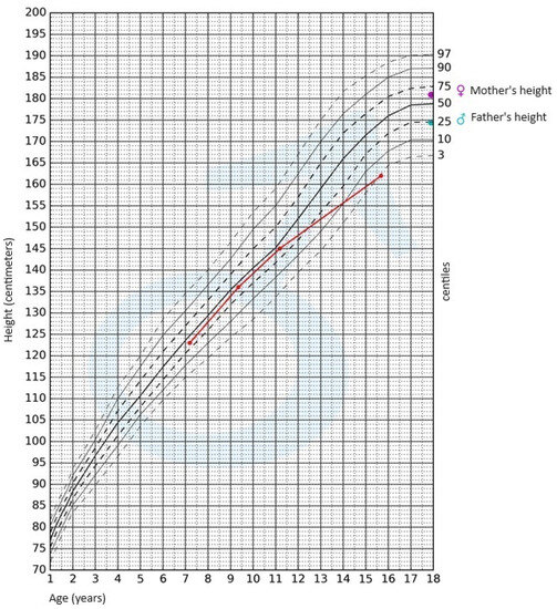
A 30-year-old female with a history of mental retardation was admitted to our emergency department (ER) with progressive weakness and fatigue. Upon taking the history, she had been having menstrual irregularities, progressive weight gain, and generalized weakness, which was significant enough to limit her physical activity and hinder her movement for the past three months. Initial vital signs showed that the patient had a body temperature of 37°C, a pulse rate of 90 beats per minute, and a blood pressure of 130/80 mmHg. On physical examination, the patient had a moon face with supraclavicular fullness, dorsocervical fat pad, purple abdominal striae, facial signs of hirsutism, and acne all over the face, shoulders, chest, and back.

Investigations

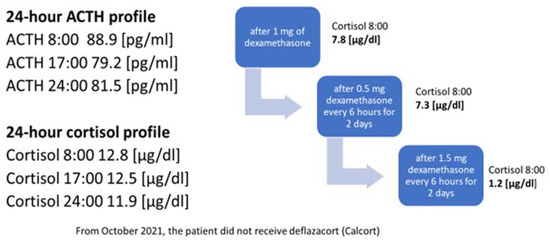
In the initial laboratory examination, hypokalemia of 2.1 mEq/L, hyperglycemia of 12.1 mmol/L, and metabolic alkalosis were detected (Table 1). The cortisol level after 1 mg dexamethasone suppression test was 2204 nmol/L (normal range 140-690), ACTH 123 pg/mL (normal range 7.2-63.3), DHEA-S 27.85 umol/L (normal range 2.6-13.9), And 24-hour urine cortisol level was 1560 mg/day (normal range 30-350) (Table 1). No suppression was observed in cortisol level with 8 mg dexamethasone suppression test.

Parameter Initial presentation Perforation presentation Refrence range
Na+ 143 mEq/L 139 mmol/L 135-147 mEq/L
Cl- 85 mEq/L 105 mmol/L 98-108 mEq/L
K+ 2.1 mEq/L 2.8 mmol/L 3.5-5.0 mEq/L
Mg2+ 0.79 mmol/L 0.77 mmol/L 0.85-1.110 mmol/L
PO3- 0.88 mmol/L 1.23 mmol/L 0.97-1.46 mmol/L
PH 7.54 7.36 7.35-7.45
PCO2 67.5 mmHg 42.7 mmHg 35-45 mmHg
PO2 27.7 mmHg 62.2 mmHg 75-100 mmHg
HCO3 49.8 mEq/L 23.6 mEq/L 22-26 mEq/L
Random blood glucose 12.1 mmol/L 24.1 mmol/L <5.5 mmol/L
Hemoglobin 13.5 g/dL 14.9 g/dL 13.7-16.8 g/dL
White blood cells 9,720 /uL 11,100 /uL 3,300-8,600 /uL
Lymphocyte 0.48% 0.33%
Neutrophil 8.55% 9.66%
Eosinophil 0.0% 0.0%
TSH 0.55 mIU/L Was not ordered 0.4-4.0 mIU/L
Cortisol 2204 nmol/L 4842 nmol/L 140-690 nmol/L
ACTH 123 pg/mL Was not ordered 7.2-63.3 pg/mL
Table 1: Laboratory findings on initial presentation and on perforation day

TSH – thyroid stimulating hormone; ACTH – adrenocorticotropic hormone

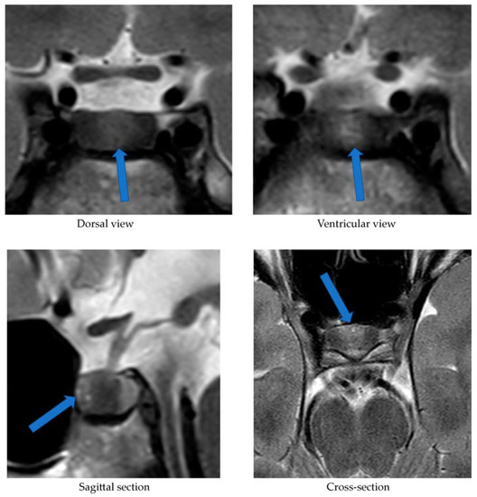
A series of CT scans for the neck, chest, abdomen, and pelvis was performed and failed to localize any tumors acting as an ectopic source. A pituitary MRI was performed, and no adenoma was found. To complete the diagnostic workup, we decided to do an inferior petrosal sinus sampling (IPSS) and PET scan with Gallium 68; however, the patient’s family refused and requested discharge and outpatient follow-ups. These results, together with the biochemical and clinical findings, supported the diagnostic hypothesis of ACTH-dependent Cushing’s syndrome.

Treatment/management

When addressing the issue of hypokalemia that the patient presented with initially, it was found to be resistant and difficult to correct. The patient was put on spironolactone 50 mg BID, and potassium chloride 20 mEq q8h, and her potassium level barely reached 3.5 mmol/L after several days. In addition, her magnesium level was corrected with magnesium oxide 800 mg every six hours. Her blood glucose level was controlled with insulin glargine 6 units daily and Novorapid as per the sliding scale. The patient was discharged on spironolactone tablets 50 mg BID (oral), potassium chloride 20 mEq q8h, cholecalciferol, calcium carbonate, insulin glargine 6 units daily, and Novorapid 4 units TID before meals.

Follow-up and outcomes

Seven days after discharge, she presented to the ER complaining of a new onset of abdominal pain, constipation, and reduced urine output. Her Glasgow Coma Scale (GCS) was 15, her blood pressure measurement was 146/90 mmHg, her pulse rate was 66 beats per minute, her respiratory rate was 21 breaths per minute, and her temperature was 36.7°C. Upon physical examination, the patient had distended non-tender abdomen without any other significant findings. Blood work was done, including renal functions, and all parameters, including potassium, were within normal limits. A chest X-ray was also performed and revealed no evidence of pneumoperitoneum. The patient was clinically stable after managing her abdominal pain with acetaminophen injection and administering fleet enema for constipation. After instructions on when to come again to the ER were given, the patient was discharged home on lactulose and paracetamol, and a close outpatient follow-up appointment was scheduled.

Five days after the ER visit, the patient presented again to the ER. She was still complaining of severe non-resolving abdominal pain, constipation, and reduced urine output. Upon physical examination in the ER, the patient was found to have developed a new onset of lower limb edema, abdominal rebound tenderness, and abdominal rigidity and guarding. She was hypotensive with a blood pressure of 91/46 mmHg, pulse rate of 80 beats per minute, respiratory rate of 16 breaths per minute, temperature of 38.2 °C, and SpO2 of 96%. The only significant laboratory finding was her potassium level dropping low to 2.8 mEq/L (Table 1). An X-ray of the chest was requested and showed a large pneumoperitoneum (Figure 1).

Posteroanterior-chest-X-ray-at-the-time-of-gastric-perforation-displaying-severe-air-under-the-diaphragm-with-bilateral-obstruction-indicating-massive-pneumoperitoneum-(red-arrow)
Figure 1: Posteroanterior chest X-ray at the time of gastric perforation displaying severe air under the diaphragm with bilateral obstruction indicating massive pneumoperitoneum (red arrow)

Abdominal CT was also urgently performed and confirmed the presence of gastric perforation likely related to an underlying perforated peptic ulcer with 0.8 cm defect at the distal greater curvature (Figures 23).

Coronal-section-CT-image-of-abdomen-and-pelvis-at-the-time-of-gastric-perforation-showing-features-of-gastric-perforation-likely-related-to-the-underlying-perforated-peptic-ulcer-with-0.8-cm-defect-at-the-distal-greater-curvature-
Figure 2: Coronal-section CT image of abdomen and pelvis at the time of gastric perforation showing features of gastric perforation likely related to the underlying perforated peptic ulcer with 0.8 cm defect at the distal greater curvature
Horizontal-section-CT-image-showing-features-of-gastric-perforation-likely-related-to-the-underlying-perforated-peptic-ulcer-with-0.8-cm-defect-at-the-distal-greater-curvature
Figure 3: Horizontal-section CT image showing features of gastric perforation likely related to the underlying perforated peptic ulcer with 0.8 cm defect at the distal greater curvature

The patient underwent an emergent gastric wedge resection for gastric perforation, and the pathology reported evidence of gastric ulcer with no evidence of malignancy. Furthermore, Helicobacter pylori test was performed on the sample, and it came back positive. The patient tolerated the surgery very well, and postoperative recovery was without any complications.

Later, the patient was prescribed metyrapone 250 mg Q4h, which was then increased to 500 mg Q4h four days after surgery, and her cortisol level significantly dropped to 634nmol/L. During that time, a gastrin level test was also performed to exclude the presence of gastrinomas, and the level was 45 pg/ml (normal range 13-115).

Discussion

A small percentage of the population suffers from Cushing’s syndrome, which is an endocrine disorder characterized by an endogenous overproduction of glucocorticoids, resulting in hypercortisolemia [1]. It is estimated to affect 0.7 to 2.4 people per million annually [1]. Hypercortisolemia alters psychologic, metabolic, and cardiovascular functions, resulting in increased mortality and morbidity rates, particularly if the diagnosis is delayed and long-term exposure to high cortisol levels occurs [2]. Women are more likely to suffer from this condition than men, and people in their 40s to 60s are most vulnerable to it [1]. Patients initially present with a wide range of symptoms, including weight gain, proximal myopathy, skin thinning, and abdominal striae [1]. Additionally, several metabolic disorders, such as diabetes mellitus, hypertension, and dyslipidemia, can occur [1]. Due to the rarity of this condition, there is often a significant delay in diagnosis and treatment, which could eventually lead to complications from prolonged hypercortisolism.

From another standpoint, in a systematic review, the incidence of peptic ulcer perforation ranges from 3.8 to 14 per 100,000 individuals in the general population [18]. In under-developed countries, patients are typically young, tobacco-using males [19]. However, patients in industrialized countries are typically older with multiple co-morbidities and are on long-term non-steroidal anti-inflammatory drugs (NSAIDs) or steroid use [19]. Patients may present with an abrupt onset of abdominal discomfort, abdominal rigidity, and tachycardia in the early stages of a perforated peptic ulcer [19]. Later, abdominal distention, pyrexia, hypotension, fever, and vomiting can occur [19]. Furthermore, when the diagnosis is made early, a perforated ulcer often has a good prognosis. However, the risk of adverse events increases if there is a delay in the diagnosis [20]. Therefore, making an early detection through different imaging modalities is crucial [20]. A history of peptic ulcer disease, NSAIDs, physiological stress, smoking, corticosteroids, and Helicobacter pylori are some of the well-established risk factors for a perforated peptic ulcer [20].

The prevalence of Helicobacter pylori among Saudi patients is high; in one study, the overall prevalence was 46.5% in patients with dyspepsia using gastric biopsy [21]. Several studies have explored the relationship between Helicobacter pylori and gastrointestinal perforation, but the results have been mixed. Some studies have suggested a higher prevalence of Helicobacter pylori infection among individuals with gastrointestinal perforation compared to those without, indicating a potential association. However, other studies have found no significant difference in the prevalence of Helicobacter pylori infection between perforated and non-perforated gastrointestinal ulcer cases [22]. Furthermore, they suggested that the presence of other risk factors like the use of NSAIDs, smoking, and alcohol may interact with Helicobacter pylori infection and contribute to the development of complications such as gastrointestinal perforation [22]. However, in our case, the patient did not have any established risk factors for gastric perforation, such as NSAIDs, smoking, or alcohol. Therefore, considering the low incidence of gastrointestinal perforation and high prevalence of Helicobacter pylori, the conflicting data regarding the association between Helicobacter pylori and gastrointestinal perforation, and the lack of established risk factors for gastrointestinal perforation in our patient, we suggest that prolonged excess glucocorticoids from Cushing’s syndrome may have contributed to the gastric perforation either independently or synergistically with Helicobacter pylori since hypercortisolism can lead to a weakened gastrointestinal wall integrity due to decreased collagen turnover and disruption of mucosal protection by prostacyclin [15]. In addition, because of hypercortisolism, perforation may not be contained or healed initially due to the immunosuppressive effects of hypercortisolism, whether endogenous or exogenous [15]. Additionally, high levels of cortisol may delay the diagnosis and treatment since it may mask the symptoms of the perforation [14]. Moreover, our patient was treated for severe hypokalemia with potassium supplementation for an extended period of time. Previous studies have linked potassium chloride supplementation to gastrointestinal ulceration and perforation, making this a possible additive cause of our patient’s condition [23,24].

A limited number of studies have addressed gastrointestinal perforations associated with endogenous hypercortisolemia [5-17]. The correlation between Cushing’s syndrome and gastrointestinal perforation is highlighted in our study and in the case reports that have been previously published (Table 2). Similar to our case, a female predominance was seen in gastrointestinal perforation among the reported cases of Cushing’s syndrome [6,7,12,13,15,16]. Additionally, the average age at which gastrointestinal perforation occurred in patients with endogenous hypercortisolism ranged from 45 to 80, which is a noticeably higher age range than the case we are presenting here (aged 30) [6-10,12]. Furthermore, unlike our case, in which gastrointestinal perforation occurred four months after the onset of Cushing’s symptoms, Intestinal perforation occurs approximately 9.8 months after Cushing’s symptoms first appear [15]. Furthermore, in our patient, gastric perforation occurred while she was hypercortisolemic and not in a remission state. Hence, in association with Helicobacter pylori infection, severe hypercortisolemia could have been a secondary contributing factor to gastric perforation. The complications of gastric ulceration, specifically with endogenous Cushing’s syndrome, have been addressed in two case reports [25,26]. It must be noted, however, that neither case is similar to ours. A case of gastric perforation was reported by Kubicka et al. in a patient who had a confirmed diagnosis of gastrinoma, and the patient was diagnosed with ectopic Cushing’s syndrome seven months after gastric perforation [25]. Therefore, since ectopic Cushing’s syndrome was diagnosed seven months after the perforation, it is more likely that the gastrinoma contributed to this complication. In contrast, our patient’s serum gastrin level was within the normal range, ruling out gastrinoma. Further, Hoshino et al. reported a case of gastrointestinal bleeding in a 39-year-old man with a confirmed diagnosis of Cushing’s disease secondary to pituitary adenoma [26]. He was found to have gastric ulceration and bleeding along with Helicobacter pylori infection and elevated cortisol levels [26]. In spite of the patient not developing a gastric perforation, it was suggested by the author that hypercortisolism might be a contributing factor for gastric ulcer complications by slowing down the ulcer healing process [26]

Reference Year of publication Age, gender Highest cortisol level plasma cortisol (PC, nmol/L) / UFC (nmol/L) Cause of Cushing’s syndrome Time from onset of Cushing’s symptoms to perforation (months) Reported site of gastrointestinal perforation
Current 2023 30, Female PC 4842 ACTH-dependant 4 Gastric perforation
Ishinoda et al. [17] 2023 24, Male PC 1647 Cushing’s disease 12 Sigmoid colon perforation
Wijewickrama et al. [16] 2021 32, Female PC 1147 Pituitary microadenoma 1 Diverticular perforation
Shahidi et al. [15] 2019 72, Female UFC 5296 Pancreatic neuroendocrine tumor 12 Diverticular perforation
Shahidi et al. [15] 2019 61, Female PC 1925 Metastatic medullary carcinoma of thyroid 12 Sigmoid colon and diverticular perforation
Shahidi et al. [15] 2019 68, Female UFC 410 Cushing’s disease 12 Sigmoid colon perforation
Shahidi et al. [15] 2019 71, Female UFC 1533 Cushing’s disease 4 Diverticular perforation
Shahidi et al. [15] 2019 54, Male UFC 374 Cushing’s disease 3 Sigmoid colon perforation
Shahidi et al. [15] 2019 52, Female UFC 885 Cushing’s disease 16 Diverticular perforation
Sater et al. [14] 2018 80, Female UFC 5601 Lung carcinoid 36 Diverticular perforation
Sater et al. [14] 2018 60, Female UFC 72726 Metastatic islet cell carcinoma 36 Diverticular perforation
Sater et al. [14] 2018 31, Male UFC 1297 Cushing’s disease 20 Diverticular perforation
Sater et al. [14] 2018 52, Female UFC 2371 Lung carcinoid 4 Diverticular perforation
Sater et al. [14] 2018 67, Male UFC 3836 Ectopic ACTH 10 Diverticular perforation
Sater et al. [14] 2018 51, Male UFC 13552 Metastatic thymic carcinoma 4 Diverticular perforation
Kaya et al. [9] 2016 70, Male PC 1432 Small cell lung cancer 1 Diverticular perforation
Dacruz et al. [12] 2016 60, Female UFC 4481 Metastatic parotid tumor 5 Sigmoid colon and diverticular perforation
Matheny et al. [10] 2016 67, Male UFC 11119 Metastatic medullary carcinoma of thyroid 4 Diverticular perforation
Flynn et al. [13]   2016 63, Female UFC 12465 Pheochromocytoma 1 Perforation at the splenic flexure
Balestrieri et al. [11] 2016 75, Male PC 2272 Neuroendocrine tumor 1 Intestinal perforation
Hara et al, [8] 2013 79, Male PC 1230 Cushing’s disease 6 Diverticular perforation
De Havenon et al. [7] 2011 71, Female PC 2593 Cushing’s disease 9 Diverticular perforation
Lutgers et al. [6] 2010 55, Female UFC 10152 Right pheochromocytoma 1 Sigmoid colon and diverticular perforation
Drake et al. [5] 1998 35, Male PC 1442 Islet cell tumor 4 Duodenal perforation and rupture of pancreatic pseudocyst
Table 2: Current case and previous reported 23 cases of patients with Cushing’s syndrome and gastrointestinal perforation

UFC – urinary free cortisol; PC – plasma cortisol; ACTH – adrenocorticotropic hormone

Conclusions

A high blood cortisol level can be associated with various clinical manifestations and diverse sets of complications. This case report sheds light on one of the less common complications of hypercortisolism in patients with Cushing’s syndrome, which is gastrointestinal perforation. Our report further supports the published evidence that gastrointestinal perforation is a rare but potentially fatal complication among patients with Cushing’s syndrome. Moreover, it highlights the possibility of developing gastric perforations in this patient group, even at younger ages than expected. This should elicit a high clinical suspicion and demand prompt investigation of Cushing’s syndrome patients in a hypercortisolism state presenting with modest gastrointestinal symptoms.

References

  1. Pivonello R, De Martino MC, De Leo M, Lombardi G, Colao A: Cushing’s syndrome. Endocrinol Metab Clin North Am. 2008, 37:135-49. 10.1016/j.ecl.2007.10.010
  2. Newell-Price J, Bertagna X, Grossman AB, Nieman LK: Cushing’s syndrome. Lancet. 2006, 367:1605-17. 10.1016/S0140-6736(06)68699-6
  3. Goethals L, Nieboer K, De Smet K, De Geeter E, Tabrizi NH, Van Eetvelde E, de Mey J: Cortisone associated diverticular perforation. JBR-BTR. 2011, 94:348-9. 10.5334/jbr-btr.705
  4. Piekarek K, Israelsson LA: Perforated colonic diverticular disease: the importance of NSAIDs, opioids, corticosteroids, and calcium channel blockers. Int J Colorectal Dis. 2008, 23:1193-7. 10.1007/s00384-008-0555-4
  5. Drake WM, Perry LA, Hinds CJ, Lowe DG, Reznek RH, Besser GM: Emergency and prolonged use of intravenous etomidate to control hypercortisolemia in a patient with Cushing’s syndrome and peritonitis. J Clin Endocrinol Metab. 1998, 83:3542-4. 10.1210/jcem.83.10.5156
  6. Lutgers HL, Vergragt J, Dong PV, de Vries J, Dullaart RP, van den Berg G, Ligtenberg JJ: Severe hypercortisolism: a medical emergency requiring urgent intervention. Crit Care Med. 2010, 38:1598-601. 10.1097/CCM.0b013e3181e47b7a
  7. de Havenon A, Ehrenkranz J: A perforated diverticulum in Cushing’s disease. Int J Surg Case Rep. 2011, 2:215-7. 10.1016/j.ijscr.2011.06.009
  8. Hara T, Akutsu H, Yamamoto T, Ishikawa E, Matsuda M, Matsumura A: Cushing’s disease presenting with gastrointestinal perforation: a case report. Endocrinol Diabetes Metab Case Rep. 2013, 2013:130064. 10.1530/EDM-13-0064
  9. Kaya T, Karacaer C, Açikgöz SB, Aydemir Y, Tamer A: Severe hypokalaemia, hypertension, and intestinal perforation in ectopic adrenocorticotropic hormone syndrome. J Clin Diagn Res. 2016, 10:OD09-11. 10.7860/JCDR/2016/17198.7127
  10. Matheny LN, Wilson JR, Baum HB: Ectopic ACTH production leading to diagnosis of underlying medullary thyroid carcinoma. J Investig Med High Impact Case Rep. 2016, 4:2324709616643989. 10.1177/2324709616643989
  11. Balestrieri A, Magnani E, Nuzzo F: Unusual Cushing’s syndrome and hypercalcitoninaemia due to a small cell prostate carcinoma. Case Rep Endocrinol. 2016, 2016:6308058. 10.1155/2016/6308058
  12. Dacruz T, Kalhan A, Rashid M, Obuobie K: An ectopic ACTH secreting metastatic parotid tumour. Case Rep Endocrinol. 2016, 2016:4852907. 10.1155/2016/4852907
  13. Flynn E, Baqar S, Liu D, et al.: Bowel perforation complicating an ACTH-secreting phaeochromocytoma. Endocrinol Diabetes Metab Case Rep. 2016, 2016:10.1530/EDM-16-0061
  14. Sater ZA, Jha S, McGlotten R, Hartley I, El Lakis M, Araque KA, Nieman LK: Diverticular perforation: A fatal complication to forestall in Cushing syndrome. J Clin Endocrinol Metab. 2018, 103:2811-4. 10.1210/jc.2018-00829
  15. Shahidi M, Phillips RA, Chik CL: Intestinal perforation in ACTH-dependent Cushing’s syndrome. Biomed Res Int. 2019, 2019:9721781. 10.1155/2019/9721781
  16. Wijewickrama PS, Ratnasamy V, Somasundaram NP, Sumanatilleke M, Ambawatte SB: A challenging case of Cushing’s disease complicated with multiple thrombotic phenomena following trans-sphenoidal surgery; a case report. BMC Endocr Disord. 2021, 21:29. 10.1186/s12902-021-00701-0
  17. Ishinoda Y, Uto A, Meshino H, et al.: Bowel perforation associated with Cushing’s disease: a case report with literature review. Endocr J. 2023, 70:933-9. 10.1507/endocrj.EJ23-0110
  18. Lau JY, Sung J, Hill C, Henderson C, Howden CW, Metz DC: Systematic review of the epidemiology of complicated peptic ulcer disease: incidence, recurrence, risk factors and mortality. Digestion. 2011, 84:102-13. 10.1159/000323958
  19. Chung KT, Shelat VG: Perforated peptic ulcer – an update. World J Gastrointest Surg. 2017, 9:1-12. 10.4240/wjgs.v9.i1.1
  20. Weledji EP: An overview of gastroduodenal perforation. Front Surg. 2020, 7:573901. 10.3389/fsurg.2020.573901
  21. Akeel M, Elmakki E, Shehata A, Elhafey A, Aboshouk T, Ageely H, Mahfouz MS: Prevalence and factors associated with H. pylori infection in Saudi patients with dyspepsia. Electron Physician. 2018, 10:7279-86. 10.19082/7279
  22. Thirupathaiah K, Jayapal L, Amaranathan A, Vijayakumar C, Goneppanavar M, Nelamangala Ramakrishnaiah VP: The association between Helicobacter pylori and perforated gastroduodenal ulcer. Cureus. 2020, 12:e7406. 10.7759/cureus.7406
  23. Farquharson-Roberts MA, Giddings AE, Nunn AJ: Perforation of small bowel due to slow release potassium chloride (slow-K). Br Med J. 1975, 3:206. 10.1136/bmj.3.5977.206
  24. Payan H, Blaustein A: Potassium chloride and small bowel perforation. Gastroenterology. 1965, 48:877-8. 10.1016/S0016-5085(65)80073-7
  25. Kubicka E, Zawadzka K, Syrycka J, Kałużny M, Pawluś A, Bolanowski M: A case of gastrinoma associated with ectopic Cushing syndrome. Pol Arch Intern Med. 2020, 130:328-9. 10.20452/pamw.15201
  26. Hoshino C, Satoh N, Narita M, Kikuchi A, Inoue M: Another ‘Cushing ulcer’. BMJ Case Rep. 2011, 2011:10.1136/bcr.02.2011.3888

From https://www.cureus.com/articles/196132-adrenocorticotropic-hormone-dependent-cushings-syndrome-complicated-with-gastric-ulcer-perforation-in-a-30-year-old-saudi-female-a-case-report-and-a-review-of-the-literature#!/

A Case Report of Cushing’s Disease Presenting With Psychosis and Muscle Weakness Postpartum

Abstract

Cushing’s syndrome is a condition leading to overproducing of cortisol by the adrenal glands. If the pituitary gland overproduces cortisol, it is called Cushing’s disease. Cushing’s syndrome and even Cushing’s disease during and after pregnancy are rare events. There is not enough literature and guidance for managing and treating these patients. The diagnosis of Cushing’s syndrome in pregnancy is often delayed because the symptoms overlap. We presented a thin 31-year-old woman, admitted 2 months after a normal-term delivery, with an atypical presentation of Cushing’s disease, unusual clinical features, and a challenging clinical course. She had no clinical discriminatory features of Cushing’s syndrome. Given that the patient only presented with psychosis and proximal myopathy and had an uncomplicated pregnancy, our case was considered unusual. The patients also had hyperpigmentation and severe muscle weakness which are among the less common presentations of Cushing’s syndrome. Our findings suggest that an early diagnosis of Cushing’s disease is important in pregnancy period for its prevalent fetal and maternal complications, and it should be treated early to optimize fetal and maternal outcomes as there is an increasing trend toward live births in treated participants.

Introduction

Cushing’s syndrome is a condition that originates from excessive production of glucocorticoids. The condition is most common in women of childbearing age and is characterized by altered distribution of the adipose tissue to the central and upper regions of the trunk (central obesity and buffalo hump), face (moon face), capillary wall integrity (easy bruising), hyperglycemia, hypertension, mental status changes and psychiatric symptoms, muscle weakness, signs associated with hyperandrogenism (acne and hirsutism), and violaceous striae among other signs. Hypercortisolism and hyperandrogenism suppress the production of the pituitary gonadotropins, which in turn leads to menstrual irregularities and infertility.13 Moreover, the main common cause of developing Cushing’s syndrome is the use of exogenic steroid.3
Cushing’s disease is a form of Cushing’s syndrome with overproduction of adrenocorticotropic hormone (ACTH) due to pituitary adenoma. The diagnosis is made using clinical features and paraclinical tests including urinary free cortisol (UFC), serum ACTH, dexamethasone suppression tests (DSTs), pituitary magnetic resonance imaging (MRI), and sometimes by inferior petrosal sinus sampling (IPSS).4 Although women with Cushing’s disease are less likely to become pregnant, timely diagnosis and appropriate management are especially important during possible pregnancy, preventing neonatal and maternal complications and death. The diagnosis is challenging due to the overlap of the disease symptoms with the changes associated with a normal pregnancy. Moreover, the hormonal milieu during pregnancy has recently been proposed as a potential trigger for Cushing’s disease in some cases; hence, the term “pregnancy-associated Cushing’s disease” has been used for the disease in the recent literature. In this study, we presented a thin 31-year-old woman who was referred to our clinic 2 months after a normal delivery, with an atypical presentation of Cushing’s disease, unusual clinical features, and a challenging clinical course.

Case Presentation

Our patient was a 31-year-old woman who presented 2 months after the delivery of her second child. She had a history of type 2 diabetes mellitus and hypertension in the past 2 years prior to her presentation. She had been admitted to another center following an episode of falling and muscle weakness. Two weeks later, she was admitted to our center with an impression of pulmonary thromboembolism due to tachypnea, tachycardia, and dyspnea. During follow-up, she was found to have leukocytosis, hyperglycemia (random blood sugar: 415 mg/d; normal level: up to 180 mg/dL) and hypokalemic metabolic alkalosis (PH: 7.5, HCO3 [bicarbonate]: 44.7 mEq/L, paO2 [partial pressure of oxygen]: 73 mm Hg, pCO2: 51.7 mm Hg, potassium: 2.7 mEq/L [normal range: 3.5-5.1 mEq/L]), which was refractory to the treatment; therefore, an endocrinology consultation was first requested. On physical examination, the patient was agitated, confused, and psychotic. Her vital signs were: blood pressure 155/100 mm Hg, heart rate: 130 bpm, and respiratory rate: 22 bpm, temperature: 39°C. As it has shown in Figure 1A, her face is not typical for moon face of Cushing’s syndrome, but facial hirsutism (Figure 1A) and generalized hyperpigmentation is obvious (Figure 1A-C). She was a thin lady and had a normal weight and distribution of adiposity (Body Mass Index [BMI] = 16.4 kg/m2; weight: 40 kg, and height: 156 cm). Aside from thinness of skin, she did not have the cutaneous features of Cushing’s syndrome (e.g. purpura, acne, and violaceous striae) and did not have supraclavicular and dorsocervical fat pad (buffalo hump), or plethora. In other words, she had no clinical discriminatory features of Cushing’s syndrome despite the high levels of cortisol, as confirmed by severely elevated UFC (5000 μg/24 h and 8000 μg/24 h; normal level: 4-40 μg/24 h). In addition, as will be mentioned later, the patient had axonal neuropathy which is a very rare finding in Cushing’s syndrome.
Figure 1. Clinical finding of our case with Cushing’s disease. (A) Hirsutism, (B) muscle atrophy seen in proximal portion of lower limbs, and (C) hyperpigmentation specially on the skin of the abdominal region.

OPEN IN VIEWER

She had a markedly diminished proximal muscle force of 1 out of 5 across all extremities; the rest of the physical examinations revealed no significant abnormalities (Figure 1B). On the contrary, based on her muscle weakness, hirsutism, psychosis and hyperpigmentation and refractory hypokalemic alkalosis, hyperglycemia, and hypertension, Cushing’s syndrome was suspected; therefore, 24-hour UFC level was checked that the results showed a severely elevated urinary cortisol (5000 μg/24 h and 8000 μg/24 h; normal level: 4-40 μg/24 h). Serum ACTH level was also inappropriately elevated (45 pg/mL; normal range: 10-60 pg/mL). High-dose dexamethasone failed to suppress plasma cortisol level and 24-hour urine cortisol level. A subsequent pituitary MRI showed an 8-mm pituitary mass, making a diagnosis of Cushing’s disease more probable. Meanwhile, the patient was suffering from severe muscle weakness that did not improve after the correction of hypokalemia. Then, a neurology consultation was requested. The neurology team evaluated laboratory data as well as EMG (Electromyography) and NCV (Nerve Conduction Velocity) of the patient, and based on their findings, “axonal neuropathy” was diagnosed for her weakness; so they ruled out the other neuromuscular diseases. A 5-day course of intravenous immunoglobulin (IVIG) was started for her neuropathy; however, the treatment did not improve her symptoms and the patient developed fungal sepsis and septic shock. Therefore, she was processed with broad-spectrum antibiotics and antifungal agents and recovered from the infection.
Mitotane was started for the patient before definitive surgical treatment to suppress hormonal production due to her poor general condition. Despite the 8-mm size of the pituitary mass which is likely to be a source of ACTH, our patient was underweight and showed the atypical clinical presentation of Cushing’s disease, making us suspect an ectopic source for the ACTH. Therefore, a Gallium dotatate scan was performed to find any probable ectopic sources; however, the results were unremarkable. The patient underwent Trans-Sphenoidal Surgery (TSS) to resect the pituitary adenoma because it was not possible to perform IPSS in our center. Finally, the patient’s condition including electrolyte imbalance, muscle weakness, blood pressure, and hyperglycemia started to improve significantly. The pathologist confirmed the diagnosis of a corticotropic adenoma. Nevertheless, the patient suddenly died while having her meal a week after her surgery; most likely due to a thromboembolic event causing a cardiac accident.

Discussion

Our patient was significantly different from other patients with Cushing’s disease because of her atypical phenotype. She was unexpectedly thin and had psychosis, hyperpigmentation, proximal myopathy, axonal neuropathy and no clinical discriminatory features of Cushing’s syndrome such as central adiposity, dorsocervical or supraclavicular fat pad, plethora or striae. She had also a history of type 2 diabetes and hypertension 2 years before her admission. The patient was diagnosed with Cushing’s later. From what was presented, the patient did not know she had Cushing’s until after her delivery and despite the highly elevated UFC, and she completed a normal-term delivery. Given that she only presented with psychosis and proximal myopathy, her pregnancy was considered unusual. Her clinical features such as hyperpigmentation and severe muscle weakness are among less common presentations.5
11β-hydroxysteroid dehydrogenase type 1 (11-βHSD1) is an enzyme responsible for converting cortisone (inactive glucocorticoid) into cortisol (active). It is speculated that this enzyme has a role in obesity (Figure 2).6,7
Figure 2. The enzymatic actions of 11β-hydroxysteroid dehydrogenase on its substrate interconverting inactive and active glucocorticoid.

OPEN IN VIEWER

In a case reported by Tomlinson, a 20-year-old female was diagnosed with Cushing’s disease despite not having the classical features of the disease. It has been suggested that the mechanism is a partial defect in 11β-HSD1 activity and concomitant increase in cortisol clearance rate. Thus, the patient did not have a classic phenotype; the defect in the conversion of cortisone to cortisol rises cortisol clearance and protects the patient from the effects of cortisol excess. This observation may help explain individual susceptibility to the side effects of glucocorticoids.6
Further studies of Tomlinson et al showed that a deficit in the function of (and not a mutation related to) 11β-HSD2 might have been responsible for the absence of typical Cushing’s symptoms. 11-HSD2 keeps safe the mineralocorticoid receptor from excess cortisol. Mutation in the HSD11B2 gene explains an inherited form of hypertension, apparent mineralocorticoid excess syndrome, in which Cushing’s disease results in cortisol-mediated mineralocorticoid excess affecting the kidney and leads to both hypokalemia and hypertension.8
It is frequent in Cushing’s syndrome that the patients usually have no mineralocorticoid hypertension; however, it is still proposed that a defect in 11β-HSD1 can be responsible for the presence of mineralocorticoid hypertension in a subgroup of patients. In fact, 11β-HSD1 is expressed in several tissues like the liver, kidneys, placenta, fatty tissues and gonads,9 meaning that this enzyme may potentially affect the results of cortisol excess in Cushing’s syndrome/disease. Abnormality in the function of this enzyme could explain the absence of the symptoms like central obesity, easy bruising, and typical striae during Cushing’s disease. Several factors affect the action of glucocorticoids. In this regard, the impact of the different types and levels of impairment in glucocorticoid receptors have been highlighted in some studies, as it can lead to different levels of response to glucocorticoids10 as well as a variety in the symptoms observed in Cushing’s disease.
The predominant reaction of the NADP(H)-dependent enzyme 11-Tukey’s honestly significant difference (HSD)1 happens through the catalysis of the conversion of inactive cortisol into receptor-active cortisol. The reverse reaction is mediated through the unidirectional NAD-dependent 11-HSD type 2 (Figure 2).11
In another case reported by Ved V. Gossein, a 41-year-old female was evaluated for hirsutism and irregular menstrual cycles. Her BMI was 22.6 kg/m2. The patient had no signs or symptoms of overnight recurrent Cushing’s syndrome, the 48-hour DST failed to suppress cortisol levels, and 24-hour urinary cortisol levels were persistently elevated on multiple occasions. Adrenocorticotropic hormone levels were unreasonably normal, suggesting ACTH-dependent hypercortisolism. Despite these disorders, she had 2 children. Magnetic resonance imaging (MRI) of the pituitary did not show any abnormalities. Moreover, abdominal MRI did not show adrenal mass or enlargement. Genetic testing to determine glucocorticoid resistance syndrome showed no mutation.12
Primary generalized glucocorticoid resistance is a rare genetic disorder characterized by generalized or partial insensitivity of target tissues to glucocorticoids.1317 There is a compensatory increase in hypothalamic-pituitary activity due to decreased sensitivity of peripheral tissues to glucocorticoids systems.1317 Excessive ACTH secretion leads to high secretion of cortisol and mineralocorticoids and/or androgens. However, the clinical features of Cushing’s syndrome do not develop after resistance to the effects of cortisol. Generalized glucocorticoid resistance is a rare condition characterized by high cortisol levels but no scarring of Cushing’s syndrome.18
An important aspect of our case was her pregnancy. Our patient had a history of hypertension and diabetes type 2, 2 years before her presentation to our center that could be because of an undiagnosed Cushing’s disease. The patient’s pregnancy terminated 2 months prior the admission and she had a normal vaginal delivery. So, we suspect that she become pregnant while involved with the disease. Aside from focusing on how this can happen in a patient with such high levels of glucocorticoids, more attention should be paid to occurring pregnancy in the background of Cushing’s disease. In fact, up to 250 patients were reported, of which less than 100 were actively treated.1922
Cushing’s disease is associated with serious complications in up to 70% of the cases coinciding with pregnancy.21 The most frequent maternal complications reported in the literature are hypertension and impaired glucose tolerance, followed by preeclampsia, osteoporosis, severe psychiatric complications, and maternal death (in about 2% of the cases). Prematurity and intrauterine growth retardation account for the most prevalent fetal complications. Stillbirth, intrauterine deaths, intrauterine hemorrhage, and hypoadrenalism have also been reported.23 Early diagnosis is especially challenging during pregnancy because of many clinical and biochemical shared features of the 2 conditions.23,24 These features include an increase in ACTH production, corticosteroid-binding globulin (CBG) 1 level, level of cortisol (urinary, plasma and free), hyperglycemia, weight gain, and an increased chance for occurrence of bruising, hypertension (mistaken with preeclampsia), gestational diabetes mellitus, weight gain, and mood swings.3 There are some suggestions proposed in the studies that help in screening and differentiation of Cushing’s from the normal and abnormal effects of pregnancy and Cushing’s disease from Cushing’s syndrome in suspected pregnant patients. Contrary to Cushing’s syndrome, the nocturnal minimum level of cortisol is preserved in pregnancy.23,25 There is not yet a diagnostic cut-off determined on mentioned level; however, a few studies elucidate the evaluation of hypercortisolemia in a pregnant patient.2628
Urinary free cortisol, a measure that reflects the amount of free cortisol in circulation, normally increases during pregnancy, and it can increase up to 8 times the normal level with Cushing’s disease during the second and the third trimesters,23,29 which is a useful tool to evaluate cortisol levels in a suspected pregnant woman. Because the suppression of both UFC and plasma cortisol is decreased in pregnancy,23,30 a low-dose DST is not very helpful for screening Cushing’s disease in pregnant patients. However, a high-dose DST with a <80% cortisol suppression might only indicate Cushing’s disease.3,31 Thus, it helps differentiating between ectopic ACTH syndrome and Cushing’s disease.32 The use of high-dose DST can distinguish between adrenal and pituitary sources of CS in pregnancy. Owing to the limited evidence available and the lack of data on normal pregnancies, the use of corticotropin-releasing hormone (CRH), desmopressin, and high-dose DST in pregnancy is not recommended yet.33 More timely diagnosis as well as timely intervention may have saved the life of our patient.
To differentiate between ectopic ACTH syndrome and Cushing’s disease, adrenal imaging should be considered. For higher plasma levels, combined employment of CRH stimulation test and an 8-mg DST can be helpful.3 Bilateral inferior petrosal sinus sampling (B-IPSS) might be needed when the findings are not in accordance with other results, but it is recommended to perform B-IPSS only if the noninvasive studies are inconclusive and only if there is enough expertise, experience, and technique for its performance.3
Although axonal neuropathy has been reported as a rare syndrome associated with paraneoplastic ectopic Cushing’s syndrome and exogenous Cushing’s syndrome, its association with Cushing’s disease has not been reported.5,32 Our patient had severe muscle weakness that we initially attributed it to myopathy and hypokalemia associated with Cushing’s syndrome. In our study, the diagnosis of axonal neuropathy was made based on electrophysiological studies by a neurology consultant and then IVIG was administered; however, the patient’s weakness did not improve after this treatment. The co-occurrence of Guillain-Barré syndrome which may also be classified as axonal neuropathy has also been reported in a pregnant woman with ectopic Cushing’s syndrome.34,35 Whether this finding is coincidental or the result of complex immune reactions driven by Cushing’s disease, or the direct effect of steroids, these results cannot be deduced from current data.36 Some data suggest that the fluctuations and inferior petrosal sinus sampling may trigger the flare of autoimmune processes, specifically when the cortisol levels start to decline during the course of Cushing’s syndrome.35,8 Also, due to COVID-19 pandemic affecting vital organs like kidney, paying attention to COVID-19 is suggested.3740

Conclusions

We presented a thin young female with psychosis, proximal myopathy, and axonal neuropathy with Cushing’s disease who had a recent pregnancy that was terminated without any fetal or maternal complications despite the repeated elevated serum cortisol and 24-hour UFC; therefore, we suggest that she might have glucocorticoid resistance. Glucocorticoid resistance is a rare disease in which the majority, but not all, of patients have a genetic mutation in the hGR-NR3C1 gene. As we did not perform genetic testing for our patient, the data are lacking.
Another clue to the absence of the classic Cushing’s disease phenotype in our case is the role of isoenzymes of 11-HSD1 and 11-HSD2. Other mechanisms, such as the defect somewhere in the glucocorticoid pathway of action such as a decreased number of receptors, a reduction in ligand affinity, or a postreceptor defect, play an important role in nonclassical clinical manifestations of Cushing’s syndrome.

Acknowledgments

The authors thank the patient for allowing us to publish this case report. The authors show their gratitude to the of the staff of the Rasool Akram Medical Complex Clinical Research Development Center (RCRDC) specially Mrs. Farahnaz Nikkhah for its technical and editorial assists.

Ethics Approval

Our institution does not require ethical approval for reporting individual cases or case series.

Informed Consent

Written informed consent was obtained from the patient and for her anonymized information to be published in this article.

Declaration of Conflicting Interests

The author(s) declared no potential conflicts of interest with respect to the research, authorship, and/or publication of this article.

Funding

The author(s) received no financial support for the research, authorship, and/or publication of this article.

References

1. Guilhaume B, Sanson ML, Billaud L, Bertagna X, Laudat MH, Luton JP. Cushing’s syndrome and pregnancy: aetiologies and prognosis in twenty-two patients. Eur J Med. 1992; 1(2):83-89.
2. Lin W, Huang HB, Wen JP, et al. Approach to Cushing’s syndrome in pregnancy: two cases of Cushing’s syndrome in pregnancy and a review of the literature. Ann Transl Med. 2019; 7(18):490.
3. Vilar L, Freitas MdC, Lima LHC, Lyra R, Kater CE. Cushing’s syndrome in pregnancy: an overview. Arq Bras Endocrinol Metabol. 2007;51(8):1293-1302.
4. Pecori Giraldi F, Cavallo LM, P, et al. The role of inferior petrosal sinus sampling in ACTH-dependent Cushing’s syndrome: review and joint opinion statement by members of the Italian Society for Endocrinology, Italian Society for Neurosurgery, and Italian Society for Neuroradiology. Neurosurg Focus. 2015;38(2):E5.
5. Molina Garrido MJ, Guillén Ponce C, Maciá Escalante S, Pons Sanz V, Carrato Mena A. Cushing’s paraneoplastic syndrome as first manifestation of an adenocarcinoma of unknown origin. Clin Transl Oncol. 2006;8(8):621-623.
6. Tomlinson JW, Draper N, Mackie J, et al. Absence of Cushingoid phenotype in a patient with Cushing’s disease due to defective cortisone to cortisol conversion. J Clin Endocrinol Metab. 2002;87(1):57-62.
7. Kobayashi T, Matsumoto T, Kamata K. IGF-I-induced enhancement of contractile response in organ-cultured aortae from diabetic rats is mediated by sustained thromboxane A2 release from endothelial cells. J Endocrinol. 2005;186(2): 367-376.
8. Stewart PM. Tissue-specific Cushing’s syndrome, 11β-hydroxysteroid dehydrogenases and the redefinition of corticosteroid hormone action. Eur J Endocrinol. 2003;149:163-168.
9. Ricketts ML, Verhaeg JM, Bujalska I, et al. Immunohistochemical localization of type 1 11β-hydroxysteroid dehydrogenase in human tissues. J Clinl Endocrinol Metabol. 1998;83:1325-1335.
10. Huizenga NA, Koper JW, De Lange P, et al. A polymorphism in the glucocorticoid receptor gene may be associated with and increased sensitivity to glucocorticoids in vivo. J Clin Endocrinol Metab. 1998;83(1):144-151.
11. Hintzpeter J, Stapelfeld C, Loerz C, Martin HJ, Maser E. Green tea and one of its constituents, Epigallocatechine-3-gallate, are potent inhibitors of human 11β-hydroxysteroid dehydrogenase type 1. PLoS ONE. 2014;9(1):e84468.
12. Gossain VV, El-Rifai M, Krishnan P, Bhavsar B. Cushing’s syndrome with no clinical stigmata—a variant of glucocorticoid resistance syndrome. Clin Diabetes Endocrinol. 2018;4:23-25.
13. Charmandari E, Kino T, Ichijo T, Chrousos GP. Generalized glucocorticoid resistance: clinical aspects, molecular mechanisms, and implications of a rare genetic disorder. J Clin Endocrinol Metab. 2008;93(5):1563-1572.
14. Cidlowski JA, Malchoff CD, Malchoff DM. Glucocorticoid receptors, their mechanism of action, and glucocorticoid resistance. In: Jameson JL, De Groot LJ, eds. Endocrinology: Adult and Pediatric. Saunders; 2016:1717-1726.
15. Charmandari E, Kino T. Chrousos syndrome: a seminal report, a phylogenetic enigma and the clinical implications of glucocorticoid signaling changes. Eur J Clin Investig. 2010;40: 932-942.
16. Nicolaides NC, Charmandari E. Chrousos syndrome: from molecular pathogenesis to therapeutic management. Eur J Clin Invest. 2015;45(5):504-514.
17. Nicolaides N, Lamprokostopoulou A, Sertedaki A, Charmandari E. Recent advances in the molecular mechanisms causing primary generalized glucocorticoid resistance. Hormones. 2016;15(1): 23-34.
18. Huizenga NATM De Lange P, Koper JW, et al. Five patients with biochemical and/or clinical generalized glucocorticoid resistance without alterations in the glucocorticoid receptor gene. J Clin Endocrinol Metab. 2000;85:2076-2081.
19. Luger A, Broersen LHA, Biermasz NR, et al. ESE clinical practice guideline on functioning and nonfunctioning pituitary adenomas in pregnancy. Eur J Endocrinol. 2021;185: G1-G33.
20. Sridharan K, Sahoo J, Palui R, et al. Diagnosis and treatment outcomes of Cushing’s disease during pregnancy. Pituitary. 2021;24(5):670-680.
21. Caimari F, Valassi E, Garbayo P, et al. Cushing’s syndrome and pregnancy outcomes: a systematic review of published cases. Endocrine. 2017;55(2):555-563.
22. Lindsay JR, Jonklaas J, Oldfield EH, Nieman LK. Cushing’s syndrome during pregnancy: personal experience and review of the literature. J Clin Endocrinol Metab. 2005;90(5):3077-3083.
23. Lindsay JR, Nieman LK. The hypothalamic-pituitary-adrenal axis in pregnancy: challenges in disease detection and treatment. Endocr Rev. 2005;26(6):775-799.
24. Buescher MA, McClamrock HD, Adashi EY. Cushing syndrome in pregnancy. Obstetr Gynecol. 1992;79:130-137.
25. Carr BR, Parker CR Jr, Madden JD, et al. Maternal plasma adrenocorticotropin and cortisol relationships throughout human pregnancy. Am J Obstetr Gynecol. 1981;139:416-422.
26. Mellor A, Harvey RD, Pobereskin LH, Sneyd JR. Cushing’s disease treated by trans-sphenoidal selective adenomectomy in mid-pregnancy. Br J Anaesth. 1998;80(6):850-852.
27. Doshi S, Bhat A, Lim K. Cushing’s syndrome in pregnancy. J Obstetr Gynaecol. 2003;23:568-569.
28. Wood PJ, Barth JH, Freedman DB, Perry L, Sheridan B. Evidence for the low dose dexamethasone suppression test to screen for Cushing’s syndrome—recommendations for a protocol for biochemistry laboratories. Ann Clin Biochem. 1997;34(pt 3):222-229.
29. Lindsay JR, Jonklaas J, Oldfield EH, Nieman LK. Cushing’s syndrome during pregnancy: personal experience and review of the literature. J Clin Endocrinol Metab. 2005;90(5): 3077-3083.
30. Wallace C, Toth EL, Lewanczuk RZ, Siminoski K. Pregnancy-induced Cushing’s syndrome in multiple pregnancies. J Clin Endocrinol Metab. 1996;81(1):15-21.
31. Invitti C, Pecori Giraldi F, de Martin M, Cavagnini F. Diagnosis and management of Cushing’s syndrome: results of an Italian multicentre study. Study Group of the Italian Society of Endocrinology on the Pathophysiology of the Hypothalamic-Pituitary-Adrenal Axis. J Clin Endocrinol Metab. 1999;84(2):440-448.
32. Vilar L, Naves LA, Freitas MdC, et al. Endogenous Cushing’s syndrome: clinical and laboratorial features in 73 cases. Arq Bras Endocrinol Metabol. 2007;51(4):566-574.
33. Hamblin R, Coulden A, Fountas A, Karavitaki N. The diagnosis and management of Cushing’s syndrome in pregnancy. J Neuroendocrinol. 2022;34(8):e13118.
34. Bressler R, Johnson CT. Cushing’s syndrome and the Guillain-Barré syndrome. Ann Intern Med. 1959;50:1298-1303.
35. Moeindarbary S, Abbasi dalooei M, Ghahremani S, et al. Guillain-Barré syndrome following Cushing’s syndrome in a pregnant woman: a case report. Int J Pediatr. 2019;7:10651-10657.
36. Hasenmajer V, Sbardella E, Sciarra F, Minnetti M, Isidori AM, Venneri MA. The immune system in Cushing’s syndrome. Trends Endocrinol Metab. 2020;31(9):655-669.
37. Besharat S, Alamda NM, Dadashzadeh N, et al. Clinical and demographic characteristics of patients with COVID-19 who died in Modarres Hospital. Open Access Maced J Med Sci. 2020;8:144-149.
38. Lotfi B, Farshid S, Dadashzadeh N, Valizadeh R, Rahimi MM. Is Coronavirus Disease 2019 (COVID-19) associated with renal involvement? A review of century infection. Jundishapur J Microbiol. 2020;13:e102899.
39. Dadashzadeh N, Farshid S, Valizadeh R, Nanbakhsh M, Rahimi MM. Acute respiratory distress syndrome in COVId-19 disease. Immunopathol Persa. 2020;6:e16.
40. Petramala L, Olmati F, Conforti MG, et al. Autoimmune diseases in patients with Cushing’s syndrome after resolution of hypercortisolism: case reports and literature review. Int J Endocrinol. 2018;2018:1464967.


Ectopic Cushing’s Syndrome From a Corticotropin-Releasing Hormone-Secreting Medullary Thyroid Carcinoma: a Rare Pitfall af Inferior Petrosal Sinus Sampling

Abstract

Summary

This case report describes a rare presentation of ectopic Cushing’s syndrome (CS) due to ectopic corticotropin-releasing hormone (CRH) production from a medullary thyroid carcinoma (MTC).

The patient, a 69-year-old man, presented with symptoms of muscle weakness, facial plethora, and easy bruising.

An inferior petrosal sinus sampling test (IPSS) demonstrated pituitary adrenocorticotrophic hormone (ACTH) secretion, but a whole-body somatostatin receptor scintigraphy (68Ga-DOTATOC PET/CT) revealed enhanced uptake in the right thyroid lobe which, in addition to a grossly elevated serum calcitonin level, was indicative of an MTC. A 18F-DOPA PET/CT scan supported the diagnosis, and histology confirmed the presence of MTC with perinodal growth and regional lymph node metastasis.

On immunohistochemical analysis, the tumor cell stained positively for calcitonin and CRH but negatively for ACTH. Distinctly elevated plasma CRH levels were documented. The patient therefore underwent thyroidectomy and bilateral adrenalectomy.

This case shows that CS caused by ectopic CRH secretion may masquerade as CS due to a false positive IPSS test. It also highlights the importance of considering rare causes of CS when diagnostic test results are ambiguous.

Learning points

  • Medullary thyroid carcinoma may secrete CRH and cause ectopic CS.
  • Ectopic CRH secretion entails a rare pitfall of inferior petrosal sinus sampling yielding a false positive test.
  • Plasma CRH measurements can be useful in selected cases.

Background

The common denominator of Cushing’s syndrome (CS) is autonomous hypersecretion of cortisol (1) and it is subdivided into ACTH-dependent and ACTH-independent causes. The majority of CS cases are ACTH-dependent (80–85%) with a pituitary corticotroph tumor as the most prevalent cause (Cushing’s disease), and less frequently an ectopic ACTH-producing tumor (2). The gold standard method to ascertain the source of ACTH secretion in CS patients is inferior petrosal sinus sampling (IPSS) with measurement of plasma ACTH levels in response to systemic corticotropin-releasing hormone (CRH) stimulation (3). The IPSS has a very high sensitivity and specificity of 88–100% and 67–100%, respectively (4), but pitfalls do exist, including the rare ectopic CRH-producing tumor, which may yield a false positive test result (3). Here, we describe a very rare case masquerading as CS including a positive IPSS test.

Case presentation

A 69-year-old man presented at a local hospital with a 6-month history of progressive fatigue, muscle weakness and wasting, easy bruising, facial plethora, and fluid retention. His serum potassium level was 2.6 mmol/L (reference range: 3.5–4.2 mmol/L) without a history of diuretics use. His previous medical history included spinal stenosis, benign prostatic hyperplasia, and hypertension. An electromyography showed no sign of polyneuropathy and an echocardiography showed no signs of heart failure with an ejection fraction of 55%. MRI of the spine revealed multiple compression fractures, and the patient underwent spinal fusion and decompression surgery; during this admission he was diagnosed with type 2 diabetes (HbA1c: 55 mmol/mol). After spine surgery, the patient developed a pulmonary embolism and initiated treatment with rivaroxaban.

Establishing the diagnosis of ACTH-dependent CS

Six months after his spine surgery, the patient was referred to the regional department of endocrinology for osteoporosis management. Blood tests revealed a low serum testosterone level with non-elevated luteinizing hormone (LH) and follicle-stimulating hormone (FSH) levels (Table 1). An overnight 1 mg dexamethasone suppression test was positive with a morning cortisol level of 254 nmol/L and three consecutive 24-h urinary cortisol levels were markedly elevated with mean level of ≈600 nmol/24 h (reference range: 12–150 nmol/24 h). A single plasma ACTH was 37 ng/L (Table 1).

Table 1Baseline endocrine assessment.

Parameters Patient’s values Reference range
ACTH, ng/L 37 7–64
UFC, nmol/day 588 12–150
Urinary cortisol, nmol/L 600 171–536
OD, nmol/L 254 <50
Free testosterone, nmol/L 0.061 0.17–0.59
HbA1c, mmol/mol 55 <48
FSH, IU/L 7.4 1.2–15.8
LH, IU/L 2.2 1.7–8.6

ACTH, adrenocorticotropin; FSH, follicle-stimulating hormone; IU, international units; LH, luteinizing hormone; OD, plasma cortisol levels after a 1 mg overnight dexamethasone suppression test; UFC, urine free cortisol hormone.

Differential diagnostic tests

The patient was referred to a tertiary center for further examinations. Ketoconazole treatment was started to alleviate the consequences of hypercortisolism. A pituitary MRI revealed an intrasellar microtumor with a maximal diameter of 6 mm and an IPSS was ordered. A whole-body somatostatin receptor scintigraphy (68Ga-DOTATOC PET/CT) was also performed to evaluate the presence of a potential neuroendocrine tumor. This revealed multiple areas of enhanced uptake including the right thyroid lobe and cervical lymph nodes in the neck (with CT correlates), as well as in the duodenum (with no CT correlate). Concomitantly, a grossly elevated serum calcitonin level of 528 pmol/L (reference range <2.79 pmol/L) was measured.

Subsequently, the IPSS revealed pituitary ACTH secretion with a central-to-peripheral ACTH ratio >3 (Table 2). The right petrosal sinus was not successfully catheterized; thus, lateralization could not be determined.

To corroborate the diagnosis MTC, a 18F-DOPA PET/CT scan (FDOPA) was performed (5), which showed pathologically enhanced uptake in the right thyroid lobe and regional lymph nodes (Fig. 1). An ultrasound-guided core needle biopsy from the thyroid nodule was inconclusive; however, the patient underwent total thyroidectomy and regional lymph node resection, from which histology confirmed the diagnosis of disseminated MTC. Standard replacement with levothyroxine, calcium, and vitamin D was initiated. A blood sample was collected, and genomic DNA was extracted. The DNA analysis for RET germline mutation was negative.

Figure 1View Full Size
Figure 1
18F-DOPA PET/CT scan with pathologically enhanced uptake in the right thyroid lobe (large blue arrow on the left side) and regional lymph nodes (small blue arrows).

Citation: Endocrinology, Diabetes & Metabolism Case Reports 2023, 3; 10.1530/EDM-23-0057

Table 2Results from the inferior petrosal sinus sampling.*

Time (min) Left IPSS Peripheral L/P
-5 42 36 1.2
-1 116 33 3.5
2 120 32 3.8
5 209 28 7.5
7 180 43 4.2
10 529 34 15.6
15 431 37 11.6

*Data represents ACTH levels in ng/L. IPSS Inferior petrosal sampling ACTH Adrenocorticotropin hormone CRH Corticotropin-releasing hormone, L/P Ratio of left (L) inferior petrosal sinus to peripheral venous ACTH concentrations.

Pathology

Total thyroidectomy and bilateral cervical lymph node dissection (level six and seven) were performed. Macroscopic evaluation of the right thyroid lobe revealed a 24 mm, irregular solid yellow tumor. Microscopically the tumor showed an infiltrating architecture with pseudofollicles and confluent solid areas. Calcification was prominent, but no amyloid deposition was seen. The tumor cells were pleomorphic with irregular nuclei and heterogenic chromatin structure. No mitotic activity or necrosis was observed. On immunohistochemical analysis, the tumor cells expressed thyroid transcription factor 1 and stained strongly for carcinoembryonic antigen and calcitonin; tumor cells were focally positive for cytokeratin 19. The tumor was completely negative for ACTH, thyroid peroxidase, and the Hector Battifora mesothelial-1 antigen. Further analysis revealed positive immunostaining for CRH (Fig. 2). The Ki-67 index was very low (0–1%), indicating a low cellular proliferation. Molecular testing for somatic RET mutation was not performed.

Figure 2View Full Size
Figure 2
Histopathological findings and immunohistochemical studies of MTC. (A) Microscopic features of medullary thyroid carcinoma. (B) Polygonal tumor cells (hematoxylin and eosin, ×40). (C) Tumor cells stain for calcitonin (×20). (D) Immunohistochemical stain (×400) for CRH showing cells being positive (brown). (E) Pituitary tissue from healthy control staining positive for ACTH in comparison to (F) ACTH-negative cells MTC tissue from the patient (×20).

Citation: Endocrinology, Diabetes & Metabolism Case Reports 2023, 3; 10.1530/EDM-23-0057

No malignancy was found in the left thyroid lobe and there was no evidence of C-cell hyperplasia. Regional lymph node metastasis was found in 13 out of 15 nodes with extranodal extension.

Outcome and follow-up

Follow-up

Serum calcitonin levels declined after neck surgery but remained grossly elevated (118 pmol/L 3 weeks post surgery) and cortisol levels remained high. Ketoconazole treatment was poorly tolerated and not sufficiently effective. Plasma levels of CRH were measured by a competitive-ELISA kit (EKX-KIZI6R-96 Nordic BioSite), according to the instructions provided by the manufacturer. The intra- and interassay %CV (coefficient of variability) were below 8% and 10%, respectively, and the assay sensitivity was 1.4 pg/mL. The plasma CRH was distinctly elevated compared to in-house healthy controls both before and after thyroid surgery (Fig. 3).

Figure 3View Full Size
Figure 3
Plasma CRH levels before and after total thyroidectomy compared to three healthy controls.

Citation: Endocrinology, Diabetes & Metabolism Case Reports 2023, 3; 10.1530/EDM-23-0057

The patient subsequently underwent uneventful bilateral laparoscopic adrenalectomy followed by standard replacement therapy with hydrocortisone and fludrocortisone. The symptoms and signs of his CS gradually subsided. Pathology revealed bilateral cortical hyperplasia as expected.

The patient continues follow-up at the Department of Oncology and the Department of Endocrinology and Internal Medicine. At 13-month follow-up, 68Ga-DOTATOC shows residual disease with pathologically enhanced uptake in two lymph nodes, whereas the previously described focal DOTATOC uptake in the duodenum was less pronounced (still no CT correlate). Serum calcitonin was 93 pmol/L at the 13-month follow-up.

Discussion

Diagnostic challenges remain in the distinction between pituitary and ectopic ACTH-dependent CS, and several diagnostic tools are used in combination, none of which is infallible, including IPSS (6). Our case and others illustrate that ectopic CRH secretion may yield a false positive IPSS test result (3). Measurement of circulating CRH levels is relevant if an ectopic CRH producing tumor is suspected, but the assay is not routinely available in clinical practice (Lynnette K Nieman M. Measurement of ACTH, CRH, and other hypothalamic and pituitary peptides https://www.uptodate.com/contents/measurement-of-acth-crh-and-other-hypothalamic-and-pituitary-peptides: UpToDate; 2019). In our case, the presence of elevated plasma CRH levels after thyroidectomy strengthened the indication for bilateral adrenalectomy.

The most common neoplasm causing ectopic CS is small-cell lung cancer, whereas MTC accounts for 2–8% of ectopic cases (7). The development of CS in relation to MTC is generally associated with advanced disease and poor prognosis of an otherwise relatively indolent cancer (8), and the clinical progression of CS is usually rapid, why an early recognition and rapid control of hypercortisolemia and MTC is necessary to decrease morbidity and mortality (79). Our case does have residual disease; however, he remains progression-free with stable and relatively low calcitonin levels within 1-year follow-up.

Only a very limited number of cases of ectopic tumors with either combined ACTH and CRH secretion or isolated CRH secretion have been reported, with ectopic CRH secretion accounting for less than 1% of CS (9).

An ACTH- or CRH-producing tumor can be difficult to localize and may include gastric ACTH/CRH-secreting neuroendocrine tumors (9). In our case, the 68Ga-DOTATOC identified a possible duodenal site, in addition to the MTC, but an upper gastrointestinal endoscopy revealed no pathological findings and there was no CT correlate. Thus, we concluded that the most likely and sole source of ectopic CRH was the MTC and its metastases.

To our knowledge, no official guidelines for managing ectopic ACTH-dependent CS have been established. In a recent publication by Alba et al. (10), the authors demonstrated a clinical algorithm (The Mount Sinai Clinical Pathway, MSCP) and recommendation for the management of CS due to ectopic ACTH secretion.

Essentially, our approach in this particular case followed these recommendations, including source location by CT and 68Ga-DOTATATE PET/CT imaging, acute management with ketoconazole, and finally, bilateral adrenalectomy as curative MTC surgery was not possible. In retrospect, performance of the IPSS could be questioned in view of the MTC diagnosis. In real time, however, a pituitary MRI performed early in the diagnostic process revealed a microadenoma, which prompted the IPSS. In parallel, a somatostatin receptor scintigraphy (68Ga-DOTATOC PET/CT) was also done, which raised the suspicion of an MTC.

Conclusion

We report a very rare case of an ectopic CS caused by a CRH-secreting MTC. Although IPSS has stood the test of time in the differential diagnosis of ACTH-dependent CS, this case illustrates a rare pitfall.

Declaration of interest

The authors declare that there is no conflict of interest that could be perceived as prejudicing the impartiality of the research reported.

Funding

This research did not receive any specific grant from any funding agency in the public, commercial, or not-for-profit sector.

Patient consent

Written informed consent for publication of their clinical details was obtained from the patient.

Author contribution statement

JOJ and MJO are the physicians responsible for the patient. LR performed the thyroidectomy and bilateral adrenalectomy. SHM and SLA assessed and reassessed the histopathology and the immunohistochemical analysis. MB measured plasma CRH. VM, JOJ, and MJO drafted the manuscript. All authors contributed to critical revision of the manuscript.

References

Hormones and High Blood Pressure: Study Reveals Endocrine Culprits and Targeted Treatments

In a recent study published in Hypertension Research, scientists examine the endocrine causes of hypertension (HTN) and investigate the efficacy of treatments to alleviate HTN.


What is HTN?

About 30% of the global population is affected by HTN. HTN is a modifiable cardiovascular (CV) risk factor that is associated with a significant number of deaths worldwide.

There are two types of HTN known as primary and secondary HTN. As compared to primary HTN, secondary HTN causes greater morbidity and mortality.

The most common endocrine causes of HTN include primary aldosteronism (PA), paragangliomas and pheochromocytomas (PGL), Cushing’s syndrome (CS), and acromegaly. Other causes include congenital adrenal hyperplasia, mineralocorticoid excess, cortisol resistance, Liddle syndrome, Gordon syndrome, and thyroid and parathyroid dysfunction.

What is PA?

PA is the most common endocrine cause of hypertension, which is associated with excessive aldosterone secretion by the adrenal gland and low renin secretion. It is difficult to estimate the true prevalence of PA due to the complexity of its diagnosis.

Typically, the plasma aldosterone-to-renin ratio (ARR) is measured to diagnose PA. The diagnosis of PA can also be confirmed using other diagnostic tools like chemiluminescent enzyme immunoassays (CLEIAs) and radio immune assay (RIA).

Continuous aldosterone secretion is associated with organ damage due to chronic activation of the mineralocorticoid (MR) receptor in many organs, including fibroblasts and cardiomyocytes. An elevated level of aldosterone causes diastolic dysfunction, endothelial dysfunction, left ventricular hypertrophy, and arterial stiffness.

Increased aldosterone secretion also leads to obstructive sleep apnea and increases the risk of osteoporosis. This is why individuals with PA are at a higher risk of cardiovascular events (CVDs), including heart failure, myocardial infarction, coronary artery disease, and atrial fibrillation.

PA is treated by focusing on normalizing potassium and optimizing HTN and aldosterone secretion. Unilateral adrenalectomy is a surgical procedure proposed to treat PA.

Young patients who are willing to stop medication are recommended surgical treatment. The most common pharmaceutical treatment for PA includes mineralocorticoid receptor antagonists such as spironolactone and eplerenone.

Pheochromocytomas and paragangliomas

PGL are tumors that develop at the thoracic-abdominal-pelvic sympathetic ganglia, which are present along the spine, as well as in the parasympathetic ganglia located at the base of the skull. The incidence rate of PGL is about 0.6 for every 100,000 individuals each year. PGL tumors synthesize excessive catecholamines (CTN), which induce HTN.

Some of the common symptoms linked to HTN associated with PGL are palpitations, sweating, and headache. PGL can be diagnosed by determining metanephrines (MN) levels, which are degraded products of CTN. Bio-imaging tools also play an important role in confirming the diagnosis of PGL.

Excessive secretion of CTN increases the risk of CVDs, including Takotsubo adrenergic heart disease, ventricular or supraventricular rhythm disorders, hypertrophic obstructive or ischaemic cardiomyopathy, myocarditis, and hemorrhagic stroke. Excessive CTN secretion also causes left ventricular systolic and diastolic dysfunction.

Typically, PGL treatment is associated with surgical procedures. Two weeks before the surgery, patients are treated with alpha-blockers. For these patients, beta-blockers are not used as the first line of treatment without prior use of alpha-adrenergic receptors.

Patients with high CTN secretion are treated with metyrosine, as this can inhibit tyrosine hydroxylase. Hydroxylase converts tyrosine into dihydroxyphenylalanine, which is related to CTN synthesis.

What is CS?

CS, which arises due to persistent exposure to glucocorticoids, is a rare disease with an incidence rate of one in five million individuals each year. The most common symptoms of CS include weight gain, purple stretch marks, muscle weakness, acne, and hirsutism. A high cortisol level causes cardiovascular complications such as HTN, hypercholesterolemia, and diabetes.

CS is diagnosed based on the presence of two or more biomarkers that can be identified through pathological tests, such as salivary nocturnal cortisol, 24-hour urinary-free cortisol, and dexamethasone suppression tests.

CS is treated through surgical procedures based on the detected lesions. Patients with severe CS are treated with steroidogenic inhibitors, such as metyrapone, ketoconazole, osilodrostat, and mitotane. Pituitary radiotherapy and bilateral adrenalectomy are performed when other treatments are not effective.

Acromegaly

Acromegaly arises due to chronic exposure to growth hormone (GH), leading to excessive insulin-like growth factor 1 (IGF1) synthesis. This condition has a relatively higher incidence rate of 3.8 million person-years. Clinical symptoms of acromegaly include thickened lips, widened nose, a rectangular face, prominent cheekbones, soft tissue overgrowth, or skeletal deformities.

Prolonged exposure to GH leads to increased water and sodium retention, insulin resistance, reduced glucose uptake, and increased systemic vascular resistance. These conditions increase the risk of HTN and diabetes in patients with acromegaly. Acromegalic patients are also at a higher risk of cancer, particularly those affecting the thyroid and colon.

Acromegaly is diagnosed using the IGF1 assay, which determines IGF1 levels in serum. After confirming the presence of high IGF1 levels, a GH suppression test must be performed to confirm the diagnosis. Bioimaging is also conducted to locate adenoma.

Acromegaly is commonly treated through surgical procedures. Patients who refuse this line of treatment are treated with somatostatin receptor ligands, growth hormone receptor antagonists, dopaminergic agonists, or radiotherapy.

Journal reference:
  • De Freminville, J., Amar, L., & Azizi, M. (2023) Endocrine causes of hypertension: Literature review and practical approach. Hypertension Research; 1-14. doi:10.1038/s41440-023-01461-1

From https://www.news-medical.net/news/20231015/Hormones-and-high-blood-pressure-Study-reveals-endocrine-culprits-and-targeted-treatments.aspx

Osteoporosis as the First Sign of Cushing’s Disease in a Thin 16-Year-Old Boy

Abstract

Cushing’s disease (CD) is an extremely rare diagnosis in children. In this report, we present the case of an almost 16-year-old, short and thin boy with CD, the first symptoms of which were spinal pain and vertebral fractures as a result of osteoporosis. In light of his growth retardation and short stature, the boy underwent diagnostics, which excluded growth hormone (GH) deficiency, hypothyroidism and celiac disease. Finally, based on cortisol profile results, dexamethasone suppression tests and bilateral sampling during catheterization of the inferior petrosal sinuses, CD was diagnosed.

1. Introduction

Cushing’s disease (CD) is an extremely rare diagnosis in children; however, if it occurs, it is more likely to present in older children [1,2]. It is a type of ACTH-dependent Cushing’s syndrome (CS), in which the pituitary gland is the source of ACTH secretion. The highest incidence of CD occurs in children aged 12.3–14.1 years [3]. The incidence of CD during this developmental age is approximately 5% of that seen in adults (with an annual incidence of 0.89–1 per million pediatric patients) [1,2,4]. The rarest form of ACTH-dependent CS in children is ectopic Cushing’s syndrome (ECS), associated with ectopic production of ACTH or CRH, most commonly by neuroendocrine tumors such as bronchial carcinoids, gastrointestinal tumors, medullary thyroid carcinoma, or pheochromocytomas [2,4,5]. Children with ECS constitute 1% of patients with CS in the developmental age [2]. An even rarer disease is ACTH-independent Cushing’s syndrome—associated with adrenal lesions (adenoma, carcinoma, bilateral macronodular adrenal hyperplasia (BMAH), or primary pigmented nodular adrenocortical disease (PPNAD)) [2].
Regarding CD, ACTH is secreted in an overwhelming majority of cases by pituitary corticotropic microadenomas and—less commonly—by macroadenomas, the latter occurring in only 10% of adult CD cases and even more rarely in children (2%) [1,3]. Long-term hypercortisolemia can also lead to bone-mineralization disorders, including osteoporosis, especially in the bones of the central skeleton [4,6,7].
In children, the most common features of CD are rapid weight gain (93–98%), growth retardation (63–100%) and/or facial changes (63–100%) [4]. Mood disturbances, muscle weakness, osteopenia, and headaches are less frequent symptoms. Limited data are available about bone mineral density (BMD) in children with CD. Lonser et al. [8] observed fractures in 7% of patients with CD that were studied. Chronic glucocorticoid excess associated with CD has negative effects on bone turnover, leading to bone-mineralization disorders in both adults and children. Multiple factors contribute to decreased bone mineral density in CD, including the direct effect of glucocorticoids on osteoclasts and osteoblasts, both impairing bone formation and enhancing bone resorption. Glucocorticoids also act to decrease gastrointestinal calcium absorption and renal calcium reabsorption. Bone loss occurs more frequently in Cushing’s syndrome caused by adrenal tumors in CD [6,7].
An additional factor involved in bone-mineralization disorders, particularly in adult patients with CD, may be hypogonadotropic hypogonadism. Reproductive and sexual dysfunctions are highly prevalent in CS, with higher frequency observed in patients with pituitary-related CS, compared to those with adrenal-related CS. Hypogonadism is identified in as much as 50–75% of men with CS and menstrual irregularities are present in 43–80% of women diagnosed with this condition. During active disease, there is a significant reduction in plasma testosterone and gonadotropin levels in men [7,9]. These testosterone levels typically normalize during remission of the disease. Pivonello et al. [7] suggest that the lack of testosterone normalization three months after CS treatment indicates the need for administration of testosterone to protect the patient’s bone mass. In children, cortisol excess can also suppress gonadotropin, TSH and growth hormone secretion, contributing to the absence of pubertal characteristics or inhibiting its progression in patients who have already entered puberty [1].
So far, to our knowledge, there have been no reports on children where bone-mineralization disorders (without weight gain and hirsutism) are the first sign of CD.

2. Case Presentation

We present the case of an almost 16-year-old boy with short stature who, in May 2021, was referred to the Osteoporosis Outpatient Clinic of the Polish Mother’s Memorial Hospital—Research Institute (PMMH-RI) in Lodz, Poland, due to severe back pain. Low bone mass was diagnosed via dual-energy X-ray absorptiometry (DXA).
Initially, it seemed that the occurrence of those symptoms might be related to steroid therapy, because in November 2020 (just after SARS-CoV-2 infection) the child had developed severe abdominal pain, accompanied by an increase in the activity of liver enzymes, and after excluding an infectious cause, autoimmune hepatitis was diagnosed. Deflazacort (Calcort) therapy was prescribed in gradually reduced doses, with the initial dose being 24 mg in the morning and 18 mg in the afternoon. This therapy was discontinued on 1 October 2021. As early as on the fifth day of glucocorticosteroid treatment, pain presented in the lumbar spine region, increasing with movement. Initially, the pain was intermittent, then it became constant. No painkillers were needed. On 7 May 2021, on the basis of DXA, low bone mass was diagnosed (Z-score Spine: −4.2, Z-score TBLH: −1.9). In June 2021 (while still undergoing treatment with steroids) the boy was admitted to the Department of Endocrinology and Metabolic Diseases PMMH-RI for further diagnostics (Table 1).
Table 1. The medical history and the course of diagnostics and treatment.
The patient was a second child, born at 40 weeks of gestational age, weighing 4150 g, measuring 56 cm, and achieving a 10-point Apgar score. During infancy, he received vitamin D supplementation in accordance with Polish recommendations at that time. However, after his first year of life, the supplementation was not taken regularly. The boy received vaccinations according to the standard immunization schedule. There was no significant family medical history.
During the physical examination, apart from the presence of short stature, no other notable abnormalities were detected. The skin was clear, without pathological lesions; no features of hyperandrogenism were observed. The boy’s body weight was 47.4 kg (3rd–10th centile); his height, 162 cm (<3rd centile); and height SDS, −2.36; while his BMI was 18.06 kg/m2 (10th–25th centile). Pubarche was assessed as stage 4 according to the Tanner scale; the volume of the testes was 10–12 mL each. After available anthropometric measurements from the patient’s medical history were plotted on the growth chart for sex and chronological age, it became evident that the boy experienced growth retardation from the age of 11 (Figure 1).
Jcm 12 05967 g001
Figure 1. Growth chart for boys. The red line represents growth retardation from the age of 11.
Apart from slightly increased calcium excretion in the 24 h urine collection (Calcium: 9.52 mmol/24 h), there were no significant abnormalities in the laboratory tests assessing calcium–phosphate metabolism (Calcium: 2.41 mmol/L, Phosphorus: 1.3 mmol/L). Serum parathormone (PTH) and vitamin D concentrations remained normal (PTH: 22.9 pg/mL, 25(OH)D: 46.7 ng/mL). Due to the described pain complaints, a thoracolumbar spine X-ray was performed. A decrease in the height of the Th5-Th9 vertebrae and central lowering of the upper border plate of the L4 and L5 were observed (Figure 2).
Jcm 12 05967 g002
Figure 2. A thoracolumbar spine X-ray with multilevel vertebral fractures. Decrease in the height of the Th5–Th9 vertebrae and central lowering of the upper border plate of the L4 and L5 were found.
Magnetic resonance imaging (MRI) of the spine confirmed multilevel vertebral fractures, which, together with the presence of low bone mass on DXA examination, allowed a diagnosis (according to ISCD guidelines) of osteoporosis to be made. Treatment included calcium supplements and cholecalciferol. The parents did not consent to treatment with bisphosphonates (sodium pamidronate), which is an off-label treatment.
In light of the patient’s short stature and growth retardation, an endocrinological assessment was conducted. The possibility of growth hormone (GH) deficiency and hypothyroidism as underlying causes for the growth retardation was ruled out. Gonadotropin and androgen levels were adequate for the pubertal stage (FSH—8.3 IU/L, LH—4.7 IU/L, testosterone—4.750 ng/mL, DHEA-S—230.30 µg/dL (normal range: 70.2–492), 17-OH-progesterone—0.78 ng/mL). The bone age was assessed to be 15 years.
Alongside continued steroid therapy for autoimmune hepatitis, profiles of cortisol and ACTH secretion were performed. Due to the patient’s elevated cortisol levels during night hours (cortisol 24:00—10.7 µg/dL), an overnight dexamethasone suppression test (DST) and low-dose dexamethasone suppression test (LDDST) were performed. After administering 1 mg dexamethasone (23:00), his morning cortisol level (8:00) still remained elevated (cortisol—3.4 µg/dL). However, after administering 0.5 mg dexamethasone every 6 h for the next 2 days, cortisol levels (8:00) normalized (cortisol—1.0 µg/dL). An MRI of the pituitary gland showed only a poorly demarcated area in the anterior part of the glandular lobe, measuring approximately 2.0 × 3.5 × 5.0 mm on T2W images (Figure 3). A follow-up MRI examination was recommended, which was performed during the child’s next hospitalization in January 2022. The previously described area was still very faint.
Jcm 12 05967 g003
Figure 3. MRI examination image depicting the poorly demarcated area in the anterior part of the glandular lobe. The arrows point to a structure suspected of being an adenoma.
In October 2021, the administration of deflazocort as a treatment was discontinued. During hospitalization in January 2022, the diurnal pattern of ACTH and cortisol secretion was re-evaluated, yet no consistent diurnal rhythm was observed; cortisol levels remained elevated at night. For this reason, overnight DST and then LDDST were carried out again (Figure 4), in which no suppression of cortisol concentrations was obtained. Only after a high-dose DST (HDDST), in which a high 1.5 mg of dexamethasone was administered every 6 h (125 µg/kg/24 h), was cortisol secretion suppressed.
Jcm 12 05967 g004
Figure 4. Laboratory findings indicating the diagnosis of ACTH-dependent Cushing’s syndrome.
Based on the above results, CD was suspected as the cause of osteoporosis and growth retardation. In February 2022, a CRH test was performed upon the patient, which revealed a four-fold increase in ACTH levels and a two-fold increase in serum cortisol levels (Table 2).
Table 2. The results of human CRH (hCRH) stimulation test and bilateral inferior petrosal sinus sampling (BIPSS).
The CRH stimulation test was administered in the morning using human synthetic CRH (Ferring) at a dose of 1 μg/kg of body weight. During the test, cortisol and ACTH levels were measured in serum at the following time points: −15, 0, 15, 30, 60, and 90 min (see Table 2). As part of the diagnostic process, urinary free cortisol excretion was also measured over two consecutive days. Only on the first day was there a slight elevation in urinary free cortisol concentration, measuring 183.60 μg/24 h (normal range: 4.3–176). The measurement performed on the second day showed a normal urinary free cortisol concentration of 145.60 μg/24 h (normal range: 4.3–176). On 2 March 2022, the patient underwent a bilateral inferior petrosal sinus sampling (BIPSS). Human CRH stimulation was also used during the procedure. The presence of ACTH-dependent hypercortisolemia of pituitary origin was confirmed. The outcome of the CRH stimulation during the BIPSS is presented in Table 2. The boy qualified for transsphenoidal surgery (TSS) of the pituitary adenoma and was successfully operated on (8 March 2022). Postoperative histopathological examination revealed features of a corticotroph-rich pituitary adenoma.

3. Discussion

Osteoporosis, like CD, is extremely rare in the developmental age population. Bone-mineralization disorders among children may be primary (e.g., osteogenesis imperfecta), or secondary to other diseases or their treatment (e.g., with glucocorticosteroids). This case report presents a boy with osteoporosis, the cause of which was originally attributed to the treatment of autoimmune hepatitis with glucocorticosteroids. Steroid therapy is the most common cause of bone-mineralization disorders in children. However, osteoporosis is a late complication of steroid treatment. Briot et al. [10] demonstrated that the risk of fractures increases as early as 3 months after initiating steroid therapy. An additional factor increasing the risk of fractures is the dose of glucocorticosteroids used, corresponding to 2.5–5 mg of prednisolone per day [10]. In the case of the present patient, the appearance of spinal pain and thus vertebral fractures could not have been related to the deflazacort treatment started 5 days earlier. The bone-mineralization disorder must therefore have occurred much earlier. For this reason, the authors considered it necessary to search for other endocrine causes of osteoporosis development, including hypogonadism, growth hormone deficiency or Cushing’s syndrome/disease.
The serum vitamin D concentration can also influence bone mineral density. Every patient with mineralization disorders, especially with osteoporosis, requires a thorough assessment of calcium–phosphate metabolism [11]. Until the initiation of steroid therapy in March 2021, the patient did not undergo regular vitamin D supplementation. At the start of deflazacort treatment, his serum 25(OH)D concentration was 12.4 ng/mL. Consequently, additional cholecalciferol supplementation at a dose of 3000 IU/day was introduced. In a subsequent measurement conducted in June 2021, the concentration was within the reference range [25(OH)D: 46.7 ng/mL].
Considering the lack of regular supplementation before March 2021, it can be assumed that in October 2020, when the boy experienced SARS-CoV-2 infection, his serum vitamin D concentration was likely decreased as well, which could have had a further negative impact on the patient’s bone mineralization. Scientific reports indicate that adequate vitamin D levels reduce the risk of viral infections, including SARS-CoV-2 [12]. Di Filippo et al. [13] demonstrated that vitamin D deficiency observed in 68.2% of SARS-CoV-2-infected individuals correlated with a more severe course of the infection. In our patient, the course of COVID-19 was asymptomatic, and the diagnosis was established based on positive IgM antibody titers against SARS-CoV-2. The vitamin deficiency was most likely associated with irregular supplementation and lack of exposure to UV radiation (due to lockdown measures in Poland at that time). A reduced serum 25(OH)D concentration could have contributed to worsened bone mineral density and increased susceptibility to SARS-CoV-2 infection; however, it is the chronic hypercortisolism characteristic of CD that most likely led to the development of osteoporosis with accompanying fractures.
Another factor necessitating further diagnostic investigation into CD was the patient’s growth retardation observed since the age of 11. Both the pubertal state of the boy, and his gonadotropin and testosterone serum levels, allowed us to exclude hypogonadism. Maximum spontaneous nocturnal secretion of the growth hormone was 31.84 ng/mL. The diagnosis of CD was established on the basis of elevated cortisol levels at night and the lack of cortisol suppression in the test after administering dexamethasone. Final confirmation of the diagnosis was obtained in a post-CRH stimulation test. In pediatric cases, the absence of typical diurnal variation in serum corticosolemia, especially the nocturnal decline, and the inability to suppress cortisol secretion at midnight, are highly sensitive indicators of hypercortisolemia [6,8]. Consequently, in our patient, osteoporosis was a complication of diagnosed CD.
The patient in question was not obese, which is the predominant symptom of CD. This symptom, according to Ferrigno et al. [1], is present in 92–98% of examined children diagnosed with CD. Storr et al. [14] showed that facial changes and facial swelling were observed in 100% of subjects with CD, whereas Lonser et al. [8] observed this in only 63% of children with CD. In our patient, no changes in facial appearance were observed. Other symptoms typical of CS, such as hirsutism, acne, or bruises, were not noticed either. These symptoms were observed in all children with CD studied by Wędrychowicz et al. [3]. Non-specific symptoms of this condition may include mood changes, depression and emotional vacillation [1,8]. However, our patient’s parents did not observe any changes in the boy’s behavior. The indication for initiating the whole diagnostic process was (in addition to osteoporosis) growth retardation. Ferrigno et al. [1] point out that chronic hypercortisolemia most often leads to growth disorders accompanied by excessive weight gain. This is an early, highly sensitive and characteristic sign of CD. Short stature is not always observed and occurs in one in two children diagnosed with CD. The patient we present was short (height—162 cm (<3rd centile hSDS: −2.36)); growth retardation was observed from the age of 11 years.
The occurrence of vertebral fractures and the accompanying pain as the initial symptoms of hypercortisolism, the absence of obesity, and the confirmation of CD, an exceedingly rare condition in the pediatric population, collectively underscore the uniqueness of our patient’s disease presentation. A case involving a child with such an atypical course of ACTH-dependent CS has not been described before. Han et al. [15] reported a case of a 28-year-old lean woman (BMI: 19 kg/m²) with ACTH-independent CS due to a left adrenal adenoma, where, similarly to our patient, the initial manifestation of hypercortisolism was compression fractures of the thoracic vertebrae. The authors emphasize that vertebral fractures may affect 30–50% of patients with Cushing’s syndrome, with a higher frequency observed in patients with ACTH-independent CS compared to those in whom hypercortisolism results from the presence of pituitary adenoma [15].
The lack of obesity in a patient with hypercortisolism could be attributed to malnutrition, which accompanies the growth process in ECS. Hence, a crucial aspect was the differential diagnosis between CD and ECS. To this end, we performed a stimulation test using hCRH. We considered cut-off points for diagnosing CD to be a 35% increase in ACTH concentration at 15 and/or 30 min, and at least a 20% increase in cortisol concentration at 30 and 45 min [16,17]. In the case of ECS, a significant rise in CRH and cortisol concentrations is not observed. Recently published reports emphasize the need to explore new cut-off points to enhance the sensitivity and specificity of this test. Detomas et al. [5] indicate that an increase in ACTH ≥ 31% and cortisol ≥ 12% in the 30th minute of CRH tests allows for a highly sensitive and specific differentiation between CD and ECS. The authors highlight that measuring these hormones at the 60 min stage of the test does not provide diagnostic benefits. Notably, the study employed ovine CRH, which exhibits stronger and more prolonged stimulatory effects compared to the hCRH available in Europe that was used to diagnose our patient [5]. Conversely, Elenius et al. [16] suggest that optimal values for distinguishing between CD and ECS in the CRH stimulation test involve an increase in ACTH and/or cortisol levels of more than 40% during the test. In our patient, an over four-fold increase in ACTH levels and a more than two-fold increase in cortisol levels were observed at the 30 min mark of the test, thus independently and definitively excluding ECS regardless of the adopted cut-off points.
Our patient’s case also demonstrates that MRI is not a perfect method of visualizing an ACTH-secreting pituitary adenoma. In the first MRI examination performed upon our patient, a poorly demarcated area (2.0 × 3.5 × 5.0 mm) was described in the anterior part of the glandular lobe; in the examination performed 6 months later, this area maintained poor visibility, while laboratory results at the time clearly indicated an ACTH-dependent form of CS. It was only the bilateral inferior petrosal sinus sampling (BIPSS) that allowed a clear diagnosis. Data from the literature indicate that microadenomas smaller than 3–4 mm are visible on MRI in only half of cases. In two large studies including children, pituitary adenomas were found on MRI in 63% and 55% of cases [18]. Among the patients with CD studied by Wędrychowicz et al. [3], pituitary adenomas were described on MRI in all of them, but in two patients (50%) this was only achieved upon follow-up. In the standard procedure, in the absence of a pituitary lesion in the MRI examination, it is recommended that a BIPSS be performed. In the case of our patient, this examination was necessary to make a definitive diagnosis.
When analyzing the results of the BIPSS with hCRH stimulation, we employed the classical cut-offs for the ACTH IPS:P (Inferior Petrosal Sinus: Peripheral) ratio (i.e., ≥2 at baseline and ≥3 after hCRH stimulation) [1]. This allowed the confirmation of CD and determination of the pituitary adenoma’s localization, followed by the procedure for its surgical removal. The optimal cut-off values for the IPS:P ratio remain controversial. There are ongoing efforts to establish new, more precise cut-off points. Detomas et al. [19] demonstrated that an IPS:P ratio ≥ 2.1 during desmopressin stimulation in the BIPSS most accurately differentiates CD from ECS. Conversely, Chen et al. [20] showed that the optimal pre-desmopressin stimulation IPS:P ratio cut-off is 1.4, and post-stimulation it is 2.8. Both studies suggest the utilization of lower cut-off values for the IPS:P ratio than those traditionally adopted. Chen et al. [20] also advocate for avoiding stimulation during BIPSS. In most cases, the IPS:P ratio before stimulation is sufficient for diagnosing CD. According to the authors, desmopressin stimulation should be reserved for patients with ambiguous MRI findings or with a pituitary adenoma with diameter less than 6 mm. However, considering that the concentration of ACTH in the right inferior petrosal sinus in our patient was over 4 times higher than in the peripheral vessel and nearly 14 times higher after hCRH stimulation, regardless of the applied criteria, CD could be unequivocally diagnosed in our patient, and the lateralization of the microadenoma could be determined with certainty.
The rarity of CD, and the diagnostic difficulties stemming from its oligosymptomatic or atypical course, encourage description in the form of case reports. Eviz et al. [21] delineate the occurrence of cerebral cortical atrophy in two children with ECS. Additionally, other researchers have underscored the potential for thyroid disorders to manifest alongside hypercorticosolemia [22]. Although obesity typically stands out as a primary symptom of CD, Pomahacova et al. [23] reported a case involving two children with CD who maintained normal body weight, mirroring our patient’s situation. The symptoms that prompted diagnostic investigation in these instances included weakness, sleep disturbances and growth retardation. Interestingly, growth retardation, along with facial changes, was observed in all examined children with CD [23]. Nonetheless, to the best of our knowledge, we have yet to encounter a case report resembling ours. Therefore, it remains crucial to share our experiences.

4. Conclusions

Cushing’s disease is an extremely rare diagnosis in children. In Poland, there is no statistical record of occurrences of this disease among children. Wędrychowicz et al. reported that in their single Polish center, between 2012 and 2018, they identified four cases of children aged 7–15 who were diagnosed with CD [3]. The case we present shows that obesity, commonly considered as a predominant symptom of CD, is not necessarily observed in patients with this diagnosis in the developmental age population. Among children, it is growth disturbance that may be the first manifestation. On the other hand, a late complication of CD may be osteoporosis, so whenever a child is diagnosed with a bone-mineralization disorder, the cause of its development should be sought.
Diagnosis should be pursued until all potential causes of the described symptoms, including the rarest ones, are definitively ruled out—even if the clinical presentation, as in the case of our patient, initially does not point towards the final diagnosis. Thus far, no case of a child with CD exhibiting such subtle symptomatology has been described in the literature. The challenges in diagnosis we encountered primarily resulted from the atypical clinical outcome of CD in our patient—normal body weight, absence of hyperandrogenism, mood disturbances not apparent to caregivers and the patient’s immediate environment, as well as normal progression of puberty, did not immediately lead to the consideration of endocrinological causes of osteoporosis. The steroid therapy employed due to autoimmune hepatitis also complicated the diagnostic process. Only after discontinuing deflazacort treatment was it possible to definitively diagnose CD.
Our patient required hydrocortisone replacement in gradually decreasing doses for a year following TSS. Considering that pituitary adenomas in children can be genetically predisposed (e.g., MEN 1 mutation, AIP mutation, USP8 mutation, and other rarer ones), genetic consultation was sought [1]. However, the conducted tests have thus far excluded the most common mutations in our patient. Due to the diagnosed osteoporosis, chronic supplementation with calcium and cholecalciferol was recommended, along with annual follow-up DXA scans. Studies indicate that patients in remission from CD experience a gradual improvement in bone mineral density [3]. While we can currently observe remission in our patient’s case, the advanced bone age of the child (indicating the completion of the growth process) left limited potential for significant improvement in final growth. The patient still requires regular endocrinological and neurosurgical follow-ups, hormonal assessments, and pituitary MRI examinations.

Author Contributions

Conceptualization—R.S. and A.Ł.; software—S.A.; formal analysis—R.S.; investigation—A.Ł. and G.Z.; data curation—A.Ł.; writing—original draft preparation—A.Ł., R.S. and S.A.; writing—review and editing—A.L.; visualization—S.A.; supervision—A.L. All authors have read and agreed to the published version of the manuscript.

Funding

This study was funded by statutory funds from the Medical University of Lodz, Lodz, Poland (503/1-107-03/503-11-001).

Institutional Review Board Statement

Not applicable.

Informed Consent Statement

Written informed consent has been obtained from the patient to publish this paper.

Data Availability Statement

Not applicable.

Conflicts of Interest

The authors declare no conflict of interest.

References

  1. Ferrigno, R.; Hasenmajer, V.; Caiulo, S.; Minnetti, M.; Mazzotta, P.; Storr, H.L.; Isidori, A.M.; Grossman, A.B.; De Martino, M.C.; Savage, M.O. Paediatric Cushing’s disease: Epidemiology, pathogenesis, clinical management and outcome. Rev. Endocr. Metab. Disord. 2021, 22, 817–835. [Google Scholar] [CrossRef]
  2. Stratakis, A. Cushing syndrome in pediatrics. Endocrinol. Metab. Clin. N. Am. 2012, 41, 793–803. [Google Scholar] [CrossRef]
  3. Wędrychowicz, A.; Hull, B.; Tyrawa, K.; Kalicka-Kasperczyk, A.; Zieliński, G.; Starzyk, J. Cushing disease in children and adolescents—Assessment of the clinical course, diagnostic process, and effects of the treatment—Experience from a single paediatric centre. Pediatr. Endocrinol. Diabet. Metab. 2019, 25, 127–143. [Google Scholar] [CrossRef]
  4. Concepción-Zavaleta, M.J.; Armas, C.D.; Quiroz-Aldave, J.E.; García-Villasante, E.J.; Gariza-Solano, A.C.; Del Carmen Durand-Vásquez, M.; Concepción-Urteaga, L.A.; Zavaleta-Gutiérre, F.E. Cushing disease in pediatrics: An update. Ann. Pediatr. Endocrinol. Metab. 2023, 28, 87–97. [Google Scholar] [CrossRef]
  5. Detomas, M.; Ritzel, K.; Nasi-Kordhishti, I.; Wolfsberger, S.; Quinkler, M.; Losa, M.; Tröger, V.; Kroiss, M.; Fassnacht, M.; Vila, G.; et al. Outcome of CRH stimulation test and overnight 8 mg dexamethasone suppression test in 469 patients with ACTH-dependent Cushing’s syndrome. Front. Endocrinol. 2022, 13, 955945. [Google Scholar] [CrossRef] [PubMed]
  6. Lodish, M.B.; Hsiao, H.P.; Serbis, A.; Sinaii, N.; Rothenbuhler, A.; Keil, M.F.; Boikos, S.A.; Reynolds, J.C.; Stratakis, C.A. Effects of Cushing disease on bone mineral density in a pediatric population. J. Pediatr. 2010, 56, 1001–1005. [Google Scholar] [CrossRef] [PubMed]
  7. Pivonello, R.; Isidori, A.M.; De Martino, M.C.; Newell-Price, J.; Biller, B.M.; Colao, A. Complications of Cushing’s syndrome: State of the art. Lancet Diabetes Endocrinol. 2016, 4 (Suppl. S7), 611–629. [Google Scholar] [CrossRef] [PubMed]
  8. Lonser, R.R.; Wind, J.J.; Nieman, L.K.; Weil, R.J.; DeVroom, H.L.; Oldfield, E.H. Outcome of surgical treatment of 200 children with Cushing’s disease. J. Clin. Endocrinol. Metab. 2013, 98, 892–901. [Google Scholar] [CrossRef]
  9. Detomas, M.; Deutschbein, T.; Tamburello, M.; Chifu, I.; Kimpel, O.; Sbiera, S.; Kroiss, M.; Fassnacht, M.; Altieri, B. Erythropoiesis in Cushing syndrome: Sex-related and subtype-specific differences. Results from a monocentric study. J. Endocrinol. Investig. 2023; epub ahead of print. [Google Scholar] [CrossRef]
  10. Briot, K.; Roux, C. Glucocorticoid-induced osteoporosis. RMD Open 2015, 1, 14. [Google Scholar] [CrossRef]
  11. Laird, E.; Ward, M.; McSorley, E.; Strain, J.J.; Wallace, J. Vitamin D and bone health; potential mechanisms. Nutrients 2010, 2, 693–724. [Google Scholar] [CrossRef]
  12. di Filippo, L.; Frara, S.; Nannipieri, F.; Cotellessa, A.; Locatelli, M.; Querini, P.R.; Giustina, A. Low vitamin D levels are associated with long COVID syndrome in COVID-19 survivors. J. Clin. Endocrinol. Metab. 2023, 1–11. [Google Scholar] [CrossRef] [PubMed]
  13. di Filippo, L.; Allora, A.; Doga, M.; Formenti, A.M.; Locatelli, M.; Rovere Querini, P.; Frara, S.; Giustina, A. Vitamin D levels are associated with blood glucose and BMI in COVID-19 patients, predicting disease severity. J. Clin. Endocrinol. Metab. 2022, 107, 348–360. [Google Scholar] [CrossRef]
  14. Storr, H.L.; Chan, L.F.; Grossman, A.B.; Savage, M.O. Pediatric Cushing’s syndrome: Epidemiology, investigation and therapeutic advances. Trends Endocrinol. Metab. 2007, 18, 167–174. [Google Scholar] [CrossRef] [PubMed]
  15. Han, J.Y.; Lee, J.; Kim, G.E.; Yeo, J.Y.; Kim, S.H.; Nam, M.; Kim, Y.S.; Hong, S. Case of Cushing syndrome diagnosed by recurrent pathologic fractures in a young woman. J. Bone Metab. 2012, 19 (Suppl. S2), 153–158. [Google Scholar] [CrossRef] [PubMed]
  16. Elenius, H.; McGlotten, R.; Nieman, L.K. Ovine CRH stimulation and 8 mg dexamethasone suppression tests in 323 patients with ACTH-dependent Cushing’s syndrome. J. Clin. Endocrinol. Metab, 2023; Epub ahead of print. [Google Scholar] [CrossRef]
  17. Nieman, L.K.; Biller, B.M.K.; Findling, J.W.; Newell-Price, J.; Savage, M.O.; Stewart, P.M.; Montori, V.M. The diagnosis of Cushing’s syndrome: An Endocrine Society Clinical Practice Guideline. J. Clin. Endocrinol. Metab. 2008, 93 (Suppl. S5), 1526–1540. [Google Scholar] [CrossRef] [PubMed]
  18. Savage, M.O.; Storr, H.L. Pediatric Cushing’s disease: Management issues. Indian J. Endocrinol. Metab. 2012, 16 (Suppl. S2), 171–175. [Google Scholar] [CrossRef]
  19. Detomas, M.; Ritzel, K.; Nasi-Kordhishti, I.; Schernthaner-Reiter, M.H.; Losa, M.; Tröger, V.; Altieri, B.; Kroiss, M.; Kickuth, R.; Fassnacht, M.; et al. Bilateral inferior petrosal sinus sampling with human CRH stimulation in ACTH-dependent Cushing’s syndrome: Results from a retrospective multicenter study. Eur. J. Endocrinol. 2023, 188 (Suppl. S5), 448–456. [Google Scholar] [CrossRef]
  20. Chen, S.; Chen, K.; Wang, S.; Zhu, H.; Lu, L.; Zhang, X.; Tong, A.; Pan, H.; Wang, R.; Lu, Z. The optimal cut-off of BIPSS in differential diagnosis of ACTH-dependent Cushing’s syndrome: Is stimulation necessary? J. Clin. Endocrinol. Metab. 2020, 105 (Suppl. S4), 1673–1685. [Google Scholar] [CrossRef]
  21. Eviz, E.; Yesiltepe, M.G.; Arduc, A.A.; Erbey, F.; Guran, T.; Hatun, S. An overlooked manifestation of hypercortisolism—Cerebral cortical atrophy and challenges in identifying the etiology of hypercortisolism: A report of 2 pediatric cases. Horm. Res. Paediatr. 2023, 27. [Google Scholar] [CrossRef]
  22. Paragliola, R.M.; Corsello, A.; Papi, G.; Pontecorvi, A.; Corsello, S.M. Cushing’s syndrome effects on the thyroid. Int. J. Mol. Sci. 2021, 22, 3131. [Google Scholar] [CrossRef]
  23. Pomahacova, R.; Paterova, P.; Nykodymova, E.; Sykora, J.; Krsek, M. Pediatric Cushing’s disease: Case reports and retrospective review. Biomed. Pap. Med. Fac. Univ. Palacky. Olomouc. Czech Repub. 2022, 166. [Google Scholar] [CrossRef] [PubMed]
Disclaimer/Publisher’s Note: The statements, opinions and data contained in all publications are solely those of the individual author(s) and contributor(s) and not of MDPI and/or the editor(s). MDPI and/or the editor(s) disclaim responsibility for any injury to people or property resulting from any ideas, methods, instructions or products referred to in the content.

Share and Cite

MDPI and ACS Style

Łupińska, A.; Aszkiełowicz, S.; Zieliński, G.; Stawerska, R.; Lewiński, A. Osteoporosis as the First Sign of Cushing’s Disease in a Thin 16-Year-Old Boy—A Case Report. J. Clin. Med. 202312, 5967. https://doi.org/10.3390/jcm12185967

AMA StyleŁupińska A, Aszkiełowicz S, Zieliński G, Stawerska R, Lewiński A. Osteoporosis as the First Sign of Cushing’s Disease in a Thin 16-Year-Old Boy—A Case Report. Journal of Clinical Medicine. 2023; 12(18):5967. https://doi.org/10.3390/jcm12185967

Chicago/Turabian StyleŁupińska, Anna, Sara Aszkiełowicz, Grzegorz Zieliński, Renata Stawerska, and Andrzej Lewiński. 2023. “Osteoporosis as the First Sign of Cushing’s Disease in a Thin 16-Year-Old Boy—A Case Report” Journal of Clinical Medicine 12, no. 18: 5967. https://doi.org/10.3390/jcm12185967

Find Other Styles

Note that from the first issue of 2016, this journal uses article numbers instead of page numbers. See further details here.

Article Metrics

Citations

No citations were found for this article, but you may check on Google Scholar

Article Access Statistics

Article access statisticsArticle Views14. Sep15. Sep16. Sep17. Sep18. Sep19. Sep20. Sep21. Sep22. Sep23. Sep24. Sep25. Sep26. Sep27. Sep28. Sep29. Sep30. Sep1. Oct2. Oct3. Oct4. Oct5. Oct6. Oct7. Oct8. Oct9. Oct10. Oct11. Oct12. Oct13. Oct14. Oct0100200300400

For more information on the journal statistics, click here.

Multiple requests from the same IP address are counted as one view.